Note: Descriptions are shown in the official language in which they were submitted.
CA 02691243 2009-12-18 PO ..' n A , -
WO 2008/155658 PCT/IB2008/002109
1
Thrombin inhibitor
The present invention relates to thrombin inhibitors derived from the salivary
glands of
haematophagous arthropods and in particular to bivalent and trivalent thrombin
inhibitors that act by interacting with thrombin at two or three different
sites.
All documents mentioned in the text and listed at the end of this description
are
incorporated herein by reference.
Blood coagulation is part of the physiological response to vascular injury, in
which
circulating zymogens of serine proteases are sequentially activated by limited
proteolysis
leading to the formation of fibrin clot. Within this network of reactions,
thrombin plays a
central role in maintaining the integrity of hemostasis. Thrombin interacts
with most of
the zymogens and their cofactors, playing multiple procoagulant and
anticoagulant roles
in blood coagulationl 2. As a procoagulant protease, the first traces of
thrombin generated
in the initiation phase activate factor V (FV) and factor VIII (FVIII) to
provide positive
feedback leading to thrombin burst. Thrombin can also activate factor XI,
triggering the
intrinsic pathway. Thrombin cleaves fibrinogen to fibrin, forming insoluble
clots. Fibrin
polymers are further strengthened and stabilized through covalent cross-
linking driven
by thrombin activated factor XIII. Thrombin also contributes to the generation
of a
platelet plug, possibly through two mechanisms: (a) it activates platelets by
interacting
with protease-activated receptors (PARs) and glycoprotein V; and (b) it
prevents
destabilization of the platelet plug, by inactivating ADAMTS 13, a disintegrin
and
metalloprotease with a thrombospondin type 1 motif, that cleaves von
Willebrand factor
(VWF). As an anticoagulant protease, thrombin activates protein C (APC) in the
presence of the cofactor thrombomodulin. APC inactivates factor Va (FVa) and
factor
VIIIa (FVIIIa), down-regulating the generation of thrombin~ 5.
Thromboembolic disorders are major causes of mortality and morbidity6.
Anticoagulants
are pivotal in the prophylaxis and treatment of these disorders. Although
heparin and
coumarin derivatives (vitamin K antagonists) are the cornerstones of
anticoagulation
therapy, both classes of drugs have well-documented limitations, such as a
narrow
therapeutic window and highly variable dose-response. These limitations drive
the
continual and intense effort to develop new anticoagulants, mainly targeting
specific
coagulation factors7 . Thrombin represents a good target owing to its central
role in the
coagulation cascade $.
6
CA 02691243 2009-12-18 PC1/ii,,,, fl Q 1 n n
WO 2008/155658 PCT/IB2008/002109 1 a` 9
2
Thrombin inhibitors such as heparin and its analogues, which have been in
widespread
therapeutic use for decades, are indirect thrombin inhibitors, that is, they
act as part of an
antithrombin complex and do not themselves interact directly with the thrombin
active
site. This means that they can only inactivate soluble thrombin but cannot
react with
fibrin-bound thrombin. Direct thrombin inhibitors are capable of inactivating
both
soluble and fibrin-bound thrombin. This confers consideirable therapeutic
benefits since
these agents can inhibit the ongoing coagulation process within the clot
itself, not just
the formation of new clot (Di Nisio, M., S. Middeldorp, and H. R. Buller.
2005. Direct
thrombin inhibitors. N Engl J Med 353: 1028-40).
Some examples of direct thrombin inhibitors include hirudin, hirulog (or
bivalirudin) and
agratroban7"9. Haematophagous animals have developed a rich reservoir of
inhibitors for
blood coagulation proteases during evolution16-2o and two known direct
thrombin
inhibitors, hirudin and hirulog, are derived from a haematophagous animal.
Hirudin is a
65-amino acids protein isolated from the salivary gland of medicinal leech
Hirudo
medicinalis7'8'10. It has a globular N-terminal domain and an acidic C-
terminal tail, both
of which bind to sites in the thrombin molecule. This C-terminal tail
interacts with
thrombin exosite-I through electrostatic and hydrophobic interactions. The N-
terminal
domain binds to an apolar site near the active site of thrombin, obstructing
its
accessibility"_I3. Hirulog (bivalirudin), a 20-mer polypeptide, is a product
of rational
design by grafting the hirudin C-terminal tail to an active site binding
moiety D-Phe-Pro-
Arg-Pro using four Gly residues as spacer14'ls Unlike hirudin and bivalirudin
which are
bivalent inhibitors that bind to the exosite I and active site of thrombin,
argatroban is a
univalent inhibitor and binds only to the active site8.
The problem with direct thrombin inhibitors that interact with the active site
of thrombin,
however, is that they may eventually be cleaved by thrombin, resulting in loss
of
inhibitory activity. There remains a need for more effective direct thrombin
inhibitors
and, in particular, for thrombin inhibitors that are less likely to lose
inhibitory activity as
a result of thrombin cleavage.
Description of the invention
According to a first aspect of the invention, there is provided a method of
inhibiting
thrombin activity by exposing thrombin to a molecule or molecules which
interact with
CA 02691243 2009-12-18
WO 2008/155658 PCT/IB2008/002109
3
exosite I and the active site on thrombin. Preferably, said molecule or
molecules interact
with all of exosite I, exosite II and the active site on thrombin.
According to a second aspect of the invention, there is provided a thrombin
inhibitor
molecule or molecules suitable for use in the methods of the first aspect of
the invention
which interact with exosite I and the active site of thrombin. Preferably, the
thrombin
inhibitor molecule or molecules interact with all of exosite I, exosite II and
the active site
of thrombin.
Preferably, the molecule or molecules of the first or second aspects of the
invention
inhibit thrombin activity by first interacting with exosites I and II and then
interacting
with the active site of thrombin.
According to a third aspect of the invention, there is also provided a complex
of a
molecule or molecules of the second aspect of the invention and thrombin,
wherein the
thrombin inhibitor molecule interacts with exosite I and the active site of
thrombin,
preferably with all of exosite I, exosite II and the active site of thrombin.
Preferably, the molecule used in the method of the first aspect of the
invention, the
thrombin inhibitor molecule of the second aspect of the invention or present
in the
complexes of the third aspect of the invention is the variegin protein having
the amino
acid sequence SDQGDVAEPKMHKTAPPFDFEAIPEEYLDDES (SEQ ID NO 1) or a
.-=Iunctional equivalent of said variegin protein.
The isolation of the variegin protein having the amino acid sequence described
above
from the saliva of the tick Amblyomma variegatum is described in W003/091284
in
which the variegin protein is termed EV445. W003/091284 discloses that the
variegin
protein inhibits thrombin-stimulated platelet aggregation. However,
W003/091284 does
not provide any experimental evidence as to whether the variegin protein is a
direct
thrombin inhibitor that exerts its effects by direct interaction with
thrombin.
Surprisingly, it has now been found that the variegin protein not only
interacts directly
with thrombin but that it does so at three separate sites. The results
presented herein
show that residues 1-7 of the variegin protein interact witll exosite II of
thrombin,
residues 8-14 of the variegin protein interact with and bind to the active
site of thrombin
and residues 15-32 of the variegin protein interact with and bind to exosite I
of thrombin.
Existing direct thrombin inhibitors, both natural and synthetic, e.g. hirudin
and hirulog,
are bivalent. They interact with an exosite on thrombin and the thrombin
active site
CA 02691243 2009-12-18 U 1 1 Q:g.
WO 2008/155658 PCT/1B2008/002109
4
itself. The variegin protein is the first example lcnown to the inventors of a
thrombin
inhibitor that interacts with both thrombin exosites and the thrombin active
site.
Interaction of residues 1-7 and 15-32 of the variegin protein with the
thrombin exosites
II and I appears to align residues 8-14 of the variegin protein for binding
with the
thrombin active site, with subsequent binding of residues 15-32 with exosite I
reinforcing the active site binding.
Unlike other thrombin inhibitors, the variegin protein is shown herein not to
cross-react
with other serine proteases, a feature that is also believed to be due to its
ability to
interact with multiple domains in thrombin.
The natural variegin protein which is glycosylated at position 14 is shown
herein to
display a high affinity for thrombin and high levels of inhibitory activity
(Ki of
approximately 10.4 pM and IC50 of approximately 0.99 nM) in an amidolytic
assay of
the type described above. A synthetic variegin protein having the same
sequence but no
glycosylation at position 14 displays a Ki of around 146 pM and an IC50 of
around 5.40
nM in an amidolytic assay of the type described above. The speed of onset of
thrombin
inhibitory action is believed to be due to the nature of variegin interaction
with thrombin
and is useful in clinical situations where rapid and potent anticoagulation
are desired,
such as emergency use following acute myocardial infarction, thrombotic
stroke,
pulmonary embolism or disseminated intravascular coagulation. The data
presented
herein show that variegin has a plasma half-life of 0.86 hours and a terminal
elimination
half-life of 117.2 hours. Autoradiography studies presented herein shown that
variegin is
rapidly excreted by the renal route confirming that it is likely to be
particularly useful for
short-term anticoagulation during surgical procedures.
The crystal structure of thrombin has been elucidated and the identities and
locations of
the active site, exosite I and exosite II of thrombin are well-known. Thrombin
is highly
homologous to other serine proteases such as chymotrypsin, and has an active
site pocket
in which the substrate binds surrounded by two charged regions, exosites I and
II. The
terms "active site", "exosite I" and "exosite II" of thrombin as used herein
are thus
intended to refer to these sites as described in the art, for example as
described in Lane et
al (Blood, 2005 Oct 15;106(8):2605-12).
In brief, the tenn "active site" is used to describe the pocket in thrombin in
which the
fibrinogen substrate binds and which contains the active serine residue (S
195) framed by
CA 02691243 2009-12-18 r`~'~~u ~ g~ U U Z 1 0 9
WO 2008/155658 PCT/1B2008/002109
the 60- and 7-loops. The 60-loop is hydrophobic with a structural rigidity
provided by
two adjacent Pro residues (P60b, P60c). It interacts with hydrophobic residues
of the
substrate, N-terminal to the cleavage site. The y -loop is more mobile,
hydrophilic, and
can make contact with residues C-terminal to the cleavage site. The term
"exosite I" as
5 used herein is the site adjacent to the active site centred on residues K36,
H71, R73, R75,
Y76, R77a, and K109/110. The term "exosite II" as used herein is the site
adjacent to the
active site centred on residues R93, K236, K240, R101, and R233 on the
opposite site of
thrombin to exosite I.
The molecule or molecules of the invention may interact with the sites on
thrombin by
electrostatic interaction. Such electrostatic interactions may be short-range
electrostatic
interactions and/or long-range electrostatic interactions. Preferably, the
electrostatic
interactions are strong enough to form an ionic bond between the molecule and
the sites
on thrombin.
The ability of the molecules of the invention to inhibit thrombin activity may
be
determined by standard assays known in the art. For example, thrombin
amidolytic
activity may be assessed by detecting the formation of p-nitroaniline
following
incubation of thrombin with postulated thrombin inhibitors in the presence of
S2238.
The molecules of the invention may have an IC50 of less than 30nM, less than
25nM, less
than 20 nM, less than 15 nM, less than 14 nM, less than 13 nM, less than 12
nM, or less
than 11 nM. Preferably, the molecules of the invention have an IC50 of less
than 10 nM,
preferably less than 9 nM, less than 8 nM, less than 7 nM, less than 6 nM,
less than 5nM,
less than 4 nM, less than 3 nM, less than 2nM or less than 1nM when assessed
in such a
thrombin amidolytic assay. The molecules of the invention may have a Ki of
less than
less than 15 nM, less than 10 nM, less than 5 nM, less than 1 nM, less than
750 pM, less
than 500pM, less than 400pM, less than 300 pM, or less than 250 pM.
Preferably, the
molecules of the invention have a Ki of less than 200 pM, preferably less than
150 pM,
less than 100 pM, less than 50 pM, less than 30 pM, less than 25 pM, less than
20 pM,
less than 15 pM when assessed in such a thrombin amidolytic assay. Preferably,
the
molecule or molecules of the first or second aspects of the invention inhibit
thrombin
activity by preventing access of fibrinogen to the active site of thrombin.
The
fibrinogenolytic activity of the molecules of the invention may be assessed by
detecting
ability to prolong fibrinogen clotting time, e.g. by incubating the molecules
with
fibrinogen and initiating clotting by the addition of thrombin.
a~.~iu,~v U B/ 0 0 2 1 0 9,
CA 02691243 2009-12-18
WO 2008/155658 PCT/IB2008/002109
6
The ability of the molecule or molecules of the first and second aspects of
the invention
to interact with sites on the thrombin molecule may be determined through
methods such
as those described in the examples herein. For example, a molecule having
amidolytic
activity in the assay described above is able to interact with the thrombin
active site,
whereas fibrinogenolytic activity requires binding of fibrinogen to both the
active site
and exosite I of thrombin. Molecules which display both amidolytic activity
and
fibrinogenlytic activity may thus be inferred to interact with both the active
site and
exosite I. The ability of the molecules to interact with exosite II may be
assessed by
analysis of a change in the binding kinetics of the reaction. The presence of
an
interaction with the exosite II appears to result in fast binding
characteristics and
deletion of residues interacting with exosite II results in a change in
binding
characteristics from fast to slow. Deletion mutants may be used to determine
the precise
locations of domains in the molecule binding to these different sites.
Preferably, the molecule or molecules used in the method of the first and the
molecule or
molecules of the second aspect of the invention inhibit thrombin specifically.
Preferably,
the molecule or molecules of the invention display very low levels of
inhibition of other
serine proteases, preferably no inhibition of other serine proteases at all.
The ability of
the molecule or molecules of the invention to inhibit thrombin specifically
may be tested
by assessing its ability to inhibit the amidolytic activities of a variety of
serine proteases
in the amidolytic assay described above, using specific chromogenic substrates
for each
serine protease. Preferably, the molecule or molecule of the invention do not
inhibit
other fibrinolytic serine proteases (such as plasmin, TPA and urokinase),
anticoagulant
protease APC or other anticoagulant serine proteases (such as FXIIa, FXIl,
FX1, FIXa,
FVIIa and kallikrein), or other classical serine proteases (such as
chymotrypsin and
trypsin).
Preferably, the molecule or molecules used in the method of the first aspect
and the
molecule or molecules of the second aspect of the invention have a random coil
structure. The random coil structure of the molecules of the invention may be
assessed
by circular dichroism spectroscopy.
The molecule or molecules used in the method of the first aspect and the
molecule or
molecules of the second aspect of the invention may have a half-life when
administered
in vivo of less than 1 hour.
CA 02691243 2009-12-18 P`'1 i ;, õ õ . -
WO 2008/155658 PCT/1B2008/002109 1 ~ 9,~= %
7
As disclosed above, the molecule used in the method of the first aspect of the
invention
and the molecule of the second aspect of the invention is preferably the
variegin protein
or a functional equivalent thereof.
"Functional equivalents" of the variegin protein invention include molecules
that show
significant structural similarity to the variegin protein and retain the
preferred
characteristics of molecules of the invention discussed above. In particular,
functional
equivalents retain the ability to interact with exosite I and the active site
on thrombin and
preferably to interact with exosite I, exosite II and the active site on
thrombin. Functional
equivalents of the variegin protein thus preferably have a random coil
structure, retain
the prefeiTed Ki and IC50 values discussed above in connection with other
molecules of
the invention and display the ability to inhibit thrombin activity
specifically.
The results presented herein show that the affinity of the variegin protein
for thrombin is
such that, unlike bivalent or univalent direct thrombin inhibitors such as
bivalirudin, the
variegin protein does not show any significant loss of thrombin activity even
when it has
been cleaved by thrombin. It is postulated that the ability of the variegin
protein to
interact at several sites leads to strong affinity of the protein to the
thrombin active site
and this strong affinity is retained by variegin cleavage products even after
cleavage by
thrombin. These cleavage products are thus considered together to be
functional
equivalents of the variegin protein. The variegin protein is cleaved by
thrombin between
amino acids 10 and 11. The method of the first aspect of the invention may
therefore
comprise inhibiting thrombin activity by exposing thrombin to the cleavage
products of
variegin having the amino acid sequences SDQGDVAEPK (SEQ ID NO 2) and
MHKTAPPFDFEAIPEEYLDDES (MH22) (SEQ ID NO 3), or functional equivalents of
these cleavage products. Additionally, the complex of the third aspect of the
invention
may comprise thrombin and the cleavage products of variegin having the amino
acid
sequences SDQGDVAEPK (SEQ ID NO 2) and MHKTAPPFDFEAIPEEYLDDES
(SEQ ID NO 3), or functional equivalents of these cleavage products.
Functional equivalents of the variegin sequence or cleavage products also
include
variants in which 1, 2, 3, 4, 5, 6, 7, 8, 9, 10 or more amino acids in the
variegin protein
sequence, or variegin protein cleavage product sequences, have been
substituted for
alternative amino acids, provided that the ability to interact with tlirombin
at exosite I
and the active site, preferably at exosite II, exosite I and the active site
is retained.
CA 02691243 2009-12-18 rLl/u"v u~ / fi U z I U~
WO 2008/155658 PCT/1B2008/002109
8
Preferably, variants will contain conservative amino acid substitutions
compared to the
original variegin protein sequence. Typical such substitutions are among Ala,
Val, Leu
and Ile; among Ser and Thr; among the acidic residues Asp and Glu; among Asn
and
Gln; among the basic residues Lys and Arg; or among the aromatic residues Phe
and
Tyr.
The results presented herein demonstrate the existence of variants of the
variegin protein
having amino acid substitutions at some or all of positions 4, 5, 6, 8, 11,
12, 13, 14, 17,
18, 25 and 31 of the variegin protein sequence. The results presented herein
also
demonstrate that mutants of the variegin protein sequence having amino acid
substitutions at positions 10 and 22 retain thrombin inhibitory activity.
Preferred
functional equivalents of the variegin protein thus include variants having
amino acid
substitutions at one or more of these positions. Preferred functional
equivalents include
variants in which Gly at position 4 is replaced by Ala or Ser, Asp at position
5 is
replaced by Gly, Val at positiori 6 is replaced by Arg, Glu at position 8 is
replaced by
Gh1, Lys at position 10 is replaced by Arg, Met at position 11 is replaced by
Leu, His at
position 12 is replaced by Pro, Lys at position 13 is replaced by Arg, Thr at
position 14
is replaced by Asn, Pro at position 17 is replaced by Gin, Phe at position 18
is replaced
by Gly, Ala at position 22 is replaced by Glu, Glu at position 25 is replaced
by Asp, or
Glu at position 31 is replaced by His. Functional equivalents include variants
containing
1, 2, 3, 4, 5, 6, 7, 8, 9, 10, 11, 12, 13 or all 14 of these changes. A
preferred variant is one
in which the Glu at position 31 is replaced by His, said variant having the
amino acid
sequence SDQGDVAEPKMHKTAPPFDFEAIPEEYLDDHS (SEQ ID NO 4). This
variant may additionally include substitutions at the positions mentioned
above and at
other positions within the molecule. Another variant of the invention is a
variant of one
of the cleavage products having an amino acid substation of a Glu for an Ala
at position
22 of the variegin sequence which thus has the sequence
MHKTAPPFDFEEIPEEYLDDES (MH22A22E) (SEQ ID NO 7).
Preferably, such variants of the variegin protein or cleavage products display
an
improved ability to inhibit thrombin activity. Such an improved ability to
iiihibit
thrombin activity may be due to improved interaction with one or more of the
exosite I,
exosite II and/or active site on thrombin. Improved inhibition of thrombin
activity may
be assessed by determination of the IC50 and Ki values of such variants using
the assays
CA 02691243 2009-12-18 rC1/1L~.v u 8/ n n 7 9
WO 2008/155658 PCT/1B2008/002109
9
described herein. Such variants may also display a similar half-life in vivo
to the variegin
protein.
The term "functional equivalent" also includes fragments of the variegin
protein or
fragments of variants thereof, provided that these fragments retain the
ability to interact
with the exosite I and active site on thrombin, preferably with the exosite I,
exosite II
and active site on thrombin. Such fragments will typically be identical to the
variegin
protein sequence or variants thereof except for the loss of 1, 2, 3, 4, 5, 6,
7, 8, 9 or 10
amino acids from the N-terminal and 1, 2, 3 or 4 amino acids from the C-
terminal of the
variegin protein sequence. Such fragments may also contain amino acid
substitutions at
one or more of the positions recited above. Examples of such fragments include
fragments having an amino acid sequence selected from:
EPKMHKTAPPFDFEAIPEEYLDDES (EP25) (SEQ ID NO 6)
EPKMHKTAPPFDFEEIPEEYLDDES (EP25A22E) (SEQ ID NO 7)
EPKMHKTAPPFDFEAIPEEYL (EP21) (SEQ ID NO 8)
MHKTAPPFDFEAIPEEYL (MH18) (SEQ ID NO 20)
DVAEPKMHKTAPPFDFEAIPEEYL (DV24) (SEQ ID NO 9)
DVAEPRMHKTAPPFDFEAIPEEYL (DV24K10R) (SEQ ID NO 10)
SDQGDVAEPKMHKTAPPFDFEAIPEEYL (SEQ ID NO 11)
SDQADRAQPKLHRNAPQGDFEAIPDEYL (SEQ ID NO 12)
SDQSGRAQPKLPRNAPQGDFEAIPDEYL (SEQ ID NO 13)
SDQGDVAEPKMHKTAPPGDFEAIPEEYLD (SEQ ID NO 14)
SDQADVAEPKMHKTAPPGDFEAIPEEYLD (SEQ ID NO 15)
Functional equivalents also include modified forms of the variegin protein and
variants
and fragments thereof that have been modified by the addition of sugar groups
or
polymer groups to amino acids within the variegin protein or variants thereof.
In
particular, functional equivalents include glycosylated forms of the variegin
protein. In
the natural form of variegin, the Thr at position 14 of the full-length
sequence is
modified by a hexose moiety. Functional equivalents thus include the variegin
protein,
and variants and fragments of the variegin protein discussed above, modified
by
CA 02691243 2009-12-18 ~` 11/~tV U 8 ~ u~ L j Q9 M1
WO 2008/155658 PCT/IB2008/002109
glycosylation at a position corresponding to position 14 of the variegin
protein sequence.
Functional equivalents also include the variegin protein, and variants and
fragments
thereof, that have been modified by glycosylation at other positions.
Preferably, the
glycosylation comprises introduction of a hexose residue. Functional
equivalents also
5 include PEGylated forms of the variegin protein and variants and fragments
thereof.
Such PEGylated forms are likely to be particularly useful to prolong the half-
life of these
molecules in certain medical applications.
A functional equivalent used according to the invention may also be a fusion
protein,
obtained, for example, by cloning a polynucleotide encoding the variegin
protein or
10 variant or fragment thereof in frame to the coding sequences for a
heterologous protein
sequence. The term "heterologous", when used herein, is intended to designate
any
polypeptide other than the variegin protein or its functional equivalent.
Examples of
heterologous sequences, comprising the fusion proteins, either at N- or at C-
terminus,
are the following: extracellular domains of membrane-bound protein,
immunoglobulin
constant regions (Fc region), multimerization domains, domains of
extracellular proteins,
signal sequences, export sequences, or sequences allowing purification by
affinity
chromatography. Many of these heterologous sequences are commercially
available in
expression plasmids since these sequences are commonly included in the fusion
proteins
in order to provide additional properties without significantly impairing the
specific
biological activity of the protein fused to them (Terpe K, Appl Microbiol
Biotechnol, 60:
523-33, 2003). Examples of such additional properties are a longer lasting
half-life in
body fluids, the extracellular localization, or an easier purification
procedure as allowed
by a tag such as a histidine or HA tag.
Fusion proteins will also have medical applications. For example, since the
variegin
protein and functional equivalents thereof are able to bind thrombin, they can
be used as
a means of conveying a therapeutic molecule to the site of a fibrin or
platelet thrombus.
The heterologous protein may therefore be a therapeutic molecule that is
useful in the
treatment of a fibrin or a platelet thrombus. Preferably, such a therapeutic
molecule is an
anti-inflammatory agent or a thrombolytic agent.
The heterologous protein may also be a marker domain. Preferably, the marker
domain
is a fluorescent tag, an epitope tag that allows purification by affinity
binding, an enzyme
tag that allows histochemical or fluorescent labelling, or a radiochemical
tag. In a
CA 02691243 2009-12-18 PC1/1L4v II ~{ / II 1 9
WO 2008/155658 PCT/1B2008/002109
11
preferred embodiment, the marker domain is a radiochemical tag. Such fusion
proteins
will be useful as diagnostic tools. For example, since the variegin protein is
able to bind
to thrombin, it can be used as a means of imaging a fibrin or platelet
thrombus when
linked to a suitable marker domain, such as a suitable radiochemical tag.
Methods for the generation of fusion proteins are standard in the art and will
be known
to the skilled reader. For example, most general molecular biology,
microbiology,
recombinant DNA technology and immunological techniques can be found in
Sambrook
et al. (2000) or Ausubel et al. (1991). Generally, fusion proteins may be most
conveniently generated recombinantly from nucleic acid molecules in which two
nucleic
acid sequences are fused together in frame. These fusion proteins will be
encoded by
nucleic acid molecules that contain the relevant coding sequence of the fusion
protein in
question.
Functional equivalents also include multimers of the variegin proteins,
variants,
fragments, modified variants or fragments, or fusion proteins described above.
These
multimers constitute a.further aspect of the invention as well as being useful
for the
method of the first aspect of the invention. It is considered that such
multimers of the
variegin protein may be particularly useful in order to bind and inhibit large
quantities of
thrombin. The variegin proteins within these multimers may all be linked to
central
linker moiety via their C-terminus. Alternatively, the variegin proteins may
be linked in
a long string N-terminus to C-terminus. Preferably, the multimers comprise 2,
3, 4, 5 or
more copies of the variegin protein or variants, fragments functional
equivalents thereof.
The variegin protein or functional equivalents thereof within the multimer may
all be
identical to one another or they may be different. For example, a multimer may
comprise
several different variants of the variegin protein.
The method of the first aspect of the invention may be carried out in vitro or
in vivo.
Where the method is carried out in vitro, it may be carried out in a cell-free
system or in
a cell comprising a nucleotide sequence encoding the molecule or molecules
that interact
with thrombin. The invention thus further provides a nucleic acid molecule
comprising a
nucleotide sequence encoding a thrombin inhibitor according to the second
aspect of the
invention that will be useful in the method of the first aspect of the
invention. Such
molecules include single- or double-stranded DNA, cDNA and RNA, as well as
synthetic nucleic acid species. Preferably, the nucleic acid sequences
comprise DNA.
CA 02691243 2009-12-18 r~.l/lli~.v 11 n n 9
WO 2008/155658 PCT/IB2008/002109
12
These nucleic acid sequences may also be used when the method of the invention
is
conducted in vivo as discussed below.
The invention also includes cloning and expression vectors comprising the
nucleic acid
molecules of this aspect of the invention. Such expression vectors may
incorporate the
appropriate transcriptional and translational control sequences, for example
enhancer
elements, promoter-operator regions, termination stop sequences, mRNA
stability
sequences, start and stop codons or ribosomal binding sites, linked in frame
with the
nucleic acid molecules of the invention. Additionally, it may be convenient to
cause the
recombinant thrombin inhibitor molecule or molecules to be secreted from
certain hosts.
Accordingly, further components of such vectors may include nucleic acid
sequences
encoding secretion, signalling and processing sequences.
Vectors according to the invention include plasmids and viruses (including
both
bacteriophage and eukaryotic viruses), as well as other linear or circular DNA
carriers,
such as those employing transposable elements or homologous recombination
technology. Many such vectors and expression systems are known and documented
in
the art (Fernandez & Hoeffler, 1998). Particularly suitable viral vectors
include
baculovirus-, adenovirus- and vaccinia virus- based vectors.
Suitable hosts for recombinant expression include commonly used prokaryotic
species,
such as E. coli, or eukaryotic yeasts that can be made to express high levels
of
recombinant proteins and that can easily be grown in large quantities.
Mammalian cell
lines grown in vitro are also suitable, particularly when using virus-driven
expression
systems. Another suitable expression system is the baculovirus expression
system that
involves the use of insect cells as hosts. An expression system may also
constitute host
cells that have the DNA incorporated into their genome. Proteins, or protein
fragments
may also be expressed in vivo, for example in insect larvae or in mammalian
tissues.
A variety of techniques may be used to introduce vectors into prokaryotic or
eukaryotic
cells. Suitable transformation or transfection techniques are well described
in the
literature (Sambrook et al, 1989; Ausubel et al, 1991; Spector, Goldman &
Leinwald,
1998). In eukaryotic cells, expression systems may either be transient (e.g.
episomal) or
permanent (chromosomal integration) according to the needs of the system.
The invention also provides antisense nucleic acid molecules which hybridise
under high
stringency hybridisation conditions to the nucleic acid molecules encoding a
thrombin
CA 02691243 2009-12-18 yL'l/~e~u li A /(i n ry~ O n
WO 2008/155658 PCT/1B2008/002109
13
inhibitor molecule according to the second aspect of the invention. High
stringency
hybridisation conditions are defined herein as overnight incubation at 42 C
in a solution
comprising 50% formamide, 5XSSC (150mM NaCI, 15mM trisodium citrate), 50mM
sodium phosphate (pH7.6), 5xDenhardts solution, 10% dextran sulphate, and 20
microgram/ml denatured, sheared salmon sperm DNA, followed by washing the
filters in
0.1X SSC at approximately 65 C. In a preferred embodiment, a label capable
of being
detected is attached to these antisense nucleic acid molecules. Preferably,
the label is
selected from the group consisting of radioisotopes, fluorescent compounds and
enzymes.
The invention also includes transformed or transfected prokaryotic or
eukaryotic host
cells comprising a nucleic acid molecule, an antisense nucleic acid molecule
or a vector
as defined above. Preferably, the host cells are prokaryotic cells, preferably
E. coli cells.
Where the method of the invention is conducted in vitro, it may be conducted
in such
cells.
A further aspect of the invention provides a method for preparing a thrombin
inliibitor
molecule according to the second aspect of the invention which comprises
culturing a
host cell containing a nucleic acid molecule according to the invention under
conditions
whereby the protein is expressed and recovering the protein thus produced. The
thrombin inhibitor thus produced may be used in the method of the first aspect
of the
invention.
Where the method of the first aspect of the invention is carried out in vivo,
it may be
used in therapy. In pai-ticular, methods carried out in vivo may be used to
treat or prevent
disorders of blood coagulation.
According to a preferred embodiment of the first aspect of the invention,
there is thus
provided a method of treating a patient suffering from a coagulopathy or
preventing a
patient developing a coagulopathy comprising inhibiting interaction of
thrombin with
fibrinogen at exosite II and the active site on the thrombin molecule.
Preferably, the
method of this embodiment of the first aspect of the invention comprises
inhibiting
interaction of thrombin with fibrinogen at all of exosite I, exosite II and
the active site of
thrombin.
Preferably, the method of this aspect of the invention comprises supplying the
patient
with a molecule or molecule of the second aspect of the invention that
inhibits thrombin
a r U U 2 1 Q 9.
CA 02691243 2009-12-18
WO 2008/155658 PCT/IB2008/002109
14
by interacting with exosite I and the active site, preferably by interacting
with a molecule
or molecules that interacts with exosite I, exosite II and the active site.
Preferably, the
molecule or molecules is the variegin protein or a functional equivalent
thereof as
described above. Alternatively, the method may comprise supplying a nucleic
acid
molecule encoding such a molecule or molecules of the second aspect of the
invention,
as described above.
By "coagulopathy" is meant any disorder of blood coagulation. The term
"therapeutically effective amount" refers to the amount of compound needed to
treat or
ameliorate a targeted disease or condition. The term "prophylactically
effective amount"
used herein refers to the amount of compound needed to prevent a targeted
disease or
condition. The exact dosage will generally be dependent on the patient's
status as the
time of administration. Factors that may be taken into consideration when
determining
dosage include the severity of the disease state in the patient, the general
health of the
patient, the age, weight, gender, diet, time and frequency of administration,
drug
combinations, reaction sensitivities and the patient's tolerance or response
to therapy.
The precise amount can be determined by routine experimentation, but may
ultimately
lie with the judgement of the clinician. Generally, an effective dose will be
from 0.01
mg/kg (mass of drug compared to mass of patient) to 50 mg/kg, preferably 0.05
mg/kg to
10 mg/kg.
Where the method of the invention is carried out in vivo, the molecule or
molecules that
interact with thrombin, or the nucleic acid molecules encoding them, are
preferably
supplied in the form of a pharmaceutical composition in conjunction with a
pharmaceutically acceptable carrier.
The term "pharmaceutically acceptable carrier", as used herein, includes
genes,
polypeptides, antibodies, liposomes, polysaccharides, polylactic acids,
polyglycolic acids
and inactive virus particles or indeed any other agent provided that the
excipient does not
itself induce toxicity effects or cause the production of antibodies that are
harmfiil to the
individual receiving the pharmaceutical composition. Pharmaceutically
acceptable
carriers may additionally contain liquids such as water, saline, glycerol,
ethanol or
auxiliary substances such as wetting or emulsifying agents, pH buffering
substances and
the like. Excipients may enable the pharmaceutical compositions to be
formulated into
tablets, pills, dragees, capsules, liquids, gels, syrups, slurries,
suspensions to aid intalce
CA 02691243 2009-12-18 I0 U Z 1 0 9 4
WO 2008/155658 PCT/IB2008/002109
by the patient. A thorough discussion of pharmaceutically acceptable carriers
is
available in Remington's Pharmaceutical Sciences (Mack Pub. Co., N.J. 1991).
Anticoagulants and thrombin inhibitors in particular have applications in the
treatment
and prevention of a wide range of diseases and conditions. The molecules and
5 compositions described above may be used in any situation in which it is
desired to
induce anticoagulation to prevent or treat a coagulopathy.
Treatment when anticoagulation is desirable include procedures involving
percutaneous,
transvascular or transorgan catheterisation for diagnostic or therapeutic
reasons. Such
procedures may include but are not confined to: Coronary angioplasty;
endovascular
10 stent procedures; direct administration of thrombolytic agents via an
arterial or venous
catheter such as following stroke or coronary thrombosis; electrical
cardioversion;
placement of cardiac pacemaker leads; intravascular and intracardiac
monitoring of
pressure, gaseous saturation or other diagnostic parameters; radiological and
other
procedures involving percutaneous or transorgan catheterisation; to ensure the
patency of
15 long-term, indwelling, intravascular parentral nutritional catheters; to
ensure the patency
of vascular access ports whether long or short term.
It has been demonstrated that the bivalent direct thrombin inhibitors such as
bivalirudin
are superior to heparin and its analogues for use during such
procedures(Lehinan, S. J.,
and D. P. Chew. 2006, Vasc Health Risk Manag 2: 357-63; Maclean, A. A. et al,
2006.
Tech Vasc Interv Radiol 9: 80-3; Lewis, B. E., and M. J. Hursting. 2007.,
Expert Rev
Cardiovasc Ther 5: 57-68.; Watson, K. et al, 2007, Pharmacotherapy 27: 647-
56.). In
particular the incidence of perioperative bleeding is substantially reduced
and in patients
with acute coronary syndrome (ACS) the incidence of subsequent MI is reduced
(Stone,
G. W. et al, 2006, N Engl J Med 355: 2203-16.; Manoukian, S. V.et al, 2007. J
Am Coll
Cardiol 49: 1362-8.; Stone, G. W.et al, 2007, Lancet 369: 907-19). It is
therefore
expected that the thrombin inhibitors discussed above will also be superior to
heparin
and its analogues for use during such procedures.
Additional in vivo applications of the methods of the first aspect of the
invention include
emergency anticoagulation after a thromboembolic event including but not
limited to:
acute myocardial infarction; thrombotic stroke; deep venous thrombosis;
thrombophlebitis; pulmonary embolism; embolic and micro-embolic episodes where
the
CA 02691243 2009-12-18 rLT~ltyLC+ (1 b r n
WO 2008/155658 PCT/IB2008/002109
16
source may be the heart, atherosclerotic plaque, valvular or vascular
prostheses or an
unknown source; disseminated intravascular coagulation (DIC).
The methods of the invention may also be used to prevent coagulation during
organ
perfusion procedures such as during cardiopulmonary bypass, hepatic bypass and
as an
adjunct to organ transplantation. The massive thrombotic reaction precipitated
by CPB
cannot fully be antagonised by indirect thrombin inhibitors such as heparin
and its
analogues (Edmunds, and Colman. 2006, Ann Thorac Surg 82: 2315-22.).
Further instances when anticoagulation is desirable include during
haemodialysis,
haemofiltration or plasma exchange procedures. Anticoagulation may also be
desirable
during surgical procedures involving cross clamping of blood vessels in order
to
minimise the risk of coagulation in the distal circulation. Such procedures
may include
but are not confined to endarterectomy, insertion of vascular prostheses,
repair of aortic
and other arterial aneurysms.
Additionally, the methods and the thrombin inhibitors of the invention may be
useful to
induce anticoagulation in heparin-resistant patients.
The methods and thrombin inhibitors may also be useful in the treatment or
prevention
of heparin-induced thrombocytopaenia. Such treatment may be administered to a
patient
with or at risk from HIT and with or without active thrombosis and may be
administered
until platelet counts have recovered to within the range of normal or until
the risk of
thrombosis has passed (Girolami and Girolami 2006, Semin Thromb Hemost 32: 803-
9;
Lewis, B. E., and M. J. Hursting. 2007. Expert Rev Cardiovasc Ther 5: 57-68.)
According to a particular aspect of the invention, the in vivo method involves
supplying
a patient suffering from a condition caused by thrombin accumulation with a
fusion
protein comprising thrombin inhibitors of the second aspect of the invention
genetically
or chemically fused to a therapeutic molecule, in a therapeutically effective
amount. The
methods of the invention involve direct interaction with thrombin. This
feature means
that they can be used to convey the therapeutic molecule to the site of
thrombin
accumulation. Preferably, the therapeutic molecule is an anti-inflammatory
agent or a
thrombolytic agent. Preferably, the condition is a fibrin or a platelet
thrombus.
The thrombin inhibitors may be administered by any suitable route. Preferred
routes of
administration include intravenous, intramuscular or subcutaneous injection
and oral
administration. The treatment may be continuously administered by intravenous
infusion
CA 02691243 2009-12-18 t ( 1~1, - õõ ,
WO 2008/155658 PCT/1B2008/0021091
17
or as a single or repeated bolus injection. The thrombin inhibitor may be
administered
individually to a patient or may be administered in combination with other
agents, drugs
or hormones. For example, the thrombin inhibitors of the invention may be
administered
with oral anticoagulants such as coumarin derivatives until such time as the
patient has
become stabilised, following which the patient may be treated with the
coumarin
derivatives alone.
The invention further provides that the methods of the first aspect of the
invention may
be used in diagnosis. Since these methods involve inhibiting thrombin activity
specifically by interaction with thrombin, they can be used to detect the
presence of
thrombin and hence to diagnose conditions caused by thrombin accumulation,
such as a
fibrin or platelet thrombus. The invention therefore provides that the method
of the first
aspect of the invention may involve diagnosing a condition caused by thrombin
accumulation by administering a thrombin inhibitor of the second aspect of the
invention
as described above to a patient or to tissue isolated from a patient, and
detecting the
presence of said thrombin inhibitor or functional equivalent thereof, wherein
the
detection of said thrombin inhibitor or functional equivalent bound to
thrombin is
indicative of said disease or condition. Preferably, the thrombin inhibitor or
functional
equivalent is in the form of a fusion protein comprising a marker domain, as
described in
more detail above, to facilitate detection. Preferably, the marker domain is a
radiochemical tag so that detection can be car-ried out using known imaging
methods.
Preferably, the disease or condition is a fibrin or platelet thrombus.
According to a further aspect of the first aspect of the invention, the in
vivo method of
the first aspect of the invention may be used to treat a malignant disease or
a condition
associated with malignant disease.
It has been recognised for decades that malignant disease is often associated
with an
increased tendency to thromboembolic episodes. Trousseau's syndrome, for
example, is
characterised by fleeting thrombophlebitis and underlying malignancy and
thrombin
inhibitors such as heparin have been used in its management (Varki A.
Trousseau's
Syndrome: multiple definitions and multiple mechanisms. Blood 2007). More
recently it
has become apparent that the generation of procoagulant factors including
thrombin may
be a cause rather than a result of certain aspects of malignant disease
(Nierodzik ML,
CA 02691243 2009-12-18 II k/ li
1D~9
WO 2008/155658 PCT/IB2008/002109
18
Karpatkin S. Thrombin induces tumor growth, metastasis, and angiogenesis:
Evidence
for a thrombin-regulated dormant tumor phenotype. Cancer Ce112006;10(5):355-
62.).
Thrombin, VEGF and IGFII have been shown to promote the survival and
invasivity of
cancer cells (Gieseler F, Luhr I, Kunze T, et al. Activated coagulation
factors in human
malignant effusions and their contribution to cancer cell metastasis and
therapy. Thromb
Haemost 2007;97(6):1023-30.). Thrombin cleavage of the COOH terminus of
osteopontin has been shown to promote breast cancer in mice (Mi Z, Oliver T,
Guo H,
Gao C, Kuo PC. Thrombin-cleaved COOH(-) terminal osteopontin peptide binds
with
cyclophilin C to CD 147 in murine breast cancer cells. Cancer Res
2007;67(9):4088-97.).
Thrombin appears to play a role in the metastasis of prostate cancer by
decreasing cell
adhesion to the extracellular matrix and positioning the malignant cell in
a`ready state'
for migration (Loberg RD, Tantivejkul K, Craig M, Neeley CK, Pienta KJ. PAR1-
mediated RhoA activation facilitates CCL2-induced chemotaxis in PC-3 cells. J
Cell
Biochem 2007). It is possible therefore that the use of a potent thrombin
inhibitor during
surgical procedures such as radical prostatectomy or prostatic biopsy might
reduce the
release of malignant cells into the systemic circulation and decrease the
survival of those
cells that are released.
The method of the first aspect of the invention and molecules of the second
aspect of the
invention may therefore be useful for the treatment of Trousseau's syndrome
particularly
when heparin and its analogues are contraindicated (eg in heparin-induced
thrombocytopaenia); for use as an anti-cancer agent; and for use during
procedures such
as surgical excision, manipulation or biopsy of malignant tumours in order to
reduce the
risk of metastasis. Where the molecule used in this aspect of the invention is
a variegin
protein or functional equivalent thereof, it is preferably in a modified form
that has been
glycosylated or PEGylated in order to increase the half-like of the molecule.
The results presented herein provide the first disclosure of the functional
domains of the
variegin protein, as well as the first disclosure of the cleavage products of
the variegin
molecule. In particular, the results presented herein disclose that residues 1-
7 of the
variegin protein interact with thrombin exosite II, residues 8-14 of the
variegin protein
interact with the active site of thrombin and residues 15-32 interact with
thrombin
exosite I binding site. These regions are believed to act together in the full-
length
variegin protein to inhibit thrombin activity. However, as discussed in the
introduction,
ub / U U Z 1 W 9,,.
CA 02691243 2009-12-18
WO 2008/155658 PCT/IB2008/002109
19
many existing thrombin inhibitors are univalent or bivalent binders. It is
therefore
expected that fragments of the variegin protein or variants thereof
interacting with only
one of these regions on thrombin will also be thrombin inhibitors. Indeed, the
results
presented herein show that a fragment containing the binding site for the
thrombin active
site and the binding site for exosite I (EP25) had an IC50 and Ki value
similar to that of
the full-length synthetic variegin protein. Fragments of the variegin protein
that interact
with just one or two sites within tliroinbin may have an advantage of the full-
length
variegin protein for medical applications in that they will be cleared more
rapidly from
the circulation. This makes them ideal for use in short procedures such as
cardiac
catheterisation where it is not desirable for anticoagulation to continue
beyond the end of
the procedure.
According to a further aspect of the invention, there is thus provided a
thrombin
inhibitor, wherein said thrombin inhibitor comprises a fragment of the
variegin sequence
and comprises an amino acid sequence selected from:
EPKMHKTAPPFDFEAIPEEYLDDES (EP25 - interaction with active site and
exosite I) (SEQ ID NO 6)
APPFDFEAIPEEYLDDES (AP 18 - interaction with exosite I) (SEQ ID NO 16)
SDQGDVAEPKMHKT (interaction with exosite II and active site) (SEQ ID NO
17)
SDQGDVA (interaction with exosite II) (SEQ ID NO 18)
EPKMHKT (interaction with active site) (SEQ ID NO 19)
APPFDFEAIPEEYLDDES (interaction with exosite I) (SEQ ID NO 16)
SDQGDVAEPK (cleavage product 1) (SEQ ID NO 2)
MHKTAPPFDFEAIPEEYLDDES (cleavage product 2; MH22) (SEQ ID NO 3)
EPKMHKTAPPFDFEAIPEEYL (EP21) (SEQ ID NO 8)
MHKTAPPFDFEAIPEEYL (MH18) (SEQ ID NO 20)
DVAEPKMHKTAPPFDFEAIPEEYL (DV24) (SEQ ID NO 9)
or a functional equivalent thereof.
CA 02691243 2009-12-18 u L
WO 2008/155658 PCT/IB2008/002109
The thrombin inhibitor of this aspect of the invention is a fragment of the
variegin
protein and does not therefore contain the complete sequence of the variegin
protein
having the amino acid sequence SDQGDVAEPKMHKTAPPFDFEAIPEEYLDDES
(SEQ ID NO 1). The thrombin inhibitor of this aspect of the invention may,
however,
5 contain additional amino acid residues from the variegin protein sequence at
the N- or C-
terminus of the specific fragment sequences recited above provided that the
thrombin
does not comprise all of the amino acids of the variegin protein.
The thrombin inhibitors of this aspect of the invention also include molecules
containing
more than one of the specific fragments recited above. For example, the
thrombin
10 inhibitor may comprise SDQGDVA (SEQ ID NO 18) (interaction with exosite II)
and
APPFDFEAIPEEYLDDES (interaction with exosite I) (SEQ ID NO 16). Preferably,
these exosite II and exosite I interacting sites are connected by a linker
molecule that is
approximately the same length as the thrombin active binding site that is
present in the
full-length variegin protein.
15 The thrombin inhibitor of this aspect of the invention may consist of one
of the
sequences recited above or a functional equivalent thereof.
Thrombin inhibitors according to the fourth aspect of the invention preferably
display
the characteristics of the thrombin inhibitors of the second aspect of the
invention
discussed above, such as the preferred Ki and IC50 values and the ability to
inhibit
20 thrombin specifically without inhibiting other serine protease.
Functional equivalents of the throinbin inhibitors of this aspect of the
invention include
molecules that show significant structural similarity to the thrombin
inhibitors of the
fourth aspect of the invention and retain the ability to interact with the
same regions of
thrombin as the thrombin inhibitors from which they are derived. Functional
equivalents
according to this aspect of the invention include variants of the specific
thrombin
inhibitors recited above containing one or more amino acid substitutions that
do not
substantially alter the interaction of the thrombin inhibitor with thrombin.
Preferably,
such amino acid substitutions are conservative amino acid substitutions such
as those
described in connection with the molecules of the first and second aspects of
the
invention above. Preferred substitutions are those occurring at the amino acid
positions
discussed above in connection with variants of the full-length variegin
protein.
CA 02691243 2009-12-18 9
WO 2008/155658 PCT/IB2008/002109
21
Examples of such functional equivalents include variants having an amino acid
sequence
selected from:
EPKMHKTAPPFDFEEIPEEYLDDES (EP25A22E) (SEQ ID NO 7)
DVAEPRMHKTAPPFDFEAIPEEYL (DV24K10R) (SEQ ID NO 10)
MHKTAPPFDFEEIPEEYLDDES (MH22A22E) (SEQ ID NO 5)
Functional equivalents of the thrombin inhibitors of this aspect of the
invention also
include fragments of the thrombin inhibitors provided that these fragments
retain the
ability to inhibit thrombin activity.
Functional equivalents also include modified forms of the thrombin inhibitors
and
fragments thereof that have been modified by the covalent attachment of
additional
groups, such as sugar groups or polymer groups. Examples of such modifications
provided above in relation to the functional equivalents variegin protein for
use in the
method of the first aspect of the invention are equally applicable to the
thrombin
inhibitors of this aspect of the invention.
Functional equivalents of this aspect of the invention also include fusion
proteins of the
thrombin inhibitors. Suitable partners for inclusion in such fusion proteins
are discussed
above in connection with fusion proteins containing the full-length variegin
sequence.
The invention further provides a complex of a thrombin inhibitor according to
this aspect
of the invention and thrombin.
The invention further provides nucleic acid molecules comprising nucleotide
sequences
encoding a thrombin inhibitor according to this aspect of the invention. Such
molecules
include single- or double-stranded DNA, eDNA and RNA, as well as synthetic
nucleic
acid species. Preferably, the nucleic acid sequences comprise DNA.
The invention further includes cloning and expression vectors comprising these
nucleic
acid molecules. Such vectors may comprise additional control sequences, such
as those
described in connection with expression vectors used in connection with the
method of
the first aspect of the invention and the thrombin inliibitors of the second
aspect of the
invention described above.
The invention further includes antisense molecules which hybridise under high
stringency conditions to the nucleic acid molecules encoding a thrombin
inhibitor
U
I 0 0 2 1 0~. 9.
CA 02691243 2009-12-18
WO 2008/155658 PCT/IB2008/002109
22
molecule according to this aspect of the invention. Examples of high
stringency
conditions are described above in connection with the molecules of the first
and second
aspects of the invention.
The invention further includes transformed or transfected prokaryotic or
eukaryotic host
cells comprising a nucleic acid molecule, an antisense nucleic acid molecule
or a vector
encoding a thrombin inhibitor molecule of this aspect of the invention.
Suitable host
cells and methods for preparing such host cells are discussed above in
connection with
the first and second aspects of the invention.
The invention further includes a method of preparing a thrombin inhibitor
molecule
according to this aspect of the invention comprising culturing a host cell
containing a
nucleic acid molecule according to the invention under conditions whereby the
protein is
expressed and recovering the protein thus produced.
The invention further includes the use of the thrombin inhibitors according to
this aspect
of the invention in therapy. The thrombin inhibitors according to this aspect
of the
invention may be in the form of a pharmaceutical composition additionally
comprising a
pharmaceutically effective carrier, as discussed above. The thrombin
inhibitors
according to this aspect of the invention may be used in the treatment or
prevention of
any of the disorders that may be treated using the method or molecules of the
first and
second aspects of the invention discussed above. The thrombin inhibitors of
this aspect
of the invention may also be used in any of the diagnostic methods discussed
in
connection with the method and molecules of the first and second aspects of
the
invention above.
Various aspects and embodiments of the present invention will now be described
in
more detail by way of example. It will be appreciated that modification of
detail may be
made without departing from the scope of the invention.
Figures
Figure 1. Purification of the thrombin inhibitor variegin isoforms. (A) In the
first
step, SGE was fractionated with a gradient of 10 - 100 % of acetonitrile over
90 min.
Protein concentrations in pooled fractions of AV-I to AV-VIII ranged from 0.08
(AV-I)
to 1.39 g/ l (AV-IV). For TT assays (control clotting time = 19 s): NC - no
clot after
adding < 0.01 g protein/50 l plasma; *** prolonged clotting of> 1 min after
adding <
0.01 gg protein/50 l plasma; ** prolonged clotting of > 40 s after adding <
0.01 g
CA 02691243 2009-12-18 ,
1'l.'1
WO 2008/155658 PCT/IB2008/002109-
23
protein/50 l plasma; * any delayed in clotting in comparison with control.
For APTT
assays (control clotting time = 40 s): NC - no clot after adding < 0.01 g
protein/50 l
plasma; === prolonged clotting of > 1 min after adding < 0.01 g protein; ==
prolonged
clotting > 1 min after adding < 0.1 g protein/50 1 plasma; = any delayed in
clotting in
comparison with control. For PT assays (control clotting time = 15 s): oo
prolonged
clotting of > 1 min after adding 0.5 g protein/50 l plasma; o any delayed in
clotting in
comparison with control. (B) Fraction AV-III was subjected to a second
purification step
with a gradient of 10 - 40 % of acetonitrile over 60 min. Protein
concentrations in
fractions ranged from 0.05 to 0.17 g/ l. The range of fractions with
anticoagulant
activities (dashed line, assayed with PT, APTT and TT) were tested for the
antithrombin
activity with S2238. Fractions indicated with asterisks inhibited thrombin
amidolytic
activity. Two fractions with the strongest activity (retention time 23.083 and
28.933 min,
indicated by arrows) were further purified with third step of purification
(gradient of 10
- 40 % of acetonitrile over 60 min) (n = 2). (C) The fraction with retention
time 23.083
min separated into two main pealcs denoted AV 3/5 and AV 5/5. (D) The fraction
with
retention time 28.933 has one main peak and with a small `shoulder peak' and
was
denoted AV 6/5.
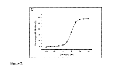
Figure 2. Amino acid sequence of variegin and its thrombin inhibitory
activity. (A)
Sequences of peptides in fraction AV 6/5 (variegin), AV 3/5 and AV 5/5 are
highly
similar. (B) Example of linear progression curves of thrombin inhibition by
variegin (M:
0.020 nM, ^: 0.039 nM, =: 0.078 nM, o: 0.156 nM, =: 0.313 nM, A : 0.625 nM, =:
1.25 nM, 0: 2.5 nM, =: 5 nM, 10 nM) using S2238 (100 M) as substrate, showing
steady state equilibrium achieved upon mixing. (C) The ability of variegin
(0.001 nM,
0.003 nM, 0.01 nM, 0.03 nM, 0.1 nM, 0.3 nM, 1 nM, 3 nM, 10 nM, 30 nM and 100
nM)
to inhibit tlirombin (3.33 nM) amidolytic activity was assayed using active
site directed
substrate S2238 (100 .M). Dose response curve of thrombin inhibition by
variegin (m)
showed significant inhibition (- 80 %) for equimolar concentration of thrombin
and
variegin (3.33 nM). IC50 of the inhibition is - 0.99 0.02 nM (n = 3) (D)
Since variegin
behaved as a tight-binding inhibitor, inhibition of thrombin (1.8 nM) by
variegin (m) at
similar concentrations (0.020 nM, 0.039 nM, 0.078 nM, 0.156 nM, 0.313 nM,
0.625 nM,
1.25 nM, 2.5 nM, 5 nM, 10 nM) was examined using S2238 (100 M) as substrate.
Data
obtained were fitted to equations (1) and (2) to derive a Ki of - 10.4 1.4
pM (n = 3).
CA 02691243 2009-12-18 PQ,,,
WO 2008/155658 PCT/1B2008/002109
24
Figure 3. Specificity of inhibition by variegin. S-variegin was screened
against 13
serine proteases: fibrinolytic serine proteases (plasmin, TPA and urokinase),
anticoagulant serine protease APC, procoagulant serine proteases (FXIIa, FXla,
FXa,
FIXa, FVIIa, kallikrein and thrombin) and classical serine proteases
(chymotrypsin and
trypsin). The final concentrations of proteases and substrates are given in
parentheses in
nM and mM, respectively: plasmin (3.61)/S2251 (1.2), TPA (36.9)/S2288 (1),
urokinase
(40 U/ml)/S2444 (0.3), APC (2.14)/S2366 (0.67), FXIta (20)/S2302 (1), FXta
(0.125)/S2366 (1), FXa (0.43)/S2765 (0.65), FIXa (333)/Spectrozyme FIXa
(0.4),
FVIfa (460)/S2288 (1), kallikrein (0.93)/S2302 (1.1), a-thrombin (3.33)/S2238
(0.1),
chymotrypsin (l.2)/S2586 (0.67) and trypsin (0.87)/S2222 (0.1). Thrombin was
tested
against three concentrations of s-variegin: %; ) represent 0.01 M, (0)
represent 0.1 M
and (EI) represent 1 M. For the other proteases, much higher concentrations
of s-
variegin were used: (m) represent 1 g.M, (0) represent 10 M and (o) represent
100 M
(n=3).
Figure 4. Inhibition of thrombin by s-variegin, EP25 and AP18. (A) The ability
of s-
variegin, EP25 and AP 18 to inhibit amidolytic activity of thrombin was
assayed using
active site directed substrate S2238 (100 M). Dose response curve of thrombin
(3.33
nM) inhibition by s-variegin (0.1 nM, 0.3 nM, 1 nM, 3 nM, 10 nM, 30 nM, 100
nM, 300
nM, 1000 nM) showed significant inhibition (- 30 %) for equimolar
concentration of
thrombin and variegin (3.33 nM). Dose-response curves and IC50 of inhibition
were
independent of incubation time: (m) represents 10 min incubation (IC50 - 5.40
0.95
nM) and (o) represents 10 min of incubation (IC50 - 5.49 0.42 nM) (n = 3).
(B) Dose-
response curves of thrombin (3.33 nM) inhibition by EP25 (0.1 nM, 0.3 nM, 1
nM, 3
nM, 10 nM, 30 nM, 100 nM, 300 nM, 1000 nM) showed an incubation time-dependent
shift. IC50 is - 139.30 7.02 nM without incubation (m), - 22.55 2.52 nM
with 1 min
incubation (o), - 10.39 1.53 nM with 2 min incubation (A), - 6.42 0.50 nM
with 5
min incubation ( V ), - 6.80 0.57 nM with 10 min incubation (+) and - 5.63
0.45 nM
with 20 min of incubation (+) (n = 3). (C) AP18 (3 gM, 10 M, 30 M, 100 M,
300
M) was unable to inhibit thrombin (3.33 nM) amidolytic activity on S2238 (100
M);
instead at high concentrations of AP 18, hydrolysis of S223 8 were slightly
enhanced (n =
3). (D) All three peptides, s-variegin (x; 0.3 nM, 1 nM, 3 nM, 10 nM, 30 nM
100 nM,
300 nM), EP25 (o; 3 nM, 10 nM, 30 nM, 100 nM, 300 nM, 1000 nM, 3000 nM) and
AP 18 (A; 0.1 M, 0.3 gM, 1 gM, 3 gM, 10 M, 30 gM, 100 gM, 300 gM) prolonged
CA 02691243 2009-12-18
WO 2008/155658 L'`1/ PCT/IB2008/002109
fibrinogen clotting times (n = 3). No pre-incubation of peptides with thrombin
was
carried out. AP 18 inhibited thrombin fibrinogenolytic activity but not
amidolytic
activity, suggesting binding to exosite-I.
Figure 5. Inhibitory constant K; of s-variegin and EP25. (A) S-variegin is a
fast and
5 tight binding inhibitor of thrombin. S-variegin (0.313 nM, 0.625 nM, 1.25
nM, 2.5 nM, 5
nM, 10 nM) was mixed with different concentrations of S2238: 12.5 M (m), 25
M (o),
50 M (A), 80 gM ( 0), 100 M (+), 150 gM (+), 200 gM (x) and 300 M ( * ) to
determine K;'. Reactions were started with the addition of thrombin (1.8 nM).
Data were
fitted to equation (1) (n = 3) (B) Plot of Ki' against substrate concentration
showed a
10 linear curve, indicating s-variegin competitively inhibited thrombin
amidolytic activity
on S2238. By fitting the data to equation (2), the inhibitory constant Ki was
shown to be
- 146.4 13.6 pM. (C) Although EP25 also inhibited thrombin at equimolar
concentrations if pre-incubated with thrombin, the initial inhibition without
pre-
incubation was weak. Ki of EP25 was determined without pre-incubation with
15 concentrations at least 8-fold greater than thrombin. Under these assay
conditions,
binding of EP25 to thrombin does not result in a significant depletion of free
EP25
concentration, thus `tight-binding' condition was not considered for data
fitting.
Progression curves of thrombin (0.9 nM) inhibition by different concentrations
of EP25:
7.8nM(m), 12.5nM(^), 15.6 nM (e), 25 nM (o), 31.3 nM (A), 50nM(V ), 62.5 nM
20 (Y), 100 nM (0) and 125 nM (+), using S2238 (100 M) as substrate. The
progression
curves are non-linear, and showed two-phase equilibria typical of slow-binding
inhibition. Data were fitted to equation (3) to obtain a k for each
concentration of EP25
used (n = 3). (D) Plot of the apparent first-order rate constant k against
EP25
concentrations is a hyperbolic curve described by equation (4) and hence was
fitted to
25 the equation to obtain a Ki' of - 529.7 76.7 pM, representing the
dissociation constant
of initial collision complex El. The overall inhibitory constant Ki was
calculated from
equation (5) and was found to be - 149.8 30.5 pM.
Figure 6. Cleavage of s-variegin and EP25 by thrombin. (A) Typical
chromatograms
of HPLC analysis of s-variegin cleavage by thrombin at 37 C. (i) At
incubation = 0 inin,
the single peak correspond to uncleaved s-variegin. (ii) After 30 min
incubation, two
new peaks appeared corresponding to cleavage product of mass 1045
(representing N-
terminal fragment SDQGDVAEPK (SEQ ID NO 2)) and of mass 2582 (representing C-
CA 02691243 2009-12-18 ii A I n " ,
WO 2008/155658 PCUIB2008/002109 9
26
terminal fragment MHKTAPPFDFEAIPEEYLDDES (SEQ ID NO 3)) while uncleaved
s-variegin decreased in quaiitity. (iii) Cleavage is almost complete after 180
min
incubation. (B) S-variegin (150 M) was incubated with thrombin (5 M) for
various
times at room temperature (n = 2). S-variegin was present in 30-fold excess of
thrombin.
Cleavage of s-variegin by thrombin was analyzed with RP-HPLC. Relative
percentage of
uncleaved s-variegin (EI), cleavage product of mass 1045 (representing N-
terminal
fragment SDQGDVAEPK) (SEQ ID NO 2) ( 0 ) and cleavage product of mass 2582
(representing C-terminal fragment MHKTAPPFDFEAIPEEYLDDES) (SEQ ID NO 3)
( 19 ) was calculated from the area under the peaks. (C) S-variegin was
incubated with
thrombin (3.33 nM) for up to 24 hr at room temperature and at various time
points
assayed for the ability to inhibit thrombin amidolytic activity on S2238 (100
M). (D)
Similar experiments were carried out replacing s-variegin with EP25.
Concentrations of
s-variegin or EP25: 10 nM (m), 100 nM (0) and 1000 nM (o) (n = 2). At 100 nM
of s-
variegin or EP25, the inhibitors were also present in 30-fold excess of
thrombin, and
hence were used primarily for comparison with cleavage data from HPLC
analysis.
Figure 7. Comparison of variegin with other thrombin inhibitors. (A) Amino
acid
sequence alignment of n-variegin, s-variegin, EP25, AP18, hirulog-1 and
hirudin show
highly similar C-terminal sequence. N-variegin is glucosylated at Thr (T),
hirulog-1
contains a D-Phe (F) and hirudin is sulfated at Tyr (Y). Sequence of TTI is
distinctly
different from variegin and was not aligned. (B) Schematic diagram showing
different
classes of thrombin inhibitors and their structural features. (i) Hirudin:
compact N-
terminus binds to active site, acidic and extended C-terminal binds to exosite-
I; (ii)
rhodniin: two Kazal-type domains in head-to-tail arrangement with the N-
terminal
domain binding to active site and the C-terminal domain binding to exosite-I;
(iii)
ornithodorin: two Kunitz-type domains in tail-to-tail arrangement with the N-
terminal
domain binding to active site and the C-terminal domain binds to exosite-I;
(iv)
haemadin: compact N-terminal domain binds to active site, acidic and extended
C-
terminus binds to exosite-II; (v) triabin: single (3-barrel domain binds to
exosite-I; (vi)
bothrojaracin: two different chains of the C-type lectin domain bind to
exosite-I and
exosite-II respectively. Other prototypic thrombin inhibitors such as theromin
and TTI
are not represented due to lack of detailed structural information. (C)
Proposed binding
mechanism of EP-25 to thrombin: (i) electrostatic charges on C-terminus steer
EP25 to
thrombin and subsequently provide specific tethering interaction, (ii) without
the
CA 02691243 2009-12-18 U~ ~ lJ U 1 1{1 9,:
WO 2008/155658 PCT/1B2008/002109
27
steering effect of N-terminal residues (SDQGDVA (SEQ ID NO 18)) the active
site
binding moiety is not orientated properly to fit the thrombin active site,
hence the initial
collision complex (EI) has a higher Ki, and (iii) in a slow step the active
site binding
moiety (EPKMHKT (SEQ ID NO 19)) adopts the correct conformation for optimum
binding and formation of a stabilized complex. (D) Proposed binding mechanism
of
variegin to thrombin: (i) complementary electrostatic charges between variegin
N-
terminus and thrombin exosite-II as well as between variegin C-terminus and
thrombin
exosite-I steer variegin to thrombin, (ii) all electrostatic interactions
occurred rapidly and
pre-orient active site binding moiety (EPKMHKT (SEQ ID NO 19)) in correct
conformation for fast binding to thrombin active site.
Figure 8. Plot of reaction velocity (Vm;,X) as a function of substrate (S2238)
concentration following the Michaelis-Menton equation. Kn, calculated with
Michaelis-Menton equation is determined to be 3.25 0.56 M, similar to
reported
values33,34
Figure 9. Far-UV spectra (260-190nm) of n-variegin, s-variegin, EP25 and AP18
dissolved in 10mM of sodium phosphate buffer (pH7.4). All spectra were typical
of a
random coil protein.
Figure 10. RP-HPLC analysis showed that s-variegin was cleaved by thrombin at
37 C and room temperature. (A) S-variegin (150 M) was incubated with thrombin
(5 M) for various time at 37 C (n=2). (B) S-variegin (150 M) was incubated
with
thrombin (5 M) for various time at room temperature (n=2). Relative
percentages of
uncleaved S-variegin (EI), cleaved product of mass 1045 (representing N-
terminal
fragment SDQGDVAEPK (SEQ ID NO 2)) (n) and cleavage product of mass 2582
(representing C-terminal fragment MHKTAPPFDFEAIPEEYLDDES) (SEQ ID NO 3)
(B) were calculated from the area under the peaks in the chromatograms.
Figui-e 11. Thrombin inhibitory activity of C-terminal fragment
MHKTAPPFDFEAIPEEYLDDES (MH22) (SEQ ID NO 3) of variegin The ability
of various concentrations of MH22 to inliibit thrombin amidolytic activity
using active
site directed substrate S2238 following incubation with thrombin at room
temperature
for 0 min (m), 10 min (=), 20 min (A), 30 inin (Y), 120 min (+), 1080 min (+)
or 1680
min (X) was assessed.
CA 02691243 2009-12-18
~ ~
WO 2008/155658 PCT/IB2008/002109"
28
Figure 12. Reversal of decrease in amidolytic activity of MH22. The decrease
in the
amidolytic activity of MH22 after prolonged incubation with thrombin (1680 min
pre-
incubation IC50 = 479.7 16.1 nM) can be reversed by including increased
concentrations of BSA (lmg/ml (m), 5mg/ml (=), 10 mg/ml) (A) in the assay
setup.
Figure 13. Ki of MH22. The K;' of MH22 at different concentrations of
substrate
(S2238) was determined through the equation describing fast and tight binding.
K;' did
not change significantly throughout the concentration range used (12.5 nM to
200 nM),
indicating that MH22 is a non-competitive inhibitor of thrombin amidolytic
activity. Ki'
= Ki and the average Ki was found to be 13.2 0.91 nM.
Figure 14. Thrombin inhibitory activity of variegin mutant fragment EP25A22E.
The ability of various concentrations of EP25A22E having the sequence
EPKMHKTAPPFDFEEIPEEYLDDES (SEQ ID NO 7) to inhibit thrombin amidolytic
activity using active site directed substrate S2238 following incubation with
tluombin at
room temperature for 0 min (m), 20 min (=) or 30 min (A) was assessed. In
EP25A22E,
alanine 22 in s-variegin (alanine 15 in EP25) was replaced with glutamic acid
since
glutamic acid is present in the same position in hirudin.
Figure 15. Ki of EP25A22E. The K; of EP25A22E was determined using the slow
binding inhibitor equation and was found to be 0.311 0.070 nM.
Figure 16. Thrombin inhibitory activity of variegin mutant fragment MH22A22E.
The ability of various concentrations of MH22A22E having the sequence
MHKTAPPFDFEEIPEEYLDDES (SEQ ID NO 5) to inhibit thrombin amidolytic
activity using active site directed substrate S2238 following incubation with
thrombin at
room temperature for 0 min (m) or 20 min (=) was assessed. MH22A22 is the C-
terminal
cleavage fragment of EP25A22E.
Figure 17. Ki of MH22A22E MH22A22E has a Ki' of 15.1 1.04 nM when tested
with
100 M of substrate (S2238).
Figure 18. Thrombin inhibitory activity of variegin fragment EP21. The ability
of
various concentrations of EP21 EPKMHKTAPPFDFEAIPEEYL (SEQ ID NO 8) to
inhibit thrombin amidolytic activity using active site directed substrate
S2238 following
incubation with tlirombin at room temperature for 0 min (m), 20 min (=) or 30
min (A)
CA 02691243 2009-12-18
PCT/IB2008/0021091 ~ ~
WO 2008/155658
29
was assessed. EP21 corresponds to EP25 except that it is missing four residues
at the C-
terminal.
Figure 19. Ki of EP21. Ki of EP21, determined by slow binding equations was
found to
be0.315 0.024 nM.
Figure 20. Thrombin inhibitory activity of variegin fragment MH18. The ability
of
various concentrations of MH18 MHKTAPPFDFEAIPEEYL (SEQ ID NO 20) to
inhibit thrombin amidolytic activity using active site directed substrate
S2238 following
incubation with thrombin at room temperature for 0 min (m) or 20 min (=) was
assessed.
MH18 corresponds to MH22 except that it is missing four residues at the C-
terminal.
Figure 21. Ki of MH18. Using fast and tight binding equation, Ki' of MH18 at
100 M
substrate (S2238) = 14.9 3.50 nM. Assuming the removal of four residues at
the C-
terminal did not alter the inhibition mechanism, MH18 is also a non-
competitive
inliibitor with K; = 14.9 3.50 nM.
Figure 22. Thrombin inhibitory activity of variegin fragment DV24. The ability
of
various concentrations of DV24 DVAEPKMHKTAPPFDFEAIPEEYL (SEQ ID NO 9)
to inhibit thrombin amidolytic activity using active site directed substrate
S2238
following incubation with thrombin at room temperature for 0 min (m) or 20 min
(=) was
assessed. DV24 corresponds to EP21 except that it contains an additional 3
residues at
the N-terminal.
Figure 23. Ki of D24. Ki' of DV24 at 100 M substrate (S2238) = 9.74 0.91 nM
and
K; of DV24 was determined to be 0.306 ::L 0.029 nM,
Figure 24. Thrombin inhibitory activity of variegin mutant fragment DV24K10R.
The ability of various concentrations of DV24K10R
DVAEPRMHKTAPPFDFEAIPEEYL (SEQ ID NO 10) to inhibit thrombin amidolytic
activity using active site directed substrate S2238 following incubation with
thrombin at
room temperature for 0 min (m) or 20 min (=) was assessed. DV2424K10R
corresponds
to DV24 except that it contains an arginine instead of a lysine at position 6
(amino acid
10 is variegin).
Figure 25. Ki of DV24KIOR. The Ki of DV24K10R was determined to be 0.259 ~
0.015 nM
Figure 26. HPLC radiochromatogram to show [3H]-Variegin dose solution
CA 02691243 2009-12-18
PC I PCT/IB2008/002109'
WO 2008/155658
Figure 27. Distribution of radioactivity in tissues at 30 minutes following a
single
intravenous administration of [3H]-Variegin to a male albino rat (0.4 mg/kg).
Levels 1 to
5 refer to successive 1 cm longitudinal sections through the rat body.
Figure 28. Distribution of radioactivity in tissues at 1 hour following a
single
5 intravenous administration of [3H]-Variegin to a male albino rat (0.4
mg/kg). Levels 1 to
5 refer to successive 1 cm longitudinal sections through the rat body.
Figure 29. Distribution of radioactivity in the kidney at 30 minutes following
a single
intravenous administration of [3H]-Variegin to a male albino rat (0.4 mg/kg)
Examples
10 EXAMPLE 1: ANALYSIS OF VARIEGIN AND EP25
Material and methods
Materials
Human citrated plasma was provided by the Department of Hematology and
Transfusiology of the Slovak Institute of Cardiovascular Diseases.
Thromboclotin
15 reagent was from Dade AG (Dudingen, Switzerland). Thromboplastin IS reagent
and
Actin FS Activated PTT reagent were from Dade International Inc. (Miami,
Florida). 9-
Fluorenylmethyloxycarbonyl (Fmoc)-L-amino acids, Fmoc-PEG-PS support resin,
N,N-
dimethylformamide (DMF), 20 % v/v piperidine in DMF, O-(7-azabenzotriazol-1-
yl)-
1,1,3,-3-tetramethyluronium hexafluorophosphate (HATU) and N,N-
20 diisopropylethylainine (DIPEA) were from Applied Biosystems (Foster City,
California). Trifluoroacetic acid (TFA), 1,2-ethanedithiol, thioanisole,
bovine
chymotrypsin and bovine serum albumin (BSA), were from Sigma Aldrich (St.
Louis,
Missouri). Human fibrinogen, FXIIa, tissue plasminogen activator (TPA),
urokinase,
kalikrein and bovine trypsin were from Merck Chemicals Ltd. (Nottingham, UK).
25 Human factor IXa (FIXa), factor Xa (FXa), factor XIa (FXIa), APC and
plasmin were
from Hematologic Technologies, Inc. (Essex Junction, Vermont). Human factor
VIIa
(FVIIa) and recombinant a-thrombin were gifts from the Chemo-Sero-Therapeutic
Research Institute (KAKETSUKEN, Japan)21'22. Chromogeiiic substrates S2222,
S2238,
S2251, S2288, S2302, S2366, S2444, S2586 and S2765 were from Chromogenix
30 (Milano, Italy). Spectrozyme FIXa was from American Diagnostica Inc.
(Stamford,
Connecticut). All other chemicals and reagents used were of analytical grade.
CA 02691243 2009-12-18 P`i/~~~,,. u o ~ n n
WO 2008/155658 PCT/1B2008/002109
31
Salivary gland extracts and estimation of protein concentrations
The extraction procedure of A. variegatum SGE and estimation of protein
concentrations
during fractionation were described previously23
Purification of variegin isoforms
Variegin was purified by a three-step reverse-phase HPLC procedure with a
Beckman
Instruments 126/168 DAD HPLC system (Fullerton, California). In the first step
(Figure
1A) SGE was loaded onto a Vydac C-4 (5 m; 250 x 4.6 mm) column (Grace Vydac,
Hesperia, California). Pooled fractions that contained the strongest
anticoagulant activity
(Figure 1A, fraction AV-III) were subjected to a second step (Figure 1B) using
a
Beckinan Ultrasphere C-18 (5 m; 250 x 4.6 mm) column. Lastly, individual
fractions
were further purified using a Vydac C-18 (5 m; 250 x 4.6 mm) column to obtain
three
fractions of potent anti-thrombin activity: AV 6/5, AV 3/5 and AV 5/5 (Figure
1 C - D).
The major component in the AV 6/5 fraction was named variegin.
Coagulation assays
Thrombin time (TT), prothrombin time (PT) and activated partial thromboplastin
time
(APTT) assays were used for the initial screens of anticoagulant activities in
SGE and
fractions. Citrated human plasma (50 l) was pre-incubated with a maximum of 5
l of
the SGE or the same volume of 150 mM NaCl (control) at 37 C for 1 min. After
adding
the corresponding reagents (TT: 50 gl of Thromboclotin reagent; PT: 100 l of
Thromboplastin IS reagent; APTT: 50 l of Actin FS Activated PTT added for 3
min
and reaction started with 50 l of 20 mM CaC12), times required for the
formation of
fibrin clots were determined visually using a stop watch.
The activities of crude SGE and the three fractions (AV 6/5, AV 3/5 and AV
515) were
verified at the Oxford Hemophilia Centre of Churchill Hospital (Oxford, UK).
TT, PT
and APTT were performed using an MDA-180 analyser (Organon Teknika Ltd.,
Cambridge, UK). 10 l of SGE or diluted fractions containing AV 6/5, AV 3/5
and AV
515 were added to 290 gl of platelet poor plasma, mixed and incubated for 5
min at 37 C.
The activities were also verified using a Thromboelastograph Analyzer
(Haemoscope
Inc., Skokie, Illinois). Five l of samples were added to 335 gl of citrated
wlzole blood,
incubated for 5 min and the sample run on the TEG following recalcification.
CA 02691243 2009-12-18 r`'1~~'~ ~~ l U(] 7
~=U = 9 ;
WO 2008/155658 PCT/IB2008/002109
32
Protein sequence analysis
The molecular weight of proteins present in AV 6/5, AV 3/5 and AV 5/5 were
determined by Eurosequence (Groningen, the Netherlands) using a BIFLEX (Bruker-
Franzen, Bremen, Germany) matrix-assisted laser desorption/ionization
reflectron time-
of-flight (MALDI-TOF) mass spectrometer equipped with a nitrogen laser (337
nm) and
gridless delayed extraction ion source. Partial amino acid sequences were
determined by
N-terminal Edman-degradation using an automated sequencer (Model 494, Applied
Biosystems). The complete sequence for AV 6/5 was determined by MALDI-MS
analysis.
Peptide synthesis and purification
Three peptides (s-variegin, EP25 and AP18) were synthesized using solid phase
peptide
synthesis methods on an Applied Biosystems Pioneer Model 433A Peptide
Synthesizer.
Fmoc groups of amino acids were removed by 20% v/v piperidine in DMF and
coupled
using HATU/DIPEA in situ neutralization chemistry. All peptides were
syiithesized on
preloaded PEG-PS resins. Cleavage by a cocktail of TFA/1,2-
ethanedithiol/thioanisole/water released peptide acids (-COOH). Synthetic
peptides were
purified by RP-HPLC on AKTATM purifier (GE Healthcare, Uppsala, Sweden) with a
SunFireTM C 18 (5 m; 250 mm x 10 mm) (Waters, Milford, Massachusetts) column.
The
purity and mass of all peptides were determined by electrospray ionization
mass
spectrometry (ESI-MS) using a Perkin-Elmer Sciex API 300 LC/MS/MS System
(Perkin-Elmer Sciex, Selton, Connecticut).
Circular dichroism (CD) spectroscopy
Far-UV CD spectra (260 - 190 nm) of variegin, s-variegin, EP25 and AP 18
dissolved in
10 mM of sodium phosphate buffer (pH 7.4) were recorded using a Jasco J-810
spectropolarimeter (Easton, Maryland). All measurements were carried out at
room
temperature using 0.1 cm path length cuvettes with a scan speed of 50 nm/min,
a
resolution of 0.2 nm and a bandwidth of 2 nm.
Inhibition of thrombin amidolytic activity
All assays for thrombin amidolytic activity on S2238 were performed in 96-
wells
microtiter plates in 50 mM Tris buffer (pH 7.4) containing 100 mM NaCl and 1
mg/ml
BSA at room temperature. Typically, 100 l of peptides and 100 l of thrombin
were
CA 02691243 2009-12-18 t n n
WO 2008/155658 PCT/1S2008/0021097
33
pre-incubated for different durations before the addition of 100 l of S2238.
The rates of
formation of colored product p-nitroaniline were followed at 405 nm for 10 min
with an
ELISA plate reader. Percentage inhibition was calculated by taking the rate of
increase
in absorbance in the absence of inhibitor as 0 %. Dose-response curves were
fitted using
Origin software (MicroCal, Northampton, Massachusetts) to calculate IC50
values.
Determination of the inhibitory constant Ki
The inhibitory constant, Ki, was determined using S2238 as substrate. When an
enzyme
is inhibited by an equimolar concentration of inhibitor, the binding of
inhibitor to
enzyme causes a significant depletion in the concentration of free inhibitors.
This tight-
binding inhibition is described by the following equation24:
Vs = (Vo/2Et) { [(Ki' + It - Et)2 + 4K;'Et] " - (K;' + It - Et)} (1)
where VS is steady state velocity, Vo is velocity observed in the absence of
inhibitor, Et is
total enzyme concentration, It is total inhibitor concentration and K;' is
apparent
inhibitory constant. For competitive inhibition, Ki is related to Ki' by
equation (2):
Ki' = K; (1 + S/Kn,) (2)
where Ki' increases linearly with S, K; is the inhibitory constant, S is the
concentration
of substrate and K,,, is the Michaelis constant for S2238 (determined to be
3.25 0.56
M, Figure 8, similar to reported values24 Z5). Both variegin and s-variegin
were found to
be tight-binding inhibitors. The data were fitted to these equations using
Origin
software.
If the rate of interaction of the inhibitor with the enzyme is slow so that
the inhibited
steady-state velocity is slowly achieved, the progress curve of product
formation of this
slow binding inhibition is described by equation (3)26:
P = VSt + (Vo - VS) (1 - e-k) / k+ Po (3)
where P is the amount of product formed, Po is initial amount of product, Vs
is final
steady state velocity, Vo is initial velocity, t is time, and k is apparent
first-order rate
constant.
There are two possible minimum kinetic mechanisms to describe such slow
binding
reactionsa6 27:
Scheme (1): E+ I Ki EI*
K2
CA 02691243 2009-12-18 ~'~.1/tL..v il H i~1 (1 ! 1 O 9~
WO 2008/155658 PCT/IB2008/002109
34
where E is enzyme, I is inhibitor and EI* is stable enzyme-inhibitor complex,
K1 is
association rate constant and K2 is dissociation rate constant. In this
scheme, slow
binding is mainly due to the slow Kl. The apparent first-order rate constant k
will
increase linearly with inhibitor concentration. Alternatively:
K, K,
Scheme (2): E + I a K EI ~- K4 EI*
where El is initial collision complex, K3 is forward isomerization rate and K4
is reverse
isomerization rate. In this scheme, binding involves rapid formation of an
initial collision
complex (EI) that subsequently undergoes slow isomerization to the final
enzyme-
inhibitor complex (EI*). k increases hyperbolically with inhibitor
concentrations.
Dissociation constant of El (denoted K;') can be calculated from equation (4):
k = Ka. + K3It / [It + Ki'(1 + S / Km)] (4)
The overall inhibitory constant Ki can be calculated from equation (5):
K;=Ki' [K4 / (K3 + K4)] (5)
EP25 was found to be a slow binding inhibitor following the Scheme 2
mechanism. The
data were fitted to these equations using Origin software.
Serine protease specificity
The selectivity profile of variegin was examined against 13 serine proteases:
fibrinolytic
serine proteases (plasmin, TPA and urokinase), anticoagulant serine protease
APC,
procoagulant serine proteases (FXIIa, FXIa, FXa, FIXa, FVIIa, kallikrein and
thrombin)
and classical serine proteases (chymotrypsin and trypsin). Effects of s-
variegin on these
serine proteases were determined by inhibition of their amidolytic activities
assayed
using specific chromogenic substrates.
Fibrinogen clotting time
The abilities of s-variegin, EP25 and AP 18 to prolong fibrinogen clotting
time were
tested using a BBL fibrometer (BD, Franklin Lakes, New Jersey). 200 l of
fibrinogen
(final concentration 3 mg/ml) were incubated with 100 l of peptides (various
concentrations) at 37 C. Clotting of fibrinogen was initiated by the addition
of 100 l of
thrombin (final concentration 20 nM). All reagents and samples were dissolved
in 50
mM Tris buffer (pH 7.4) containing 100 mM NaCI.
CA 02691243 2009-12-18
WO 2008/155658 PCT/IB2008/002109
I U U L i -g'q:
Cleavage of s-variegin by thrombin
S-variegin and EP25 (final concentrations: 150 M) were incubated with
thrombin (final
concentration: 5 M) at both room temperature and 37 C. After various
incubation
times, the reactions were quenched with 0.1 % TFA buffer (pH 1.8) and loaded
onto a
5 SunFireTM C 18 column attached to an AKTATM purifier. New peaks other than
those
present in the chromatogram of 0 min incubation were identified as cleavage
products
and subjected to ESI-MS to verify their masses. The pealcs were integrated to
calculate
the area under the peaks and relative percentage of each peak.
Results
10 Purification of variegin isoforms
Crude SGE of A. l~ariegatum exhibited potent anticoagulant activity in all
three
coagulation assays (PT, APTT and TT) (Figure 9). Potency was in the order TT
>>
APTT > PT, indicating that SGE is a promising source of potent thrombin
inhibitor(s).
To purify this inhibitor(s), SGE was fractionated by RP-HPLC (Figure 1A).
After the
15 first step of purification, the most potent anticoagulant fraction (AV-III)
was subjected to
a second purification step (Figure 1 B). The resulting fractions were screened
for
antithrombin activity in coagulation and chromogenic substrate assays. Two
fractions
with the strongest activity (retention time 23.083 and 28.933 min) were
further purified
in separate runs. The fraction with retention time 23.083 min was separated
into two
20 main peaks denoted AV 3/5 and AV 5/5 (Figure 1 C). The fraction with
retention time
28.933 has one main peak and with a small `shoulder peak' and was denoted AV
6/5
(Figure 1 D). The anticoagulant activities of these three fractions (AV 3/5,
AV 5/5 and
AV 6/5) along with crude SGE were verified by PT, APTT, TT and TEG assays. All
four assays revealed that AV 6/5 contained the most potent anticoagulant
activity,
25 followed by AV 3/5 and AV 5/5 (Table 1).
CA 02691243 2009-12-18
WO 2008/155658 t'Cj/1PCT/1B2008/002109J -0 ~9
36
Table 1. Anticoagulation activities of Anablyomma variegatutn SGE (females fed
for
9 days). Results show the mean of duplicate values. In controls 150 mM NaCl
was
substituted for SGE.
TT (s) APTT (s) PT (s)
Control 17 28 15
SGE protein ( g)
0.025 50
0.05 105
0.10 >180
0.25 28 15
0.50 38 19
1.00 45 22
2.50 >180 40
5.00 >180
Protein sequence analysis
Partial sequences of all three fractions were determined by Edman degradation.
For AV
6/5 the sequence and molecular weight were completed by MALDI-TOF. MALDI
spectrum of AV 6/5 revealed a major m/z signal of 3769.96 Da (monoisotopic
mass =
3768.96 Da) and a minor m/z signal of 3777.79 Da (monoisotopic mass = 3776.79
Da).
The main component has the sequence
SDQGDVAEPKMHKT(hex)APPFDFEAIPEEYLDDES (SEQ ID NO 1), where the
Thr14 is modified by a hexose moiety. This was named variegin and was further
characterized. The minor component (3776.79 Da) is almost identical to
variegin, with
G1u31 replaced by His. Partial sequences determined by Edman degradation
revealed
two components in the AV 3/5 fraction (m/z 3953.54 and 3409.57 Da) and three
components in AV 5/5 (m/z 3680.23, 3368.94 and 3173.62 Da). All the sequences
determined are highly similar to variegin (Figure 2A). CD spectrum of variegin
is typical
of a random coil protein (Table 2).
CA 02691243 2009-12-18
WO 2008/155658 r~-1I'"PCT/1B2008/002109
37
Table 2. Anticoagulation activities of A. variegatum SGE and RP-HPLC
fractions.
AV 6/5 is the most potent fraction determined in all assays. The major
component in AV
6/5 was sequenced and named variegin. Since APTT, PT and TT were performed in
citrated platelet poor plasma (PPP), and thus represent a non-physiological
milieu in
which to assess its anticoagulant potential, the activity of the samples were
also verified
with TEG, which permits coagulation monitoring in whole blood using
viscoelastic
assessment of clot formation as an endpoint. (PNP: pooled normal plasma; r: r
phase,
the period of time of latency from the time that blood was placed in the TEG
until the
initial fibrin formation; k: k phase, a measure of the speed to reach a
certain level of clot
strength).
Sample TEG PT (s) APTT (s) TT (s)
Crude SGE Complete inhibition No clot No clot No clot
PNP Normal 13.6 25.6 12.2
AV 6/5 Inhibited - - -
1:200 dilution - 15.3 59.2 78.9
1:500 dilution - 14.4 48.3 39.2
AV 3/5 Prolonged r/k - - -
1:200 dilution - 14.1 46.7 30.8
1:500 dilution - 14.1 38.8 20.6
AV 5/5 Prolonged r/k - - -
1:200 dilution - 13.8 38.5 21.4
1:500 dilution - 13.8 33.7 15.6
BLAST results indicate that variegin does not show similarity to any known
proteins in
the database. Interestingly, its C-terminus (DFEAIPEEYL) (SEQ ID NO 21) is
almost
identical to the C-terminus of hirudin (residues 55 to 64: DFEEIPEEYL (SEQ ID
NO
22)). Thus, we hypothesized that variegin C-terminus plays a similar role to
hirudin C-
terminus in binding to thrombin. However, Tyr63 of hirudin is sulfated28 z9
while the
corresponding Tyr in variegin is not.
CA 02691243 2009-12-18 rL1/1L~~ ~; p! n n~ 1 n, 9
WO 2008/155658 PCT/IB2008/002109,
38
Inhibition of thrombin amidolytic activity by variegin and its K;
The ability of variegin to inhibit thrombin amidolytic activity was assayed
with S2238, a
small peptidyl substrate that binds only to the active site. Variegin
inhibited the
amidolytic activity and progress curves of inhibition showed that steady state
equilibrium was achieved upon mixing (Figure 2B). Significant inhibition (- 80
%) was
observed for equimolar concentrations of thrombin and variegin (3.33 nM). IC50
of the
inhibition is - 0.99 0.02 nM (Figure 2C). Variegin is a fast and tight-
binding
competitive inhibitor of thrombin with a Ki of - 10.4 ~ 1.4 pM (Figure 2D).
Synthesis of s-variegin and variants
For further characterization, three peptides were synthesized, purified and
characterized.
Synthetic variegin (SDQGDVAEPKMHKTAPPFDFEAIPEEYLDDES (SEQ ID NO 1),
s-variegin) has the complete sequence of variegin, while EP25
(EPKMHKTAPPFDFEAIPEEYLDDES) (SEQ ID NO 6) and AP 18
(APPFDFEAIPEEYLDDES) (SEQ ID NO 16) have seven and 14 residues truncated
from the N-terminus. Unlike native variegin (n-variegin) Thr is not
glycosylated in s-
variegin and EP25. CD spectra of s-variegin, EP25 and AP 18 are all similar to
that of n-
variegin, typical of random coil proteins (Figure 9).
Specificity of inhibition by variegin
To determine the specificity, s-variegin was .. screened against 13 serine
proteases
including thrombin. Apart from thrombin, no other serine proteases showed
significant
inhibition (<_ 5 %) even at 1 M of s-variegin. Inhibition of > 10 % was
observed at
much higher concentrations of s-variegin. The most susceptible proteases are
plasmin,
trypsin and FXIa, which were inhibited - 20 to 30 % by 100 M of s-variegin.
In
contrast, against thrombin, similar - 30 % inhibition was observed at a
concentration at
least 4 orders of magnitude lower (-- 3.3 nM) (Figure 3). Therefore, s-
variegin is a
specific and potent thrombin inhibitor.
Inhibition of thrombin amidolytic activity by s-variegin, EP25 and AP18
S-variegin is similar to n-variegin in that steady state equilibrium of
inhibition was
achieved upon mixing. It was 5-fold less active than n-variegin and - 30 %
inhibition
was observed at equimolar concentrations of thrombin and s-variegin (3.33 nM).
Dose-
response curves showed an IC50 value of 5.40 0.95 nM, independent of
incubation time
CA 02691243 2009-12-18
WO 2008/155658 PCT/IB2008/002109 . ~7
39
(0 min and 10 min) (Figure 4A). Hence, s-variegin is also a fast and tight
binding
inhibitor of thrombin. The absence of Thr glycosylation in s-variegin might
account for
its weaker activity.
EP25 also inhibited amidolytic activity of thrombin. However, unlike n-
variegin and s-
variegin, progress curves of inhibition showed two-phase equilibria in the
absence of
pre-incubation. The steady state equilibrium inhibition was achieved
relatively slowly
after -10 min pre-incubation. Dose-response curves of EP25 were dependent on
incubation times. Thus the deletion of seven N-terminal residues (SDQGDVA (SEQ
ID
NO 18)) turned the binding mode from fast to slow. However, potency of EP25
was not
affected by the deletion. When the final steady state equilibrium was achieved
(20 min
pre-incubation) EP25 inhibited thrombin to the same extent as s-variegin (IC50
values for
EP25 and s-variegin are 5.63 0.45 nM and 5.40 10.95 nM, respectively)
(Figure 4B).
In contrast, AP18 did not inhibit thrombin amidolytic activity even at 300 M,
suggesting that it did not bind to the active site. Instead, AP 18 enhanced
thrombin
amidolytic activity slightly in a dose-dependent manner (Figure 4C). This is
consistence
with the reported behavior of hirudin C-terminus28. In summary, these results
suggest
that the active site binding moiety on variegin resides within position 8 to
14
(EPKMHKT).
Inhibition of thrombin fibrinogenolytic activity
S-variegin, EP25 and AP 18 all prolonged fibrinogen clotting time in a dose-
dependent
manner (Figure 4D). Fibrinogen binds to both the active site and exosite-I of
thrombin1'2.
AP 18 inhibited fibrinogenolytic but not amidolytic activity of thrombin, and
hence we
concluded that C-terminus of variegin binds to exosite-I. This observation is
consistence
with that of hirudin C-terminus28'29. The difference in activity between s-
variegin and
EP25 is likely to be due to the slow binding mode of EP25.
Inhibitory constant K; of s-variegin and EP25
Ki of s-variegin and EP25 was determined using S2238 as substrate. S-variegin
is a fast
and tight binding inhibitor. Ki' was determined in the presence of different
concentrations of S2238 (Figure 5A). S-variegin is a competitive inhibitor of
thrombin
and its Ki' increased linearly with increasing concentrations of S2238
(equation 2)
(Figure 5B). The true inhibitory constant, Ki was found to be - 146.4 13.6
pM, which
is 14-fold higher than n-variegin (-10.4 1.4 pM). In contrast, EP25 is a
slow binding
CA 02691243 2009-12-18 rL1j1j WO 2008/155658 PCT/IB2008/002109 Q 9-'
inhibitor of thrombin. Progress curves of inhibition were fitted to equation 3
to obtain k
for each concentration of EP25 (Figure 5C). k, the apparent first-order rate
constant for
the establishment of the equilibrium between initial collision complex (EI)
and final
stable complex (EI*), increased hyperbolically with EP25 concentration (Figure
5D), as
5 described by Scheme (2). Thus, the binding between EP25 and thrombin
involves the
isomerization of El to EI*. The dissociation constant of El (Ki', equation 4)
was - 529.7
76.7 pM, while the overall inhibitory constant K; (equation 5) was - 149.8
30.5 pM.
Thus, Ki of EP25 is essentially the same as Ki of s-variegin (- 146.4 13.6
pM). These
results confirmed that the deletion of seven N-terminal residues did not
affect potency
10 but switched the binding mode from fast to slow.
Cleavage of s-variegin by thrombin
Since variegin binds to the thrombin active site, it may be cleaved by
thrombin, similar
to other serine protease inhibitors30. Therefore we exainined the cleavage of
s-variegin
by thrombin and its effects on inhibition. RP-HPLC analysis showed that s-
variegin was
15 indeed cleaved by thrombin at room temperature and 37 C. At 0 min of
incubation only
peaks corresponding to uncleaved s-variegin and thrombin were present. Two new
peaks
of cleavage products appeared and increased with increasing incubation time
(Figure
6A). These new peaks had molecular weights of 1045 Da (SDQGDVAEPK (SEQ ID
NO 2)) and 2582 Da (MHKTAPPFDFEAIPEEYLDDES (SEQ ID NO 3)) respectively,
20 and corresponded to cleavage at the Lys10-Metll peptide bond. Cleavage
proceeded
faster at 37 C than at room temperature (Figure 9).
To verify the effect of variegin cleavage, s-variegin and EP25 were incubated
with
thrombin up to 24 h and at various time points assayed for the ability to
inhibit thrombin
amidolytic activity. The results showed that both s-variegin and EP25 lost
their activity
25 only after prolonged incubation with thrombin (Figure 6B - D).
Interestingly, at the same
temperature (24 C) and molar ratios (30-fold excess of s-variegin), after 60
min of
incubation, - 30 % of s-variegin was cleaved, yet no loss of inhibitory
activity of s-
variegin and EP25 was observed. 24 h of incubation was needed for - 30 % loss
of
inhibitory activity of s-variegin and EP25. In the case of the slow binding
inhibition of
30 EP25, percentage iiihibition increased with incubation time up to 20 min
and then
decreased due to cleavage by thrombin (Figure 6D). Thus, it is likely that the
cleavage
product(s) retain strong binding to the thrombin active site.
CA 02691243 2009-12-18 0 0 2 1.0- 9
WO 2008/155658 PCT/IB2008/002109
41
Discussion
Variegin is one of the smallest thrombin inhibitors found in nature. Despite
its small size
and flexible structure, variegin binds to thrombin with strong affinity.
Structure-activity
studies indicate that variegin binds over an extended surface area of
thrombin. The seven
N-terminal residues affected the binding kinetics; when removed, the binding
characterisitics of variegin changed from fast to slow. Residues 8 to 14
appear to bind to
the thrombin active site, and residues 15 to 32 appear to bind to exosite-I.
Although
variegin is cleaved by thrombin, its inhibitory activity was largely retained
after
cleavage.
Over the years, many thrombin inhibitors have been isolated from hematophagous
animals and snake venom. However, no similarities were found in the primary
structure
of variegin and other thrombin inhibitors. The absence of cysteines,
suggesting a flexible
structure, also differs from prototypic thrombin inhibitors such as hirudin
(compact N-
terminus, acidic and extended C-terminus)6 I1-13, rhodniin (double domain
Kazal-type
inhibitor)3i'3Z, ornithodorin (double domain Kunitz-type inhibitor)33 and
theromin (acidic
and antitastin-like N-terminus, compact C-terminus)34, even though they all
bind to the
same sites on thrombin (active site and exosite-I) (Figure 7A). Although
variegin
residues 19 to 28 are almost identical to hirudin C-terminus, their N-termini
are
completely different (Figure 7B). Unlike hirudin, variegin is not sulfated at
the Tyr
residue and has three extra residues at the end. Desulfation of hirudin24 or
its C-terminal
peptide (hirugen)29 retained anti-thrombin activity despite a 10-fold
reduction in
affinity24 and activity29. Our results indicated that AP 18 binds to exosite-I
and slightly
enhanced thrombin amidolytic activity, comparable to the reported behavior of
hirudin
C-terminus28'29, suggesting similar roles for these two sequences. This
appears to be an
example of convergent evolution in two phylogenetically distant lineages.
Variegin is also distinct from other thrombin inhibitors such as
haemadin35,36, triabin37'3s
and bothrojaracin39. Haemadin has a similar structure to hirudin, binding to
the thrombin
active site with its N-terminus, and to exosite-II with the extended C-
terminus35'36
Triabin only inhibits exosite-I and has a similar structure to
lipocalins37'38. Bothrojaracin,
a C-type lectin protein, binds to both exosite-I and exosite-II39. Only two
other thrombin
inhibitors of similar size have been reported to date, but they appear to be
unrelated to
variegin. Despite also having 32 residues, tsetse tlirombin inhibitor (TTI),
isolated from
PCllu,-) 0 8/ 0 0 2 1 0 9
CA 02691243 2009-12-18
WO 2008/155658 PCT/IB2008/002109
42
tsetse fly Glossina morsitans morsitansao'41, does not share any sequence
similarity with
variegin (Figure 7A). Another low molecular weight thrombin inhibitor (3.2
kDa) was
isolated from the camel tick, Hyalomma dromedarii (NTI-1)42. Unlike variegin,
NTI-1 is
a weak (K; = 11.7 M) and non-competitive inhibitor of thrombin, binding to
only one
site on thrombin (Figure 7A). Currently, no detailed structural information
for NTI-1 is
available.
Perhaps variegin is best compared with hirulogs, synthetic thrombin inhibitors
designed
by grafting the hirudin C-terminus to the active site binding moiety D-Phe-Pro-
Arg-Pro
through a linker of four Gly residues' 4 (Figure 7A). While development of
hirulogs
(marketed as bivalirudin) represents successful rational drug design, variegin
demonstrates the ability of nature to produce similar `designs' through
evolution. Thus,
variegin can be described as a`natural' hirulog. S-variegin and EP25 have
several
advantages over hirulogs as throinbin inhibitors. Firstly, variegin and EP25
comprise
natural amino acids (hirulogs generally have D-Phe). Secondly, even without
Thr
glycosylation, their affinity for thrombin is higher than that of hirulog-1.
EP25
(comparable to hirulog-1 in length) inhibits thrombin with a much stronger
affinity (K;
values of EP25 and hirulog-1 are - 149.8 30.5 pM and -2500 pM43
respectively).
Lastly, although both hirulogs and variegin are cleaved by thrombin, variegin
(and
EP25) loses its inhibitory activity towards thrombin at a much slower rate
than hirulogs.
For example, at an inhibitor to thrombin ratio of 3:1, hirulog-1 lost all
inhibitory activity
towards thrombin amidolytic activity after - 15 min43 while s-variegin and
EP25 lost >
90 % inhibitory activity only after 24 h incubation. Thus, variegin and EP25
appear to be
superior to hirulogs.
Since the C-termini of hirulogs and variegin are highly similar (Figure 7A),
we propose
that the improved affinity and delayed loss of activity of variegin are mainly
due to the
N-terminus. Our results showed that the active site binding moiety on variegin
has the
sequence EPKMHKT (SEQ ID NO 19), and thrombin cleaves variegin between K-M.
This substrate sequence appears to be different from sequences of most natural
substrates
of thrombin. For example, Lys at Pl, although possible, is very rarely
observed44. Also,
the presence of Glu at P3, Met at Sl', His at S2' and glucosylated Thr at P4'
are all
uncommon44'4s Therefore, the identification of this unique active site binding
moiety
could have strong implications in both understanding thrombin substrate
preference and
the discovery of new leads for developing direct thrombin inhibitors.
CA 02691243 2009-12-18 ~~'lllL~.v ~J 8 1 0 0 2 1 0 9--
WO 2008/155658 PCT/IB2008/002109
43
Site-directed mutation and intrinsic fluorescence studies suggest the
following events
during binding of hirudin to thrombin2s 46: (1) electrostatic steering due to
the
complementary electric fields of hirudin C-terminus and thrombin exosite-I,
(2) ionic
tethering through direct interactions between specific residues of hirudin C-
terminus
inducing confonnational changes and stabilization of the thrombin-hirudin C-
terminal
complex, and (3) subsequent binding of hirudin N-terminus to the apolar site
near the
active site. The conformational changes upon binding of hirudin C-terminus
(step 2)
detected with intrinsic fluorescence studies were observed to be the rate
limiting step46
Hirudin behaved as a slow binding inhibitor in high ionic strength solution (>
0.2 M)
where ionic interactions were impaired24. Interestingly, in variegin, the
deletion of seven
N-terminal residues led to a switch from a fast binding inhibitor to a slow
binding
inhibitor without significant loss of binding affinity. This slow binding
observed for
EP25 is presumably due to the loss of N-terminal residues instead of impaired
ionic
tethering observed for hirudin, suggesting a different rate limiting step. The
kinetic
studies indicate that the slow binding mode of EP25 probably involves
isomerization of
the thrombin-EP25 complex. We propose that long-range electrostatic
interactions
between the C-terminus of EP25 and thrombin exosite-I allow rapid formation of
initial
collision complex (EI). This leads to subsequent binding of EPKMHKT (SEQ ID NO
19) to the active site in a slow step to form the stabilized enzyme-inhibitor
complex
(EI*) through short range interactions (step 3 is the rate limiting step)
(Figure 7B). By
contrast, in the full-length variegin, the N-terminus, possibly through two
negatively
charged residues in SDQGDVA (SEQ ID NO 18), provides an additional
electrostatic
steering effect to pre-orientate the N-terminus close to the active site
allowing rapid
formation of short-range interactions. The electrostatic steering effect of
the N-terminus
is facilitated by the presence of highly basic exosite-II. Exosite-II is
located about 10 A
away from the active site, a distance that can theoretically be covered by the
seven N-
terminal residues in an extended conformation (Figure 7C).
In summary, we present the isolation, characterization and structure-function
relationships of a potent bivalent thrombin inhibitor, variegin. It is a novel
class of
thrombin inhibitor and provides an excellent platform for the development of
new
thrombin inhibitors.
CA 02691243 2009-12-18 rl'1~1L.V Q 8 / Q Q 2 1 9
WO 2008/155658 PCT/IB2008/002109
44
EXAMPLE 2: ANALYSIS OF ACTIVITY OF VARIANTS AND FRAGMENTS
OF VARIEGIN
The assays described above to determine the IC50 and Ki of s-variegin and EP25
were
repeated as described in Example 1 except that 1.65 nM human plasma derived
thrombin
(from KAKETSUKEN, Japan) was used, instead of 3.33 nM recombinant human alpha-
thrombin (from KAKETSUKEN, Japan).
In these experiments, s-variegin was found to have an IC50 of around 9nM and a
Ki or
around 0.318 nM. EP25 was found to have an IC50 of around 13 nM and a Ki or
around
0.365nM. The reason for the difference between the IC50 and Ki values in this
experiment compared to the results obtained in Example 1 was identified as
being the
use of human plasma derived thrombin instead of recombinant human alpha-
thrombin.
Experiments were also conducted to assess the IC50 and Ki of a variety of
variegin
fragments and mutants of these fragments, as discussed below, and to compare
the IC50
and Ki values of these fragments and mutants with the IC50 and Ki values of
the known
thrombin inhibitor hirulog-1 (bivalirudin). All of these experiments were also
conducted
using human plasma derived thrombin so that the results would be directly
comparable.
A summary of these results is presented in Table 3 below.
Analysis of MH22 - MHKTAPPFDFEAIPEEYLDDES
Considering that s-variegin largely retains its activity after cleavage, we
hypothesized
that the cleavage product(s) remained tightly bond to thrombin. A peptide,
MH22, that
represents the C-terminal fragment after s-variegin cleavage was synthesized.
Cleavage by thrombin
1
s-variegin (SEQ ID NO 1): SDQGDVAEPKMHKTAPPFDFEAIPEEYLDDES
MH22 (SEQ ID NO 3): MHKTAPPFDFEAIPEEYLDDES
Without any pre-incubation with thrombin, MH22 was found to inhibit thrombin
amidolytic activity with an IC50 of 11.5 10.71 nM (Figure 11). No significant
change of
inhibitory activity was observed when MH22 was pre-incubated with tllrombin
for a
short period of time (10 min pre-incubation IC50 = 13.4 0.76 nM; 20 min pre-
incubation IC50 = 12.3 1.89 nM), indicating that MH22 is fast binding.
CA 02691243 2009-12-18 YC'ljuõ_,, n p] n n 7 1 ~ 9 r
WO 2008/155658 PCT/IB2008/002109
MH22 shows decreased amidolytic activity after prolonged incubation with
thrombin
(1680 min pre-incubation IC50 = 479.7 16.1 nM). This lost of activity can be
reverted
by increasing concentrations of BSA in assays setup (Figure 12), indicating
that activity
lost was largely due to absorption of peptides to the reaction plates. With
higher
5 concentrations of BSA used, after 1680 min pre-incubation, IC50 decreases
from 479.7
16.1 nM (1 mg/ml BSA) to 60.9 3.05 nM (5 mg/ml BSA) and 62.9 10.9 nM (10
mg/ml BSA).
The apparent K;' of MH22 at different concentrations of substrate (S2238) was
deteirnined tlirough equation describing fast and tight binding. Ki' did not
change
10 significantly throughout the concentration range used (12.5 nM to 200 nM),
indicating
that MH22 is a non-competitive inhibitor of thrombin amidolytic activity
(Figure 13).
For non-competitive inhibitors, K;' = Ki and in this case the average Ki was
found to be
13.2 0.91 nM.
Analysis of EP25A22E - EPKMHKTAPPFDFEifIPEEYLDDES (SEQ ID NO 3)
15 Next, peptide EP25A22E was synthesized. In this peptide, alanine 22 in s-
variegin
(alanine 15 in EP25) was replaced with glutamic acid since glutamic acid is
present in
the same position in hirudin.
EP25 (SEQ ID NO 6): EPKMHKTAPPFDFEAIPEEYLDDES
20 EP25A22E (SEQ ID NO 7): EPKMHKTAPPFDFE_L, IPEEYLDDES
Similar to EP25, EP25A22E is a slow binding inhibitor, with IC50 = 124.3 ::L
22.7 nM
without pre-incubation with thrombin, IC50 = 13.5 2.08 nM with 20 min of pre-
incubation and IC50 = 13.6 3.15 nM (Figure 14). Compared to EP25, the
replacement
did not adversely affect the amidolytic activity.
25 K; of EP25A22E was determined using the slow binding inhibitor equation and
was
found to be 0.311 0.070 nM (Figure 15). Compared to the Ki of EP25, the
replacement
did not therefore adversely affect binding affinity to thrombin.
Analysis of MH22A22E - MHKTAPPFDFEEi IPEEYLDDES (SEQ ID NO 5)
The C-terminal fragment of EP25A22E cleavage, represented by peptide MH22A22E
30 was synthesized.
CA 02691243 2009-12-18 YL1/1, ri h~õ
WO 2008/155658 PCT/1B2008/002109~
46
EP25A22E (SEQ ID NO 7): EPKMHKTAPPFDFEEIPEEYLDDES
MH22A22E (SEQ ID NO 5): MHKTAPPFDFEt, IPEEYLDDES
Similar to MH22, IC50 of MH22A22E is 13.6 0.45 nM without pre-incubation
with
thrombin and IC50 = 15.6 0.36 with 20 min pre-incubation (Figure 16).
Again similar to MH22, MH22A22E has a Ki' of 15.1 1.04 nM when tested with
100
M of substrate (S2238). Assuming the single residue replacement from alanine
to
glutamic acid did not alter the inhibition mechanism, MH22A22E is also a non-
competitive inhibitor with Ki = 15.1 1.04 nM (Figure 17).
Analysis of EP21 having the sequence EPKMHKTAPPFDFEAIPEEYL (SEQ ID
NO 8) and MH18 having the sequence MHKTAPPFDFEAIPEEYL (SEQ ID NO
20)
Results from both EP25A22E and MH22A22E showed that replacement of alanine 22
with glutamic acid did not alter peptide activities. Next, peptides were
synthesized by
retaining the alanine residue.
Considering that s-variegin has an additional four residues on the C-terminal
when
compared to the known thrombin iiihibitor hirulog, peptides EP21 and MH 18
were
synthesized to determine the role of the four additional residues.
EP21 (SEQ ID NO 8): EPKMHKTAPPFDFEAIPEEYL
MH18 (SEQ ID NO 20): MHKTAPPFDFEAIPEEYL
The ability of these two fragments to inhibit thrombin activity was assessed.
No
significant activity was lost when the four residues were removed. EP21 is
also a slow
binding inhibitor, with IC50 of 176.9 6.77 nM without pre-incubation with
thrombin,
IC50 = 16.2 2.93 nM with 20 min pre-incubation and IC50 = 16.20 2.93 nM
with 30
min pre-incubation (Figure 18). K; of EP21, determined by slow binding
equations was
found to be 0.315 0.024 nM (Figure 19).
Similarly, no significant loss of activity was observed for MH18. IC50 = 10.9
1.20 nM
without pre-incubation with tlirombin and IC50 = 11.7 1.88 nM with 20 min
pre-
incubation (Figure 20).
Using fast and tight binding equation, Ki' of MH18 at 100 M substrate (S2238)
= 14.9
:J= 3.50 nM. Assuming the removal of four residues at the C-terminal did not
alter the
CA 02691243 2009-12-18 Q I n n 7
WO 2008/155658 PCT/IB2008/002109
47
inhibition mechanism, MH18 is also a non-competitive inhibitor with Ki = 14.9
:L 3.50
nM (Figure 21).
Analysis of DV24 - DVAEPKMHKTAPPFDFEAIPEEYL (SEQ ID NO 9)
Since we have postulated that the charged residues in the N-terminal of s-
variegin are
responsible for its fast binding kinetic, we synthesized a peptide DV24 with
three extra
residues on the N-terminal of EP21 to test if the peptide will switch to a
fast binding
mode.
EP21 (SEQ ID NO 8): EPKMHKTAPPFDFEAIPEEYL
DV24 (SEQ ID NO 9): DVAEPKMHKTAPPFDFEAIPEEYL
As predicted, DV24 is a fast and tight binding inhibitor, with IC50 = 7.49 ~-_
0.28 nM
without pre-incubation with thrombin and IC50 = 10.1 0.60 nM with 20 min pre-
incubation (Figure 22). DV24 is cleaved by thrombin and the activity observed
after
cleavage is due to the C-terminal fragment of the cleavage product (the
fragment is
represented by peptide MH18).
Using fast and tight binding equation, K;' of DV24 at 100 M substrate (S2238)
= 9.74 ~
0.91 nM and K; of DV24 was determined to be 0.306 0.029 nM, assuming the
peptide
is a competitive inhibitor (Figure 23).
Analysis of DV24K10R - DVAEPX2MHKTAPPFDFEAIPEEYL (SEQ ID NO 10)
Considering most thrombin inhibitors have an arginine at the P 1 position
instead of
lysine in s-variegin, we synthesized a peptide DV24Kl OR with the same
replacement.
DV24 (SEQ ID NO 9): DVAEPKMHKTAPPFDFEAIPEEYL
DV24KlOR (SEQ ID NO 10): DVAEPRMHKTAPPFDFEAIPEEYL
DV24K10R is also a fast and tight binding inhibitor, with IC50 = 6.98 ~: 0.76
nM without
pre-incubation with thrombin and IC50 = 12.01 1 0.41 nM with 20 min pre-
incubation
(Figure 24). DV24K10R is also cleaved by thrombin and the activity observed
after
cleavage is due to the C-terminal fragment of the cleavage product (the
fragment is
represented by peptide MH18).
Using fast and tight binding equation, Ki' of DV24K10R at 100 M substrate
(S2238)
8.22 0.48 nM and Ki of DV24K10R is determined to be 0.259 0.015 nM,
assuming
CA 02691243 2009-12-18 9.
WO 2008/155658 PCT/IB2008/002109
48
the peptide is a competitive inhibitor (Figure 25). Replacement of lysine with
arginine
thus improves the activity of the fragment.
Conclusion
These experimental results confirm the finding that fragments of variegin and
mutants of
these fragments are effective inhibitors of thrombin activity. Information
resulting from
these molecular substitution experiments also confirmed that interaction with
exosite 2 is
important in conferring the most rapid binding to thrombin.
Table 3: Comparison of IC50 and Ki values
Peptide Sequence Pre- IC50 (nM) Ki (nM)
incubation
time (min)
s-variegin SDQGDVAEPKMHKTAPPF 0 8.50 10.16 0.318 ~ 0.020
DFEAIPEEYLDDES (SEQ ID
NO 1)
9.62 f 0.30
10.59 0.30
11.54 1.28
120 13.15 1.25
1080 21.79 5.64
1680 504.22 :h 27.98
(1mg/ml
BSA)
1680 53.41 12.16
(5 mg/ml
BSA)
1680 38.99 f 0.43
(10 mg/mI
B SA)
EP25 EPKMHKTAPPFDFEAIPEE 0 173.13 25.86 0.365 10.109
YLDDES (SEQ ID NO 6)
CA 02691243 2009-12-18
*9
WO 2008/155658 PCT/1S2008/002109
49
14.09 1.12
13.12 0.67
13.59 1.34
120 12.43 1.83
1080 26.80 4.09
1680 437.92 4.90
(Img/m1
BSA)
1680 39.06 ~ 9.37
(5 mg/mi
BSA)
1680 38.43 ~ 5.39
(10 mg/ml
BSA)
MH22 MHKTAPPFDFEAIPEEYLD 0 11.46 0.71 13.16 ~ 0.91
DES (SEQ ID NO 3)
10 13.36 0.76
20 12.34 1.89
30 14.94 0.77
120 14.63 t 2.32
1080 34.52 4.59
1680 479.74 16.05
(1 mg/ml
BSA)
1680 60.90 3.05
(5 mg/ml
BSA)
1680 62.89 t 10.90
(10 mg/ml
BSA)
CA 02691243 2009-12-18 ." . h" " , 0119
WO 2008/155658 PCT/IB2008/002109
Hirulog-1 DFPRPGGGGNGDFEEIPEEY 0 72.58 3.90 2.94 ~ 0.12
L (SEQ ID NO 23)
10 101.62 t 12.92
45 133.85 15.78
120 258.77 25.72
(I mg/ml
BSA)
120 279.72 4.74
(5mg/ml
BSA)
120 281.84 f 6.21
(10mg/ml
BSA)
EP25A22E EPKMHKTAPPFDFEEIPEEY 0 124.32 22.74 0.311 t 0.070
LDDES (SEQ ID NO 7)
20 13.49 2.08
30 13.55 ~ 3.15
MH22A22E MHKTAPPFDFEEIPEEYLD 0 13.62 t 0.45 15.07 1.04
DES (SEQ ID NO 5)
20 15.63 t 0.36
EP21 EPKMHKTAPPFDFEAIPEE 0 176.87 6.77 0.315 0.024
YL (SEQ ID NO 8)
20 16.20 f 2.93
30 13.85 :L 1.29
MH I 8 MHKTAPPFDFEAIPEEYL 0 10.93 1.20 14.94 3.50
(SEQ ID NO 20)
CA 02691243 2009-12-18 2 I Q''9 !
WO 2008/155658 PCT/IB2008/002109
51
20 11.73 1.88
DV24 DVAEPKMHKTAPPFDFEAI 0 7.49 0.28 0.306 0.029
PEEYL (SEQ ID NO 9)
20 10.07 0.60
DV24KI OR DVAEPRMHKTAPPFDFEAI 0 6.98 0.76 0.259 0.015
PEEYL (SEQ ID NO
10)
20 12.01 t 0.41
EXAMPLE 3: QUANTITATIVE WHOLE BODY AUTORADIOGRAPHY
STUDIES IN RATS
The distribution of Variegin, was investigated in the rat, using [3H]-labelled
test
substance. Experiments were conducted at a dose level of 0.4 mg/kg.
Experimental procedures:
Dose preparation and evaluation
A solution of 1 mg of Variegin dissolved in 1 mL of dialysis buffer (50 mM
sodium
phosphate, 200 mM sodium chloride (pH 8.0)) was prepared and incubated with
[3H]-
NSP (400 Ci).
The solution was transferred to a dialysis tube (1000 kda) and dialysed for
approximately 96 hours the dialysis buffer was changed three times per day.
The
solution was then loaded onto a NAP5 column (pre-equilibrated with 10 mL
buffer
solution at pH8) and the eluate discarded. Buffer was then added and the
eluate
collected to provide a [3 H]-labelled protein solution at approximately 0.5
mg/mL.
Aliquots of the [3H]-Variegin solution were removed for radioassay by liquid
scintillation counting. Further aliquots of the [3H]-Variegin solution were
analysed by
HPLC before dosing to confirm efficiency of protein labelling (see Figure 26).
CA 02691243 2009-12-18
WO 2008/155658 PCT/IB2008/0021091
52
Dose administration
Single intravenous doses were administered to each animal using a syringe and
needle,
by volume, at a dose level of 0.4 mg/kg (0.8 mL/kg bodyweight). The
formulation was
dispensed as a single pulse dose into a tail vein of the rat. The amount of
dose
administered to each rat was determined by volume dosed, and the stated
radioactive
concentration and specific activity of the dose solution.
Phaf=macokinetic study
[3H]-Variegin was administered to three male rats as a single intravenous dose
at a
nominal dose level of 0.4 mg/kg. Serial blood samples were taken for plasma
preparation, at the following times post dose: 0.5, 1, 2, 4, 6, 24 and 48
hours
To obtain plasma, samples were centrifuged as soon as possible after
collection. Plasma
was harvested and an aliquot retained for radioactivity measurement. Blood
cells were
discarded.
Measurement of radioactivity
The radioactivity associated with plasma was determined directly by liquid
scintillation
counting of la-iown volumes of samples. Samples were mixed with Ultima Gold
scintillant and counted using a Packard liquid scintillation counter with
automatic
external standard quench correction. After choosing the optimal channel
setting, quench
correction curves were prepared from radiochemical standards. The validity of
the
curves was checked throughout the experiments. Radioactivity with less than
twice
background counts was considered to be below the limit of accurate
quantification.
Pharmacokinetics
The concentration of Variegin in the plasma following intravenous
administration was
analysed using PCModfit (Version 3.0). The kinetic data was characterised by a
non-
compartmental analysis (NCA). The following pharmacokinetic parameters were
derived from the data: maximum peak plasma blood concentration (C,,,aX); the
time of
maximum observed concentration (Tinax); the terminal half-life (t'/2), and the
area under
the curve (AUC).
The AUC was determined using the linear/log trapezoidal method. A value of
zero was
used for any plasma concentrations recorded as below the limits of
quantification (BLQ).
~'t;lju~4v U b 1 U U L CA 02691243 2009-12-18
WO 2008/155658 PCT/IB2008/002109
53
The AUC;,,f (observed) was calculated as the area under the curve from the
time of
dosing extrapolated to time infinity based on the observed concentrations. The
AUC;nf
parameter therefore is an extrapolated parameter which gives a more
representative
estimate of exposure as it contains the additional portion of the time-
concentration
profile from the last data point to a time (in the future) when the
concentration is
estimated to be zero.
Tissue distribution study
[3H]-Variegin was administered to three male rats as a single intravenous dose
at a
nominal dose level of 0.4 mg/kg. At 0.5, 1 and 24 hours after dose
administration, one
rat was killed by CO2 overdose. After sacrifice, the animals were frozen
rapidly by total
immersion in a bath of hexane cooled to ca. -80 C with solid carbon dioxide.
Following removal of the whiskers, legs and tail, each frozen carcass was set
in a block
of 1% (w/v) aqueous carboxymethylcellulose and mounted onto a stage of a Leica
CM3600 cryomicrotome maintained at ca. -20 C. Sagittal sections (nominally 30
m)
were then obtained from five levels through the carcass so as to include all
major tissues
and organs.
Level A: exorbital lachrymal gland
Level B: intra-orbital lachrymal gland
Level C: Harderian gland/adrenal gland
Level D: thyroid
Level E: brain and spinal cord
The sections, mounted on autoradiography tape, were placed in contact with
FUJI
imaging plates (type BAS-I11, Raytek Scientific Ltd, Sheffield). These
procedures are
based on the work of Ullberg (Acta. Radiol. Suppl. 118, 22).
Image analysis of whole-body autoradiograrns
After exposure in a lead container stored in a freezer at ca. -75 C for at
least 14 days, the
imaging plates were processed using a FUJI BAS 1500 Bio-image analyser (Raytek
Scientific Ltd).
rc1,lb'~U U b I
CA 02691243 2009-12-18
WO 2008/155658 PCT/IB2008/002109
54
The electronic images were analysed using a validated PC-based image analysis
package
(SeeScan Densitometry software, LabLogic, Sheffield). A set of [3H]-labelled
blood
standards were prepared and used to construct calibration lines over a range
of
radioactivity concentrations.
The lower limit of quantification for this procedure was defined as the lowest
quantifiable standard included in the microscale (36.6 nCi/g). Individual
tissue
concentrations of radioactivity were expressed in nCi/g and converted to g
equivalents
Variegin/g ( g equiv/g) using the calculated specific activity of test
material in the dose
formulation. This gave a lower limit of quantification of 6.83 g equiv/g.
Wherever possible, the maximum area within a single autoradiograph was defined
for
each tissue for measurement. For some tissues this was impractical and so one
particular
region was selected for measurement. These tissues, along with the
corresponding areas
of measurement, are listed as follows:
Tissue Region defined for measurement
Blood Heart
Bone marrow Pelvic girdle
Brown fat Hibernating gland
Lymph nodes Mandibular
Muscle Rump
Non-pigmented skin Lower back
Stomach mucosa Non-fundic
White fat Peri-renal area
The electronic images of the autoradiograms were used to prepare Figures 27-
29. Levels
1 to 5 in Figure 27-29 refer to successive 1 cm longitudinal sections through
the rat
body.
Yl:'ljL.õ ub j Uuz 'I.u
CA 02691243 2009-12-18
WO 2008/155658 PCT/IB2008/002109
Results and discussion
Where concentrations are reported as g or ng equivalents/g (mL),
radioactivity is
assumed to be associated with Variegin or with compounds of the same molecular
weight. The specific activity of the dose solution was used for the
calculation of
5 concentrations ( g or ng equiv/g (mL)) in all cases.
Pharmacokinetic study:
A summary of the mean pharmacokinetic parameters of total radioactivity
observed
following intravenous administration of [3H]-Variegin to three male Sprague
Dawley
rats are given in Tables 4 and 5 below:
10 Table 4: Concentrations of total radioactivity in plasma obtained from male
rats
after intravenous administration of [3H]-Variegin at a nominal dose level of
0.4
mg/kg
Time (hours) 1M 2M 3M Mean sd
(n=3)
0.5 425.9 540.3 441.0 469.1 62.15
1 245.6 316.7 260.5 274.3 37.50
2 127.7 139.2 144.4 137.1 8.546
4 92.97 104.6 100.3 99.29 5.880
6 98.61 122.6 101.4 107.5 13.12
24 96.47 92.32 102.2 97.00 4.961
48 83.17 84.89 83.62 83.89 0.892
BLD Below Limit of detection (<2x background dpm)
sd standard deviation
15 Results expressed as ng equivalents/g
PC`i%l.t~~v b 8 1 U U 2 1 0 9:
CA 02691243 2009-12-18
WO 2008/155658 PCT/IB2008/002109
56
Table 5: Summary of mean pharmacokinetic parameters (total radioactivity)
measured in plasma obtained from male rats following a single intravenous
administration of [3H]-variegin.
Parameter Total Radioactivity
Cmax (ng equiv./g) 469.1
Tmax (hours) 0.5
AUCO_48 (ng equiv./g.h) 4943.4
AUC;,,f(ng equiv./g.h) 19147
t'/2a (hours) 117.2
t%26 (hours) 0.86
C,,,aX = maximum plasma concentration
Tmax = time of maximum plasma concentration
AUCO_48 = area under curve from time of dosing to last measurable
concentration
AUCiõf = area under curve from time of dosing extrapolated to infinity
t%za = apparent terminal elimination half life
t'/2b = apparent distribution half life
Tissue distribution study:
The results of the tissue distribution study are shown in Tables 6 and 7
below.
CA 02691243 2009-12-18 y~.111bt.V t) ~ ~ 0 0 1 1 U 9
WO 2008/155658 PCT/IB2008/002109
57
Table 6: Concentrations of radioactivity in tissues of male albino rats
following a
single intravenous administration of [3H]-Variegin at a nominal dose level of
0.4
mg/kg
Tissue 0.5 hours 1 hour 24 hours
Kidney 25.7 18.8 BLQ
Kidney cortex 29.1 BLQ BLQ
Kidney medulla 8.15 BLQ BLQ
Skin (non-pigmented) 17.0 BLQ BLQ
Urinary bladder 63.6 43.8 BLQ
Results expressed as g equivalents /g
BLQ Below limit of quantification (<6.83 g equivalents/g)
Table 7: Concentrations of radioactivity measured in tissues of male albino
rats
following a single intravenous administration of [3H]-Variegin at a nominal
dose
level of 0.4 mg/kg
Tissue Rat 4M Rat 5M Rat 6M
0.5 hours 1 hour 24 hours
Kidney 137.8 101.0 BLQ
Kidney cortex 156.0 BLQ BLQ
Kidney medulla 43.7 BLQ BLQ
Skin (non-pigmented) 91.2 BLQ BLQ
Urinary bladder 341.0 234.5 BLQ
Results expressed as nCi/g
BLQ Below limit of quantification (36.6 nCi/g)
At 0.5 hours (the first sampling time point), radioactivity was distributed
throughout
limited tissues. Concentrations of radioactivity were observed in the kidney
(25.7 g
equiv./g), (kidney cortex: 29.1 g equiv./g and kidney medulla: 8.15 g
equiv./g), skin
(17.0 g equiv./g) and the urinary bladder (63.6 g equiv./g). All other
tissues were at
levels below the limit of detection (<6.83 g equiv./g). At 1 hour,
concentrations were
CA 02691243 2009-12-18 ~b I~~~ A.
9
WO 2008/155658 PCT/IB2008/002109
58
observed in the kidney (18.8 g equiv./g) and the urinary bladder (43.8 g
equiv./g)
only. By 24 hours, radioactivity in all tissues had declined to below the
limit of
detection.
Conclusion:
The results indicate that after dosing, absorbed radioactivity was distributed
throughout
limted tissues. Radioactivity concentrations in the brain were at levels below
the limit of
quantification at all time points, which would suggest that there is no
transfer of test
compound across the blood-brain barrier. Maximal concentrations in tissues
were
observed at 0.5 hours, the first sampling time point. Greatest concentrations
of
radioactivity were observed in the kidney and urinary bladder. After 24 hours,
radioactivity in all tissues had declined to below the limit of detection.
These data indicate that [3H]-Variegin is eliminated very rapidly from the
rat. The data
obtained is also consistent with the published behaviour of hirudin in the rat
where 80%
of the radioactivity was recovered in the kidney (Bichler, Baynes and Thorpe,
Biochem J
(1993) 296, 771-776).
These studies thus confirm that variegin, like other small peptide anti-
thrombin agents
such as bivalirudin, is rapidly excreted by the renal route. This property
makes it suitable
for short-terin intravenous anticoagulation during surgical procedures. Since
direct
thrombin inhibitors, unlike heparin which is an indirect thrombin inhibitor,
cannot be
reversed by the use of vitamin K, having a short half-life is an advantage as
in the event
of haemolThage the drug will be eliminated rapidly making other measures to
remove
residual drug such as ultrafiltration or dialysis less necessary. If prolonged
anticoagulation is needed the drug can be administered by continuous
intravenous
infusion but on cessation, assuming normal renal function, almost all residual
drug will
be cleared in a period of between 1 and 2 hours. For short procedures such as
coronary
arthroplasty which typically last about 30 minutes a single bolus injection
should provide
adequate cover and be eliminated without the need for reversal.
CA 02691243 2009-12-18 2 1~
WO 2008/155658 PCT/IB2008/002109
59
References:
(1) Huntington JA. Molecular recognition mechanisms of thrombin. J Thromb
Haemost.
2005;3:1861-1872.
(2) Di Cera E. Thrombin interactions. Chest. 2003;124:11S-17S.
(3) Davie EW, Fujikawa K, Kisiel W. The coagulation cascade: initiation,
maintenance,
and regulation. Biochemistry. 1991;30:10363-10370.
(4) Davie EW. A brief historical review of the waterfall/cascade of blood
coagulation. J
Biol Chem. 2003;278:50819-50832.
(5) Lane DA, Philippou H, Huntington JA. Directing thrombin. Blood.
2005;106:2605-
2612.
(6) Schwienhorst A. Direct thrombin inhibitors - a survey of recent
developments. Cell
Mol Life Sci. 2006;63 :2773-2791.
(7) Hirsh J, O'Donnell M, Weitz JI. New anticoagulants. Blood. 2005;105:453-
463.
(8) Gurm HS, Bhatt DL. Thrombin, an ideal target for pharmacological
inhibition: a
review of direct thrombin inhibitors. Am Heart J. 2005;149:S43-S53.
(9) Bates SM, Weitz JI. The status of new anticoagulants. Br J Haematol.
2006;134:3-
19.
(10) Markwardt F. The development of hirudin as an antithrombotic drug. Thromb
Res.
1994;74:1-23.
(11) Grutter MG, Priestle JP, Rahuel J et al. Crystal structure of the
thrombin-hirudin
complex: a novel mode of serine protease inhibition. EMBO J. 1990;9:2361-
2365.
(12) Rydel TJ, Ravichandran KG, Tulinsky A et al. The structure of a complex
of
recombinant hirudin and human alpha-thrombin. Science. 1990;249:277-280.
(13) Rydel TJ, Tulinsky A, Bode W, Huber R. Refined structure of the hirudin-
thrombin
complex. J Mol Biol. 1991;221:583-601.
(14) Maraganore JM, Bourdon P, Jablonski J, Ramachandran KL, Fenton JW. Design
and characterization of hirulogs: a novel class of bivalent peptide inhibitors
of
thrombin. Biochemistry. 1990;29:7095-7101.
CA 02691243 2009-12-18 t,~1~11 õ k"1 ~ J n n ~ 1 0 9~
WO 2008/155658 PCT/1B2008/002109 '
(15) Skrzypczak-Janlcun E, Carperos VE, Ravichandran KG et al. Structure of
the
hirugen and hirulog 1 complexes of alpha-thrombin. J Mol Biol. 1991;221:1379-
1393.
(16) Champagne DE. Antihemostatic molecules from saliva of blood-feeding
arthropods.
5 Pathophysiol Haemost Thromb. 2005;34:221-227.
(17) Mans BJ, Neitz AW. Adaptation of ticks to a blood-feeding environment:
evolution
from a functional perspective. Insect Biochem Mol Biol. 2004;34:1-17.
(1S) Kazimirova M, Sulanova M, Trimnellt AR et al. Anticoagulant activities in
salivary
glands of tabanid flies. Med Vet Entomol. 2002;16:301-309.
10 (19) Subburaju S, Kini RM. Isolation and purification of superbins I and II
from
Austrelaps superbus (copperhead) snake venom and their anticoagulant and
antiplatelet effects. Toxicon. 1997;35:1239-1250.
(20) Banerjee Y, Mizuguchi J, Iwanaga S, Kini RM. Hemextin AB complex, a
unique
anticoagulant protein complex from Hemachatus haemachatus (African Ringhals
15 cobra) venom that inhibits clot initiation and factor VIIa activity. J Biol
Chem.
2005;280:42601-42611.
(21) Soejima K, Mimura N, Yonemura H et al. An efficient refolding method for
the
preparation of recombinant human prethrombin-2 and characterization of the
recombinant-derived alpha-thrombin. J Biochein (Tokyo). 2001;130:269-277.
20 (22) Yoneinura H, Imamura T, Soejima K et al. Preparation of recombinant
alpha-
thrombin: high-level expression of recombinant human prethrombin-2 and its
activation by recombinant ecarin. J Biochem (Tokyo). 2004;135:577-582.
(23) Kazimirova M, Jancinova V, Petrikova M et al. An inhibitor of thrombin-
stimulated
blood platelet aggregation from the salivary glands of the hard tick Amblyomma
25 variegatum (Acari: Ixodidae). Exp Appl Acarol. 2002;28:97-105.
(24) Stone SR, Hofsteenge J. Kinetics of the inhibition of thrombin by
hirudin.
Biochemistry. 1986;25:4622-4628.
(25) Myles T, Le Bonniec BF, Betz A, Stone SR. Electrostatic steering and
ionic
tethering in the formation of thrombin-hirudin complexes: the role of the
30 thrombin anion-binding exosite-I. Biochemistry. 2001;40:4972-4979.
CA 02691243 2009-12-18 Y~1/~GV U~, 0 O L ~ O J
WO 2008/155658 PCT/IB2008/002109
61
(26) Morrison JF, Walsh CT. The behavior and significance of slow-binding
enzyme
inhibitors. Adv Enzymol Relat Areas Mol Biol. 1988;61:201-301.
(27) Rezaie AR. Kinetics of factor Xa inhibition by recombinant tick
anticoagulant
peptide: both active site and exosite interactions are required for a slow-
and
tight-binding inhibition mechanism. Biochemistry. 2004;43:3368-3375.
(28) Naski MC, Fenton JW, Maraganore JM, Olson ST, Shafer JA. The COOH-
terminal
domain of hirudin. An exosite-directed competitive inhibitor of the action of
alpha-thrombin on fibrinogen. J Biol Chem. 1990;265:13484-13489.
(29) Maraganore JM, Chao B, Joseph ML, Jablonski J, Ramachandran KL.
Anticoagulant activity of synthetic hirudin peptides. J Biol Chem.
1989;264:8692-8698.
(30) Bode W, Huber R. Natural protein proteinase inhibitors and their
interaction with
proteinases. Eur J Biochem. 1992;204:433-451.
(31) Friedrich T, Kroger B, Bialojan S et al. A Kazal-type inhibitor with
thrombin
specificity from Rhodnius prolixus. J Biol Chem. 1993;268:16216-16222.
(32) van de LA, Lamba D, Bauer M et al. Two heads are better than one: crystal
structure of the insect derived double domain Kazal inhibitor rhodniin in
complex with thrombin. EMBO J. 1995;14:5149-5157.
(33) van de LA, Stubbs MT, Bode W et al. The ornithodorin-thrombin crystal
structure,
a key to the TAP enigma? EMBO J. 1996;15:6011-6017.
(34) Salzet M, Chopin V, Baert J, Matias I, Malecha J. Theromin, a novel leech
thrombin inhibitor. J Biol Chem. 2000;275:30774-30780.
(35) Strube KH, Kroger B, Bialojan S, Otte M, Dodt J. Isolation, sequence
analysis, and
cloning of haemadin. An anticoagulant peptide from the Indian leech. J Biol
Chem. 1993;268:8590-8595.
(36) Richardson JL, Kroger B, Hoefflcen W et al. Crystal structure of the
human alpha-
thrombin-haemadin complex: an exosite II-binding inhibitor. EMBO J.
2000;19:5650-5660.
CA 02691243 2009-12-18
WO 2008/155658 PCTIIbLu PcT/IS2oo8/002~0~9
62
(37) Fuentes-Prior P, Noeske-Jungblut C, Donner P et al. Structure of the
thrombin
complex with triabin, a lipocalin-like exosite-binding inhibitor derived from
a
triatomine bug. Proc Natl Acad Sci U S A. 1997;94:11845-11850.
(38) Noeske-Jungblut C, Haendler B, Donner P et al. Triabin, a highly potent
exosite
inhibitor of thrombin. J Biol Chem. 1995;270:28629-28634.
(39) Zingali RB, Jandrot-Perrus M, Guillin MC, Bon C. Bothrojaracin, a new
thrombin
inhibitor isolated from Bothrops jararaca venom: characterization and
mechanism
of thrombin inhibition. Biochemistry. 1993;32:10794-10802.
(40) Cappello M, Bergum PW, Vlasuk GP et al. Isolation and characterization of
the
tsetse thrombin inhibitor: a potent antithrombotic peptide from the saliva of
Glossina morsitans morsitans. Am J Trop Med Hyg. 1996;54:475-480.
(41) Cappello M, Li S, Chen X et al. Tsetse thrombin inhibitor: bloodmeal-
iilduced
expression of an anticoagulant in salivary glands and gut tissue of Glossina
morsitans morsitans. Proc Nati Acad Sci U S A. 1998;95:14290-14295.
(42) Ibrahim MA, Ghazy AH, Maharem T, Khalil M. Isolation and properties of
two
forms of thrombin inhibitor from the nymphs of the camel tick Hyalomma
dromedarii (Acari: Ixodidae). Exp Appl Acarol. 2001;25:675-698.
(43) Witting JI, Bourdon P, Brezniak DV, Maraganore JM, Fenton JW. Thrombin-
specific inhibition by and slow cleavage of hirulog-l. Biochem J. 1992;283 (
Pt
3):737-743.
(44) Page MJ, Macgillivray RT, Di Cera E. Determinants of specificity in
coagulation
proteases. J Thromb Haemost. 2005;3:2401-2408.
(45) Bode W, Turk D, Karshikov A. The refined 1.9-A X-ray crystal structure of
D-Phe-
Pro-Arg chloroinethylketone-inhibited human alpha-thrombin: structure
analysis,
overall structure, electrostatic properties, detailed active-site geometry,
and
structure-function relationships. Protein Sci. 1992;1:426-471.
(46) Jackman MP, Parry MA, Hofsteenge J, Stone SR. Intrinsic fluorescence
changes
and rapid kinetics of the reaction of thrombin with hirudin. J Biol Chem.
1992;267:15375-153 83.