Note: Descriptions are shown in the official language in which they were submitted.
CA 02691412 2015-05-25
=
SURGICAL MESH IMPLANT COATED WITH COLLAGEN AND A
POLYSACCHARIDE
BACKGROUND
The present disclosure relates to medical implants. More particularly, the
present
disclosure relates to medical implants having a mesh configuration that are
useful in
tissue repair. In the present disclosure, the terms "mesh implant" and
"medical implant"
are used interchangeably to designate the medical implants of the invention.
Implantable meshes may be inserted into a patient's body during a surgical
procedure to reinforce, at least temporarily, deficient musculo-aponeurotic
substrates.
For example, implantable meshes may be utilized to treat hernias, urinary
incontinence,
uterovaginal prolapses, and other similar injuries.
Implanted meshes may be produced from non-absorbable or absorbable materials
and may be constructed of monofilament threads or multifilament yarns. Some
commercially available implantable meshes are made of monofilaments threads,
the
resulting mesh having relatively small pores, in some cases less than about 1
mm, and
almost all are relatively rigid. This rigidity results in a mechanical
mismatch between the
implant and the host tissues which, in turn, may result in irritation of the
tissue at the site
of the implant. This irritation, combined with a lack of porosity, may lead to
the
formation of a pseudo fibrous capsule around the mesh implant which may cause
discomfort, chronic pain, and increase the risk of recurrence.
Recently, some monofilament polypropylene meshes have been demonstrated to
be oxidized in vivo when infection or acute inflammation occurs, resulting in
some
1
CA 02691412 2009-12-18
WO 2009/010879
PCT/1B2008/002695
degradation of the material which could also be responsible for mesh
stiffening, impaired
abdominal wall movement when used to repair a hernia, and chronic pain.
Multifilament meshes are usually softer and more compliant than monofilament
meshes. A multifilament mesh may possess a larger, more developed surface,
which
could be beneficial with respect to tissue integration, but could be
detrimental with
respect to increased bacterial contamination.
One way to attempt to minimize the risk of infection associated with the use
of
meshes in vivo is to apply antimicrobial coatings thereto. For example, U.S.
Patent
Application Publication No. 2005/0085924 and U.S. Patent No. 5,217,493 both
disclose
meshes with coatings possessing antimicrobial agents. However, while these
meshes
may exhibit an antibacterial effect on a local and diffuse basis by inhibiting
bacterial
adhesion and proliferation as a result of the antibiotics and antiseptics
included in the
coatings, they may also damage the cytocompatibility of the material, thereby
inhibiting
and/or delaying the integration of the mesh with tissue. This inhibition or
delay of the
integration of the mesh material may generate adverse effects such as local
necrosis,
seroma, pseudocapsule formation, secondary infection, and the like.
Meshes with long term biocompatibility and infection resistance remain
desirable.
SUMMARY
The present disclosure provides mesh implants which are tissue-friendly, with
an
initial rigidity providing easy handling and positioning of the mesh. In
embodiments, the
mesh implants may possess biological active agents capable of providing the
mesh with
desirable properties during the key phase of tissue integration, while
maintaining for the
2
CA 02691412 2009-12-18
WO 2009/010879
PCT/1B2008/002695
long term a minimal amount of material possessing suitable mechanical
properties. The
strands of the mesh may include monofilament threads or multifilament yarns.
In embodiments, a suitable medical implant may include a mesh having strands
and pores, with a coating on at least a portion of the mesh. The coating on
the mesh, in
embodiments, may include at least one collagen in combination with at least
one
polysaccharide which, in turn, may include fucans, dextrans, dextran
derivatives,
chitosan, cellulose, oxidized cellulose, polyglucuronic acid, hyaluronic acid
and
combinations thereof.
In other embodiments, a medical implant of the present disclosure may include
a
mesh having strands and pores and a coating on at least a portion of the mesh,
wherein
the coating includes at least one collagen in combination with at least one
fucan.
In some embodiments, the strands of the mesh may include a synthetic non-
absorbable material such as polyethylene, polypropylene, copolymers of
polyethylene
and polypropylene, blends of polyethylene and polypropylene, polyethylene
terephthalate, polyamides, aramides, expanded polytetrafluoroethylene,
polyurethane,
polyvinylidene difluoride, polybutester, copper alloy, silver alloy, platinum,
medical
grade stainless steel, and combinations thereof.
Methods for forming such meshes and uses thereof are also provided.
BRIEF DESCRIPTION OF THE DRAWINGS
Various embodiments of the present disclosure will be described herein below
with reference to the figures wherein:
3
CA 02691412 2009-12-18
WO 2009/010879
PCT/1B2008/002695
FIG. 1 is a graph of the results of HPLC analysis depicting the amount of
fucan
released from a collagen film in accordance with the present disclosure;
FIG. 2 is a graph depicting the adhesion of S. aureus on collagen and collagen-
fucan films in accordance with the present disclosure (a), and adhesion of S.
aureus on
polypropylene (PP, I"), collagen films, and collagen-fucan films in the
presence of an
extract obtained from a collagen-fucan film in accordance with the present
disclosure (b);
FIG. 3 is a graph depicting the growth of fibroblasts on polypropylene (PP),
polyethylene terephthalate (PET), collagen films with varying concentrations
of fucan,
and collagen films with varying concentrations of fucan on a textile;
FIG. 4 is a depiction of a Boyden Chamber Assay utilized to test coated
implants
of the present disclosure;
FIG. 5 is a graph depicting the chemotactic response of fucans on fibroblasts
(varying concentrations of fucan in collagen films, with and without textile,
with
polypropylene and polyethylene terephthalate as a control);
FIG. 6 is a graph depicting the anti-complement activity of heparin, fucan
precursor P240 RED, and fucan TH9ORED A2 0305 PUF 30 in solution; and
FIG. 7 are histological pictures obtained after intraperitoneal implantation
of
implants of the present disclosure in rats at various times after
explantation.
DETAILED DESCRIPTION
According to the present disclosure there is provided a surgical mesh implant
made for example of a biocompatible material. The mesh implants of the present
disclosure may be suitable for soft tissue repair, for example when a
permanent
4
CA 02691412 2009-12-18
WO 2009/010879
PCT/1B2008/002695
reinforcement is necessary. The implants of the present disclosure can also be
used as an
in-vitro support for biological evaluations, for example, cell cultures,
microbiological
assays, anticomplement and anticoagulant activity assays, and the like.
To support tissue ingrowth, it may be desirable to minimize the invasiveness
of a
mesh implant. At the same time, while it may be desirable for the implant to
possess
mechanical properties as close as possible to those of healthy tissue, the
stiffer the mesh,
the easier for the surgeon it is to handle the mesh, to spread it
homogeneously on the
defect, and adhere the mesh to the defect, thus decreasing the time required
for a surgical
procedure to repair a defect. Thus, a suitable mesh implant in accordance with
the
present disclosure may possess large pores, a limited amount of permanent, non-
absorbable material, and isoelastic behavior. The mesh of the present
disclosure may
also, in embodiments, possess a coating which enhances its integration in vivo
while at
the same time minimizing bacterial colonization of the mesh. Such a coating
may also, in
embodiments, provide a stiffness to the mesh thereby facilitating its handling
by a
surgeon during implantation.
The mesh of the medical implant of the present disclosure may be made of
strands
which, in turn, may be made of filaments of any suitable biocompatible
material.
Suitable materials from which the mesh can be made should have the following
characteristics: biocompatibility; sufficient tensile strength; sufficiently
inert to avoid
foreign body reactions when retained in the human body for long periods of
time; exhibit
minimal allergic and/or inflammatory response; non-carcinogenic; easily
sterilized to
prevent the introduction of infection when the mesh is implanted in the human
body;
minimal elasticity; minimal shrinkage; and easy handling characteristics for
placement in
5
CA 02691412 2009-12-18
WO 2009/010879
PCT/1B2008/002695
the desired location in the body. Meshes of the present disclosure may be of
monofilament or multi-filament in construction.
In some embodiments the filaments may be made of a plastic or similar
synthetic
non-absorbable material. Some examples of suitable non-absorbable materials
which
may be utilized include polyolefins, such as polyethylene, polypropylene,
copolymers of
polyethylene and polypropylene, and blends of polyethylene and polypropylene.
Other
non-absorbable materials which may be utilized include polyesters such as
polyethylene
terephthalate (PET), polyamides, aramides, expanded polytetrafluoro ethylene,
polyurethane, polyvinylidene difluoride (PVDF), polybutester, copper alloy,
silver alloy,
platinum, medical grade stainless steels such as 316L medical grade stainless
steel,
combinations thereof, and the like. Examples of commercially available
polypropylene-
based textile supports which may be utilized include those sold under the
brand name
PARIETENE6 from Sofradim, and examples of commercially available PET-based
textile supports which may be utilized include those sold under the brand name
PARIETEX from Sofradim.
In other embodiment the filaments of the mesh may be made of an absorbable
material. Suitable absorbable materials include, but are not limited to,
trimethylene
carbonate, caprolactone, dioxanone, glycolic acid, lactic acid, glycolide,
lactide,
homopolymers thereof, copolymers thereof, and combinations thereof Specific
absorbable materials which may be suitable include, for example chitosan,
cellulose,
oxidized cellulose, combinations thereof, and the like.
In embodiments, the filaments described above may be utilized to form strands
which, in turn, may be utilized to form a mesh implant of the present
disclosure.
CA 02691412 2009-12-18
WO 2009/010879
PCT/1B2008/002695
In embodiments, the strands comprise a synthetic non-absorbable material
selected from the group consisting of polyethylene, polypropylene, copolymers
of
polyethylene and polypropylene, blends of polyethylene and polypropylene,
polyethylene
terephthalate, polyamides, aramides, expanded polytetrafluoroethylene,
polyurethane,
polyvinylidene difluoride, polybutester, copper alloy, silver alloy, platinum,
medical
grade stainless steel, and combinations thereof.
In embodiments, the strands comprise an absorbable material selected from the
group consisting of trimethylene carbonate, caprolactone, dioxanone, glycolic
acid, lactic
acid, glycolide, lactide, chitosan, cellulose, oxidized cellulose,
homopolymers thereof,
copolymers thereof, and combinations thereof.
For example, the strands may be warp knit or woven into a variety of different
mesh shapes. Thus, the mesh may include strands, with pores formed between the
strands. In some embodiments the strands may be arranged to form a net mesh
which has
isotropic or near isotropic tensile strength and elasticity.
In embodiments, the strands may comprise monofilament threads. In such
embodiments, the monofilaments utilized to produce the strands of the mesh
implant may
have a diameter of from about 0.07 mm to about 0.1 mm, in embodiments from
about
0.08 mm to about 0.09 mm. In embodiments, the mesh strands comprise
monofilament
threads having a diameter of from about 0.07 mm to about 0.1 mm.
In other embodiments, the mesh strands comprise multifilament yarns.
In embodiments, the pores have a size of from about 1.5 mm to about 4 mm.
7
CA 02691412 2009-12-18
WO 2009/010879
PCT/1B2008/002695
In embodiments, a mesh implant of the present disclosure may possess large
hexagonal pores of more than about 1.5 mm in size, in embodiments from about
1.5 mm
to about 4 mm in size. In some embodiments, the pores in a mesh implant in
accordance
with the present disclosure may be square in shape having dimensions of from
about 1.2
mm to about 2.5 mm in size, in embodiments about 1.5 mm x 1.5 mm in size.
A multifilament yarn in accordance with the present disclosure may possess a
mass in grams per 10,000 meters (decitex or dtex) of from about 33 dtex to
about 76 dtex,
in embodiments from about 35 dtex to about 50 dtex.
As would be apparent to one of skill in the art, the surface density of a mesh
can
be decreased while maintaining its mechanical properties in an adequate range
by
selecting a monofilament thread having the right size and strength. For
example, for a
thread having the same diameter, a PET monofilament thread may have better
mechanical properties compared to a polypropylene monofilament, so a smaller
diameter
PET monofilament thread can be used to obtain similar mechanical properties as
the
polypropylene monofilament, thus decreasing the amount of material implanted
and
enlarging pore sizes. Similarly, in other embodiments a PET monofilament
thread having
the same diameter as a polypropylene monofilament can be used with a more open
textile
structure to get similar mechanical properties as the polypropylene
monofilament, thus
decreasing the amount of material implanted and enlarging pore sizes. In both
cases the
surface density may not be lower because the PET specific weight is higher
than the
polypropylene specific weight. However, the developed surface will be lower
and the
pore size greater, thereby enhancing tissue ingrowth.
8
CA 02691412 2009-12-18
WO 2009/010879
PCT/1B2008/002695
Moreover, for the same yarn count, a high tenacity polyester multifilament
yarn
may have better mechanical properties than a standard polyester multifilament
yarn, so a
thinner high tenacity polyester such as a high tenacity PET multifilament yarn
could be
used to obtain similar mechanical properties, thus decreasing the mesh surface
density. A
same count high tenacity PET multifilament yarn can be combined with a more
open
textile structure to get similar mechanical properties, thus decreasing the
mesh surface
density. In both cases the surface density will be lower, thereby limiting
foreign body
implantation and promoting mesh integration.
The mesh of the medical implants of the present disclosure may have a surface
density of less than about 50 g/m2, in embodiments from about 20 g/m2 to about
50 g/m2,
in other embodiments from about 25 g/m2 to about 35 g/m2.
The mesh of the medical implants may also possess compliance and mechanical
properties matching or very similar to native tissues, for example from about
10% to
about 50% of elongation under a force of about 20 N of load in warp and weft
direction,
in embodiments from about 10 % to about 40% of elongation under a force of
about 20 N
in warp direction and from about 20% to about 50% of elongation under a force
of about
N in weft direction, as determined according to ISO 13394-1. Thus, in
embodiments,
a mesh of the disclosure may possess isoelastic behavior wherein the ratio of
longitudinal
elastic properties to transverse elastic properties is from about 0.7:1 to
about 1.3:1, in
20 embodiments of about 0.75:1 under a force of about 20 Newtons of load.
The pattern and the density of the strands forming the mesh provide the mesh
implant with its necessary strength. The mesh of the medical implant in
accordance with
the present disclosure may possess a tensile strength of more than about 80
Newtons, in
9
CA 02691412 2009-12-18
WO 2009/010879
PCT/1B2008/002695
embodiments from about 80 Newtons to about 200 Newtons, in other embodiments
from
about 90 Newtons to about 150 Newtons, as determined according to ISO 13934-1
in
both the warp and well direction.
The shape of the mesh implant of the present disclosure may be varied
depending
upon the condition to be treated with the mesh implant. Mesh implants of the
present
disclosure may be circular, rectangular, trapezoidal, and the like. Due to the
variability in
patient morphology and anatomy, the implant may be of any suitable size. The
mesh
implant may have a width from about 50 mm to about 500 mm, in embodiments from
about 75 mm to about 200 mm, and a length from about 50 mm to about 500 mm, in
embodiments from about 90 mm to about 250 mm.
The thickness of the surgical mesh of the present disclosure may also vary,
but
may be less than about 5 mm. In some embodiments, the thickness of the mesh
can be
from about 0.05 mm to about 0.8 mm.
In embodiments a mesh may be formed utilizing a polyester monofilament, of a
diameter of from about 0.07 mm to about 0.1 mm. In other embodiments, a
multifilament polyester may be utilized to form a mesh, with a mass of about
49 dtex. In
other embodiments, the multifilament yarn comprises a polyester selected from
the group
consisting of polyethylene terephthalate, high tenacity polyethylene
terephthalate, and
combinations thereof. In other embodiments, a multifilament high tenacity
polyester, for
example, a high tenacity PET, may be utilized to form a mesh, with a mass of
about 49
dtex. In either embodiment, the mesh may have a low surface density of from
about 20
g/m2 to about 35 g/m2.
CA 02691412 2015-05-25
, =
Methods and apparatus suitable for forming meshes are within the purview of
those skilled in the art. Suitable apparatus and methods include, for example,
those
disclosed in U.S. Patent Nos. 6,408,656 and 6,478,727. In embodiments, a
suitable mesh
may be formed utilizing a tricot warp knitting machine or Rachel warp knitting
machine
with 2 or 3 guide bars. The gauge of needles utilized to form these meshes may
be from
about E22 to about E28 (i.e., about 22 to about 28 needles/inch), in
embodiments from
about E22 to about E24, in some embodiments about E24. In some embodiments, a
mesh
may be formed with two half threaded guide bars, being moved symmetrically for
forming an open mesh according to the following graphics / bar movement.
In embodiments, to obtain pores with no specific shape and several pore sizes:
Guide-bar BI: 5.4/ 4.3/ 2.1/ 0.1/1.2/ 3.4//
Guide-bar BII: 0.1/ 1.2/ 3.4/ 5.4/ 4.3/ 2.I//
or
Guide-bar BI: 3.2/ 2.1/ 0.11/
Guide-bar BIT: 0.1/ 1.2/3,21/
In some embodiments, a mesh may be formed with several guide bars using
adequate threading diagrams and adequate bar movement to form an open mesh
according to the following graphics.
11
CA 02691412 2009-12-18
WO 2009/010879
PCT/1B2008/002695
In embodiments, to obtain single size square pores:
Guide-bar BI: 1.0/ 0.1//
Guide-bar BII: 6.6/ 0.0/ 2.2/ 0.0/ 6.6/ 4.4//
Guide-bar BIII: 0.0/ 6.6/ 4.4/ 6.6/ 0.0/ 2.21/
In other embodiments, to obtain single size hexagonal pores:
Guide-bar BI: 1.0/0.1/ 1.0/ 2.3/ 3.2/ 2.3//
Guide-bar BIT: 0.0/ 1.1/ 0.0/ 3.3/ 2.2/ 3.3//
In embodiments, it may be desirable for a mesh to possess single size
hexagonal
pores, but any configuration of pores, or multiple pore configurations, may be
utilized.
In order to facilitate handling by a surgeon during implantation, the meshes
of the
present disclosure may possess a coating thereon. Suitable coatings include,
but are not
limited to, collagens, chitosan, polyethylene glycol (PEG), polyglycolic acid
(PGA),
oxidized cellulose, polyarylates, polysiloxanes, combinations thereof, and the
like.
In embodiments, a suitable coating may include collagen. The term "collagen"
as
used herein refers to all forms of collagen from any source including, but not
limited to,
collagen extracted from tissue or produced recombinantly, collagen analogues,
collagen
derivatives, modified collagens, and denatured collagens such as gelatin. For
example,
collagen may be extracted and purified from animal tissue including human or
other
mammalian sources, such as bovine or porcine corium and human placenta, or may
be
recombinantly or otherwise produced. The preparation of purified,
substantially non-
12
CA 02691412 2009-12-18
WO 2009/010879
PCT/1B2008/002695
antigenic collagen in solution from animal sources such as bovine and porcine
sources is
within the purview of those skilled in the art. For example, collagen,
including Type I
collagen, may be extracted from pig dermis via an acid pH solubilization or
via a pepsin
digestion and purified with saline precipitations, utilizing processes within
the purview of
those skilled in the art. Moreover, U.S. Patent No. 5,428,022 discloses
methods of
extracting and purifying collagen from the human placenta, and U.S. Patent No.
5,667,839 discloses methods of producing recombinant human collagen in the
milk of
transgenic animals, including transgenic cows. Non-transgenic, recombinant
collagen
expression in yeast and other cell lines is described in U.S. Patent Nos.
6,413,742,
6,428,978, and 6,653,450.
Collagen of any type, including, but not limited to, types I, H, III, IV, or
any
combination thereof, may be used in the coating of a mesh implant of the
present
disclosure. Either atelopeptide or telopeptide-containing collagen may be
used; however,
when collagen from a xenogenic source, such as bovine collagen or porcine
collagen, is
used, atelopeptide collagen may be suitable because of its reduced
immunogenicity
compared to telopeptide-containing collagen.
Collagen that has not been previously crosslinked by methods such as heat,
irradiation, or chemical crosslinking agents may be utilized in some
embodiments; in
other embodiments previously crosslinked collagen may be used.
Collagens for use in coatings of mesh implants of the present disclosure may
generally be in aqueous suspensions at a concentration of from about 20 mg/ml
to about
120 mg/ml, in embodiments from about 30 mg/ml to about 90 mg/ml.
13
CA 02691412 2009-12-18
WO 2009/010879
PCT/1B2008/002695
Collagen for use in forming a coating on a mesh implant of the present
disclosure
may be fibrillar or nonfibrillar. Collagens for use in the compositions of the
present
invention may start out in fibrillar form, then can be rendered nonfibrillar
by the addition
of one or more fiber disassembly agent(s). Where utilized, a fiber disassembly
agent may
be present in an amount sufficient to render the collagen substantially
nonfibrillar at a pH
of about 7. Suitable fiber disassembly agents include, without limitation,
various
biocompatible alcohols, amino acids, inorganic salts, and carbohydrates.
Suitable
biocompatible alcohols include glycerol and propylene glycol. Suitable amino
acids
include arginine. Suitable inorganic salts include sodium chloride and
potassium
chloride.
In embodiments, collagen type I and/or collagen type III, the main molecules
of
native extracellular matrix (ECM), may be utilized as the coating. Collagen
types I and
III are known to facilitate cellular adhesion, proliferation and
differentiation.
The collagen coating leaves the pores empty for rapid colonization of the
macrostructure of the mesh. Hence, the coating of the present disclosure
should provide
a better handling of the mesh and will also hide the main part of the surface
of the
synthetic yarns utilized to construct the mesh during the early integration
phase.
In some embodiments, in addition to the collagen described above, a coating on
a
mesh implant of the present disclosure may also include additional absorbable
materials.
Such additional absorbable materials are within the purview of those skilled
in the art and
include, but are not limited to, trimethylene carbonate, caprolactone,
dioxanone, glycolic
acid, lactic acid, glycolide, lactide, polysaccharides including but not
limited to, chitosan,
polyglucuronic acid, hyaluronic acid, homopolymers thereof, copolymers
thereof, and
=
14
CA 02691412 2009-12-18
WO 2009/010879
PCT/1B2008/002695
combinations thereof. In embodiments, the coating further comprises an
absorbable
material selected from the group consisting of trimethylene carbonate,
caprolactone,
dioxanone, glycolic acid, lactic acid, glycolide, lactide, homopolymers
thereof,
copolymers thereof, and combinations thereof. When present, such absorbable
materials
may be present in a coating in an amount from about 20% to about 80% by weight
of the
coating, in embodiments from about 40% to about 60% by weight of the coating.
The coating of the present disclosure, in embodiments, may also include a
bioactive molecule, such as a natural vegetal or synthetic polysaccharide.
Suitable
natural or synthetic polysaccharides include Ricans, also called fucoidans,
dextrans,
dextran derivatives, cellulose, oxidized cellulose, chitosan, polyglucuronic
acid,
hyaluronic acid, combinations thereof, and the like.
In embodiments, a fucan may be utilized as the polysaccharide in the coating
of a
mesh implant of the present disclosure. As used herein, "fucan" includes any
natural
fucoidans, including those produced by recombinant techniques, as well as any
fucoidan
precursors, fucoidan derivatives or modified fucoidans and fucoidan
derivatives, and
depolymerized fucans. "Fucan" and "fucoidan" are used interchangeably herein.
Sulfated fucans, also referred to simply as fucans, include natural sulfated
polysaccharides extracted from the cell wall of brown algae, or the egg jelly
coat of sea
urchins, or from the body wall of sea cucumbers. Fucoidans are mainly absent
from green
algae (Chlorophyceae), red algae (Rhodophyceae), golden algae (Xanthophyceae)
and
from fresh water algae and terrestrial plants. In embodiments, suitable fucans
may be
extracted from brown algae. Suitable fucans include, for example, TH9ORED A2
0305
CA 02691412 2009-12-18
WO 2009/010879
PCT/1B2008/002695
PUF30 (extracted from Ascophyllum Nodosum brown algae) which is a low
molecular
weight fucan of about 17,000 g/mol with a polydispersity index of about 1.78.
Methods for extracting fucans from natural vegetal sources, including brown
algae, are within the purview of those skilled in the art. Once obtained, the
fucan may
then be combined with collagen as described above to form a coating on a mesh
implant
of the present disclosure.
In embodiments, the polysaccharide comprises at least one fucan. In
embodiments, the
collagen is selected from the group consisting of Type I collagen, Type III
collagen, and
combinations thereof, and the polysaccharide comprises a fucan.
The addition of a fucan as part of a coating may permit quicker integration of
the
mesh in host tissue by enhancing fibroblastic and mesothelial cell
proliferation and
migration (respectively an increase of about 45% to 70% and about 50% to 80%
of
stimulation), inhibiting bacterial adhesion proliferation (about 20% to 40% of
inhibition)
and generating a favorable environment after implantation as evidenced by
reduced
anticomplement, limiting the immune response of the host, reducing
anticoagulant
activity, and enhancing the integration of the mesh without generating any
adverse
hemophilic effect. Biological properties of the fucans may be increased with a
low
molecular weight, low polydispersity index and a high sulfate rate.
A coating of the present disclosure may possess collagen in an amount from
about
2% to about 5% by weight of the coating solution, in embodiments from about
2.5% to
about 3.2% by weight of the coating solution, with a polysaccharide like a
fucan present
in the coating in an amount from about 0.001% to about 1% by weight of the
coating
16
CA 02691412 2009-12-18
WO 2009/010879
PCT/1B2008/002695
solution, in embodiments from about 0.005% to about 0.05% by weight of the
coating
solution.
As noted above, in embodiments the collagen may be in a suspension. The
polysaccharide described above may be added to this suspension which, in turn,
may then
be applied to a mesh implant. In other embodiments the collagen and
polysaccharide
may be placed into a solvent to form a solution, which may then be applied to
a mesh.
Any biocompatible solvent may be used to form such a solution. In embodiments,
suitable solvents include, but are not limited to, methylene chloride, hexane,
ethanol
acetone, combinations thereof, and the like.
The coating may encapsulate an entire filament, strand or mesh. Alternatively,
the coating may be applied to one or more sides of a filament, strand or mesh.
Such a
coating may improve the desired therapeutic characteristics of the mesh.
The coating may be applied to the mesh implant utilizing any suitable method
known to those skilled in the art. Some examples include, but are not limited
to,
spraying, dipping, layering, calendaring, etc.
In some embodiments, the coating may add bulk to the mesh such that it is
easier
to handle. As the coating includes collagen and a polysaccharide, the coating
should be
released into the body after implantation and therefore should not contribute
to the
foreign body mass retained in the body. Thus, the advantages of a surgical
implant
having minimal mass may be retained.
The coating may be released into the body within a period of time from about 0
days to about 28 days following implantation, in embodiments from about 1 day
to about
5 days following implantation.
17
CA 02691412 2009-12-18
WO 2009/010879
PCT/1B2008/002695
As noted above, in embodiments a mesh implant in accordance with the present
disclosure may possess initial handling properties which facilitate surgeon
use, including
use through a laparoscopic approach. Such handling properties may include, for
example, initial memory, relative stiffness, surface smoothness, and
combinations
thereof.
Mesh implants of the present disclosure may also possess a tissue friendly
surface
capable of enhancing quick cellular adhesion, proliferation and connective
tissue
differentiation, while minimizing foreign body inflammation and decreasing the
risk of
bacterial adhesion and proliferation.
In embodiments, the mesh implant of the present disclosure may possess
additional bioactive agents in its coatings. The term "bioactive agent", as
used herein, is
used in its broadest sense and includes any substance or mixture of substances
that have
clinical use. Consequently, bioactive agents may or may not have
pharmacological
activity per se, e.g., a dye. Alternatively, a bioactive agent could be any
agent which
provides a therapeutic or prophylactic effect; a compound that affects or
participates in
tissue growth, cell growth, and/or cell differentiation; a compound that may
be able to
invoke a biological action such as an immune response; or a compound that
could play
any other role in one or more biological processes.
Any agent which may produce therapeutic benefits, i.e., tissue repair, cell
proliferation, limit the risk of sepsis, may be added in the coating
formulation. Such
agents include, for example, fucans, dextrans, dextran derivatives,
carrageenan, alginate,
hyaluronic acid, keratin sulfate, keratan sulfate, dermatan sulfate, chitin,
chitosan,
combinations thereof, and the like. For example, chitosan is biodegradable,
has good
18
CA 02691412 2009-12-18
WO 2009/010879
PCT/1B2008/002695
biocompatibility, has been demonstrated to be hemostatic and bacteriostatic,
and it also
plays an important role in cell proliferation and tissue regeneration.
Examples of classes of bioactive agents which may be utilized in accordance
with
the present disclosure include antimicrobials, analgesics, antiadhesive
agents,
antipyretics, anesthetics, antiepileptics, antihistamines, anti-
inflammatories,
cardiovascular drugs, diagnostic agents, sympathomimetics, cholinomimetics,
antimuscarinics, antispasmodics, hormones, growth factors, muscle relaxants,
adrenergic
neuron blockers, antineoplastics, immunogenic agents, immunosuppressants,
gastrointestinal drugs, diuretics, steroids, lipids, narcotics,
lipopolysaccharides,
polysaccharides, polypeptides, proteins, hormones, enzymes. It is also
intended that
combinations of bioactive agents may be used.
Suitable antimicrobial agents which may be included as a bioactive agent in
the
coating include quaternary ammonium, including triclosan also known as 2,4,4'-
trichloro-
T-hydroxydiphenyl ether, diallyldimethylaminocarbonate (also known as DADMAC),
chlorhexidine and its salts, including chlorhexidine acetate, chlorhexidine
gluconate,
chlorhexidine hydrochloride, and chlorhexidine sulfate, silver and its salts,
including
silver acetate, silver benzoate, silver carbonate, silver citrate, silver
iodate, silver iodide,
silver lactate, silver laurate, silver nitrate, silver oxide, silver
palmitate, silver protein, and
silver sulfadiazine, polymyxin, tetracycline, amino glycosides, such as
tobramycin and
gentamicin, rifampicin, bacitracin, neomycin, chloramphenicol, miconazole,
quinolones
such as oxolinic acid, norfloxacin, nalidixic acid, pefloxacin, enoxacin and
ciprofloxacin,
penicillins such as oxacillin and pipracil, nonoxynol 9, fusidic acid,
cephalosporins, and
19
CA 02691412 2009-12-18
WO 2009/010879
PCT/1B2008/002695
combinations thereof. In addition, antimicrobial proteins and peptides such as
bovine
lactoferrin and lactoferricin B may be included as a bioactive agent in the
coating.
Other bioactive agents which may be included in the coating of a mesh implant
of
the present disclosure include: local anesthetics; non-steroidal antifertility
agents;
parasympathomimetic agents; psychotherapeutic agents; tranquilizers;
decongestants;
sedative hypnotics; steroids; sulfonamides; sympathomimetic agents; vaccines;
vitamins;
antimalarials; anti-migraine agents; anti-parkinson agents such as L-dopa;
anti-
spasmodics; anticholinergic agents (e.g. oxybutynin); antitussives;
bronchodilators;
cardiovascular agents such as coronary vasodilators and nitroglycerin;
alkaloids;
analgesics; narcotics such as codeine, dihydrocodeinone, meperidine, morphine
and the
like; non-narcotics such as salicylates, aspirin, acetaminophen, d-
propoxyphene and the
like; opioid receptor antagonists, such as naltrexone and naloxone; anti-
cancer agents;
anti-convulsants; anti-emetics; antihistamines; anti-inflammatory agents such
as
hormonal agents, hydrocortisone, prednisolone, prednisone, non-hormonal
agents,
allopurinol, indomethacin, phenylbutazone and the like; prostaglandins and
cytotoxic
drugs; estrogens; antibacterials; antibiotics; anti-fungals; anti-virals;
anticoagulants;
anticonvulsants; antidepressants; antihistamines; and immunological agents.
Other examples of suitable bioactive agents which may be included in the
coating
of a mesh implant of the present disclosure include viruses and cells,
peptides,
polypeptides and proteins, analogs, muteins, and active fragments thereof,
such as
immunoglobulins, antibodies, beta glycans, cytokines (e.g. lymphokines,
monokines,
chemokines), blood clotting factors, hemopoietic factors, interleukins (IL-2,
IL-3, IL-4,
IL-6), interferons (fl-IFN, (a-IFN and 7-IFN), erythropoietin, nucleases,
tumor necrosis
CA 02691412 2009-12-18
WO 2009/010879
PCT/1B2008/002695
factor, colony stimulating factors (e.g., GCSF, GM-CSF, MCSF), insulin, anti-
tumor
agents and tumor suppressors, blood proteins, gonadotropins (e.g., FSH, LH,
CG, etc.),
hormones and hormone analogs (e.g., growth hormone), vaccines (e.g., tumoral,
bacterial
and viral antigens); somatostatin; antigens; blood coagulation factors; growth
factors
(e.g., nerve growth factor, insulin-like growth factor); protein inhibitors,
protein
antagonists, and protein agonists; nucleic acids, such as antisense molecules,
DNA and
RNA; oligonucleotides; and ribozymes.
Any combination of bioactive agents may be utilized as part of a coating of
the
mesh implant of the present disclosure.
A coating may be applied to the mesh as a composition containing one or more
bioactive agents, or bioactive agent(s) dispersed in a suitable biocompatible
solvent.
Suitable solvents for particular bioactive agents are within the purview of
those skilled in
the art.
The rate of release of a bioactive agent from the coating on a mesh of the
present
disclosure can be controlled by any means within the purview of one skilled in
the art.
Some examples include, but are not limited to, the depth of the bioactive
agent from the
surface of the coating; the size of the bioactive agent; the hydrophilicty of
the bioactive
agent; and the strength of physical and physical-chemical interaction between
the
bioactive agent, the coating and/or the mesh material. By properly controlling
some of
these factors, a controlled release of a bioactive agent from the mesh of the
present
disclosure can be achieved.
In embodiments, filaments utilized to produce the strands of the mesh implant
of
the present disclosure may be made of bicomponent microfibers. Bicomponent
21
CA 02691412 2009-12-18
WO 2009/010879
PCT/1B2008/002695
microfibers typically include a core material and a surface material. In
embodiments, the
bicomponent microfibers may include a non-absorbable or long lasting
absorbable core
and a shorter lasting absorbable surface material. The surface material of the
bicomponent microfiber may be absorbed by the body within a number of hours,
such
that only the core portion is left in the body for an extended period of time,
typically for a
long enough period of time to enable tissue ingrowth. Although a variety of
materials
may be used in forming these bicomponent microfibers, suitable materials
include
polypropylene for the core and polylactic acid or polyglycolic acid for the
surface
material. In another embodiment, the bicomponent microfibers may be made of a
core
material which may be rapidly absorbed by the body and a surface material
which is not
rapidly absorbed, but instead is absorbed for a longer period of time than the
core.
In embodiments, the surface material of the bicomponent microfibers may
provide the mesh implant with enhanced characteristics required for surgical
handling.
After insertion in the body, the surface material of the bicomponent
microfiber may be
absorbed by the body leaving behind the reduced mass of the core material as
the strands
of the mesh. For example, suitable bicomponent microfibers include a
polypropylene
non-absorbable portion as the core and a polylactic acid absorbable portion as
the surface.
The surface material is present during the surgical procedure when the mesh is
being
inserted and located in the patient, and provides the mesh with
characteristics desirable
for surgical handling. Following a period of insertion in the body, typically
a few hours,
the surface material is absorbed into the body leaving only the core material
of the
filaments in the body.
22
CA 02691412 2009-12-18
WO 2009/010879
PCT/1B2008/002695
It may be desirable to provide a variety of implants having different sizes
and
dimensions so that a surgeon can select an implant of suitable size to treat a
particular
patient. This allows implants to be completely formed before delivery,
ensuring that the
smooth edge of the implant is properly formed under the control of the
manufacturer.
The surgeon would thus have a variety of differently sized and/or shaped
implants to
select the appropriate implant to use after assessment of the patient.
Methods of reducing fraying of the filaments to maintain a smooth edge of the
mesh implant are within the purview of those skilled in the art and include,
but are not
limited to, heat treatment, laser treatment, combinations thereof, and the
like. In some
embodiments a heat treatment may be desirable, as such a treatment may promote
adhesion of the strands forming the mesh, thereby facilitating removal of the
mesh
implant if required for any reason.
In another embodiment the mesh can be cut to any desired size. The cutting may
be carried out by a surgeon or nurse under sterile conditions such that the
surgeon need
not have many differently sized implants on hand, but can simply cut a mesh to
the
desired size of the implant after assessment of the patient. In other words,
the implant
may be supplied in a large size and be capable of being cut to a smaller size,
as desired.
Even where the cutting of the mesh causes an unfinished edge of the mesh to be
produced, this unfinished mesh is not likely to cause the same problems as the
rough and
jagged edges of implants of the prior art, due to the coating, which protects
the tissue
from the mesh during the surgical procedure when damage to the tissue is most
likely to
occur.
23
CA 02691412 2009-12-18
WO 2009/010879
PCT/1B2008/002695
Medical implants of the disclosure may include, but are not limited to,
incontinence tapes and slings, and meshes, patches and/or implants for use in
fascial
repair, hernia repair, prolapse repair, and the like. Different shapes are
suitable for
repairing different defects. Thus, by providing a mesh implant which can be
cut to a
range of shapes, a wide range of defects, including those found in fascial
tissue, can be
treated.
In some embodiments, it may be desirable to secure the mesh in place once it
has
been suitably located in the patient. The mesh implant can be secured in any
manner
within the purview of those skilled in the art. Some examples include suturing
the mesh
to strong lateral tissue, gluing the mesh in place using a biocompatible glue,
using a
surgical fastener, or combinations thereof.
Any biocompatible glue within the purview of one skilled in the art may be
used.
In embodiments useful glues include fibrin glues, cyanoacrylate glues,
combinations
thereof, and the like. In other embodiments, the mesh implant of the present
disclosure
may be secured to tissue using a surgical fastener such as a surgical tack.
Other surgical
fasteners which may be used are within the purview of one skilled in the art,
including
staples, clips, helical fasteners, tissue anchors, suture anchors, bone
anchors, hooks,
combinations thereof, and the like.
Surgical fasteners useful with the mesh implant herein may be made from
bioabsorbable materials, non-bioabsorbable materials, and combinations
thereof.
Examples of suitable absorbable materials which may be utilized to form a
fastener
include trimethylene carbonate, caprolactone, dioxanone, glycolic acid, lactic
acid,
glycolide, lactide, homopolymers thereof, copolymers thereof, and combinations
thereof.
24
CA 02691412 2009-12-18
WO 2009/010879
PCT/1B2008/002695
Examples of non-absorbable materials which may be utilized to form a fastener
include
stainless steel, titanium, nickel, chrome alloys, and other biocompatible
implantable
metals. In embodiments, a shape memory alloy, such as nitinol, may be utilized
as a
fastener.
Surgical fasteners utilized with the mesh implant of the present disclosure
may be
made into any size or shape to enhance their use depending on the size, shape
and type of
tissue located at the repair site for attachment of the mesh implant. The
surgical
fasteners, e.g., tacks, may be used alone or in combination with other
fastening methods
described herein to secure the mesh to the repair site. For example, the mesh
implant
may be tacked and glued, sutured and tacked, or only tacked, into place.
The surgical fasteners may be attached to the mesh implant in various ways. In
embodiments, the ends of the mesh may be directly attached to the fastener(s).
In other
embodiments, the mesh may be curled around the fastener(s) prior to
implantation. In yet
another embodiment, the fastener may be placed inside the outer edge of the
mesh and
implanted in a manner which pinches the mesh up against the fastener and into
the site of
the injury.
A mesh in accordance with the present disclosure possesses several desirable
characteristics. In embodiments, where a non-absorbable material is utilized
to form the
strands of the mesh, the low surface density of a mesh of the present
disclosure enhances
the integration of the mesh with tissue, especially upon implantation in vivo.
The
collagen component of the coating minimizes the formation of adhesions and
reduces the
inflammation response to the mesh, while also improving the handling
characteristics of
the mesh for implantation by providing the mesh with stiffness. Moreover, the
bioactive
CA 02691412 2009-12-18
WO 2009/010879
PCT/1B2008/002695
agent, in embodiments a fucan polysaccharide, may confer desirable properties
to the
mesh, for example the enhancement of cell proliferation and migration for
enhanced and
faster integration, antibacterial properties including the inhibition of both
gram positive
and gram negative bacteria, and the inhibition of inflammation, as evidenced
by a
decrease in complement activity. The bioactive agent, in embodiments a
polysaccharide
such as a fucoidan, may be released by the collagen coating immediately upon
implantation, as well as for an extended period over several days.
A variety of different surgical approaches are contemplated herein for
introducing
the mesh implant of the present disclosure into a patient, including through
an incision,
laparoscopically, or through a natural approach such as, for example, vaginal
approach,
and the like.
The following Examples are being submitted to illustrate embodiments of the
present disclosure. These Examples are intended to be illustrative only and
are not
intended to limit the scope of the present disclosure. Also, parts and
percentages are by
weight unless otherwise indicated.
26
CA 02691412 2009-12-18
WO 2009/010879
PCT/1B2008/002695
EXAMPLES
EXAMPLE 1
A mesh was prepared with the following parameters. A high tenacity PET
multifilament yarn, about 49 dtex was utilized to form the mesh. A tricot warp
knitting
machine utilizing gauge E24 needles (i.e., 24 needles/inch) was utilized. The
mesh
included hexagonal pores, which were formed using 2 guide-bars, with the
following bar
movement:
Guide-bar B1: 1.0/ 0.1/ 1.0/ 2.3/ 3.2/ 2.3//
Guide-bar BII:0.0/ 1.1/ 0.0/ 3.3/ 2.2/ 3.3//
The resulting mesh had a low surface density of from about 20 g/m2 to about 35
g/m2,
large pores of about 1.5 mm x 1.5 mm, a ratio of longitudinal elastic
properties/transversal elastic properties of from about 0.7:1 to about 1.3:1,
and a breaking
strength measured according to ISO 13934-1 in warp and weft direction of from
about 80
Newtons to about 150 Newtons.
EXAMPLE 2
The high tenacity PET mesh produced in Example 1 above was coated with a
porcine collagen solution (about 0.8% m/V), which was a Type I collagen
extracted from
pig dermis. Dried collagen fibers were used, obtained after precipitation of
an acid
collagen solution and adjunction of NaCI, followed by washings and dryings of
the
27
CA 02691412 2009-12-18
WO 2009/010879
PCT/1B2008/002695
resulting precipitate with acetone aqueous solution with concentrations of
from about
80% up to about 100%.
The mesh was coated by immersion in the solution, followed by wringing and
drying the textile under a laminar air flow. At the end of the enduction
process, the
collagen coating on the textile was reticulated by an aqueous solution of
glutaraldehyde
at about 0.5% m/V (Fluka, Glutaraldehyde about 25%), at pH about 6.5 to about
7.5, over
a period of about 2 hours. A reduction with sodium borohydrate was then
performed.
The reagents in excess were washed several times with water and rinsed.
EXAMPLE 3
The molecular weight, polydispersity and structure of the fucan TH90 RED A2
0305 PUF30, was physicochemically characterized via Gel Permeation
Chromatography
(GPC, on a Column Zorbax G-F450 associated with a column TSK G2000 SW XL),
Infra
Red analysis (FTIR, on a Perkin Elmer 1600) and elemental analysis. This fucan
had a
low molecular weight (M,, about 12,000 to 17,000 g/mol), and a polydispersity
index of
about 1.78. The FTIR showed that the extraction process was reproducible and
stable.
Elemental analysis indicated that the sulfate content was about 25%.
Furthermore, the
final depyrogenation process utilized to obtain a pharmaceutical grade fucan
did not alter
the main molecule, as confirmed with GPC, FTIR and elemental analysis.
28
CA 02691412 2009-12-18
WO 2009/010879
PCT/1B2008/002695
EXAMPLE 4
In order to use the fucan with a mesh, the fucan of Example 3 was mixed with
the
collagen solution of Example 2 prior to application to the mesh of Example 1.
Two
concentrations of fucan were incorporated in the collagen solution: about 0.1%
(m/V),
sometimes referred to herein as "High Dose", and about 0.01% (m/V), sometimes
referred to herein as "Low Dose". The coating of the yarns was performed as
described
above in Example 2.
In vitro assays were conducted in which about 1.5 mm diameter collagen-fucan
disc shaped samples were prepared as models. The collagen-fucan films at a
fucan
concentration of about 0.1% contained about 250 ttg of fucan, while the films
at a fucan
concentration of about 0.01% contained about 25 lug of fucan.
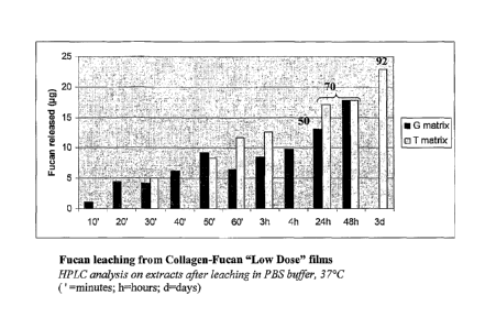
Fucan leaching from the collagen film was studied using High Pressure Liquid
Chromatography (HPLC on a Dionex Carbo Pac 100).
Measurements were performed on the extracts of the collagen in combination
with the collagen-fucan Low Dose after several hydration times of from about
20 minutes
to about 96 hours in PBS buffer solution (Na2HPO4, 7H20 at about 0.726 g/L,
NaCl at
about 9g/L, KH2PO4 t about 0.21 g/L, [PBS Gibco, Invitrogen ref 20012-019]
from
Gibco, Life Sciences), at about 37 C. The results are set forth in Figure 1.
As can be seen in Figure 1, about 50% and about 70% of the incorporated fucan
was released during the first 24 and 48 hours, respectively, of hydration in
the PBS
medium.
29
CA 02691412 2009-12-18
WO 2009/010879
PCT/1B2008/002695
From these results, it can be seen that the fucan on the mesh may possess both
local and diffuse effects during the first phase of implantation, which is the
critical phase,
in terms of immune and adverse reaction due to the surgery.
Moreover, incorporation of the fucan in a collagen film did not significantly
alter
its physico-chemical properties, in the case of fucan concentrations of less
than about
0.1% (m/V).
EXAMPLE 5
A mediated bacterial adhesion assay involving the fucan in collagen as
described
above in Example 4 was conducted. Cultures of the bacterial strain S. aureus
(ATCC
6538; Gram +) were prepared by incubating a well-isolated representative
colony
selected from an agar plate in about 1 ml of broth at about 37 C overnight.
Bacteria were harvested from this saturated bacterial suspension by
centrifugation
at about 3500 revolutions per minute (rpm) for about 15 minutes. After
discarding the
supernatant, the bacterial pellet, about 107 colony forming units (cfu)/ml,
was suspended
in about 1 ml of fresh broth and about 100 [it of tritiated thymidine (from
Amersham,
activity about 1 mCi/m1) was added. The resulting bacterial suspensions were
incubated
for about 3 hours at about 37 C to obtain bacteria in the exponential growth
phase. After
the incubation period, the bacterial suspension was harvested twice at about
3500 rpm for
about 15 minutes to remove the excess unbound radioactive thymidine.
A solution of PBS with Ca ++ and Mg++ was then added to the bacterial pellet
to
obtain suitable bacterial dilutions (about 106-107 cfu/ml) and the bacterial
suspension was
homogenized using a vortex-mixer.
CA 02691412 2009-12-18
WO 2009/010879
PCT/1B2008/002695
Collagen from Example 2 and collagen-fucan Low Dose samples from Example 4
were utilized to prepare films. The films were first coated with plasma
constituents and
then incubated with about 500 p.1 of PBS for about 50 hours under stirring.
About 500111 of the washed-log phase radiolabeled bacterial suspension (about
106-107 cfu/ml) described above was then added to the films. The bacterial
suspension
on the film was incubated for about 3 hours at about 37 C. After about 5
washings with
PBS buffer, each sample was transferred to counting vials; about 10 ml of
scintillation
fluid (Optiphase Hisahe, EG and G) were added; the amount of bacteria which
adhered
onto the implants was measured using an automatic 13-liquid scintillation
analyser model
(Tri CARB 2100 TR (Packard [ND 1401)).
In order to check that the investigated bacteriophobic activity was due to the
fucan, additional collagen films (with and without fucan) were first coated
with plasma
constituents and incubated with a mixture of about 500 1 of the above washed-
log phase
radiolabeled bacterial suspension (about 106-107 cfu/ml) in combination with
about 500
.1 of a solution of collagen-fucan Low Dose implant extracts obtained after
about 50
hours of incubation in PBS buffer at about 37 C. The resulting mixture was
incubated
for about 3 hours at about 37 C. After about 5 washings with PBS buffer, each
sample
was transferred to counting vials; about 10 ml of scintillation fluid
(Optiphase Hisahe,
EG and G) were added; the amount of bacteria which adhered onto the implants
was
measured using an automatic 13-liquid scintillation analyser model Tri CARB
2100 TR
(Packard IND 1401).
The results are set forth in Figure 2, which shows the bacterial adhesion on
collagen and collagen-fucan films. In Figure 2, the two bar graphs for (a)
demonstrate
31
CA 02691412 2009-12-18
WO 2009/010879
PCT/1B2008/002695
the adhesion of S. aureus on collagen (C) and collagen-fucan Low Dose (CF)
films; the
three bar graphs for (b) demonstrate the adhesion of S. aureus on a control of
porous
polypropylene (T'), collagen films (C') and collagen-fucan Low Dose films
(CF') in the
presence of collagen-fucan Low Dose extracts.
As can be seen in Figure 2, the bacterial adhesion was more prevalent on the
control and was statistically different than bacterial adhesion obtained on
collagen films.
Moreover, as can be seen in Figure 2(a), films possessing fucan incorporated
into
collagen demonstrated a decrease in bacterial adhesion. The inhibition rate
reached an
average value of about 37 % (after a period of incubation of about 50 hours in
buffer).
In the case of the extract diffusion, an inhibition of bacterial adhesion on
the three
types of implants was observed (see Figure 2(b)). Bacterial adhesion
inhibition reached
an average value of about 40 %, which was nearly equal the rate obtained for
the first
experiment (about 37 %). The bacterial inhibition obtained on T' (textile
alone) was less
(about 31 % inhibition) as compared to the one observed on C' (collagen film)
and CF'
(Low Dose film).
The above results demonstrate that the fucan was released from the collagen-
fucan Low Dose film during the first 50 hours, and was responsible for the
inhibition of
adhesion.
32
CA 02691412 2009-12-18
WO 2009/010879
PCT/1B2008/002695
EXAMPLE 6
In vivo experiments were conducted to check the antibacterial properties of a
collagen-fucan implant in a rat contaminated model.
About 2.5 x 3.5 cm shaped composite implants were constructed with the two-
dimensional non biodegradable textile of Example 1. Multiple implants were
prepared;
some possessed a collagen film coating as described in Example 2, while others
possessed a collagen-fucan film coating as described in Example 4. The
implants were
implanted in rat peritoneal cavities at the site of a preformed 1.5 x 2.5 cm
parietal defect.
The implants were sutured with 6 points and the surgery was ended with suture
strand.
High virulence E. coli bacteria were inoculated (109 bacteria in 2 mL of
phosphate buffer
Na2HPO4, 7I420; 0.1M; pH 7.2 [PBS, Invitrogen 20012-068 ]) by means of a
percutaneous injection in the region of the implant/defect.
After time periods of about 2 days and about 30 days, the rats were sacrificed
and
the meshes explanted. The proliferated bacteria were detached from the
explants and
cultured on agar gelose before being counted. Immunohistology was also
performed in
order to identify the bacteria.
Fucan, at high dose, inhibited bacterial proliferation after 30 days (2 logs
of
inhibition). No significant effect was observed after 2 days of incubation.
The results are
summarized in Table 1 below.
33
CA 02691412 2009-12-18
WO 2009/010879
PCT/1B2008/002695
Table 1
# of Rats Mesh reference Timing E. Coli
7 Textile* J2 5.43E+08
7 Textile High Dose** J2 2.73E+08
7 Textile Low Dose*** J2 5.17E+08
7 Textile* J30 9.43E+10
7 Textile High Dose** J30 1.74E+09
Textile Low Dose*** J30 8.28E+10
J2 = rats sacrificed after 2 days
J30 = rats sacrificed after 30 days
5 * Textile ¨ mesh with collagen coating
** Textile High Dose = mesh with collagen and High Dose fucan coating
*** Textile Low Dose = mesh with collagen and collagen-fucan Low Dose coating
EXAMPLE 7
In-vitro cell culture characterization. The effects of fucans, incorporated in
the
collagen films as described above in Example 4, were analyzed at several
concentrations
on several different cells and their effects on cell proliferation were
studied. The cells
tested included fibroblasts, mesothelial cells, mesenchymal stem cells,
urothelial cells,
endothelial cells and smooth muscle cells (SMCs).
Normal human dermal fibroblasts (NHDF, Cambrex CC2511) were cultured in
Dulbecco's Modified Eagle's Medium (DMEM, Cambrex CC3132) supplemented with
about 10% fetal calf serum (FCS, Fischer 10270106), about 1% Fungizone
(Fischer
15290026) and about 1% Penicillin/Streptomycin (Fischer 15140122). Cells were
maintained in a controlled atmosphere (about 37 C, about 95% relative humidity
and
about 5% CO2). All the experiments were carried out using cells with passage
numbers
of less than about 25 (passage = treatment with trypsin-ethylenediamine
tetraacetic acid
(EDTA)).
34
CA 02691412 2009-12-18
WO 2009/010879
PCT/1B2008/002695
NHDF were grown onto the collagen-fucan film of Example 4, which included a
collagen based gel associated with different concentrations of fucan,
optionally
associated with a 2D textile of Example 1. Cell growth was studied for about 7
days.
Each experiment was repeated 3 times. The results of this experiment are set
forth in
Figure 3, which shows the fibroblast growth on collagen-fucan TH90 RED A2 0305
PUP
30. As depicted in Figure 3, GO, G0.01, G0.05 were collagen film without
textile
containing respectively about 0%, about 0.01%, and about 0.05% (m/V) of fucan;
TO,
TO.01, TO .05 were composite collagen films/2D textiles containing
respectively about
0%, about 0.01%, and about 0.05% (m/V) of fucan.
The fibroblasts demonstrated an affinity for the collagen-fucan surfaces (see
Figure 3). The optimal concentration was evaluated at about 0.01% (rn/V) of
fucan in the
collagen solution, i.e., a non degrading concentration for the physical
integrity of the
film.
The presence of the textile reduced the cell adhesion and proliferation rate.
This
may be due to the surface properties (e.g., planarity) induced by the presence
of the
textile, as well as differences in the degradation rate of the film and its
impact on the cell
adhesion and proliferation.
EXAMPLE 8
Cell migration is a major process in tissue repair and wound healing. Cell
migration was studied using a Boyden chamber assay through inserts with 8 um
pores. A
depiction of a Boyden chamber is set forth in Figure 4.
CA 02691412 2009-12-18
WO 2009/010879
PCT/1B2008/002695
Cells were suspended in culture medium and added to the upper chamber of the
assay wells. Migration assays were performed in the presence of fucan matrices
(TO,
TO.01, GO, G0.01 as described above in Example 7), polypropylene (PP) or
polyethylene
terephthalate (PET) in the lower chamber. The chemotactic response to fucan
was
determined for fibroblasts. Positive controls were performed (migration in
presence of
about 20% fetal calf serum (FCS, Fischer 10270106)).
The results are presented in Figure 5, which demonstrates the chemotactic
response of fucans on fibroblasts. As can be seen in Figure 5, the migration
of fibroblasts
was stimulated in presence of fucans (comparison between G0.01 and GO and
between
TO.01 and TO). The same effect was observed when fucan was released from the
matrix
including polypropylene and collagen (T0.01) or from the sole collagen matrix
(G0.01),
and reached about 60%.
No statistical difference was observed when the fibroblasts or mesothelial
cells
migrated in the presence of PP or PET matrices in the medium.
EXAMPLE 9
The anti-complement activity of fucans was tested via a CH50 test, a standard
hemolytic assay in total human serum.
Complement activation in human serum was induced by the introduction of sheep
erythrocytes coated with rabbit antibodies and then recognized as foreign
elements. This
led to the activation of the classical pathway of the complement system, and
hence to the
lysis of erythrocytes.
36
CA 02691412 2009-12-18
WO 2009/010879
PCT/1B2008/002695
The amount of released hemoglobin was then determined by an optical density
(OD) measurement at about 414 nm. The human serum dilution was adjusted for a
known amount of erythrocytes, in order to lyse about 50% of the red blood
cells (CH50).
In order to check the impact of the fucan on the complement activation in
solution, the fucan was added with the sheep erythrocytes. A decrease in cell
lysis, as
evidenced by a decrease of the OD at about 414 nm, demonstrated that the fucan
inhibited the complement activation.
Heparin (heparin H108 173 UI/mg, Choay-Sanofi) and fucan P240 RED
synthesized in the Laboratoire de Recherche sur les Macromolecules (LRM, CNRS
UMR
7540, France) were also tested instead of fucan.
The results are set forth in Figure 6, which depicts the anti-complement
activity of
heparin H108, P240 RED and fucan TH9ORED A2 0305 PUF 30 in solution.
As can be seen in Figure 6, the fucan TH9ORED A2 0305 PUF30 like its
precursor P240 RED, presented dose-dependent anti-complement activity; both
had an
IC50 (median inhibition concentration) of about 4 Rg/mL, evidencing a strong
anti-
complement activity as compared with the reference heparin H108 (IC50 about 30
g/mL
as measured in this test).
EXAMPLE 10
In order to check the in-vivo integration of composite implants made of a 3D
non
biodegradable textile (PET) associated with a collagen film and collagen-fucan
film, an
intraperitoneal implantation in rat peritoneal cavity was performed. 2 sites
in 25 rats
37
CA 02691412 2015-05-25
=
were implanted with 3 kinds of implants: collagen/textile implant, as a
control; collagen-
fucan Low Dose/textile implant; and collagen-fucan High Dose/textile implant.
The mesh integration and associated adherences were observed after about 3
days,
about 5 days, about 7 days, and about 6 weeks, by both macroscopic and
immunohistological observations.
The results of the histological analysis of explanted composite implants is
set
forth in Figure 7 which depicts images of tissue obtained by histological
observation. As
can be seen in Figure 7, after 3 days of implantation better integration of
the mesh
associated with the collagen-fucan Low Dose was observed compared with the
composite
control. The mesh containing the collagen-fucan Low Dose (0.01% (m/V)) showed
multiple layers of fibroblastic cells after about 3 days of implantation.
Statistical
differences were observed for tissue integration between the collagen-fiican
Low Dose
and control.
The following days (see day 5 data and day 7 data on Figure 7) presented a
faster
integration of the mesh associated with the collagen-fucan Low Dose compared
to the
composite control, with comparable inflammatory reactions. Integration of all
the
meshes was observed about 7 days after implantation. The moderate inflammatory
reactions observed did not prevent the final integration of the mesh.
No data were available for the collagen-fuean Low Dose implant at 6 weeks,
because the rat died during the experiment. This death was not due to the
experiment.
While the invention has been described in connection with specific embodiments
thereof, it will be understood that the scope of the claims should not be
limited by the
38
CA 02691412 2015-05-25
preferred embodiments set forth in the examples, but should be given the
broadest
interpretation consistent with the description as a whole.
39