Note: Descriptions are shown in the official language in which they were submitted.
CA 02691664 2009-12-18
WO 2009/006146 PCT/US2008/068141
Macrophage-Enhanced MRI (MEMRI)
Cross Reference to Related Applications
The present application claims priority from provisional patent application
U.S.
Application Serial Number 60/947,259, filed June 29, 2007, which is hereby
incorporated by
reference herein.
Technical Field
The present invention relates to whole body MRI scanning and cancer staging
using
macrophage-seeking MRI agents to perform Macrophage-Enhanced MRI or "MEMRI".
Back2round Art
Cancer is one of the leading causes of death in the developed world, resulting
in over
500,000 deaths per year in the United States alone. Over one million people
are diagnosed
with cancer in the U.S. each year, and overall it is estimated that more than
1 in 3 people will
develop some form of cancer during their lifetime. Though there are more than
200 different
types of cancer, four of them - breast, lung, colorectal, and prostate -
account for over half
of all new cases (Jemal et al., CA Cancer J. Clin. 53:5-26 (2003)). Cancer
metastasis is
considered to be due to the distribution of cancer cells via the blood-with
liver, lung, bone,
and CNS as common sites at risk, or the lymphatics with lymph node and bone as
metastatic
risk sites.
Breast cancer is the most common cancer in women, with an estimate 12% of
women
at risk of developing the disease during their lifetime. Although mortality
rates have
decreased due to earlier detection and improved treatments, breast cancer
remains a leading
cause of death in middle-aged women. Furthermore, metastatic breast cancer is
still an
incurable disease. On presentation, most patients with metastatic breast
cancer have only one
or two organ systems affected, but as the disease progresses, multiple sites
usually become
involved. The most common sites of metastatic involvement are locoregional
recurrences in
the skin and soft tissues of the chest wall, as well as in regional lymph
nodes. The most
1
CA 02691664 2009-12-18
WO 2009/006146 PCT/US2008/068141
common site for distant metastasis is the bone (30-40% of distant metastasis),
followed by
the lungs and liver. And although only approximately 1-5% of women with newly
diagnosed
breast cancer have distant metastasis at the time of diagnosis, approximately
50% of patients
with local disease eventually relapse with metastasis within five years. At
present the median
survival from the manifestation of distant metastases is about three years.
Current methods of diagnosing and staging breast cancer include the tumor-node-
metastasis (TNM) system that relies on tumor size, tumor presence in lymph
nodes, and the
presence of distant metastases as described in the American Joint Committee on
Cancer,
AJCC Cancer Staging Manual, Philadelphia, Pa., Lippincott-Raven Publishers,
6th ed.
(2006), pp 221-240, and in Harris, J R: "Staging of breast carcinoma" in
Harris, J. R., et al.,
eds., Breast Diseases, Philadelphia, Lippincott (1991). These parameters are
used to provide
a prognosis and select an appropriate therapy. The morphologic appearance of
the tumor can
also be assessed but because tumors with similar histopathologic appearance
can exhibit
significant clinical variability, this approach has serious limitations.
Finally assays for cell
surface markers can be used to divide certain tumors types into subclasses.
For example, one
factor considered in the prognosis and treatment of breast cancer is the
presence of the
estrogen receptor (ER) as ER-positive breast cancers typically respond more
readily to
hormonal therapies such as tamoxifen or aromatase inhibitors than ER-negative
tumors. Yet
these analyses, though useful, are only partially predictive of the clinical
behavior of breast
tumors, and there is much phenotypic diversity present in breast cancers that
current
diagnostic tools fail to detect and current therapies fail to treat.
Prostate cancer is the most common cancer in men in the developed world,
representing an estimated 33% of all new cancer cases in the U.S., and is the
second most
frequent cause of death (Jemal et al., CA Cancer J. Clin. 53:5-26 (2003)).
Since the
introduction of the prostate specific antigen (PSA) blood test, early
detection of prostate
cancer has dramatically improved survival rates, and the five year survival
rate for patients
with local and regional stage prostate cancers at the time of diagnosis is
nearing 100%. Yet
more than 50% of patients will eventually develop locally advanced or
metastatic disease
(Muthuramalingam et al., Clin. Oncol. 16:505-516 (2004)).
Currently radical prostatectomy and radiation therapy provide curative
treatment for
the majority of localized prostate tumors. However, therapeutic options are
very limited for
2
CA 02691664 2009-12-18
WO 2009/006146 PCT/US2008/068141
advanced cases. For metastatic disease, androgen ablation with luteinizing
hormone-
releasing hormone (LHRH) agonist alone or in combination with anti-androgens
is the
standard treatment. Yet despite maximal androgen blockage, the disease nearly
always
progresses with the majority developing androgen-independent disease. At
present there is
no uniformly accepted treatment for hormone refractory prostate cancer, and
chemotherapeutic regimes are commonly used (Muthuramalingam et al., Clin.
Oncol.
16:505-516 (2004); Trojan et al., Anticancer Res. 25:551-561 (2005)).
Colorectal cancer is the third most common cancer and the fourth most frequent
cause of cancer deaths worldwide (Weitz et al., 2005, Lancet 365:153-65).
Approximately 5-
10% of all colorectal cancers are hereditary with one of the main forms being
familial
adenomatous polyposis (FAP), an autosomal dominant disease in which about 80%
of
affected individuals contain a germline mutation in the adenomatous polyposis
coli (APC)
gene. Colorectal carcinoma has a tendency to invade locally by circumferential
growth and
elsewhere by lymphatic, hematogenous, transperitoneal, and perineural spread.
The most
common site of extralymphatic involvement is the liver, with the lungs the
most frequently
affected extra-abdominal organ. Other sites of hematogenous spread include the
bones,
kidneys, adrenal glands, and brain.
The current staging system for colorectal cancer is based on the degree of
tumor
penetration through the bowel wall and the presence or absence of nodal
involvement. This
staging system is defined by three major Duke's classifications: Duke's A
disease is confined
to submucosa layers of colon or rectum; Duke's B disease has tumors that
invade through the
muscularis propria and may penetrate the wall of the colon or rectum; and
Duke's C disease
includes any degree of bowel wall invasion with regional lymph node
metastasis. While
surgical resection is highly effective for early stage colorectal cancers,
providing cure rates
of 95% in Duke's A patients, the rate is reduced to 75% in Duke's B patients
and the presence
of positive lymph node in Duke's C disease predicts a 60% likelihood of
recurrence within
five years. Treatment of Duke's C patients with a post surgical course of
chemotherapy
reduces the recurrence rate to 40%-50%, and is now the standard of care for
these patients.
Lung cancer is the most common cancer worldwide, the third most commonly
diagnosed cancer in the United States, and by far the most frequent cause of
cancer deaths
(Spiro et al., Am. J. Respir. Crit. Care Med. 166:1166-1196 (2002); Jemal et
al., CA Cancer
3
CA 02691664 2009-12-18
WO 2009/006146 PCT/US2008/068141
J. Clin. 53:5-26 (2003)). Cigarette smoking is believed responsible for an
estimated 87% of
all lung cancers making it the most deadly preventable disease. Lung cancer is
divided into
two major types that account for over 90% of all lung cancers: small cell lung
cancer (SCLC)
and non-small cell lung cancer (NSCLC). SCLC accounts for 15-20% of cases and
is
characterized by its origin in large central airways and histological
composition of sheets of
small cells with little cytoplasm. SCLC is more aggressive than NSCLC, growing
rapidly
and metastasizing early and often. NSCLC accounts for 80-85% of all cases and
is further
divided into three major subtypes based on histology: adenocarcinoma, squamous
cell
carcinoma (epidermoid carcinoma), and large cell undifferentiated carcinoma.
The most
common metastatic sites are pleura, lung, bone, liver, brain, and pericardium.
Lung cancer typically presents late in its course, and thus has a median
survival of
only 6-12 months after diagnosis and an overa115 year survival rate of only 5-
10%. Although
surgery offers the best chance of a cure, only a small fraction of lung cancer
patients are
eligible with the majority relying on chemotherapy and radiotherapy. Despite
attempts to
manipulate the timing and dose intensity of these therapies, survival rates
have increased
little over the last 15 years (Spiro et al., Am. J. Respir. Crit. Care Med.
166:1166-1196
(2002)).
Cancer arises from dysregulation of the mechanisms that control normal tissue
development and maintenance, and increasingly stem cells are thought to play a
central role
(Beachy et al., Nature 432:324 (2004)). During normal animal development,
cells of most or
all tissues are derived from normal precursors, called stem cells (Morrison et
al., Cell
88:287-298 (1997); Morrison et al., Curr. Opin. Immunol. 9:216-221 (1997);
Morrison et al.,
Annu. Rev. Cell. Dev. Biol. 11:35-71 (1995)). Stem cells are cells that: (1)
have extensive
proliferative capacity; (2) are capable of asymmetric cell division to
generate one or more
kinds of progeny with reduced proliferative and/or developmental potential;
and (3) are
capable of symmetric cell divisions for self-renewal or self-maintenance.
Solid tumors are composed of heterogeneous cell populations. For example,
breast
cancers are a mixture of cancer cells and normal cells, including mesenchymal
(stromal)
cells, inflammatory cells, and endothelial cells. Classic models of cancer
hold that
phenotypically distinct cancer cell populations all have the capacity to
proliferate and give
rise to a new tumor. In the classical model, tumor cell heterogeneity results
from
4
CA 02691664 2009-12-18
WO 2009/006146 PCT/US2008/068141
environmental factors as well as ongoing mutations within cancer cells
resulting in a diverse
population of tumorigenic cells. This model rests on the idea that all
populations of tumor
cells would have some degree of tumorigenic potential. (Pandis et al., Genes,
Chromosomes
& Cancer 12:122-129 (1998); Kuukasjrvi et al., Cancer Res. 57:1597-1604
(1997); Bonsing
et al., Cancer 71:382-391 (1993); Bonsing et al., Genes Chromosomes & Cancer
82:173-183
(2000); Beerman H. et al., Cytometry. 12:147-154 (1991); Aubele M & Werner M,
Analyt.
Cell. Path. 19:53 (1999); Shen L et al., Cancer Res. 60:3884 (2000)).
An alternative model for the observed solid tumor cell heterogeneity is that
solid
tumors result from a "solid tumor stem cell" that subsequently undergoes
chaotic
development through both symmetric and asymmetric rounds of cell divisions. In
this stem
cell model, solid tumors contain a distinct and limited (possibly even rare)
subset of cells that
share the properties of normal "stem cells", in that they extensively
proliferate and efficiently
give rise both to additional solid tumor stem cells (self-renewal) and to the
majority of tumor
cells of a solid tumor that lack tumorigenic potential. Indeed, mutations
within a long-lived
stem cell population may initiate the formation of cancer stem cells that
underlie the growth
and maintenance of tumors and whose presence contributes to the failure of
current
therapeutic approaches.
MRI is noninvasive, tomographic, nonionizing, and able to generate images with
high
resolution and excellent soft tissue contrast. Whole body MRI has recently
been used for
evaluation of metastasis in bone in the absence of contrast agents. When MRI
has been used
in tumor staging, it has been by taking advantage of inherent tissue
differences in MR
properties that could be imaged by varying the MR image sequences. To obtain
whatever
information is contained in these inherent tissue differences for all tissues
at cancer risk has
required selection of different imaging sequences for each potential host
tissue as well as the
repeated programming and positioning of the patient within the MR instrument.
It is known
that the administration of some agents can change tissue MR properties in a
useful way. The
most popular agents for assessing the primary tumor are small gadolinium
chelates. Given
intravenously, these agents are distributed by blood perfusion and can
identify regions of
excess vascular leakiness or enlarged extracellular spaces that may herald the
presence of
cancer. However, these agents have no cell targeting capabilities and their
distribution and
accumulation are not specific for cancer nor for the tissue at risk for cancer
metastasis.
5
CA 02691664 2009-12-18
WO 2009/006146 PCT/US2008/068141
Bolus administration and dynamic MRI may provide some additional information
about the
degree of vascular leakiness, but such information can only be obtained for
one body region
of interest. As is described in example 1(vide infra), the use of such
contrast-enhanced MRI
may be insufficient to characterize the cancer stage even with respect to the
primary tumor.
Positive contrast agents cause a reduction in the Tl relaxation time
(increased signal
intensity on T 1 weighted images). They are typically small molecular weight
compounds
containing as their active element Gadolinium, Manganese, or Iron. All of
these elements
have unpaired electron spins in their outer shells and long relaxivities.
Negative contrast agents (appearing predominantly dark on MRI) are small
particulate aggregates often termed superparamagnetic iron oxides (SPIO) or
ultrasmall
superparamagnetic iron oxides (USPIO). USPIO typically are less than about 100
nanometers in diameter, and often have a mean diameter of less than 50 nm.
These agents
produce predominantly spin spin relaxation effects (local field
inhomogeneities), which
results in shorter Tl and T2 relaxation times. SPIO's and ultrasmall
superparamagnetic iron
oxides (USPIO) usually consist of a crystalline iron oxide core containing
thousands of iron
atoms and a shell of polymer, dextran, polyethyleneglycol, and produce very
high T2
relaxivities.
For the lymph, liver and spleen images, ferumoxtran-10 is useful because it
identifies
normal healthy tissues that are enriched with macrophages. The reduced
population of
macrophage in the tumor in these particular types of tissue permits
visualization of the tumor
by the absence of enhancement relative to the normal tissue. In contrast, for
brain tumors, it
has been proposed that ferumoxtran-10 is useful to image brain tumors because
of its
association with reactive cells surrounding the tumor, including astrocytes
and dendrites,
which are found only in the brain and other nerve tissue and macrophages. This
study
(Nixon et al. Neuropath and Appl. Neurobiol (2004), 30, 456-471) focused on
primary
tumors in the brain, and the authors report that there was a pattern of
sharply delimited cells
without processes that were histologically identifiable as macrophages and
another pattern of
stellate-shaped cells typical of reactive astrocytes. This lead the authors to
conclude that
uptake in primary brain tumors of the iron oxide contrast agent studied is
primarily
concentrated in reactive cells in and around the brain tumor, rather than the
tumor cells
6
CA 02691664 2009-12-18
WO 2009/006146 PCT/US2008/068141
themselves. Thus, they could not conclude that all of the lesions imaged with
the iron oxide
contrast agent were actually tumors (id., at 464).
A special group of negative contrast agents (appearing dark on MRI) are
perfluorocarbons (perfluorochemicals), because their presence excludes the
hydrogen atoms
responsible for the signal in MR imaging. These agents are reported to allow
enhanced,
sensitive detection and quantification of occult microthrombi within the
intimal surface of
atherosclerotic vessels in symptomatic patients and provide direct evidence to
support acute
therapeutic intervention, particularly if used in combination with gadolinium
(see Flacke et
al. Circulation. 2001;104:1280).
In sum, the prior art teaches the use of contrast agents that are not specific
to cancer
at all, namely gadolinium chelates and manganese compounds, or contrast agents
including
perfluorocarbon compounds and biofunctionalized nanoparticles containing
perfluorocarbons
and gadolinium for imaging arterial plaques and atherosclerotic vessels, or
SPIO and USPIO
contrast agents such as ferumoxtran-10 that are used only for lymph, liver,
spleen and
recently brain imaging. But even as recently as June 28, 2007, an update on
the Magnetic
Resonance - Technology Information Portal by Robert R. Edelman, Professor and
Chairman, Department of Radiology at Northwestern University's Feinberg School
of
Medicine states that "The design objectives for the next generation of MR
contrast agents
will likely focus on prolonging intravascular retention, improving tissue
targeting, and
accessing new contrast mechanisms .... Technical advances in MR imaging will
further
increase the efficacy and necessity of tissue-specific MRI contrast agents."
(Emphasis
added, see Google's cache of http://www.mr-
tip.com/servl.php?type=dbl&dbs=Contrast%20Agents as retrieved on Jun 28, 2007
21:51:08 GMT. To link to or bookmark this page, use the following url:
http://www. o~ogle.com/search?q=cache:ornByMzsGVgJ:www.mr-
tip. com/servl .php%3Ftype%3Ddb 1
%26dbs%3DContrast%2520Agents+tissue+specific+MR
I+contrast+agents&hl=en&ct=clnk&cd=2&gl=us).
Summary of the Invention
In a first embodiment of the invention there is provided a method of assessing
stage
of cancer in a subject, the method comprising administering a macrophage
imaging agent to
7
CA 02691664 2009-12-18
WO 2009/006146 PCT/US2008/068141
the subject, making a magnetic resonance image of regions of the subject's
body at cancer
risk, and using the image to assess macrophage density and displacement
associated with any
primary cancer or metastatic cancer in the subject, such density and
displacement being
indicative of neoplasia.
Another embodiment provides a method as described, wherein using the image
includes observing macrophage activity associated with a primary tumor or with
any
metastatic tumor in bone, lymph node, spleen, liver, central nervous system,
lung, or other
organ. In particular embodiments, the regions collectively include the entire
body. In other
particular embodiments, the macrophage imaging agent is an ultrasmall
superparamagnetic
iron oxide particle and in still more particular embodiments, the macrophage
imaging agent
has a blood half-life sufficient to permit microphage trapping throughout the
regions at
cancer risk. In related particular embodiments the macrophage imaging agent is
a complex of
ultrasmall superparamagnetic iron oxide and a polysaccharide. In still other
related
embodiments, the polysaccharide is selected from the group consisting of
dextran, reduced
dextran and a derivative thereof.
Another particular embodiment provides a method of assessing efficacy of an
anticancer treatment in a subject comprising administering a macrophage
imaging agent to
the subject before the anticancer treatment, making a magnetic resonance image
of regions of
the subject's body to be targeted by the anti-cancer treatment to establish a
pre-treatment
image, administering the anticancer treatment to the subject, administering
the macrophage
imaging agent to the subject after the anti-cancer treatment, making a
magnetic resonance
image of the regions of the subject's body targeted by the anticancer
treatment to establish a
post-treatment image, and assessing any change in the post-treatment image
compared to the
pre-treatment image with respect to macrophage density and displacement
associated with a
primary cancer or metastatic cancer in the subject, wherein assessment of such
change in
macrophage density and displacement is indicative of the efficacy of the anti-
cancer
treatment. In more particular embodiments, the anticancer treatment may be
attempted
extirpation or in situ ablation, chemotherapy, radiation therapy, or a
combination of any of
the individual treatment modalities. In still more particular embodiments, the
macrophage
density and displacement associated with a primary cancer or metastatic cancer
is reduced or
shows no change in the post-treatment image compared to the pre-treatment
image. In a
8
CA 02691664 2009-12-18
WO 2009/006146 PCT/US2008/068141
related embodiment, the macrophage density and displacement associated with a
primary
cancer or metastatic cancer is increased in the post-treatment image compared
to the pre-
treatment image, suggesting progression. In yet a more particular embodiment,
the
macrophage density and displacement associated with a primary cancer or
metastatic cancer
shows regression or is progression free in the post-treatment image compared
to the pre-
treatment image.
Another particular embodiment provides a method of determining frequency of
follow-up MEMRI evaluation in a subject, the method comprising performing a
first whole
body MEMRI evaluation of the subject at date one to determine a first level of
macrophage
density at a tumor site of interest, performing a second whole body MEMRI
evaluation of the
subject at date two to determine a second level of macrophage density at the
tumor site of
interest, and determining a date three for performing a third whole body MEMRI
evaluation
of the subject, thereby determining the frequency of follow-up MEMRI
evaluation in the
subject at the tumor site of interest.
Still another particular embodiment provides a method for determining
metastatic
potential of cancer foci in a subject, the method comprising using whole body
MEMRI
evaluation to identify macrophage density at a tumor site of interest, the
macrophage density
at the tumor site of interest being an indicator of metastatic potential of
the cancer foci and
assessing the macrophage density at the tumor site of interest, thereby
determining metastatic
potential for the cancer foci in the subject based on the macrophage density.
Still another embodiment provides a method for determining prognosis of cancer
in a
subject, the method comprising performing a whole body MEMRI evaluation of the
subject
to identify macrophage density at a tumor site of interest, assessing the
macrophage density
to identify primary and/or metastatic tumors in the subject, and determining
the prognosis of
the cancer in the subject based on macrophage density of the primary and/or
metastatic
tumors, the macrophage density being an indicator of the prognosis of the
cancer whereby
low macrophage density relative to normal cells is an indicator of a more
favorably
prognosis and high macrophage density relative to normal cells is an indicator
of a less
favorable prognosis.
In yet another particular embodiment there is provided a report card for
follow-up
assessment of cancer status, the report card comprising fillable space for
patient information;
9
CA 02691664 2009-12-18
WO 2009/006146 PCT/US2008/068141
fillable space for date information; fillable space for initial MEMRI
information; fillable
space for follow-up MEMRI evaluation information; fillable space for next
scheduled
MEMRI evaluation; optionally, fillable space for initial diagnosis;
optionally, fillable space
for initial stage information; optionally; and optionally, fillable space for
TNM Stage.
Still another particular embodiment provides a method for directing site of
biopsy in
a subject, the method comprising performing a whole body MEMRI evaluation of
the subject
to identify macrophage density at a tumor site of interest and assessing the
macrophage
density to identify the site of biopsy in the subject, macrophage density
being an indicator of
tumor growth.
Another particular embodiment provides a method for providing individualized
cancer treatment to a subject in need thereof using whole body MEMRI
evaluation, the
method comprising performing a whole body MEMRI evaluation of the subject to
identify
macrophage density at a primary and/or tumor site of interest, assessing the
macrophage
density to identify characteristics (type, location, phenotypic and
morphological) of the
primary and/or metastatic tumors in the subject, assessing the characteristics
of the primary
and/or metastatic tumors in the subject to determine optimal treatment, and
providing
individualized cancer treatment to the subject based on the assessment of the
primary and/or
metastatic tumors in the subject, as determined using whole body MEMRI
evaluation.
And in still another particular embodiment there is provided a macrophage
biomarker
capable of being administered to a subject from between 12 and 168 hours prior
to whole
body MEMRI evaluation.
Brief Description of the Drawin2s
The foregoing features of the invention will be more readily understood by
reference
to the following detailed description, taken with reference to the
accompanying drawings, in
which:
Fig. 1 shows an illustration of a patient placed within a whole-body MRI
system for
scanning, here used with a currently-approved contrast agents to visualize the
arterial vessels
throughout the body. (see Nael et al. (2007) Am. J. Radiol, 188, 529-39.
CA 02691664 2009-12-18
WO 2009/006146 PCT/US2008/068141
Fig. 2A-C show three variations of a cancer report card that may be used when
practicing embodiments of the present invention.
Figure 3 is an illustration showing a tumor region with increased macrophage
density,
and the process whereby tumor-associated macrophages produce chemotactic
factors (CC-
Chemokines, e.g. CCL2), macrophage colony stimulatory factor (M-CSF) and
vascular
endothelial growth factor (VEGF) to generate new blood vessels and facilitate
further growth
of the tumor (angiogenesis) (illustration from Allavena et al., (2006) Eur. J.
Cancer, 42, 717-
727).
Figures 4A and 4B show a patient with breast cancer with macrophages around
the
primary tumor and displaced from the metastatic tumor in an adjacent lymph
node tumor.
Figure 4A is an in vivo MRI of the patient's breast with a contrast agent of
the invention;
Figure 4B is an in vitro MRI of the removed specimen containing the tumor and
a metastatic
lymph node, also with a contrast agent of the invention. The arrow in Figure
4A shows the
very clear presence of a dark accumulation of macrophages indicating a tumor.
Figure 4B
shows the lymph node tumor indicated by a dark outline of macrophages where
the center
area of the tumor is light, because the cancer cells have displaced the
macrophages from this
central region of the tumor.
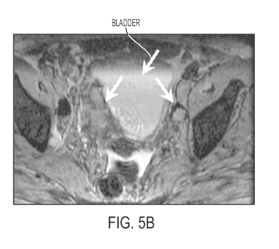
Figures 5A and 5B show a patient with bladder cancer with macrophages around
the
primary tumors. The bladder is indicated generally with an arrow in Figures 5A
and 5B as
the large central light area in the center of the pelvis region. Fig. 5A is
the MRI without
contrast agent and Figure 5B is the image with contrast agent of the
invention. In Figure 5A,
the tumor's presence is only hinted at by the "crease" in the bladder (shown
with an arrow)
that seems to be an indication of pressure on or displacement of the bladder
along this
juncture. Figure 5B, with contrast agent, clearly shows the line of
demarcation for the tumor
along that "crease", with the massive tumor showing as a dark mass directly to
the left of this
line, continuing down to a point and back again. A second smaller tumor is
indicated with an
arrow to the right of the bladder, appearing as a bulls eye type node. This
second tumor is
outlined with a dark ring of macrophages. The center of the tumor shows up
lighter where
the cancer cells have displaced the macrophages. As an indication of the power
of this
contrast agent in cancer diagnosis and staging, this tumor is not identifiable
as a tumor at all
in the image (5A) with no contrast agent.
11
CA 02691664 2009-12-18
WO 2009/006146 PCT/US2008/068141
Figures 6A, 6B and 6C show MRI depictions of a patient with prostate cancer.
The
prostate is indicated generally as the large central circular space in the
center of the pelvis
region. Figure 6A is the MRI without contrast agent, and Figures 6B and 6C are
the MRIs
with a contrast agent of the invention. The presence of a tumor is not
indicated at all in
Figure 6A, the MRI without contrast agent. In stark comparison, Figures 6B and
6C indicate
the presence of a very large tumor, and possibly multiple large tumors, within
the prostate, as
indicated by the three arrows pointing out regions of the tumor (or tumors)
that are
particularly enhanced with macrophage, in the presence of contrast agent. In
Figure 6C, one
can more clearly see the large size of the tumor, as well as its amorphous
nature (indicated
by an arrow to the central left portion of the prostate), where macrophage
have infiltrated the
tumor and cause the tumor to appear mottled dark and light grey in this image.
The presence
of the macrophages provides important information about the aggressive nature
of this
prostate cancer.
Detailed Description of Specific Embodiments
Definitions. As used in this description and the accompanying claims, the
following
terms shall have the meanings indicated, unless the context otherwise
requires:
As used herein, the terms "cancer" and "cancerous" refer to or describe the
physiological condition in mammals in which a population of cells are
characterized by
unregulated cell growth. Examples of cancer include, but are not limited to,
carcinoma,
lymphoma, blastoma, sarcoma, and leukemia. More particular examples of such
cancers
include squamous cell cancer, small-cell lung cancer, non-small cell lung
cancer,
adenocarcinoma of the lung, squamous carcinoma of the lung, cancer of the
peritoneum,
hepatocellular cancer, gastrointestinal cancer, pancreatic cancer,
glioblastoma, cervical
cancer, ovarian cancer, liver cancer, bladder cancer, hepatoma, breast cancer,
colon cancer,
colorectal cancer, endometrial or uterine carcinoma, salivary gland carcinoma,
kidney
cancer, liver cancer, prostate cancer, vulval cancer, thyroid cancer, hepatic
carcinoma,
melanoma and various types of head and neck cancer.
"Tumor" and "neoplasm" as used herein refer to any mass of tissue that result
from
excessive cell growth or proliferation, either benign (noncancerous) or
malignant (cancerous)
including pre-cancerous lesions.
12
CA 02691664 2009-12-18
WO 2009/006146 PCT/US2008/068141
"Metastasis" as used herein refers to the process by which a cancer spreads or
transfers from the site of origin to other regions of the body with the
development of a
similar cancerous lesion at the new location. A "metastatic" or
"metastasizing" cell is one
that loses adhesive contacts with neighboring cells and migrates via the
bloodstream or
lymph from the primary site of disease to invade neighboring body structures.
As used herein, the term "subject" refers to any animal (e.g., a mammal),
including,
but not limited to humans, non-human primates, rodents, and the like, which is
to be the
recipient of a particular treatment. Typically, the terms "subject" and
"patient" may be used
interchangeably herein in reference to a human subject.
The terms "cancer cell", "tumor cell" and grammatical equivalents refer to the
total
population of cells derived from a tumor including both non-tumorigenic cells,
which
comprise the bulk of the tumor cell population, and tumorigenic cells.
As used herein, " assessing stage of cancer " or "staging cancer" refers to
any MRI
information that is useful in determining whether a patient has a primary
cancer or tumor,
and/or metastatic cancer or tumor, and/or information that is useful in
classifying the stage of
the cancer into a phenotypic category or any category having significance with
regards to the
prognosis of or likely response to anticancer treatment (either anticancer
treatment in general
or any particular anticancer treatment) of the primary or metastatic tumor(s).
Similarly,
assessing stage of cancer refers to providing any type of information,
including, but not
limited to, whether a subject is likely to have a condition (such as a tumor),
and information
related to the nature or classification of a tumor as for example a high risk
tumor or a low
risk tumor, information related to prognosis and/or information useful in
selecting an
appropriate treatment. Selection of treatment can include the choice of a
particular
chemotherapeutic agent or other treatment modality such as surgery or
radiation or a choice
about whether to withhold or deliver therapy.
As used herein, the terms "providing a prognosis", "prognostic information",
or
"predictive information" refer to providing information regarding the impact
of the presence
of cancer (e.g., as determined by the staging methods of the present
invention) on a subject's
future health (e.g., expected morbidity or mortality, the likelihood of
getting cancer, and the
risk of metastasis).
13
CA 02691664 2009-12-18
WO 2009/006146 PCT/US2008/068141
MRI. Nuclear Magnetic Resonance (NMR) Imaging, or Magnetic Resonance
Imaging (MRI) as it is commonly known, is a non-invasive imaging modality that
can
produce high resolution, high contrast images of the interior of the human
body. Magnetic
resonance imaging (MRI) has proven useful in the diagnosis of many diseases
such as
hepatic steatosis, cancer, multiple sclerosis, sports related injury, and bone
marrow disorders.
MRI provides unique imaging capabilities which are not attainable in any other
imaging
method. For example, MRI can provide detailed images of soft tissues, abnormal
tissues such
as tumors, and other structures which cannot be readily imaged using
techniques like X-rays.
Further, MRI operates without exposing patients to ionizing radiation
experienced in X-rays.
For these and other reasons, MRI is commonly utilized in the medical field.
MRI involves the interrogation of the nuclear magnetic moments of a subject
placed
in a strong magnetic field with radio frequency (RF) magnetic fields. An MRI
system
typically comprises a fixed magnet to create the main strong magnetic field, a
gradient coil
assembly to permit spatial encoding of signal information, a variety of RF
resonators or RF
coils as they are commonly known, to transmit RF energy to, and receive
signals emanating
back from the subject being imaged, and a computer to control overall MRI
system operation
and create images from the signal information obtained. The large majority of
RF coils used
in MR imaging are tuned to 1 H due to the high abundance of this paramagnetic
nucleus in the
body, and the resulting ability to produce detailed structural images of body
and tissue
structure, although MRI using other nuclei (13C, 3iP, 23Na, 19F) is also
possible.
Whole body magnetic resonance imaging (MRI) technology has been known and
used for a number years. For example, US Patent No. 6,963,768 to V.B. Ho and
T.K.F. Foo
issued November 8, 2005 (Whole body MRI scanning with moving table and
interactive
control), US Patent No. 6,681,132 issued January 20, 2004 to J. Katz et al.
(Sodium magnetic
reasonance imaging used in diagnosing tumors and assessing response to
treatment), U.S.
Patent No. 6,975,113 issued on December 13, 2006 to D. Gurr (Method and system
for
moving table MRI with partial Fourier imaging) and U.S. Patent No. 7,227,359
issued June
5, 2007 to J. Ma (Method and apparatus for phase-sensitive magnetic resonance
imaging) all
describe various methods and systems that can be used for performing
continuous whole
body MRI. Similarly, US Publication No. 20050171423 to V.B. Ho and T.K.F. Foo
published August 4, 2005 (Whole body MRI scanning with moving table and
interactive
14
CA 02691664 2009-12-18
WO 2009/006146 PCT/US2008/068141
control) and US Publication No. 20050154291 to L. Zhao et al. published July
14, 2005
(Method of using a small MRI scanner) also disclose whole body MRI methods and
apparatus. It is envisioned that any one or more of the above-disclosed
methodologies and
apparatus may be useful to carry out various embodiments of the presently
claimed
invention. With that in mind, the entire contents of the above-referenced U.S.
Patents
(6,963,786; 6,681,132; 6,975,113; 7,227,359) and US Published Applications
(20050171423
and 20050154291) are hereby incorporated by reference herein in their
entirety.
The major hardware that comprises an MRI system includes the magnet, cryogenic
systems, gradient coils, RF coils, patient table, the various amplifiers and
image acquisition
and processing subsystems. A whole body scanner typically requires a large
enough magnet
opening to accommodate whole body scans with sufficient magnetic field
homogeneity, RF
field homogeneity and enough RF power over large volumes to generate
sufficient
excitation, sufficient gradient linearity over a large volume, strength and
slew rate to
generate images of acceptable clarity and quality to make diagnosis of
diseased organs and
tissues. These in turn depend on the magnetic field strength and patient
opening which
determine to a large extent the overall system design, power consumption and
demand on the
complexity of the electronics and image acquisition and processing systems.
Traditional magnet systems for MRI scanners have to accommodate the insertion
of a
human being and generate a homogeneous region large enough to cover a
cylindrical area
with a diameter between about 20 to about 50 cm, preferably about 40 cm,
spherical volume
(DSV) over the subject. For sufficient image quality, the magnets are
typically made from
permanent magnets in low-field systems (<5,000 gauss; <0.5 T) and
superconducting magnet
systems in high field systems (>10,000 gauss; >IT). Figure 1 shows an
illustration of a
patient placed within a whole-body MRI system for scanning with the use of
contrast agents
(see Nael et al. (2007) Am. J. Radiol, 188, 529-39).
MRI Contrast Agents. nts. Magnetic Resonance Imaging (MRI) uses NMR (nuclear
magnetic resonance) to visualize internal features of a living subject, and is
useful to produce
for prognosis, diagnosis, treatment, and surgery. Generally, the differences
related to
relaxation time constants Tl and T2 of water protons in different environments
are used to
generate an image. However, these differences can be insufficient to provide
sharp high
resolution images with adequate depiction of health or disease.
CA 02691664 2009-12-18
WO 2009/006146 PCT/US2008/068141
The differences in the relaxation time constants can be enhanced by contrast
agents.
Examples of such contrast agents include a number of magnetic agents
paramagnetic agents
(which primarily alter Tl) and ferromagnetic or superparamagnetic (which
disproportionately alter T2 response). Chelates (e.g., EDTA, DTPA and NTA
chelates) can
be used to attach (and reduce toxicity) of some paramagnetic substances (e.g.,
Fe+3, Mn+2,
Gd+3). Other agents can be in the form of particles, e.g., less than 10 m to
about 10 nM in
diameter). Particles can have ferromagnetic, antiferromagnetic or
superparamagnetic
properties. Particles can include, e.g., magnetite (Fe304), gamma-Fe203,
ferrites, and other
magnetic mineral compounds of transition elements. Magnetic particles may
include: one or
more magnetic crystals with and without nonmagnetic material. The nonmagnetic
material
can include synthetic or natural polymers (such as sepharose, dextran,
dextrin, starch and the
like.
Embodiments of the present invention provide methods for staging, diagnosing,
characterizing, and assessing cancer progression, growth and potential for
and/or actual
metastasis using MRI and a contrast agent. Some MRI contrast agents that may
be useful in
carrying out the presently claimed invention are summarized in EP0502814B1,
the contents
of which are hereby incorporated by reference herein.
For all cancers, staging requires information on the status of the primary
tumor, the
regional lymph nodes, and the evaluation of possible metastatic sites. At each
of these
locations, usually evaluated using the TNM system as described for breast
cancer above, the
activity of local macrophages provides diagnostic information. In primary
tumors or
metastatic sites, increased macrophage density identifies a local region of
concern. In
addition, the displacement of normal macrophages from lymph nodes, liver, or
spleen, when
appropriate to the primary tumor, identifies potential metastasis. In
particular embodiments
of the present invention methods of staging cancer involves whole body MRI
using a
macrophage-seeking contrast agent.
With the feasibility of whole body MRI scanning and the availability of an MR
biomarker that accumulates in local macrophages, it becomes feasible to
conduct whole body
TNM staging in a single examination. One particularly useful class of MR
biomarkers
providing this utility are iron oxide nanoparticles. An important attribute
facilitating their
utility is a long blood life so that better macrophage accumulation is
achieved. Two such
16
CA 02691664 2009-12-18
WO 2009/006146 PCT/US2008/068141
agents are ferumoxytol and ferumoxtran- 10, contrast agents that are
particularly suited for
use in embodiments of the presently claimed invention. Ferumoxytol and
ferumoxtran-l0 are
MRI agents that are superparamagnetic, and fall within a class known as
ultrasmall
superparamagnetic particles iron oxide particles (USPIOs). In one study,
useful iron oxide
nanoparticles such as ferumoxtran- 10 were studied for their effect on
macrophages in vitro
and found to be non-toxic to human monocyte-macrophages (see Gillard et al.,
Biomaterials
28 (2007) 1629-1642). In general, USPIOs that comprise polyols, polyethers
and/or
polysaccharides, particularly reduced polysaccharides, more particularly
carboxyalkylated
reduced polysaccharides are useful for embodiments of the whole body MRI
scanning
described here. In a particular embodiment, the polysaccharide of the USPIO is
a
carboxyalkylated reduced dextran iron oxide complex.
More particularly, MRI agents useful for embodiments of the presently claimed
invention will be macrophage-seeking agents, such as the USPIOs disclosed in
the following
patents and applications, the contents of which are all hereby incorporated by
reference
herein in their entirety: US Patent No 5,160,726 issued November 3, 1992 to
Josephson et al.
(Filter Sterilization for Production of Colloidal Superparamagnetic MR
Contrast Agents); US
Patent No. 5,262,176 issued November 16, 1993 to Palmacci et al. (Synthesis of
Polysaccharide Covered Superparamagnetic Oxide Colloids); US Patent No.
6,599,498
issued on July 29, 2003 to Groman et al. (Heat Stable Colloidal Iron Oxides
Coated With
Reduced Carbohydrates and Carbohydrate Derivatives); and US Publication No.
2003/0225033 Al, published December 4, 2003 to Groman et al. (Heat Stable
Colloidal Iron
Oxides Coated With Reduced Carbohydrates and Carbohydrate Derivatives); and US
Publication No. 2003/0232084 Al, published December 18, 2003 to Groman et al.
(Polyol
and Polyether Iron Oxides Complexes Coated With Reduced Carbohydrates and
Carbohydrate Derivatives). In particular embodiments the contrast agent is
used as a single
contrast agent. In related embodiments, the contrast agent is used in
combination with
another contrast agent.
Traditionally, MRI is used to evaluate tumor morphology at a single site. For
example, Combidex, a monocrystalline iron oxide complex useful for practicing
the present
invention, has been used experimentally to evaluate metastasis to lymph nodes -
visualizing
17
CA 02691664 2009-12-18
WO 2009/006146 PCT/US2008/068141
the displacement of the rich macrophage population in normal nodes (see
Weissleder et al.,
N Engl J. Med., 2003).
We have determined, surprisingly, that administration of any one of a class of
macrophage-seeking contrast agents followed by a whole-body MRI enables
visualization of
tissue surrounded by or associated with macrophages, which tissue will be
enhanced in the
MR image by the macrophage-seeking contrast agent. This in turn permits
staging of any
solid tumor, with the identification of both primary and metastatic cancers.
In addition, such
MRI methods allow an assessment of anticancer therapy, by comparison of tumor
number,
size, morphology and location, among other characteristics, observed with MRI
before
treatment, between treatment cycles and after the anticancer treatment.
Using macrophage-seeking contrast agents and whole body MRI to perform a
MEMRI evaluation as described above unexpectedly and surprisingly allows a
physician to
efficiently stage cancer for a variety of tumor types as well as assess
metastasis at a much
earlier point in the patient's cancer management because any tissue or organ
in the entire
body that has become surrounded by or associated with macrophages - a marker
of the
tumorigenic capabilities of that tumor - will be visualized by the whole-body
MEMRI
performed with any of the macrophage-seeking contrast agents described in
particular
embodiments of the present invention. By taking advantage of this effect in
embodiments of
the present invention, the physician can (a) provide a more accurate
assessment of the
metastatic potential of the primary tumor, (b) determine the degree of
metastasis that may
have already begun, (c) identify the location of the metastatic tumors, (d)
customize the
anticancer treatment based on the characteristics and metastatic extent of the
primary tumor
(or metastatic tumors already present), and (e) assess the efficacy of such
treatment
Recently it has become known by those specializing in MRI that whole-body
imaging
is becoming more feasible and will be useful in oncology, including staging.
Thus, in
addition to the patents for whole body MRI described above, Paula Gould, in an
"Overread"
article in "Diagnostic Imaging" magazine discusses how whole-body MR imaging
"should
now be regarded as the test of choice for staging skeletal metastatic disease"
(Whole-Body
MR Imaging Outclasses Bone Scans" in Diagnostic Imaging "April 1, 2007).
However, the
whole-body MR imaging advocated for staging skeletal metastatic disease does
not propose
using macrophage-seeking contrast agents to perform a MEMRI, and more
importantly,
18
CA 02691664 2009-12-18
WO 2009/006146 PCT/US2008/068141
misses the reason it would be advantageous to do so, not just for skeletal
changes, but for the
unexpected presence of macrophages. In fact, the article continues to stress
that positron
emission topography (PET)/computer tomography (CT), i.e. PET/CT, "is currently
the best
option for staging soft-tissue metastatic disease." And, although
acknowledging that whole-
body MRI (again, in the absence of macrophage-enhancing contrast agents) is
showing
promise, a noted professor of musculoskeletal radiology in Dublin is quoted as
stating that,
while the emergence of diffusion-weighted techniques with whole-body MRI
produce a
PET-like map of the molecular movements of water, "sclerotic metastases do not
have
increased diffusion and will be missed using this technique." And, as recently
as Apri12007
Quon et al. (Radiology, 243, pp. 204-211) continue to advocate the use of
integrated FDG
PET/CT imaging for detection, monitoring and its positive predictive value
(PPV) for
patients with bone metastases, mentioning only in the last sentence that "an
additional
adjunctive examination (e.g. MR imaging or biopsy) may be necessary" for
patients with
solitary bone lesions with discordant PET and CT findings. As with other
reports, the
authors do not suggest MR imaging with contrast agents, and in this case, do
not suggest
whole body imaging, and never disclose use of macrophage biomarkers to perform
a
MEMRI evaluation.
Also, Ruehm et al. (JAMA, 2003, 290, pp 3199-3206) compared the strengths of
whole-body fluorine 18 fluorodeoxyglucose (FDG) PET/CT and whole-body MRI for
tumor
staging in oncology (for a variety of malignant diseases), but do not disclose
or suggest using
MR contrast agents, particularly macrophage-seeking contrast agents, to stage
cancer.
Moreover, the authors concluded that "[r]eflecting the more precise definition
of the T-stage
and N-stage status, staging of malignancies was considerably more accurate
when based on
whole-body PET/CT imaging compared with whole-body MRI. Based on our data, FDG-
PET/CT can be recommended as a first-line modality for whole-body tumor
staging." (see
Ruehm et al, p. 3204, col. 1).
Nixon et al. compared MR imaging in patients with malignant brain tumors using
iron oxide nanoparticles versus gadolinium agents as the contrast agents,
concluding that the
iron oxide contrast agent appears to enhance areas that do not enhance with
gadolinium
agents and may improve post-operative imaging problems associated with
gadolinium. This
study focused on primary tumors in the brain, and the authors report that
there was a pattern
19
CA 02691664 2009-12-18
WO 2009/006146 PCT/US2008/068141
of sharply delimited cells without processes that were histologically
identifiable as
macrophages and another pattern of stellate-shaped cells typical of reactive
astrocytes,
leading them to conclude that uptake in primary brain tumors of the iron oxide
contrast agent
studied is primarily concentrated in reactive cells in and around the brain
tumor, rather than
the tumor cells themselves and so could not conclude that all of the lesions
imaged with the
iron oxide contrast agent were actually tumors. The authors also hypothesized
that changes
in residual post-operative enhancement by the iron oxide contrast agent in
brain lesions
compared with what is observed with gadolinium contrast agents may be caused
by trauma
from surgery. Nothing in the study suggests that the iron oxide nanoparticles
could be useful
for whole body imagining and staging of cancer in general using MEMRI
evaluation.
A study in the New England Journal of Medicine by Weissleder et al. (N. Engl.
J.
Med. 2003, 348, pp. 2491-2499) discloses the use of MRI for detection of
clinically occult
lymph node metastases in prostate cancer, reporting that MRI "is relatively
insensitive for
the detection of lymph-node metastases [but] can be improved by using
different imaging
agents and acquisition techniques" particularly the use of lymphotrophic
superparamagnetic
nanoparticles. The authors report 100% sensitivity in identifying patients
with metastases
using this technique, and 96% accuracy in correctly diagnosing patients that
are free of
lymph node metastases (see Weissleder at 2495). However, the technique is
described for
detecting lymph node metastases only, and nowhere do the authors suggest that
this
technique is generally applicable to other metastatic diseases. A follow-up
study published
in 2006 by Siemens Medical Solutions USA, Inc. (Harisinghani et al., 09 2006,
Siemens
Medical Solutions USA Inc., Order No. A9119-61365-C1-4A00, MR lymphangiography
-
Molecular Imaging Perspective with MR) confirmed the value of MR
lymphangiography
using lymphotrophic superparamagnetic nanoparticles for detecting/identifying
lymph node
metastases, but again did not suggest the techniques as generally applicable
to other
metastatic diseases other than lymph node metastases.
It has also become known by those in the area of cancer that macrophages are
closely
associated with tumor cells and are associated with metastasis. For example,
Allavena et al.,
in a paper about tumor-associated macrophages as potential targets of
anticancer therapy,
discuss that "accumulation of leukocyte subpopulations is the hallmark of
several
pathological conditions, including tumors, and that a major component of the
leukocytes
CA 02691664 2009-12-18
WO 2009/006146 PCT/US2008/068141
found in tumors is macrophages.(Eur. J. Cancer (2006), 42, pp. 717-727 at
717). They go on
to explain that these macrophages located in and around tumors are known as
tumor-
associated macrophages, abbreviated as TAM, and that immunologists see the
presence of
TAM as evidence of a host response against the growing tumor (id.). Others
(e.g. Wyckoff
et al., in Cancer Res. 2007, 67, pp. 2649-2656) report that the presence of
macrophages in
tumors has been correlated with poor prognosis, but until their study, there
was no direct
observation of how macrophages were involved in metastasis (id, at 2649).
But Applicant is the first to have the insight that the macrophage-seeking
properties
of certain MR contrast agents can be combined with whole-body MR imaging and
surprisingly permit initial staging of a wide variety of soft tissue cancers,
identification of
primary and metastatic tumors with MRI using a single contrast agent, permit
assessment of
anticancer therapy and development of individualized therapy based on the
morphology of
the tumors identified, identify a site for biopsy, and provide a prognosis,
because of the
knowledge that macrophages associate with tumors and are an indicator of poor
prognosis.
Applicant is the first to understand the surprising benefit that can be
obtained by
performing whole body MEMRI to stage soft tissue cancers, allowing earlier,
more sensitive,
and more accurate evaluation of a wide variety of metastatic tumors using an
MR contrast
agent that accumulates in macrophages. None of the studies summarized above
realized the
potential for whole body MEMRI in cancer diagnosis, staging, anticancer
therapy, biopsy,
prognosis, and follow-up therapy. Until Applicant's surprising discovery, it
was not
understood that certain contrast agents, such as the lymphotrophic iron oxide
nanoparticles
disclosed in Weissleder at al. and the Siemens Medical Solutions USA Inc.
report had all the
properties required for such improved cancer evaluation using MEMRI. The prior
art
teaches particular contrast agents for particular tissue imaging, whole-body
imaging in the
absence of contrast agents to stage bone cancer, MRI with USPIOs to assess
lymph nodes for
cancer metastasis, and compares gadolinium contrast agents with USPIOs in
brain cancer
with MRI, but no where does the prior art suggest or teach that a general, non-
tumor-, but
macrophage-seeking contrast agent with a long half life might be used
effectively with whole
body MEMRI for staging, diagnosing, assessing, providing prognosis, (and more)
of soft
tissue primary and metastatic tumors. In fact, a study by Guerbet of France
using a USPIO
contrast agent - Sineram, also known as Combidex in the U.S. A. - teaches away
from
21
CA 02691664 2009-12-18
WO 2009/006146 PCT/US2008/068141
Applicants surprising insight. The Guerbet study reported that
characterization of breast
tumors using MRI after administration of the contrast agent Sineram was not
useful because
no enhancement shortly after Sineram administration was seen in any of the
assessed breast
tumors by MRI, but all were detected using a gadolinium contrast agent
(unpublished study,
attached as Appendix A).
However, applicants surprisingly show that the disclosed contrast agents,
which have
been used primarily to image macrophage displacement in lymph node, liver,
spleen, can be
exploited because of their general, non-tumor-specific macrophage seeking
properties and
long in vivo half life, to be used with whole-body MEMRI to identify
macrophage enriched
regions associated with cancer foci, thereby enabling the physician to stage
cancer, follow
metastasis, assess prognosis, and assess anticancer treatments, among other
benefits.
A possible mechanism for utility of ME-MRI is described below.
Macrophage enhancement, as described in this application, is based upon the
ability
of the biomarker to identify anatomic regions where normal macrophage
populations are
numerous, such as liver, spleen, lymph node and bone marrow as well as
abnormal anatomic
regions where accumulations of macrophages represents a pathophysiologic
process. In the
best studied class of useful biomarkers, the ultrasmall superparamagnetic
nanoparticles, such
as ferumoxtran-10, ferumoxytol, and ferucarbotran, their size and coating
create a long-lived
vascular distribution following administration. This is due to the very slow
transit from the
vascular space in regions that is characteristic of most of the body tissues.
But there are
normal tissues, such as liver, spleen, lymph node and bone marrow that have
high vascular
permeability. Macrophage populations in these tissues have access to the ME-
MRI
biomarker and trap the effective agent for subsequent imaging. Some of the
pathophysiological processes that are effectively imaged also have increased
vascular
permeability and this is facilitated by cytokines released from the local
macrophages that
have accumulated in the diseased tissues. The long vascular phase and limited
vascular
permeability sustains a vascular reservoir of biomarker for a sufficient time
to allow the
improved targeting of the effective MR biomarker to the macrophages in regions
of high
permeability for ME-MRI.
22
CA 02691664 2009-12-18
WO 2009/006146 PCT/US2008/068141
Example 1 MEMRI Evaluation of Patient with Suspected Cancer in Single Breast
This situation involves a patient presenting with known or suspected cancer of
one
breast but a normal mammogram of the opposite breast. Recently, it has been
suggested that
such patients should undergo contrast-enhanced breast MRI to rule out other
cancer foci [See
Lehman, et al (2007) N Eng. J Med 356, 1295-303. In such an evaluation, the
contrast agent
is usually a gadolinium chelate and abnormal breast tissue is expected to show
a focal
accumulation of gadolinium in the expanded extracellular space associated with
the cancer
that was not clinically or mammographically evident. Though sensitive, this
procedure is
fraught with false positives - the abnormal regions must be biopsied and four
of five such
regions will not be cancerous - and does not provide information on the
possible metastasis
to local lymph nodes. In the present invention, the patient at risk is
administered the
macrophage biomarker and the breast and axilla are imaged with MRI at a time
when
macrophage labeling is evident - usually 12-168 hours, but preferably 24-72
hours later.
Normal regional lymph nodes will accumulate the macrophage agent whereas nodal
tissue
replaced by metastasis will not. Most aggressive breast cancers or those with
a poor
prognosis show a region of accumulated excess macrophages. The presence of
these
macrophages is detected by the MRI examination. This detection is more
specific than
excessive gadolinium accumulation.
Figures 4A and 4B show a patient with breast cancer with macrophages around
the
primary tumor and displaced from the metastatic tumor in an adjacent lymph
node tumor.
Figure 4A is an in vivo MRI of the patient's breast with a contrast agent of
the invention;
Figure 4B is an in vitro MRI of the removed specimen containing the tumor and
a metastatic
lymph node, also with a contrast agent of the invention. The arrow in Figure
4A shows the
very clear presence of a dark accumulation of macrophages indicating a tumor.
Figure 4B
shows the lymph node tumor indicated by a dark outline of macrophages where
the center
area of the tumor is light, because the cancer cells have displaced the
macrophages from this
central region of the tumor.
This patient was imaged following the administration of Combidex. The in vivo
image in Figure 4A identifies a primary breast tumor (arrow) and a metastatic
lymph node
tumor. The tissue was removed and a high resolution T2 weighted in vitro MRI
performed.
(Figure 4B). With this MR pulse sequence, the macrophage enhancing agent is
identified by
23
CA 02691664 2009-12-18
WO 2009/006146 PCT/US2008/068141
the dark rim surrounding the primary breast cancer. Within the lymph node,
normal
macrophages similarly identified the lymph node tumor, but in this case there
is a central
zone where the macrophages have been displaced by metastatic cancer.
Histopathology
confirms the primary and metastatic tumors. This example shows the utility of
identifying
macrophages in regions where they represent pathology and the absences of
macrophages
from normal structures where they should be abundant.
If desired, other regions of the body can be imaged at the same time without
an
additional contrast administration to evaluate the presence of cancer in, for
example, brain,
lung, liver, or bone.
It is evident that gadolinium enhancement and macrophage enhancement can also
be
combined. Where desirable, the MEMRI exam is done as above, followed
immediately or
later by the gadolinium-enhanced MRI.
Example 2-Patient with breast cancer and bone pain
When metastatic breast cancer is suspected, it is important to rule out the
most
common sites of metastasis, as well as recurrence or new cancer in the breast.
If whole body
MRI is performed during macrophage enhancement, in other words, if a whole
body MEMRI
is performed, identification of soft tissues where there is excessive
macrophage density will
identify where metastasis may be present. In addition, bone MRI is the best
examination for
bone metastasis, and the identification of macrophage dense areas by MEMRI
would
increase diagnostic accuracy in bone. Finally, displacement of normal
macrophages in liver,
spleen, or lymph nodes would suggest the presence of metastasis in these
sites. One
additional use of the macrophage imaging technique MEMRI is to identify
regions where a
tissue biopsy may be obtained for pathological information.
Example 3. Patient with bladder cancer and/or bone pain
This example is similar to the above examples with breast cancer, with the
additional
advantage that regional nodes can be reexamined along with liver and lung, or
other sites of
potential metastasis.
Figures 5A and 5B show a patient with bladder cancer with macrophages around
the
primary tumors. The bladder is indicated generally with an arrow in Figures 5A
and 5B as
24
CA 02691664 2009-12-18
WO 2009/006146 PCT/US2008/068141
the large central light area in the center of the pelvis region. Fig. 5A is
the MRI without
contrast agent and Figure 5B is the image with contrast agent of the
invention. In Figure 5A,
the tumor's presence is only hinted at by the "crease" in the bladder (shown
with an arrow)
that seems to be an indication of pressure on or displacement of the bladder
along this
juncture. Figure 5B, with contrast agent, clearly shows the line of
demarcation for the tumor
along that "crease", with the massive tumor showing as a dark mass directly to
the left of this
line, continuing down to a point and back again. A second smaller tumor is
indicated with an
arrow to the right of the bladder, appearing as a bulls eye type node. This
second tumor is
outlined with a dark ring of macrophages. The center of the tumor shows up
lighter where
the cancer cells have displaced the macrophages. As an indication of the power
of this
contrast agent in cancer diagnosis and staging, MRI prior to MEMRI (Figure 5A)
merely
hints at a large lesion adjacent to the bladder. Following MEMRI (Figure 5B)
the lesion is
seen to be large with a substantial content of macrophages and invasion of the
bladder wall.
The macrophage content suggests a high degree of angiogenicity and likely
aggressive local
tumor growth.
Once diagnosis of the primary tumor is made, whole body MRI is appropriate. If
a
whole body MEMRI is performed, identification of soft tissues where there is
excessive
macrophage density will identify where metastasis may be present. In addition,
bone MRI is
the best examination for bone metastasis, and the identification of macrophage
dense areas
by MEMRI would increase diagnostic accuracy in bone. Finally, displacement of
normal
macrophages in liver, spleen, or lymph nodes would suggest the presence of
metastasis in
these sites. One additional use of the macrophage imaging technique MEMRI is
to identify
regions where a tissue biopsy may be obtained for pathological information.
Example 4 Patient with aggressive prostate cancer and possible bone pain
This example is similar to the above examples with breast cancer, with the
additional
advantage that regional nodes can be reexamined along with liver and lung, or
other sites of
potential metastasis.
Figures 6A, 6B and 6C show MRI depictions of a patient with prostate cancer.
The
prostate is indicated generally as the large central circular space in the
center of the pelvis
region. Figure 6A is the MRI without contrast agent, and Figures 6B and 6C are
the MRIs
CA 02691664 2009-12-18
WO 2009/006146 PCT/US2008/068141
with a contrast agent of the invention. The presence of a tumor is not
indicated at all in
Figure 6A, the MRI without contrast agent. In stark comparison, Figures 6B and
6C indicate
the presence of a very large tumor, and possibly multiple large tumors, within
the prostate, as
indicated by the three arrows pointing out regions of the tumor (or tumors)
that are
particularly enhanced with macrophage, in the presence of a contrast agent of
the invention.
In Figure 6C, one can more clearly see the seemingly massive size of the
tumor, as well as its
amorphous nature (indicated by an arrow to the central left portion of the
prostate), where
macrophage have infiltrated the tumor and cause the tumor to appear mottled
dark and light
grey in this image.
The MRI prior to MEMRI (Figure 6A) shows an enlarged prostate gland with
little
cellular discrimination. Following MEMRI (Figure 6B and 6C), the prostate
cancer is seen to
include multiple zones with surrounding macrophages. This finding is believed
to reflect
poor prognosis.
Once diagnosis of the primary tumor is made, whole body MRI is appropriate. If
a
whole body MEMRI is performed, identification of soft tissues where there is
excessive
macrophage density will identify where metastasis may be present. In addition,
bone MRI is
the best examination for bone metastasis, and the identification of macrophage
dense areas
by MEMRI would increase diagnostic accuracy in bone. Finally, displacement of
normal
macrophages in liver, spleen, or lymph nodes would suggest the presence of
metastasis in
these sites. Again, an additional use of the macrophage imaging technique
MEMRI is to
identify regions where a tissue biopsy may be obtained for pathological
information.
Example 5 Patient with metastatic disease expressing excess macrophage density
undergoing
treatment
Prior to the initiation of chemotherapy with expected dose-related side
effects, the
sites of metastases are determined with delayed macrophage-enhanced MRI
(MEMRI). As
chemotherapy progresses, the reduction in macrophage density indicates
efficacy whereas
the unabated presence of the same or increased macrophage density indicates
incomplete
therapeutic response.
26
CA 02691664 2009-12-18
WO 2009/006146 PCT/US2008/068141
Example 6 General Whole body MEMRI Protocol
Either T 1-weighted and fast-spin echo T2-weighted images, complimented with
gradient-recalled-echo (GRE) T2*-weighted sequences, or T2 and T2*-weighted
sequences,
are examples of imaging methods that are used with a suitable USPIO. Depending
on the
particular USPIO chosen, however, Tl-weighted sequences alone may be
sufficient. To
capture a primary tumor and possible associated metastatic tumors, high
resolution images
are essential. Therefore, preferably, acquired images have a resolution of at
least about 1-3
mm isotropic ideally, with at least 2-5 mm through plane, nominally. Siemens
Medical
Solutions and TIM (total imaging matrix) technology is one example system that
may be
utilized to acquire such high resolution images with a suitable USPIO such as
ferumoxtran-
10. Other useful imaging systems include the PoleStar N-10 system (Odin
Medical
Technologies, Yokneam Elit, Israel), the Magnetom Vision system (Siemens), the
Sonata
System (Siemens) using a rolling table platform (Body SURF, MR Innovation,
Essen,
Germany) and the Horizon system (GE Medical Systems).
Because macrophage seeking biomarkers such as ferumoxtran-10 slowly escape
from
the blood vessels after administration over the course of 12 to 168 hours or
more, they leak
into the interstitial space. They encounter monocytes that have been recruited
through
cytokine signals to the tumor and have been differentiated into macrophages.
It is there that
the macrophages will internalize the biomarker, enabling imaging of these
TAMs.
T2- or T2*-weighted images appear as dark images for benign lymph, liver and
spleen tumors because of the biomarker uptake by the macrophages, whereas
malignant
tumors of the lymph, liver and spleen appear as brighter regions on the images
due to lack of
uptake of the particles by the tumor cells. Such images are referred to as
displacement
images, and the process is sometimes also referred to by us as negative MEMRI
evaluation
because the normal cells are displaced by the tumor and only the normal cells
are directly
imaged by the USPIO biomarkers.
By contrast, the T2- or T2*-weighted images for malignant tumors in other
tissues
will be identifiable by a dark band of TAMs which have accumulated the USPIO.
First, non-contrast enhanced Tl-weighted and T2-weighted sequences may be
taken
using, for example, a section width of about 7 mm. Repetition times and echo
times are, for
27
CA 02691664 2009-12-18
WO 2009/006146 PCT/US2008/068141
example, 124 ms and 1.8 ms, respectively, for the T 1-weighted sequences, and
1200 ms and
60 ms, respectively, for the T2-weighted sequences. Subsequently, the USPIO is
administered to the patient, and after approximately 12-168 h, 5-10
successive, contrast-
enhanced 3-dimensional data sets are acquired as the patient is moved through
the imaging
cavity. With certain advances, the whole-body MRI can be acquired
continuously.
It is also possible to perform MEMRI evaluations of isolated, or partial
regions of the
body, such as of the torso, the legs, or excluding the head and neck, etc., as
needed or
indicated, and as instructed by the physician.
7. Sta6n Cancer and Providin _a I'mgnosis Using MEMRI
A patient identified as having a malignant tumor (such as by clinical exam,
other
imaging or biopsy) is administered a macrophage biomarker and the suspected
primary is
tumor imaged with MEMRI, as described in Example 5, at a time when macrophage
labeling is evident - usually 12-168 hours, but preferably 24-72 hours later.
TAMs will
accumulate the macrophage-seeking biomarker agent in tumor sites and so these
sites will
thus be visible by MEMRI. Depending on the morphology of the primary tumor and
the
presence, number and morphology of metastatic tumors, a physician can stage
the cancer.
The presence of TAMs indicates sites of tumor growth, and the density of the
TAMs at the
sites of tumor is an indication of tumor prognosis. Aggressive cancers and/or
those with a
poor prognosis show a region of accumulated excess macrophages. The presence
of these
macrophages is detected by the MRI examination and provides an indication of
tumor stage
and prognosis.
Multiple regions of the body can be imaged in this way, without administration
of an
additional contrast agent at the same time, to evaluate the presence of cancer
in tissues such
as, for example, breast, brain, lung, liver, muscle or bone.
Gadolinium enhancement and macrophage enhancement can also be combined, when
desirable, in which case the MEMRI exam is done as above, followed immediately
or later
by the gadolinium-enhanced MRI. These same techniques can be used to identify
and assess
the metastatic potential of cancer foci.
28
CA 02691664 2009-12-18
WO 2009/006146 PCT/US2008/068141
8. Determination of Individualized Anticancer Therapy and Assessment of
Anticancer
Therapy Using MEMRI Evaluation
A patient identified as having a malignant tumor (such as by biopsy) is
administered
a macrophage biomarker and the patient is imaged with MEMRI, as described in
Example 5,
at a time when macrophage labeling is evident - usually 12-168 hours, but
preferably 24-72
hours later. TAMs will accumulate the macrophage-seeking biomarker agent at
tumor sites
and these sites will thus be visible by MEMRI. Depending on the morphology of
the
primary tumor and the presence, number and morphology of metastatic tumors, a
physician
can determine the aggressiveness of the cancer and whether it has a good or a
poor
prognosis, based on the density of TAMs visible with the initial MEMRI, with
tumors having
a poor prognosis exhibiting a region of accumulated excess macrophages. The
presence of
these macrophages is detected by the MRI examination and provides an
indication of tumor
stage and prognosis. This, in turn, is used to determine an individualized
anticancer therapy
for that patient, which may comprise chemotherapy, radiation therapy, surgery,
and
immunotherapy, in various combinations, sequences, or alone.
Then, at predetermined times during and after the anticancer therapy, follow-
up
MEMRI evaluations may be directed performed, as directed by the physician, and
compared
to the base-line MEMRI evaluation performed prior to the anticancer therapy. A
decrease in
the number of tumors or the size of the tumors, as evidenced by the observance
of TAMs
using MEMRI, or evidence that the macrophage density and displacement
associated with a
primary cancer or metastatic cancer shows regression or is progression free in
the post-
treatment image compared to the pre-treatment image is evidence of the
efficacy of the
anticancer therapy. In contrast, evidence that the number of tumors or the
size of the tumors
is increasing, based on the observance of TAMs using MEMRI, or evidence that
the
macrophage density and displacement associated with a primary or metastatic
tumor is still
progressing, post-treatment, instructs the physician to modify or suspend the
current
anticancer therapy in favor of an alternate/additional treatment regimen.
As described above, multiple regions of the body can be imaged in this way,
without
administration of an additional contrast agent at the same time, to evaluate
the presence of
cancer in tissues such as, for example, breast, brain, lung, liver, muscle or
bone. In addition,
as described above, gadolinium enhancement and macrophage enhancement can also
be
29
CA 02691664 2009-12-18
WO 2009/006146 PCT/US2008/068141
combined, when desirable, in which case the MEMRI exam is done as above,
followed
immediately or later by the gadolinium-enhanced MRI to establish baseline
scans, after
which the anticancer therapy is administered, and follow-up MEMRI and
gadolinium-
enhanced MRI is again performed and compared to the pre-treatment images.
These same techniques may be used to determine the frequency of follow-up
MEMRI
evaluations in a subject, to assess ongoing treatment, determine whether the
patient is in
remission, determined whether a secondary cancer has emerged in a patient,
and/or look for
metastasis, among other things.
9. Use of MEMRI to Determine a Site for Biopsy
A patient identified as being at risk for a malignant tumor because of
physical
indicators and evidence, such as observation of a strange mole, lump, pain, or
other
indicators, is administered a macrophage biomarker and the patient is imaged
with MEMRI,
as described in Example 5, at a time when macrophage labeling is evident -
usually 12-168
hours, but preferably 24-72 hours later. TAMs will accumulate the macrophage-
seeking
biomarker agent at tumor sites and thus be visible by MEMRI. Depending on the
morphology of the observed tumors and the presence, number and morphology of
possible
metastatic tumors, a physician determines a site for biopsy. Potential biopsy
sites will be
chosen, for example, if there is evidence of one tumor being the primary
tumor. Such
evidence may include an accumulation of TAMs at one suspected tumor site over
another, as
observed by MEMRI evaluation. Other evidence may be the morphology, size and
position
of a suspected tumor, as observed by MEMRI. Since the presence of TAMs
indicates sites
of tumor growth, the density of the TAMs at a given site will enable a
physician to determine
a site for biopsy. Aggressive cancers and/or those with a poor prognosis show
a region of
accumulated excess macrophages. In addition to providing a physician to
determine a site
for biopsy, the presence of tumor-associated macrophages is detected by the
MRI
examination through the use of macrophase-seeking biomarkers, and so MEMRI
evaluation
also provides an indication of tumor stage and prognosis, once the biopsy
confirms that the
tumor is malignant. The biopsy so obtained from a region with active TAMs may
be
analyzed for genetic or compositional information that may inform therapy.
CA 02691664 2009-12-18
WO 2009/006146 PCT/US2008/068141
Multiple regions of the body can be imaged in this way, without administration
of an
additional contrast agent at the same time, to evaluate the presence of cancer
in tissues such
as, for example, breast, brain, lung, liver, muscle or bone. Gadolinium
enhancement and
macrophage enhancement can also be combined, when desirable, in which case the
MEMRI
exam is done as above, followed immediately or later by the gadolinium-
enhanced MRI.
These same techniques can be used to identify and assess the metastatic
potential of cancer
foci.
Example 10. Use of a report card to follow up treatment
As an aid to a physician in assessing and following treatment for a given
cancer
patient, a report card, such as shown in Figures 2A, 2B and 2C made be used.
The report
card may include fillable spaces for patient information, the date original
and new
information is entered, spaces for information and date regarding an initial
MEMRI, and for
the next scheduled MEMRI evaluation and all follow-on MEMRI evaluations. A
report card
may also contain fillable spaces for information relating to the initial
diagnosis, information
relating to the initial stage or staging of the cancer, for information
relating to the nature or
cell type of the primary tumor, additional space for information relating to
secondary tumors
found or suspected, space for adding information relating to follow-up MEMRI
evaluation
information, and fillable space for information relating to standard TNM Stage
procedures.
Of course, any combination of categories are envisioned for such a physician
report card, as
well as additional categories for the report card, depending on the patient,
the nature and type
of cancer, and the needs of the physician. For example, the report card may be
organized
and/or designed to aid primarily the patient. In such an embodiment, the
report card serves
to provide information and a succinct summary or snapshot of the ongoing
progress of the
patient's disease and treatment plan, prognosis, in layperson's terms and
designed to provide
information that will be helpful and informative from the patient's point of
view. Other
embodiments may be organized and/or designed to aid primarily the physician
and
healthcare providers, providing a similar succinct summary or snapshot of the
ongoing
progress of the patient's disease and treatment plan, prognosis, but presented
more
technically and clinically, i.e. such a report card would be designed to
include information
helpful and informative from a physician's or other healthcare provider's
point of view.
31
CA 02691664 2009-12-18
WO 2009/006146 PCT/US2008/068141
The embodiments of the invention described above are intended to be merely
exemplary; numerous variations and modifications will be apparent to those
skilled in the art.
All such variations and modifications are intended to be within the scope of
the present
invention as defined in any appended claims.
32