Note: Descriptions are shown in the official language in which they were submitted.
CA 02691801 2009-12-17
WO 2009/004445 PCT/IB2008/001688
POROUS COMPOSITE MATERIAL, PREPARATION PROCESS THEREOF
AND USE TO REALIZE TISSUE ENGINEERING DEVICES
DESCRIPTION
The present invention refers to a porous composite
material, the preparation process thereof and its use
for osseous bone and/or bone-cartilage regeneration
and to realize tissue engineering devices.
As it is understood, bone tissue is an extremely
complex biomineralized composite, material, mainly
consisting of inorganic components such as
hydroxyapatite (HA) and water (70-800) and of organic
components such as type I collagen, proteoglycans and
other non-collagen proteins (20-30 s). During bone
formation, low cristallinity hydroxyapatite
nanocrystals accumulate and intimately associate on
the organic component in its fibrous form, such that
they form a nanostructured composite material of
excellent mechanic and elastic properties.
The bone defect and relative need of missing volume
reintegration or need of existing volume increment
constitutes a major challenge in the orthopaedic,
maxillo-facial and neurosurgical field. Various
biomaterials have been investigated and proposed as
bone substitutes, which have to show high
biocompatibility properties and concurrently such
biomimetic characteristics as to activate biological
CA 02691801 2009-12-17
WO 2009/004445 PCT/IB2008/001688
2
mechanisms with host bone tissues and their cellular
components, promoting the new-formation and bone
consolidation processes. When this function has been
completed, these materials are usually completely
reabsorbed, leaving exclusive space to new-formed
bone. This regeneration process is usually indicated
as "guided bone regeneration".
For example, in the International Patent Application
WO 03/071991 a porous matrix is described, which can
be used as bone regeneration material, consisting of a
fibrillar polymer, insoluble in water, especially an
insoluble collagen, a collagen derivate or a modified
gelatin derivate, mineralized with calcium phosphate.
The biopolymer may be used mixed with a water soluble
ligand, for example soluble collagen, gelatin,
polylactic acid, polyglycolic acid and others. The
mineralization has been obtained by. treating the
polymer fibres with a calcium ions and phosphate ions
aqueous solution with basic pH. Then the water soluble
ligand has been added to and mixed with the
mineralized biopolymer aqueous solution; then the
resulting mixture has been, cooled and freeze-dried.
The porous matrix may be crosslinked by adding, for
example, glutaraldehyde.
International Patent Application WO 06/031196
discloses a porous composite consisting of a
CA 02691801 2009-12-17
WO 2009/004445 PCT/IB2008/001688
3
biomaterial and a mineral charge. The biomaterial may
be selected from a wide range of products, comprising
proteins (for example, collagen, elastin, gelatin and
others), peptides, polysaccharides. The mineral charge
may be calcium phosphate, for example apatite or
substituted apatite, or brushite, tricalcium
phosphate, octacalcium phosphate. The biomineral may
be-crosslinked with various crosslinking agents, such
as acrylamides, dions, glutaraldehyde, acetaldehyde,
formaldehyde or ribose. The composite may be prepared
by mixing the biomaterial and mineral charge in water
according to various methods, to obtain a suspension,
which is then freeze-dried.
Chun-Hsu Yao et al. "Calvarial bone response to
tricalcium phosphate-genipin crosslinked gelatin
composite", Biomaterials 26 (2005), p. 3065-3074,
reports a study on the biological response in vivo of
a porous biodegradable composite obtained from
crosslinked gelatin with genipin and tricalcium
phosphate ceramic particles. A 0,5 w/o genipin aqueous
solution has been added to a 18% gelatin aqueous
solution to produce gelatin crosslinking.
Subsequently, tridalcium phosphate has been added in
the form of particles, with a size of 200-300 pm (from
Merck). After solidification, the composite has been
freezed at -80OC and freeze-dried.
CA 02691801 2009-12-17
WO 2009/004445 PCT/IB2008/001688
4
Yoshitake Takahashi et al. "Osteogenic differentiation
of mesenchymal stem ce11s in biodegradable sponges
composed of gelatin and P-tricalcium phosphate",
Biomaterials 26 (2005), p. 3587-3596, describes the
preparation of biodegradable porous materials
consisting of gelatin and P-tricalcium phosphate and
their use for in vitro osteogenic differentiation of
mesenchymal stem cells. These materials have been
prepared by crosslinking of gelatin with
glutaraldehyde in the presence of P-tricalcium
phosphate-and subsequent freeze-drying.
Hae-Won Kim et al. "Stimulation of osteoblast
responses to biomimetic nanocomposites of gelatin-
hydroxyapatite for tissue engineering scaffolds",
Biomaterials 26 (2005), p. 5221-5230, finally regards
a study on the in vitro response of osteoblastic cells
in presence of a collagen/hydroxyapatite based
nanocomposite. The nanocomposite has been prepared by
co-precipitation of hydroxyapatite with a gelatin
solution and subsequent freeze-drying. The
hydroxyapatite may be obtained by adding calcium ions
and phosphate ions to the gelatin solution, or by
mixing directly hydroxyapatite as a powder with the
gelatin solution.
The Applicants have aimed to obtain a porous composite
material usable to accelerate bone and/or bone-
CA 02691801 2009-12-17
WO 2009/004445 PCT/IB2008/001688
cartilage regeneration, showing a suitable in vivo
resorption rate, proportioned to the.processes of
rapid new tissue formation, so that said composite
material is especially suited to carry out bone and/or
5 bone-cartilage regeneration techniques and to realize
tissue engineering devices.
The Applicants have surprisingly found that this
problem may be solved by a porous composite material
as claimed by the following claims, wherein at least
one interdispersed biopolymer is present having a
calcium-phosphatic mineral component comprising from
50 w/o to 95 w/% of a-tricalcium phosphate (a-TCP, a-
Ca3(PO4) 2) and from 5 w/% --to 50 w/% of octacalcium
phosphate (OCP, Ca$H2(PO4) 6'5H20) to the total weight of
the mineral component. Said combination of a-TCP and
OCP allows for an increased in vivo resorption rate
and thereby for a faster formation of new bone tissue
having a low cristallinity mineral component with
nanocrystalline structure, said features being very
similar to the features of biologic apatites.
Therefore, according to a first aspect, the present
invention refers to a porous composite material
comprising at least one interdispersed biopolymer with
a mineral component comprising from 50 w/% to 95 w/o
of a-tricalcium phosphate (a-TCP) and from 5 w/o to 50
w/o of octacalcium phosphate (OCP), to the total
CA 02691801 2009-12-17
WO 2009/004445 PCT/IB2008/001688
6
weight of the mineral component.
Preferably, the mineral component comprises from 60
w/o to 85 w/o of a-TCP and from 15 /% to 40 w/o of
OCP. More preferably, the mineral component comprises
from 70 w/% to 80 w/o of a-TCP and from 20 w/o to 30
w/% of OCP.
Preferably, the biopolymer is a protein or a
polysaccharide. More preferably, the biopolymer is a
water soluble protein, in particular animal gelatin
obtained, for example, by extraction from biological
tissue such as muscle, connective tissue, for example
bone, tendon, ligament or cartilage, or skin or derma.
The porous composite material preferably comprises
from 30 w/o to 99 w/o, more preferably from 55 w/o to
95 w/%, of said at least one biopolymer, and from 1
w/o to 70 w/o, more preferably from 5 w/% to 45 w/a of
the mineral component as defined above, the percentage
being expressed with regard to the total weight of the
porous composite material.
According.to a further aspect, the present invention
refers to a process for preparing a porous composite
material as disclosed above comprising:
mixing the at least one biopolymer with a mineral
component substantially consisting of a-tricalcium
phosphate (a-TCP) in an aqueous medium so as to obtain
a foam;
CA 02691801 2009-12-17
WO 2009/004445 PCT/IB2008/001688
7
allowing the so obtained foam to stay for a sufficient
time to obtain gelation of the biopolymer;
cooling the foam at a temperature lower than -20 C,
preferably lower than -90 C;
freeze-drying the cooled foam.
According to a further aspect, the present invention
refers to the use of a porous composite material as
disclosed above as a material for bone and/or bone-
cartilage regeneration.
According to a further aspect, the present invention
refers to the use of a porous composite material as
disclosed above as a material for the production of
tissue engineering devices.
According to a further aspect, the present invention
refers to the use of a porous composite material as
disclosed above as a bone and/or bone-cartilage
substitute (scaffold).
In accordance with the process according to the
present invention, the a-TCP and OCP combination as
defined above is obtained from a-TCP since a-TCP in an
aqueous environment is partially hydrolyzed to OCP. To
this regard, see A. Bigi et al. "a-Tricalcium
phosphate hydrolysis to octacalcium phosphate: effect
of sodium polyacrylate", Biomaterials 23 (2002),p.
1849-1854.
It is to be noted that such result is not obtainable
CA 02691801 2009-12-17
WO 2009/004445 PCT/IB2008/001688
8
from other calcium phosphates, for example from (3-TCP,
which is a crystallographic form of TCP completely
different from a-TCP, neither from hydroxyapatite;
which is the main constituent of commercial products
generically marketed as TCP, as showed from the
experimentation carried out by the Applicants and
reported hereinafter.
According to a preferred embodiment, said at least one
biopolymer present in the porous composite material
according to the present invention is crosslinked.
Thereby, it is possible to modulate the characteristic
of high mechanic pressure resistance and greater
degradation resistance following the application
needs.
The crosslinking of the biopolymer may be obtained by
adding at least one crosslinking agent during the
preparation. The crosslinking agent may be selected,
for example, from: amides, such as acrylamide;
aldhehydes, such as glutaraldehyde; dions.
A particularly preferred crosslinking agent is
genipin, a biodegradable natural product having very
low cytotoxicity. Genipin is the product of hydrolysis
of geniposide, usually obtained from the fruit of
Gardenia jasminoides Ellis.
The crosslinking agent is added to the aqueous medium
in which biopolymer and a-TCP are dispersed, while
CA 02691801 2009-12-17
WO 2009/004445 PCT/IB2008/001688
9
stirring for a sufficient time to obtain the
crosslinking of the biopolymer. The crosslinking agent
amount is usually from 0.5 w/% to S w/o, preferably
from 1.5 w/o and 3.0 w/o, to the weight of the
biopolymer.
Before cooling and freeze-drying, the obtained foam is
allowed to stay for a sufficient time to achieve
gelation of the biopolymer.
To modulate shape and size of the final material so
that it is suitable for the desired use, the gelation
phase may be carried out in a suitably shaped die.
Thereby, wastes are minimized. Alternatively, to
obtain the desired shape and size, it is possible to
cut the material after freeze-drying.
The freeze-drying phase may be carried out by known
techniques, at a temperature usually not over -20 C,
preferably between -40 and -60 C, for a time usually
not less than 18 hours, preferably from 24 hours to 3
days, under reduced pressure, usually lower than 10
millibar, preferably from 0,1 and 1,0 millibar.
The composite material according to the present
invention shows a porous structure having a mean
particle size from 1 to 500 pm. By SEM analysis,` the
porous structure shows both macro-porosity and micro-
porosity, with interconnected macropores having a mean
particles size from 1.00 to 200 }lm. The macropores
CA 02691801 2009-12-17
WO 2009/004445 PCT/IB2008/001688
walls are microporous, the micropores mean particle
size being a few pm.
The porous composite material according to the present
invention may comprise cells for in situ e/o in vitro
5 tissue engineering. These cells, differentiated (such
as osteoblasts, osteocytes, chondroblasts,
chondrocytes) and/or undifferentiated (such as
mesenchymal stem cells) autologous or homologous, may
be associated with the porous composite material
10 during the surgical implant phase or they may be
cultivated thereon to obtain in vitro engineered
structures which will be implanted in vivo. Growth
factors or other proteins and/or biological
stimulators (both synthesized and biological,
autologous or homologous), may be associated with the
porous composite material during the production phase
thereof, concurrently with the surgical implant with
or without cells, and during the in vitro construct
engineering phase before the implant.
The, present inventioh will be presently illustrated by
a few examples, which are not to be considered in' any
way limitating the scope of the invention.
The appended Figures illustrate:
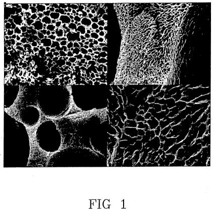
Fig. 1 Various enlargements of SEM images of a gelatin
porous material which does not contain the mineral
component;
CA 02691801 2009-12-17
WO 2009/004445 PCT/IB2008/001688
11
Fig. 2, 3, 4 e 5: various enlargements of SEM images
of the porous composite material according to the
present invention, which contains the mineral
component in an amount of 9 w/o (Fig.2), 23 w/o (Fig.
3), 33 w/% (Fig. 4), 42 w/ o(Fi:g. 5) respectively, to
the total amount of the porous composite material;
Fig. 6: X-ray diffraction diagram of a-TCP powders
used for the preparation of the porous composite
material of the present invention;
Fig. 7, 8, 9: X-ray diffraction diagram of powders of
the mineral component, which is isolated from porous
composite materials of the present invention
containing the mineral component in an amount of 23
w/% (Fig. 7), 33 w/o (Fig. 8), 42 w/o (Fig. 9)
respectively, to the total amount of the porous
composite material;
Fig. 10: X-ray diffraction diagram of P-TCP powders
used for preparing a porous composite material
according to known art;
Fig. 11:,X-ray diffraction diagram of powders of the
mineral component isolated from porous composite
material obtained from P-TCP according to the known
art, said mineral component being present in an amount
of 33 w/%, to the total weight of the porous composite
material;
Fig. 12: X-ray diffraction diagram of powders of the
CA 02691801 2009-12-17
WO 2009/004445 PCT/IB2008/001688
12
commercial product TCP (Merck) used for preparing a
porous composite material according to the known art;
Fig. 13: X-ray diffraction diagram of powders of the
mineral component isolated from the porous composite
material obtained from commercial product TCP (Merck)
according to the known art, said mineral component
being present in an amount of 42 w/%, to the total
weight of the porous composite material.
EXAMPLES 1-5
Materials employed
Pig skin gelatin has been used, obtained by acid
extraction.
a-TCP has been prepared by solid state reaction of a
mixture of CaCO3 with CaHPO4'2H2O with a molar ratio of
1:2 at 1300 C for 5 hours. The solid product has been
finely grinded before using.
Porous composite material preparation
The various samples preparation was conducted
according..-to the following phases.
a) The gelatin was dissolved in water containing a-
TCP at concentrations such as to obtain a mineral
component amount, in the final composite material, of
9 w/o (Example 2) , of 23 w/% (Example 3), of 33 w/o
(Example 4) and of 42 w/o (Example 5). Dissolution was
obtained by mechanic stirring at 40 C for 50 minutes
at 1,000 rpm. At the end of the stirring a foam was
CA 02691801 2009-12-17
WO 2009/004445 PCT/IB2008/001688
13
obtained.
b) The foam was gelled while keeping it in a Petri
dish at ambient temperature for a time ranging from 10
to 40 minutes.
c) The obtained gel was frozen by immersion in
liquid nitrogen (-195 C) for 10 minutes.
d) The frozen gel was freeze-dried at -50 C for 24
hours at a 1 millibar pressure.
A porous material was also prepared as a reference,
using gelatin without adding the a-TCP, following the
same methods as noted before. (Example 1)
If samples of crosslinked composite material are
desired, after the (a) phase, a genipin aqueous
solution may be added so,as to obtain a genipin amount
of 1,5 w/o to the gelatin weight. The so obtained
composition is then kept under stirring for 10
minutes.
Composite materials characterization
Figg. 2-5 images are SEM micrographs of porous
composite material according to the present invention
comprising the mineral component in amounts of: 9 w/o
(Example 2, Fig. 2), 23 w/o (Example 3, Fig. 3); 33
w/o (Example 4, Fig. 4), 42 w/o (Example 5, Fig. 5).
As a reference, Fig. 1 illustrates._SEM images of the
gelatin porous material according to Example 1, not
containing the mineral component, at various
CA 02691801 2009-12-17
WO 2009/004445 PCT/IB2008/001688
14
enlargements.
As can be noted, the porous structure exhibits a
macro- and micro-porosity. The macropores, which
seemed interconnected, had a mean particle size of
100-200 }im. The images do not show details pertaining
to the inorganic phase, showing an excellent
homogenization of the composite material components
The characterization of the crystalline structure of
the mineral component was carried out by X-ray
diffraction analysis of powders, using.a PANalytical
X'Pert PRO diffractometer.
Fig. 6 shows the X-ray diffraction diagram of powders
obtained from a-TCP used for preparing porous
composite material samples. All the diffraction peaks
coincide with those characteristic of a-TCP (in the
diagram the a-TCP reference file ICDD is reported by
segments corresponding to characteristic peaks).
Fig. 7 shows the X-ray diffraction diagram of powders
obtained from the mineral component isolated from the
composite material (by gelatin solubilization)
containing 23 w/% of the mineral component immediately
after freeze drying (Example 3). The diagram shows, as
well as the typical a-TCP peaks, the presence of other
diffraction peaks typical of OCP (in the diagram the
ICDD reference file of OCP is reported by segments
corresponding to characteristic peaks).
CA 02691801 2009-12-17
WO 2009/004445 PCT/IB2008/001688
Similar results have been obtained for samples with
different mineral component contents, as seen in Fig.
8 (33 w/o, Example 4) and Fig. 9 (42 w/%, Example 5).
The relative amount of the two a-TCP and OCP phases,
5 has been calculated by structural refinement of the
whole diffraction diagram, realized by using the
QUANTO program. Data obtained are very similar for all
the examined samples; and mediated values of composite
materials with different mineral component contents,
10 examined at various time after preparation up to a
month, are 26 5% OCP and 74 5% a-TCP.
The composite materials samples have been subjected to
pressure by a INSTRON 4465 dynamometer equipped with a
1 KN load cell with a 1 mm/min bar speed. The results
15 show how the mineral component content affects the
mechanic properties under pressure. In fact, the
mechanic properties increase as a function of the
mineral component content: the stress value under
pressure increases from the mean value of 0,08 2
MPa, for samples free of mineral component, to the
value of 0,21 3 MPa for the samples with a 70 w/%
mineral component content. Concurrently
(simultaneously), the Young modulus value increases
from 0,9 1 MPa to 4 1 MPa.
An intrusion porosimetric analysis was carried out on
the above composite material samples by ThermoFinnigan
CA 02691801 2009-12-17
WO 2009/004445 PCT/IB2008/001688
16
Pascal 140 and Pascal 240 apparatus, using a maximum
pressure of 240 MPa and a contact angle mercury-sample
of 1400. Pore size has been also measured by SEM
images. Results indicate interconnected porosity due
to micro- and macropores, ranging in size from 1 to
500 }lm.
EXAMPLES 6-7 (according to known art)
Some preparing tests of a porous composite material
have been carried out by the same methods as noted
above for Examples 1-5, but using (3-TCP as inorganic
component, instead of a-TCP (Example 6) or the Merck
commercial product indicated as TCP (Example 7).
R-TCP has been prepared by solid state reaction of a
mixture of CaCO3 with CaHPO4'2H2.O with a molar ratio
1:2 at 1000 C for 15 hours. The X-ray diffraction
diagram of the objective product is illustrated in
Fig. 10. All diffraction peaks coincide with those
characteristic of P-TCP (in the diagram the ICDD
reference file of P-TCP is reported by segments
corresponding to characteristic, peaks). Fig. 11 shows
the X-ray diffraction diagram of powders obtained from
the mineral component isolated from the composite
material (by gelatin solubilizaton) containing 33 w/%
of mineral component immediately after freeze-drying.
All diffraction peaks coincide with those
characteristic of P-TCP (the ICDD reference file of ~-
CA 02691801 2009-12-17
WO 2009/004445 PCT/IB2008/001688
17
TCP is also reported by segments corresponding to
characteristic peaks). A significant amount of OCP and
a-TCP is not observed.
The X-ray diffraction diagram of the TCP commercial
product (Merck) is reported in Fig. 12. All
diffraction peaks coincide in fact with HA and not TCP
characteristic peaks (the ICDD reference file of HA is
reported in the diagram by segments corresponding to
characteristic peaks). Fig. 13 shows the X-ray
diffraction diagram of powders obtained from the
mineral component isolated from the composite material
(by gelatin solubilization) containing 42 w/o of the
mineral component immediately after freeze-drying. All
diffraction peaks coincide with HA characteristic
peaks (in this case also the ICDD reference file of HA.
is reported in the diagram by segments corresponding
to characteristic peaks). A significant amount of OCP
and a-TCP is not observed.