Note: Descriptions are shown in the official language in which they were submitted.
CA 02692171 2015-05-05
WO 2009/002931 PCT/US2008/067899
METHODS AND USES THEREOF OF PROSAPOSIN
FIELD OF THE INVENTION
[0002] The present invention relates to methods for treating of tumor
metastasis, as well as =
methods for preventing, inhibiting, and predicting tumor metastasis. The
invention further
relates to treating angiogenesis-dependent diseases and disorders, screening
methods for tumor
cell derived anti-angiogenic factors and methods for cancer prognosis
evaluation.
BACKGROUND OF THE INVENTION
[0003] The spread of cancer cells from a primary tumor site to distant
organs is known as
metastasis. The progression of human cancer to metastatic disease is the major
contributing
factor to its lethality. Metastasis has been considered one of the most
intriguing aspects of the
pathogenesis of cancer. Cancer tumor metastasis, or otherwise known as
metastatic disease, is
responsible for most therapeutic failures in treating the disease, as patients
succumb to the
multiple tumor growth, accounting for more than 90% of human cancer related
deaths. See, for
example, Cancer, A Comprehensive Treatise, F. F. Becker (editor), Volume 4,
Chapter 3,
Plenum Press, New York, 1975.
[0004] In order for a tumor to form lethal metastases it must acquire the
ability to carry out
a complex series of steps. These steps include: gaining access to the
vasculature or lymphatic
system (intravasation), surviving during transit, exiting the vascular or
lymphatic channels
(extravasation), and proliferating at the metastatic site. One of the rate
limiting steps in the
proliferation of tumors, both at the primary and metastatic sites, is the
acquisition of the
angiogenic phenotype (Folkman, 1971). The induction of angiogenesis not only
allows tumors
to grow beyond the size limitation imposed by the diffusion limit of oxygen,
but also provides a
conduit through which the tumor cells can travel and colonize distant organs
(Brown et al.,
1999; MacDougall and Matrisian, 1995). Once the tumor cells arrive at the
metastatic site they
must also induce neovascularization in order to grow beyond a microscopic
size. It has been
documented, however, that metastatic colonies can remain in a microscopic or
dormant state and
1
CA 02692171 2009-12-17
WO 2009/002931 PCT/US2008/067899
not progress beyond this size for months or years following the initial
colonization (Fidler,
2003).
[0005] The presence of dormant or micro-metastases indicates that tumor
growth and
proliferation is not governed solely by cell-autonomous processes and that the
conditions present
in the microenvironment that permitted proliferation at the primary site can
not exist at the
metastatic site. Thus, the ability of a tumor to communicate with the
surrounding stroma,
composed of fibroblasts, immune cells and endothelium must be reestablished
upon arrival at
the metastatic site. One way in which heterotypic tumor-stromal signaling
could affect tumor
growth is through the regulation of the production and secretion of pro- and
anti-angiogenic
proteins by the surrounding stromal fibroblasts and endothelial cells.
[0006] The molecular and genetic events that facilitate escape from the
primary site and
homing to the metastatic site have been well studied. It has been demonstrated
in a murine
model of breast cancer metastasis that escape from the primary site was
largely dependent on the
activity of the transcription factor Twist (Yang et al., 2004). Furthermore,
microarray analyses
of metastatic human breast cancer cells, derived by serial injection into
immuno-compromised
mice, revealed sets of genes whose expression correlated with their preferred
metastatic
destination of bone or lung (Kang et al., 2003; Minn et al., 2005). These
studies, though
yielding key insights into two critical steps of tumor metastasis, namely
intravasation and
homing, did not address the requirements for tumor establishment and growth at
the metastatic
site.
[0007] It has been previously demonstrated that tumor cells can stimulate
the expression of
the pro-angiogenic protein VEGF in the surrounding stroma (Dong et al., 2004;
Fukumura et al.,
1998). However, the regulation of Thrombospondin (Tsp-1), one of the most
potent
endogenous anti-angiogenic proteins, in the tumor-associated stroma have not
been as well
studied (Kalas et al., 2005).
[0008] New research into the cell-to-cell signaling events between
metastatic tumors and
their surrounding stroma can yield novel strategies for treating metastatic
disease. There is still a
need for methods of treating metastatic disease that have less systemic
toxicity than the current
standard treatments comprising chemotherapy and/or radiation therapy.
SUMMARY OF THE INVENTION
2
CA 02692171 2009-12-17
WO 2009/002931 PCT/US2008/067899
[0009] In cancer patients, tumor and micrometastases can remain for
prolonged periods of
time in a dormant asymptotic state before diagnosis and development of
disease. Embodiments
of the present invention are based on the discovery that such dormant, non- or
weakly metastatic
tumor cells secrete a protein, prosaposin (Psap), that stimulates the
expression of
thrombospondin (Tsp-1) in the surrounding environment of the tumor cells,
namely the stroma
comprised of fibroblasts and endothelial cells. Tsp-1 is also activated in
distant environments
such as the lymph nodes. Tsp-1 is a potent endogenous anti-angiogenic factor,
and its activation
by the tumor-derived protein is via the activation of the tumor suppressor
p53. P53 is a
transcription activator of Tsp-1. The present discovery is contrary to current
scientific literature
wherein the prosaposin and its metabolite derivative saposin C is a potent
growth factor for
promoting prostate cancer.
[0010] As such, embodiments of the present invention provide methods of
treating an
angiogenesis-dependent disease or disorder, the method comprises administering
to a subject in
need of treatment thereof, a therapeutically effective amount of Psap protein
or a vector
comprising a nucleic acid encoding Psap protein and a pharmaceutically
acceptable carrier.
[0011] In one embodiment, provided herein is a method of inhibiting the
recurrence of an
angiogenesis-dependent disease or disorder, the method comprises administering
to a subject in
need thereof, a therapeutically effective amount of a Psap protein or a vector
comprising the
nucleic acid encoding a Psap protein, and a pharmaceutically acceptable
carrier.
[0012] Angiogenesis-dependent disease or disorder to which the methods
described herein
are applicable include, for example, cancer, psoriasis, age-related macular
degeneration, thyroid
hyperplasia, preeclampsia, rheumatoid arthritis, Alzheimer's disease, obesity,
pleura effusion,
atherosclerosis, endometriosis, diabetic/other retinopathies, neovascular
glaucoma, age-related
macular degeneration, hemangiomas, and corneal neovascularization. In one
embodiment, the
age-related macular degeneration is wet macular degeneration.
[0013] In another embodiment, the invention provides a method for
inhibiting metastasis of
cancer in a subject diagnosed with cancer, the method comprises administering
to the individual,
a therapeutically effective amount of Psap protein or a vector comprising a
nucleic acid
encoding Psap protein and a pharmaceutically acceptable carrier. The subject
can be diagnosed
with a benign or malignant cancer.
3
CA 02692171 2009-12-17
WO 2009/002931 PCT/US2008/067899
[0014] In some aspect, the invention provides a method of inhibiting
recurrence of cancer in
a subject diagnosed with cancer, the method comprises administering to a
subject in need
thereof, a therapeutically effective amount of a Psap protein or a vector
comprising the nucleic
acid encoding a Psap protein, and a pharmaceutically acceptable carrier. The
subject can
diagnosed with a benign or malignant cancer.
[0015] In one aspect, the invention provides for a method of preventing
cancer development
in a subject at risk of cancer development, the method comprises administering
to a subject in
need thereof, a therapeutically effective amount of a Psap protein or a vector
comprising the
nucleic acid encoding a Psap protein, and a pharmaceutically acceptable
carrier. The subject can
have a family history of cancer, e. g. early on-set colon rectal cancer,
and/or carry some gene
mutations that are shown to be associated with certain cancers, e. g. BRAC1
and BRAC2 for
breast cancer.
[0016] In one aspect, the invention provides for a method of preventing
cancer metastasis in
a subject previously diagnosed with cancer, the method comprises administering
to a subject in
need thereof, a therapeutically effective amount of a Psap protein or a vector
comprising the
nucleic acid encoding a Psap protein, and a pharmaceutically acceptable
carrier. The subject can
be diagnosed with a benign or malignant cancer.
[0017] In one aspect, the invention provides for a method of preventing
development of
cancer malignancy in a subject previously diagnosed with cancer, the method
comprises
administering to a subject in need thereof, a therapeutically effective amount
of a Psap protein or
a vector comprising the nucleic acid encoding a Psap protein, and a
pharmaceutically acceptable
carrier. The subject can be diagnosed with a benign or malignant cancer.
[0018] In one embodiment, the Psap protein is saposin A (SEQ. ID. No. 13)
or smaller
functional fragments and variants thereof. In another embodiment, the smaller
functional
fragment or variant of saposin A is at least 10 amino acid residues and is
capable of activating
p53 and inducing Tsp-1 expression. In yet another embodiment, the smaller
functional fragment
or variant of saposin A is fused to other protein or portions thereof, or
conjugated with a
polymer, wherein the fragment or variant can still activate p53 and induce Tsp-
1 expression.
For examples, the other protein or portions thereof or polymer can be
transferrin, Fc portion of
IgG, albumin, and PEG, for the purpose of improving serum half-life in vivo.
Other examples
include to thioredoxin and six histidine tag for facilitating recombinant
protein expression and
purification; and to angiotensin and endostatin for improving anti-angiogenic
activity.
4
CA 02692171 2009-12-17
WO 2009/002931 PCT/US2008/067899
[0019] In the methods described herein, Psap protein can be administered
after the detection
and diagnosis of an angiogenesis-dependent disease or disorder, such as
cancer. The treatment
can administered in conjunction with chemotherapy, radiation therapy, a
cytostatic agent, an
anti-VEGF agent and/or a p53 reactivation agent.
[0020] An embodiment of the invention also provides a method for prognostic
evaluation of
an individual diagnosed with cancer, the method comprises determining the
level of Psap
expression in a tumor sample from an individual diagnosed with cancer and
comparing the level
to a reference Psap level, e.g. from a non-tumor sample from the individual. A
level of Psap in
the tumor sample lower than a reference Psap level indicates that there is an
increased likelihood
of cancer metastasis and/or recurrence of neoplastic disease, and thus a poor
prognosis.
[0021] In one embodiment, the method described herein further comprises:
(a) determining
the level of Tsp-1 expression in the tumor stroma, and comparing the level to
a reference Tsp-1
level. When the level of Tsp-1 in the tumor stroma are lower than the
reference Tsp-1 level,
there is an increased likelihood of cancer metastasis and/or recurrence of
neoplastic disease, and
thus a poor prognosis.
[0022] In one embodiment, the invention provides a method of treating an
individual
diagnosed with cancer, the method comprises: (a) determining a level of Psap
in a tumor sample
from said individual; (b) comparing the Psap level determined in (a) with a
reference Psap level;
and (c) when the Psap level determined in (a) is lower than 95% of said
reference Psap level,
administering a therapeutically effective amount of Psap protein or a vector
comprising a nucleic
acid encoding Psap protein and a pharmaceutically acceptable carrier. The
treatment can be
administered in conjunction with chemotherapy, radiation therapy, a cytostatic
agent, an anti-
VEGF agent, an anti-angiogenesis factor, and/or a p53 reactivation agent.
[0023] In one embodiment, the invention provides a method of screening and
identifying
tumor secreted factors that promote angiogenesis and metastasis, the method
comprises: (a)
contacting fibroblasts and/or endothelial cells with a cancer cell derived
factor; (b) determining
the levels of angiogenic growth factors and/or angiogenesis inhibitors; and
(c) comparing with
reference levels of angiogenic growth factors and/or angiogenesis inhibitor of
fibroblasts and/or
endothelial cells not treated with cancer cell derived factors. A decrease in
the level of an
angiogenesis inhibitor and/or an increase in the level of angiogenic growth
factors in comparison
to the respective reference levels indicate that the cancer cell derived
factor contains factors that
promote angiogenesis and metastasis.
CA 02692171 2009-12-17
WO 2009/002931 PCT/US2008/067899
[0024] In one embodiment, the invention provides a method of screening for
a compound,
drug, or small molecule that inhibits angiogenesis and metastasis, the method
comprises: (a)
contacting fibroblasts and/or endothelial cells with a cancer cell derived
factor in the presence of
a compound, drug, or small molecule; (b) determining the levels of angiogenic
growth factors
and angiogenesis inhibitors; and (c) comparing with reference levels of
angiogenic growth
factors and angiogenesis inhibitors of fibroblasts and/or endothelial cells
not treated with a
compound, drug, or small molecule. An increase in the level of an angiogenesis
inhibitor and/or
a decrease in the level of an angiogenic growth factor in comparison to the
respective reference
levels indicate that the compound, drug, or small molecule can inhibit
angiogenesis and
metastasis.
[0025] In one embodiment, the invention provides a method of screening for
a compound,
drug, or small molecule that promotes anti-angiogenic and anti-metastatic
activities, the method
comprises: (a) contacting fibroblasts and/or endothelial cells with a
compound; (b) determining
the expression levels of p53 and Tsp-1; and (c) comparing with reference
levels of p53 and Tsp-
1 not treated with the compound, drug, or small molecule. Increases in the
levels of p53 and
Tsp-1 expression in the treated cells indicate that the tested compound has
anti-angiogenesis and
anti-metastatic activity.
[0026] In one embodiment, the invention provides a method of predicting the
metastatic
tissue specificity of cancer cells in an individual diagnosed with cancer, the
method comprises:
(a) contacting test fibroblasts and/or endothelial cells with a cancer cell
derived factor; (b)
determining the levels of Tsp-1, Psap, and/or c-Myc in the fibroblasts and/or
endothelial cells;
and (c) comparing to the reference levels of Tsp-1, Psap, and c-Myc of
fibroblasts and/or
endothelial cells not treated with cancer cell derived factors; wherein
repression of Tsp-1 and
Psap expressions, and/or an activation of c-Myc expression in the tested
fibroblasts and/or
endothelial cells indicate that the cancer cells are likely to metastasize to
the type of tissue from
which tested fibroblast and/or endothelial cells had originated.
[0027] In one embodiment, the invention provides a method for determining
the likelihood
of metastasis an individual diagnosed with cancer, the method comprises: (a)
determining the
level of Psap expression in a sample from an individual diagnosed with cancer;
and (b)
comparing to a reference level of Psap. The reference Psap level is that of a
Psap expression in a
normal individual not diagnosed with any cancer or an average Psap expression
level of a group
of normal individuals not diagnosed with any cancer. When the level of Psap in
the sample from
6
CA 02692171 2009-12-17
WO 2009/002931 PCT/US2008/067899
an individual diagnosed with cancer is the same or lower than the reference
Psap level, this
indicates that there is an increased likelihood of cancer metastasis.
[0028] In one embodiment, the invention provides a method for prognostic
evaluation in an
individual diagnosed with cancer, the method comprises: (a) determining the
level of Psap
expression in a sample from an individual diagnosed with cancer at a first
time point; (b)
determining the level of Psap expression in a sample from an individual
diagnosed with cancer
at a second time point, the first time point being before the second time
point; and (c) comparing
the levels of Psap from the time points with a reference Psap level. When the
level of Psap at the
second time point becomes lower than the reference Psap level, the cancer has
likely spread.
[0029] In one embodiment, the sample is blood, preferably platelet, serum
or plasma.
[0030] In one embodiment, the invention provides an isolated chimeric
polypeptide
comprising a first portion and a second portion, wherein the first portion is
saposin A (SEQ. ID.
No. 13) or a functional fragment thereof, and the second portion comprises an
amino acid
sequence or a polymer that enhances the serum half life of the first portion.
The second portion
is not a Psap protein, and the first portion is capable of activating p53 and
inducing Tsp-1
expression.
[0031] In one embodiment, the invention provides an isolated chimeric
polypeptide
comprising a first portion and a second portion, wherein the first portion is
saposin A (SEQ. ID.
No. 13) or a functional fragment thereof, and the second portion comprises an
amino acid
sequence that facilitates protein expression and/or purification of the first
portion. The second
portion is not a Psap protein, and the first portion is capable of activating
p53 and inducing Tsp-
1 expression.
[0032] In one embodiment, the invention provides an isolated chimeric
polypeptide
comprising a first portion and a second portion, wherein the first portion is
saposin A (SEQ. ID.
No. 13) or a functional fragment thereof, and the second portion is a
therapeutic molecule. The
second portion is not a Psap protein, and the first portion is capable of
activating p53 and
inducing Tsp-1 expression.
BRIEF DESCRIPTION OF THE DRAWINGS
[0033] Fig.1A. ELISA of VEGF secretion by PC3 and PC3M-LN4 (LN4) prostate
cancer
cells and MDA-MB-231 (231) and MDA-MET (MET) breast cancer cells cultured
under 20%
7
CA 02692171 2009-12-17
WO 2009/002931 PCT/US2008/067899
oxygen (normoxia) or 1% oxygen (hypoxia) Error bars represent SEM (Standard
Error of Mean)
of 3 independent experiments performed in triplicate).
[0034] Fig.1B. Western blot analysis of Tsp-1, c-Myc, and 13-Actin
expression by PC3,
PC3M-LN4 (LN4), MDA-MB-231 (231) and MDA-MET (MET) cells.
[0035] Fig. 1C. Western blot analysis of phosphorylated c-Myc (phospho-Myc)
and ¨Actin
expression by PC3, PC3M-LN4 (LN4), MDA-MB-231 (231) and MDA-MET (MET) cells.
[0036] Fig.1D. Western blot analysis of Tsp-1, c-Myc, and 13-Actin
expression in prostate
tumors formed by PC3 (P1-P5) and PC3M-LN4 (L1-L4).
[0037] Fig.1E. ELISA of VEGF secretion from murine stromal cells present in
PC3 (P1-P5)
and PC3M-LN4 (L1-L5) prostate tumors.
[0038] Fig.1F. Tabular depiction of Tsp-1 expression in primary tumors
formed by PC3 and
PC3M-LN4 and the incidence of metastases in mice bearing these tumors.
[0039] Fig.2A. Western blot analyses of Tsp-1, c-Myc, and 13-Actin
expression in prostate
fibroblasts that were untreated (-) or treated with the conditioned media from
PC3, or PC3M-
LN4 (LN4) cells.
[0040] Fig.2B. Western blot analyses of Tsp-1 and -Actin expression in
normal human
mammary fibroblasts that were treated with the conditioned media (CM) from MDA-
MB-231
(231) and MDA-MET (MET) cells or co-cultured in transwell apparati (TW) with
MDA-MB-
231 (231) and MDA-MET (MET) cells.
[0041] Fig.2C. Western blot analyses of ELISA of VEGF secretion from
prostate
fibroblasts treated with the conditioned media from PC3, PC3M-LN4 (LN4) cells.
Error bars
represent SEM (Standard Error of Mean) of 3 independent experiments performed
in triplicate.
[0042] Fig.2D. Western blot analyses of Tsp-1 and 13-Actin expression in
WI38 lung
fibroblasts and bone marrow stromal cells that were untreated (-) or treated
with the conditioned
media from PC or PC3M-LN4 (LN4) cells.
[0043] Fig.2E. Western blot analyses of Tsp-1 and 13-Actin expression in
WI38 lung
fibroblasts and bone marrow stromal cells that were untreated (-) or treated
with the conditioned
media from MDA-MB-231 (231) or MDA-MET (MET) cells.
8
CA 02692171 2009-12-17
WO 2009/002931 PCT/US2008/067899
[0044] Fig.3A. Western blot analyses of Tsp-1 and 13-actin expression in
prostate fibroblasts
treated with fractions of conditioned media from PC3 cells eluted from a Cu2+-
heparin column.
[0045] Fig.3B. Western blot analyses of Tsp-1 and 13-actin expression in WI
38 lung
fibroblasts treated with fractions of conditioned media from PC3 cells eluted
from a Cu2+-
heparin column.
[0046] Fig.3C. Western blot analyses of Psap and -actin expression in PC3
and PC3M-
LN4 (LN4) cells.
[0047] Fig.3D. Western blot analyses of Psap and 13-actin expression in MDA-
MB-231
(231), MDA-Bone (Bone), MDA-Brain (Brain), MDA-MB-LM2-4 (LM), MDA-MET (MET),
MDA-MB-231-1833 (1833) and MDA-MB-231-4175 (4175) cells.
[0048] Fig.3E. Western blot analyses of Psap, Tsp-1 and 13-actin expression
in PC3 cells
that were transduced with five shRNA constructs for Psap or an empty pLKO
vector (V).
[0049] Fig.3F. Western blot analyses of Tsp-1 and 13-actin expression in
prostate fibroblasts
treated with conditioned media (CM) from pLKO vector transduced PC3, and PC3
cells
transduced with 5 shRNA sequences specific for Psap.
[0050] Fig.3G. Western blot analyses of Tsp-1 and 13-actin expression in WI
38 lung
fibroblasts treated with conditioned media (CM) from pLKO vector transduced
PC3, and PC3
cells transduced with 5 shRNA sequences specific for Psap.
[0051] Fig.3H. Western blot analyses of Psap and 13-actin expression in
PC3M-LN4 cells
that were uninfected (LN4), infected with control pLNCX vector (V) or pLNCX-
Psap (Psap).
[0052] Fig.3I. Western blot analyses of Tsp-1 and 13-actin expression in
untreated prostate
fibroblasts (-), or treated with conditioned media from PC3M-LN4 (LN4), PC3M-
LN4-pLNCX
(V) or PC3M-LN4-Psap (Psap) cells.
[0053] Fig.3J. Western blot analyses of Tsp-1 and 13-actin expression in
untreated prostate
fibroblasts (-), or treated with 51..tg of purified recombinant human Psap.
9
CA 02692171 2009-12-17
WO 2009/002931 PCT/US2008/067899
[0054] Fig. 4A. Western blot analyses of p53 and 13¨Actin expression in
prostate tissue
from non-tumor bearing mice (N), PC3 prostate tumor tissue (P) and PC3M-LN4
tumor tissue
(L).
[0055] Fig. 4B. Western blot analyses of p53 and 13¨Actin expression in
normal lymph node
tissue (N), lymph node tissue from PC3 tumor bearing mice (P) and lymph node
metastases
from PC3M-LN4 tumor bearing mice (L).
[0056] Fig. 4C. Western blot analyses of p53 and 13- Actin expression in
prostate fibroblasts
(PrF) that were untreated (-) or treated with the conditioned media from PC3
or PC3M-LN4
(LN4) cells;
[0057] Fig. 4D. Western blot analyses of Tsp-1, p53 and 13- Actin
expression in prostate
fibroblasts containing empty pLKO vector, (V) or p53 shRNA that were untreated
(-) or treated
with the conditioned media from PC3 or PC3M-LN4 (LN4) cells.
[0058] Fig. 4E. Western blot analyses of Tsp-1, p53 and 13¨Actin expression
in WI38 lung
fibroblasts containing empty pLKO vector (V) or p53 shRNA that were untreated
(-) or treated
with the conditioned media from PC3 or PC3M-LN4 (LN4).
[0059] Fig. 4F. Western blot analyses of p53 and 13-actin expression in
prostate fibroblasts
treated with conditioned media (CM) from pLKO vector transduced PC3, and PC3
cells
transduced with 5 shRNA sequences specific for Psap.
[0060] Fig. 4G. Western blot analyses of p53 and I3-actin expression in WI
38 lung
fibroblasts treated with conditioned media (CM) from pLKO vector transduced
PC3, and PC3
cells transduced with 5 shRNA sequences specific for Psap.
[0061] Fig. 4H. Western blot analyses of p53 and 13-actin expression in
untreated prostate
fibroblasts (-), or treated with conditioned media from PC3M-LN4 (LN4), PC3M-
LN4-pLNCX
(V) or PC3M-LN4-Psap (Psap) cells.
[0062] Fig. 41. Western blot analyses of p53 and 13-actin expression in
untreated prostate
fibroblasts (-), or treated with 51..tg of purified recombinant human Psap.
[0063] Fig. 5A. Western blot analyses of Psap and 13-actin expression in
pBabepuro vector
transduced PC3 or PC3-MycER cells that were untreated (-) or treated with 4-HT
(+).
CA 02692171 2009-12-17
WO 2009/002931 PCT/US2008/067899
[0064] Fig. 5B. Western blot analyses of Tsp-1, p53 and 13-actin expression
in prostate
fibroblasts and WI 38 lung fibroblasts that were untreated (-), treated with 4-
HT alone (-/+) or
treated with the conditioned media from 4-HT treated PC3-MycER cells
(MycER/+).
[0065] Fig. 5C. Western blot analyses of Myc and 13-actin expression in
wild-type PC3M-
LN4 cells (-), as well as PC3M-LN4 cells transduced with empty pLKO vector (V)
or
transduced with pLKO lentivirus specifying two different shRNA sequences for
Myc (sh1, sh2).
[0066] Fig. 5D. Western blot analyses of Psap and 13-actin expression in
PC3M-LN4 cells
containing empty pLKO vector (V) or expressing two different shRNA sequences
for Myc (sh1,
sh2).
[0067] Fig. 5E. Western blot analyses of Tsp-1, p53 and 13-actin expression
in prostate
fibroblasts and WI 38 lung fibroblasts that were untreated (-) or treated with
the conditioned
media from PC3M-LN4-shMyc cells (sh1).
[0068] Fig.6A. Plot of tumor mass of PC3shPsap tumors and PC3pLKO tumors
from SCID
mice.
[0069] Fig.6B. Western blot analyses of Tsp-1, p53, Psap and 13-actin
expression in normal
prostate (N) and prostate tumor formed by PC3pLKO (P) and PC3shPsap (sh) tumor
bearing
mice.
[0070] Fig.6C. Western blot analyses of Tsp-1, p53 and 13-actin expression
in normal lymph
node (N) and lymph node from PC3pLKO (P) or PC3shPsap (sh) tumor bearing mice.
[0071] Fig.6D. Western blot analyses of Tsp-1, p53 and 13-actin expression
in normal lung
tissue (N) and lung tissue from PC3pLKO (P) or PC3shPsap (sh) tumor bearing
mice.
[0072] Fig.7A. Plot of metastases/lung in the mice described in (K),
PC3=PC3pLKO.
[0073] Fig.7B. Relative mRNA expression levels of Psap in localized human
prostate
tumors and metastatic human prostate tumors. Each bar represents the mean of
each group. The
difference in Psap expression between localized and metastatic prostate tumors
has a p-
value<0.0001 based on one way ANOVA.
[0074] Fig.7C. Relative mRNA expression levels of Tsp-1 in localized human
prostate
tumors and metastatic human prostate tumors. Each bar represents the mean of
each group. The
11
CA 02692171 2009-12-17
WO 2009/002931 PCT/US2008/067899
difference in Tsp-1 expression between localized and metastatic prostate
tumors has a p-
value<0.0001 based on one way ANOVA.
[0075] Fig.7D. Relative mRNA expression levels of p53 in localized human
prostate
tumors and metastatic human prostate tumors. Each bar represents the mean of
each group. The
difference in p53 expression between localized and metastatic prostate tumors
has a p-
value=0.0004 based on one way ANOVA.
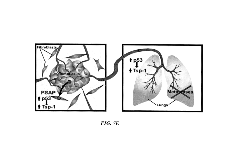
[0076] Fig.7E. Schematic depiction of Prosaposin-mediated inhibition of
tumor metastasis
(blue hexagons=Prosaposin).
[0077] Fig. 8. Western blot analysis of the expression levels of
Thrombospondin-2 (Tsp-2),
murine endostatin (m-endostatin) and 13-actin protein in normal mouse prostate
(N) tumors
formed by PC3 cells (P), and tumor formed by PC3shPsap cells (sh).
[0078] Fig. 9. Western blot analysis of Tsp-1 and -Actin expression in
normal human
dermal fibroblasts (NHDF), normal human astrocytes (NHA), normal human
prostate fibroblasts
(prostate) and normal human mammary fibroblasts that were untreated (-) or
treated with the
conditioned media from PC3, PC3M-LN4 (LN4), MDA-MB-231 (231), MDA-MB-231-4175
(MDA-L), MDA-MB-1833 (MDA-B) or MDA-MET (MET) cells as denoted.
[0079] Fig. 10. Western blot analysis of murine Tsp-2 and -Actin expression
in normal
lung tissue (-) and lungs of wild-type and tsp-1-/- C57B1/6J mice injected
with 1x10⁶ Lewis
Lung Carcinoma cells and treated with serum free RPMI media (L+M), conditioned
media from
empty vector containing PC3pLKO (L+P) or PC3shPsap cells (L+S).
[0080] Fig. 11. Relative mRNA expression levels of Tsp-2 in localized human
prostate
tumors (PCA) and metastatic human prostate tumors (MET). Each bar represents
the mean of
each group. The difference in Tsp-2 expression between encapsulated and
metastatic prostate
tumors has a p-value=0.6797 based on one way ANOVA.
[0081] Fig. 12. Saposin A stimulates Tsp-1. Western blot of Tsp-1 and 13-
Actin expression
in prostate fibroblasts treated with conditioned media from PC3M-LN4 cells
(LN4) or PC3M-
LN4 cells transiently transfected with an expression vector (pCMV) specifying
Saposin A
(L+A), Saposin AB (L+AB), Saposin ABC (L+ABC), or control empty vector.
12
CA 02692171 2015-05-05
WO 2009/002931 PCT/US2008/067899
[0082] Fig. 13. Psap expression in plasma from colorectal cancer patients.
Western blot
analysis of prosaposin and Tsp-1 protein levels in plasma samples from normal
patients, and
colon cancer patients with low grade (Ti, NO ,M0), high grade without
metastases (3 T3, 1 T4
NO, MO) and high grade with metastasis (3 T3, 1 T4, Ni, M1).
[0083] Fig. 14A. Correlation of endogenous expression of Prosaposin and
biochemical
failure in patients after radical pro statectomy.
[0084] Fig. 14B. Kaplan Meyer plot of survival of patients from the series
of men described
above as a function of time elapsed subsequent to radical prostatectomy and
endogenous
expression of Prosaposin.
DETAILED DESCRIPTION OF THE INVENTION
[0085] Unless otherwise stated, the present invention was performed using
standard
procedures that are well known to one skilled in the art, for example, in
Maniatis et al.,
Molecular Cloning: A Laboratory Manual, Cold Spring Harbor Laboratory Press,
Cold Spring
Harbor, N.Y., USA (1982); Sambrook et al., Molecular Cloning: A Laboratory
Manual (2 ed.),
Cold Spring Harbor Laboratory Press, Cold Spring Harbor, N.Y., USA (1989);
Davis et al.,
Basic Methods in Molecular Biology, Elsevier Science Publishing, Inc., New
York, USA
(1986); Methods in Enzymology: Guide to Molecular Cloning Techniques Vol.152,
S. L. Berger
and A. R. Kimmerl Eds., Academic Press Inc., San Diego, USA (1987); Current
Protocols in
Molecular Biology (CPMB) (Fred M. Ausubel, et al. ed., John Wiley and Sons,
Inc.), Current
Protocols in Protein Science (CPPS) (John E. Coligan, et. al., ed., John Wiley
and Sons, Inc.);
Current Protocols in Immunology (CPI) (John E. Coligan, et. al., ed. John
Wiley and Sons, Inc.);
Current Protocols in Cell Biology (CPCB) (Juan S. Bonifacino et. al. ed., John
Wiley and Sons,
Inc.); Culture of Animal Cells: A Manual of Basic Technique by R. Ian
Freshney, Publisher:
Wiley-Liss; 5th edition (2005); and Animal Cell Culture Methods (Methods in
Cell Biology,
Vol 57, Jennie P. Mather and David Barnes editors, Academic Press, 1st
edition, 1998).
[0086] Unless otherwise defined herein, scientific and technical terms used
in connection
with the present application shall have the meanings that are commonly
understood by those of
ordinary skill in the art. Further, unless otherwise required by context,
singular terms shall
include pluralities and plural terms shall include the singular.
13
CA 02692171 2015-05-05
WO 2009/002931 PCT/US2008/067899
[0087] Definitions of common terms in molecular biology are found in
Benjamin Lewin,
Genes IX, published by Jones & Bartlett Publishing, 2007 (ISBN-13:
9780763740634);
Kendrew et al. (eds.), The Encyclopedia of Molecular Biology, published by
Blackwell Science
Ltd., 1994 (ISBN 0-632-02182-9); and Robert A. Meyers (ed.), Molecular Biology
and
Biotechnology; a Comprehensive Desk Reference, published by VCH Publishers,
Inc., 1995
(ISBN 1-56081-569-8).
[0088] It should be understood that this invention is not limited to the
particular
methodology, protocols, and reagents, etc., described herein and as such may
vary. The
terminology used herein is for the purpose of describing particular
embodiments only, and is not
intended to limit the scope of the present invention, which is defined solely
by the claims.
[0089] Other than in the operating examples, or where otherwise indicated,
all numbers
expressing quantities of ingredients or reaction conditions used herein should
be understood as
modified in all instances by the term "about." The term "about" when used in
connection with
percentages may mean 1%.
[0090] All patents and other publications identified are provided for the
purpose of
describing and disclosing, for example, the methodologies described in such
publications that
might be used in connection with the present invention. These publications are
provided solely
for their disclosure prior to the filing date of the present application.
Nothing in this regard should
be construed as an admission that the inventors are not entitled to antedate
such disclosure by
virtue of prior invention or for any other reason. All statements as to the
date or representation as
to the contents of these documents is based on the information available to
the applicants and
does not constitute any admission as to the correctness of the dates or
contents of these
documents.
[0091] The singular terms "a," "an," and "the" include plural referents
unless context clearly
indicates otherwise. Similarly, the word "or" is intended to include "and"
unless the context
clearly indicates otherwise. It is further to be understood that all base
sizes or amino acid sizes,
and all molecular weight or molecular mass values, given for nucleic acids or
polypeptides are
approximate, and are provided for description. Although methods and materials
similar or
equivalent to those described herein can be used in the practice or testing of
this disclosure,
suitable methods and materials are described below. The term "comprises" means
"includes."
The abbreviation, "e.g." is derived from the Latin exempli gratia, and is used
herein to indicate a
non-limiting example. Thus, the abbreviation "e.g." is synonymous with the
term "for example."
14
CA 02692171 2009-12-17
WO 2009/002931 PCT/US2008/067899
[0092] Embodiments of the present invention are based on the discovery that
non- or
weakly metastatic tumor cells secrete a protein that stimulates the expression
of thrombospondin
(Tsp-1) in the surrounding environment of the tumor cells, namely the stroma
comprised of
fibroblasts and endothelial cells. While not wishing to be bound by theory,
the increase in
expression of Tsp-1 in the stroma keeps the tumor cells from metastasizing.
Tsp-1 is a potent
endogenous anti-angiogenic factor, and the stimulation of Tsp-1 expression by
the tumor-
derived protein is via the activation of the tumor suppressor p53. The tumor
suppressor p53 is a
transcription activator of Tsp-1 expression. This tumor-associated protein
secreted by non- or
weakly metastatic tumor cells is prosaposin (Psap).
[0093] It was found that non- or weakly metastatic tumor cells express a
high amount of
Psap and Tsp-1, in addition to stimulating p53 and Tsp-1 expression in the
surrounding tumor
stroma. In contrast, metastatic tumor cells express low amounts of Psap and
Tsp-1, and
metastatic tumor cells also repress the expression of p53 and Tsp-1 in the
tumor stroma. There
is a strong correlation between metastasis, Psap and Tsp-1 expression in the
tumor cells, and
Psap and Tsp-1 expression in the tumor stroma. In addition, there is also a
strong correlation
between metastasis and the Psap level in the plasma and/or platelets of
patients with metastatic
cancers. Both the plasma and platelets of patients with non-metastatic cancers
contained
elevated levels of Psap compared to normal individuals not diagnosed with
cancers. In contrast,
the plasma and platelets of patients with metastatic cancers contain Psap
levels that are
comparable to normal individuals with no diagnosed cancers. While not wishing
to be bound by
theory, the shift from elevated levels of Psap levels to normal or lower than
normal Psap levels
indicates the transition from non-metastatic to metastatic cancer.
[0094] While not wishing to be bound by theory, high expression of Psap
from a dormant
primary tumor prevents the tumor from metastasis and also prevents the
establishment
secondary tumors at sites away from the primary tumor site. Conversely, low or
no expression
Psap in a primary tumor allows the tumor to metastasize and establish
secondary tumors at sites
far away from the primary tumor site. The Psap secreted by a tumor affects its
local and distant
environment via paracrine and endocrine signaling mechanisms, affecting
whether a tumor cell
can grow bigger and/or implant and grow at a different and distant location
from the primary
tumor site. Psap functions as a repressor of both lymphatic and vascular
metastasis by inducing
p53 and consequently Tsp-1 expression in stromal fibroblasts via both
paracrine and endocrine
signaling mechanisms.
CA 02692171 2009-12-17
WO 2009/002931 PCT/US2008/067899
[0095] These new discoveries are contrary to existing reports that Psap
and/or its known
active molecular derivatives (e.g., saposin C) function as a pluripotent
growth factor with
diverse biological activities that favor malignant phenotypes in prostate
cancer (Koochekpour S,
et.al., J Cell Biochem. 2007,101:631-41; J Cell Biochem. 2008, May, in press).
[0096] In cancer patients, tumor and micrometastasis can remain for
prolonged periods of
time in a dormant asymptotic state before diagnosis and development of
disease. It is unknown
how this dormant state is maintained or how and why the dormant tumor changes
to a metastatic
form. This discovery of Psap expression by dormant tumors explains how the
dormant non-
metastatic tumor state can be maintained. Moreover, changes in Psap expression
can account for
how and why the dormant tumor changes to a metastatic form. Therefore,
measurements of Psap
in organism can provide valuable information regarding the status of a tumor
or cancerous
growth, such as dormant non-metastatic tumor state versus progressively
metastatic or
likelihood of metastasis.
[0097] Prosaposin (Psap) is the saposin precursor protein made up of
approximately 524-
527 amino acids which includes a 16 amino acids signal peptide. The full-
length precursor 53-
kDa polypeptide undergoes co-translational glycosylation and modification in
the endoplasmic
reticulum and Golgi system to yield a 70-72 kDa precursor protein. After
transport to the
lysosome, cathepsin D participates in its proteolytic processing to yield
intermediate molecular
forms of 35 to 53 kDa and then to a 13-kDa glycoprotein and finally to the
mature 8-11 kDa
partially glycosylated forms of individual saposin molecules (O'Brien J. S.,
and Kishimoto Y,
The FASEB J., 5: 301-8, 1991; Kishimoto Y. et. al., J. Lipid Res. 33:1255-67,
1992). There are
currently three known splice variants of the precursor protein; isoforms a, b
and c.
[0098] Psap and the individual saposin proteins are expressed by a wide
variety of cells
types originating from ectodermal, mesodermal, and endodermal germ layers
including but not
limited to lung, skin, fibroblast, stromal cells, bone, smooth muscle,
skeletal muscle, cardiac
muscle, placenta, red and white blood cells, pancreas, placenta,
lymphoreticular system (spleen,
thymus, liver), micro and macrovascular system, genitourinary system (e.g.,
prostate, testes,
seminal vesicle), central and peripheral nervous system. Prosaposin and
saposins are also
present as soluble proteins in extracellular space/fluid including pleural
fluid, cerebrospinal
fluid, seminal fluid, milk, and serum (Campana WM., et. al., 1999; Kishimoto
Y. et. al., 1992).
[0099] Psap is overexpressed in breast adenocarcinoma cell lines, non small-
cell lung
adenocarcinoma, neuroblastoma, and schwannoma cell lines, glioma cell lines,
adult and
16
CA 02692171 2009-12-17
WO 2009/002931 PCT/US2008/067899
pediatric brain tumors (e.g., medulloblastoma-, astrocytoma-, glioblastoma
multiforme-cell
lines), fibrosarcoma, osteosarcoma, and prostate cancer cell lines, different
types of tumors
(brain, colon, lung, pancreas, rectum, ovary, parotid, skin, bladder, small
intestine, thymus, and
uterus), including human prostate cancer cell lines. However the overall the
expression and
biofunctional significances of prosaposin and saposins in cancer are largely
unknown
(Koochekpour S. March 2006; Koochekpour S. September 2006).
[0100] In the cell, prosaposin is a dual function molecule; as the
precursor of intracellular
lysosomal saposin proteins involved in sphingolipid hydrolysis activity and as
a secreted soluble
protein with neurotrophic activities, including growth, development, and
maintenance of the
peripheral and central nervous system, nerve regeneration and plasticity,
stimulation of neurite
outgrowth, stimulation of neuroblastoma cells proliferation, protection from
cell-death or
apoptosis, and activation of MAPK- and PI3K/Akt-signaling pathways (Morales
and Badran,
2003; Misasi R, et. al., 2001; Campana WM, et. al., 1998; Hiraiwa M, et.
al.,1997; Hiraiwa M,
et. al., 1997; Kotani Y., et. al., 1996; Campana WM., et. al., 1996; O'Brien
JS., et. al.,1995;
O'Brien JS., et. al., 1994). The use of prosaposin and its cytokine-derived
peptide in neurite
growth and cell myelination is described in US Pat. No. 5700909.
[0101] Definitions
[0102] As used herein, the term "stroma" or "tumor stroma" refers to the
connective tissue
framework and non-tumor cells of a tumor. Examples of some non-tumor cells
found in a tumor
stroma are fibroblasts and endothelial cells.
[0103] As used herein, the term "tumor" means a mass of transformed cells
that are
characterized, at least in part, by containing angiogenic vasculature. The
transformed cells are
characterized by neoplastic uncontrolled cell multiplication which is rapid
and continues even
after the stimuli that initiated the new growth has ceased. The term "tumor"
is used broadly to
include the tumor parenchymal cells as well as the supporting stroma,
including the angiogenic
blood vessels that infiltrate the tumor parenchymal cell mass. Although a
tumor generally is a
malignant tumor, i.e., a cancer having the ability to metastasize (i.e. a
metastatic tumor), a tumor
also can be nonmalignant (i.e. non-metastatic tumor). Tumors are hallmarks of
cancer, a
neoplastic disease the natural course of which is fatal. Cancer cells exhibit
the properties of
invasion and metastasis and are highly anaplastic.
17
CA 02692171 2009-12-17
WO 2009/002931 PCT/US2008/067899
[0104] As used herein, the term "metastases" or "metastatic tumor "refers
to a secondary
tumor that grows separately elsewhere in the body from the primary tumor and
has arisen from
detached, transported cells, wherein the primary tumor is a solid tumor. The
primary tumor, as
used herein, refers to a tumor that originated in the location or organ in
which it is present and
did not metastasize to that location from another location.
[0105] As used herein, a "malignant tumor" is one having the properties of
invasion and
metastasis and showing a high degree of anaplasia. Anaplasia is the reversion
of cells to an
immature or a less differentiated form, and it occurs in most malignant
tumors.
[0106] As used herein, the term "recurrence" of an angiogenic disease or
disorder refers to
the re-manifestation/re-development of known symptoms associated with the
angiogenic disease
or disorder after previous successful treatment of the angiogenic disease or
disorder. For
example, a "recurrence" of a tumor refers to the enlargement of an existing
tumor whose growth
had stopped or reduced during an anti-cancer therapy, or the emergence of a
tumor at the
original (primary) site of tumor discovery after the original tumor had been
excised or reduced
in size. The recurrence of a tumor can also mean new tumor growth(s) of the
same tumor type as
the original tumor at a site different from the original site of tumor
discovery. This can be an
indication that the original primary tumor has spread to other locations, or
the primary tumor has
emerged as an anti-angiogenic resistant form. For example, a recurrence of
rheumatoid arthritis
can include localized swelling/ pain/ joint stiffness, and elevated leukocyte
ingression after a
period of disease remission and symptom free.
[0107] As used herein, the term "inhibit" or "inhibition" means the
reduction or prevention
of tumor growth and /or tumor metastasis in cancers. Inhibition include
slowing the rate of
tumor growth and metastasis. The tumor growth rate can be reduced by about
20%, about 30%,
about 40%, about 50%, about 60%, about 70%, about 80%, about 90%, about 100%,
about
125%, about 150% or more compared to a control, untreated tumor of the same
type. Inhibition
also means a reduction in the size of the tumor of at least 10%, at least 20%,
at least 30%, at
least 40%, at least 50%, at least 60%, at least 70%, at least 80%, at least
90%, at least 100% or
more compared to a control, untreated tumor of the same type. The prevention
of tumor growth
and /or metastasis means no further increase in the size of the tumors from
the time of start of
treatment administration. Prevention also means status quo of no new
metastatic tumors
detected (i.e. no further spread of cancer) and/or an increase amount of tumor
markers detected
by methods known in the art.
18
CA 02692171 2009-12-17
WO 2009/002931 PCT/US2008/067899
[0108] As used herein, the term "therapeutically effective amount" refers
to the amount that
is safe and sufficient to prevent or delay the development and further spread
of metastases in
cancer patients. The amount can also cure or cause the cancer to go into
remission, slow the
course of cancer progression, slow or inhibit tumor growth, slow or inhibit
tumor metastasis,
slow or inhibit the establishment of secondary tumors at metastatic sites, or
inhibit the formation
of new tumor metastasis.
[0109] The term "treat" or "treatment" refer to both therapeutic treatment
and prophylactic
or preventative measures, wherein the object is to prevent, slow down, and/or
halt the
development or spread of cancer. Beneficial or desired clinical results
include, but are not
limited to, alleviation of symptoms, diminishment of extent of disease,
stabilized (i.e., not
worsening) state of disease, delay or slowing of disease progression,
amelioration or palliation
of the disease state, and remission (whether partial or total), whether
detectable or undetectable.
"Treatment" can also mean prolonging survival as compared to expected survival
if not
receiving treatment. Those in need of treatment include those already
diagnosed with cancer as
well as those likely to develop secondary tumors due to metastasis.
[0110] The term "angiogenesis", as used herein refers to the sprouting of
new blood vessels
from pre-existing blood vessels, characterized by endothelial cell
proliferation and migration
triggered by certain pathological conditions, such as the growth of solid
tumors and metastasis.
[0111] As used herein, the term "angiogenesis-dependent disease or
disorder" refers to
diseases or disorders that are dependent on a rich blood supply and blood
vessel proliferation for
the disease pathological progression (e. g. metastatic tumors) or diseases or
disorders that are the
direct result of aberrant blood vessel proliferation (e.g. diabetic
retinopathy and hemangiomas).
Examples include abnormal vascular proliferation, ascites formation,
psoriasis, age-related
macular degeneration, thyroid hyperplasia, preeclampsia, rheumatoid arthritis
and osteoarthritis,
Alzheimer's disease, obesity, pleura effusion, atherosclerosis, endometriosis,
diabetic/other
retinopathies, ocular neovascularizations such as neovascular glaucoma and
corneal
neovascularization.
[0112] As used herein, the term "nucleic acid" refers to DNA or RNA. The
term
encompasses sequences that include any of the known base analogs of DNA and
RNA.
[0113] The term "vector", as used herein, refers to a nucleic acid
construct comprising the
complete or partial cDNA of Psap (SEQ. ID. No. 2, 4, or 6) (Genbank Accession
No.
19
CA 02692171 2009-12-17
WO 2009/002931 PCT/US2008/067899
NM 002778, NM 001042466, or NM 001042465), wherein the nucleic acid construct
is
designed for delivery to a host cell, transfer between different host cells,
or for the expression of
Psap or functional fragments or variants thereof, in cells. As used herein, a
vector can be viral or
non-viral.
[0114] As used herein, the term "viral vector," refers to a nucleic acid
vector construct that
includes at least one element of viral origin and includes elements sufficient
for or permissive of
packaging into a viral vector particle. A viral vector can contain the coding
sequence for a Psap
protein in place of non-essential viral genes. The vector and/or particle can
be utilized for the
purpose of transferring DNA, RNA or other nucleic acids into cells either in
vitro or in vivo.
Numerous forms of viral vectors are known in the art.
[0115] As used herein, the term "prognosis" encompass predictions and
likelihood analysis
of disease progression, particularly tumor recurrence, metastatic spread, and
disease relapse.
The prognosis method described herein is intended for clinical use in making
decision
concerning treatment modalities, including therapeutic interventions,
diagnostic criteria such as
disease staging, and disease monitoring and surveillance for metastasis or
recurrence of
neoplastic disease.
[0116] As used herein, a "tissue sample" refers to a portion, piece, part,
segment, or fraction
of a tissue which is obtained or removed from an intact tissue or organ of a
subject, preferably a
human subject.
[0117] As used herein, a "subject" refers to a mammal, preferably a human.
The term
"individual", "subject", and "patient" are used interchangeably.
[0118] As used herein, a "tumor sample" refers to a portion, piece, part,
segment, or
fraction of a tumor, for example, a tumor which is obtained or removed from a
subject (e. g.,
removed or extracted from a tissue of a subject), preferably a human subject.
[0119] As used herein, the term "functional" refers to the fragments and
variants of Psap
protein having cellular functions substantially similar to that of the parent
full-length Psap. At
the minimum, "functional" refers to the capability of stimulating Tsp-1 and/or
p53 expressions.
Other cellular functions including capability of being glycosylated, of being
proteolytically
processed to give the smaller saposins which are required for the hydrolysis
of
CA 02692171 2009-12-17
WO 2009/002931 PCT/US2008/067899
glycosphingolipids by lysosomal lysozmes, ability to bind to membrane lipids,
and have
neurotrophic activities.
[0120] As used herein, the term "substantially similar" refers to no change
to the course of
direction of biological effects resulting from the actions of the fragments
and variants of Psap in
the cell. For example, the fragment and variant forms of Psap are still
capable of stimulating
Tsp-1 and p53 expression and activation, and they can still be processed
proteolytically to give
saposins, and these saposins can be used by the cell in the hydrolysis of
glycosphingolipids. A
"substantially similar" functional fragment of saposin A has an amino acid
sequence differing
from SEQ. ID. No 13 by having one or more conservative amino acid substitution
and/or
modification but is still capable of stimulating Tsp-1 and p53 expressions and
activation, the
methods of assaying are described herein and are well known in the art.
[0121] As used herein, the term "variant" refers the splice variant of Psap
protein (also
known as isoforms) encoded by the nucleic acids of Psap (Genbank Accession No.
NM 002778,
SEQ. ID. No. 2), NM 001042465 (SEQ. ID. No. 4), or NM 001042466 (SEQ. ID. No.
6)).
"Variant" also refers to Psap protein or molecule modified at one or more base
pairs, codons,
introns, exons, or amino acids, respectively, yet still retain the biological
activity and cellular
functions of a Psap protein. Thus the polypeptide sequence of the variant Psap
protein is slightly
different from that prescribed by the Psap coding nucleic acid (SEQ. ID. No.
2, 4, and 6). There
is one or more amino acid mutations in the Psap protein. Conservative amino
acid substitution
can produced variant Psap proteins. For example, the amino acid serine can be
substituted for
threonine and the amino acid aspartate can be substituted for glutamate. The
variant Psap
proteins have comparable or greater Tsp-1 and p53 expression stimulation
activity than the
parent Psap protein. The variant Psap protein can have at least 80%, at least
90 %, at least 100%,
at least 110%, at least 120%, at least 130%, at least 140%, or at least 150%
of the Tsp-1 and p53
expression stimulation activity of the parent Psap protein. Variants can be
produced by a number
of means including methods such as, for example, error-prone PCR, shuffling,
oligonucleotide-
directed mutagenesis, recursive ensemble mutagenesis, exponential ensemble
mutagenesis, site-
specific mutagenesis, gene reassembly, GSSM and any combination thereof.
[0122] As used herein, the term "conservative amino acid substitution" is
one in which the
amino acid residue is replaced with an amino acid residue having a side chain
with a similar
charge. Families of amino acid residues having side chains with similar
charges have been
defined in the art. These families include amino acids with basic side chains
(e.g., lysine,
21
CA 02692171 2009-12-17
WO 2009/002931 PCT/US2008/067899
arginine, histidine), acidic side chains (e.g., aspartic acid, glutamic acid),
uncharged polar side
chains (e.g., glycine, asparagine, glutamine, serine, threonine, tyrosine,
cysteine), nonpolar side
chains (e.g., alanine, valine, leucine, isoleucine, proline, phenylalanine,
methionine, tryptophan),
beta-branched side chains ( e.g., threonine, valine, isoleucine) and aromatic
side chains (e.g.,
tyrosine, phenylalanine, tryptophan, histidine).
[0123] As used herein, the term "fragment" refers to an amino acid sequence
which is
shorter than the original polypeptide encoded by the cDNA of Psap (Genbank
Accession No.
NM 002778, SEQ. ID. No. 2), NM 001042465.1 (SEQ. ID. No. 4), or NM 001042466.1
(SEQ.
ID. No. 6) thus presenting an incomplete Psap protein. The Psap protein is
shortened or
truncated. The term "functional fragment" as used herein refers to the
truncated Psap protein that
has cellular functions including the stimulation of Tsp-1 and p53 expressions.
Examples of
fragments include fragments consisting of amino acids 1-300, amino acids 1-
150, and amino
acids 1-490. These fragments contain the Saposin A- and Saposin B- domains and
can be
cleaved to give the smaller saposins of even smaller fragments thereof. For
example: Saposin A:
(protein)
SLPCDICKDVVTAAGDMLKDNATEEEILVYLEKTCDWLPKPNMSASCKEIVDSYLPVIL
DIIKGEMSRPGEVCSALNLCES (SEQ. ID. No. 13); Saposin B: (Protein)
GDVCQDCIQMVTDIQTAVRTNSTFVQALVEHVKEECDRLGPGMADICKNYISQYSEIAI
QMMMHMQPKEICALVGFCDE (SEQ. ID. No. 14); Saposin C (Protein)
SDVYCEVCEFLVKEVTKLIDNNKTEKEILDAFDKMCSKLPKSLSEECQEVVDTYGSSILS
ILLEEVSPELVCSMLHLCSG (SEQ. ID. No. 15); Saposin D (Protein)
DGGFCEVCKKLVGYLDRNLEKNSTKQEILAALEKGCSFLPDPYQKQCDQFVAEYEPVLI
EILVEVMDPSFVCLKIGACPS (SEQ. ID. No. 16); SLPCDICKDVVTAAG (SEQ. ID. No.
18); VTAAGDMLKDNATEE (SEQ. ID. No. 19); NATEEEILVYLEKTC (SEQ. ID. No. 20);
LEKTCDWLPKPNMSA (SEQ. ID. No. 21); PNMSASCKEIVDSYL (SEQ. ID. No. 22);
VDSYLPVILDIIKGE (SEQ. ID. No. 23); IIKGEMSRPGEVCSA (SEQ. ID. No. 24);
SRPGEVCSALNLCES (SEQ. ID. No. 25); SLPCDICKDVVTAAGDMLKD (SEQ. ID. No.
26); VTAAGDMLKDNATEEEILVY (SEQ. ID. No. 27); NATEEEILVYLEKTCDWLPK
(SEQ. ID. No. 28); LEKTCDWLPKPNMSASCKEI (SEQ. ID. No. 29);
PNMSASCKEIVDSYLPVILD (SEQ. ID. No. 30); VDSYLPVILDIIKGEMSRPG (SEQ. ID.
No. 31); and IIKGEMSRPGEVCSALNLCES (SEQ. ID. No. 32).
[0124] As used herein, the term "Psap protein" refers to the splice variant
full-length human
prosaposin isoform a preproprotein (Genbank Accession Nos.: NM 002778 (SEQ.
ID. No. 2),
22
CA 02692171 2009-12-17
WO 2009/002931 PCT/US2008/067899
NP 002769.1 (SEQ. ID. No. 1)); UniProtKB/Swiss-Prot P07602, UniProtKB/TrEMBL
Q53Y86), the splice variant full-length human prosaposin isoform b
preproprotein (Genbank
Accession Nos.: NM 001042465.1 (SEQ. ID. No. 4), NP 001035930.1 (SEQ. ID. No.
3));
UniProtKB/Swiss-Prot entry P07602), the splice variant full-length human
prosaposin isoform c
preproprotein (Genbank Accession Nos.: NM 001042466.1 (SEQ. ID. No. 6), NP
001035931.1
SEQ. ID. No. 5)); GenPept/UniProtKB/TrEMBL: 075905, P07602.2, Q53FJ5, Q59EN5,
Q5BJH1, Q5JQ36, and Q5JQ37; the secreted forms of these splice variants,
functional
fragments and variants thereof, the functional fragments and variants of the
isoforms,
differentially glycosylated forms of the full-length splice variant Psap
protein, secreted
differentially glycosylated forms of the Psap protein, differentially
glycosylated functional
fragments with less than 524 amino acids, and/or differentially glycosylated
functional variants
thereof. Psap protein includes substantially similar Psap proteins, saposin A
and functional
fragments thereof.
[0125] As used herein, the term "differentially glycosylated" refer to
different glycosylation
sites of full-length Psap are glycosylated. There are five glycosylation sites
on the full-length
protein. Accordingly, a full-length Psap can have anywhere from zero and up to
five
glycosylated groups. In addition, the term also refer to different sugar
groups added to the
glycosylation backbone.
[0126] As used herein, the term "cancer" refers to any of various malignant
neoplasms
characterized by the proliferation of anaplastic cells that tend to invade
surrounding tissue and
metastasize to new body sites as well as to the pathological conditions
characterized by such
malignant neoplastic growths. The term "cancer" also refers to cells and
tissue with neoplasms
characteristics of anaplastic proliferation that are not invasive of
surrounding tissue, i. e.
anaplastic cells that are benign.
[0127] As used herein, the term "promptness" refers to any time within one
month of
positive laboratory test results confirming presence of cancer cells.
[0128] As used herein, the phrase "development of cancer" or "cancer
development" refers
to the development of primary or initial cancer, the development of metastasis
from benign
and/or malignant tumors , and/or the development of malignancy from benign
tumors.
[0129] As used herein, the term "fusion protein" or "fusion polypeptide"
refers to a protein
created by joining two genes or two proteins / peptides together. In the
laboratory, this is
23
CA 02692171 2009-12-17
WO 2009/002931 PCT/US2008/067899
achieved through the creation of a fusion gene which is done through the
removal of the stop
codon from a DNA sequence of the first protein and then attaching the DNA
sequence of the
second protein in frame. The resulting DNA sequence will then be expressed by
a cell as a single
protein. In a fusion protein, the two proteins that will be joined together
with a linker or spacer
peptide added between the two protein. This linker or spacer peptide often
contain protease
cleavage site to facilitate the separation of the two proteins after
expression and purification The
making of fusion protein as a technique is commonly used for the
identification and purification
of proteins through the fusion of a GST protein, FLAG peptide or a hexa-his
peptide.
[0130] In one embodiment, the invention provides a method of treating an
angiogenesis-
dependent disease or disorder, the method comprises administering to a subject
in need thereof,
a therapeutically effective amount of a Psap protein or a vector comprising
the nucleic acid
encoding a Psap protein, and a pharmaceutically acceptable carrier.
[0131] In one embodiment, the invention provides a method of inhibiting the
recurrence of
an angiogenesis-dependent disease or disorder, the method comprises
administering to a subject
in need thereof, a therapeutically effective amount of a Psap protein or a
vector comprising the
nucleic acid encoding a Psap protein, and a pharmaceutically acceptable
carrier.
[0132] The angiogenesis-dependent disease or disorder is selected from, but
is not limited
to, a group consisting of cancer, ascites formation, psoriasis, age-related
macular degeneration,
thyroid hyperplasia, preeclampsia, rheumatoid arthritis and osteoarthritis,
Alzheimer's disease,
obesity, pleura effusion, atherosclerosis, endometriosis, diabetic/other
retinopathies, neovascular
glaucoma, age-related macular degeneration, hemangiomas, and corneal
neovascularization.
[0133] In one embodiment, the angiogenesis-dependent disease or disorder is
cancer, where
the rapidly dividing neoplastic cancer cells require an efficient blood supply
to sustain their
continual growth of the tumor. As used herein, cancer refers to any of various
malignant
neoplasms characterized by the proliferation of anaplastic cells that tend to
invade surrounding
tissue and metastasize to new body sites and also refers to the pathological
condition
characterized by such malignant neoplastic growths. The blood vessels provide
conduits to
metastasize and spread elsewhere in the body. Upon arrival at the metastatic
site, the cancer
cells then work on establishing a new blood supply network. Administration of
Psap proteins
and/or the overexpression of Psap proteins lead to the activation of the
potent angiogenesis
inhibitor Tsp-1 in the tumor stroma. By inhibiting angiogenesis at the primary
tumor site and
secondary tumor site, embodiments of the invention serve to halt, prevent and
limit the
24
CA 02692171 2009-12-17
WO 2009/002931 PCT/US2008/067899
progression of the disease. Any solid tumor that requires an efficient blood
supply to keep
growing is a candidate target. For example, candidates for the treatment
described herein include
carcinomas and sarcomas found in the anus, bladder, bile duct, bone, brain,
breast, cervix,
colon/rectum, endometrium, esophagus, eye, gallbladder, head and neck, liver,
kidney, larynx,
lung, mediastinum (chest), mouth, ovaries, pancreas, penis, prostate, skin,
small intestine,
stomach, spinal marrow, tailbone, testicles, thyroid and uterus. The types of
carcinomas include
papilloma/carcinoma, choriocarcinoma, endodermal sinus tumor, teratoma,
adenoma/adenocarcinoma, melanoma, fibroma, lipoma, leiomyoma, rhabdomyoma,
mesothelioma, angioma, osteoma, chondroma, glioma, lymphoma/leukemia, squamous
cell
carcinoma, small cell carcinoma, large cell undifferentiated carcinomas, basal
cell carcinoma
and sinonasal undifferentiated carcinoma. The types of sarcomas include soft
tissue sarcoma
such as alveolar soft part sarcoma, angiosarcoma, dermatofibrosarcoma, desmoid
tumor,
desmoplastic small round cell tumor, extraskeletal chondrosarcoma,
extraskeletal osteosarcoma,
fibrosarcoma, hemangiopericytoma, hemangiosarcoma, Kaposi's sarcoma,
leiomyosarcoma,
liposarcoma, lymphangiosarcoma, lymphosarcoma, malignant fibrous histiocytoma,
neurofibrosarcoma, rhabdomyosarcoma, synovial sarcoma, and Askin's tumor,
Ewing's sarcoma
(primitive neuroectodermal tumor), malignant hemangioendothelioma, malignant
schwannoma,
osteosarcoma, and chondrosarcoma. Abnormal build up and growth of blood
vessels in the skin
or internal organs in the form of hemangiomas can also be treated according to
the methods
described herein.
[0134] In one embodiment, the angiogenesis-dependent disease or disorder is
age-related
macular degeneration. It is known that VEGF contributes to abnormal blood
vessel growth from
the choroid layer of the eye into the retina, similar to what occurs during
the wet or neovascular
form of age-related macular degeneration. Macular degeneration, often called
AMD or ARMD
(age-related macular degeneration), is the leading cause of vision loss and
blindness in
Americans aged 65 and older. New blood vessels grow (neovascularization)
beneath the retina
and leak blood and fluid. This leakage causes permanent damage to light-
sensitive retinal cells,
which die off and create blind spots in central vision or the macula.
[0135] In one embodiment, the angiogenic disease or disorder is diabetic
retinopathy-
abnormal blood vessel growth associated with diabetic eye diseases. The
activation of Tsp-1 via
prosaposin serves to antagonize VEGF, a substance naturally produced in the
body that
promotes blood vessel formation. Released by the retina (light-sensitive
tissue in back of the
eye) when normal blood vessels are damaged by tiny blood clots due to
diabetes, VEGF turns on
CA 02692171 2009-12-17
WO 2009/002931 PCT/US2008/067899
its receptor, igniting a chain reaction that culminates in new blood vessel
growth. However, the
backup blood vessels are faulty; they leak, bleed and encourage scar tissue
that detaches the
retina, resulting in severe loss of vision. Such growth is the hallmark of
diabetic retinopathy, the
leading cause of blindness among young people in developed countries. In one
embodiment, the
subject in need of treatment can be a mammal, such as a dog or a cat,
preferably a human.
[0136] In one embodiment, the angiogenesis-dependent disease or disorder is
rheumatoid
arthritis. Rheumatoid arthritis (RA) is characterized by synovial tissue
swelling, leucocyte
ingress and angiogenesis, or new blood vessel growth. The disease is thought
to occur as an
immunological response to an as yet unidentified antigen. The expansion of the
synovial lining
of joints in rheumatoid arthritis (RA) and the subsequent invasion by the
pannus of underlying
cartilage and bone necessitate an increase in the vascular supply to the
synovium, to cope with
the increased requirement for oxygen and nutrients. Angiogenesis is now
recognised as a key
event in the formation and maintenance of the pannus in RA (Paleolog, E. M.,
2002). Even in
early RA, some of the earliest histological observations are blood vessels. A
mononuclear
infiltrate characterizes the synovial tissue along with a luxuriant
vasculature. Angiogenesis is
integral to formation of the inflammatory pannus and without angiogenesis,
leukocyte ingress
could not occur (Koch, A. E., 2000). Disruption of the formation of new blood
vessels would
not only prevent delivery of nutrients to the inflammatory site, it could also
reduce joint swelling
due to the additional activity of VEGF, a potent pro-angiogenic factor in RA,
as a vascular
permeability factor.
[0137] In one embodiment, the angiogenesis-dependent disease or disorder is
Alzheimer's
disease. Alzheimer's disease (AD) is the most common cause of dementia
worldwide. AD is
characterized by an excessive cerebral amyloid deposition leading to
degeneration of neurons
and eventually to dementia. The exact cause of AD is still unknown. It has
been shown by
epidemiological studies that long-term use of non-steroidal anti-inflammatory
drugs, statins,
histamine H2-receptor blockers, or calcium-channel blockers, all of which are
cardiovascular
drugs with anti-angiogenic effects, seem to prevent Alzheimer's disease and/or
influence the
outcome of AD patients. Therefore, it has been speculated that in AD
angiogenesis in the brain
vasculature may play an important role in AD. In Alzheimer's disease, the
brain endothelium
secretes the precursor substrate for the beta-amyloid plaque and a neurotoxic
peptide that
selectively kills cortical neurons. Moreover amyloid deposition in the
vasculature leads to
endothelial cell apoptosis and endothelial cell activation which leads to
neovascularization.
Vessel formation could be blocked by the VEGF antagonist SU 4312 as well as by
statins,
26
CA 02692171 2009-12-17
WO 2009/002931 PCT/US2008/067899
indicating that anti-angiogenesis strategies can interfere with endothelial
cell activation in AD
(Schultheiss C., el. al., 2006; Grammas P., et. al., 1999) and can be used for
preventing and/or
treating AD.
[0138] In one embodiment, the angiogenesis-dependent disease or disorder is
obesity. It has
been shown that the angiogenesis inhibitor, TNP-470 was able to prevent diet-
induced and
genetic obesity in mice (Ebba Brakenhielm et. al., Circulation Research,
2004;94:1579). TNP-
470 reduced vascularity in the adipose tissue, thereby inhibiting the rate of
growth of the adipose
tissue and obesity development.
[0139] In one embodiment, the angiogenesis-dependent disease or disorder is
endometriosis. Excessive endometrial angiogenesis is proposed as an important
mechanism in
the pathogenesis of endometriosis (Healy, DL., et. al., 1998). The endometrium
of patients with
endometriosis shows enhanced endothelial cell proliferation. Moreover there is
an elevated
expression of the cell adhesion molecule integrin v133 in more blood vessels
in the endometrium
of women with endometriosis when compared with normal women. Strategies that
inhibit
angiogenesis can be used to treat endometriosis.
[0140] In one embodiment, the method of treating cancer is applicable to
all carcinomas
and sarcomas. Preferably, the method is applicable to cancers selected from
the group
consisting of papilloma/carcinoma, choriocarcinoma, endodermal sinus tumor,
teratoma,
adenoma/adenocarcinoma, melanoma, fibroma, lipoma, leiomyoma, rhabdomyoma,
mesothelioma, angioma, osteoma, chondroma, glioma, lymphoma/leukemia, squamous
cell
carcinoma, small cell carcinoma, large cell undifferentiated carcinomas, basal
cell carcinoma,
sinonasal undifferentiated carcinoma, soft tissue sarcoma such as alveolar
soft part sarcoma,
angiosarcoma, dermatofibrosarcoma, desmoid tumor, desmoplastic small round
cell tumor,
extraskeletal chondrosarcoma, extraskeletal osteosarcoma, fibrosarcoma,
hemangiopericytoma,
hemangiosarcoma, Kaposi's sarcoma, leiomyosarcoma, liposarcoma,
lymphangiosarcoma,
lymphosarcoma, malignant fibrous histiocytoma, neurofibrosarcoma,
rhabdomyosarcoma,
synovial sarcoma, and Askin's tumor, Ewing's sarcoma (primitive
neuroectodermal tumor),
malignant hemangioendothelioma, malignant schwannoma, osteosarcoma, and
chondrosarcoma, that are found in the anus, bladder, bile duct, bone, brain,
breast, cervix,
colon/rectum, endometrium, esophagus, eye, gallbladder, head and neck, liver,
kidney, larynx,
lung, mediastinum (chest), mouth, ovaries, pancreas, penis, prostate, skin,
small intestine,
stomach, spinal marrow, tailbone, testicles, thyroid and uterus.
27
CA 02692171 2009-12-17
WO 2009/002931 PCT/US2008/067899
[0141] In one embodiment, the method of treating cancer is administered
promptly after the
detection of cancer. As used herein, promptness refers to any time within one
month of positive
laboratory test results confirming presence of cancer cells. Diagnosis and
detection of cancer
cells are well known to one skilled in the art. Laboratory tests can be in the
form of histology
and/or biomarkers that are known in the art but are not limited to these
examples. Some
laboratory tests include testing for cancer biomarkers such as cancer antigen
(CA) 15-3,
carcinoembryonic antigen (CEA) and HER-2 for breast cancer, human
papillomavirus (HPV) E6
and E7 oncoproteins for cervical cancer, alpha-fetoprotein (AFP), AFP
fractions L3, P4/5, and
the +II band, and ultrasonography for hepatocellular carcinoma (HCC), prostate-
specific
antigen (PSA) for prostate cancer, and serum CA-125 for ovarian and HCC.
Tissue biopsy and
histology are usually performed for confirmation and tissue typing of the
original of cancer cell
type.
[0142] In one embodiment, the invention provides a method of inhibiting
metastasis of
cancer in a subject diagnosed with cancer, the method comprises administering
to a subject in
need thereof, a therapeutically effective amount of a Psap protein or a vector
comprising the
nucleic acid encoding a Psap protein, and a pharmaceutically acceptable
carrier. The subject can
be diagnosed with a benign or malignant cancer. Psap protein can be
administered to inhibit the
establishment of secondary tumor from the initially discovered benign or
malignant tumor.
[0143] In one embodiment, the subject is a mammal, such as a dog or a cat,
preferably a
human, who has previously been diagnosed with cancer. The cancer can be benign
or malignant,
and it may or may not have metastatised. As used herein, individual and
subject are used
interchangeably. In one embodiment, the method of treatment is administered
promptly after the
diagnosis of cancer.
[0144] In one embodiment, the invention provides a method of inhibiting
recurrence of
cancer in a subject diagnosed with cancer, the method comprises administering
to a subject in
need thereof, a therapeutically effective amount of a Psap protein or a vector
comprising the
nucleic acid encoding a Psap protein, and a pharmaceutically acceptable
carrier. The subject can
be diagnosed with a benign or malignant cancer. Psap protein can be
administered to inhibit the
re-growth of the primary tumor, development of tumors not related to the
primary tumor, and/or
establishment of secondary tumors from the initially discovered benign or
malignant tumor.
[0145] In one embodiment, the invention provides a method of preventing
cancer
development in a subject, the method comprises administering to a subject in
need thereof, a
28
CA 02692171 2009-12-17
WO 2009/002931 PCT/US2008/067899
therapeutically effective amount of a Psap protein or a vector comprising the
nucleic acid
encoding a Psap protein, and a pharmaceutically acceptable carrier. In one
embodiment, Psap
protein can be administered to prevent the development of cancer, the
development of
metastasis, and/or the development of malignancy. For example, for a subject
who is
predisposed to, or at risk of developing cancer (e.g. family history of early
onset colon-rectal
cancer, previous exposure to hepatitis B or C, or the subject carries some
gene mutations that are
associated with certain cancer types, e. g. BRCA1 and BRCA2), Psap can be
administered to the
subject for preventing cancer development in this subject. For a subject who
has been diagnosed
with a benign tumor, the benign tumor can be removed by surgery. Psap can be
administered to
the subject for preventing any remaining existing benign tumor cells from
developing into a
malignant cancer as well as to prevent the development of metastasis. For a
subject who has
been diagnosed with a malignant tumor, Psap can be administered to the subject
for preventing
the malignant tumor from further metastasis.
[0146] In one embodiment, in the methods described herein, the Psap protein
can be the
full-length human prosaposin isoform a preproprotein, isoform b preproprotein,
isoform c
preproprotein, the secreted forms of these splice variants, functional
fragments and variants
thereof, the functional fragments and variants of the isoforms, differentially
glycosylated forms
of the full-length splice variant Psap protein, secreted differentially
glycosylated forms of the
Psap protein, differentially glycosylated functional fragments with less than
524 amino acids,
and/or differentially glycosylated functional variants thereof. Examples of
functional fragments
of Psap include Saposin A:
SLPCDICKDVVTAAGDMLKDNATEEEILVYLEKTCDWLPKPNMSASCKEIVDSYLPVIL
DIIKGEMSRPGEVCSALNLCES (SEQ. ID. No. 13); Saposin B:
GDVCQDCIQMVTDIQTAVRTNSTFVQALVEHVKEECDRLGPGMADICKNYISQYSEIAI
QMMMHMQPKEICALVGFCDE (SEQ. ID. No. 14); Saposin C
SDVYCEVCEFLVKEVTKLIDNNKTEKEILDAFDKMCSKLPKSLSEECQEVVDTYGSSILS
ILLEEVSPELVCSMLHLCSG (SEQ. ID. No. 15); Saposin D
DGGFCEVCKKLVGYLDRNLEKNSTKQEILAALEKGCSFLPDPYQKQCDQFVAEYEPVLI
EILVEVMDPSFVCLKIGACPS (SEQ. ID. No. 16); SLPCDICKDVVTAAG (SEQ. ID. No.
18); VTAAGDMLKDNATEE (SEQ. ID. No. 19); NATEEEILVYLEKTC (SEQ. ID. No. 20);
LEKTCDWLPKPNMSA (SEQ. ID. No. 21); PNMSASCKEIVDSYL (SEQ. ID. No. 22);
VDSYLPVILDIIKGE (SEQ. ID. No. 23); IIKGEMSRPGEVCSA (SEQ. ID. No. 24);
SRPGEVCSALNLCES (SEQ. ID. No. 25); SLPCDICKDVVTAAGDMLKD (SEQ. ID. No.
29
CA 02692171 2009-12-17
WO 2009/002931 PCT/US2008/067899
26); VTAAGDMLKDNATEEEILVY (SEQ. ID. No. 27); NATEEEILVYLEKTCDWLPK
(SEQ. ID. No. 28); LEKTCDWLPKPNMSASCKEI (SEQ. ID. No. 29);
PNMSASCKEIVDSYLPVILD (SEQ. ID. No. 30); VDSYLPVILDIIKGEMSRPG (SEQ. ID.
No. 31); and IIKGEMSRPGEVCSALNLCES (SEQ. ID. No. 32). The functional fragments
of
saposin A, saposin B, saposin C, or saposin D, and the substantially similar
functional fragments
thereof described herein are capable of activating p53 and inducing Tsp-1
expression. Short
peptides of at least 10 amino acid residues of saposin A, B, C, or D, and
their peptidomimetics
are also encompassed herein. Such a peptide mimetic can have different amino
acids from the
peptide that it mimics but retains the p53 and Tsp-1 activation and induction
activity of the
peptide that it mimics. Conservative amino acid substitution of these Psap
proteins are also
envisioned. The methods for determining p53 activating activity and Tsp-1
expression induction
activity are described herein and are also well known to one skilled in the
art.
[0147] In one embodiment, in the methods described herein, the Psap protein
is saposin A:
SLPCDICKDVVTAAGDMLKDNATEEEILVYLEKTCDWLPKPNMSASCKEIVDSYLPVIL
DIIKGEMSRPGEVCSALNLCES (SEQ. ID. No. 13). In another embodiment, the Psap
protein
is a functional fragment of saposin A that is less than 81 amino acid
residues. In one
embodiment, the functional fragment of saposin A is any of SEQ. ID. No. 18-
31.Conservative
amino acid substitution of saposin A and of functional fragments thereof are
also envisioned.
The functional fragments of saposin A and substantially similar fragments of
saposin A induce
p53 activation and/or Tsp-1 expression. In yet another embodiment, the Psap
protein is a
functional fragment of saposin A that comprises at least 10 amino acid
residues. In another
embodiment, the Psap protein is a peptidomimetic of a functional fragment of
saposin A.
[0148] In one embodiment, the invention provides an isolated chimeric
polypeptide
comprising a first portion and a second portion, wherein the first portion is
saposin A (SEQ. ID.
No. 13) or a functional fragment thereof, and the second portion comprises an
amino acid
sequence or a polymer that enhances the serum half life of the first portion.
The second portion
is not a Psap protein, and the first portion is capable of activating p53 and
inducing Tsp-1
expression. In other embodiments, the first portion is a conservative amino
acid substitution
variant of saposin A, a functional fragment of saposin A, or a functional
peptide mimetic of a
functional fragment of saposin A. Examples of the second portion are serum
transferrin or
portions thereof, albumin, transthyretin, Fc of IgG (See G. M. Subramanian,
(2007), Nature
Biotechnology 25, 1411 ¨ 141), and polymers such as polyethylene glycol for
the purpose of
enhancing the serum half life. The suitable polymers include, for example,
polyethylene glycol
CA 02692171 2015-05-05
WO 2009/002931 PCT/US2008/067899
(PEG), polyvinyl pyrrolidone, polyvinyl alcohol, polyamino acids, divinylether
maleic
anhydride, N-(2-Hydroxypropy1)-methacrylamide, dextran, dextran derivatives
including
dextran sulfate, polypropylene glycol, polyoxyethylated polyol, heparin,
heparin fragments,
polysaccharides, cellulose and cellulose derivatives, including
methylcellulose and
carboxymethyl cellulose, starch and starch derivatives, polyalkylene glycol
and derivatives
thereof, copolymers of polyalkylene glycols and derivatives thereof, polyvinyl
ethyl ethers, and
a,13-Po1y[(2-hydroxyethyl)-DL-aspartamide, and the like, or mixtures thereof.
A polymer may or
may not have its own biological activity. The polymers can be covalently or
non-covalently
conjugated to the first portion. Methods of conjugation for increasing serum
half life and for
radiotherapy are known in the art, for example, in U. S. Pat. Nos.: 5,180,816,
6,423,685,
6,884,780, and 7,022,673..
[0149] In one embodiment, the invention provides an isolated chimeric
polypeptide
comprising a first portion and a second portion, wherein the first portion is
saposin A (SEQ. ID.
No. 13) or a functional fragment thereof, and the second portion comprises an
amino acid
sequence that facilitates protein expression and/or purification of the first
portion. The second
portion is not a Psap protein, and the first portion is capable of activating
p53 and inducing Tsp-
1 expression. For example, a short peptide of saposin A, a peptidomimetic
thereof or
conservative amino acid substitution variant thereof, can be fused with other
proteins or short
amino acid residues for the purposes of facilitating protein expression and
purification, e.g.
thioredoxin and six histidine tags.
[0150] In one embodiment, the invention provides an isolated chimeric
polypeptide
comprising a first portion and a second portion, wherein the first portion is
saposin A (SEQ. ID.
No. 13) or a functional fragment thereof, and the second portion is a
therapeutic molecule. The
second portion is not a Psap protein, and the first portion is capable of
activating p53 and
inducing Tsp-1 expression. In one embodiment, the first portion is conjugated
to a therapeutic
molecule. In one embodiment, the therapeutic molecule is an anti-angiogenic
therapeutic
molecule, e. g. angiostatin and endostatin. In another embodiment, the
therapeutic molecule can
be a toxin or a radiotherapy molecule. The functional fragment of Psap, for
example, a short
peptide of saposin A, a peptidomimetic thereof, or conservative amino acid
substitution variant
thereof can be fused with other anti-angiogenic factors, e.g. angiostatin and
endostatin to
enhance anti-angiogenic potency. Fusions or conjugates of such Psap peptides
have dual
functions: activate p53 and induce Tsp-1 expression as well as anti-angiogenic
activity. Methods
of determining p53 activating activity and Tsp-1 expression inducing activity
are described
31
CA 02692171 2009-12-17
WO 2009/002931 PCT/US2008/067899
herein. Determining anti-angiogenic activity are well known to one skilled in
the art, for
example by, a chick chorioallantoic membrane assay.
[0151] In one embodiment, the Psap proteins, peptides, peptidomimetics,
fusion protein of
saposin A or conservative amino acid substitution variant thereof include
modification within
the sequence, such as, modification by terminal-NH2 acylation, e.g.,
acetylation, or thioglycolic
acid amidation, by terminal-carboxylamidation, e.g., with ammonia,
methylamine, and the like
terminal modifications. Terminal modifications are useful, and is well known,
to reduce
susceptibility to proteinase digestion, and therefore serve to prolong half
life of the polypeptides
in solutions, particularly biological fluids where proteases may be present.
[0152] In another embodiment, the methods described herein can be used in
combination
with other treatment options available for the angiogenesis-dependent disease
or disorder. For
example, the treatment methods described herein can be administered in
conjunction with
chemotherapy, radiation therapy, and/or a cytostatic agent. The treatment
methods described
herein are administered in conjunction with anti-VEGF or anti-angiogenic
factor, and/or p53
reactivation agent. Examples of cancer chemotherapeutic agents include, but
are not limited to,
irinotecan (CPT-11); erlotinib; oxalipatin; anthracyclins- idarubicin and
daunorubicin;
doxorubicin; alkylating agents such as melphalan and chlorambucil; cis-
platinum, methotrexate,
and alkaloids such as vindesine and vinblastine. A cytostatic agent is any
agent capable of
inhibiting or suppressing cellular growth and multiplication. Examples of
cytostatic agents used
in the treatment of cancer are paclitaxel, 5-fluorouracil, 5-fluorouridine,
mitomycin-C,
doxorubicin, and zotarolimus. Other cancer therapeutics include inhibitors of
matrix
metalloproteinases such as marimastat, growth factor antagonists, signal
transduction inhibitors
and protein kinase C inhibitors.
[0153] In another embodiment, the methods described herein are administered
in
conjunction with an anti-VEGF agent. Some examples of anti-VEGF agents include
bevacizumab(AvastinTm), VEGF Trap, CP-547,632, AG13736, AG28262, 5U5416,
5U11248,
5U6668, ZD-6474, ZD4190, CEP-7055, PKC 412, AEE788, AZD-2171, sorafenib,
vatalanib,
pegaptanib octasodium, IM862, DC101, angiozyme, Sirna-027, caplostatin,
neovastat,
ranibizumab, thalidomide, and AGA-1470, a synthetic analog of fumagillin
(alternate names:
Amebacilin, Fugillin, Fumadil B, Fumadil) (A. G. Scientific, catalog #F1028),
an angio-
inhibitory compound secreted by Aspergillus fumigates.
32
CA 02692171 2009-12-17
WO 2009/002931 PCT/US2008/067899
[0154] As used herein the term "anti-VEGF agent" refers to any compound or
agent that
produces a direct effect on the signaling pathways that promote growth,
proliferation and
survival of a cell by inhibiting the function of the VEGF protein, including
inhibiting the
function of VEGF receptor proteins. The term "agent" or "compound" as used
herein means any
organic or inorganic molecule, including modified and unmodified nucleic acids
such as
antisense nucleic acids, RNAi agents such as siRNA or shRNA, peptides,
peptidomimetics,
receptors, ligands, and antibodies. Preferred VEGF inhibitors, include for
example,
AVASTIN (bevacizumab), an anti-VEGF monoclonal antibody of Genentech, Inc. of
South
San Francisco, CA, VEGF Trap (Regeneron / Aventis). Additional VEGF inhibitors
include
CP-547,632 (3-(4-Bromo-2,6-difluoro- benzyloxy)-5-[3-(4-pyrrolidin 1-yl-
butyl)-ureido]-
isothiazole-4- carboxylic acid amide hydrochloride; Pfizer Inc. , NY),
AG13736, AG28262
(Pfizer Inc.), 5U5416, SU11248, & 5U6668 (formerly Sugen Inc., now Pfizer, New
York, New
York), ZD-6474 (AstraZeneca), ZD4190 which inhibits VEGF-R2 and -R1
(AstraZeneca), CEP-
7055 (Cephalon Inc., Frazer, PA), PKC 412 (Novartis), AEE788 (Novartis), AZD-
2171),
NEXAVAR (BAY 43-9006, sorafenib; Bayer Pharmaceuticals and Onyx
Pharmaceuticals),
vatalanib (also known as PTK-787, ZK-222584: Novartis & Schering: AG), MACUGEN
(pegaptanib octasodium, NX-1838, EYE-001, Pfizer Inc./Gilead/Eyetech), IM862
(glufanide
disodium, Cytran Inc. of Kirkland, Washington, USA), VEGFR2-selective
monoclonal antibody
DC101 (ImClone Systems, Inc.), angiozyme, a synthetic ribozyme from Ribozyme
(Boulder,
Colorado) and Chiron (Emeryville, California), Sirna-027 (an siRNA-based
VEGFR1 inhibitor,
Sirna Therapeutics, San Francisco, CA) Caplostatin, soluble ectodomains of the
VEGF
receptors, Neovastat (iEterna Zentaris Inc; Quebec City, CA) and combinations
thereof.
[0155] Anti-angiogenesis factors include any agent that directly or
indirectly inhibits,
prevents, and stops angiogenesis and/or neovascularization. Anti-angiogenesis
factors include
anti-VEGF agent. Other anti-angiogenesis factors include, but are not limited
to angiostatin,
endostatin and cleaved antithrombin III, alpha-2 antiplasmin (fragment),
angiostatin
(plasminogen fragment), antiangiogenic antithrombin III, cartilage-derived
inhibitor (CDI),
CD59 complement fragment, endostatin (collagen XVIII fragment), fibronectin
fragment, gro-
beta ( a C-X-C chemokine), heparinases heparin hexasaccharide fragment, human
chorionic
gonadotropin (hCG), interferon alpha/beta/gamma, interferon inducible protein
(IP-10),
interleukin-12, kringle 5 (plasminogen fragment), beta-thromboglobulin, EGF
(fragment),
VEGF inhibitor, endostatin, fibronection (45 kD fragment), high molecular
weight kininogen
(domain 5), NK1, NK2, NK3 fragments of HGF, PF-4, serpin proteinase inhibitor
8, TGF-beta-
33
CA 02692171 2015-05-05
WO 2009/002931 PCT/US2008/067899
1, p53, angioarrestin, metalloproteinase inhibitors (TIMPs), 2-
Methoxyestradiol, placental
ribonuclease inhibitor, plasminogen activator inhibitor, prolactin 16k.D
fragment, proliferin-
related protein (PRP), retinoids, tetrahydrocoitisol-S transforming growth
factor-beta (TGF-13),
vasculostatin, and vasostatin (calreticulin fragment), pamidronate
thalidomide, TNP470, the
bisphosphonate family such as amino-bisphosphonate zoledronic acid.
bombesin/gastrin-
releasing peptide (GRP) antagonists such as RC-3095 and RC-3940-II (Bajol AM,
et. al.,
British Journal of Cancer (2004) 90, 245-252), monoclonal antibody therapies
directed against
specific pro-angiogenic growth factors and/or their receptors: example:
bevacizumab
(Avastin ), cetuximab (Erbitux ), panitumumab (VectibixTm), and trastuzumab
(HerceptinO);
small molecule tyrosine kinase inhibitors (TKIs) of multiple pro-angiogenic
growth factor
receptors. The three TKIs that are currently approved as anti-cancer therapies
are erlotinib
(TarcevaO), sorafenib (NexavarO), and sunitinib (Sutent ); and inhibitors of
mTOR
(mammalian target of rapamycin) such as temsirolimus (ToricelTm) and
bortezomib (Velcade()).
thalidomide (Thalomi.410). Doxycyclin,
[0156] Methods of determining anti-VEGF activity and/or anti-angiogenesis
activity are
well known to one skilled in the art. For example, the human umbilical vein
endothelial cell
phosphorylation assay and the VEGF-induced proliferation assay as described by
Holash et. al.,
2002, in Proc. Natl. Acad. Sci. USA, 99:11393-98, can be used to determine the
anti-VEGF
inhibitory activity of an anti-VEGF agent.
The human VEGF165 can be used as the positive control in the cell
phosphorylation and
proliferation assays. The cell phosphorylation assay detects tyrosine
phosphorylation which is
an indicator of the activation of the VEGF signaling pathway. The
proliferation assay detects
cell proliferation induced by the activation of the VEGF signaling pathway. An
anti-VEGF
agent that blocks the activation of the VEGF signaling pathway will give
reduced tyrosine
phosphorylation and reduced cell proliferation in these assays compared to the
results when the
human VEGF165 is used as a positive control.
[0157] In yet another embodiment, the methods described herein are
administered in
conjunction with a p53 reactivation agent. Around half of all human tumors
carry p53 mutations,
mostly point mutations that abrogate p53's specific DNA binding and
transactivation activity.
p53 mutation is associated with poor therapeutic response and prognosis.
Tumors that carry wild
type p53 often have other alterations in the p53 pathway that ablate the p53
tumor suppression
response. Several strategies have been designed to restore p53 function in
human tumors,
including p53 gene therapy, reactivation of mutant p53, and activation of wild
type p53 by
34
CA 02692171 2009-12-17
WO 2009/002931 PCT/US2008/067899
inhibition of the p53 antagonist MDM2. In all cases, the aim is to eliminate
the tumor through
induction of massive apoptosis (Bykov VJ and Wiman KG. 2003).
[0158] A p53 reactivation agent is any organic or inorganic chemical,
compound, including
protein and nucleic acid molecule that can restore the p53 response of a tumor
cell. The p53
reactivation agent can be a gene therapy agent, such as a vector, carrying a
wild-type p53 gene
for reconstitution into tumor cells with deletions in the p53 gene, that is,
introduction of an intact
cDNA copy of the p53 gene using a suitable viral vector, typically one based
on adenovirus
(Adp53) (Wiman, 2006) or ADVEXIN (Introgen Inc.). The end result is to have
functional p53
protein expression in the tumor cells. Functional p53 will perform the tumor
suppression
activities that is well known in the art.
[0159] Some cancer cells carry the wild-type p53 gene and should express
theoretically
functional p53 protein yet tumor growth is not regulated by the expressed p53
(Gurova, et. al.
2004). It is speculated that p53 is somehow deactivated. A frequent
observation in wild-type
p53 gene carrying tumors is the overexpression of MDM2. The deactivation of
p53 has been
shown to be the result of MDM2-mediated p53 ubiquitination and the
deregulation of HDM-2,
which binds to p53 and targets it for proteasomal degradation. The
deactivation of p53 has been
shown to be also mediated by suppression of NF-KB activity as it was shown
that p53 tumor
suppressor activity was restored by ectopic expression of a super-repressor of
IKB such as 9-
aminoacridine (9AA), its derivatives, and the anti-malaria drug quinacrine
(Gurova, et. al. 2004).
P53 reactivation agents that activate p53 by blocking the p53/MDM2 and the
p53/HDM-2
protein-protein interactions to prevent p53 degradation are MDM-2 inhibitors
and HDM-2
inhibitors. Some examples include a group of imidazoline compounds dubbed
Nutlins (Vassilev
LT et al., 2004) which fit neatly into the small pocket where MDM-2 contacts
p53 and prevent
the interaction between the two proteins.
[0160] Mutant p53 proteins have point mutations that abrogate p53's
specific DNA binding
and transactivation activity. These mutant p53 often fold abnormally and thus
lose the ability to
regulate their target genes. New small molecules that help these mutant p53
proteins fold more
normally have been successful in reactivating the mutant p53 protein. Examples
are the novel
compounds RITA (Issaeva N., et. al., 2004; Espinoza-Fonseca LM. 2005), the
related PRIMA-1
and MIRA-1 (Rehman, A. 2005), and CP-31398 (Tanner S and Barberis A., 2004; Ho
CK and
Li G., 2005). For tumors with mutations in p53 that abolish the DNA binding
activity in p53, a
p53 reactivation agent can be one that facilitates DNA binding of the mutant
p53 thus enabling
CA 02692171 2009-12-17
WO 2009/002931 PCT/US2008/067899
the mutant p53 to function again as an activator of transcription. An example
of such a p53
reactivation agent is described in Roth, J. et. al., 2003, where a chimeric
adaptor protein made of
the DNA-binding and tetramerizing portions of the p53-homologue p73 (i.e.
having tumor
suppressive effects) fused to the oligomerization domain of p53 enables the
mutant p53 to bind
to its respective p53 response elements and initiate apoptosis. In addition,
drugs that mimic
p53's effects in activating gene transcription are also contemplated.
Furthermore, agents that
increase the production, expression, and/or stability of p'73, the p53
homologue, can also be used
in combination with the methods described herein. The increase of p73
production, expression,
and/or stability in tumor cells serves to promote apoptosis.
[0161] In yet another embodiment, the methods described herein are
administered in
conjunction with therapeutics, physiotherapy and/or behavioral psychotherapy
used in the
treatment of rheumatoid arthritis, obesity, endometriosis, and Alzheimer's
disease.
[0162] For examples of treatments of rheumatoid arthritis, there are
therapeutic drugs that
decrease pain and local inflammation including aspirin and non-steroidal anti-
inflammatory
drugs or NSAIDS (such as ibuprofen or naproxen) and other immunosuppressive
drugs that
decrease pain and inflammation while decreasing the growth of abnormal
synovial tissue (the
tissue that lines the inside of the joint). These drugs include methotrexate
and low doses of
corticosteroids (such as prednisone or cortisone). Other medications used to
treat rheumatoid
arthritis include: anti-malarial medications (such as hydroxychloroquine),
gold, sulfasalazine,
penicillamine, cyclophosphamide, cyclosporine, minocycline, interleukin
receptor antagonist
and anti-I12 antibodies.
[0163] Treatment for Alzhemier's disease include, but are not be limited
to, nonsteroidal
anti-inflammatory drugs (NSAIDs), estrogen, steroids such as prednisone,
vitamin E, menantine,
donepezil, rivastigmine, tacrine, and galantamine. Holistic medicine include
example such as
gingko nuts extracts.
[0164] Treatment of endometrosis include, but should not be construed as
limited to, a
combination oral contraceptives (estrogen plus a progestin), progestins (such
as
medroxyprogesterone, danazol (a synthetic hormone related to testosterone,
gonadotropin-
releasing hormone agonists (GnRH agonists¨such as buserelin, goserelin,
leuprolide and
nafarelin), and nonsteroidal anti-inflammatory drugs (NSAIDs) for pain
control.
36
CA 02692171 2009-12-17
WO 2009/002931 PCT/US2008/067899
[0165] Examples of treatment options for obesity include dieting and
nutritional counseling,
exercise regime, gastric-bypass surgery, and drugs such as a combination of
fenfluramine and
phentermine (often called fen-phen), orlistat, sibutramine , phentermine,
benzphetamine,
diethylpropion, mazindol, and phendimetrazine.
[0166] Functional Fragments, peptides, variants, and peptidomimetics
[0167] Functional peptides of Psap or saposin A, variants and
peptidomimetics thereof can
be chemically synthesized and purified by biochemical methods that are well
known in the art
such as solid phase peptide synthesis using t-Boc (tert-butyloxycarbonyl) or
FMOC (9-
flourenylmethloxycarbonyl) protection group described in "Peptide synthesis
and applications"
in Methods in molecular biology Vol. 298, Ed. by John Howl and "Chemistry of
Peptide
Synthesis" by N. Leo Benoiton, 2005, CRC Press, (ISBN-13: 978-1574444544) and
"Chemical
Approaches to the Synthesis of Peptides and Proteins" by P. Lloyd-Williams,
et. al., 1997, CRC-
Press, (ISBN-13: 978-0849391422). Solid phase peptide synthesis, developed by
R. B.
Merrifield, 1963, J. Am. Chem. Soc. 85 (14): 2149-2154, was a major
breakthrough allowing for
the chemical synthesis of peptides and small proteins. An insoluble polymer
support (resin) is
used to anchor the peptide chain as each additional alpha-amino acid is
attached. This polymer
support is constructed of 20-50 iim diameter particles which are chemically
inert to the reagents
and solvents used in solid phase peptide synthesis. These particles swell
extensively in solvents,
which makes the linker arms more accessible.
[0168] Organic linkers attached to the polymer support activate the resin
sites and
strengthen the bond between the (-amino acid and the polymer support.
Chloromethyl linkers,
which were developed first, have been found to be unsatisfactory for longer
peptides due to a
decrease in step yields. The PAM (phenylacetamidomethyl) resin, because of the
electron
withdrawing power of the acid amide group on the phenylene ring, provides a
much more stable
bond than the classical resin. Another alternative resin for peptides under
typical peptide
synthesis conditions is the Wang resin. This resin is generally used with the
FMOC labile
protecting group.
[0169] A labile group protects the alpha-amino group of the amino acid.
This group should
be easily removed after each coupling reaction so that the next alpha-amino
protected amino
acid may be added. Typical labile protecting groups include t-Boc and FMOC t-
Boc is a very
satisfactory labile group which is stable at room temperature and easily
removed with dilute
solutions of trifluoroacetic acid (TFA) and dichloromethane. FMOC is a base
labile protecting
37
CA 02692171 2009-12-17
WO 2009/002931 PCT/US2008/067899
group which is easily removed by concentrated solutions of amines (usually 20-
55% piperidine
in N-methylpyrrolidone). When using FMOC alpha-amino acids, an acid labile (or
base stable)
resin, such as an ether resin, is desired.
[0170] The stable blocking group protects the reactive functional group of
an amino acid
and prevents formation of complicated secondary chains. This blocking group
must remain
attached throughout the synthesis and may be removed after completion of
synthesis. When
choosing a stable blocking group, the labile protecting group and the cleavage
procedure to be
used should be considered.
[0171] After generation of the resin bound synthetic peptide, the stable
blocknig groups are
removed and the peptide is cleaved from the resin to produce a "free" peptide.
In general, the
stable blocking groups and organic linkers are labile to strong acids such as
TFA. After the
peptide is cleaved from the resin, the resin is washed away and the peptide is
extracted with
ether to remove unwanted materials such as the scavengers used in the cleavage
reaction. The
peptide is then frozen and lyophilized to produce the solid peptide. This is
then characterized by
HPLC and MALDI before being used. In addition, the peptide should be purified
by HPLC to
higher purity before use.
[0172] Commercial peptide synthesizing machines are available for solid
phase peptide
synthesis. For example, the Advanced Chemtech Model 396 Multiple Peptide
Synthesizer and
an Applied Biosystems Model 432A Peptide synthesizer. There are commercial
companies that
make custom synthetic peptide to order, e.g. Abbiotec, Abgent, AnaSpec Global
Peptide
Services, LLC. Invitrogen and rPeptide, LLC.
[0173] Designing peptide mimetics
[0174] Methods of designing peptide mimetics and screening of functional
peptide
mimetics are well known in the art. One basic method of designing a molecule
which mimics a
known protein or peptide, is first to identifies the active region(s) of the
known protein (for
example in the case of an antibody-antigen interaction one identifies which
region(s) of the
antibody enable binding to the antigen), and then searches for a mimetic which
emulates the
active region. Since the active region of the known protein is relatively
small, it is hoped that a
mimetic will be found which is much smaller (e.g. in molecular weight) than
the protein, and
correspondingly easier and cheaper to synthesis. Such a mimetic could be used
as a convenient
substitute for the protein, as an agent for interacting with the target
molecule.
38
CA 02692171 2015-05-05
WO 2009/002931 PCT/US2008/067899
[0175] For example, Reineke et al. (1999, Nature Biotechnology, 17;271-275)
designed a
mimic molecule which mimics a binding site of the interleuldn-10 protein using
a large library
of short peptides were synthesized, each of which corresponded to a short
section of interleukin
10. The binding of each of these peptides to the target (in this case an
antibody against
interleukin-10) was then tested individually by an assay technique, to
identify potentially
relevant peptides. Phage display libraries of peptides and alanine scanning
method can be used.
[0176] Other methods for designing peptide mimetic to a particular peptide
or protein
include The Chemical Computing Group's Molecular Operating Environment"
(M.O.E.)
software, European Patent EP1206494, the SuperMimic program by Andrean Goede
et. al. 2006
BMC Bioinformatics, 7:11; and MIMETIC program by W. Campbell et. al.,2002,
Microbiology
and Immunology 46:211-215. The SuperMimic program is designed to identify
compounds that
mimic parts of a protein, or positions in proteins that are suitable for
inserting mimetics. The
application provides libraries that contain peptidomimetic building blocks on
the one hand and
protein structures on the other. The search for promising peptidomimetic
linkers for a given
peptide is based on the superposition of the peptide with several conformers
of the mimetic.
New synthetic elements or proteins can be imported and used for searching. The
MIMETIC
computer program, which generates a series of peptides for interaction with a
target peptide
sequence is taught by W.Campbell et. al.,2002. In depth discussion of the
topic is reviewed in
"Peptide Mimetic Design with the Aid of Computational Chemistry" by James R.
Damewood
Jr. in Reviews in Computational Chemistry Reviews in Computational Chemistry,
Jan 2007,
Volume 9 Book Series: Reviews in Computational Chemistry, Editor(s): Kenny B.
Lipkowitz,
Donald B. BoydPrint ISBN: 9780471186397 ISBN: 9780470125861 Published by John
Wiley
&Sons, Inc.; and in T. Tselios, et. al., Amino Acids, 14: 333-341, 1998.
[0177] Methods for preparing libraries containing diverse populations of
peptides, peptoids
and peptidomimetics are well known in the art and various libraries are
commercially available
(see, for example, Ecker and Crooke, Biotechnology 13:351-360 (1995), and
BlondeIle et al.,
Trends Anal. Chem. 14:83-92 (1995), and the references cited therein; see,
also, Goodman and
Ro, Peptidomimetics for Drug Design, in "Burger's Medicinal Chemistry and Drug
Discovery"
Vol. 1 (ed. M. E. Wolff; John Wiley & Sons 1995), pages 803-861, and Gordon et
al., J. Med.
Chem. 37:1385-1401 (1994). One skilled in the art understands that a peptide
can be produced in
vitro directly or can be expressed from a nucleic acid, which can be produced
in vitro. Methods of
synthetic peptide and nucleic acid chemistry are well known in the art.
39
CA 02692171 2015-05-05
WO 2009/002931 PCT/US2008/067899
[0178] A library of peptide molecules also can be produced, for example, by
constructing a
cDNA expression library from mRNA collected from a tissue of interest. Methods
for producing
such libraries are well known in the art (see, for example, Sambrook et al.,
Molecular Cloning:
A laboratory manual (Cold Spring Harbor Laboratory Press 1989).
Preferably, a peptide encoded by the cDNA is expressed on the surface of a
cell or a virus
containing the cDNA.
[0179] Synthesis of Psap proteins
[0180] Functional fragments of Psap or saposin A, functional variants and
functional
peptide mimetics thereof and fusion proteins thereof can also be synthesized
and purified by
molecular methods that are well known in the art. Preferably molecular biology
methods and
recombinant heterologous protein expression systems be used. For example,
recombinant
protein may be expressed in bacteria, mammal, insects, yeast, or plant cells.
[0181] The Psap proteins can be synthesized and purified by protein and
molecular methods
that are well known in the art. Preferably molecular biology methods and
recombinant
heterologous eukaryotic protein expression systems are used. An example of
expression and
purification of the human prosaposin is described in Gopalakrishnan, M. M.,
et. al., 2004 and in
US Pat. No. 5,700,909. The purification of rat prosaposin is described in
Morales, CR., 1998.
The approach can be applied to the purification of human Psap proteins by one
skilled in the art.
[0182] Standard techniques known to those of skill in the art can be used
to introduce
mutations in the nucleotide sequence encoding prosaposin, including, for
example, site-directed
mutagenesis and PCR-mediated mutagenesis which result in amino acid
substitutions.
Preferably, the variants encode less than 50 amino acid substitutions, less
than 40 amino acid
substitutions, less than 30 amino acid substitutions, less than 25 amino acid
substitutions, less
than 20 amino acid substitutions, less than 15 amino acid substitutions, less
than 10 amino acid
substitutions, less than 5 amino acid substitutions, less than 4 amino acid
substitutions, less than
3 amino acid substitutions, or less than 2 amino acid substitutions relative
to the prosaposin
protein.
[0183] A "conservative amino acid substitution" is one in which the amino
acid residue is
replaced with an amino acid residue having a side chain with a similar charge.
Families of amino
acid residues having side chains with similar charges have been defined in the
art. These
CA 02692171 2009-12-17
WO 2009/002931 PCT/US2008/067899
families include amino acids with basic side chains (e.g., lysine, arginine,
histidine), acidic side
chains (e.g., aspartic acid, glutamic acid), uncharged polar side chains
(e.g., glycine, asparagine,
glutamine, serine, threonine, tyrosine, cysteine), nonpolar side chains (e.g.,
alanine, valine,
leucine, isoleucine, proline, phenylalanine, methionine, tryptophan), beta-
branched side chains
(e.g., threonine, valine, isoleucine) and aromatic side chains (e.g.,
tyrosine, phenylalanine,
tryptophan, histidine). Alternatively, mutations can be introduced randomly
along all or part of
the coding sequence, such as by saturation mutagenesis, error-prone PCR,
shuffling,
oligonucleotide-directed mutagenesis, recursive ensemble mutagenesis,
exponential ensemble
mutagenesis, site-specific mutagenesis, gene reassembly, GSSM and any
combination thereof.
The resultant mutants can be screened for Tsp-1 and p53 expression stimulating
activity by the
assays described herein to identify mutants that retain or have enhanced Tsp-1
and p53
expression stimulating activity.
[0184] Methods of assaying the effects of mutant, fragment or variant Psap
on Tsp-1 and
p53 expression are described herein. Briefly, full-length, mutant, fragment
and variant Psap are
applied to the cell cultures prostate fibroblast. The conditioned media from
PC3M-LN4 (LN4)
and PC3 cells are used as controls, with PC3 as positive/stimulating control
and LN4 as
negative/inhibiting controls. After a period of included (-16 h), the cells
are harvested, rinsed,
and lysed. The lysates are analyzed for the level of Tsp-1 and p53 expression
by western blot
analyses, with 13-actin as the internal lysate protein loading control.
[0185] Functional fragments of Psap are incomplete proteins of Psap and
will therefore
have less than the 524 amino acids in the polypeptide. The full-length
polypeptide can be
truncated at the amino terminus or the carboxyl terminus or at both ends. The
polypeptide can
also have an internal deletion of the amino acids such as the deletion of the
SapB or SapA
coding regions. Preferably, the functional fragments has less than 50 amino
acid deletion, less
than 40 amino acid deletion, less than 30 amino acid deletion, less than 25
amino acid deletion,
less than 20 amino acid deletion, less than 15 amino acid deletion, less than
10 amino acid
deletion, less than 5 amino acid deletion, less than 4 amino acid deletion,
less than 3 amino acid
deletion, or less than 2 amino acid deletion, relative to the parent Psap
protein.
[0186] As used herein, the Tsp-1 and p53 expression stimulating activity
refers to Psap
protein's ability to induce an increase in the expression levels of Tsp-1 and
p53 in surrounding
tumor stroma or fibroblast cells. The stimulating activity also include the
effects on tumor and
non-tumor cells.
41
CA 02692171 2009-12-17
WO 2009/002931 PCT/US2008/067899
[0187] The introduced mutations can be silent or neutral missense
mutations, i.e., have no,
or little, effect on Psap protein's Tsp-1 and p53 expression stimulating
activity. These types of
mutations can be useful to optimize codon usage, or improve recombinant Psap
protein
expression and production. Alternatively, non-neutral missense mutations can
alter Psap
protein's ability to stimulate Tsp-1 and p53 expression. One of skill in the
art would be able to
design and test mutant molecules for desired properties such as no alteration
of Psap protein's
ability to stimulate Tsp-1 and p53 expression. Following mutagenesis, the
encoded protein can
routinely be expressed and the functional and/or biological activity of the
encoded protein, (e.g.,
ability to stimulate Tsp-1 and p53 in tumor-derived fibroblasts) can be
determined using
techniques described herein or by routinely modifying techniques known in the
art.
[0188] Embodied in the invention is a vector carrying a cDNA encoding
prosaposin, or
coding cDNA fragments of prosaposin. Conventional polymerase chain reaction
(PCR) cloning
techniques can be used to generate the complete cDNA sequence, using, e.g. the
PCR primers:
5'-CGGGCTACGTAATGTACGCCCTCTTCCTCCTGG (SEQ. ID. No. 7) and 3'-
GGCGGGGTCGACCTAGTTCCACACATGGCG (SEQ. ID. No. 8).
[0189] In one embodiment, the amplified cDNA of Psap is:
S'ATGTACGCCCTCTTCCTCCTGGCCAGCCTCCTGGGCGCGGCTCTAGCCGGCCCGGT
CCTTGGACTGAAAGAATGCACCAGGGGCTCGGCAGTGTGGTGCCAGAATGTGAAGA
CGGCGTCCGACTGCGGGGCAGTGAAGCACTGCCTGCAGACCGTTTGGAACAAGCCA
ACAGTGAAATCCCTTCCCTGCGACATATGCAAAGACGTTGTCACCGCAGCTGGTGA
TATGCTGAAGGACAATGCCACTGAGGAGGAGATCCTTGTTTACTTGGAGAAGACCT
GTGACTGGCTTCCGAAACCGAACATGTCTGCTTCATGCAAGGAGATAGTGGACTCC
TACCTCCCTGTCATCCTGGACATCATTAAAGGAGAAATGAGCCGTCCTGGGGAGGT
GTGCTCTGCTCTCAACCTCTGCGAGTCTCTCCAGAAGCACCTAGCAGAGCTGAATCA
CCAGAAGCAGCTGGAGTCCAATAAGATCCCAGAGCTGGACATGACTGAGGTGGTGG
CCCCCTTCATGGCCAACATCCCTCTCCTCCTCTACCCTCAGGACGGCCCCCGCAGCA
AGCCCCAGCCAAAGGATAATGGGGACGTTTGCCAGGACTGCATTCAGATGGTGACT
GACATCCAGACTGCTGTACGGACCAACTCCACCTTTGTCCAGGCCTTGGTGGAACAT
GTCAAGGAGGAGTGTGACCGCCTGGGCCCTGGCATGGCCGACATATGCAAGAACTA
TATCAGCCAGTATTCTGAAATTGCTATCCAGATGATGATGCACATGCAACCCAAGG
AGATCTGTGCGCTGGTTGGGTTCTGTGATGAGGTGAAAGAGATGCCCATGCAGACT
CTGGTCCCCGCCAAAGTGGCCTCCAAGAATGTCATCCCTGCCCTGGAACTGGTGGA
GCCCATTAAGAAGCACGAGGTCCCAGCAAAGTCTGATGTTTACTGTGAGGTGTGTG
42
CA 02692171 2009-12-17
WO 2009/002931 PCT/US2008/067899
AATTCCTGGTGAAGGAGGTGACCAAGCTGATTGACAACAACAAGACTGAGAAAGA
AATACTCGACGCTTTTGACAAAATGTGCTCGAAGCTGCCGAAGTCCCTGTCGGAAG
AGTGCCAGGAGGTGGTGGACACGTACGGCAGCTCCATCCTGTCCATCCTGCTGGAG
GAGGTCAGCCCTGAGCTGGTGTGCAGCATGCTGCACCTCTGCTCTGGCACGCGGCT
GCCTGCACTGACCGTTCACGTGACTCAGCCAAAGGACGGTGGCTTCTGCGAAGTGT
GCAAGAAGCTGGTGGGTTATTTGGATCGCAACCTGGAGAAAAACAGCACCAAGCA
GGAGATCCTGGCTGCTCTTGAGAAAGGCTGCAGCTTCCTGCCAGACCCTTACCAGA
AGCAGTGTGATCAGTTTGTGGCAGAGTACGAGCCCGTGCTGATCGAGATCCTGGTG
GAGGTGATGGATCCTTCCTTCGTGTGCTTGAAAATTGGAGCCTGCCCCTCGGCCCAT
AAGCCCTTGTTGGGAACTGAGAAGTGTATATGGGGCCCAAGCTACTGGTGCCAGAA
CACAGAGACAGCAGCCCAGTGCAATGCTGTCGAGCATTGCAAACGCCATGTGTGGA
ACTAG-3'(SEQ. ID. No. 17). To generate various functional fragments of
prosaposin, specific
primers will be designed to correspond to the desired coding region of the
protein's cDNA
(SEQ. ID. No. 17;. The cDNAs can be cloned into a general purpose cloning
vector such as
pUC19, pBR322 , pBluescript vectors (Stratagene Inc.) or pCR TOPO from
Invitrogen Inc. In
the example below, the cDNA is subcloned into the vector pDNR-dual. The
resultant
recombinant vector carrying cDNA sequence encoding prosaposin can then be used
for further
molecular biological manipulations such as site-directed mutagenesis to
enhance Tsp-1 and/or
p53 expression stimulating activity, or can be subcloned into protein
expression vectors or viral
vectors for protein synthesis in a variety of protein expression systems using
host cells selected
from the group consisting of mammalian cell lines, insect cell lines, yeast,
and plant cells. In the
example below, Cre recombinase to move the cDNA's into pCMVneo for expression.
[0190] Examples of other expression vectors and host cells are the pET
vectors (Novagen),
pGEX vectors (Amersham Pharmacia), and pMAL vectors (New England labs. Inc.)
for protein
expression in E. coli host cell such as BL21, BL21(DE3) and AD494(DE3)pLysS,
Rosetta
(DE3), and Origami(DE3) (Novagen); the strong CMV promoter-based pcDNA3.1
(Invitrogen)
and pCIneo vectors (Promega) for expression in mammalian cell lines such as
CHO, COS,
HEK-293, Jurkat, and MCF-7; replication incompetent adenoviral vector vectors
pAdeno X,
pAd5F35, pLP-Adeno-X-CMV (Clontech), pAd/CMV/V5-DEST, pAd-DEST vector
(Invitrogen) for adenovirus-mediated gene transfer and expression in mammalian
cells;
pLNCX2, pLXSN, and pLAPSN retrovirus vectors for use with the RetroXTM system
from
Clontech for retroviral-mediated gene transfer and expression in mammalian
cells; pLenti4/V5-
DESTTm, pLenti6/V5-DESTTm, and pLenti6.2/V5-GW/lacZ (Invitrogen) for
lentivirus-mediated
43
CA 02692171 2009-12-17
WO 2009/002931 PCT/US2008/067899
gene transfer and expression in mammalian cells; adenovirus-associated virus
expression vectors
such as pAAV-MCS, pAAV-IRES-hrGFP, and pAAV-RC vector (Stratagene) for adeno-
associated virus-mediated gene transfer and expression in mammalian cells;
BACpak6
baculovirus (Clontech) and pFastBacTM HT (Invitrogen) for the expression in
Spodopera
frugiperda 9 (Sf9) and Sf11 insect cell lines; pMT/BiP/V5-His (Invitrogen) for
the expression
in Drosophila Schneider S2 cells; Pichia expression vectors pPICZcc, pPICZ,
pFLDcc and pFLD
(Invitrogen) for expression in Pichia pastoris and vectors pMETcc and pMET for
expression in P.
methanolica; pYES2/GS and pYD1 (Invitrogen) vectors for expression in yeast
Saccharomyces
cerevisiae. Recent advances in the large scale expression heterologous
proteins in
Chlamydomonas reinhardtii are described by Griesbeck C. et. al. 2006 Mol.
Biotechnol. 34:213-
33 and Fuhrmann M. 2004, Methods Mol Med. 94:191-5. Foreign heterologous
coding
sequences are inserted into the genome of the nucleus, chloroplast and
mitochondria by
homologous recombination. The chloroplast expression vector p64 carrying the
most versatile
chloroplast selectable marker aminoglycoside adenyl transferase (aadA), which
confer resistance
to spectinomycin or streptomycin, can be used to express foreign protein in
the chloroplast.
Biolistic gene gun method is used to introduce the vector in the algae. Upon
its entry into
chloroplasts, the foreign DNA is released from the gene gun particles and
integrates into the
chloroplast genome through homologous recombination.
[0191] Specific site-directed mutagenesis of Psap cDNA sequence in a vector
can be used
to create specific amino acid mutations and substitutions. Site-directed
mutagenesis can be
carried out using the QuikChange site-directed mutagenesis kit from
Stratagene according to
manufacture's instructions or any method known in the art.
[0192] In one embodiment, the invention provides expression vectors
carrying the Psap
cDNA that encodes prosaposin or fragments, derivatives, or variants thereof
for the expression
and purification of the recombinant Psap proteins produced from a eukaryotic
protein expression
system using host cells selected from the group consisting of mammal, insects,
yeast, or plant
cells.
[0193] Envisioned in the methods described herein are fusion Psap proteins.
For example,
Psap protein can be fused to transferrin, IgG, or albumin, to name a few, to
enhance serum half
life and pharmacokinetics in the individual being treated. Psap protein can
also be fused to a tag
protein such as tandem histidine residues(6xHis), GST, myc, thioredoxin first
105 amino acids
or HA tag for the purification and/or enhance solubility of the expressed
recombinant protein in
44
CA 02692171 2009-12-17
WO 2009/002931 PCT/US2008/067899
heterologous system. Enzymatic digestion with serine proteases such as
thrombin and
enterokinase cleave and release the Psap protein from the histidine or myc
tag, releasing the
recombinant Psap protein from the affinity resin while the histidine-tags and
myc-tags are left
attached to the affinity resin. Other reasons for tagging the Psap protein
include monitoring the
distribution of the protein over time in the individual, since the tagged Psap
is distinguishable
from the native Psap protein.
[0194] In one embodiment, the recombinant vector that expresses prosaposin
is a viral
vector. The viral vector can be any viral vector known in the art including
but not limited to
those derived from adenovirus, adeno-associated virus (AAV), retrovirus, and
lentivirus.
Recombinant viruses provide a versatile system for gene expression studies and
therapeutic
applications.
[0195] A simplified system for generating recombinant adenoviruses is
presented by He
TC. et. al. Proc. Natl. Acad. Sci. USA 95:2509-2514, 1998. The gene of
interest is first cloned
into a shuttle vector, e.g. pAdTrack-CMV. The resultant plasmid is linearized
by digesting with
restriction endonuclease Pme I, and subsequently co-transformed into E. coli.
BJ5183 cells with
an adenoviral backbone plasmid, e.g. pAdEasy-1 of Stratagene's AdEasyTM
Adenoviral Vector
System. Recombinant adenovirus vectors are selected for kanamycin resistance,
and
recombination confirmed by restriction endonuclease analyses. Finally, the
linearized
recombinant plasmid is transfected into adenovirus packaging cell lines, for
example HEK 293
cells(E1-transformed human embryonic kidney cells) or 911 (El-transformed
human embryonic
retinal cells) (Human Gene Therapy 7:215-222, 1996). Recombinant adenovirus
are generated
within the HEK 293 cells.
[0196] In one embodiment, the invention provides a recombinant lentivirus
for the delivery
and expression of prosaposin protein in either dividing or non-dividing
mammalian cells. The
HIV-1 based lentivirus can effectively transduce a broader host range than the
Moloney
Leukemia Virus (MoMLV)-base retroviral systems. Preparation of the recombinant
lentivirus
can be achieved using the pLenti4/V5-DESTTm, pLenti6/V5-DESTTm or pLenti
vectors together
with ViraPowerTM Lentiviral Expression systems from Invitrogen.
[0197] In one embodiment, the invention provides a recombinant adeno-
associated virus
(rAAV) vector for the expression of a prosaposin protein. In one embodiment,
the rAAV vector
encoding a prosaposin protein is administered to slow, inhibit, or prevent the
growth of cancer
and tumors such as glioma. Using rAAV vectors, genes can be delivered into a
wide range of
CA 02692171 2009-12-17
WO 2009/002931 PCT/US2008/067899
host cells including many different human and non-human cell lines or tissues.
Because AAV is
non-pathogenic and does not elicit an immune response, a multitude of pre-
clinical studies have
reported excellent safety profiles. rAAVs are capable of transducing a broad
range of cell types,
and transduction is not dependent on active host cell division. High titers, >
108 viral
particles/ml, are easily obtained in the supernatant and 1011 -1012 viral
particles/ml can be
obtained with further concentration. The transgene is integrated into the host
genome so
expression is long term and stable.
[0198] The use of alternative AAV serotypes other than AAV-2 (Davidson et
al (2000),
PNAS 97(7)3428-32; Passini et al (2003), J. Virol 77(12):7034-40) has
demonstrated different
cell tropisms and increased transduction capabilities. With respect to brain
cancers, the
development of novel injection techniques into the brain, specifically
convection enhanced
delivery (CED; Bobo et al (1994), PNAS 91(6):2076-80; Nguyen et al (2001),
Neuroreport
12(9):1961-4), has significantly enhanced the ability to transduce large areas
of the brain with an
AAV vector.
[0199] Large scale preparation of AAV vectors is made by a three-plasmid
cotransfection
of a packaging cell line: AAV vector carrying the Psap DNA coding sequence,
AAV RC vector
containing AAV rep and cap genes, and adenovirus helper plasmid pDF6, into 50
x 150 mm
plates of subconfluent 293 cells. Cells are harvested three days after
transfection, and viruses are
released by three freeze-thaw cycles or by sonication.
[0200] AAV vectors are then purified by two different methods depending on
the serotype
of the vector. AAV2 vector is purified by the single-step gravity-flow column
purification
method based on its affinity for heparin (Auricchio, A., et. al., 2001, Human
Gene therapy
12;71-6; Summerford, C. and R. Samulski, 1998, J. Virol. 72:1438-45;
Summerford, C. and R.
Samulski, 1999, Nat. Med. 5: 587-88). AAV2/1 and AAV2/5 vectors are currently
purified by
three sequential CsC1 gradients.
[0201] Therapeutic compositions and administration
[0202] In one embodiment, the invention provides for a pharmaceutical
composition
comprising prosaposin, functional fragments or variants thereof and a
pharmaceutically
acceptable carrier. In another embodiment, the invention also provides for a
pharmaceutical
composition comprising the expression vector carrying the cDNA that encodes
prosaposin,
functional fragments or variants thereof and a pharmaceutically acceptable
carrier.
46
CA 02692171 2009-12-17
WO 2009/002931 PCT/US2008/067899
[0203] As used herein, the term "pharmaceutical composition" refers to the
Psap proteins in
combination with a pharmaceutically acceptable carrier. The pharmaceutical
composition does
not include tissue culture media, water, and serum.
[0204] When used in mammalian therapy, the therapeutic composition of the
invention can
be administered in any convenient vehicle that is physiologically acceptable.
The compounds
can be formulated for a variety of modes of administration, including
systemic, topical or
localized administration. Techniques and formulations generally can be found
in Remington's
Pharmaceutical Sciences, Mack Publishing Co., Easton, Pa., latest edition. In
each case, a
therapeutically effective amount of Psap, functional fragments or variants, or
vector comprising
the cDNA encoding Psap or fragments or variants thereof is administered in
order to prevent or
inhibit the progression of the angiogenesis-dependent disease or disorder. The
Psap, functional
fragments or variants, or vector comprising a cDNA encoding Psap or fragments
or variants
thereof are generally combined with a carrier such as a diluent or excipient
which can include
fillers, extenders, binding, wetting agents, disintegrants, surface-active
agents, or lubricants,
depending on the nature of the mode of administration and dosage forms.
Typical dosage forms
include tablets, powders, liquid preparations including suspensions, emulsions
and solutions,
granules, capsules and suppositories, as well as liquid preparations for
injections.
[0205] For angiogenesis-dependent diseases or disorders that are accessible
externally on
the skin, such as dermal hemangiomas and skin cancer lesions (melanoma), gene
therapy virus,
expression vectors, or Psap, fragments or variants can be preferably applied
topically to the
hemangioma or cancer lesion site in a therapeutically effective amount in
admixture with
pharmaceutical carriers, in the form of topical pharmaceutical compositions.
The gene therapy
virus can be in the form of an adenovirus, adeno-associated virus or
lentivirus. Such
compositions include solutions, suspensions, lotions, gels, creams, ointments,
emulsions, skin
patches, etc. All of these dosage forms, along with methods for their
preparation, are well known
in the pharmaceutical and cosmetic art. HARRY'S COSMETICOLOGY (Chemical
Publishing,
7th ed. 1982); REMINGTON'S PHARMACEUTICAL SCIENCES (Mack Publishing Co., 18th
ed. 1990). Typically, such topical formulations contain the active ingredient
in a concentration
range of 0. 1 to 100 mg/ml, in admixture with suitable vehicles. For gene
therapy viruses, the
dosage ranges from 10 6 to 10 14 particle per application. Other desirable
ingredients for use in
such preparations include preservatives, co-solvents, viscosity building
agents, carriers, etc. The
carrier itself or a component dissolved in the carrier can have palliative or
therapeutic properties
of its own, including moisturizing, cleansing, or anti-inflammatory/anti-
itching properties.
47
CA 02692171 2009-12-17
WO 2009/002931 PCT/US2008/067899
Penetration enhancers can, for example, be surface active agents; certain
organic solvents, such
as di-methylsulfoxide and other sulfoxides, dimethyl-acetamide and
pyrrolidone; certain amides
of heterocyclic amines, glycols (e.g. propylene glycol);propylene carbonate;
oleic acid; alkyl
amines and derivatives; various cationic, anionic, nonionic, and amphoteric
surface active
agents; and the like.
[0206] Topical administration of a pharmacologically effective amount can
utilize
transdermal delivery systems well known in the art. An example is a dermal
patch. Alternatively
the biolistic gene gun method of delivery can be used. The gene gun is a
device for injecting
cells with genetic information, originally designed for plant transformation.
The payload is an
elemental particle of a heavy metal coated with plasmid DNA. This technique is
often simply
referred to as biolistics. Another instrument that uses biolistics technology
is the PDS-1000/He
particle delivery system. The Psap, functional fragments, or variants,
expression vector, and/or
gene therapy virus can be coated on minute gold particles, and these coated
particles are "shot"
into biological tissues such as hemangiomas and melanoma under high pressure.
An example of
gene gun-based method is described for DNA based vaccination of cattle by
Loehr B. I. et. al. J.
Virol. 2000, 74:6077-86.
[0207] In one embodiment, the compositions described herein can be
administered directly
by intratumoral injection. If the solid tumors and hemangiomas are accessible
by injection, the
Psap, functional fragments, or variants, expression vector, and/or viral
vector can be
administered by injection directly to the tumor mass as a pharmaceutical
formulation. The
preferred formulation is also sterile saline or Lactated Ringer's solution.
Lactated Ringer's
solution is a solution that is isotonic with blood and intended for
intravenous administration.
[0208] In the treatment and prevention of diabetic retinopathy and wet
macular
degeneration, pharmaceutical formulation of the present invention can be
applied to the eye by
intra-vitral or intraocular injection. In one embodiment, the invention can be
formulated as an
eye drop solution for direct application on the eyes.
[0209] In addition to topical therapy, the compositions described herein
can also be
administered systemically as a pharmaceutical formulation. Systemic routes
include but are not
limited to oral, parenteral, nasal inhalation, intratracheal, intrathecal,
intracranial, and intrarectal.
The pharmaceutical formulation is a liquid, preferably in sterile saline,
lactated Ringer's or
Hank's solution. In addition, the pharmaceutical formulation can be in solid
forms and re-
dissolved or suspended immediately prior to use. Lyophilized forms are also
included.
48
CA 02692171 2009-12-17
WO 2009/002931 PCT/US2008/067899
[0210] For therapeutic applications, the compositions described herein are
administered to a
mammal, preferably a human, in a pharmaceutically acceptable dosage form,
including those
that can be administered to a human intravenously as a bolus or by continuous
infusion over a
period of time, by intramuscular, intraperitoneal, intracerebrospinal,
subcutaneous, intra-arterial,
intrasynovial, intrathecal, oral, topical, or inhalation routes. The
pharmaceutical formulation can
be infused upstream from the site of the cells whose activity is to be
modulated. Implantable
drug pumps, as for example Infusaid® pumps (Infusaid, Inc.), are useful
for delayed-release
intraarterial administration. The preferred embodiment is the intramuscular
injection of AAV
viral vectors encoding the cDNA of Psap, functional fragments or variants
thereof.
[0211] The compositions described herein are also suitably administered by
intratumoral,
peritumoral, intralesional or perilesional routes, to exert local as well as
systemic effects. The
intraperitoneal route is expected to be particularly useful, for example, in
the treatment of
ovarian tumors. For these uses, additional conventional pharmaceutical
preparations such as
tablets, granules, powders, capsules, and sprays can be preferentially
required. In such
formulations further conventional additives such as binding-agents, wetting
agents, propellants,
lubricants, and stabilizers can also be required.
[0212] In one embodiment, the composition described herein takes the form
of a cationic
liposome formulation such as those described for intratracheal gene therapy
treatment of early
lung cancer treatment (Zou Y. et. al., Cancer Gene Ther. 2000 Can;7(5):683-
96). The liposome
formulations are especially suitable for aerosol use in lung cancer patients.
The pharmaceutical
formulation can be contained within a lipid particle or vesicle, such as a
liposome or
microcrystal, which is also suitable for parenteral administration. The
particles can be of any
suitable structure, such as unilamellar or plurilamellar, so long as Psap,
fragments, variants
thereof, or vector carrying the cDNA of Psap fragments, variants thereof are
contained therein.
Vector DNA and/or virus can be entrapped in 'stabilized plasmid-lipid
particles' (SPLP)
containing the fusogenic lipid dioleoylphosphatidylethanolamine (DOPE), low
levels (5-10
mol%) of cationic lipid, and stabilized by a polyethyleneglycol (PEG) coating
(Zhang Y. P. et.
al. Gene Ther. 1999, 6:1438-47). Positively charged lipids such as N-[1-(2,3-
dioleoyloxi)propyl]-N,N,N-trimethyl-amoniummethylsulfate, or "DOTAP," are
particularly
preferred for such particles and vesicles. The preparation of such lipid
particles is well known.
See, e.g., U.S. Pat. Nos. 4,880,635 to Janoff et al.; 4,906,477 to Kurono et
al.; 4,911,928 to
Wallach; 4,917,951 to Wallach; 4,920,016 to Allen et al.; and 4,921,757 to
Wheatley et al. Other
non-toxic lipid based vehicle components can likewise be utilized to
facilitate uptake of the
49
CA 02692171 2015-05-05
WO 2009/002931 PCT/US2008/067899
vector carrying the cDNA encoding Psap, fragments, or variant thereof by the
cell. Other
techniques in formulating expression vectors and virus as therapeutics are
found in "DNA-
Pharmaceuticals: Formulation and Delivery in Gene Therapy, DNA Vaccination and
Immunotherapy" by Martin Schleef (Editor) December 2005, Wiley Publisher, and
"Plasmids
for Therapy and Vaccination" by Martin Schleef (Editor) Can 2001.
In one embodiment, the dosage for viral vectors is 106 to 1 x 10⁴ viral vector
particles per
application per patient.
[0213] Systemic administration can also be by transmucosal or transdermal
means, or the
compounds can be administered orally. For transmucosal or transdermal
administration,
penetrants appropriate to the bather to be permeated are used in the
formulation. Such
penetrants are generally known in the art, and include, for example, bile
salts and fusidic acid
derivatives for transmucosal administration. In addition, detergents can be
used to facilitate
permeation. Transmucosal administration can be through use of nasal sprays,
for example, as
well as formulations suitable for administration by inhalation, or
suppositories. For oral
administration, the Psap proteins or vector are formulated into conventional
as well as delayed
release oral administration forms such as capsules, tablets, and tonics.
[0214] The route of administration, dosage form, and the effective amount
vary according
to the potency of the Psap proteins, expression vectors and viral vectors,
their physicochemical
characteristics, and according to the treatment location. The selection of
proper dosage is well
within the skill of an ordinary skilled physician. Topical formulations can be
administered up to
four-times a day.
[0215] In one embodiment, dosage forms of the compositions described herein
include
pharmaceutically acceptable carriers that are inherently non-toxic and non-
therapeutic.
Examples of such carriers include ion exchangers, alumina, aluminum stearate,
lecithin, serum
proteins, such as human serum albumin, buffer substances such as phosphates,
glycine, sorbic
acid, potassium sorbate, partial glyceride mixtures of saturated vegetable
fatty acids, water, salts,
or electrolytes such as protamine sulfate, disodium hydrogen phosphate,
potassium hydrogen
phosphate, sodium chloride, zinc salts, colloidal silica, magnesium
trisilicate, polyvinyl
pyrrolidone, cellulose-based substances, and polyethylene glycol. Carriers for
topical or gel-
based forms of Psap proteins include polysaccharides such as sodium
carboxymethylcellulose or
methylcellulose, polyvinylpyrrolidone, polyacrylates, polyoxyethylene-
polyoxypropylene-block
polymers, polyethylene glycol and wood wax alcohols. For all administrations,
conventional
CA 02692171 2009-12-17
WO 2009/002931 PCT/US2008/067899
depot forms are suitably used. Such forms include, for example, microcapsules,
nano-capsules,
liposomes, plasters, inhalation forms, nose sprays, sublingual tablets, and
sustained release
preparations. For examples of sustained release compositions, see U.S. Pat.
No. 3,773,919, EP
58,481A, U.S. Pat. No. 3,887,699, EP 158,277A, Canadian Patent No. 1176565, U.
Sidman et
al., Biopolymers 22:547 (1983) and R. Langer et al., Chem. Tech. 12:98 (1982).
The Psap
proteins will usually be formulated in such vehicles at a concentration of
about 0.1 mg/ml to 100
mg/ml and the vector should be in the range of 106 to 1 x 10¹⁴ viral vector
particles per
application per patient.
[0216] In one embodiment, other ingredients can be added to the
pharmaceutical
formulations as described herein, such as anti-oxidants, e.g., ascorbic acid;
low molecular
weight (less than about ten residues) polypeptides, e.g., polyarginine or
tripeptides; proteins,
such as serum albumin, gelatin, or immunoglobulins; hydrophilic polymers such
as
polyvinylpyrrolidone; amino acids, such as glycine, glutamic acid, aspartic
acid, or arginine;
monosaccharides, disaccharides, and other carbohydrates including cellulose or
its derivatives,
glucose, mannose, or dextrins; chelating agents such as EDTA; and sugar
alcohols such as
mannitol or sorbitol.
[0217] In one embodiment, the pharmaceutical formulation used for
therapeutic
administration must be sterile. Sterility is readily accomplished by
filtration through sterile
filtration membranes (e.g., 0.2 micron membranes). Alternatively,
preservatives can be used to
prevent the growth or action of microorganisms. Various preservatives are well
known and
include, for example, phenol and ascorbic acid. The Psap proteins ordinarily
will be stored in
lyophilized form or as an aqueous solution if it is highly stable to thermal
and oxidative
denaturation. The pH of the Psap proteins preparations typically will be about
from 6 to 8,
although higher or lower pH values can also be appropriate in certain
instances.
[0218] The localized concentration or amount administered to a subject can
be determined
empirically and will depend upon the purpose of the administration, the area
to be treated, the
effectiveness of the composition, and the manner of administration. The
localized concentration
at the site of the targeted cells will desirably be in the range of about 0.05
to 501.1M, or more
particularly 0.2 to 51.1M, although higher or lower dosages can be employed as
appropriate. For
administration to a subject such as a human, a dosage of from about 0.01, 0.1,
or 1 mg/kg up to
50, 100, or 150 mg/kg or more can typically be employed.
[0219] Screening and prognosis applications of the invention.
51
CA 02692171 2009-12-17
WO 2009/002931 PCT/US2008/067899
[0220] In one embodiment, the invention described herein provides a method
of screening
and identifying tumor secreted factors such as proteins that promote
angiogenesis and
metastasis, the method comprises: (a) contacting fibroblasts and/or
endothelial cells with a
cancer cell derived factor; and (b) determining the levels of angiogenic
growth factors and/or
angiogenesis inhibitors and comparing with reference levels of angiogenic
growth factors and/or
angiogenesis inhibitors, wherein a decrease in the level of an angiogenesis
inhibitor and/or an
increase in the level of an angiogenic growth factors indicates that the
cancer cell derived factor
contains factors (e.g. proteins, peptides, carbohydrates, sugars, lipids,
inorganic and organic
compounds) that promote angiogenesis and metastasis.
[0221] Cancer cell derived factor as used herein include, but are not
limited to conditioned
media from cultured cancer cells obtained from an individual diagnosed with
cancer, lysate of
cancer cells, homogenates of cancer cells, blood plasma from an individual
diagnosed with
cancer, whole blood from an individual diagnosed with cancer, biopsy tissue
samples from an
individual diagnosed with cancer and fractionated samples thereof.
Fractionation of cancer cell
derived factors can be performed, for example, with size-exclusion gel
chromatography and
copper-heparin affinity chromatography, although other fractionation
approaches can be used.
The cultured cancer cells can be non-angiogenic, angiogenic, non-metastatic,
or metastatic
cancer. Methods of determining whether a cancer is metastatic or angiogenic
are well known to
one skilled in the art, e. g. measurement of biomarkers such as
metalloproteinase pump-1 (U. S.
Pat. No. 5,726,015), CA125, or CEA.
[0222] The fibroblasts and/or endothelial cells can also be co-cultured
with cancer cells
obtained from the individual diagnosed with cancer. In another embodiment, the
screening
method comprises co-culturing fibroblasts and/or endothelial cells with cancer
cells, determining
the levels of angiogenic growth factors and/or angiogenesis inhibitors in the
tested fibroblasts
and/or endothelial cells, and comparing with reference levels of angiogenic
growth factors
and/or angiogenesis inhibitors, wherein a decrease in the level of an
angiogenesis inhibitor
and/or an increase in the level of an angiogenic growth factors in the tested
fibroblasts and/or
endothelial cells indicates that the tested cancer secretes factors that
promote angiogenesis and
metastasis.
[0223] Reference levels of angiogenic growth factors and angiogenesis
inhibitors are those
obtained from fibroblasts and/or endothelial cells not in contact with the
tested cancer cell
derived factor, e.g. fibroblasts and/or endothelial cells in culture media
only. The reference
52
CA 02692171 2009-12-17
WO 2009/002931 PCT/US2008/067899
levels are normalized to 100%. A decrease in the level of an angiogenesis
inhibitor compared
with a reference level of the same angiogenesis inhibitor is at least 95% to
0% of the reference
angiogenesis inhibitor level, including all percentages between 95% and 0%,
i.e. at least 95%,
80%, 70% .. ,20%......,10 % ,.....5%,.....2%....0% of the reference
angiogenesis inhibitor
level. An increase in the level of an angiogenic growth factor compared with a
reference level
of the same angiogenic growth factor is at least 105% or more of the reference
angiogenic
growth factor level.
[0224] The angiogenic growth factors are any factors that promote, bring
about, facilitate
and lead to angiogenesis. Angiogenic growth factors include, but are not
limited to EGFR, E-
cadherin, VEGF, angiogenin, angiopoietin-1, del-1, fibroblast growth factors:
acidic (aFGF) and
basic (bFGF), follistatin, granulocyte colony-stimulating factor (G-CSF),
hepatocyte growth
factor (HGF), scatter factor (SF), interleukin-8 (IL-8), leptin, midkine,
placental growth factor,
platelet-derived endothelial cell growth factor (PD-ECGF), platelet-derived
growth factor-BB
(PDGF-BB), pleiotrophin (PTN), progranulin, proliferin, transforming growth
factor-alpha
(TGF-alpha), transforming growth factor-beta (TGF-beta), tumor necrosis factor-
alpha (TNF-
alpha) and c-Myc.
[0225] The angiogenesis inhibitors are any factors that inhibit, impede,
arrest, block, stop,
prevent the onset and progression of angiogenesis. Angiogenesis inhibitors
include, but are not
limited to Tsp-1, Psap, p53, angioarrestin, angiostatin (plasminogen
fragment), antiangiogenic
antithrombin III, cartilage-derived inhibitor (CDI), CD59 complement fragment,
endostatin
(collagen XVIII fragment), fibronectin fragment, gro-beta, heparinases heparin
hexasaccharide
fragment, human chorionic gonadotropin (hCG), interferon alpha/beta/gamma,
interferon
inducible protein (IP-10), interleukin-12, kringle 5 (plasminogen fragment),
metalloproteinase
inhibitors (TIIVIPs), 2-Methoxyestradiol, placental ribonuclease inhibitor,
plasminogen activator
inhibitor ,platelet factor-4 (PF4), prolactin 16kD fragment, proliferin-
related protein (PRP),
retinoids, tetrahydrocortisol-S, thrombospondin-1v transforming growth factor-
beta (TGF-b),
vasculostatin, and vasostatin (calreticulin fragment).
[0226] The fractions that contained secreted factors that promote
angiogenesis and
metastasis can then be further analyzed by tandem LC/MS to identify the
factors.
[0227] In one embodiment, the invention described herein provides a method
of screening
and identifying compounds, drugs, and/or small molecules that inhibit,
prevent, arrest, impede,
stop, and/or block angiogenesis and metastasis comprising: (a) contacting
fibroblasts and/or
53
CA 02692171 2009-12-17
WO 2009/002931 PCT/US2008/067899
endothelial cells with a cancer cell derived factor in the presence of
compounds, drugs, and/or
small molecules; and (b) determining the levels of angiogenic growth factors
and angiogenesis
inhibitors, and comparing with reference levels of angiogenic growth factors
and angiogenesis
inhibitors, wherein an increase in the level of an angiogenesis inhibitor
and/or a decrease in the
level of an angiogenic growth factor indicate that the said compounds, drugs,
and/or small
molecules can inhibit, prevent, arrest, stop, and/or block the angiogenesis
and metastasis.
[0228] Cancer cell derived factor as used herein include, but are not
limited to conditioned
media from cultured cancer cells obtained from an individual diagnosed with
cancer, lysate of
cancer cells, homogenates of cancer cells, blood plasma from an individual
diagnosed with
cancer, whole blood from an individual diagnosed with cancer, biopsy tissue
samples from an
individual diagnosed with cancer and fractionated samples thereof. In
particular, the cancer cell
derived factor is metastatic cancer or tumors. Fractionation of cancer cell
derived factors can be
performed, for example, with copper-heparin affinity chromatography, although
other
fractionation approaches scan be used. The cultured cancer cells can be non-
angiogenic,
angiogenic, non-metastatic, or metastatic cancer. Methods of determining
whether a cancer is
metastatic or angiogenic are well known to one skilled in the art, e. g.
measurement of
biomarkers such as metalloproteinase pump-1 (U. S. Pat. No. 5,726,015), CA125,
or CEA.
[0229] The fibroblasts and/or endothelial cells can also be co-cultured
with cancer cells
obtained from the individual diagnosed with cancer.
[0230] Reference levels of angiogenic growth factors and angiogenesis
inhibitors are those
obtained from fibroblasts and/or endothelial cells not treated with the cancer
cell derived factor,
e.g. fibroblasts and/or endothelial cells in culture media. The reference
levels are normalized to
100%. A decrease in the level of an angiogenic growth factor compared with a
reference level of
the same angiogenic growth factor is at least 95% to 0% of the reference
angiogenic growth
factor level, including all percentages between 95% and 0%, i.e. at least 95%,
80%, 70% ,
20%,.....,10 % ,.....5%,.....2%....0% of the reference angiogenesis inhibitor
level. An increase
in the level of an angiogenesis inhibitor compared with a reference level of
the same
angiogenesis inhibitor is at least 105% or more of the reference angiogenesis
inhibitor level.
[0231] In one embodiment, the invention described herein provides a method
of screening
and identifying compounds, drugs, and/or small molecules that promote anti-
angiogenic and
anti-metastatic activities comprising (a) contacting fibroblasts and/or
endothelial cells with a
compound; and (b) determining the expression levels of p53 and Tsp-1 comparing
with
54
CA 02692171 2009-12-17
WO 2009/002931 PCT/US2008/067899
reference levels of p53 and Tsp-1, wherein increases in the levels of p53 and
Tsp-1 expression
in the treated cells indicate that the tested compound have anti-angiogenesis
and anti-metastatic
activity. The method further comprises determining the amount of secreted Psap
protein in the
treated cells and comparing with a reference level of secreted Psap protein
wherein an increase
in the secreted Psap protein from the treated cells further indicates that the
tested compound
have anti-angiogenesis and anti-metastatic activity. Reference levels of p53
and Tsp-1
expression and secreted Psap protein are those obtained from fibroblasts
and/or endothelial not
treated with any compound, drug, and/or small molecules. The reference levels
are normalized
to 100%. An increase in the level of p53 and Tsp-1 expression and secreted
Psap protein
compared with a reference level of the respective p53 and Tsp-1 expression and
secreted Psap
protein is at least 105% or more of the reference respective p53 and Tsp-1
expression and
secreted Psap protein. Methods of determining the levels of p53, Tsp-1 and
secreted Psap
protein are disclosed herein and are known in the art, such as western blot
analysis and ELISA.
[0232] In one embodiment, a variety of different fibroblasts can be used
for the screening
method described herein, each fibroblast originating from a different organ or
tissue type in the
body, such as bone-marrow derived fibroblast or lung-derived fibroblast. Other
types of cells
include but are not limited to, stromal cells, immune inflammatory cells, and
endothelial cells.
These cells can be derived from a various tissues such as the brain, lung,
liver, bone, breast,
prostate, pancreas, kidney, skin, and colon. Different compound, drugs, and/or
small molecules
can have different effects on different cells types, depending on the origin
of the cells. It is
envisioned that a small molecule that have demonstrated anti-angiogenic and
anti-metastatic
activity in pancreas-derived fibroblast can be useful in treating pancreatic
cancer.
[0233] In one embodiment, the libraries of compounds, drugs, and/or small
molecules used
for screening can be obtained from commercial sources such as Biomol Inc.
which a variety of
compound libraries: bioactive lipid library, endocannabinoid library, fatty
acid library, Harvard
Institute of Chemistry and Cell Biology (ICCB) known bioactives library, ion
channel ligand
library, kinase inhibitor library, kinase/phosphatase inhibitor library
neurotransmitter library,
LOPAC 1280 compound library, natural products library, nuclear receptor ligand
library, orphan
ligand library, protease inhibitor library, phosphatase inhibitor library, and
rare natural products
library; TimTec, Inc, and many others in the Molecular Libraries Screening
Centers Network
(MLSCN). Alternately, custom libraries can be made using services of companies
such as
AsisChem, Inc. Existing screening library are also available among the
community of academic
CA 02692171 2009-12-17
WO 2009/002931 PCT/US2008/067899
researchers at Harvard University, Cambridge, USA and Whitehead Institute for
Biomedical
Research at MIT, Cambridge, USA.
[0234] In one embodiment, the invention described herein provides a method
of predicting
the metastatic tissue specificity of cancer cells in an individual diagnosed
with cancer
comprising: (a) contacting fibroblasts and/or endothelial cells with a cancer
cell derived factor;
(b) determining the levels of Tsp-1, Psap, and/or c-Myc in the fibroblasts
and/or endothelial
cells and comparing to the reference levels of Tsp-1, Psap, and c-Myc of
fibroblasts and/or
endothelial cells not treated with cancer cell derived factors, wherein
repression of Tsp-1 and
Psap expressions, and/or an activation of c-Myc expression in the treated
fibroblasts and/or
endothelial cells indicate that cancer is likely to metastasize to the type of
tissue from which the
test fibroblast and/or endothelial cells had originated. Cancer cell derived
factors as used herein
include, but are not limited to conditioned media from cultured cancer cells
obtained from an
individual diagnosed with cancer, preferably a metastatic form of cancer,
lysates of cancer cell,
homogenates of cancer cells, blood plasma from individual diagnosed with
cancer, whole blood
from an individual diagnosed with cancer, and biopsy tissue samples from an
individual
diagnosed with cancer. For example, to determine the metastatic tissue
specificity of liver cancer
in an individual, a sample of liver cancer cells is obtained from said
individual and cultured in
vitro. Alternatively, the blood plasma from the individual can be used. The
conditioned media of
the cultured liver cancer cells are collected and added to a variety of
different fibroblast tissue
cultures. A variety of different fibroblasts are used, each fibroblast
originating from a different
organ or tissue type in the body, such as bone-marrow derived fibroblast or
lung-derived
fibroblast. Alternatively, the fibroblasts and/or endothelial cells can be co-
cultured with the liver
cancer cells in transwell apparati. The fibroblasts and/or endothelial cells
are then analyzed for
the levels of Tsp-1, Psap, or c-Myc expression. Lower levels of Tsp-1 and
Psap, or a higher level
of c-Myc in a treated fibroblast and/or endothelial cells, for example, bone
marrow derived
fibroblast, compared to the reference levels of Tsp-1, Psap, and c-Myc
indicate that said
fibroblasts and/or endothelial cells are responsive to factors from said liver
cancer cells and are
conducive to the establishment of said liver cancer cells once the cancer
cells have metastasized
to the bone marrow. Therefore, liver cancer in the individual is likely to
metastasize to the bone
marrow. A skilled clinician can then proceed to screen the bone marrow of that
individual for
cancer. Reference levels of Tsp-1, Psap, and c-Myc are those obtained from a
control sample of
non-treated fibroblasts with any cancer cells derived factors. The reference
Tsp-1, Psap, and c-
Myc levels are normalized to 100%. Lower levels of Tsp-1 and Psap determined
in the
56
CA 02692171 2009-12-17
WO 2009/002931 PCT/US2008/067899
fibroblasts and/or endothelial cells treated with cancer cells derived factors
compared to
reference Tsp-1 and Psap levels are at least 95% to 0% of the reference Tsp-1
and Psap levels,
including all percentages between 95% and 0%, i.e. at least 95%, 80%, 70% ,
20%,.....,10
% ,.....5%,.....2%....0% of the reference Tsp-1 and Psap levels. A higher
level of c-Myc in the
fibroblasts treated with cancer cells derived factors compared to the
reference c-Myc level is at
least 105% or more of the reference c-myc level.
[0235] In one embodiment, the method described herein is not restricted to
the analyses of
Tsp-1, Psap, and c-Myc. The levels of a variety of angiogenic growth factors
and angiogenesis
inhibitors are known to one skilled in the art.
[0236] In one embodiment, the invention provides a method for prognostic
evaluation of an
individual diagnosed with cancer comprising determining the level of Psap
expression in a
tumor sample from an individual diagnosed with cancer, wherein when the level
of Psap in the
tumor sample is lower than a reference Psap level, there is an increased
likelihood of cancer
metastasis and/or recurrence of neoplastic disease, and thus a poor prognosis.
[0237] The method described herein makes a prediction on the likelihood of
cancer
metastasis, recurrence, and relapse of neoplastic disease in a subject
diagnosed with cancer by
comparing the level of Psap in the tumor to a reference level of Psap. A
reference level of Psap
is that obtained from a control sample of non-tumor, healthy cells in the same
tissue type or
organ type from which a tumor sample was excised. The reference Psap level is
normalized to
100%. A lower level of Psap determined in a tumor sample compared to a
reference Psap level is
at least 95% to 0% of the reference Psap level, including all percentages
between 95% and 0%,
i.e. at least 95%, 80%, 70% ,20%......,i0 % ,.....5%,.....2%....0% of the
reference Psap
level. For example, if the prognosis is for breast cancer in a female subject,
the reference Psap
level is determined using healthy breast tissue from a female subject. This
reference breast Psap
level is compared with a level of Psap determined in a breast cancer tissue
sample. If the breast
cancer tissue sample has a Psap level of 65% of a reference Psap level found
in healthy breast
tissue sample, the prognosis is an increased likelihood of cancer metastasis
and/or recurrence of
neoplastic disease, and thus a poor prognosis.
[0238] In one embodiment, the method for prognostic evaluation of an
individual diagnosed
with cancer further comprises: (a) determining the level of Psap expression in
the tumor stroma;
and (b) determining the level of Tsp-1 expression in the tumor stroma, wherein
when the levels
of Psap and Tsp-1 in the tumor stroma are lower than a reference Psap level
and a reference Tsp-
57
CA 02692171 2009-12-17
WO 2009/002931 PCT/US2008/067899
1 level respectively, there is an increased likelihood of cancer metastasis
and/or recurrence of
neoplastic disease, and thus a poor prognosis. The method described herein
makes a prediction
on the likelihood of cancer metastasis, recurrence, and relapse of neoplastic
disease in a subject
diagnosed with cancer by comparing the levels of Psap and Tsp-1 in the tumor
stroma with
reference levels of Psap and Tsp-1. Reference levels of Psap and Tsp-1 are
those obtained from
a control sample of non-tumor, healthy cells in the same tissue type or organ
type from which a
tumor sample was excised. The reference Psap andTsp-1 levels are normalized to
100%. Lower
levels of Psap and Tsp-1 in a tumor sample compared to the reference Psap and
Tsp-1 levels are
at least 95% to 0% of the reference Psap level, including all percentages
between 95% and 0%,
i.e. at least 95%, 80%, 70% ,20%......,i0 % ,.....5%,.....2%....0% of the
reference Psap or
Tsp-1 levels. For example, if the prognosis is for lung cancer in a male
subject, the reference
Psap and Tsp-1 levels are determined using healthy lung tissue from a male
subject. These
reference lung Psap and Tsp-1 levels are then compared with levels of Psap and
Tsp-1
determined in a lung cancer tissue sample. If the lung cancer tissue sample
has a Psap level of
25% and a Tsp-1 level of 5% compared to the respective reference Psap and Tsp-
1 levels found
in healthy lung tissue, the prognosis is an increased likelihood of cancer
metastasis and/or
recurrence of neoplastic disease, and thus a poor prognosis. Since highly
metastatic tumors have
virtually no detectable Tsp-1 and Psap, extremely low levels (i.e. at least
30% - 0% of the
reference levels) or undetectable amounts of Psap and Tsp-1 in the cancer
tissue sample strongly
indicate definite cancer metastasis and/or recurrence of neoplastic disease,
and thus a poor
prognosis, and would require an aggressive treatment plan. On the basis of the
prognosis and
the levels of Tsp-1 and Psap in a cancer tissue sample, a clinician skilled in
the art can design a
customized treatment plan for an afflicted individual. The treatment plan can
include
administering Psap protein and/or vector expressing Psap protein in
conjunction with surgical
removal of tumors or tissue with cancerous lesions, chemotherapy, radiation
therapy, a cytostatic
agent, an anti-VEGF agent, an anti-angiogenesis factor and/or a p53
reactivation agent.
Administering a composition comprising of Psap protein and or vector
expressing Psap protein
systemically raises the level of Psap and consequently the Tsp-1 and p53 in
the cancer cells,
surrounding tissue, and potential metastatic sites to which metastatic cancer
cell can target. This
can prevent future metastasis and also establishment of secondary tumors.
Compositions
comprising of Psap proteins and/or vector expressing Psap proteins can also be
injected
intratumorly.
58
CA 02692171 2009-12-17
WO 2009/002931 PCT/US2008/067899
[0239] In one embodiment, the methods described herein provide a method of
treating an
individual diagnosed with cancer comprising making a prognosis evaluation
based on the levels
of Psap in the tumor sample and tumor stroma, and administering a
therapeutically effective
amount of Psap protein or a vector comprising a nucleic acid encoding Psap
protein and a
pharmaceutically acceptable carrier if the Psap level is lower than 95% of a
reference Psap level
and the prognosis is poor.
[0240] In one embodiment, the invention provides a method for diagnosing
metastasis an
individual diagnosed with cancer comprising determining the level of Psap
expression in a
sample from an individual diagnosed with cancer, wherein when the level of
Psap in the sample
is same or lower than a reference Psap level, there is an increased likelihood
of cancer metastasis
and/or recurrence of neoplastic disease, and thus a poor prognosis. The sample
can be blood,
preferably platelet, serum or plasma. Methods of collecting and isolating
platelet, serum or
plasma are well known in the art. The reference Psap level is the average Psap
level in the
corresponding platelet, serum or plasma of normal healthy individuals not
diagnosed with any
cancer. The reference Psap levels are normalized to 100%. The Psap levels in
the platelet, serum
or plasma of patients having non-metastatic cancer are higher than the
reference Psap levels, at
least 5% higher. On the other hand, the Psap levels in platelet, serum or
plasma of patients
having metastatic cancer tend to be comparable, and can even be lower than the
reference Psap
levels. Hence, when a sample from an individual recently diagnosed with cancer
has a slightly
lower Psap level in the plasma compared to the reference Psap level, there is
an increased
likelihood that individual's cancer has already metastasized.
[0241] In another embodiment, the invention provides a method for
monitoring or
surveillance for the development of metastasis in an individual diagnosed with
cancer
comprising determining the level of Psap expression in a sample from an
individual at a first
time point, determining the level of Psap expression in a sample from an
individual at a second
time point, the first time point being before the second time point; comparing
the levels of Psap
from the time points with a reference Psap level, wherein the levels of Psap
at the second time
point become the lower than the reference Psap level, the cancer is deemed
likely to have
developed into a metastasis cancer and thus a poor prognosis. The sample can
be blood,
preferably platelet, serum or plasma. The reference Psap level is the average
Psap levels in the
corresponding platelet, serum or plasma of normal healthy individuals not
diagnosed with any
cancer. The reference Psap levels are normalized to 100%. The Psap levels in
the platelet, serum
or plasma of patients diagnosed having non-metastatic cancer are higher than
the reference Psap
59
CA 02692171 2009-12-17
WO 2009/002931 PCT/US2008/067899
levels, at least 5% higher. On the other hand, the Psap levels in platelet,
serum or plasma of
patients having metastatic cancer can be the same, and/or lower than the
reference Psap levels.
The Psap level in the sample can be used as a biomarker for the progression of
the disease into
the metastatic form. For example, a patient has been newly diagnosed with
breast cancer. A
single tumor mass was found and excised. There was no indication that the
tumor had
metastasized. A sample of her plasma is collected at this initial diagnosis
and the Psap level in
her plasma is determined and compared to the reference Psap level. Over the
next few years,
periodic sampling of her plasma Psap level can be performed, e. g. every three
months initially
for the first two years, then every six months for the next five years
thereafter if she remains
cancer free in the first two years. These samplings of plasma Psap level can
be compared to the
reference Psap level and charted over time. When there is a drop in her plasma
Psap level
compared to the reference Psap level, at least 5%, this is an indication that
possibly the cancer
has recurred and is of the metastatic form. Her physician can then perform a
thorough screening
for the cancer recurrence. The method described herein provides a method of
prognosis
evaluation in an individual diagnosed with cancer.
[0242] In one embodiment, the methods described herein provide a method of
treating an
individual diagnosed with cancer comprising: (a) determining a level of Psap
in a tumor sample
from said individual; (b) comparing the Psap level determined in (a) with a
reference Psap level;
and (c) when said Psap level determined in (a) is lower than 95% of said
reference Psap level,
administering a therapeutically effective amount of Psap protein or a vector
comprising a nucleic
acid encoding Psap protein and a pharmaceutically acceptable carrier.
Reference levels of Psap
is that obtained from a control sample of non-tumor, healthy cells in the same
tissue type or
organ type from which a tumor sample was excised. The reference Psap level is
normalized to
100%.
[0243] In one embodiment, the reference Psap level is the average of the
Psap levels
obtained from a population of healthy individuals and the reference Psap level
is normalized to
100%.
[0244] In one embodiment, the average Psap level from a population of
healthy individuals
is for a specific tissue type or organ type, e. g. the liver or lungs. For
example, the average Psap
level is from obtained from the liver Psap levels of a population of healthy
individuals. The
reference Psap level is normalized to 100%.
CA 02692171 2009-12-17
WO 2009/002931 PCT/US2008/067899
[0245] As used herein, the term "prognosis" is intended to encompass
predictions and
likelihood analyses of disease progression, particularly tumor recurrence,
metastatic spread and
disease relapse. The prognostic methods of the invention are intended to be
used clinically in
making decisions concerning treatment modalities, including therapeutic
intervention, diagnostic
criteria such as disease staging, and disease monitoring and surveillance for
metastasis or
recurrence of neoplastic disease.
[0246] In one embodiment, the method for prognosis evaluation is carried
out on tissue
samples removed from a subject in a surgical procedure, for example, in a
biopsy. Preferably,
the method is carried out using human cancer patient tumor samples, or samples
from human
patients suspected of having cancer or having abnormal growth or lesions.
Various methods of
harvesting a tissue sample are known to those skilled in the art and include,
for example, fine
needle aspiration, image-guided needle core aspiration, liposuction, laser
capture
microdissection, and ultrasound guided needle core aspiration, to name a few.
Preferably, the
samples are preserved, for example, in paraffin, and prepared for histological
and
immunohistochemical analysis. Alternatively, the samples can be prepared for
other methods of
determining and quantifying protein expression levels that are well known in
the art. Tissues
samples are often dissolved in TrizolTm reagent to prevent the breakdown and
to preserve the
integrity of the nucleic acids and proteins. Nucleic acid molecules can then
be extracted and
isolated from the TrizolTm dissolved sample using any of a number of
procedures, which are
well-known in the art. For example, the most common approach is the alcohol
salt precipitation
of nucleic acids.
[0247] Plasma, serum, and platelet sampling.
[0248] The patient's blood can be drawn directly into anti-coagulants
containing citrate,
EDTA, PGE, and theophylline. The whole blood should be separated into the
plasma portion,
the cells, and platelets portion by refrigerated centrifugation at 3500 g, 2
minutes. Since
platelets have a tendency to adhere to glass, it is preferred that the
collection tube be siliconized.
After centrifugation, the supernatant is the plasma. The plasma is filtered
though a 0.2 iim filter
to remove residual platelets and is kept at -20 C before measurements are
performed.
[0249] Alternately, the serum can be collected from the whole blood.
Collect the blood in a
hard plastic or glass tube; blood will not clot in soft plastic. Draw 15 mL of
whole blood for 6
mL of serum. The whole blood is allowed to stand at room temperature for 30
minutes to 2
hours until a clot has formed. Carefully separate clot from the sides of the
container using a
61
CA 02692171 2015-05-05
WO 2009/002931 PCT/US2008/067899
glass rod or wooden applicator stick and leave overnight at 4 C. After which,
decant serum,
centrifuge, and/or using a Pasteur pipette, remove serum into a clean tube.
Clarify the serum by
centrifugation at 2000-3000 rpm for 10 minutes. The serum is stored at ¨20 or
¨80 C
measurement is performed. Detailed described of obtaining serum using
collection tubes can be
found in U.S. Patent No. 3,837,376. Blood collection tubes can also be
purchased from BD
Diagnostic Systems, Greiner Bio-One, and Kendall Company.
[0250] Platelets can be separated from whole blood. When whole blood is
centrifuged as
described herein to separate the blood cells from the plasma, a pellet is
formed at the end of the
centrifugation, with the plasma above it. Centrifugation separates out the
blood components (red
blood cells, white blood cells, and platelets) by their various densities. The
red blood cells
(RBCs) are denser and will be the first to move to the bottom of the
collection/centrifugation
tube, followed by the smaller white blood cells, and finally the platelets.
The plasma fraction is
the least dense and is found on top of the pellet. The "buffy coat" which
contains the majority of
platelets will be sandwiched between the plasma and above the RBCs.
Centrifugation of whole
blood (with anti-coagulant, PGE and theophylline) can produce an isolated a
platelet rich "buffy
coat" that lies just above the buoy. The "buffy" coat contains the
concentrated platelets and
white blood cells.
[0251] Platelets can be separated from blood according to methods described
in United
States Patent No. 4,656,035 using lectin to agglutinate the platelets in whole
blood.
Alternatively, the methods and apparatus described in United States Patent No.
7,223,346 can
be used involving a platelet collection device comprising a centrifugal spin-
separator container
with a cavity having a longitudinal inner surface in order to collect the
"buffy coat" enriched
with platelets after centrifugation. As another alternative, the methods and
apparatus as
described in WO/2001/066172 can be used.
[0252] Platelets can be isolated by the two methods described in A. L.
Conley and R B.
Houlihan, Blood, 1947,2:170-181.
Both methods are based on the principle that the platelet layer can be
obtained by repeated
fractional centrifugation.
[0253] The whole blood can be first separated into platelet-rich plasma and
cells (white and
red blood cells). Platelet rich plasma (PRP) can be isolated from the blood
centrifugation of
citrated whole blood at 200xG for 20 minutes. The platelet rich plasma is then
transferred to a
62
CA 02692171 2009-12-17
WO 2009/002931 PCT/US2008/067899
fresh polyethylene tube. This PRP is then centrifuged at 800x G to pellet the
platelets and the
supernatant (platelet poor plasma [PPP]) can be saved for analysis by ELIZA at
a later stage.
Platelets can be then gently re-suspended in a buffer such as Tyrodes buffer
containing 1U/m1
PGE2 and pelleted by centrifugation again. The wash can be repeated twice in
this manner
before removing the membrane fraction of platelets by centrifugation with
Triton X, and lysing
the pellet of platelet for Psap analyses. Platelets can be lysed using 50 mM
Tris HCL, 100-120
mM NaCl, 5 mM EDTA, 1% Igepal and Protease Inhibitor Tablet (complete TM
mixture,
Boehringer Manheim, Indianopolis, IN). For the analysis of Psap mRNA, the
pellet of platelets
can be dissolved in Trizol immediately after separation from the plasma.
[0254] Determining expression level by measuring mRNA
[0255] Real time PCR is an amplification technique that can be used to
determine levels of
mRNA expression. (See, e.g., Gibson et al., Genome Research 6:995-1001, 1996;
Heid et al.,
Genome Research 6:986-994, 1996). Real-time PCR evaluates the level of PCR
product
accumulation during amplification. This technique permits quantitative
evaluation of mRNA
levels in multiple samples. For mRNA levels, mRNA is extracted from a
biological sample, e.g.
a tumor and normal tissue, and cDNA is prepared using standard techniques.
Real-time PCR
can be performed, for example, using a Perkin Elmer/Applied Biosystems (Foster
City, Calif.)
7700 Prism instrument. Matching primers and fluorescent probes can be designed
for genes of
interest using, for example, the primer express program provided by Perkin
Elmer/Applied
Biosystems (Foster City, Calif.). Optimal concentrations of primers and probes
can be initially
determined by those of ordinary skill in the art, and control (for example,
beta-actin) primers and
probes can be obtained commercially from, for example, Perkin Elmer/Applied
Biosystems
(Foster City, Calif.). To quantitate the amount of the specific nucleic acid
of interest in a sample,
a standard curve is generated using a control. Standard curves can be
generated using the Ct
values determined in the real-time PCR, which are related to the initial
concentration of the
nucleic acid of interest used in the assay. Standard dilutions ranging from
101-106 copies of the
gene of interest are generally sufficient. In addition, a standard curve is
generated for the control
sequence. This permits standardization of initial content of the nucleic acid
of interest in a tissue
sample to the amount of control for comparison purposes.
[0256] Methods of real-time quantitative PCR using TaqMan probes are well
known in the
art. Detailed protocols for real-time quantitative PCR are provided, for
example, for RNA in:
63
CA 02692171 2015-05-05
WO 2009/002931 PCT/US2008/067899
Gibson et al., 1996, A novel method for real time quantitative RT-PCR. Genome
Res., 10:995-
1001; and for DNA in: Heid et al., 1996, Real time quantitative PCR. Genome
Res., 10:986-994.
[0257] The TaqMan based assays use a fluorogenic oligonucleotide probe that
contains a 5'
fluorescent dye and a 3' quenching agent. The probe hybridizes to a PCR
product, but cannot
itself be extended due to a blocking agent at the 3' end. When the PCR product
is amplified in
subsequent cycles, the 5' nuclease activity of the polymerase, for example,
AmpliTaq, results in
the cleavage of the TaqMan probe. This cleavage separates the 5' fluorescent
dye and the 3'
quenching agent, thereby resulting in an increase in fluorescence as a
function of amplification
(see, for example, www2.perkin-elmer.com).
[0258] In another embodiment, detection of RNA transcripts can be achieved
by Northern
blotting, wherein a preparation of RNA is run on a denaturing agarose gel, and
transferred to a
suitable support, such as activated cellulose, nitrocellulose or glass or
nylon membranes.
Labeled (e.g., radiolabeled) cDNA or RNA is then hybridized to the
preparation, washed and
analyzed by methods such as autoradiography.
[0259] Detection of RNA transcripts can further be accomplished using known
amplification methods. For example, it is within the scope of the present
invention to reverse
transcribe mRNA into cDNA followed by polymerase chain reaction (RT-PCR); or,
to use a
single enzyme for both steps as described in U.S. Pat. No. 5,322,770, or
reverse transcribe
mRNA into cDNA followed by symmetric gap lipase chain reaction (RT-AGLCR) as
described
by R. L. Marshall, et al., PCR Methods and Applications 4: 80-84 (1994). One
suitable method
for detecting enzyme mRNA transcripts is described in reference Pabic et. al.
Hepatology, 37(5):
1056-1066,2003,
[0260] Other known amplification methods which can be utilized herein
include but are not
limited to the so-called "NASBA" or "3SR" technique described in PNAS USA 87:
1874-1878
(1990) and also described in Nature 350 (No. 6313): 91-92 (1991); Q-beta
amplification as
described in published European Patent Application (EPA) No. 4544610; strand
displacement
amplification (as described in G. T. Walker et al., Clin. Chem. 42: 9-13
(1996) and European
Patent Application No. 684315; and target mediated amplification, as described
by PCT
Publication WO 9322461.
[0261] In situ hybridization visualization can also be employed, wherein a
radioactively
labeled antisense RNA probe is hybridized with a thin section of a biopsy
sample, washed,
64
CA 02692171 2015-05-05
WO 2009/002931 PCT/US2008/067899
cleaved with RNase and exposed to a sensitive emulsion for autoradiography.
The samples can
be stained with haematoxylin to demonstrate the histological composition of
the sample, and
dark field imaging with a suitable light filter shows the developed emulsion.
Non-radioactive
labels such as digoxigenin can also be used.
[0262] Alternatively, mRNA expression can be detected on a DNA array, chip
or a
microarray. Oligonucleotides corresponding to enzyme are immobilized on a chip
which is then
hybridized with labeled nucleic acids of a test sample obtained from a
patient. Positive
hybridization signal is obtained with the sample containing enzyme
transcripts. Methods of
preparing DNA arrays and their use are well known in the art. (See, for
example U.S. Patent
Nos: 6,618,6796; 6,379,897; 6,664,377; 6,451,536; 548,257; U.S. 20030157485
and Schena et
al. 1995 Science 20:467-470; Gerhold et al. 1999 Trends in Biochem. Sci. 24,
168-173; and
Lennon et al. 2000 Drug discovery Today 5: 59-65. Serial Analysis of Gene
Expression (SAGE)
can also be performed (See for example U.S. Patent Application 20030215858).
[0263] To monitor mRNA levels, for example, mRNA is extracted from the
tissue sample
to be tested, reverse transcribed, and fluorescent-labeled cDNA probes are
generated. The
microarrays capable of hybridizing to enzyme cDNA are then probed with the
labeled cDNA
probes, the slides scanned and fluorescence intensity measured. This intensity
correlates with the
hybridization intensity and expression levels.
[0264] Methods of "quantitative" amplification are well known to those of
skill in the art.
For example, quantitative PCR involves simultaneously co-amplifying a known
quantity of a
control sequence using the same primers. This provides an internal standard
that can be used to
calibrate the PCR reaction. Detailed protocols for quantitative PCR are
provided, for example,
in Innis et al. (1990) PCR Protocols, A Guide to Methods and Applications,
Academic Press,
Inc. N.Y.
[0265] Determining expression level by measuring protein
[0266] In one embodiment, the levels of Psap and Tsp-1 proteins are
measured by
contacting the tissue sample with an antibody-based binding moiety that
specifically binds to
Psap or Tsp-1, or to a fragment of Psap or Tsp-1. Formation of the antibody-
protein complex is
then detected by a variety of methods known in the art.
CA 02692171 2009-12-17
WO 2009/002931 PCT/US2008/067899
[0267] The term "antibody-based binding moiety" or "antibody" includes
immunoglobulin
molecules and immunologically active determinants of immunoglobulin molecules,
e.g.,
molecules that contain an antigen binding site which specifically binds
(immunoreacts with) to
the Psap proteins. The term "antibody-based binding moiety" is intended to
include whole
antibodies, e.g., of any isotype (IgG, IgA, IgM, IgE, etc), and includes
fragments thereof which
are also specifically reactive with the Psap proteins. Antibodies can be
fragmented using
conventional techniques. Thus, the term includes segments of proteolytically-
cleaved or
recombinantly-prepared portions of an antibody molecule that are capable of
selectively reacting
with a certain protein. Non limiting examples of such proteolytic and/or
recombinant fragments
include Fab, F(ab')2, Fab' , Fv, dAbs and single chain antibodies (scFv)
containing a VL and
VH domain joined by a peptide linker. The scFv's can be covalently or non-
covalently linked to
form antibodies having two or more binding sites. Thus, "antibody-base binding
moiety"
includes polyclonal, monoclonal, or other purified preparations of antibodies
and recombinant
antibodies. The term "antibody-base binding moiety" is further intended to
include humanized
antibodies, bispecific antibodies, and chimeric molecules having at least one
antigen binding
determinant derived from an antibody molecule. In a preferred embodiment, the
antibody-based
binding moiety detectably labeled.
[0268] "Labeled antibody", as used herein, includes antibodies that are
labeled by a
detectable means and include, but are not limited to, antibodies that are
enzymatically,
radioactively, fluorescently, and chemiluminescently labeled. Antibodies can
also be labeled
with a detectable tag, such as c-Myc, HA, VSV-G, HSV, FLAG, V5, or HIS. The
detection and
quantification of Psap or Tsp-1 present in the tissuel samples correlate to
the intensity of the
signal emitted from the detectably labeled antibody.
[0269] In one preferred embodiment, the antibody-based binding moiety is
detectably
labeled by linking the antibody to an enzyme. The enzyme, in turn, when
exposed to it's
substrate, will react with the substrate in such a manner as to produce a
chemical moiety which
can be detected, for example, by spectrophotometric, fluorometric or by visual
means. Enzymes
which can be used to detectably label the antibodies of the present invention
include, but are not
limited to, malate dehydrogenase, staphylococcal nuclease, delta-V-steroid
isomerase, yeast
alcohol dehydrogenase, alpha-glycerophosphate dehydrogenase, triose phosphate
isomerase,
horseradish peroxidase, alkaline phosphatase, asparaginase, glucose oxidase,
beta-galactosidase,
ribonuclease, urease, catalase, glucose-VI-phosphate dehydrogenase,
glucoamylase and
acetylcholinesterase.
66
CA 02692171 2015-05-05
WO 2009/002931 PCT/US2008/067899
[0270] Detection can also be accomplished using any of a variety of other
immunoassays.
For example, by radioactively labeling an antibody, it is possible to detect
the antibody through
the use of radioimmune assays. The radioactive isotope can be detected by such
means as the
use of a gamma counter or a scintillation counter or by audioradiography.
Isotopes which are
particularly useful for the purpose of the present invention are 3H, 1311,
35s, 14C, and preferably
1251.
[0271] It is also possible to label an antibody with a fluorescent
compound. When the
fluorescently labeled antibody is exposed to light of the proper wavelength,
its presence can then
be detected due to fluorescence. Among the most commonly used fluorescent
labeling
compounds are CYE dyes, fluorescein isothiocyanate, rhodamine, phycoerytherin,
phycocyanin,
allophycocyanin, o-phthaldehyde and fluorescamine.
[0272] An antibody can also be detectably labeled using fluorescence
emitting metals such
as 152Eu, or others of the lanthanide series. These metals can be attached to
the antibody using
such metal chelating groups as diethylenetriaminepentaacetic acid (DTPA) or
ethylenediaminetetraacetic acid (EDTA).
[0273] An antibody also can be detectably labeled by coupling it to a
chemiluminescent
compound. The presence of the chemiluminescent-antibody is then determined by
detecting the
presence of luminescence that arises during the course of a chemical reaction.
Examples of
particularly useful chemiluminescent labeling compounds are luminol,
luciferin, isoluminol,
theromatic acridinium ester, imidazole, acridinium salt and oxalate ester.
[0274] As mentioned above, levels of enzyme protein can be detected by
immunoassays,
such as enzyme linked immunoabsorbant assay (ELISA), radioimmunoassay (RIA),
Immunoradiometric assay (IRMA), Western blotting, immunocytochemistry or
immunohistochemis try, each of which are described in more detail below.
Immunoassays such
as ELISA or RIA, which can be extremely rapid, are more generally preferred.
Antibody arrays
or protein chips can also be employed, see for example U.S. Patent Application
Nos:
20030013208A1; 20020155493A1; 20030017515 and U.S. Patent Nos: 6,329,209;
6,365,418.
[0275] Immunoassays
67
CA 02692171 2009-12-17
WO 2009/002931 PCT/US2008/067899
[0276] The most common enzyme immunoassay is the "Enzyme-Linked
Immunosorbent
Assay (ELISA)." ELISA is a technique for detecting and measuring the
concentration of an
antigen using a labeled (e.g. enzyme linked) form of the antibody. There are
different forms of
ELISA, which are well known to those skilled in the art. The standard
techniques known in the
art for ELISA are described in "Methods in Immunodiagnosis", 2nd Edition, Rose
and Bigazzi,
eds. John Wiley & Sons, 1980; Campbell et al., "Methods and Immunology", W. A.
Benjamin,
Inc., 1964; and Oellerich, M. 1984, J. Clin. Chem. Clin. Biochem., 22:895-904.
[0277] In a "sandwich ELISA", an antibody (e.g. anti-enzyme) is linked to a
solid phase
(i.e. a microtiter plate) and exposed to a biological sample containing
antigen (e.g. enzyme). The
solid phase is then washed to remove unbound antigen. A labeled antibody (e.g.
enzyme linked)
is then bound to the bound-antigen (if present) forming an antibody-antigen-
antibody sandwich.
Examples of enzymes that can be linked to the antibody are alkaline
phosphatase, horseradish
peroxidase, luciferase, urease, and B-galactosidase. The enzyme linked
antibody reacts with a
substrate to generate a colored reaction product that can be measured.
[0278] In a "competitive ELISA", antibody is incubated with a sample
containing antigen
(i.e. enzyme). The antigen-antibody mixture is then contacted with a solid
phase (e.g. a
microtiter plate) that is coated with antigen (i.e., enzyme). The more antigen
present in the
sample, the less free antibody that will be available to bind to the solid
phase. A labeled (e.g.,
enzyme linked) secondary antibody is then added to the solid phase to
determine the amount of
primary antibody bound to the solid phase.
[0279] In an "immunohistochemistry assay" a section of tissue is tested for
specific proteins
by exposing the tissue to antibodies that are specific for the protein that is
being assayed. The
antibodies are then visualized by any of a number of methods to determine the
presence and
amount of the protein present. Examples of methods used to visualize
antibodies are, for
example, through enzymes linked to the antibodies (e.g., luciferase, alkaline
phosphatase,
horseradish peroxidase, or beta-galactosidase), or chemical methods (e.g.,
DAB/Substrate
chromagen). The sample is then analysed microscopically, most preferably by
light microscopy
of a sample stained with a stain that is detected in the visible spectrum,
using any of a variety of
such staining methods and reagents known to those skilled in the art.
[0280] Alternatively, "Radioimmunoassays" can be employed. A
radioimmunoassay is a
technique for detecting and measuring the concentration of an antigen using a
labeled (e.g..
radioactively or fluorescently labeled) form of the antigen. Examples of
radioactive labels for
68
CA 02692171 2009-12-17
WO 2009/002931 PCT/US2008/067899
antigens include 3H, 14C, and 1251. The concentration of antigen enzyme in a
biological
sample is measured by having the antigen in the biological sample compete with
the labeled
(e.g. radioactively) antigen for binding to an antibody to the antigen. To
ensure competitive
binding between the labeled antigen and the unlabeled antigen, the labeled
antigen is present in a
concentration sufficient to saturate the binding sites of the antibody. The
higher the
concentration of antigen in the sample, the lower the concentration of labeled
antigen that will
bind to the antibody.
[0281] In a radioimmunoassay, to determine the concentration of labeled
antigen bound to
antibody, the antigen-antibody complex must be separated from the free
antigen. One method
for separating the antigen-antibody complex from the free antigen is by
precipitating the
antigen-antibody complex with an anti-isotype antiserum. Another method for
separating the
antigen-antibody complex from the free antigen is by precipitating the antigen-
antibody complex
with formalin-killed S. aureus. Yet another method for separating the antigen-
antibody complex
from the free antigen is by performing a "solid-phase radioimmunoas say" where
the antibody is
linked (e.g., covalently) to Sepharose beads, polystyrene wells,
polyvinylchloride wells, or
microtiter wells. By comparing the concentration of labeled antigen bound to
antibody to a
standard curve based on samples having a known concentration of antigen, the
concentration of
antigen in the biological sample can be determined.
[0282] An "Immunoradiometric assay" (IRMA) is an immunoassay in which the
antibody
reagent is radioactively labeled. An IRMA requires the production of a
multivalent antigen
conjugate, by techniques such as conjugation to a protein e.g., rabbit serum
albumin (RSA). The
multivalent antigen conjugate must have at least 2 antigen residues per
molecule and the antigen
residues must be of sufficient distance apart to allow binding by at least two
antibodies to the
antigen. For example, in an IRMA the multivalent antigen conjugate can be
attached to a solid
surface such as a plastic sphere. Unlabeled "sample" antigen and antibody to
antigen which is
radioactively labeled are added to a test tube containing the multivalent
antigen conjugate coated
sphere. The antigen in the sample competes with the multivalent antigen
conjugate for antigen
antibody binding sites. After an appropriate incubation period, the unbound
reactants are
removed by washing and the amount of radioactivity on the solid phase is
determined. The
amount of bound radioactive antibody is inversely proportional to the
concentration of antigen in
the sample.
69
CA 02692171 2015-05-05
WO 2009/002931 PCT/US2008/067899
[0283] Other techniques can be used to detect Psap and Tsp-1 in the tissue
samples obtained
in a biopsy, according to a practitioner's preference, and based upon the
present disclosure. One
such technique is Western blotting (Towbin et at., Proc. Nat. Acad. Sci.
76:4350 (1979)),
wherein a suitably treated sample is run on an SDS-PAGE gel before being
transferred to a solid
support, such as a nitrocellulose filter. Detectably labeled anti- enzyme
antibodies can then be
used to assess enzyme levels, where the intensity of the signal from the
detectable label
corresponds to the amount of enzyme present. Levels can be quantified, for
example by
densitometry.
[0284] In one embodiment, Psap and Tsp-1, and /or their mRNA levels in the
tissue sample
can be determined by mass spectrometry such as MALDI/TOF (time-of-flight),
SELDI/TOF,
liquid chromatography-mass spectrometry (LC-MS), gas chromatography-mass
spectrometry
(GC-MS), high performance liquid chromatography-mass spectrometry (HPLC-MS),
capillary
electrophoresis-mass spectrometry, nuclear magnetic resonance spectrometry, or
tandem mass
spectrometry (e.g., MS/MS, MS/MS/MS, ESI-MS/MS, etc.). See for example, U.S.
Patent
Application Nos: 20030199001, 20030134304, 20030077616 .
[0285] Mass spectrometry methods are well known in the art and have been
used to
quantify and/or identify biomolecules, such as proteins (see, e.g., Li et al.
(2000) Tibtech
18:151-160; Rowley et al. (2000) Methods 20: 383-397; and Kuster and Mann
(1998) Curr.
Opin. Structural Biol. 8: 393-400). Further, mass spectrometric techniques
have been developed
that permit at least partial de novo sequencing of isolated proteins. Chait et
al., Science 262:89-
92 (1993); Keough et al., Proc. Natl. Acad. Sci. USA. 96:7131-6 (1999);
reviewed in Bergman,
EXS 88:133-44 (2000).
[0286] In certain embodiments, a gas phase ion spectrophotometer is used.
In other
embodiments, laser-desorption/ionization mass spectrometry is used to analyze
the sample.
Modern laser desorption/ionization mass spectrometry ("LDI-MS") can be
practiced in two main
variations: matrix assisted laser desorption/ionization ("MALDI") mass
spectrometry and
surface-enhanced laser desorption/ionization ("SELDI"). In MALDI, the analyte
is mixed with a
solution containing a matrix, and a drop of the liquid is placed on the
surface of a substrate. The
matrix solution then co-crystallizes with the biological molecules. The
substrate is inserted into
the mass spectrometer. Laser energy is directed to the substrate surface where
it desorbs and
CA 02692171 2009-12-17
WO 2009/002931 PCT/US2008/067899
ionizes the biological molecules without significantly fragmenting them. See,
e.g., U.S. Pat. No.
5,118,937 (Hillenkamp et al.), and U.S. Pat. No. 5,045,694 (Beavis & Chait).
[0287] In SELDI, the substrate surface is modified so that it is an active
participant in the
desorption process. In one variant, the surface is derivatized with adsorbent
and/or capture
reagents that selectively bind the protein of interest. In another variant,
the surface is derivatized
with energy absorbing molecules that are not desorbed when struck with the
laser. In another
variant, the surface is derivatized with molecules that bind the protein of
interest and that contain
a photolytic bond that is broken upon application of the laser. In each of
these methods, the
derivatizing agent generally is localized to a specific location on the
substrate surface where the
sample is applied. See, e.g., U.S. Pat. No. 5,719,060 and WO 98/59361. The two
methods can
be combined by, for example, using a SELDI affinity surface to capture an
analyte and adding
matrix-containing liquid to the captured analyte to provide the energy
absorbing material.
[0288] For additional information regarding mass spectrometers, see, e.g.,
Principles of
Instrumental Analysis, 3rd edition., Skoog, Saunders College Publishing,
Philadelphia, 1985;
and Kirk-Othmer Encyclopedia of Chemical Technology, 4th ed. Vol. 15
(John Wiley &
Sons, New York 1995), pp. 1071-1094.
[0289] Detection of the presence of Psap or Tsp-1 mRNA or protein will
typically depend
on the detection of signal intensity. This, in turn, can reflect the quantity
and character of a
polypeptide bound to the substrate. For example, in certain embodiments, the
signal strength of
peak values from spectra of a first sample and a second sample can be compared
(e.g., visually,
by computer analysis etc.), to determine the relative amounts of particular
biomolecules.
Software programs such as the Biomarker Wizard program (Ciphergen Biosystems,
Inc.,
Fremont, Calif.) can be used to aid in analyzing mass spectra. The mass
spectrometers and their
techniques are well known to those of skill in the art.
[0290] Antibodies or antisera against Psap and Tsp-1 proteins
[0291] In one embodiment, the diagnostic method of the invention uses
antibodies or anti-
sera for determining the expression levels of Psap and Tsp-1. The antibodies
for use in the
present invention can be obtained from a commercial source such as Novus
Biologicals (anti-
prosaposin, Clone 1D1-C12, catalog #H00005660-M01), Santa Cruz Biotechnology
(Anti-
saposin B (E-15), catalog #sc-27014; anti-Tsp-1, Clone CSI 002-65, catalog #
sc-59888), and
Labvision (anti-Tsp-1, clone Ab-2, catalog #MS-419-B) . The antibodies can be
polyclonal or
71
CA 02692171 2015-05-05
WO 2009/002931 PCT/US2008/067899
monoclonal antibodies. Alternatively, antibodies can be raised against Psap
protein (Genbank
Accession No. NM_002778) or Tsp-1 (Genbank Accession No. NM_003246). Methods
for the
production of enzyme antibodies are disclosed in PCT publication WO 97/40072
or U.S.
Application. No. 2002/0182702j.
[0292] Antibodies for use in the present invention can be produced using
standard methods
to produce antibodies, for example, by monoclonal antibody production
(Campbell, A.M.,
Monoclonal Antibodies Technology: Laboratory Techniques in Biochemistry and
Molecular
Biology, Elsevier Science Publishers, Amsterdam, the Netherlands (1984); St.
Groth et al., J.
Immunology, (1990) 35: 1-21; and Kozbor et al., Immunology Today (1983) 4:72).
Antibodies
can also be readily obtained by using antigenic portions of the protein to
screen an antibody
library, such as a phage display library by methods well known in the art. For
example, U.S.
patent 5,702,892 (U.S.A. Health & Human Services) and WO 01/18058 (Novopharm
Biotech
Inc.) disclose bacteriophage display libraries and selection methods for
producing antibody
binding domain fragments.
[0293] Detection of Psap and Tsp-1 antibodies can be achieved by direct
labeling of the
antibodies themselves, with labels including a radioactive label such as 3H,
14C, 35s, 125/, or 1311,
a fluorescent label, a hapten label such as biotin, or an enzyme such as horse
radish peroxidase
or alkaline phosphatase. Alternatively, unlabeled primary antibody is used in
conjunction with
labeled secondary antibody, comprising antisera, polyclonal antisera or a
monoclonal antibody
specific for the primary antibody. In one embodiment, the primary antibody or
antisera is
unlabeled, the secondary antisera or antibody is conjugated with biotin and
enzyme-linked
strepavidin is used to produce visible staining for histochemical analysis.
[0294] This invention is further illustrated by the following example which
should not be
construed as limiting.
[0295] It should be understood that this invention is not limited to the
particular
methodology, protocols, and reagents, etc., described herein and as such can
vary. The
terminology used herein is for the purpose of describing particular
embodiments only, and is not
intended to limit the scope of the present invention, which is defined solely
by the claims.
[0296] Other than in the operating examples, or where otherwise indicated,
all numbers
expressing quantities of ingredients or reaction conditions used herein should
be understood as
72
CA 02692171 2009-12-17
WO 2009/002931 PCT/US2008/067899
modified in all instances by the term "about." The term "about" when used in
connection with
percentages can mean 1%.
EXAMPLE
[0297] Experimental Procedures
[0298] Cell lines
[0299] The cell lines PC3 and MDA-MB-231 were obtained from the American
Type
Culture Collection (ATCC, Manassas, VA). PC3M-LN4 was the generous gift of Dr.
Isaiah
Fidler (MD Anderson Cancer Center, Houston, TX), MDA-MET was the generous gift
of Dr.
Larry Suva (University of Arkansas for Medical Sciences, Little Rock, AR) and
MDA-MB-231-
LM2 and 1833 were generously provided by Dr. Joan Massague (Memorial Sloan-
Kettering
Cancer Center, New York, NY) and have been previously described (Bendre et
al., 2002; Kang
et al., 2003; Minn et al., 2005; Pettaway et al., 1996). The prostate cancer
cell lines were grown
in RPMI medium containing 10 % fetal bovine serum (FBS). The prostate
fibroblasts and MDA-
MB-231 and MDA-MET cells were grown in DMEM medium with 10% FBS. The WI 38 and
MRC 5 lung fibroblasts were also obtained from the ATCC and were grown in MEM
medium
with 10 % FBS. The bone marrow derived stromal cells were the generous gift of
Dr. Michael
Rosenblatt (Tufts Medical School, Boston, MA) and were grown in high-glucose
DMEM with
% heat-inactivated FBS, 2501.1M L-ascorbic acid and 1 %
penicillin/streptomycin.
PC3MycER cells were generated by retroviral infection of PC3 cells with
pBabepuro-MycER
(Littlewood et al., 1995; Watnick et al., 2003) using the previously described
protocol. PC3-
shPsap and PC3M-LN4-shMyc cells were generated by lentiviral infection of PC3
cells with
shRNA-Psap and PC3M-LN4 cells with shRNA-Myc respectively (Sigma, St. Louis,
MO).
Human Prostate Fibroblast (PrF) and lung fibroblasts (MRCS) were also infected
with pLKO-
shRNAp53 (Sigma, St. Louis, MO) to silence expression of p53 in these
fibroblast cells. PC3M-
LN4-Psap cells were generated by PCR cloning the Prosaposin cDNA sequence from
total
cDNA generated from PC3 cells using the following primers: 5'-
CGGGCTACGTAATGTACGCCCTCTTCCTCCTGG (SEQ. ID. No. 7) and 3' -
GGCGGGGTCGACCTAGTTCCACACATGGCG (SEQ. ID. No. 8). The resultant PCR
product was digested with gel purified and digested with SnaB1 and Sall and
ligated into the
pBabePuro retroviral vector. PC3M-LN4 cells were then transduced with
pBabePuro-Psap as
described previously (Elenbaas et al., 2001) to generate PC3M-LN4-Psap. Cre-
lox site-specific
recombination cloning of the pDNR-Dual Prosaposin cDNA into pLP-LNCX vector
(Clontech,
73
CA 02692171 2009-12-17
WO 2009/002931 PCT/US2008/067899
Moutainview, CA) was also performed to generate an independent line of PC3M-
LN4-Psap
cells.
[0300] Animal studies
[0301] All animal work was done in accordance with a protocol approved by
the
Institutional Animal Care and Use Committee. Male SC1D mice 6-8 weeks old were
injected
with 2 x 10⁶ viable cells in the prostate gland. The cells were washed and
harvested in PBS
prior to injection into the prostate glands of anaesthetized mice (2% avertin,
0.5 ml per mouse)
in a volume of 0.8 ml. Endpoint assays were conducted at 5 weeks after
injection unless
significant morbidity required that the mouse be euthanized earlier.
[0302] In vitro conditioned media assays and co-culture assays
[0303] For the conditioned media assay, 1.5x10⁶ tumor cells and fibroblasts
cells were
grown in the tumor cell media containing 0.1% FBS for 12 hours at which point
the conditioned
media from the tumor cells was centrifuged to remove any cells or cell debris
and transferred to
the fibroblasts subsequent to removal of the low serum fibroblast growth
media. All assays were
performed a minimum of five times and representative samples depicted. The
fibroblasts and
media were harvested after 12 to 16 hours after addition of the tumor cell
conditioned media and
lysed for the western blot analysis while the conditioned media was collected
for ELISA
analysis. For the co-culture assays, lx106 fibroblasts were seeded in the
bottom chamber of
transwell tissue culture plates (Corning Inc., Corning, NY) and 1x10⁶ tumor
cells were seeded
on the membrane in the top chamber of the plates. The two cell types were co-
cultured for 40 to
44 hours in tumor cell media containing 0.1% FBS before harvesting and lysing
the fibroblasts
for western blot analysis and collecting the media for ELISA analysis.
[0304] Western blot analyses and ELISA analysis
[0305] For Western blotting, cells were harvested by mechanical scraping
into 4 C PBS,
cell pellets were obtained by removing PBS after centrifugation at 13,000rpm
in a Heraeus
Biofuge Fresco (Thermo Electron, Asheville, NC) for 2 min. Cells were then
lysed in 50 mM
Tris-HC1 (pH 7.4), 150 mM NaCl, 1 % NP40, 1 mM sodium orthovanadate, 5 mM NaF,
20 mM
13-glycerophosphate, and complete protease inhibitor (Roche, Indianapolis, IN)
and protein
concentrations were determined by the BioRad protein assay (BioRad, Hercules,
CA). Fifty
micrograms of protein were loaded per well onto a 4-12 % pre-cast Bis-Tris gel
(BioRad,
74
CA 02692171 2009-12-17
WO 2009/002931 PCT/US2008/067899
Hercules, CA). The protein extracts were electrophoresed and transferred to an
immobilon-P
membrane (Millipore, Bedford, MA). The membranes were blocked in 5 % nonfat
milk and
incubated in primary antibody to Tsp-1 (Ab-11, Lab Vision, Fremont, CA), c-Myc
and phospho-
Myc (Cell Signaling Technology, Beverly, MA), Prosaposin (Santa Cruz
Biotechnology, Santa
Cruz, CA), 13-actin (AbCam, Cambridge, UK). The membranes were then washed in
PBS + 0.1
% Tween-20 and incubated with either HRP-conjugated goat anti-mouse or anti-
rabbit
secondary antibody (Jackson Immunoresearch Laboratories, West Grove, PA)
followed by
another wash. The membrane was then developed with ECL reagent (Pierce,
Rockford, IL) and
exposed on film. Protein volume intensity was determined using a Bio-Rad Chemi-
Doc XRS
system (Bio-Rad, CA).
[0306] For ELISA analysis, the conditioned media were centrifuged to remove
cell debris
and the supernatant retained to measure VEGF levels using an ELISA kit that
was specific for
human VEGF (R & D, Minneapolis, MN). VEGF levels were normalized against total
protein
from the cells used in the assay. Xenograft tumors and tissue samples were
homogenized and
suspended in PBS and murine VEGF levels were determined using an ELISA kit
that was
specific for murine VEGF (R & D, Minneapolis, MN) results were normalized
against the mass
of the tumors. All error bars included in the graphical depiction of ELISA
data represent SEM
(Standard Error of Mean).
[0307] Immunohistochemistry
[0308] Thin paraffin sections (5 m) from formalin fixed and paraffin
embedded prostate
tumors and lung tissue were dewaxed with xylene/ethanol before heat induced
microwave
epitope retrieval in Tris-HCL (pH 1.0) for 15 minutes. Immunostaining of
tissue sections was
performed on the DAKO Autostainer with the DAKO ARK (animal research kit)
peroxidase
(Dako Cytomation, Copenhagen, Denmark) as detection system. The slides were
incubated with
a mouse monoclonal thrombospondin antibody (TSP-Ab-4, clone A6.1) (Neomarkers,
CA),
diluted 1:50 for 60 minutes at room temperature. Antigen localization was
achieved using the
DAB diaminobenzidine peroxidase reaction, counterstained with hematoxylin.
Sections
incubated with isotypic mouse IgG1 (Dako Cytomation, Denmark) served as
negative controls.
Thrombospondin expression was evaluated subjectively by estimating the
staining intensity and
percentage positivity. A staining index (values 0-9), obtained as a product of
staining intensity
(0-3) and proportion of immunopositive tumor cells 0_0%=1, 10-50%=2, >50%=3),
was
calculated. Cases with a TSP staining index < 2 were defined as weak/negative.
CA 02692171 2009-12-17
WO 2009/002931 PCT/US2008/067899
[0309] Regarding p53, sections were stained with a mouse monoclonal
antibody (p53-
PAB1802; Santa Cruz Biotechnology, CA, USA), incubated overnight (4 C),
dilution 1:10, after
heat induced microwave epitope retrieval in TRS (pH 6.0) for 15 minutes. For
p53, only nuclear
staining was considered, and staining intensity was recorded as either
negative/weak or
moderate/strong. For both antibodies, immunostaining of tissue sections was
performed on the
DAKO Autostainer with the DAKO ARK (animal research kit) peroxidase (Dako
Cytomation,
Copenhagen, Denmark) as detection system. Antigen localization was achieved
using the DAB
diaminobenzidine peroxidase reaction, counterstained with hematoxylin.
[0310] Fractionation of Conditioned Media and Identification of Prosaposin
[0311] Volumes of 500mL were collected of PC3 and PC3M-LN4 conditioned
media from
cells grown in serum free RPMI media for 16 hours. The conditioned media was
centrifuged to
remove any cells or cell debris for 5 minutes at 3,000rpm in a Sorvall SS-34
rotor. Conditioned
media was then loaded onto a 10mL Heparin-Cu2+ sepharose column at 1m1/min.
The column
was washed with 100mL 10mM NaPO4, 150mM NaCl, pH 7.4. The column was
subsequently
washed with 50mL 10mM NaPO4, 2M NaCl, pH 7.4; 50mL 10mM NaPO4, 150mM NaCl, pH
7.4; 50mM 10mM NaPO4, 150mL NaCl, 10mM imidazole, pH 7.4, and finally 50mL
10mM
NaPO4, 150mM NaCl, pH 7.4. Following these wash steps, the remaining bound
proteins were
eluted at 1mL/min in 10mM NaPO4, 10mM imidazole, pH 7.4 with a linear gradient
of NaCl
ranging from 0.3M to 2M with fractions collected in 0.05M NaCl increments. The
fractions
were then dialyzed against 10mM NaPO4, 150mL NaCl, pH 7.4 to remove excess
NaCl and
imidazole.
[0312] The fractions were then tested for activity by adding 1mL to serum
starved prostate
or lung fibroblasts in 9mL of DMEM containing 0.1% FBS for 12 hours. The
fibroblasts were
then prepped for western blot analysis as described above. Fractions
containing Tsp-1
stimulating activity were concentrated using centriplus-10 columns (Millipore,
MA, USA) and
submitted to the Children's Hospital Proteomics Center for tandem liquid
chromatography/mass
spectroscopy analysis. The lists of proteins present in all the active
fractions was then cross
compared to identify proteins present in all active fractions.
[0313] Prosaposin Purification
[0314] Prosaposin was cloned into pLNCX acceptor vector (Clontech)
including a C-
terminal 6X-HN tag. PC3M-LN4 cells were then retrovirally transduced with this
construct and
76
CA 02692171 2009-12-17
WO 2009/002931 PCT/US2008/067899
subsequent to selection using G418 we serum starved cells for 12 hours and
collected 500mL of
conditioned media. The conditioned media was then incubated with 5mL Talon
metal affinity
resin (Clontech) for thr at 4 C. The solution was then centrifuged at 2,000xG
in an SS-34 rotor
for 10 minutes and the supernatant removed. The beads were then resuspended in
10mM
NaPO4, 150mM NaCl, pH8.0 and loaded into a 10mL Econo-column (Bio-Rad). The
column
was then washed first with 100mL 10mM NaPO4, 150mM NaCl, pH8.0 and
subsequently with
10mM NaPO4, 150mM NaCl, 25mM imidazole pH8Ø The remaining bound proteins
were then
eluted from the column with a step gradient of 50-500mM imidazole in 10mM
NaPO4, 150mM
NaCl, pH 7.4. Fractions were analyzed for Prosaposin expression and purity by
western blot
and SYPRO Ruby staining, respectively. Prosaposin containing fractions were
then dialyzed
against 10mM NaPO4, 150mM NaCl, pH 7.4 and concentrated as described above.
[0315] Tail vein metastasis
[0316] Wild-type and Tsp-1-/- C57BL/6J mice were pretreated with 5001.L of
serum-free
conditioned media from PC3 or PC3shPsap cells or serum-free RPMI media for 10
days via
intraperitoneal (i.p.) injection. On the 10th day mice were injected via tail
vein, with 1x10⁶
Lewis Lung Carcinoma cells. Subsequently, i.p. injections of serum-free tumor
cell conditioned
media or control RPMI were performed for 19 additional days, at which point
the animals were
sacrificed. The lungs were photographed and the number of visible metastatic
nodules were
counted by eye.
[0317] Example 1. Tsp-1 expression is inversely related to metastatic
potential.
[0318] The initial step of metastasis is dependent on access to the
vasculature or lymphatic
system. Metastatic human tumors may differ in the relative of expression of
pro- and anti-
angiogenic proteins compared to non-metastatic tumors. Thus, the level of VEGF
secretion by
the weakly metastatic prostate cancer cell line PC3 and a metastatic
derivative PC3M-LN4 as
well as by the breast cancer cell line MDA-MB-231 and a bone-specific
metastatic derivative
MDA-MET were measured. PC3M-LN4 cells have been documented to metastasize to
multiple
organs, including lymph node, liver, lung and bone, while MDA-MET was derived
to
metastasize only to bone via intra-cardiac injection (Bendre et al., 2002;
Pettaway et al., 1996).
[0319] Highly metastatic PC3M-LN4 cells and MDA-MET cells secreted lower
levels of
VEGF than their parental counterparts, as measured by ELISA (Figure 1A). Even
under
hypoxic conditions (1% 02), the relative levels remained unchanged with the
parental cells
77
CA 02692171 2009-12-17
WO 2009/002931 PCT/US2008/067899
secreting more VEGF than their more metastatic derivatives (Figure 1A). The
levels of other
angiogenic proteins such as bFGF, TGF-13, and PDGF secreted by the tumor cells
were analyzed
but no significant differences was found between the metastatic and non-
metastatic cells (data
not shown).
[0320] Next, an endogenous inhibitor of angiogenesis, Tsp-1, was determined
to ascertain
whether metastatic tumor cells express lower levels of Tsp-1 than their non-
metastatic
counterparts. As determined by western blot analysis, Tsp-1 levels decreased
as the metastatic
potential of the cell lines increased (Figure 1B). The non-metastatic PC3
cells expressed high
levels of Tsp-1 while their metastatic PC3M-LN4 derivatives expressed no
detectable Tsp-1.
This analysis was extended to the breast cancer cell lines MDA-MB-231 and MDA-
MET.
Consistent with the expression levels in the prostate cancer cells, the weakly
metastatic MDA-
MB-231 breast cancer cell line also expressed higher levels of Tsp-1 than its
bone-specific
metastatic derivative MDA-MET (Figure 1B). This indicated an inverse
relationship between
metastatic potential and Tsp-1 expression.
[0321] Example 2. Myc expression is directly related to metastatic
potential.
[0322] It has been demonstrated that c-myc is often amplified or
overexpressed in several
types of human cancer, including prostate and breast cancer (Escot et al.,
1986; Nag and Smith,
1989). Furthermore, c-Myc represses the expression of Tsp-1 (Janz et al.,
2000; Ngo et al.,
2000; Tikhonenko et al., 1996; Watnick et al., 2003). Therefore, the levels of
c-Myc was
analyzed to determine whether the levels of c-Myc increase as tumors progress
to the metastatic
phenotype. The levels of c-Myc increase with the metastatic potential of both
prostate and breast
cancer cells were examined. Protein expression of c-Myc was significantly
increased in the
highly metastatic PC3M-LN4 and MDA-MET cells as compared to the parental PC3
and MDA-
MB-231 cells (Figure 1B). Furthermore, levels of phosphorylated c-Myc, which
have been
shown to be directly involved in Tsp-1 repression (Watnick et al., 2003), were
also significantly
increased in the metastatic cell lines (Figure 1C). Hence, the levels of Tsp-1
could be
attributable to the effects of Myc protein expression.
[0323] Example 3. Expression of Tsp-1 in primary tumors.
[0324] In order to determine whether levels of Tsp-1 observed in in vitro
culture conditions
were representative of their expression levels in vivo, 2x10⁶ PC3 and their
metastatic
derivatives, PC3M-LN4 cells, were injected orthotopically into the prostate
glands of SCID
78
CA 02692171 2009-12-17
WO 2009/002931 PCT/US2008/067899
mice. Five weeks after injection the tumors and surrounding tissue were
analyzed for Tsp-1
expression via western blot analysis and immunohistochemistry. The prostate
tumors formed by
both PC3 and PC3M-LN4 cells were histologically characterized by significant
nuclear atypia
and a diffuse infiltrating growth pattern, i.e. poorly differentiated, often
surrounding benign
prostate glands. Strong Tsp-1 staining was observed in fibrous stroma
surrounding the invading
tumor cells in 14 out of 17 PC3 tumors (Figure 1F) but in only one out of 16
PC3M-LN4
prostate tumors (Figure 1F). Conversely, undetectable Tsp-1 staining was
observed in the
fibrous stroma surrounding the remaining tumors (3/17 PC3 and 15/16 PC3M-LN4
tumors
(Figure 1F). Significantly, analysis of Thrombospondin-2 (Tsp-2) and
Endostatin expression by
western blot analysis revealed no elevation in PC3 primary tumors relative to
normal tissue
(Figure 8).
[0325] Consistent with the immunohistochemistry results, four of five PC3
tumors
examined expressed high levels of Tsp-1 and low/undetectable, levels of c-Myc
by western blot
(Figure 1D). Conversely, western blot analyses revealed that four out of four
representative
PC3M-LN4 tumors expressed low levels of Tsp-1 and high levels of c-Myc (Figure
1D).
However, one of the PC3 tumors analyzed by western blot expressed low levels
of Tsp-1 and
high levels of c-Myc (Figure 1D, P5). Strikingly, this PC3 tumor was one of
only two PC3
tumors that formed lung metastases (data not shown).
[0326] The level of VEGF secreted by the host stroma in these tumors was
analyzed using a
murine specific VEGF ELISA that does not recognize the human form of the
protein secreted by
the tumor cells themselves. Surprisingly, but in keeping with the in vitro
observations, 4 out of
PC3 tumors sampled contained higher levels of stromal (murine) VEGF than the 4
sampled
tumors formed by the PC3M-LN4 cells (Figure 1E). Significantly, the one
metastatic PC3
tumor that did not express higher levels of VEGF also expressed low levels of
Tsp-1 as
determined by immunohistochemistry and western blot (Figure 1D), thus
indicating that
repression of Tsp-1 obviates the requirements for high levels of VEGF in tumor
metastasis.
[0327] Example 4. Expression of Tsp-1 in metastases.
[0328] Having determined that reduced Tsp-1 expression favored primary
growth, the
expression of Tsp-1 in distant tissue sites was determine to ascertain if
similar reduction of
expression of Tsp-1 in distant tissue sites was correlated with the growth of
metastases. The
lungs from mice injected orthotopically with PC3 and PC3M-LN4 cells was
analyzed. The lung
metastases were distributed in both central and peripheral lung tissues, and
in a few cases as
79
CA 02692171 2009-12-17
WO 2009/002931 PCT/US2008/067899
small subpleural tumor cell clusters. The lung metastases harbored only a
minimal amount of
stroma compared with corresponding prostate tumors, but scattered stromal
cells were identified
within the metastases.
[0329] As determined by immunohistochemical analyses, metastatic colonies
formed by the
PC3 and PC3M-LN4 cells contained undetectable to very low levels of Tsp-1 in
the infiltrating
stroma (data not shown). Moreover, the PC3M-LN4 cells formed metastases with
much greater
efficiency than the parental PC3 cells, 10/16 mice injected with PC3M-LN4
cells had detectable
metastatic colonies in their lungs while only 2/17 mice injected with PC3
cells had detectable
metastatic colonies in their lungs. The frequency of lymph node metastases was
virtually
identical. Large lymph node metastases were detected in 12/16 mice injected
with PC3M-LN4
cells and the two PC3 tumors that metastasized to lung also metastasized to
lymph node (Figure
1F, table 1).
[0330] Significantly, the metastatic colonies formed by the two cell types
were almost
identical with respect to their expression levels of Tsp-1. We observed that
the two mice with
lung metastases from PC3 tumors were in the group of three mice that harbored
primary tumors
with low levels of stromal Tsp-1 expression (Figure 1F, PC3-5). Furthermore,
10/15 PC3M-LN4
tumors that expressed low levels of stromal Tsp-1 gave rise to metastatic
colonies. Strikingly, of
the fifteen primary tumors that expressed high levels of Tsp-1 (14 PC3 tumors
and 1 PC3M-LN4
tumor, designated #9) none gave rise to metastatic colonies, suggesting that
stromal Tsp-1
expression at the primary site is an indicator of metastatic potential (Figure
1D and F, table 1).
Moreover, all metastases that formed showed low levels of Tsp-1 expression in
the metastasis-
associated stroma. Indeed, the few metastases formed by the generally non-
metastatic PC3 cells
also lacked stromal Tsp-1 expression.
[0331] Example 5. Effects of tumor cells on fibroblasts in vitro.
[0332] The regulation of stromal Tsp-1 by human tumor cells could be the
result of a
unidirectional paracrine signaling event or a reciprocal signaling system
involving both tumor
cells and nearby stromal fibroblasts. In order to determine which signaling
system was
involved, it was necessary to created an in vitro tissue culture system that
recapitulate the in vivo
observations. To that end, human prostate (PrF) and mammary fibroblasts (a
gift of Dr.
Kornelia Polyak, Dana Farber Cancer Institute, Boston, MA) was treated with
conditioned
media from metastatic and non-metastatic prostate and breast cancer cell
lines, respectively.
CA 02692171 2009-12-17
WO 2009/002931 PCT/US2008/067899
[0333] Treatment of fibroblasts with conditioned media from non-metastatic
PC3 prostate
and MDA-MB-231 breast cancer cell lines stimulated Tsp-1 expression 4-fold (as
determined by
band volume density) (Figure 2A and B). Conversely, treatment of prostate
fibroblasts with
conditioned media from the metastastic PC3M-LN4 line resulted in a 3-fold
suppression of Tsp-
1 protein expression (Figure 2A). Hence, non-metastatic PC-3 cells actively
induced Tsp-1
expression above its normal basal levels, while metastatic PC3M-LN4 actively
suppressed Tsp-1
below its normal levels.
[0334] The conditioned media from MDA-MET cells stimulated Tsp-1 expression
in
mammary fibroblasts. Furthermore, when these tumor cells were co-cultured with
mammary
fibroblasts in a transwell apparatus, the level of Tsp-1 expression in the
fibroblasts was still not
repressed by MDA-MET cells (Figure 2B). While the MDA-MET cells are portrayed
as being
metastatic, in fact they do not metastasize from orthotopic sites of injection
and instead do so
only following intracardiac injection. Thus, in consonance with the fact that
MDA-MET cells do
not metastasize when injected into the mammary fat pad, these cells are unable
to repress Tsp-1
expression in mammary fibroblasts.
[0335] The role of stromal Tsp-1 repression was strengthened by the
observation that the
level of VEGF secretion induced in PrFs by the conditioned media from the PC3M-
LN4 tumor
cells was lower than that induced by the conditioned media from the PC3 cells
(Figure 2C).
These data recapitulate the in vivo observations and suggest that repression
of Tsp-1 at both the
primary and metastatic sites may be a more critical event for tumor growth
than the stimulation
of VEGF secretion. Furthermore, the stimulation of stromal Tsp-1 by PC3 cells,
despite the
concomitant stimulation of VEGF, suggests that Tsp-1 is a potent inhibitor of
metastasis even in
such cases when the tumor itself is angiogenic.
[0336] In addition to repressing stromal Tsp-1 production in the primary
tumor, it was
demonstrated that Tsp-1 expression was also suppressed in sites of pulmonary
metastasis
formation. The ability of metastasizing cancer cells to also repress Tsp-1
production in the
fibroblasts forming the stroma was determined in two favored sites of
metastasis ¨ the lungs and
the bone marrow. Human lung and bone marrow derived fibroblasts were treated
with
conditioned media from PC3, PC3M-LN4, MDA-MB-231 and MDA-MET cells. Consistent
with their ability to metastasize to both lung and bone, western blot analysis
revealed that
PC3M-LN4 cells repressed Tsp-1 expression in both types of fibroblasts (Figure
2D). Also,
consistent with their bone-specific metastatic potential, MDA-MET cells only
repressed Tsp-1
81
CA 02692171 2009-12-17
WO 2009/002931 PCT/US2008/067899
in bone marrow- derived fibroblasts and not in lung fibroblasts (Figure 2E).
Strikingly, rather
than repressing Tsp-1 expression, the non-metastatic PC3 and MDA-MB-231 cells
stimulated
Tsp-1 expression in both lung and bone marrow- derived fibroblasts (Figure 2D
and E). Finally,
neither PC3M-LN4 nor MDA-MET was able to repress Tsp-1 in fibroblasts from
tissues to
which they do not metastasize, such as skin, brain, and in the case of PC3M-
LN4, breast (Figure
9). These observations held two implications. First, cancer cells can
successfully repress Tsp-1
expression in the stromal fibroblasts of tissues in which they succeed in
forming metastases.
Second, the fibroblasts in different tissues, although superficially similar,
are biologically
heterogeneous, in that some are and some are not responsive to these Tsp-1-
repressing signals.
[0337] Example 6. Identification of a Prosaposin as a Tsp-1 stimulating
protein.
[0338] In order to determine the mechanism by which PC3 cells were able to
stimulate the
expression of p53 and consequently Tsp-1 in stromal fibroblasts, a proteomic
analysis of the
proteins secreted by PC3 cells was undertaken. The first step was to
fractionate the conditioned
media over a heparin-Cu2+ sepharose column with increasing concentrations of
NaCl in the
presence of imidazole (Shing, 1988). Subsequently, we treated both prostate
and lung
fibroblasts with the fractionated media and analyzed Tsp-1 expression by
western blot analysis.
Three fractions, eluting between 0.7 and 0.9M NaCl in the presence of 10mM
imidazole
contained an activity that stimulated Tsp-1 expression in prostate and lung
fibroblasts (Figure
3A and B).
[0339] The Tsp-1-inducing fractions were concentrated and submitted them
for tandem
liquid chromatography/mass spectrometry (LC/MS) analysis. The LC/MS analysis
revealed that
only two proteins were present in all of the active fractions, Prosaposin and
Fetuin A (Table 3).
Western blot analysis of cell lysates and conditioned media from both PC3 and
PC3M-LN4 cells
revealed that Prosaposin (Psap), was expressed at ¨10-fold higher levels in
PC3 cells than in
PC3M-LN4 cells (Figure 3C), while there was no significant difference between
the two cell
populations in Fetuin A expression (data not shown). Significantly, it was
observed that
metastatic derivatives of the MDA-MB-231 cell line also expressed
significantly lower levels of
Psap (Figure 3D) than the parental, weakly metastatic, MDA-MB-231 cells
[0340] To determine whether Prosaposin was the protein responsible for
stimulating Tsp-1
expression in stromal fibroblasts, PC3 cells were transduced with lentiviral
constructs specifying
five different shRNAs targeted to Psap. It was confirmed via western blot
analysis that four of
these hairpin sequences suppressed the expression of Psap expressed by PC3
cells (Figure 3E,
82
CA 02692171 2009-12-17
WO 2009/002931 PCT/US2008/067899
#1, 2, 4, and 5). Significantly, suppression of Psap expression had no effect
on the expression of
Tsp-1 by PC3 cells themselves (Figure 3E). Normal human prostate and WI-38
lung fibroblasts
were subsequently treated with the conditioned media from the five cell
populations expressing
the various Psap shRNA sequences and assessed Tsp-1 expression. Western blot
analysis
revealed that the conditioned media from the four cell populations in which
Psap had been
suppressed no longer stimulated Tsp-1 in PrF and WI-38 cells (Figure 3F and
G). In contrast,
treatment of prostate and lung fibroblasts with conditioned media from PC3
cells with the
weakest knockdown of Prosaposin (#3) still stimulated Tsp-1 expression (Figure
3F and G).
[0341] In order to confirm the role of Psap as a stimulator of Tsp-1, the
PC3M-LN4 cells,
which normally do not express significant levels of Psap, was transduced with
a retroviral
construct specifying Psap (Figure 3H). Treatment of normal human prostate
fibroblasts with
conditioned media from these Psap-overexpressing PC3M-LN4 cells reversed their
normal
ability to repress Tsp-1 (Figure 31). Finally, a 6x-HN-tagged version of
prosaposin was purified
using a Talon metal affinity resin. Treatment of prostate fibroblasts with
purified prosaposin
resulted in the stimulation of Tsp-1 expression (-5-fold), confirming that
Prosaposin sufficed, on
its own, to elicit the previously observed increase in Tsp-1 expression
(Figure 3J).
[0342] Example 7. Role of p53 in stromal Tsp-1 regulation.
[0343] In order to determine the mechanism by which tumor cells regulate
Tsp-1, the tumor
suppressor p53 was studied. It has been demonstrated that the tumor suppressor
p53 is a
transcriptional activator of Tsp-1 in human fibroblasts (Dameron et al.,
1994). Thus, the relative
p53 expression levels in the stroma of PC3 and PC3M-LN4 tumors were determined
via western
blot analysis. Both PC3 and PC3M-LN4 contain deletion mutations in the p53
gene (Isaacs et
al., 1991), resulting in the absence of detectable p53 protein in both cell
types. The
immunohistochemical analysis of p53 tumor expression is therefore simplified
by the lack of
contribution from the tumor cells themselves.
[0344] The analyses of PC3 and PC3M-LN4 tumors revealed a significant
association
between p53 and Tsp-1 expression levels in tumor- associated stromal cells. Of
7 cases with
strong Tsp-1 staining, 6 cases had strong p53 staining. Correspondingly, of 7
cases with weak or
negative Tsp-1 staining, 6 cases had negative/weak p53 staining (Fisher's
exact test, one-sided:
p=0.015) (data not shown). Consistent with the immunohistochemical
observations, western
blot analysis revealed that p53 expression was markedly elevated in the tumor
stroma of PC3
primary tumors but undetectable in PC3M-LN4 tumors (Figure 4A). Hence, the non-
83
CA 02692171 2009-12-17
WO 2009/002931 PCT/US2008/067899
metastasizing PC3 tumors could induce both p53 and Tsp-1 expression in the
tumor-associated
stroma, while the metastasizing PC3M-LN4 cells had no such effect.
[0345] Significantly, the p53 expression was stimulated in the proximal
lumbar lymph
nodes from mice bearing PC3 tumors despite the fact that there were no lymph
node metastases.
In contrast, lymph nodes from mice bearing PC3M-LN4 tumors expressed p53 at
levels similar
to normal lymph nodes (Figure 4B). These observations provided the first
suggestion that PC3
tumors secrete a protein that not only stimulates p53 in a paracrine fashion
in the stroma of the
primary tumor but also stimulates p53 expression in distal tissues via some
type of endocrine
signaling, a mechanism that is described in more detail below.
[0346] Having determined that p53 protein levels were elevated in the
stroma of PC3
tumors, the inventors determined if p53 stimulation could be responsible for
the increased
expression of Tsp-1. Cultured prostate fibroblasts (PrFs) were treated with
conditioned media
from either the PC3 or PC3M-LN4 cells and assessed the change in p53 protein
levels via
western blot analysis. Consistent with the immunohistochemical and western
blot results of the
tumor-associated stroma, PC3- conditioned media stimulated p53 protein levels
in PrFs, while
PC3M-LN4- conditioned media induced a modest repression of p53 in PrF's
(Figure 4C).
Hence, in parallel with earlier analyses of Tsp-1 expression, non-metastatic
cells induced p53
levels while metastatic cells caused their repression.
[0347] The p53 expression was silenced in both prostate and lung
fibroblasts via lentiviral
transduction of a short-hairpin RNA (shRNA) sequence targeted to p53
(Brummelkamp et al.,
2002). As expected, treatment of these cells with conditioned media from PC3
cells failed to
stimulate p53 (Figure 4D and E). More significantly conditioned media from PC3
cells failed to
stimulate Tsp-1 expression in fibroblasts in which p53 had been silenced
(Figure 4D and E).
These results demonstrate that stimulation of Tsp-1 in fibroblasts by PC3
cells is p53-dependent.
[0348] Could Prosaposin also functioned as a p53-stimulating protein?
Prostate and lung
fibroblasts were treated with conditioned media from the five populations of
PC3shPsap cells
described above. As observed for Tsp-1 expression, silencing of Psap also
reversed the ability
of PC3 cells to stimulate p53 expression (Figure 4F and G). Of note is the
observation that the
one shRNA sequence that did not suppress Psap in PC3 cells (#3) still
stimulated p53 in both
prostate and lung fibroblasts (Figure 4F and G). Moreover, overexpression of
Psap in PC3M-
LN4 cells reversed the ability to repress p53 in prostate fibroblasts (Figure
4H) and treatment of
prostate fibroblasts with purified recombinant Psap stimulated p53 expression
(Figure 41).
84
CA 02692171 2009-12-17
WO 2009/002931 PCT/US2008/067899
These data confirm that Psap stimulates p53 expression and as a result induces
the stimulation of
Tsp-1 expression.
[0349] Example 8. Prosaposin expression is negatively regulated by Myc.
[0350] The metastatic PC3M-LN4 cells express lower levels of Psap than the
non-
metastatic parental PC3 cells. In addition, the PC3 cells express lower levels
of Myc than their
metastatic derivative PC3M-LN4, indicating that Psap expression might be
negatively regulated
by Myc. To examine this possibility, PC3 cells were transduced with a
retroviral construct
specifying a Myc-Estrogen Receptor (ER) fusion protein, which is activated
upon administration
of 4-hydroxy-tamoxifen (4-HT) (Littlewood et al., 1995). Upon treatment of PC3-
MycER cells
with 4-HT and resulting induction of Myc function, there was a significant
reduction (4-fold) in
Psap levels (Figure 5A). Furthermore, conditioned media from the 4-HT-treated
PC3MycER
cells failed to stimulate the expression of either p53 or Tsp-1 in lung or
prostate fibroblasts,
while conditioned media from untreated PC3MycER cells stimulated both p53 and
Tsp-1
expression in these two cell types (Figure 5B).
[0351] To further explored the connection between Myc and Psap expression,
Myc
expression in PC3M-LN4 cells were silenced via lentiviral transduction of an
shRNA construct
that specifically antagonizes c-Myc expression. Two shRNA sequences directed
against Myc
that were able to achieve significant knockdown of Myc protein expression were
identified (4-
and 8-fold, respectively) (Figure 5C). Indeed, these two cell lines expressed
higher levels (>3-
and 6-fold, respectively) of Psap than PC3M-LN4 cells, as determined via
western blot analysis
(Figure 5D). Consistent with Psap overexpression, conditioned media from the
PC3M-
LN4shMyc cells no longer repressed Tsp-1 expression in lung and prostate
fibroblasts (Figure
5E). Taken together these data indicate that Myc is a negative regulator of
Psap expression in
these tumor cells.
[0352] Example 9. Loss of Prosaposin stimulates tumor metastasis.
[0353] Given Psap's ability to stimulate p53 and Tsp-1 expression in
prostate and lung
fibroblasts, it was speculated that it might also inhibit the metastatic
potential of PC3 cells in
vivo. To test this hypothesis, 2x10⁶ PC3shPsap cells that demonstrated the
greatest knockdown
of Psap expression (Figure 3E: #4) and PC3pLKO control cells were injected,
independently,
into the prostate glands of SCID mice. It was observed that six of seven
tumors formed by
PC3shPsap cells gave rise to large lymph node metastases whereas none of the
eight tumors
CA 02692171 2009-12-17
WO 2009/002931 PCT/US2008/067899
generated from PC3pLKO vector controls cells gave rise to metastases (data not
shown).
Significantly, the tumors that arose from PC3shPsap cells formed metastases
with a similar
frequency to PC3M-LN4 cells (6/7 vs. 12/15), albeit with increased latency of
8-9 weeks as
compared to 5-6 weeks. Of greater significance is the fact that, although one
of the shPsap
tumors grew significantly larger than the PC3pLKO tumors, three of the smaller
shPsap tumors
(Figure 6A: #4, 5, and 7), which grew to approximately the same size as the
PC3pLKO control
tumors, formed visible lymph node metastases (data not shown). These data
indicated that
reduction of Psap expression, achieved in this instance by use of an shRNA,
significantly
potentiated the ability of otherwise-non-metastatic tumor cells to form lymph
nodes metastases.
[0354] Could the suppression of Prosaposin expression abrogated the
stimulation of p53
and Tsp-1 previously observed in PC3 tumors. Immunohistochemical analysis of
p53 and Tsp-1
expression in the primary tumors formed by PC3 and PC3shPsap tumors revealed
that p53
expression in the tumor-associated stromal cells of PC3shPsap tumors was
completely negative
(n=4) (data not shown), in contrast to the behavior of PC3 control tumors in
which p53
expression was significantly stimulated. Regarding Tsp-1 staining, most
PC3shPsap tumors
were weak or negative within the central parts of the tumors, whereas
occasional moderate
staining was observed in the periphery, corresponding to the invasive border,
but independent of
p53 expression (n=4/10) (data not shown).
[0355] These results were supported by western blot analysis of the tumors
formed by
PC3shPsap cells, which revealed no stimulation of either p53 or Tsp-1 in the
tumor stroma
(Figure 6B). Western blot analysis of 4 representative tumors revealed that
PC3shPsap tumors
no longer stimulated p53 or Tsp-1 expression in lymph node or lung tissue of
mice bearing these
tumors (Figure 6C and D). Taken together these findings strongly indicate that
Psap functions
as a repressor of both lymphatic and vascular metastasis by inducing p53 and
consequently Tsp-
1 expression in stromal fibroblasts via both paracrine and endocrine signaling
mechanisms. In
addition, when Psap-suppressed cancer cells metastasized to lymph nodes, they
were surrounded
by stromal cells that similarly lacked p53 and Tsp-1 expression.
[0356] Example 10. Tsp-1 expression is required for Prosaposin-mediated
suppression of
metastasis.
[0357] Is Tsp-1 expression required for Prosaposin-mediated suppression
of lung
metastasis in vivo? Tsp-1-/- mice (Lawler et al., 1998) were used to answer
this question.
Earlier observations have shwon that Prosaposin could act systemically to
influence the levels of
86
CA 02692171 2009-12-17
WO 2009/002931
PCT/US2008/067899
both Tsp-1 and p53 in distant organs (Fig 6C and D). Both wild-type C57BL/6J
and Tsp-1-/-
mice from the same genetic background were pretreated with RPMI media or
conditioned media
from PC3pLKO or PC3shPsap cells for 10 days; these serum-free media were
injected at daily
intervals into the peritoneal space of these animals. On the tenth day, 1x10⁶
syngeneic Lewis
Lung Carcinoma (LLC) cells were injected via tail vein into wild-type and Tsp-
1-/- mice that
were treated as described above or with control RPMI media alone. Nineteen
days after tail vein
injection, these mice were sacrificed and examined the lungs, in which many of
the LLC cells
were presumably trapped.
[0358] It
was observed that the lungs of wild-type mice treated with RPMI alone were
riddled with metastases, containing an average of 30 metastatic colonies per
lung (Figure 7A).
Strikingly, it was also observed that the lungs of wild-type mice treated with
PC3pLKO-
conditioned media contained, on average only 2 metastatic colonies, more than
15-fold fewer
than control media-treated mice (Figure 7A). Conversely, the lungs of mice
treated with
conditioned media from PC3shPsap cells resembled the control mice with respect
to the number
of metastases, containing 27 metastatic nodules per lung on average (Figure
7A). This supported
the notion that the PC3 cells released a substance that could actively
suppress metastasis
formation by these LLC cells.
[0359] Consistent with a requirement for Tsp-1, the number of metastases
observed in
the lungs of tsp-1-/- mice treated with conditioned media from PC3pLKO and
PC3shPsap cells
was virtually identical (10.25 vs 10.7) compared to 9.7 in the control mice
(Figure 7A). Hence,
in the absence of Tsp-1 production by the host, the factor(s) released by PC3
cells failed to
suppress metastasis formation by LLC cells. The observed lower numbers of
metastases in the
Tsp-1 KO mice compared with their wild-type counterparts was apparently
related to
compensatory Tsp-2 production induced by LLC cells in these mice, a phenomenon
not
observed in human tumors (Figures 10 and 11).
[0360] Of interest is whether Prosaposin is differentially expressed in
metastatic and
localized human prostate cancer. A microarray gene expression data set was
gathered from 55
patient samples consisting of normal prostate, benign prostatic hyperplasia
(BPH), localized
primary prostate tumors and metastatic prostate tumors (Dhanasekaran et al.,
2001) and the data
were analyzed. Consistent with the xenograft experiments, it was discovered
that relative psap
mRNA expression (normalized to normal, benign prostate tissue) was ¨40% lower,
on average,
in the metastatic tumors as compared with localized primary tumors with a p
value <0.0001
87
CA 02692171 2009-12-17
WO 2009/002931 PCT/US2008/067899
(Figure 7B). Analysis of this same data set revealed that the relative tsp-1
mRNA expression
was, on average, ¨50% lower in the metastatic tumors, also with a p value
<0.0001 (Figure 7C).
Finally, the relative expression of p53 was ¨35% lower in metastatic tumors
compared to
localized tumors, with a p value of 0.0004 (Figure 7D). These expression data
support the
results obtained in the experiments and demonstrate that in metastatic human
tumors expression
of Prosaposin is suppressed compared to non-metastatic tumors. Taken together
these data
indicate that Prosaposin functions as a suppressor of tumor metastasis (Figure
7E).
[0361] Example 11. Saposin A stimulates Tsp-1.
[0362] Truncation mutants of Prosaposin comprised of SaposinA, SaposinAB,
and
SaposinABC downstream of the native signal sequence of Prosaposin that
mediates secretion of
the protein. These mutants were created by PCR of Prosaposin using the
following primers:
Saposin A: 5' Primer: 5'-ggcggcTCAGTCGACGGTACCGG-3' (SEQ. ID. No.9) which
primes
at the 5' region of the pDNR-Dual MCS, the vector where the cDNA of Psap is
subcloned into,
3' Primer:5'-ggcgcctctagaAGAGACTCGCAGAGGTTGAG-3' (SEQ. ID. No.10). Saposin AB:
5' Primer: 5'-ggcggcTCAGTCGACGGTACCGG-3'(SEQ. ID. No. 9) and 3' Primer:5'-
ggcgcctctagaACCTCATCACAGAACCC-3'(SEQ. ID. No. ii). Saposin ABC: 5' Primer: 5'-
ggcggcTCAGTCGACGGTACCGG-3' (SEQ. ID. No.9) and 3' Primer:5'-
ggcgcctctagaGCCAGAGCAGAGGTGCAGC-3'(SEQ. ID. No. i2).
[0363] The resulting PCR products were cloned into the Sall and XbaI sites
of pDNR-dual.
The Saposin constructs were then transferred via Cre-recombinase mediated
cloning into
pCMVneo for transient expression. These pCMVneoSaposin constructs were
transiently
transfected into the PC3M-LN4 prostate cancer cells. After 48 hours the
conditioned media
from these cells as well as cells infected with pCMVneo alone were transferred
to prostate and
lung fibroblasts. After 12 hours the treated fibroblasts were harvested and
lysed and Tsp-1
expression assessed by western blot analysis.
[0364] It has been demonstrated that Psap inhibits metastasis via
stimulation of Tsp-1
expression and this stimulation is via the tumor suppressor p53. In order to
determine the active
region of the full-length Psap, constructs were created expressing truncation
mutants specifying
Saposin A, Saposin AB, and Saposin ABC. Following transient transfection of
PC3M-LN4 cells
with these vectors, prostate and lung fibroblasts were treated with the
conditioned media from
these cells. It was determined that Saposin A is the minimal domain that is
functionally required
for the stimulation of Tsp-1 in prostate fibroblasts (Fig. 12).
88
CA 02692171 2009-12-17
WO 2009/002931 PCT/US2008/067899
[0365] Example 12. Psap in platelets and plasma as biomarker for
metastasis.
[0366] Since the amount of Psap secreted by tumors depended on whether the
tumor was
metastatic or non-metastatic, the Psap in platelets and plasma were determined
for patients with
either tumor type. There was also a strong correlation between metastasis and
the Psap level in
the plasma and/or platelets of patients with non-metastatic and metastatic
cancers. Both plasma
and platelets of patients with non-metastatic cancers contained elevated
levels of Psap compared
to normal individuals with no diagnosed cancers. Conversely, the plasma and
platelets of
patients with metastatic cancers contain Psap levels that are comparable to
normal individuals
with no diagnosed cancers (Fig. 13).
[0367] Moreover, high protein levels of endogenously expressed prosaposin
in human
prostate cancer patients, as determined by immunohistochemical analysis
significantly correlated
with increased survival and delayed time to cancer recurrence after radical
prostatectomy (Fig.
14A and B).
[0368] Representative tumor areas from a tissue microarray were identified
on HE slides,
and three tissue cylinders (diameter of 0.6 mm) were punched from the donor
block and
mounted into a recipient paraffin block. Sections were stained for expression
of Prosaposin
using the antibody from Santa Cruz Biotechnology described above. The tissue
microarray was
assembled from a consecutive series of 104 men treated by radical
prostatectomy for clinically
localized prostate cancer during 1988 - 1994, with long and complete follow-
up, was included.
Clinical stage T1/T2 disease, negative bone scan and generally good health
were the
prerequisites for radical retropubic prostatectomy. The majority of cancers in
this series are
clinical stage T2 and presented before the PSA era started in Norway in the
mid-1990s.
Consequently, the prevalence of adverse prognostic factors like capsular
penetration, seminal
vesicle invasion and positive surgical margins is rather high compared with
most contemporary
series. No patients treated by radical prostatectomy received radiotherapy
prior to biochemical
failure or clinical recurrence.
[0369] Postoperatively, s-PSA, loco-regional tumor recurrences, distant
metastases, and
patient survival were recorded. Time from surgery until biochemical failure
(defined as
persistent or rising s-PSA level of > 0.5 ng/ml in two consecutive blood
samples) was noted.
Further, a tumor in the prostatic fossa or evidence of distant metastasis on
bone scan, X-ray or
MRI was recorded as clinical recurrence. The last time of follow-up was
December 2001.
Median follow-up time was 95 months (7.9 yrs). No patients were lost because
of insufficient
89
CA 02692171 2009-12-17
WO 2009/002931 PCT/US2008/067899
data. 67 patients experienced biochemical failure, 31 patients had clinical
recurrence, and 9
patients died of prostate cancer.
[0370] Over 100 years ago Stephen Paget published his "seed and soil"
hypothesis, in
which he noted that tumors from certain organs tend to preferentially
metastasize to specific
organs due to the compatibility between 'seed and soil' (Paget, 1889). The
tissue specificity, or
homing, of tumor cells to their metastatic destination has since been
demonstrated to be
mediated in part by discrete chemokine ligand-receptor interactions (Muller et
al., 2001).
However, the contributions of the tumor stroma at both the primary and
metastatic sites that
regulate the growth of metastases subsequent to colonization has not been as
well documented.
[0371] It has been previously demonstrated that, at the primary site,
tumor growth
beyond the microscopic size is regulated by the levels of Tsp-1 expressed by
the epithelial tumor
cells (Watnick et al., 2003). Metastatic human tumor cells should be more
angiogenic than their
non/weakly metastatic counterparts in the primary site, so as to have better
access to their
conduit, the vasculature and lymphatics, as well as at metastatic sites to
allow their growth
beyond the microscopic size. Described herein 14 of 17 tumors formed by the
weakly metastatic
PC3 cell line expressed high levels of Tsp-1. Of the three PC3 tumors that
expressed low levels
of Tsp-1 two formed lung metastases. Furthermore, 15 of 16 tumors formed by
the highly
metastatic PC3M-LN4 cell line expressed low levels of Tsp-1. Of the fifteen
PC3M-LN4
tumors that expressed low levels of Tsp-1 ten, or two-thirds, developed lung
metastases.
Finally, no primary tumors that expressed high levels of Tsp-1 (0/15 combined)
formed lung
metastases. Thus, high levels of Tsp-1 in the stroma of primary tumors is a
potent barrier to
metastasis.
[0372] Described herein is a novel suppressor of tumor metastasis. It was
demonstrated
that secretion of Psap by tumor cells inhibits metastasis by stimulating the
expression of p53
and, consequently, Tsp-1 in stromal fibroblasts. Significantly, Psap, secreted
by the primary
tumor, also was able to stimulate the expression of p53 in distal tissues,
such as lymph node and
lung. Furthermore, the Psap-mediated stimulation of Tsp-1 was a direct
function of p53
activation as shRNA knockdown of p53 abolishes this stimulation. Additionally,
it was
determined that expression of Psap was not only repressed in metastatic
prostate cancer cells,
but in several independently derived metastatic versions of the MDA-MB-231
cell line. These
lines were derived to metastasize specifically to bone and lung, however they
all share the
common trait of reduced expression of Psap. These observations indicate that
repression of Psap
CA 02692171 2009-12-17
WO 2009/002931 PCT/US2008/067899
expression can be a common mechanism to enhance tumor metastasis, and can be a
component
of a "metastatic switch".
[0373] Additionally, by culturing tumor cells and fibroblasts from
various tissues in a
trans-well tissue culture apparatus, it was possible to recapitulate our in
vivo observations and
extend them to demonstrate that the ability to repress Tsp-1 in stromal
fibroblasts strictly
correlates with the ability to metastasize to the tissue from which the
fibroblasts were derived.
Specifically, PC3M-LN4 cells, which have been reported to be able to
metastasize to both lung
and bone (Pettaway et al., 1996), can repress Tsp-1 in both lung fibroblasts
and bone marrow
derived stromal cells, while they are unable to repress Tsp-1 in mammary or
dermal fibroblasts,
or astrocytes. Coordinately, MDA-MET cells, which were selected to metastasize
solely to the
bone (Bendre et al., 2002), are only able to repress Tsp-1 in bone marrow-
derived stromal cells.
These observations indicate that while repression of Tsp-1 in the stroma of
the primary tumor is
necessary for metastasis, repression of Tsp-1 at the distal site determines
tissue specificity.
[0374] The role of Psap in the inhibition of metastasis is confirmed by the
formation of
metastases by tumors in which Psap expression has been suppressed via shRNA.
Significantly,
shPsap tumors, with the exception of one, did not grow substantially larger
than the parental
PC3 tumors. These findings indicate that Psap functions as an inhibitor of
tumor metastasis and
not of primary tumor growth. The observation that repression of Psap
expression also results in
the formation of lymph node metastases, accompanied by decreased Tsp-1
expression in the
node itself, is somewhat surprising as lymph-angiogenesis and lymph node
metastases have been
demonstrated not to be affected by Tsp-1 in a murine model of skin cancer
(Hawighorst et al.,
2002).
[0375] It was demonstrated here that escape from the primary site and
growth at the
metastatic site is not only a function of tumor-secreted Tsp-1 but also of the
level of Tsp-1
secreted by the tumor-associated stromal fibroblasts.
[0376] The references cited herein and throughout the specification are
incorporated herein
by reference.
REFERENCES
Bendre, M.S., Gaddy-Kurten, D., Mon-Foote, T., Akel, N.S., Skinner, R.A.,
Nicholas, R.W.,
and Suva, L.J. (2002). Expression of interleukin 8 and not parathyroid hormone-
related
91
CA 02692171 2009-12-17
WO 2009/002931 PCT/US2008/067899
protein by human breast cancer cells correlates with bone metastasis in vivo.
Cancer Res 62,
5571-5579.
Brown, L.F., Guidi, A.J., Schnitt, S.J., Van De Water, L., Iruela Arispe,
M.L., Yeo, T.K.,
Tognazzi, K., and Dvorak, H.F. (1999). Vascular stroma formation in carcinoma
in situ,
invasive carcinoma, and metastatic carcinoma of the breast. 5, 1041-1056.
Brummelkamp, T.R., Bernards, R., and Agami, R. (2002). A System for Stable
Expression
of Short Interfering RNAs in Mammalian Cells. Science 296, 550-553.
Bykov VJ, Wiman KG. (2003) Novel cancer therapy by reactivation of the p53
apoptosis
pathway. Ann Med. 35(7):458-65.
Campana WM, Hiraiwa M, Addison KC, O'Brien JS. Induction of MAPK
phosphorylation
by prosaposin and prosaptide in PC12 cells. Biochem Biophys Res Commun 1996;
229(3):
706-712.
Campana WM, Hiraiwa M, O'Brien JS, Prosaptide activates the MAPK pathway by a
G-
protein-dependent mechanism essential for enhanced sulfatide synthesis by
Schwann cells.
FASEB J 1998; 12(3): 307-314.
Campana WM, O'Brien JS, Hiraiwa M, Patton S. Secretion of prosaposin, a
multifunctional
protein, by breast cancer cells. Biochim Biophys Acta 1999; 1427(3): 392-400.
Dameron, K.M., Volpert, 0.V., Tainsky, M.A., and Bouck, N. (1994). Control of
angiogenesis in fibroblasts by p53 regulation of thrombospondin-1. Science
265, 1582-1584.
Dong, J., Grunstein, J., Tejada, M., Peale, F., Frantz, G., Liang, W.C., Bai,
W., Yu, L.,
Kowalski, J., Liang, X., et al. (2004). VEGF-null cells require PDGFR alpha
signaling-
mediated stromal fibroblast recruitment for tumorigenesis. EMBO J 23, 2800-
2810.
Ebba Brakenhielm, Renhai Cao, Bihu Gao, Bo Angelin, Barbara Cannon, Paolo
Parini,
Yihai Cao, 2004, Angiogenesis Inhibitor, TNP-470, Prevents Diet-Induced and
Genetic
Obesity in Mice. Circulation Research. 94:1579
Elenbaas, B., Spirio, L., Koerner, F., Fleming, M.D., Zimonjic, D.B., Donaher,
J.L.,
Popescu, N.C., Hahn, W.C., and Weinberg, R.A. (2001). Human breast cancer
cells
92
CA 02692171 2009-12-17
WO 2009/002931 PCT/US2008/067899
generated by oncogenic transformation of primary mammary epithelial cells.
Genes Dev 15,
50-65.
Escot, C., Theillet, C., Lidereau, R., Spyratos, F., Champeme, M.H., Gest, J.,
and Callahan,
R. (1986). Genetic alteration of the c-myc protooncogene (MYC) in human
primary breast
carcinomas. Proc Natl Acad Sci U S A 83, 4834-4838.
Espinoza-Fonseca LM. (2005) Targeting MDM2 by the small molecule RITA: towards
the
development of new multi-target drugs against cancer. Theor Biol Med Model.
Sep 20;2:38.
Gurova, K. V., Hill, J. E., Razorenova, 0. V., Chumakov, P. M. & Gudkov, A. V.
(2004)
Cancer Res. 64:, 1951-1958.
Fidler, I.J. (2003). The pathogenesis of cancer metastasis: the 'seed and
soil' hypothesis
revisited. Nat Rev Cancer 3, 453-458.
Folkman, J. (1971). Tumor angiogenesis: therapeutic implications. N Engl J Med
285, 1182-
1186.
Fukumura, D., Xavier, R., Sugiura, T., Chen, Y., Park, E.C., Lu, N., Selig,
M., Nielsen, G.,
Taksir, T., Jain, R.K., et al. (1998). Tumor induction of VEGF promoter
activity in stromal
cells. Cell 94, 715-725.
Grammas P, Moore P, Weigel PH., 1999, Microvessels from Alzheimer's disease
brains kill
neurons in vitro. Am. J. Path., 154(2):337-42
Gopalakrishnan, M. M., HW. Grosch, S.Locatelli-Hoops, N. Werth, E. Smolenova,
M.
Nettersheim, K. Sandhoff, and A. Hasilik, Purified recombinant human
prosaposin forms
oligomers that bind procathepsin D and affect its autoactivation, Biochem J.
2004 November
1; 383(Pt 3): 507-515.
Hawighorst, T., Oura, H., Streit, M., Janes, L., Nguyen, L., Brown, L.F.,
Oliver, G., Jackson,
D.G., and Detmar, M. (2002). Thrombospondin-1 selectively inhibits early-stage
carcinogenesis and angiogenesis but not tumor lymphangiogenesis and lymphatic
metastasis
in transgenic mice. Oncogene 21, 7945-7956.
Healy DL, PAW Rogers, L Hii and M Wingfield, 1998, Angiongenesis: a new theory
for
endometriosis, Human Reproduction Update, 4:736-740.
93
CA 02692171 2009-12-17
WO 2009/002931 PCT/US2008/067899
Hiraiwa M, Campana WM, Martin BM, O'Brien JS. Prosaposin receptor: evidence
for a G-
protein-associated receptor. Biochem Biophys Res Commun 1997; 240(2): 415-418.
Hiraiwa M, Taylor EM, Campana WM, Darin SJ, O'Brien JS. Cell death prevention,
mitogen-activated protein kinase stimulation, and increased sulfatide
concentrations in
Schwann cells and oligodendrocytes by prosaposin and prosaptides. Proc Natl
Acad Sci U S
A 1997; 94(9): 4778-4781.
Ho CK, Li G. 2005. Mutant p53 melanoma cell lines respond differently to CP-
31398-
induced apoptosis. Br J Dermatol. 2005 Nov;153(5):900-10.
Issaeva N, Bozko P, Enge M, Protopopova M, Verhoef LG, Masucci M, Pramanik A,
Selivanova G. Small molecule RITA binds to p53, blocks p53-HDM-2 interaction
and
activates p53 function in tumors. Nat Med. 2004 Dec;10(12):1321-8.
Janz, A., Sevignani, C., Kenyon, K., Ngo, C.V., and Thomas Tikhonenko, A.
(2000).
Activation of the myc oncoprotein leads to increased turnover of
thrombospondin-1 mRNA.
Nucleic Acids Res 28, 2268-2275.
Kalas, W., Yu, J.L., Milsom, C., Rosenfeld, J., Benezra, R., Bornstein, P.,
and Rak, J.
(2005). Oncogenes and Angiogenesis: down-regulation of thrombospondin-1 in
normal
fibroblasts exposed to factors from cancer cells harboring mutant ras. Cancer
Res 65, 8878-
8886.
Kang, Y., Siegel, P.M., Shu, W., Drobnjak, M., Kakonen, S.M., Cordon-Cardo,
C., Guise,
T.A., and Massague, J. (2003). A multigenic program mediating breast cancer
metastasis to
bone. Cancar Cell 3, 537-549.
Kishimoto Y, Hiraiwa M, O'Brien JS. Saposins: structure, function,
distribution, and
molecular genetics. J Lipid Res 1992; 33(9): 1255-1267.
Koch, A. E. The role of angiogenesis in rheumatoid arthritis: recent
developments. Ann.
Rheum. Dis. 2000;59(Suppl 1):i65-i71
Koochekpour S . PSAP (Prosaposin (variant Gaucher disease and variant
metachromatic
leukodystrophy)). Atlas Genet Cytogenet Oncol Haematol. March 2006.
94
CA 02692171 2009-12-17
WO 2009/002931 PCT/US2008/067899
Koochekpour S . PSAP (Prosaposin (variant Gaucher disease and variant
metachromatic
leukodystrophy)). Atlas Genet Cytogenet Oncol Haematol. September 2006
Kotani Y, Matsuda S, Wen TC, Sakanaka M, Tanaka J, Maeda N, Kondoh K, Ueno S,
Sano
A. A hydrophilic peptide comprising 18 amino acid residues of the prosaposin
sequence has
neurotrophic activity in vitro and in vivo. J Neurochem 1996; 66(5): 2197-
2200.
Littlewood, T.D., Hancock, D.C., Danielian, P.S., Parker, M.G., and Evan, G.I.
(1995). A
modified oestrogen receptor ligand-binding domain as an improved switch for
the regulation
of heterologous proteins. Nucleic Acids Res 23, 1686-1690.
MacDougall, J.R., and Matrisian, L.M. (1995). Contributions of tumor and
stromal matrix
metalloproteinases to tumor progression, invasion and metastasis. Cancer
Metastasis Rev 14,
351-362.
Martins, C.P., Brown-Swigart, L., and Evan, G.I. (2006). Modeling the
therapeutic efficacy
of p53 restoration in tumors. Cell 127, 1323-1334.
Minn, A.J., Gupta, G.P., Siegel, P.M., Bos, P.D., Shu, W., Gin, D.D., Viale,
A., Olshen,
A.B., Gerald, W.L., and Massague, J. (2005). Genes that mediate breast cancer
metastasis to
lung. Nature 436, 518-524.
Misasi R, Sorice M, Di Marzio L, Campana WM, Molinari S, Cifone MG, Pavan A,
Pontieri
GM, O'Brien JS. Prosaposin treatment induces PC12 entry in the S phase of the
cell cycle
and prevents apoptosis: activation of ERKs and sphingosine kinase. FASEB J
2001; 15(2):
467-474.
Morales, CR. Role of sialic acid in the endocytosis of prosaposin by the
nonciliated cells of
the rat efferent ducts, Molecular Reproduction and Development (1998), 51:156-
166.
Morales, CR, Badran H. Prosaposin ablation inactivates the MAPK and Akt
signaling
pathways and interferes with the development of the prostate gland. Asian J
Androl 2003;
5(1): 57-63.
Muller, A., Homey, B., Soto, H., Ge, N., Catron, D., Buchanan, M.E.,
McClanahan, T.,
Murphy, E., Yuan, W., Wagner, S.N., et al. (2001). Involvement of chemokine
receptors in
breast cancer metastasis. Nature 410, 50-56.
CA 02692171 2009-12-17
WO 2009/002931 PCT/US2008/067899
Nag, A., and Smith, R.G. (1989). Amplification, rearrangement, and elevated
expression of
c-myc in the human prostatic carcinoma cell line LNCaP. Prostate 15, 115-122.
Naumov, G.N., MacDonald, I.C., Weinmeister, P.M., Kerkvliet, N., Nadkarni,
K.V., Wilson,
S.M., Morris, V.L., Groom, A.C., and Chambers, A.F. (2002). Persistence of
solitary
mammary carcinoma cells in a secondary site: a possible contributor to
dormancy. Cancer
Res 62, 2162-2168.
Ngo, C.V., Gee, M., Akhtar, N., Yu, D., Volpert, 0., Auerbach, R., and
Thomas Tikhonenko, A. (2000). An in vivo function for the transforming Myc
protein:
elicitation of the angiogenic phenotype. Cell Growth Differ 11.
O'Brien JS, Carson GS, Seo HC, Hiraiwa M, Kishimoto Y. Identification of
prosaposin as a
neurotrophic factor. Proc Natl Acad Sci U S A. 1994; 91(20): 9593-9596.
O'Brien JS, Carson GS, Seo HC, Hiraiwa M, Weiler S, Tomich JM, Barranger JA,
Kahn M,
Azuma N, Kishimoto Y. Identification of the neurotrophic factor sequence of
prosaposin.
FASEB J 1995; 9(8): 681-685.
Paget, S. (1889). The distribution of secondary growths in cancer of the
breast. Lancet 1,
571-573.
Paleolog, E. M., Angiogenesis in rheumatoid arthritis. Arthritis Res 2002,
4(Suppl 3):581-
S90)
Pettaway, C.A., Pathak, S., Greene, G., Ramirez, E., Wilson, M.R., Killion,
J.J., and Fidler,
I.J. (1996). Selection of highly metastatic variants of different human
prostatic carcinomas
using orthotopic implantation in nude mice. Clinical Cancer Research: An
Official Journal
of the American Association For Cancer Research 2, 1627-1636.
Rehman Abdur, Manpreet S. Chahal , Xiaoting Tang, James E Bruce , Yves
Pommier, and
Sayed S Daoud, Proteomic identification of heat shock protein 90 as a
candidate target for
p53 mutation reactivation by PRIMA-1 in breast cancer cells, Breast Cancer
Research 2005,
7:R765-R774.
Roth, J., Lenz-Bauer, C., Contente, A., Liihr, K., Koch, P., Bernard, S., and
Dobbelstein, M.
(2003) Reactivation of mutant p53 by a one-hybrid adaptor protein. Can. Res.
63: 3904-
3908.
96
CA 02692171 2009-12-17
WO 2009/002931 PCT/US2008/067899
Schultheiss C, Blechert B, Gaertner FC, Drecoll E, Mueller J, Weber GF,
Drzezga A, Essler
M. (2006) In vivo characterization of endothelial cell activation in a
transgenic mouse model
of Alzheimer's disease. Angiogenesis. 9(2):59-65.
Shing, Y. (1988). Heparin-copper biaffinity chromatography of fibroblast
growth factors. J
Biol Chem 263, 9059-9062.
Tanner S, Barberis A. (2004). CP-31398, a putative p53-stabilizing molecule
tested in
mammalian cells and in yeast for its effects on p53 transcriptional activity.
J Negat Results
Biomed. Nov 17;3:5.
Tikhonenko, A.T., Black, D.J., and Linial, M.L. (1996). Viral Myc oncoproteins
in infected
fibroblasts down-modulate thrombospondin-1, a possible tumor suppressor gene.
Journal of
Biological Chemistry 271, 30741-30747.
Vassilev, L.T., Vu, B.T., Graves, B., Carvajal, D., Podlaski, F., Filipovic,
Z., Kong, N.,
Kammlott, U., Lukacs, C., Klein, C., et al. (2004). In vivo activation of the
p53 pathway by
small-molecule antagonists of MDM2. Science 303, 844-848.
Ventura, A., Kirsch, D.G., McLaughlin, M.E., Tuveson, D.A., Grimm, J.,
Lintault, L.,
Newman, J., Reczek, E.E., Weissleder, R., and Jacks, T. (2007). Restoration of
p53 function
leads to tumour regression in vivo. Nature 445, 661-665.
Vagnucci AH, Li WW, (2003) Alzheimer's disease and angiogenesis. Lancet,
361(9357):605-8.
Watnick, R., Cheng, Y.-N., Rangaraj an, A., Ince, T., and Weinberg, R.A.
(2003). Ras
modulates Myc activity to repress thrombospondin-1 expression and increase
tumor
angiogenesis. Cancer Cell 3.
Wiman, K. G., Strategies for therapeutic targeting of the p53 pathway in
cancer. Cell Death
and Differentiation (2006) 13, 921-926.
Xue, W., Zender, L., Miething, C., Dickins, R.A., Hernando, E., Krizhanovsky,
V., Cordon-
Cardo, C., and Lowe, S.W. (2007). Senescence and tumour clearance is triggered
by p53
restoration in murine liver carcinomas. Nature 445, 656-660.
Yang, J., Mani, S.A., Donaher, J.L., Ramaswamy, S., Itzykson, R.A., Come, C.,
97
CA 02692171 2009-12-17
WO 2009/002931 PCT/US2008/067899
Savagner, P., Gitelman, I., Richardson, A., and Weinberg, R.A. (2004). Twist,
a master
regulator of morphogenesis, plays an essential role in tumor metastasis. Cell
117, 927-939.
98
CA 02692171 2009-12-17
WO 2009/002931 PCT/US2008/067899
Table 2. Summary of thrombospondin-1 expression induced in stromal cells by
metastatic and
non-metastatic tumor cells.
Stromal cells Prostate Lung Bone Breast Skin
Marrow
Tumor cells
PC3 TTT TTT TTT T T
PC3M-LN4 11, U. U. T T
MDA-MB-231 TT TT TT TTT T
MDA-MET - T 11 - T
99
CA 02692171 2009-12-17
WO 2009/002931 PCT/US2008/067899
Table 3. List of human proteins secreted from metastatic and non-metastatic
tumors as analyzed
by tandem LC/MS analysis. The Tsp-1 stimulating fractions of Heparin/Cu2+
fractionated PC3
or LN4 conditioned media are the salt elution fractions at 0.7 M and 0.9 M
NaCl respectively.
Proteins present in all active fractions are highlighted in bold.
PC3 0.7M PC3 0.9M LN4 0.7M LN4 0.9M
Actin, cytoplasmic 1 Actin, cytoplasmic 1 Ribonucleases
P/MRP Actin, cytoplasmic 1
protein subunit POP1
HSP 90-beta HSP 90-beta Splice Isoform 1 of 106 Splice
Isoform 1 of
kDa 0-GIcNAc Fibronectin precursor
transferase-interacting
protein
Splice Isoform Sap-mu-0 of Splice Isoform Sap-mu-0 Splice
Isoform Sap-mu-0 Splice Isoform Sap-mu-0
Proactivator polypeptide of Proactivator of Proactivator of
Proactivator
precursor polypeptide precursor polypeptide precursor
polypeptide precursor
HSP 90-alpha 2 HSP 90-alpha 2 Lumican precursor Pyruvate kinase 3
isoform 2
Elongation factor 2 Elongation factor 2 Peroxiredoxin 1
Cathepsin D precursor Cathepsin D precursor
.. Nucleoside diphosphate
kinase B
Alpha-2-HS-glycoprotein Alpha-2-HS-glycoprotein Alpha-2-HS-glycoprotein Alpha-
2-HS-glycoprotein
precursor precursor precursor precursor
Splice Isoform 1 of Quiescin Q6, isoform a Quiescin Q6, isoform a
Nucleophosmin
29 kDa protein 58 kDa protein Serotransferrin precursor
Serotransferrin precursor
Hypothetical protein FLJ45525 Hypothetical protein
Alcadein alpha-1
FLJ45525
lmportin beta-1 subunit Fructose-bisphosphate lmportin beta-1
subunit Fructose-bisphosphate
aldolase A aldolase A
Alpha-fetoprotein precursor Alpha-fetoprotein Alpha-fetoprotein
precursor
precursor
DNA-(apurinic or apyrimidinic Complement C3 precursor Rab proteins
Complement C3 precursor
site) lyase geranylgeranyltransferase
component A 1
Annexin AS Thrombospondin 1 Heparin cofactor II Thrombospondin 1
precursor
precursor precursor
Transitional endoplasmic Alpha 2 macroglobulin Eukaryotic
translation Alpha 2 macroglobulin
reticulum ATPase variant initiation factor 2C, 2 variant
Metalloproteinase inhibitor 1 Metalloproteinase inhibitor 1 Uveal
autoantigen Urokinase-type plasminogen
precursor precursor activator precursor
Protein disulfide-isomerase A3 Splice Isoform APP770
of Splice Isoform APP770 of
precursor Amyloid beta A4 protein Amyloid beta A4
protein
precursor precursor
PREDICTED: similar to ATP- EEF1A1 protein
EEF1A1 protein
dependent DNA helicase II, 70
kDa subunit
Hypothetical protein Keratin, type II
cytoskeletal Keratin, type II cytoskeletal 1
LOC345651 1
Splice Isoform 1 of Heat shock ALB protein ALB protein ALB
protein
cognate 71 kDa protein
Pentraxin-related protein PTX3 Pentraxin-related protein Lactotransferrin
precursor
precursor PTX3 precursor
Tubulin beta-3 chain Tubulin beta-3 chain
Tubulin beta-3 chain
Tubulin alpha-6 chain Tubulin alpha-6 chain
Alpha enolase Splice Isoform 1 of
Solute
carrier family 12 member 7
Alpha-actinin 1 Alpha-actinin 1
Peptidyl-prolyl cis-trans Glia derived nexin
precursor
isomerase A
100