Note: Descriptions are shown in the official language in which they were submitted.
CA 02692244 2009-12-18
WO 2009/022230 PCT/IB2008/002706
DURAL REPAIR MATERIAL
TECHNICAL FIELD
Composite materials having a non-porous layer, a porous layer and a
reinforceinent
member are useful as a patch for repair or partial replacement of dura mater.
DESCRIPTION OF THE RELATED ART
Dura mater refers to the membranes found between the skull and the brain and
between
the vertebral column and the spinal cord. Defects of the dura mater can
produce a variety of
midesirable consequences such as brain hemiation, adhesion formation between
the neural tissue
and the overlying structures, pseudomeningocele, cortical scarring,
cerebrospinal fluid fistulas
and wound infection with potential propagation to the brain parenchyma.
Duraplasty is a plastic or reconstructive operation on the dura mater. Repair
of a dural
defect may require application of a dural substitute (commonly referred to as
a dural patch),
especially, for example, wllen a large defect is created in the dural envelope
in the course of a
surgical procedure (e.g., tumor removal) or as a result of trauma. Also,
congenital anomalies such
as Arnold Chiari malformation and myelomeningoceles and spinal dysrapllic
states may require a
duraplasty as part of the repair.
There remains a need in the repair of dural defects for a material that can
mimic the
functionality characteristics of the dura mater and that possesses
satisfactory handling
characteristics.
SUMMARY
The present dural repair materials include a non-porous layer, a porous layer
and a fiber
reinforcement ineinber. In embodiments, the fiber reinforcement member is
coated with a
biologic component. In embodiments, said biologic component is selected from
the group
consisting of oxidized collagen, glutaraldehyde cross-linlced collagen,
polysaccharides such as
fucan, and mixtures thereof. In embodiments, the non-porous layer is a
collagen containing film
1
CA 02692244 2009-12-18
WO 2009/022230 PCT/IB2008/002706
possessing anti-adhesion properties. In embodiments, the porous layer is a
collagen containing
foam that provides hemostatic properties. In embodiments, the reinforcement
member is formed
from fibers, such as, for example, monofilaments, inultifilament braids, or
staple fibers. In
embodiments, the reinforcement member is a mesh. In embodiments, the fiber
reinforcement
member is embedded within the non-porous layer.
In embodiments, said dural repair material further comprises an additional non
porous
layer, said porous layer being sandwiched between said fiber-reinforced non
porous layer and
said additional non porous layer.
In embodiments, said porous layer comprises a collagenic constituent and said
fiber-
reinforced non-porous layer comprises a collagenic constituent.
In embodiments, said additional non-porous layer coinprises a collagenic
constituent.
Methods for producing the present dural repair materials are also described.
In
embodiments, a liquid solution based on a collagenic constituent destined to
form the non-porous
layer is cast on a substrate. The reinforcement member is applied to the
solution, in
embodiments becoming completely embedded therein by the application of
additional solution
on top of the original volume of solution. Prior to complete gelling, a pre-
formed porous layer is
laid on the surface of the gelling solution. Upon drying, the various
components adhere to form a
dural repair material.
BRIEF DESCRIPTION OF THE DRAWINGS
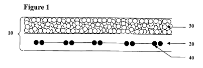
FIG. 1 is a schematic representation of a composite dural repair product in
accordance with
one embodiment of the present disclosure; and
FIG. 2 is a schematic representation of a composite dural repair product in
accordance with
another embodiment of the present disclosure.
DETAILED DESCRIPTION
The present dural repair materials include at least a non-porous layer, a
porous layer and a
fiber reinforcement member. As seen in Figure 1, composite implant 10 includes
non-porous
layer 20, porous layer 30 and reinforcement members 40, which in this
illustrative embodiment
2
CA 02692244 2009-12-18
WO 2009/022230 PCT/IB2008/002706
are embedded within non-porous layer 20. Each of these layers and processes
for preparing each
layer and the composite implant are described in greater detail below.
The Non-Porous La yer
The non-porous layer may retard or prevent tissue ingrowth from surrounding
tissues
thereby acting as an adhesion barrier and preventing the formation of unwanted
scar tissue.
Thus, in embodiments, the non-porous layer possesses anti-adhesion properties.
The non-porous layer of the present dural repair materials may be made from
any
bioabsorbable biocompatible natural or synthetic material. It should of course
be understood that
any combination of bioabsorbable materials may be used to form the non-porous
layer.
Techniques for forming non-porous layers from such materials are within the
purview of
those skilled in the art and include, for example, casting, molding and the
like.
Some non-limiting examples of bioabsorbable materials from which the non-
porous layer
may be made include but are not limited to poly(lactic acid), poly (glycolic
acid), poly
(hydroxybutyrate), polydioxanone, polyalkylene oxides, polyvinyl alcohols,
polycaprolactone,
poly(amino acids), polyalkylene oxalates, polyoxaesters, polyorthoesters, and
copolymers, block
copolyiners, homopolymers, blends and combinations thereof.
In embodiments, natural biological polymers are used in forming the non-porous
layer of
the present dural repair materials. Suitable natural biological polymers
include, but are not
limited to, collagen, gelatin, fibrin, fibrinogen, elastin, keratin, albumin,
hydroxyethyl cellulose,
cellulose, oxidized cellulose, hydroxypropyl cellulose, carboxyethyl
cellulose, carboxymethyl
cellulose, and combinations thereof. In addition, the natural biological
polymers may be
combined with any of the other polymeric materials described herein to produce
the non-porous
layer of the present dural repair materials.
In embodiments, the non porous layer comprises a collagenic constituent.
In embodiments, an aqueous solution of a collagenic constituent is used to
form the non-
porous layer of the present dural repair materials. As used herein, the term
"collagenic
constituent" designates collagen which has at least partially lost its helical
structure through
heating or any other method, or gelatine. The term "gelatine" here includes
commercial gelatine
made of collagen which has been denatured by heating and in which the chains
are at least
3
CA 02692244 2009-12-18
WO 2009/022230 PCT/IB2008/002706
partially hydrolyzed (molecular weight lower than 100 kDa). The collagenic
constituent used
may advantageously be formed of non-hydrolyzed collagen, mainly coinposed of a
chains
(molecular weight around 100 kDa). In the context of the present disclosure, a
chains means
complete a chains or fragments of these complete a chains produced by the loss
of a small
number of amino acids. The term "non-hydrolyzed" as used herein means that
less than 10% of
the collagenic chains have a molecular weight below about 100 kDa. If heating
is used to
denature the helical structure of the collagen, the heating should be moderate
and provided under
gentle conditions so as to avoid degradation by hydrolytic cleavage of the
gelatine thus formed.
Suitable gelatine materials are commercially available.
The collagen used can be of human or animal origin. It may particularly be
type I porcine
or bovine collagen, or type I or type III human collagen or mixtures in any
proportions of the last
two types. Native collagen may advantageously be used, in acid solution or
after processing, to
eliminate the telopeptides, notably by pepsin digestion. The collagen can also
be modified by
oxidative cleavage using any technique know to those skilled in the art,
including, but not limited
to the use of periodic acid or one of its salts as described by Tardy et al.
in U.S. Pat. No.
4,931,546. Briefly, this technique involves mixing the collagen in acid
solution with a solution
of periodic acid or one of its salts at a concentration of between 1 and 10"5
M, in embodiments
between 5 10-3 and 10"1 M, at a temperature of between 10 and 25 C. for 10
minutes to 72
hours. This process breaks down hydroxylysine and the sugars of the collagen,
thus creating
reactive sites without causing crosslinking. The oxidative cleavage of
collagen allows moderate
cross-linking later in the collagenic material. It should of course be
understood that this function
may be provided by other means of moderate cross-linlcing, for example by beta
or gamma
irradiation, or other agents of moderate cross-linking, for example chemical
reagents at suitably
low and non-toxic doses.
In embodiments, the non-porous layer of the composite material according to
the present
disclosure is made of collagen which is oxidized or a mixture in any
proportions of non-oxidized
and oxidized collagens.
In embodiments, a solution of collagenic constituent as defined above is used
to form the
non-porous layer. Typically, a collagen concentration from about 5 g/1 to
about 50 g/1, in
embodiments from about 25 g/l to about 35 g/1 is used.
4
CA 02692244 2009-12-18
WO 2009/022230 PCT/IB2008/002706
The solution of oxidized collagen, non-oxidized collagen or a mixture thereof,
thus
prepared, may be heated, for example to a teinperature in excess of 37 C., in
embodiments to a
temperature of between 40 and 50 C., for at least one hour. This results in
at least partial
denaturing of the collagen's helical structure. Other physical or chemical
techniques for
denaturing collagen (e.g., ultrasonication, or by the addition of chaotropic
agents) are within the
purview of those skilled in the art may also be used.
In embodiments, at least one macromolecular hydrophilic additive that is
chemically
unreactive with the collagenic constituent may be added to the solution used
to form the non-
porous layer. "Chemically unreactive with the collagenic constituent" as used
herein means a
hydrophilic compound which is not likely to react with the collagenic
constituent, notably which
does not form covalent bonds with it during cross-linking.
The macromolecular hydrophilic additive advantageously has a molecular weight
in
excess of 3,000 Daltons, in embodiments from about 3,000 to about 20,000
Daltons. Illustrative
examples of suitable macromolecular hydrophilic additives include polyalkylene
glycols (such as
polyethylene glycol), polysaccharides (e.g., starch, dextran and/or
cellulose), oxidized
polysaccharides, and mucopolysaccharides. It should of course be understood
that combinations
of macromolecular hydrophilic additives may be used. The concentration of
hydrophilic
additive(s) can typically be from about 2 to about 10 times less than that of
the collagenic
constituent.
Typically, the macromolecular hydrophilic additive is eliminated by diffusion
through the
non-porous layer, in a few days. The swelling of this material may
advantageously promote
degradation of a collagenic non-porous layer in less than a month.
Optionally, glycerine may be added to the solution used to form the non-porous
layer.
When present, the concentration of glycerine in the solution can typically be
from about 2 to
about 10 times less than that of the collagenic constituent, in embodiments
less than about one-
third of the collagenic constituent concentration.
In illustrative embodiments of the solution used to form the non-porous layer,
the
concentrations of collagenic constituent, hydrophilic additive(s) and
glycerine, when present, can
be from about 2 to about 10% for the collagenic constituent, from about 0.6 to
about 4% for the
hydrophilic additive(s) and from about 0.3 to about 2.5% for glycerine,
respectively.
CA 02692244 2009-12-18
WO 2009/022230 PCT/IB2008/002706
The solution used to form the non-porous layer may be prepared by adding
collagenic
constituent, hydrophilic additive(s) and glycerine, when present, to water or
a water/alcohol
(e.g.,ethanol) mixture at a temperature of 30 to 50 C. The solution may
advantageously be
neutralized to a neutral pH to avoid hydrolyzing the collagenic constituent by
heating and to
obtain a film of physiological pH while permitting pre-cross-linking of the
collagenic constituent
if the mixture contains oxidized collagen as indicated previously.
In embodiments, the non-porous layer is a collagen film made from either non
heated oxidized
collagen or heated oxidized collagen. The following table gives the
concentration of illustrative
collagen solutions that may be used to form the non-porous layer(s) of the
present dural repair
materials.
Non heated oxidized collagen content 0.1 %--1 % (w/w)
Heated Oxidized collagen content 0.1%--6% (w/w)
In embodiments; the dural repair material comprises an additional non porous
layer. For example,
the porous layer may be sandwiched between a first non porous layer and an
additional non
porous layer.
The Porous Layer
The porous layer of the present dural repair materials has openings or pores
over at least a
portion of a surface thereof. As described in more detail below, suitable
materials for forming
the porous layer include, but are not limited to foams (e.g., open or closed
cell foams). In
embodiments, the pores may be in sufficient number and size so as to
interconnect across the
entire thickness of the porous layer. In other embodiments, the pores do not
interconnect across
the entire thickness of the porous layer. Closed cell foams are illustrative
examples of structures
in which the pores may not interconnect across the entire thickness of the
porous layer. In yet
other embodiments, the pores do not extend across the entire thickness of the
porous layer, but
rather are present at a portion of the surface tllereof. In einbodiments, the
openings or pores are
located on a portion of the surface of the porous layer, with other portions
of the porous layer
6
CA 02692244 2009-12-18
WO 2009/022230 PCT/IB2008/002706
having a non-porous texture. Those skilled in the art reading the present
disclosure will envision
other pore distribution patterns and configurations for the porous layer.
The porous layer of the present dural repair materials may be made from any
bioabsorbable natural or synthetic material. It should of course be understood
that any
combination of bioabsorbable materials may be used to form the porous layer.
Some non-
limiting examples of materials from which the porous layer may be made include
but are not
limited to poly(lactic acid), poly (glycolic acid), poly (hydroxybutyrate),
polydioxanone,
polyalkylene oxides, polyvinyl alcohols, polycaprolactone, poly(amino acids),
polyalkylene
oxalates, polyoxaesters, polyorthoesters, and copolymers, block copolymers,
homopolymers,
blends and combinations thereof. In embodiments, natural biological polymers
are used in
forming the porous layer of the implant. Suitable natural biological polymers
include, but are not
limited to, collagen, gelatin, fibrin, fibrinogen, elastin, keratin, albumin,
hydroxyethyl cellulose,
cellulose, hydroxypropyl cellulose, carboxyethyl cellulose, and combinations
thereof.
Alternatively, the polymer constituent may be a polysaccharide, or
polysaccharides modified by
oxidation of alcohol functions into carboxylic functions such as oxidized
cellulose. In addition,
the natural biological polymers may be combined with any of the other
polymeric materials
described herein to produce the porous layer of the present dural repair
materials.
Where the porous layer is a foam, the porous layer may be formed using any
method
suitable to forming a foam or sponge including, but not limited to the
lyophilization or freeze-
drying of a composition. Suitable techniques for making foams are within the
purview of those
skilled in the art.
The porous layer can be at least 0.1 cm thick, in embodiments from about 0.2
to about 1.5
cm thick. The porous layer can have a density of not more than about 75 mg
collagen/cm2 and,
in embodiments below about 7 mg collagen/cm2. The size of the pores in the
porous layer can be
from about 20 m to about 300 m, in embodiments from about 100 m to about
200 m.
In embodiments, the porous layer possesses haemostatic properties.
Illustrative examples
of materials which may be used in providing the porous layer with the capacity
to assist in
stopping bleeding or hemorrhage include, but are not limited to, poly(lactic
acid), poly(glycolic
acid), poly(hydroxybutyrate), poly(caprolactone), poly(dioxanone),
polyallcyleneoxides,
copoly(ether-esters), collagen, gelatin, thrombin, fibrin, fibrinogen,
fibronectin, elastin, albumin,
7
CA 02692244 2009-12-18
WO 2009/022230 PCT/IB2008/002706
hemoglobin, ovalbumin, polysaccharides, hyaluronic acid, chondroitin sulfate,
hydroxyethyl
starch, hydroxyethyl cellulose, cellulose, oxidized cellulose, hydroxypropyl
cellulose,
carboxyethyl cellulose, agarose, maltose, maltodextrin, alginate, clotting
factors, methacrylate,
polyurethanes, cyanoacrylates, platelet agonists, vasoconstrictors, alurn,
calciuin, RGD peptides,
proteins, protamine sulfate, epsilon amino caproic acid, ferric sulfate,
ferric subsulfates, ferric
chloride, zinc, zinc chloride, aluminum cl-Aoride, aluminum sulfates, aluminum
acetates,
permanganates, tannins, bone wax, polyethylene glycols fucans and combinations
thereof.
The haemostatic agents from which the porous layer can be made or which can be
included in the porous layer can be in the form of foams, fibers, filaments,
meshes, woven and
non-woven webs, compresses, pads, powders, flakes, particles and combinations
thereof. For
example, the implant may include commercially available types of hemostatic
porous layers, such
as materials based on oxidized cellulose (Surgicel or Interceed ).
In embodiments, the porous layer comprises a collagenic constituent.
In embodiments, the porous layer is made from non-denatured collagen or
collagen which
has at least partially lost its helical structure through heating or any other
method, consisting
mainly of non-hydrolyzed a chains, of molecular weight close to 100 kDa. The
term "non-
denatured collagen" means collagen which has not lost its helical structure.
The collagen used
for the porous layer of present iinplant may be native collagen or
atelocollagen, notably as
obtained through pepsin digestion and/or after moderate heating as defined
previously. The
collagen may have been previously chemically modified by oxidation,
methylation, ethylation,
succinylation or any other known process. The origin and type of collagen may
be as indicated
for the non-porous layer described above.
In embodiments, the porous layer can be obtained by freeze-drying an aqueous
acid
solution of collagen at a concentration of 2 to 50 g/l and an initial
temperature of 4 to 25 C. The
concentration of collagen in the solution can be from about 1 g/1 to about 30
g/l, in embodiments
about 10 g/l. This solution is advantageously neutralized to a pH of around 6
to 8.
The porous layer can also be obtained by freeze-drying a fluid foam prepared
from a
solution of collagen or heated collagen, emulsified in the presence of a
volume of air in variable
respective quantities (voluine of air to water varying from about 1 to about
10).
8
CA 02692244 2009-12-18
WO 2009/022230 PCT/1B2008/002706
In embodiments, a collagen sponge is obtained by freeze-drying a collagen
suspension,
resulting from the mixing of oxidized collagen and glutaraldehyde (GTA) cross-
linked collagen,
at different concentrations. Glutaraldehyde (GTA) cross-linked collagen is
obtained by the
incubation of a 1% neutralized collagen solution with a glutaraldehyde
solution at a final
concentration of 0.5%, at room temperature, during 1 hour. The suspension is
then filtered and
washed to remove the excess of GTA. Then, it is treated with sodium
borohydride at room
temperature until removal of the yellow coloration. The suspension is
filtered, washed, and
neutralized. The precipitate is washed several times, by acetone, to remove
salts and water. The
fmal precipitate is dried under vacuum or air flow, and stored at - 20 C.
Oxidized collagen is
obtained by the oxidation of a 3%(w/w) collagen solution by periodic acid (C=8
mM) at room
teinperature, during 3 hours, in the manner described in Example 4 of US
Patent No. 6,596,304,
the entire disclosure of which is incorporated herein by this reference. The
concentration of the
two collagen types and the total amount of collagen in the suspension are
detailed in the table
below.
(A) GTA cross-linked collagen content 20%--100% (w/w total collagen)
(B) Oxidized collagen content 80%--0% (w/w total collagen)
Total collagen concentration in the 0.2%--5% (w/w)
suspension
The ratio (A/B) of concentration of the two collagen types may advantageously
be between 1 and
5. The collagen sponge optionally can be then compacted by using a press, a
calendar or any
other appropriate means.
The Reinforcement Member
The present dural repair materials also include a reinforcement member. The
reinforcement member may be positioned between the non-porous layer and the
porous layer.
Alternatively, the reinforcement member may be positioned entirely within the
non-porous layer.
It is also envisioned that the reinforcement member may be positioned at the
surface of one of the
layers making up the present multilayer dural repair materials and, in
embodiments, may be
positioned at an exterior surface of the present multilayer dural repair
materials.
9
CA 02692244 2009-12-18
WO 2009/022230 PCT/IB2008/002706
Some suitable non-limiting examples of the reinforcement member include
fabrics,
meshes, monofilaments, multifilament braids, chopped fibers (sometimes
referred to in the art as
staple fibers) and combinations thereof.
Where the reinforcement member is a mesh, it may be prepared using any
technique
known to those skilled in the art, such as knitting, weaving, tatting,
knipling or the like.
Illustrative examples of suitable meshes include any of those that are
presently coinmercially
available for hernia repair. In embodiments where a mesh is used as the
reinforcement member,
the mesh will aid in affixing the composite to tissue without tearing of the
porous or non-porous
layers.
Where monofilainents or multifilament braids are used as the reinforcement
member, the
monofilaments or multifilament braids may be oriented in any desired manner.
For example, the
monofilaments or multifilament braids may be randoinly positioned with respect
to each other
within the present dural repair materials. As another example, the
monofilaments or
multifilament braids may be oriented in a common direction within the present
dural repair
materials. In embodiments, monofilainents or multifilament braids are
associated with botll the
porous layer and with the non-porous layer. In an illustrative embodiment of
this type, the
present dural repair materials include a first reinforcement member having a
plurality of
reinforcement members oriented in a first direction within the non-porous
layer and a second
reinforcement layer having a plurality of reinforcement members oriented in a
second direction
within the porous layer. In embodiments, the first and second directions may
be substantially
perpendicular to each other.
Where chopped fibers are used as the reinforcement member, the chopped fibers
may be
oriented in any desired manner. For example, the chopped fibers may be
randomly oriented or
may be oriented in a common direction. The chopped fibers can thus form a non-
woven
material, such as a mat or a felt. The chopped fibers may be joined together
(e.g., by heat fusing)
or they may be unattached to each other. The chopped fibers may be of any
suitable length. For
example, the chopped may be from 0.1 mm to 100 mm in length, in embodiments,
0.4 mm to 50
mm in length. In an illustrative embodiment, the implant has randomly oriented
chopped fibers
that have not been previously fused together embedded within in the non-porous
layer.
CA 02692244 2009-12-18
WO 2009/022230 PCT/IB2008/002706
It is envisioned that the reinforcement member may be formed from any of the
bioabsorbable, natural or synthetic materials previously described herein
including derivatives,
salts and combinations thereof. In embodiments, the reinforcement member is a
surgical mesh
made from polylactic acid fibers. Where monofilaments or multifilament braids
are used as the
reinforcement member, any cominercially available bioabsorbable suture
material may
advantageously be employed as the reinforcement member.
In embodiments, the reinforcement member is a textile knitted with fully
bioresorbable
polylactic acid (PLA) threads designed to achieve suturability and
reinforcement of the dural
implant. The following table gives the technical data of illustrative PLA
textiles that may be
used as the reinforcement member in the present dural repair materials.
PLA textile technical data
Thread Multifilament 84*/240
Weight per surface m2 20-40
Pore sizes 0.5--2 x 0.5--2 mm
Thiclcness 0.2-0.4 mm
Filament diameter
Multifilament 18 m
Cleaning procedure Methanol-ether
Sterilization rays
* yarn count : 84 g for 10 000 m
0 number of filaments
In other embodiments, a textile reinforcement member may be knitted by
combining two
different chemically fibers, such as PLA and oxidized cellulose.
In embodiments, the fibers of the reinforcement member may advantageously be
coated
by a biologic component so as to decrease the risk of inflammatory reaction
and sepsis,
particularly in already contaminated surgical sites. In embodiments, said
biologic component is
selected from the group consisting of oxidized collagen, glutaraldehyde cross-
linked collagen,
polysaccharides such as fucan, and mixtures thereof.
As used in the present application, "fucan" includes any natural fucoidans,
including
those produced by recombinant techniques, as well as any fucoidan precursors,
fucoidan
derivatives or modified fucoidans and fucoidan derivatives, and depolymerized
fucans. "Fucan"
and "fucoidan" are used interchangeably herein.
11
CA 02692244 2009-12-18
WO 2009/022230 PCT/IB2008/002706
The solution used for the textile coating may be composed of any product which
may
limit the risk of inflammatory reaction and sepsis, such as, for example,
oxidized collagen,
glutaraldehyde cross-linked collagen, or polysaccharides (such as fucans).
Advantageously, the
fibers of the reinforcement member may be then processed by a surface
treatment (for example, a
plasma treatment with N2) so as to impart hydrophilic properties and/or a
positive charged at the
surface of the reinforcement member. Such a treatment will facilitate coating
of the
reinforcement member, e.g., with collagen and/or polysaccharide solutions.
Optional Bioactive Agents
h1 some embodiments, at least one bioactive agent may be combined with the
present
dural repair materials and/or any of the individual coinponents (the porous
layer, the non-porous
layer(s) and/or the reinforcement member) used to construct the present dural
repair materials. In
these embodiments, the present dural repair material can also serve as a
vehicle for delivery of
the bioactive agent. The term "bioactive agent", as used herein, is used in
its broadest sense and
includes any substance or mixture of substances that have clinical use.
Consequently, bioactive
agents may or may not have pharmacological activity per se, e.g., a dye, or
fragrance.
Alternatively a bioactive agent could be any agent which provides a
therapeutic or prophylactic
effect, a compound that affects or participates in tissue growth, cell growth,
cell differentiation,
an anti-adhesive compound, a compound that may be able to invoke a biological
action such as
an immune response, or could play any other role in one or more biological
processes. It is
envisioned that the bioactive agent may be applied to the present dural repair
materials in any
suitable form of matter, e.g., films, powders, liquids, gels and the like.
Examples of classes of bioactive agents which may be utilized in accordance
with the
present disclosure include anti-adhesives, antimicrobials, analgesics,
antipyretics, anestlietics,
antiepileptics, antihistamines, anti-inflammatories, cardiovascular drugs,
diagnostic agents,
sympathomimetics, cholinomimetics, antimuscarinics, antispasmodics, hormones,
growth
factors, muscle relaxants, adrenergic neuron blockers, antineoplastics,
immunogenic agents,
iininunosuppressants, gastrointestinal drugs, diuretics, steroids, lipids,
lipopolysaccharides,
polysaccharides, and enzymes. It is also intended that combinations of
bioactive agents may be
used.
12
CA 02692244 2009-12-18
WO 2009/022230 PCT/IB2008/002706
Anti-adhesive agents can be used to prevent adhesions from forming between the
present
dural repair materials and the surrounding tissues opposite the target tissue.
In addition, anti-
adhesive agents may be used to prevent adhesions from forming between the
present dural repair
materials and the packaging material. Some exainples of these agents include,
but are not limited
to poly(vinyl pyrrolidone), carboxymethyl cellulose, hyaluronic acid,
polyethylene oxide, poly
vinyl alcohols and combinations thereof.
Suitable antimicrobial agents which may be included as a bioactive agent in
the dural
repair materials of the present disclosure include triclosan, also known as
2,4,4'-trichloro-2'-
hydroxydiphenyl ether, chlorhexidine and its salts, including chlorhexidine
acetate, chlorhexidine
gluconate, chlorhexidine hydrochloride, and chlorhexidine sulfate, silver and
its salts, including
silver acetate, silver benzoate, silver carbonate, silver citrate, silver
iodate, silver iodide, silver
lactate, silver laurate, silver nitrate, silver oxide, silver palmitate,
silver protein, and silver
sulfadiazine, polymyxin, tetracycline, aminoglycosides, such as tobramycin and
gentamicin,
rifainpicin, bacitracin, neomycin, chloramphenicol, miconazole, quinolones
such as oxolinic
acid, norfloxacin, nalidixic acid, pefloxacin, enoxacin and ciprofloxacin,
penicillins such as
oxacillin and pipracil, nonoxynol 9, fusidic acid, cephalosporins, and
combinations thereof. In
addition, antimicrobial proteins and peptides such as bovine lactoferrin and
lactoferricin B and
antimicrobial polysaccharides such as fucans and derivatives may be included
as a bioactive
agent in the dural repair materials of the present disclosure.
Other bioactive agents which may be included as a bioactive agent in the dural
repair
materials in accordance with the present disclosure include: local
anesthetics; non-steroidal
antifertility agents; parasympathomimetic agents; psychotherapeutic agents;
tranquilizers;
decongestants; sedative hypnotics; steroids; sulfonamides; sympathomimetic
agents; vaccines;
vitamins; antimalarials; anti-migraine agents; anti-parkinson agents such as L-
dopa; anti-
spasmodics; anticholinergic agents (e.g. oxybutynin); antitussives;
bronchodilators;
cardiovascular agents such as coronary vasodilators and nitroglycerin;
alkaloids; analgesics;
narcotics such as codeine, dihydrocodeinone, meperidine, morphine and the
like; non-narcotics
such as salicylates, aspirin, acetaininophen, d-propoxyphene and the like;
opioid receptor
antagonists, such as naltrexone and naloxone; anti-cancer agents; anti-
convulsants; anti-emetics;
antihistamines; anti-inflammatory agents such as hormonal agents,
hydrocortisone, prednisolone,
13
CA 02692244 2009-12-18
WO 2009/022230 PCT/IB2008/002706
prednisone, non-hormonal agents, allopurinol, indomethacin, phenylbutazone and
the like;
prostaglandins aiid cytotoxic drugs; estrogens; antibacterials; antibiotics;
anti-fungals; anti-virals;
anticoagulants; anticonvulsants; antidepressants; antihistainines; and
immunological agents.
Other examples of suitable bioactive agents which may be included in the
present dural
repair materials include viruses and cells, peptides, polypeptides and
proteins, analogs, muteins,
and active fragments thereof, such as immunoglobulins, antibodies, cytokines
(e.g. lymphokines,
monokines, chemokines), blood clotting factors, hemopoietic factors,
interleukins (IL-2, IL-3, IL-
4, IL-6), interferons ((3-IFN, (a-IFN and y-IFN), erythropoietin, nucleases,
tumor necrosis factor,
colony stimulating factors (e.g., GCSF, GM-CSF, MCSF), insulin, anti-tumor
agents and tumor
suppressors, blood proteins, gonadotropins (e.g., FSH, LH, CG, etc.), hormones
and hormone
analogs (e.g., growth hormone), vaccines (e.g., tumoral, bacterial and viral
antigens);
somatostatin; antigens; blood coagulation factors; growth factors (e.g., nerve
growth factor,
insulin-like growth factor); protein inhibitors, protein antagonists, and
protein agonists; nucleic
acids, such as antisense molecules, DNA and RNA; oligonucleotides;
polynucleotides; and
ribozymes.
Assembling the Composite
The multilayer dural repair materials described herein may be formed using any
method
known to those skilled in the art capable of connecting one or more non-porous
layer(s) to a
porous layer. It is envisioned that the non-porous layer(s) and the porous
layer may be adhered to
one another using chemical bonding, surgical adhesives, surgical sealants, and
surgical glues. In
addition, the layers may be bound together using mechanic means such as pins,
rods, screws,
clips, etc. Still further, the layers may naturally or through chemical or
photoinitiation may
interact and crosslink or provide covalent bonding between the layers.
In the illustrative embodiment shown in Figure 1, composite dural repair
material 10
includes non-porous layer 20, porous layer 30 and reinforcement members 40,
which are
embedded within non-porous layer 20. In an alternative embodiment shown in
Figure 2,
coinposite dural repair material 100 includes porous layer 130 sandwiched
between fiber
reinforced non-porous layer 120, and a second or additional non-porous layer
150. Those skilled
14
CA 02692244 2009-12-18
WO 2009/022230 PCT/IB2008/002706
in the art reading the present disclosure will readily envision other
combinations of porous and
non-porous layers suitable for use as dural repair materials
In embodiments, the inultilayer dural repair materials described herein are
prepared by
attaching the individual layers of materials together to form a multiple layer
implant. The porous
layer may be formed separate and apart from the non-porous layer(s).
Alternatively, the porous
and non-porous layers may be forined together.
In an illustrative embodiment, the present dural repair materials are prepared
by first
pouring a solution of collagenic constituent, destined to form the film,
possibly containing the
hydrophilic additive(s) and glycerine, onto an adequate, substantially flat
support and distributing
it evenly.
The support is inert in that it does not react with the above-mentioned
components and is
not involved in the cross-linking process. The support may advantageously be
made from a
hydrophobic material such as, for example, PVC or polystyrene. However, this
support can also
consist of a strippable material which will remain slightly adhesive and which
can then be
separated from the implant at the time of surgical use. This support may
itself also consist of a
film, for example dried collagen, onto which the solution is poured, or a
layer of collagenic
material gel in a distinctly more advanced state of gelification.
The density of the thin layer initially applied as a solution to the substrate
can be from
about 0.1 g solution/cm2 to about 0.3 g solution/cm2. This collagenic solution
advantageously
may be poured at a temperature from about 4 C. to about 30 C., and in
embodiments from
about 18 C. to about 25 C. Once applied to the substrate, the collagen
solution is allowed to
partially gel. Partial gelling results from cooling of the collagen solution,
and not from drying of
the solution.
A mesh reinforcement member is then applied to the solution. Application of
the
reinforcement member onto the solution means simply laying the reinforcement
member onto the
solution or partially gelled solution, and optionally applying slight
pressing. The pressing should
be insufficient to cause any significant disruption of the portion of the
layer of solution in contact
with the substrate thereby helping to maintain the integrity and anti-adhesion
characteristics of
the non-porous layer. The pressing may leave the surface of the reinforcement
member exposed
CA 02692244 2009-12-18
WO 2009/022230 PCT/IB2008/002706
at the surface of the solution or may embed the reinforcement member
completely within the
layer of solution.
Following application of the mesh reinforcement member, but before complete
gellification of the initially applied solution, additional solution may be
applied in an amount
sufficient to cover the mesh, so that it is completely embedded within the
solution. Where
pressing has already embedded the reinforcement member in the solution,
application of
additional solution may be eliminated.
This solution containing the embedded mesh reinforcement member is left to gel
and a
porous layer prepared as indicated above is applied to the solution during
gelification.
Application of the porous layer onto the solution during gelification means
simply laying
the porous layer onto the gel, and optionally applying slight pressing. The
pressing should be
insufficient to cause any significant compaction of the porous layer. In
embodiments where the
porous layer has been pre-formed, the porous layer will become joined to the
solution, but will
not become interlocked with the mesh reinforcement member.
The moment at wl7ich the porous layer is applied to the solution during
gelification will
depend upon the nature of the solution employed, the conditions under which
the solution is
maintained during gelification and the nature of the porous layer. Generally,
the solution will
allowed to gellify for a period of time prior to application of the porous
layer such that the gel is
still soft and allows the porous layer to penetrate over a distance which is
advantageously from
about 0.05 mm to about 2 mm and, in embodiments from about around 0.1 mm to
about 0.5 mm.
The appropriate moment for application of the porous layer for any given
combination of
materials/conditions can be determined empirically, for example by applying
small samples of
the porous layer to the gel at various times and evaluating the degree of
penetration and
adherence. Generally, when the solution which is gelling is at a temperature
of between 4 and
30 C., the porous layer can be applied 5 to 30 minutes after the solution has
been poured over
the surface holding it.
At this step, an additional or second non porous layer may be distributed on
the porous layer, for
exainple under the form of a solution of collagenic constituent.
The composite implant is left to dry or dried in order to obtain the final
implant. When
the collagenic solution destined to form the film or non porous layer includes
oxidized collagen,
16
CA 02692244 2009-12-18
WO 2009/022230 PCT/IB2008/002706
it is polymerized while the material is drying. This drying occurs favorably
at a temperature of
from about 4 C. to about 30 C., in embodiments from about 18 C. to about 25
C. The
material can be dried in a jet of sterile air if desired.
After drying, the implant can be separated from its support, packaged and
sterilized using
conventional techniques, e.g., irradiation with beta (electronic irradiation)
or gamma (irradiation
using radioactive cobalt) rays. In embodiments where hydrolytically unstable
materials are used
in forming the composite, such as polyglycolic acid, polylactic acid the
composites are packaged
under sufficiently dry conditions to ensure that no degradation of the
composite takes place
during storage.
The present dural repair materials are stable at ambient temperature and
remains stable
for long enough to be handled at temperatures which may rise to 37-40 C. The
thickness of the
non-porous layer is not critical, but typically can be less than about 100 m
thick, and in
embodiments from about 30 m to about 75 m thick. Likewise, the thickness of
the porous
layer is not critical, but typically caii be from about 0.1 mm to about 1.4 mm
thick. The overall
thickness of the dural repair material is not critical, but typically can be
from about 0.2 mm to
about 1.5 mm thick, and in embodiments from about 0.3 mm to about 0.8 mm
thick. The dural
repair materials in accordance witli this disclosure can be produced at a
desired size or produced
in large sheets and cut to sizes appropriate for the envisaged application.
The present dural repair materials may be implanted using open surgery or in a
laparoscopic procedure. When implanted laparoscopically, the present dural
repair materials
should be rolled with the porous side on the inside before trocar insertion.
The following non-limiting example illustrates the preparation of dural repair
materials in
accordance witriz the present disclosure.
EXAMPLES
EXAMPLE 1:
1 ) Preparation of the fiber reinforcement member
17
CA 02692244 2009-12-18
WO 2009/022230 PCT/IB2008/002706
Hereinbelow are given three examples of coating of a textile or mesh with a
biologic component
in order to obtain a fiber reinforcement member suitable for the dural repair
materials of the
invention:
a ) Preparation of textile reinforcement member coated with oxidized collagen
Oxidized collagen is obtained by the oxidation of a 3 % collagen solution by
periodic
acid, at a final concentration of 8 mM, at room temperature, during 3 hours as
described in
Example 4 of US Patent No. 6,596,304. To a 3 % oxidized collagen solution, a
sterile
concentrated solution of PEG 4000 (polyethylene glycol having a molecular
weight of 4000
g/mol) and glycerol, in order to achieve a PEG concentration of 1% and a
glycerol concentration
of 0.6 %. The pH of the solution is adjusted to 7.0 by adding concentrate
sodium hydroxide
solution. The volume of the solution is then adjusted with sterile water to
obtain final
concentrations of collagen, PEG and glycerol of 2.7 %, 0.9 % and 0.54 %
respectively. A two-
dimensional textile (or mesh) of polylactic (PLA) fibers is soaked once or
twice into the oxidized
collagen solution, then dried, so as to cover as much as possible the overall
accessible surface of
PLA fibres of the 2D textile.
b , Preparation of textile reinforcement member coated with GTA cross-linked
collagen
A textile (or mesh), for example a textile of PLA fibers, is coated with GTA
cross-linked
collagen, in two steps. It is first soaked once or twice into a collagen
solution (1 1o w/w) and then
dried. Then, the coated textile is cross-linked in a solution of
glutaraldehyde with a concentration
of 0.5 % for 1 hour. It is further treated with sodium borohydride during at
least two hours, until
the initial yellowish appearance of fibers was completely removed to give
white fibers. The
textile is then washed several times in sterile water and finally dried.
c ) Preparation of textile reinforcement member coated with GTA cross-linked
collagen
A textile (or mesh), for example a textile of PLA fibers, is coated with GTA
cross-linked
collagen, in two steps. It is first sprayed with a collagen solution (1% w/w),
several times up to
ten times. After each series of spraying, the collagen laid on the mesh is
completely dried in an
oven, at +50 C. Then, the coated textile is cross-linleed in a solution of
glutaraldehyde with a
concentration of 0.5 % for 1 hour. It is further treated with sodium
borohydride during at least
18
CA 02692244 2009-12-18
WO 2009/022230 PCT/IB2008/002706
two hours, until the initial yellowish appearance of fibers was completely
removed to give white
fibers. The textile is then washed several times in sterile water and finally
dried.
2 ) Preparation of calendered collagen porous layer
A collagen suspension is obtained by mixing GTA cross-linked collagen and
oxidized
collagen in relative concentrations of 80 % / 20 % respectively. The total
collagen concentration
in the aqueous solution is fixed at 1.5 % w/w. Then, the suspension is poured
in Petri dishes and
freeze-dried. Finally the collagen sponges are calendered to obtain a maximal
thickness of 0.15
mm.
3 ) Preparation of a solution in order to make non porous layers suitable for
the dural repair
materials of the invention:
Hereinbelow are given three examples of preparation of solutions for preparing
non porous layers
comprising a collagenic constituent suitable for the dural repair materials of
the invention
a ) Preparation of oxidized collagen solution/suspension
To a 3.9 % oxidized collagen solution, an ultra-filtered concentrated solution
of PEG
4000 (polyethylene glycol having a molecular weight of 4000 g/mol) and
glycerol is added, in
order to achieve a PEG concentration of 1% and a glycerol concentration of 0.6
%. The pH of
the solution is adjusted to 7.0 by adding concentrate sodium hydroxide
solution. The volume of
the solution is then adjusted with sterile water to obtain final
concentrations of collagen, PEG
and glycerol of 2.7 %, 0.9 % and 0.54 %, respectively.
b Preparation of the oxidized collagen solution/suspension
To a 3.9 % oxidized collagen solution, an ultra-filtered concentrated solution
of PEG
4000 (polyethylene glycol having a molecular weight of 4000 g/mol) and
glycerol is added, in
order to achieve a PEG concentration of 1 % and a glycerol concentration of
0.6 %. To the
solution is added one part of dry GTA cross-linked collagen for 5 parts of
oxidized collagen by
weight. The pH of the suspension is adjusted to 7.0 by adding concentrate
sodium hydroxide
19
CA 02692244 2009-12-18
WO 2009/022230 PCT/IB2008/002706
solution. The volume of the solution is then adjusted with sterile water to
obtain final
concentrations of collagen, GTA cross-linked collagen, PEG and glycerol of 2.7
%, 0.55 %, 0.9
% and 0.54 %, respectively.
c Preparation of the oxidized collagen solution/suspension
To a 3.9 % oxidized collagen solution, an ultra-filtered concentrated solution
of PEG
4000 (polyethylene glycol having a molecular weight of 4000 g/mol) and
glycerol is added, in
order to achieve a PEG concentration of 1% and a glycerol concentration of 0.6
%. To the
solution is added one part of dry GTA cross-linked collagen for 20 parts of
oxidized collagen by
weight. The pH of the suspension is adjusted to 7.0 by adding concentrate
sodium hydroxide
solution. The volume of the solution is then adjusted with sterile water to
obtain final
concentrations of collagen, GTA cross-linked collagen, PEG and glycerol of 2.7
%, 0,13 %, 0.9
% and 0.54 %, respectively.
4 Assembly of a two-layer dural implant
An oxidized collagen solution, as prepared in one of points 3 )a-c above, is
poured in a
thin layer on a flat hydrophobic support of the PVC or polystyrene type, with
a density of 0.266 g
solution /cm2, then a coated mesh, as prepared in one of points 1 )a-c above,
is laid over the
collagen solution, pressed into the solution and the application of additional
solution on top of
the original volume of solution. The surfaces are then exposed to a sterile
stream of air at
ambient temperature, during less than half of an hour. A calendered sponge, as
prepared in point
2 ) above, is then gently applied on the gelling layer of oxidized collagen
and the two layers are
exposed to a sterile stream of air at ambient temperature. The two layers
composite is exposed to
a sterile stream of air at ambient temperature, leading to complete
evaporation in at least
approximately 18 hours.
EXAMPLE 2:
Assembly of a three-layer dural implant
An oxidized collagen solution, as prepared in one of points 3 )a-c of EXAMPLE
1
aboveis poured in a thin layer on a flat hydrophobic support of the PVC or
polystyrene type, with
CA 02692244 2009-12-18
WO 2009/022230 PCT/IB2008/002706
a density of 0.400 g solution /cin2, and then a textile reinforcement member,
as prepared in one of
points 1 )a-c of EXAMPLE 1 above, is laid over the collagen solution, pressed
into the solution
and the application of additional solution on top of the original volume of
solution. The surfaces
are then exposed to a sterile stream of air at ambient temperature, during
less than one hour. A
calendered sponge, as prepared in point 2 ) of EXAMPLE 1 above, is then gently
applied on the
gelling layer of oxidized collagen and the two layers are exposed to a sterile
stream of air at
ambient temperature, overnight. At this step, a second or additional layer of
oxidized collagen
solution, as prepared in one of points 3 )a-c of EXAMPLE 1 above, is
distributed on the bi-layer
composite with a reduced density, 0.133 g solution / cma. The three layers
composite is exposed
to a sterile stream of air at ainbient temperature, leading to complete
evaporation in at least
approximately 18 hours.
A dural repair material is thus obtained, comprising a fiber reinforced non
porous layer, a
porous layer and and an additional non porous layer, in which the fiber
reinforcement member is
coated with a biological component and is embedded in the non porous layer,
said porous layer
being sandwiched between the fiber reinforced non porous layer and the
additional non porous
layer, all layers, ie the fiber reinforced non porous layer, the porous layer
and the additional non
porous layer, comprising a collagenic constituent.
It will be understood that various modifications may be made to the
embodiments
disclosed herein. Therefore, the above description should not be construed as
limiting, but
merely as an exemplification of preferred embodiments. Those skilled in the
art will envision
other modifications within the scope and spirit of the present disclosure.
Such modifications and
variations are intended to come within the scope of the following claims.
21