Note: Descriptions are shown in the official language in which they were submitted.
CA 02693296 2010-02-18
HETERODIMERIC FUSION PROTEINS USEFUL FOR TARGETED IMMUNE
THERAPY AND GENERAL IMMUNE STIMULATION
Field of the Invention
The present invention relates generally to fusion proteins. More specifically,
the present
invention relates to heterodimeric fusion proteins useful for targeted immune
therapy and general
immune stimulation.
Background of the Invention
One of the key immune regulators is the T helper cell which reacts to antigens
presented
on HLA class II molecules. This CD4+ cell differentiates in response to
antigenic stimulation and
becomes a type 1 or type 2 helper (Thl or Th2) according to the type of
cytokines that it secretes.
Mosmann and Coffman, Ann. Rev. Immunol. 7:145-173 (1989). A Thl response leads
to the
secretion of interleukin-2 (IL-2) and interferon-7 (IFN-y) which stimulates
cell-mediated immune
reactions against intracellular pathogens. A Th2 response leads to the
secretion of IL-4, IL-5 and
IL-10 which stimulates antibody responses to extracellular pathogens. The most
interesting
component of this system of regulation is that one response inhibits the other
through the negative
regulatory activities of the cytokines that are produced. Thus, IL-4 and IL-10
can down-regulate
Thl responses while IFN-y can down-regulate Th2 responses.
The regulatory activity of T helper cells and their differentiation following
exposure to
antigen is regulated by cytokines as well. IL-12, a disulfide-linked
heterodimeric cytokine with a
40 kDa subunit and 35 kDa subunit, exerts a powerful positive regulatory
influence on the
development of Thl helper T-cell immune responses. See review by Trinchieri,
Blood 84:
4008-4027 (1994). IL-12 also has a powerful synergistic effect in the
induction of IFNay from
both T helpers and natural killer (NK) cells (Eur. Patent Appl. 90123670.3).
Secreted IFN-y then
inhibits any Th2 cell proliferation and polarizes the response to favor cell-
mediated immunity.
One way of changing the outcome of an immune response would be to administer
the
appropriate cytokine at the time of antigen stimulation. If II.,-4 was the
major cytokine present
during antigen stimulation, the Th2 response would be enhanced and the Thl
response would be
inhibited. In contrast, if IL-12 was the major cytokine present during antigen
stimulation, the Thl
CA 02693296 2010-02-18
- 2 -
response would be enhanced and the Th2 response would be inhibited. However,
systemic administration of cytokines is difficult due to their very short
circulating half-
lives and their deleterious side effects.
A better approach is to target the effect of the cytokine to a cell surface
antigen
by fusing it to an antibody (or fragment derived therefrom) having specificity
and
affinity for that antigen. See Gillies, et al., Proc. Natl. Acad ScL 89: 1428-
1432
(1992); U.S. Patent No 5,650,150. Alternatively, the stimulatory cytokine can
be
linked to a protein antigen via a peptide linkage in the form of a fusion
protein. See
Hazama, et al., Vaccine 11: 629-636 (1993). However, the complex structure of
IL-12
makes it more difficult to express as a fusion protein due to the necessity of
expressing
exactly the same molar ratio of each subunit in the final product. In fact, IL-
12 itself is
naturally expressed and secreted as a mixture of p40 homodimer. D'Andrea, et
al., J.
Exp. Med, 176: 1387-1398 (1992).
Therefore, there is a need in the art for methods of producing fusion proteins
with heterodimeric cytokines and an antibody or an antigen that maintain the
natural
heterodimeric structure of the cytokine and secretes the molecules with
equimolar
ratios of the subunits.
Summary of the Invention
The present invention provides heterodimeric fusion proteins useful for
targeted
immune therapy and general immune stimulation and methods for producing these
heterodimeric fusion proteins. Specifically, the present invention provides
methods for
the production of fusion proteins with IL-12 that maintain its natural
heterodimeric
structure, and provide for the secretion of the molecules with equimolar
ratios of IL-12
subunits.
In one aspect of the invention, the fusion proteins comprise a heterodimeric
cytokine linked to an antibody, or a portion thereof. The fusion protein may
comprise
two chimeric chains linked by a disulfide bond. Each chimeric chain may
comprise a
different subunit of the heterodimeric cytokine linked through a peptide bond
to a
portion of an Ig heavy chain.
Alternatively, the fusion protein may comprise a first chimeric chain
comprising
one of the subunits of the heterodimeric cytokine linked by a peptide bond to
a portion
of an Ig heavy chain. This subunit may be linked by a disulfide bond to the
other
CA 02693296 2010-02-18
- 3 -
subunit of the heterodimeric cytokine. In another alternative, this first
chimeric chain
may be linked by a disulfide bond to a second chimeric chain comprising one of
the
subunits of the heterodimeric cytokine linked by a peptide bond to a portion
of an Ig
heavy chain and by a disulfide bond to the other subunit of the heterodimeric
cytokine.
In yet another alternative, the fusion protein may be a trimeric fusion
protein
comprising a first and a second chimeric chain linked by a disulfide bond.
Each
chimeric chain may comprise a subunit of the heterodimeric cytokine linked by
a
peptide bond to a portion of an Ig heavy chain. The subunit of one of the
chimeric
chains may further be linked by a disulfide bond to a different subunit of the
heterodimeric cytokine.
Fusion proteins of the invention may be considered chimeric by virtue of two
aspects of their structure. First, the fusion protein is chimeric in that it
includes an
immunoglobulin chain (typically but not exclusively a heavy chain) of
appropriate
antigen-binding specificity fused to a given heterodimeric cytokine. Second,
an
immunoconjugate of the invention may be chimeric in the sense that it includes
a
variable region and a constant region which may be the constant region
normally
associated with the variable region, or a different one and thus a V/C
chimera; e.g.,
variable and constant regions from different species. Also embraced within the
term
"fusion protein" are constructs having a binding domain comprising framework
regions
and variable regions (L e., complementarity determining regions) from
different species,
such as are disclosed by Winter, et al., GB2, 188, 638.
The heterodimeric cytokine-antibody fusion protein of the present invention
preferably displays antigen-binding specificity. In a preferred embodiment,
the
heterodimeric cytokine-antibody fusion protein comprises a heavy chain. The
heavy
chain can include a CH1, CH2, and/or CH3 domains. In an alternative preferred
embodiment, the heterodimeric cytokine-antibody fusion protein comprises a
light
chain. The invention thus provides fusion proteins in which the antigen
binding
specificity and activity of an antibody are combined with the potent
biological activity
of a heterodimeric cytokine. A fusion protein of the present invention can be
used to
deliver selectively a heterodimeric cytokine to a target cell in vivo so that
the
heterodimeric cytokine can exert a localized biological effect.
CA 02693296 2010-02-18
- 4 -
Preferably, the fusion protein of the present invention displays cytokine
biological activity. The preferred heterodimeric cytokine of the fusion
protein is IL-12.
Fusions with antibodies capable of binding antigens are useful for co-
localizing the
immune stimulatory activity of IL-12 either to target cells or target protein
antigens.
Further, the fusion protein of the present invention preferably has a longer
circulating half-life than an unlinked heterodimeric cytokine. Fusions with
the Fc
portion of antibodies and IL-12 are useful for altering the pharmacology and
biodistribution of the molecule by increasing its circulating half-life and
its affinity for
Fc-receptor bearing cells, e.g., antigen presenting cells. Changes in
biodistribution may
also alter its systemic toxicity by changing the mechanism by which it is
cleared from
the circulation.
In another aspect of the invention, the fusion proteins comprise a
heterodimeric
cytokine linked to an antigen. The heterodimeric cytokine-antigen fusion
protein of the
present invention may display cytokine biological activity and antigenic
activity.
Further, the fusion protein of the present invention preferably has a longer
circulating
half-life than an unlinked heterodimeric cytokine. The preferred heterodimeric
cytokine
of the fusion protein is IL-12.
The fusion protein may comprise two chimeric chains linked by a disulfide
bond. Each chimeric chain comprises a different subunit of the heterodimeric
cytokine,
either of which is linked through a peptide bond to an antigen.
Alternatively, the fusion protein may comprise a first chimeric chain
comprising
one of the subunits of the heterodimeric cytokine linked by a peptide bond to
an
antigen. This subunit may be linked by a disulfide bond to the other subunit
of the
heterodimeric cytokine. In another alternative, this first chimeric chain may
be linked
by a disulfide bond to a second chimeric chain comprising one of the subunits
of the
heterodimeric cytokine linked by a peptide bond to an antigen and by a
disulfide bond
to the other subunit of the heterodimeric cytokine.
In another alternative, the fusion protein may be a trimeric fusion protein
comprising a first and a second chimeric chain linked by a disulfide bond.
Each
chimeric chain may comprise a subunit of the heterodimeric cytokine linked by
a
peptide bond to an antigen. The subunit of one of the chimeric chain may
further be
linked by a disulfide bond to a different subunit of the heterodimeric
cytokine.
CA 02693296 2010-02-18
- 5 -
In another aspect, there is provided a fusion protein comprising a first
chimeric
Ig chain comprising a portion of an Ig heavy chain linked by a peptide bond to
a p40
subunit of IL-12, the P40 subunit of IL-12 being linked to a p35 subunit of IL-
12.
There is also disclosed a fusion protein comprising a first chimeric Ig chain
comprising a portion of an Ig heavy chain linked by a peptide bond to a p35
subunit of
IL-12, said p35 subunit of IL-12 being linked to a p40 subunit of IL-12.
The invention also features DNA constructs encoding the above-described
fusion proteins, and cell lines, e.g., myelomas, transfected with these
constructs.
The invention also includes a method for selectively targeting a heterodimeric
cytokine. The method may comprise linking at least one subunit of a
heterodimeric
cytokine by a peptide bond to a portion of an Ig heavy chain. In an
alternative, the
method may comprise linking each of the two subunits of a heterodimeric
cytokine by a
peptide bond to a portion of an Ig heavy chain, thereby forming two chimeric
chain.
The two chimeric chains may be linked by a disulfide bond, thereby forming a
heterodimeric fusion protein. In yet another preferred embodiment, the method
comprises (1) linking one of the two subunits of a first heterodimeric
cytokine by a
peptide bond to an Ig heavy chain, thereby forming a first chimeric chain; (2)
linking
one of the two subunits of a second heterodimeric cytokine by a peptide bond
to an Ig
heavy chain, thereby forming a second chimeric chain; and (3) linking the
first and
second chimeric chains by a disulfide bond, thereby forming a fusion protein.
The
resulting fusion proteins can display binding specificity for a predetermined
antigen
and cytokine biological activity.
The invention also includes a method of selectively delivering a heterodimeric
cytokine to a target cell. The method includes providing a heterodimeric
cytokine
fusion protein including a chimeric Ig chain including an Ig heavy chain
having a
variable region specific for an epitope on the target cell and a constant
region joined at
its carboxy terminus by a peptide bond to a cytokine, and an Ig light chain
combined
with the chimeric Ig heavy chain, forming a functional antigen-binding site,
and
administering the fusion protein in an amount sufficient to reach the target
cell to a
subject harboring the target cell.
Further, the invention features a method of increasing the circulating half-
life of
a heterodimeric cytokine. The method may comprise linking at least one subunit
of a
I
CA 02693296 2010-02-18
- 6 -
heterodimeric cytokine by a peptide bond to a polypeptide. In an alternative,
the
method may comprise linking each of the two subunits of a heterodimeric
cytokine by a
peptide bond to a polypeptide, thereby forming two chimeric chain. The two
chimeric
chains are linked by a disulfide bond, thereby forming a heterodimeric fusion
protein.
In yet another preferred embodiment, the method comprises (1) linking one of
the two
subunits of a first heterodimeric cytokine by a peptide bond to a polypeptide,
thereby
forming a first chimeric chain; (2) linking one of the two subunits of a
second
heterodimeric cytokine by a peptide bond to a polypeptide, thereby forming a
second
chimeric chain; and (3) linking the first and second chimeric by a disulfide
bond,
thereby forming a fusion protein. The polypeptide can be serum albumin, an
antigen,
and a portion of an Ig heavy chain. The resulting fusion proteins display
cytokine
biological activity.
There is also disclosed a method of increasing the circulating half-life of IL-
12,
comprising the steps of: (a) linking a p35 subunit of IL-12 by a peptide bond
to a
polypeptide, thereby forming a first chimeric chain, said p35 subunit of 1L-12
being
linked to a p40 subunit of IL-12 by a disulfide bond; (b) linking (i) a p35
subunit of IL-
12 by a peptide bond to a polypeptide, thereby forming a second chimeric
chain, said
p35 subunit of IL-12 being linked to a p40 subunit of IL-12 by a disulfide
bond; or
linking (ii) a p40 subunit of IL-12 by a peptide bond to a polypeptide,
thereby forming
a second chimeric chain being linked by a disulfide bond; and (c) linking said
first and
said second chimeric chain by a disulfide bond, thereby forming a dimeric
fusion
protein, said fusion protein having a longer circulating half-life than
unlinked p35 and
p40 subunits of IL-12.
The IL-12 fusion proteins of the present invention may be useful for specific
targeting or immune stimulation when it is important to generate a cell-
mediated
immune response, such as in cancer immunotherapy or antiviral responses. They
may
also be useful for specifically downregulating Th2 responses which often lead
to the
overproduction of IL-4. This cytokine has been shown to be essential for the
development of allergy through the induction of a Th2 response and the
resulting
overproduction of IgE antibody.
CA 02693296 2010-02-18
- 7 -
Brief Description of the Drawings
The foregoing and other objects of the present invention, and the various
features thereof, may be more fully understood from the following description,
when
read together with the accompanying drawings, in which
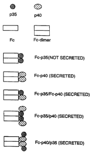
FIG. 1 is a diagrammatic representation of the predicted protein structure of
heterodimeric fusion proteins; A: representation of p35 and p40 subunits; B:
representation of chimeric chains linked to free subunits; C-E: representation
of various
heterodimers and homodimers;
FIG. 2 is a diagrammatic representation of an SDS-PAGE showing an analysis,
under reducing conditions, of proteins secreted by cells transfected with
vectors
expressing the Fc-p35 fusion protein (lane 1), the Fc-p40 fusion protein (lane
2), the
Fc-p35 fusion protein and the Fc-p40 fusion protein (lane 3), the Fc-p35
fusion protein
and the p40 subunit (lane 4), and the p35 subunit and the Fc-p40 fusion
protein (lane
5);
FIG. 3 is a diagrammatic representation of the predicted protein structure of
expressed fusion proteins;
FIG. 4 is a bar graph depicting the ability of various fusion proteins to
stimulate
IFN-y production;
CA 02693296 2010-02-18
- 8 -
FIG. 5A is a diagrammatic representation of an SDS-PAGE showing an analysis of
whole
antibody-1L-12 fusion proteins produced by two independent transfectants,
under non-reducing
(lanes I and 2) and reducing conditions (lanes 3 and 4);
FIG. 5B-D are line graphs depicting the effects of human 1L-12 (X), Hu-KS-IL-
12 fusion
protein with both human IL-12 chains (closed squares), and Hu-KS-1/4-mouse p35
human p40
fusion protein (open squares) on proliferation of mitogen-activated human PBMC
(Panel B);
induction of 1FN-y secretion from PHA-activated PBMC (Panel C) and from mouse
effector cells,
pre-stimulated with Concanavalin A (Panel D);
FIG. 6A-B are line graphs depicting the effects of 1L-12 (X), single-chain
fusion protein
with human p35 and p40 subunits (closed squares), and single-chain fusion
protein with a mouse
p35 subunit and a human p40 subunit (open squares) on induction of1FN-y
secretion;
FIG. 6C is line graphs depicting the antigen binding activity of whole Hu-KS-
1/4-IL-12
fusion protein (open squares), single-chain fusion protein with human 1L-12
(open diamond),
single-chain fusion protein with mouse p35 human p40 (open and free circles),
and human IL-12
(open triangles);
FIG. 7 is a graph depicting the serum half-life of Hu-KS-IL-12 (mouse p35
human p40),
as measured by an ELBA using a capture step with anti-human H and L chain and
a second
detection with either anti-human Fc antibody (closed diamonds) or anti-human
IL-12 p40
antibody (open squares);
FIG. 8 (top and bottom panels) are line graphs depicting the immunogenicity of
fusion proteins. Serum dilutions from animals injected with either Hu-KS-1/4
antibody or Hu-
KS-1/4-11.-12 (mouse p353 human p40) were tested for reactivity to Hu-KS-1/4
antibody.
Detailed Description of the Invention
The present invention describes fusion proteins between heterodimeric
cytokines and other
proteins. Heterodimeric cytokines can be fused to, for example, proteins with
targeting or
antigenic properties. Fusion proteins between heterodimeric cytokines and
proteins with targeting
or antigenic properties may have a longer circulating half life than unlinked
heterodimeric
cytokines. Targeting or antigenic properties are not required for the
increased circulating half life
CA 02693296 2010-02-18
- 9 -
as this property can also be achieved by fining a heterodimeric cytokine with
a protein that lacks
targeting or antigenic properties such as, for example, serum albumin.
The fusion proteins of this invention can be produced by genetic engineering
techniques.
As depicted in FIG. 1, various fusion protein constructs can be produced by
the methods of the
present invention. In one embodiment, one of the subunit of the heterodimeric
cytokine fused to a
polypeptide is co-expressed with a free subunit of the other type. Once
expressed, the chimeric
chain is linked by a disulfide bond to the free subunit (FIG. 1B). In another
embodiment, the
polypeptide fused with one of the subunit can be linked to another such
polypeptide. Since each
polypeptide is linked to a heterodimeric cytokine, the resulting construct has
two molecules of the
heterodimeric cytokine (FIG. 1C). In yet another embodiment, each of the
subunit of the
heterodimeric cytokine is fused to a polypeptide and the two chimeric chains
are linked by a
disulfide bond. The resulting construct has only one molecule of the
heterodimeric cytokine
(FIG. 1D). In yet another embodiment, two subunits of the heterodimeric
cytokine fused to a
polypeptide are co-expressed with a free subunit. The resulting construct has
three subunits of
the heterodimeric cytokine (FIG. 1E).
At present, the only known heterodimeric cytokine is 1L-12. However, as novel
heterodimeric cytokines are identified and sequenced, a skilled artisan will
be able to use methods
of the present invention to produce fusion proteins with these novel
heterodimeric cytokines.
Methods for synthesizing useful embodiments of the invention are described, as
well as
assays useful for testing their pharmacological activities, both in vitro and
in pre-clinical in vivo
animal models. The preferred gene construct encoding a chimeric chain (Le., a
subunit of the
heterodimeric cytokine fused to a polypeptide) includes, in 5' to 3'
orientation, a DNA segment
which encodes a polypeptide and DNA coding for one subunit of the
heterodimeric cytokine. An
alternative preferred gene construct includes, in 5' to 3' orientation, a DNA
segment which
encodes one subunit of the heterodimeric cytokine and DNA coding for a
polypeptide. The fused
gene is assembled in or inserted into an expression vector for transfection of
the appropriate
recipient cells where it is expressed.
The invention is illustrated further by the following non-limiting examples:
CA 02693296 2010-02-18
- 10 -
Example 1 Cloning cDNAs encoding human and mouse IL-12 subunits
Htunan peripheral blood monocytes (PBMC) were obtained from a healthy
volunteer and were purified by centrifugation on a Ficoll-HypaqueTM
(Pharmacia)
gradient (1700 rpm for 20 min). The "buffy" coat containing the PBMC was
diluted
with serum-free culture medium (SF-RPMI) to a volume of 50 ml and collected by
centrifugation at 1500 rpm for 5 min. Cells were resuspended in AIM-V cell
culture
medium (GIBCO) at a density of 5 x 10⁶ cells/ml and were cultured for 2 days
at 37 C
in a humidified CO2 incubator. The attached cells were selected by gently
agitating the
culture flask to remove non-adherent cells. Fresh medium containing phorbol
ester (100
nM) and the calcium ionophore, ionomycin (0.1 ilg/m1) was added. After three
days,
the cells were collected by gentle scraping and centrifugation. Poly A+ mRNA
was
prepared using oligo dT-coated beads (Dynal, Inc.).
Subunit cDNAs were cloned using polymerase chain reactions (PCR). First
strand cDNA was synthesized in a 50 gl reaction containing oligo dT primer (50
1.tg/m1), reaction buffer, RNAsin (10 U/ml) and reverse transcriptase.
Incubation was at
43 C for 2 hrs, followed by extraction with phenol, phenol: chloroform (50:
50) and
precipitation with ethanol. The cDNA product was used as template for PCR
reactions
containing Taq polymerase and reaction buffer (10x buffer; Perkin Elmer),
sense and
antisense primers (0.2 to 0.5 p.M each), and 10% of the cDNA reaction. Primer
sequences were 5'-CCAGAAAGCAAGAGACCAGAG-3' (SEQ ID NO: 1) for the
sense primer, and 5'-GGAGGGACCTCGAGTTTTAGGAAGCATTCAG-3' (SEQ ID
NO: 2) for the antisense primer of the p35 subunit cDNA. The sense primer is
derived
from a sequence in the 5' untranslated region of the p35 message just upstream
of a
XmaI site, while the antisense primer encodes a translational stop codon
followed
shortly thereafter by a convenient XhoI site for directional subcloning in an
expression
vector. The primers for the p40 subunit cDNA were 5'-
CTCCGTCCTGTCTAGAGCAAGATGTGTC-3' (SEQ ID NO: 3) for the sense and 5'-
GCTTCTCGAGAACCTAACTGCAGGGCACAG-3' (SEQ ID NO: 4) for the
antisense primer. The sense primer encodes a unique XbaI site upstream of the
translation start site while the antisense primer encodes a stop codon and
unique XhoI
site as above. Both subunit sequences, cloned with these PCR primers, will be
expressed as single proteins and thus require native (or other) secretory
leader
CA 02693296 2010-02-18
- 11 -
sequences for proper heterodimer assembly and secretion. PCR reactions
consisted of
40 cycles including: 1 min at 92 C, 2 min at 52 C, and 3 min at 72 C,
following an
initial denaturation step at 94 C for 2 min. Products were gel purified and
cloned in the
SK cloning vector (Strategene) for sequence verification. DNA sequencing using
a
commercial kit (U. S. Biochemical) was carried out on each of the subunit
cDNA. The
same procedure can be used to clone the mouse p35 subunit cDNA from spleen
cells
activated with Concanavalin A (51.1g/m1 in culture medium for 3 days).
Recommended
primers are 5'-CCTCTACTAACATGTGTCAATCACGCTACCTC-3' (SEQ ID NO: 5)
for the sense and 5'-CCCTCGAGTCAGGCGGAGCTCAGATAGCC-3' (SEQ ID NO:
it) 6) for the antisense primers encoding the same restriction sites as
described above for
the human p35 subunit.
Example 2 Expression of fusion protein combinations in transfected mammalian
cells
In order to make the fused versions of each subunit, the DNAs encoding the
mature protein sequence of each were adapted as follows. The p40 subunit DNA
was
digested with NdeI which cuts very close to the junction of the mature protein
and
leader sequence, and XhoI. An adapter oligonucleotide was synthesized with the
sequence 5'-CCGGGCAAGTCCA-3' (SEQ ID NO: 7) hybridized to a second, partly
complementary oligonucleotide with the sequence 5'-TATGGACTTGC-3' (SEQ ID
NO: 8). The double stranded DNA contains overhanging sequence compatible with
ligation to an XmaI site at the 5' end and an NdeI site at the 3' end. This
fragment was
ligated to the NdeI-XhoI fragment of the p40 cDNA and cloned as an XmaI to
XhoI
fragment in vector pdC-Fc-X, cut with XmaI and XhoI. This vector already
contains a
human IgG1 Fc encoding DNA fragment in its genomic configuration (containing
introns and exons) and fused downstream of a leader sequence derived from a
mouse
light chain. See, Gillies, et al., J. Immunol. Methods 125: 191-202 (1989).
The addition
of a DNA fragment to its unique XmaI site allows for the production of fusion
proteins
joined directly to the carboxyl terminus of the Fc, provided that the reading
frame
between the two sequences is maintained (Lo, et al., U.S. Patent No.
5,541,087). Other
proteins (e.g., antigen, serum albumin) can be fused to the amino termini of
these
subunits in the same manner. The advantages of this method include the large
quantities
CA 02693296 2010-02-18
- 12 -
of product produced and the ease of purification of the product by binding to
and
elution from protein A SepharoseTm.
The same general strategy was used to fuse the p35 subunit DNA to human Fc.
In this case, a Xmal-Ball linker was synthesized using the oligonucleotides 5'-
CCGGGAAGAAACCTCCCCGTGG-3' (SEQ ID NO: 9) and
CA 02693296 2010-02-18
- 13 -
5'-CCACGGGGAGGTTTCTTC-3' (SEQ ID NO: 10), which were ligated to a p35 subunit
DNA, cut with Ball and Xhol, and subcloned as an Xrnal¨Xhol fragment in the
pdC-Fc-X vector,
as described above. The human p35 subunit has been shown to be active for
human cells but not
mouse cells, in terms of1L-12 activity, whereas the human p40 subunit does not
show species
specificity. Therefore, the human p40 subunit can be used to make either all
human EL-12 fusion
proteins or hybrid human/mouse fusion proteins.
The resulting constructs encode Fc¨p35 or Fc¨p40 fusion proteins which are
expected to
spontaneously dimerize into proteins of 120 kD (50 Kd from the Fc) and 130 kD
respectively and
to migrate after reduction on denaturing SDS gels as proteins of 60 kD and 65
kD. The
individual subunit cDNAs were subcloned in the pdC expression vector (without
the Fc) for their
expression as independent proteins. This vector provides promoter sequences
for expression of
mRNA, transcribed from the cDNA insert, following the transfection of
mammalian cells. It also
provides for a 3' untranslated region and poly A addition site, downstream of
the 3' XhoI
insertion site. There are also sequences necessary for the propagation of the
plasmid in E. coli
and selection with ampicillin, as well as a selectable marker gene, such as
dihydrofolate reductase
(dhfr), for conferring resistance to methotrexate. These same components are
also used in the
pdC-Fc-X vector for expression of the fusion proteins.
For expression of biologically-active IL-12 fusion protein heterodimers,
different
combinations of the individual vectors encoding fusion and non-fusion forms of
the subunits were
transiently expressed by co-transfection of human 293 epidermal carcinoma
cells. DNA was
purified using preparative kits (Wizard, Promega Inc.), ethanol precipitated
for sterilization and
resuspension in sterile water. Calcium phosphate precipitates were prepared by
standard methods
using 10 ttg of DNA per ml (5 pg of each when two plasmids were co-
transfected) and 0.5
ml/plate were added to cultures of 293 growing in 60 mm plates at
approximately 70%
confluency. MOLECULAR CLONING A LABORATORY MANUAL, 2nd Ed. (Sambrook, Fritsch
and
Maniatis, eds., Cold Spring Harbor Laboratory Press, 1989). After 16 hr, the
medium containing
. the precipitate was removed and replaced with fresh medium. After 3 days,
the supernatant was -
..
removed and analyzed for production of transfected gene expression by ELISA,
biological
determination of M-12 activity, or immunoprecipitation and analysis on SDS
gels of radioactively
labeled proteins. For labeling, medium without methionine was used to replace
the growth
CA 02693296 2010-02-18
- 14 -
medium on the second day of culture and 35S-methionine (100 Ci/m1) is added.
After an
additional 16 hr incubation, the media was harvested, clarified by
centrifugation (5 min at 13,000
rpm in a table top microcentrifuge) and incubated with protein A Sepharose
beads (10 I of bead
volume per ml of culture supernatant). After 1 hr at room temperature, the
beads were washed by
repeated centrifugation and resuspension in PBS buffer containing 1% NP-40.
The final pellet
was resuspended in SDS-containing gel buffer and boiled for 2 min. After
removing the beads by
centrifugation, the supernatant was divided into two aliquots. Reducing agent
(5%
2-mercaptoethanol) was added to one sample and both are boiled for 5 min prior
to loading on an
SDS polyacrylarnide gel. After electrophoresis the gel was exposed to X-ray
film
(autoradiography).
An example of an analysis of the co-expression of various fusion proteins and
individually
expressed proteins, under reducing conditions, is shown in FIG. 2. The results
show that the p35
subunit cannot be secreted from the cell, even when expressed as a fusion
protein with the Fc
fragment (lane 1). The p40 subunit, on the other hand, was readily secreted
when fused to Fc
(lane 2). The p35 subunit was secreted when it could pair with the p40
subunit, either as an Fc¨
p35 fusion pairing with an Fc¨p40 fusion protein (lane 3), the Fc-p35 pairing
with free p40 (lane
4), or free p35 pairing with the Fc¨p40 fusion protein (lane 5). In all cases
of expression of a free
subunit, together with a fusion protein, the free subunit assembles with the
other subunit and
forms a covalent, disulfide bond. A diagram of these various combinations is
shown in FIG. 1.
Note that the construct with each subunit fused to Fc and co-expressed in the
same cell has one
molecule of IL-12 per Fc (FIG. ID), whereas the constructs with a single
subunit fusion to Fc
paired with a free subunit (of the other type) has two molecules of 113-12 per
Fc (FIG. 1C).
Expression in stably transfected cells is expected to be different from
transient expression since
the expression and secretion is independent of p35. Thus, overexpression of
p40 is possible and
more advantageous to the cell since it can easily be exported. This could lead
to an
overabundance of Fc¨p40 subunits relative to Fc¨p35 and result in a mixture of
heterothmer and
p40 homodimer secretion from the cell. This would be inefficient and lead to
purification
problems. Expression of p35 is likely to have a growth disadvantage, since
excess protein is likely
degraded in the endoplasmic reticulum, unless it is effectively paired with
the p40 subunit. Thus,
it is possible to take advantage of this situation to ensure the balanced
secretion of only
heterodixner fusion product, by expressing the p35 subunit as a fusion protein
together with free
CA 02693296 2010-02-18
- 15 -
p40 subunit. Only p35 fusion protein paired with an equimolar amount of p40
subunit can be
secreted. Purification of this product on protein A results in a homogeneous
preparation of
heterodimer. A diagrammatic representation of the predicted protein structure
of expressed
fusion proteins is provided in FIG. 3.
Example 3 Activity of Fusion Proteins on in an EFN-y Induction Assay
Biological activity was measured in an IFN-y induction assay using mitogen-
activated
human PBMC, purified as described in Example 1. After gradient centrifugation,
cells were
resuspended in cell culture medium containing 10% fetal bovine serum (RPMI-10)
and
phytohemaglutinin (PHA; 10 g/m1) at a density of 5 x 10⁶ cells/ml and were
cultured for 3 days
at 37 C in a humidified CO2 incubator. The PHA-activated cells were collected
by centrifugation,
washed three times with an equal volume of SF-RPM' and resuspended in fresh
RPMI-10 (1 x
106 cells /m1). Aliquots (100 I) were dispensed into the wells of multiple 96-
well plates to give a
final cell number of 105 per well. Test samples from culture medium were
serially diluted in fresh
culture medium and added to wells of the 96-well plate. Stimulation medium (50
I/well)
containing 10% serum and IL-2 (25 Il/m1) was added. Control wells received
only IL-2 (negative
control) or both IL-2 and commercial IL-12 (R & D Systems) but no sample
(positive control).
The plates were incubated for 48 hr at 37 C in a CO2 incubator at which time
aliquots (20 I)
were removed for analysis of IFNI concentration by ELISA.
The same assay was used to determine the activity of mouse forms of1L-12
fusion
proteins, except that spleen cells from Balb/c mice activated for 3 days with
Concanavalin A,
were used instead of PHA-activated human PBMC. A mouse-specific ELISA was used
to
quantitate the amount of IFN-y induced by the human p40/mouse p35 hybrid
molecules from
mouse cells.
For the human system, a quantitative ELISA was developed by coating 96-well
plates
(Nunc-Immuno plate F96 Cert. Maxisorb) with a mouse monoclonal antibody
against human =
IFN-y (1 g/m1) in phosphate buffered saline (PBS; Pestka Biological
Laboratories) overnight at
4 C, washing unbound antibody three times with PBS, and blocking with a
solution of 1% bovine
serum albumin (BSA) and 1% goat serum in PBS (150 l/well for 2 hr at 37 C).
After washing
the blocked plates four times with PAS, test samples and dilutions of the IFNI
standard were
CA 02693296 2010-02-18
- 16 -
added in a final volume of 100 Following an overnight incubation at 4 C,
the plates were
washed four times with PBS, and a polyclonal rabbit antiserum against human
IFN-y (1/10000
dilution; Petska Biological Laboratories) was added. After an additional
incubation for 1 hr at
37 C and four washes with PBS, a polyclonal donkey anti-rabbit detecting
antibody, conjugated to
horse radish peroxidase (1/700 dilution; Petska Biological Laboratories) was
added for 1 hr at
37 C. The plates are then washed four times with PBS and 100 ill of K-blue
substrate (ELISA
Technologies, Neogen Corp.) was added until the color in the wells containing
the standard curve
was sufficiently developed, at which time 100 1.t1 of Red-stop solution (ELISA
Technologies) was
added. The plate was read at 650 nrn using an ELISA plate reader (Dynatech
MR7000) and the
amount of IFN-y was calculated by comparing the optical density of the test
sample with a
standard curve derived from the dilutions of the control IFN-y. The amount of
!FN-y that was
induced in the presence of both IL-2 and IL-12 generally ranges from 1200-2000
pg/rtil while the
amount produced in the absence of IL-12 was generally less than 50 pg/ml.
The biological activity of the culture supernatants described in Example 2
were compared
for their ability to stimulate IFN-y production. As depicted in FIG. 4, the
highest activity was
obtained with the Fo¨p35 fusion protein co-expressed with free p40 subunit,
although the other
combinations with both subunits were also active. More accurate measurements
with purified
proteins are described below.
Example 4 Expression of Antibody-IL-12 Fusion Proteins
The experiments described in Example 2 demonstrate that a convenient way to
express
fusion proteins with the IL-12 heterodimeric cytokine is to co-express a fused
p35 subunit protein
together with the free p40 subunit in the same cell. This can be done by two
approaches: the first
is achieved by co-transfecting the fusion protein vector and the p40
expression vector
simultaneously (i.e., simultaneous transfection); the second is to first
transfect a cell with p40
alone and select for high level, stable secretors of this protein, and then
use this cell as a recipient
.for transfection by the fusion protein expressing construct (i.e., sequential
transfection). The
latter method is particularly useful when the fusion protein is an antibody
molecule with both a
heavy and light chain that need to be assembled properly for correct assembly
and secretion.
Theoretically, the fusion of p35 subunit could be to the heavy or light chain,
but the preferred
embodiment would be to the carboxyl terminus of the heavy chain, where it can
be more free to
CA 02693296 2010-02-18
- 17 -
interact with the IL-12 receptor on cells. It is also possible to fuse the p35
subunit via its
carboxyl terminus to the amino terminus of the heavy or light chain. In this
case, a leader
sequence would be required for p35 expression, since it would be at the amino
terminus of the
fusion protein, thus requiring its direction to the endoplasmic reticulum for
assembly and secretion
from the cell.
The nucleic acid construct can also include the endogenous promoter and
enhancer for the
variable region-encoding gene to regulate expression of the chimeric
immunoglobulin chain. For
example, the variable region encoding genes can be obtained as DNA fragments
comprising the
leader peptide, the VJ gene [functionally rearranged variable (V) regions with
joining (J) segment]
for the light chain or VDJ gene for heavy chitin, and the endogenous promoter
and enhancer for
these genes. Alternatively, the gene coding for the variable region can be
obtained apart from
endogenous regulatory elements and used in an expression vector which provides
these elements.
Variable region genes can be obtained by standard DNA cloning procedures from
cells
that produce the desired antibody. Screening of the genomic library for a
specific functionally
rearranged variable region can be accomplished with the use of appropriate DNA
probes such as
DNA segments containing the J region DNA sequence and sequences downstream.
Identification
and confirmation of correct clones are then achieved by DNA sequencing of the
cloned genes and
comparison of the sequence to the corresponding sequence of the full length,
properly spliced
mRNA.
4.1 Simultaneous Transfeetion
Simultaneous transfection can be achieved by constructing a vector with two
transcription
units and a selectable marker gene. Such vectors are described for the
expression of recombinant
antibodies in mammalian cells. Mies, et al., J. Immunol. Methods 125: 191-202
(1989). An
alternative method is to use two independent plasmid vectors (one with a
transcription unit for the
fusion protein and one with a transcription unit for the p40 subunit) with
their own selectable
marker genes, and to select for successfully transfected, expressing cells by
culturing in the
presence of the drugs to which the cells have become resistant (e.g.,
methotrexate in cells
transfected with the dhfr gene). Still another approach would be to use an
expression vector for
the fusion protein to the p35 subunit containing a selectable marker gene and
co-transfecting a
second vector with no selectable marker gene and a transcription unit for the
p40 subunit. Any
CA 02693296 2010-02-18
- 18 -
drug resistant clone obtained by the latter method could not secrete the
fusion protein in the
absence of the p40 subunit and thus, would not be detected by an ELISA assay
of the culture
supernatant. Only cells transfected with both vectors would secrete the intact
fusion protein-p40
heterodimer.
A plasmid vector (pdHL7-14.18-p35) was constructed, as described in Gillies,
et al., J.
Immunol. Methods 125: 191-202 (1989), that contains a dhfr-selectable marker
gene, a
transcription unit encoding a humanized 14.18 anti-GD2 antibody light chain,
and a transcription
unit encoding a humanized heavy chain fused to the p35 subunit of hutnan1L-12.
The fusion was
achieved by ligation of the Xmal to Xh.oI fragment of the adapted p35 subunit
cDNA, as
described in Example 2, to a unique Xmal site at the end of the CH3 exon of
the human IgG1 H
chain gene. Both the H and L chain transcription units include a
cytomegalovirus (CMV)
promoter (in place of the metallothionein promoter in the original reference)
at the 5' end and a
poly adenylation site at the 3' end. A similar vector (pC-p40) was constructed
for expression of
the free p40 subunit but did not include a selectable marker gene (dhfr or
other) but still used the
CMV promoter for transcription. The coding region in this case included the
natural leader
sequence of the p40 subunit for proper trafficking to the endoplasmic
reticulum and assembly with
the fusion protein. Another version of this vector (pNC-p40), which includes
the neomycin
resistance gene, was constructed for use in sequential transfection.
For simultaneous tran.sfection, plasmid DNAs (approximately 10 14 of each
plasmid;
pdHL7-14.18-p35 and pC-p40) were linearized by digestion with Sall restriction
enzyme, purified
using PCR Cleanup kit (Wizard, Promega), and electroporated into 5 x 10⁶
myeloma cells (in 0.5
ml ice cold PBS) using a setting of 0.25 volts and 500 F. After a recover)?
for 10 min on ice,
cells were transferred to fresh medium and plated in 96-well dishes at
approximately 105 cells/ml.
After 48 hr, cells were fed with medium containing methotrexate (0.1 M).
Fresh medium was
added by exchange of half the fluid volume every 4 days until clones appeared.
Expression of the
desired antibody-IL-12 fusion protein was assayed using an ELISA based on
antibody Fc
detection. The capture antibody reacted with human H and L chains, and the
detection utilized an =
antibody specific for human Fc. Positive clones were expanded in selection
medium and the
product was purified by binding to and elution from protein A Sepharose as
described above.
Eluted proteins were analyzed by PAGE and detected by staining with Coomassie
Blue.
CA 02693296 2010-02-18
- 19 -
4.2 Sequential Transfection
For sequential transfection, plasmid pNC-p40 was electroporated into cells, as
described
above, and cells were plated and selected in G418-containing medium. Culture
supernatants from
drug-resistant clones were tested by ELISA for production of p40 subunit. The
capture antibody
was a mouse anti-human IL-12 p40 and the detecting antibody was directed to
human IL-12
p40/p70. Commercial ELISA kits are available from several manufacturers for
this purpose
(Pharminogen, San Diego; R & D Systems, MN). The highest producing cell clones
were tested
for the stable expression of p40. One such clone was transfected with pdHL7-
14.18-p35 plasmid
DNA, as described above, and clones were selected in methotrexate-containing
medium.
Expression of the desired antibody-IL-12 fusion protein was assayed using an
ELISA based on
antibody Fc detection. The capture antibody reacted with human H and L chains,
and the
detection utilized an antibody specific for human Fc. Positive clones were
expanded in selection
medium and the product was purified by binding to and elution from protein A
Sepharose as
described above. Eluted proteins were analyzed by PAGE and detected by
staining with
Coomassie Blue.
4.3 Activities of Antibody-IL-12 Fusion Proteins
As summarized in Table 1, fusion protein-expressing cell clones were obtained
by either
simultaneous transfection and sequential transfection but more highly
productive clones were
obtained using sequential transfection. The product secreted by two individual
transfectants were
analyzed for chain composition. The SDS-PAGE analysis is shown in FIG. 5A.
Clearly, both
clones secrete the same relative amount of each of the three chains: light
chain, H chain-p35, and
covalently bound p40, indicating coinplete and proper assembly of this 6-chain
molecule. The
same process was repeated with a second antibody, KS-1/4, reactive with the
EpCAM antigen
expressed on virtually all epidermal carcinoma cells (colon, lung, breast,
prostate, pancreatic,
ovarian, and bladder carcinoma). Exactly the same results were obtained,
including normal
binding activities of the antibodies to their respective antigens.
The biological activities of the whole antibody-114-.12 fusion proteins are
shown in FIG. 5.
When assayed for ability to stimulate proliferation of mitogen-activated human
PBMC, the
Hu-KS-IL-12 fusion protein with both human IL-12 chains was nearly as active
on a molar basis
as the human IL-12 standard (FIG. 5B). The same construct containing the mouse
p35 subunit
CA 02693296 2010-02-18
- 20 -
fused to Hu-KS-1/4 was significantly less active in the stimulation of human
PBMC. When
assayed for ability to induce IFN-y secretion from PHA-activated PBMC, the Hu-
KS-IL-12
protein with human IL-12 chains was about 6-fold less active than the IL-12
standard, while the
hybrid form was an additional 4-fold less active (FIG. 5C). When mouse
effector cells
(pre-stimulated with Concanavalin A) were used, the hybrid form was about 50-
fold less active
than the mouse IL-12 standard. The all-human form was inactive (FIG. 5D), as
expected from
the literature. See, Schoenhaut, et al., J Immunol. 148: 3433-3340 (1992).
Table 1
Comparison of Co-transfection and Sequential
Transfection of IL-12 Fusion Protein Expression
Method Frequency of Positive Clones Expression Level (ng/m1)
Co-transfection 4/22 20, 22, 244, 386
Sequential 26/37 18, 19, 19, 45, 48, 60,
67, 93,
97, 128, 177, 244, 256, 345,
348, 366, 371, 386, 504, 554,
731, 757, 821, 2000
Example 5 Expression of Single Chain IL-12 Fusion Proteins
The methods just described for the production of dimeric antibody and Fc-based
fusion
proteins can also be used in its simpler form to express single chain fusion
proteins with IL-12
(those not forming dimers). In this case, a single polypeptide encoding
sequence is joined to the
sequence for the p35 subunit and co-expressed in the same cell as the free p40
subunit. Either of
the two methods, simultaneous or sequential transfection, can be used to
produce single-chain
heterodimeric fusion proteins. The purpose of such fusion proteins can be
either to target IL-12
to an antigen bearing cell, through the fusion of a single-chain Fv (sc-Fv)
antibody (Huston and
Oppermann, WO 88/09344) or to combine the very specific immtmostimulatory
effect of IL-12
together with a protein antigen as an adjuvant. The linking of stimulatory
protein and antigen
ensures their co-localization following injection into an animal. The antigen
can be any
polypeptide. These can induce antibodies in animals capable of reacting with
tumor, viral or other
antigens that have therapeutic value. For example, sc-Fv can be used as it is
often advantageous
=
CA 02693296 2010-02-18
-21-
to induce immune responses to antibody V regions including the idiotype
(specific antigen binding
region) for the purpose of stimulating idiotype networks.
The type of antigen used for such fusion proteins can also be one that
normally induces an
allergic response, such as the Der p I and Der p II from dust mites, or
tropomyosin from several
types of shellfish, which can be fused at the DNA level to the p35 subunit
of1L-12 and expressed
in the same cell with the p40 subunit. Immunization with such fusion proteins
would induce
strong Thl helper cell responses that would be useful in desensitizing the
disease-causing Th2
response in atopic patients with allergy.
To demonstrate the expression of a single chain fusion protein, a scFv version
of the
KS-1/4 antibody was constructed. The 5' end of the protein-encoding portion of
fusion gene (an
Xbal to AO fragment) consists of a leader sequence derived from a mouse k
light chain, fused to
the mature protein sequence of the KS-1/4 L chain V region. The end of the V
region is fused, in
frame, to a DNA encoding the simple linker sequence, (Gly4Ser)3, described by
others (Huston
and Oppermann, WO 88/09344) followed, in frame, by the sequence encoding the H
chain V
region of KS-1/4. The 3' end of this scFv contains a XmaI site, compatible
with ligation to the 5'
end of the human and mouse versions (Xmal to XhoI fragments) of the p35
subunit of IL-12.
The final XbaI to 'Choi fragments were inserted into the corresponding sites
of the same
expression vector (pdC) used to express the free IL-12 subunits to give
vectors pdC-SCA-hu-p35
and pdC-SCA-mu-p35.
These vectors were introduced into a human p40 expressing cell line and grown
in
medium containing methotrexate (0.1 AM). Fusion protein-expressing, drug-
resistant clones were
identified by ELISA assays specific for the species of p35 utilized in the
construct (i.e., an IL-12
human p40.antibody was used for antigen capture, and specific anti-mouse or
human-p35
antibodies were used for detection). Culture media from each type of single-
chain fusion protein
were used to determine their amounts so that relative specific activities
could be calculated.
Serial dilutions of each sample were tested for the ability to induce IFN-y
secretion as detailed
*above in Example 2. The results are shown in FIG. 6, which compares the
activity of single-chain
1L-12 fusion proteins made with either both human subunits or with mouse p35
and human p40,
as well as the species specificity of the fusion proteins. The data show that
the human IL-12
single chain fusion protein is as active as the whole antibody fusions in its
ability to induce IFN-y
CA 02693296 2010-02-18
- 22 -
but that it is not as potent as the human IL-12 standard when human PBMC were
used
(FIG. 6A). The hybrid mouse/human form was approximately 50-fold less than the
mouse IL-12 control as was seen with the whole antibody construct (FIG. 68).
FIG. 6C
shows an antigen binding assay of the single-chain IL-12 proteins. Plates were
coated
with the KS antigen recognized by the KS-1/4 antibody and used to capture any
reactive antibody or antibody fusion protein. After washing several times, the
bound
fusion protein was detected using an anti-human IL-12 p40 antibody. The data
show
that the single-chain fusion proteins bound to the antigen coated plate and
could be
detected with an antibody against IL-12, thus demonstrating that the fused
molecules
retain antigen binding activity. The intensity of binding was roughly 3-fold
lower than
that seen with the whole KS-1/4 antibody but this is not unexpected, due to
the
monovalency of the single chain construct.
The activity results with both whole antibody and single chain IL-12 fusion
proteins suggest that the amino terminus of the p35 chain may be important to
receptor
binding since fusions appear to reduce activity. Nonetheless, the antibody-IL-
12
molecules are still very potent inducers of IFN-y at concentrations above 1
ng/ml. The
concentration of such molecules in treated animals is expected to be several
orders of
magnitude higher than this both in the circulation, and at the target site of
action.
A possible way to increase the specific activity of antibody-IL-12 fusion
proteins would be to insert a flexible peptide linker between the antibody and
p35
sequences thus giving more freedom to the amino terminal sequences of this
subunit. A
sequence such as the (Gly4Ser)3 linker, described above, could be used in this
manner.
One possible problem with this approach is that such a linker could be
immunogenic,
especially when fused to a powerful immune stimulator such as IL-12.
Example 6 Pharmacoldnetic Properties of IL-12 Fusion Proteins
The antibody-IL-12 fusion proteins were tested for their phannacokinetic
behavior following intravenous injection into Balb/c mice. Blood was collected
from
mice by retro-orbital bleeding and stored at 4 C in Eppendorfrm micro-
centrifuge tubes.
ELISA methods were used to measure the amount of human antibody, as well as
the
amount of intact IL-12 fusion protein, remaining in the blood at increasing
time points.
The first ELISA measuring human antibody utilizes an antibody against human H
and
L chains for capture and an anti-human Fc antibody for
CA 02693296 2010-02-18
- 23 -
detection. The fusion protein-specific assay uses the same first capture step,
but an anti-p40
subunit antibody for detection. As depicted in FIG. 7, both the antibody and
IL-12 fusion protein
had a prolonged half-life but the half-life of the fusion protein was somewhat
shorter. This
suggests that the circulating fusion protein is cleaved over time to release
IL-12 while the
antibody remains in the circulation. Earlier-reported experiments with other
antibody-cytokine
fusion proteins demonstrate that cytoldnes can be released by protease
cleavage. See, Gillies, et
aL, Bioconj. Chem. 4: 230-235 (1993). Nonetheless, the half-lives of the
fusion proteins are far
longer than the 3 hr value reported for native IL-12. In fact, the serum
concentration at 72 hr is
still much higher than the level required to induce IFNI secretion. Trincieri,
Blood 84:
4008-4027 (1992).
Example 7 Treatment of established colon carcinoma with antibody-IL-12 fusion
protein.
The murine colon carcinoma, CT26, is particularly insensitive to treatment
with systemic
administration with mouse IL-12 at non-toxic doses. Martinotti, et al., Eur.
J. ImmunoL 25:
137-146 (1995). Some efficacy has been found when systemic 11,-12
administration has been
combined together with repeated vaccination of irradiated CT26 cells,
engineered to secrete IL-2.
Vagliani, et aL, Cancer Res. 56: 467-470 (1996). An alternative approach to
successful therapy
involved the engineering CT26 to secrete low levels of IL-12. This was
ineffective unless mica
were first treated with antibodies to deplete CD4+ cells, Martinotti, et aL,
Eur. J. ImmunoL 25:
137-146 (1995), presumably due to an immunosuppressive effect of these cells
after exposure to
the engineered tumors in vivo. Still another approach of engineering much
higher IL-12 secretors
was far more successful, thus indicating that the amount of loc,a11L-12 was
critical in establishing
an immune response to subcutaneous tumors, Colombo, et aL, Cancer Res. 56:
2531-2534
(1996). In this case, however, there was no demonstration of treatment of
established,
disseminated tumors similar to what would be seen in the clinical setting. The
purpose of the
present experiment was to evaluate the efficacy of antibody-IL-12 fusion
proteins for the
treatment of murine colon carcinoma, CT26.
CT26 cells were transfected with a cDNA encoding the antigen recognized by the
KS-1/4
antibody, referred to as either KS antigen (KSA) or epithelial cell adhesion
molecule (EpCAM).
Clones expressing this protein on their surface were identified by
immunostaining with KS-l/4
CA 02693296 2010-02-18
- 24. -
and fluorescence activated cell sorting (FACS) analysis. Cells from one clone,
stably expressing
KSA (clone 21.6), were injected into the tail vein of Balb/c mice (1 x 10⁵ per
mouse). Untreated
mice formed extensive pulmonary metastases by day 28 and died within 40 days
of inoculation.
This growth rate was virtually the same as the parental cells indicating that
the expression of the
human KSA had no effect on CT26 immunogenicity or ability to form tumors.
The efficacy of the antibody-IL-12 fusion protein for therapy of CT26
metastases was
tested in this mouse model using the hybrid human/mouse form which has
activity on mouse cells.
Following tumor cell injection, mice received injections of either PBS (no
treatment control), the
KS-1/4-IL-2 fusion protein (positive control), KS-1/4 antibody with free IL-2
(negative control)
or the KS-1/4-IL-12 fusion protein (test sample). Treatment began on day 4, a
time when
established metastases are readily detectable by histological staining in the
lungs of animals, and
continued daily for 5 days. On day 28 after tumor cell inoculation, animals
were euthanized and
their lungs examined for the presence of tumor. The weights of the lungs were
also measured to
determine the amount of tumor mass, relative to tumor-free mice. The results
are summarized in
Table 2. Untreated animals had extensive metastatic disease characterized by
near complete
surface coverage of the organ with tumor via fusion of individual metastatic
nodules. The
weights of the lungs increased by an average of three-fold, indicating that
the tumor masses
actually made up the majority of the organ. Treated animals had little if any
evidence of
metastases, with some animals completely free of tumor. None of the animals
showed any overt
sign of toxicity during the treatment process. Thus, unlike treatment with
systemic IL-12,
antibody-IL-12 fusion protein therapy can eradicate established metastatic
CT26 colon carcinoma
CA 02693296 2010-02-18
- 25 -
Table 2
Treatment of Murine Colon Carcinoma Lung Metastases
in SC1D Mice with Antibody-IL-12 Fusion Proteins
Treatment Metastatic Score Organ Weights
PBS 3, 3, 3,3, 3, 3 0.52
Hu-KS1/4 3, 3, 3, 3, 3 0.48
Hu-KS-1/4 + IL-2 3, 3, 3, 3, 3 0.40
Hu-KS-1L-2 2, 1, 1, 1, 1 0.22
Hu-KS-1L-12 1, 1, 1, 1, 1 0.20
Experimental lung metastases were induced by intravenous injection of 105 CT26-
KSA
cells. Treatmait began three days later with intravenous injection of 10 pg of
the
humanized KS-1/4 antibody or the indicated fusion protein for five consecutive
days.
Animals were sacrificed and the metastatic score was determined by the extent
of surface
coverage: 0= no visible metastatic foci; 1= less than 5% of the surface
covered; 2= 5 to
50% of the surface covered; and 3= more than 50% of the lung surface is
covered with
metastatic foci.
Example 8 IL-12 fusion proteins as vaccines.
The humanized KS-1/4 antibody IL-12 fusion protein in PBS buffer, made with
the murine
p35 subunit (HuICS-1/4-mIL-12), was injected into Balb/c mice intravenously (5
g/day x 5).
Control mice received the same antibody, in the same amounts, but with no
attached IL-12.
Neither injection solution contained any other type of adjuvant. On day 10,
blood samples were
collected into microcentrifuge tubes by retro-orbital bleeding and plasma were
prepared by
collecting blood samples in plastic tubes containing sodium citrate, followed
by centrifugation at
fun speed in an Eppendorf tabletop microcentrifuge. ELISA plates (96-well)
were coated with
the HuKS-1/4 protein containing human constant region and used to capture any
mouse
antibodies made in response to the immunization. After washing away unbound
material, the
bound mouse antibodies were detected with goat anti-mouse Fc antibody (Jackson
InununoResearch) coupled to horse-radish peroxidase. Any bound antibodies
could be directed
to either the human constant regions or the variable region, both of which are
shared between the
HU-KS-1/4 and the fusion proteins.
As depicted in FIG. 8, there was little or no reactivity to Hu-KS-1/4 without
fused 1L-12.
The fusion protein, on the other hand, induced a strong antibody response in
the absence of
exogenous adjuvants and despite the fact that the intravenous route of
administration is highly
CA 02693296 2012-09-21
- 26 -
unfavorable for inducing such responses, compared to either subcutaneous or
intraperitoneal administration. Antibodies of the IgG2a isotype, which are
typical of IL-
12 enhanced responses, were seen in the antibody-IL-12 injected group but not
the group
injected with the Hu-KS-1/4 antibody.
The immunogenicity of IL-12 fusion proteins administered by various routes is
tested by injecting a solution of the fusion protein (such as that described
above) in PBS
or other biocompatible buffer, or a known adjuvant such as Freund's incomplete
or
complete adjuvant. For example, single or multiple subcutaneous, intradermal
or
intraperitoneal injections can be given every two weeks. Alternatively, the
fusion protein
can be administered first by subcutaneous injection and then followed by
intraperitoneal
injection. Freund's adjuvant cannot be used for human use, due to the
irritation at the
injection site. Alternative adjuvants such as precipitates of aluminum
hydroxide (Alum)
are approved for human use and can be used in the present invention. New
organic
chemical adjuvants based on squalenes and lipids can also be used for
injections into the
skin.
Equivalents
The invention may be embodied in other specific forms without departing from
the essential characteristics thereof. The foregoing embodiments are therefore
to be
considered in all respects illustrative rather than limiting on the invention
described
herein. Scope of the invention is thus indicated by the appended claims and
all changes
which come within the meaning and range of equivalency of the claims are
intended to be
embraced therein.
I
CA 02693296 2010-02-18
- 27 -
Sequence Listing In Electronic Form
In accordance with section 111(1) of the Patent Rules, this description
contains
a sequence listing in electronic form in ASCII text format (file: 93200-1D seq
10-02-18
vl.txt).
A copy of the sequence listing in electronic form is available from the
Canadian
Intellectual Property Office.
The sequences in the sequence listing in electronic form are reproduced in the
following table.
Sequence Table
<110> MERCK PATENT GMBH
<120> Heterodimeric Fusion Proteins Useful for Targeted Immune Therapy
and General Immune Stimulation
<130> 93200-1D
<150> US 08/986,997
<151> 1997-12-08
<160> 10
<170> PatentIn Ver. 2.0
<210> 1
<211> 21
<212> DNA
<213> Artificial Sequence
<220>
<223> Description of Artificial Sequence: Synthetic
Oligonucleotide
<400> 1
ccagaaagca agagaccaga g
21
<210> 2
<211> 31
<212> DNA
<213> Artificial Sequence
<220>
<223> Description of Artificial Sequence: Synthetic
Oligonucleotide
<400> 2
ggagggacct cgagttttag gaagcattca g
31
<210> 3
CA 02693296 2010-02-18
-28-
<211> 28
<212> DNA
<213> Artificial Sequence
<220>
<223> Description of Artificial Sequence: Synthetic
Oligonucleotide
<400> 3
ctccgtcctg tctagagcaa gatgtgtc 28
<210> 4
<211> 30
<212> DNA
<213> Artificial Sequence
<220>
<223> Description of Artificial Sequence: Synthetic
Oligonucleotide
<400> 4
gcttctcgag aacctaactg cagggcacag 30
<210> 5
<211> 32
<212> DNA
<213> Artificial Sequence
<220>
<223> Description of Artificial Sequence: Synthetic
Oligonucleotide
<400> 5
cctctactaa catgtgtcaa tcacgctacc tc 32
<210> 6
<211> 29
<212> DNA
<213> Artificial Sequence
<220>
<223> Description of Artificial Sequence: Synthetic
Oligonucleotide
<400> 6
ccctcgagtc aggcggagct cagatagcc 29
<210> 7
<211> 13
<212> DNA
<213> Artificial Sequence
<220>
<223> Description of Artificial Sequence: Synthetic
Oligonucleotide
<400> 7
ccgggcaagt cca 13
1
CA 02693296 2010-02-18
-29-
<210> 8
<211> 11
<212> DNA
<213> Artificial Sequence
<220>
<223> Description of Artificial Sequence: Synthetic
Oligonucleotide
<400> 8
tatggacttg c
11
<210> 9
<211> 22
<212> DNA
<213> Artificial Sequence
<220>
<223> Description of Artificial Sequence: Synthetic
Oligonucleotide
<400> 9
ccgggaagaa acctccccgt gg
22
<210> 10
<211> 18
<212> DNA
<213> Artificial Sequence
<220>
<223> Description of Artificial Sequence: Synthetic
Oligonucleotide
<400> 10
ccacggggag gtttcttc
18