Note: Descriptions are shown in the official language in which they were submitted.
CA 02693326 2010-01-22
WO 2008/014612 PCT/CA2007/001356
-1-
GM-CSF AND IL-15 FUSOKINES AND METHODS FOR MODULATION OF THE IMMUNE RESPONSE
FIELD OF THE INVENTION
[0001] The invention relates to fusion proteins useful in the modulation
of immune response.
BACKGROUND OF THE INVENTION
[0002] Immune stimulatory cytokines can be exploited to treat human
ailments including cancer. Amongst cytokines identified for such use,
Gran u locyte-Macrophage-Colony Stimulating Factor (GM-CSF) has been
under much scrutiny since it acts directly on the adaptive immune system by
enhancing antigen presentation as well as costimulation'2. Furthermore,
second generation strategies linking innate and adaptive immunity using GM-
CSF delivered as a fusion cytokine (fusokine) with other immune stimulatory
proteins such as Interleukin-2 (IL-2) and IL-3 have been developed3, 4. GM-
CSF was first described as a growth factor for granulocyte and macrophage
progenitor cells. However, GM-CSF is also an important mediator for
inflammatory reactions produced by T lymphocytes, macrophages and mast
cells present at sites of inflammation5. GM-CSF is a strong chemoattractant
for neutrophils. It enhances microbicidal activity, phagocytotic activity and
cytotoxicity of neutrophils and macrophages. An important feature of GM-CSF
is that it greatly enhances the state of antigen presentation on dendritic
cells,
known to be crucial mediators of acquired immunity. The DNA and protein
sequences of GM-CSF have been protected under PCT application
W08600639 and the derived patents.
[0003] IL-15 is a pleiotropic cytokine that plays an important role in both
the innate and adaptive immune system. IL-15 promotes the activation of
neutrophils and macrophages, and is critical to dendritic cell function. In
addition, IL-15 is essential to the development, homeostasis, function and
survival of natural killer (NK) cells, NK T (NKT) cells and CD8+ T cells.
Based
on these properties, IL-15 has been proposed as a useful cytokine for
immunotherapy. It is currently being investigated in settings of immune
CA 02693326 2010-01-22
WO 2008/014612 PCT/CA2007/001356
-2-
deficiency, for the in vitro expansion of T and NK cells, as well as an
adjuvant
for vaccines6. The only stimulatory IL-15 molecule has been described in the
form of the cDNA of IL-15 in US patent 5,552,303.
[0004] IL-15 is expressed in several inflammatory disorders, including
rheumatoid arthritis, psoriasis, pulmonary inflammatory diseases and
diabetes. The beneficial effect of IL-15 neutralisation in autoimmune disease
models of psoriasis and diabetes has been proposed in the literature'. IL15
antagonists, such as IL-15 "muteins", Fc derivatives, or antibodies directed
against IL-15 or IL-15 Receptor (IL-15R) have been developed for
immunosuppression". US patent 6,013,480 refers to an antagonist of IL-15
encoded by a DNA of IL-15 mutated in Asp56 or GIn156 via addition,
substitution, or deletion that still binds to the IL-15 R a-subunit but no
longer
to the P or y-subunits thus preventing any signal transduction. US patent
6,165,466 describes an IL-15 specific monoclonal antibody directed against
the epitopes containing Asp56/GIn156 preventing signal transduction via the
IL-15 R. This antibody is protected in its humanized form under US patent
6177079.
[0005] IL-2 and IL-15 have pivotal roles in the control of the life and
death of lymphocytes. Although their heterotrimeric receptors have two
receptor subunits in common, these two cytokines have contrasting roles in
adaptive immune responses. The unique role of IL-2 is in the elimination of
self-reactive T cells to prevent autoimmunity. By contrast, IL-15 is dedicated
to the prolonged maintenance of memory T-cell responses to invading
pathogens.
[0006] Therefore, both cytokines could affect the immune system as
complimentary fusion proteins in the development of novel therapies for
malignancy and autoimmune diseases, as well as the design of vaccines
against infectious diseases'o
SUMMARY OF THE INVENTION
CA 02693326 2010-01-22
WO 2008/014612 PCT/CA2007/001356
-3-
[0007] The present inventors have prepared a conjugate that
comprises Granulocyte-Macrophage-Colony Stimulating Factor (GM-CSF)
and interieukin-15 (IL-15) and have surprisingly shown that the conjugate acts
as an immune suppressant. This is completely unexpected as both GM-CSF
and IL-15 are immune stimulatory molecules.
[0008] Consequently, in one aspect, the present invention provides a
method of suppressing an immune response comprising administering an
effective amount of a GM-CSF and IL-15 conjugate protein, or a nucleic acid
sequence encoding a GM-CSF and IL-15 conjugate protein, to an animal in
need of such treatment.
[0009] In one embodiment, the present invention provides a method of
suppressing an immune response to a transplanted organ, tissue or cell
comprising administering an effective amount of a GM-CSF or IL-15 conjugate
protein or a nucleic acid sequence encoding a GM-CSF or IL-15 conjugate
protein to an animal in need thereof.
[0010] In another embodiment, the present invention provides a
method of preventing or inhibiting an autoimmune disease comprising
administering an effective amount of a GM-CSF and IL-15 conjugate protein
or a nucleic acid sequence encoding a GM-CSF and IL-15 conjugate protein
to an animal in need thereof.
[0011] In yet another embodiment, the present invention provides a
method of inducing angiogenesis comprising administering an effective
amount of a GM-CSF and IL-15 conjugate protein or a nucleic acid sequence
encoding a GM-CSF and IL-15 conjugate protein to an animal in need thereof.
[0012] The invention also includes novel conjugates comprising GM-
CSF and IL-15. As specific embodiment, the novel conjugate has a sequence
shown in SEQ ID NOs:1-4 or a homolog or analog thereof.
[0013] The invention further includes pharmaceutical compositions
comprising GM-CSF and IL-15 conjugate proteins or nucleic acids encoding
CA 02693326 2010-01-22
WO 2008/014612 PCT/CA2007/001356
-4-
GM-CSF and IL-15 conjugate proteins for use in suppressing an immune
response, inducing angiogenesis, and inhibiting cell death.
[0014] Other features and advantages of the present invention will
become apparent from the following detailed description. It should be
understood, however, that the detailed description and the specific examples
while indicating preferred embodiments of the invention are given by way of
illustration only, since various changes and modifications within the spirit
and
scope of the invention will become apparent to those skilled in the art from
this detailed description.
BRIEF DESCRIPTION OF THE DRAWINGS
[0015] Further features and advantages of the present invention will
become apparent from the following detailed description, taken in
combination with the appended drawings, in which:
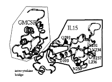
[0016] Figure 1 Structure and function of the recombinant (r) mouse
(m) GIFT15 fusokine. (a) Schematic representation of the mGIFT15 amino
acid (aa) sequence comprising GM-CSF (green), an intercytokine bridge
(grey), and IL-15 (cyan). (b) Predicted structural model of mGIFT15
interacting with IL-15Ra, IL2R(3 and IL2Ry via the aa residues in yellow
(E231,
V234, and H237), purple (D193 and N250), and red (Q293), respectively. (c)
mGIFT15 expression in B16 FO transduced cancer cells confirmed in a
Western blot using antibodies specific for mouse GM-CSF and IL-15. (d)
Confirmation of the biological activity of GIFT15 in IL-15 dependent CTLL-2
and GM-CSF dependent JAWSII cell lines. (e) Human (h) GIFT15 expression
in CHO cells confirmed in a Western blot using antibodies specific for human
GM-CSF and IL-15.
[0017] Figure 2 Immunosuppressive Properties of GIFT15 in a
syngeneic model. (a) Increased tumor growth as in vivo effect of GIFT15 in
syngeneic immunocompetent C57BI/6 mice inoculated with genetically
modified B16F0 cancer cells. (b) Decreased number of NK and NKT cells in
the in vitro analysis of tumor infiltrates.
CA 02693326 2010-01-22
WO 2008/014612 PCT/CA2007/001356
-5-
[0018] Figure 3 Allogeneic and xenogeneic transplantation of tumors
into immunocompetent mice facilitated by GIFT15. (a) Uninhibited tumor
growth of GIFT15 transduced B16F0 cancer cells in allogeneic
immunocompetent BALB/c mice. (b) Splenomegaly arising in BALB/c mice
described in (a). (c) Increased absolute numbers of T and NK cells in BALB/c
mice described in (a) determined by flow cytometry. (d) Tumor growth of
GIFT15 transduced human U87GM cancer cells in immunocompetent BALB/c
mice and xenograft survival compared to control, i.e. Green Fluorescent
Protein (GFP) trasnduced cancer cells. (e) Survival of xenograft described in
(d) in WT C57B1/6, CD4 and CD8 knock-out (KO) mice, and beige mice
stressing the importance of CD4 positive cells for the GIFT15 effect in
recipients.
[0019] Figure 4 Phenotypic analysis of cells involved in the GIFT15
induced immunosuppression. (a) IFN-y secretion by splenocytes activated by
rmGM-CSF, rmlL-15, both cytokines or mGIFT15. (b) Schematic presentation
of the gates set for the flow cytometry analysis of splenocytes described in
(a). (c) Increased MHCI-MHCII co-expression on GIFT15 treated splenocytes
described in (a). (d) Increased MHCII-CD2 co-expression on GIFT15 treated
splenocytes described in (a). (e) Elimination of B cells as splenocytes
described in (d) by CD19 staining. (f) Profiling of splenocytes described in
(a)
with antibodies for CD3, CD4, CD8, NKT cell markers, CD11b, Gr1, CTLA-4,
FasL, B7H1, CD80 and CD86.
[0020] Figure 5 Effects of mGIFT15 and hGIFT15 in direct and indirect
Mixed Lymphocyte Reaction (MLR) assays. (a) IFN-y secretion in a MLR
between equal numbers of splenocytes from BALB/c and C57B1/6 mice in the
presence or absence of 180 nM mGIFT15. (b) IFN-y secretion in a MLR
between equal numbers of peripheral blood lymphocytes (PBL) from 2 human
donors in the presence or absence of hGIFT15. (c) Indirect
immunosuppressive effect of mGIFT15 in a MLR between C57B1/6 (B6)
splenocytes pre-treated with mGIFT15 and subsequently added to BALB/c
splenocytes in a 1:1 ratio in the absence of mGIFT15
CA 02693326 2010-01-22
WO 2008/014612 PCT/CA2007/001356
-6-
[0021] Figure 6 The indirect effect of GIFT15 on antigen presentation
and IFN-y production in syngeneic in vitro systems. (a) Unhampered antigen
presentation by GIFT15 treated C57BI/6 splenocytes to an ovalbumin (OVA)
specific MHCII restricted T-cell hybridoma cell line subsequentiy secreting
IFN-y. (b) Suppression of IFN-y secretion by primary OTII T-cells in the
presence of GIFT15 pre-treated C57BI/6 splenocytes. N.B. The OTII mouse
strain is transgenic for a T cell receptor (TCR) recognizing the OVA323-339
peptide in the context of MHCII I-Ab, i.e. C57BI/6.
[0022] Figure 7 Inhibition of antigen dependent T cell activation by
GIFT15 treated splenocytes as bystander cells and not as antigen presenting
cells. (a) Suppression of activation and IFN-y secretion by OTII T cells
recognizing rOVA presented by C57B1/6 peritoneal macrophages in the
presence of mGIFT15 treated C57BI/6 splenocytes. (b) Suppression of
activation and IFN-y secretion by OTII T cells recognizing rOVA presented by
fixed C57BI/6 peritoneal macrophages in the presence of mGIFT15 treated
C57BI/6 spienocytes. (c) Suppression of activation and IFN-y secretion by
MOG35-55 specific primary T-cells derived from Myelin Oligodendrocyte
Glycoprotein (MOG) induced Experimental Autoimmune Encephalitis (EAE)
mice in culture conditions as in (a).
[0023] Figure 8 Partially blocked T cell activation by mGIFT15 through
IL10 secretion. (a) mGIFT15 induced suppression based on soluble factors.
(b) Identification of IL-10 as the soluble factor involved in mGIFT15 induced
immunosuppression by ELISA. (c) Confirmation of IL-10 as the soluble factor
involved in mGIFT15 induced immunosuppression by neutralization with an
IL-10 specific antibody.
[0024] Figure 9 Suppression of humoral in vivo responses by mGIFT15
by antibody titer analysis. (a) Lack of influence on an OVA directed IgM B
cell
response by mGIFT15. (b) Induction of transient immunosuppression of the
secondary IgG B cell response by mGIFT15.
CA 02693326 2010-01-22
WO 2008/014612 PCT/CA2007/001356
-7-
[0025] Figure 10 Syngeneic suppression of allogeneic activation in
vitro by mGIFT15 treated BALB/c splenocytes. (a) Preliminary calibration of
mGIFT15 immunosuppressive effects of BALB/c splenocytes challenged by
allogeneic immunostimulation by C57BI/6 macrophages. (b) The
immunosuppressive effect of supernatant from mGIFT15 treated splenocytes
added to BALB/c splenocytes cultured in a 1:1 ratio with C57B1/6
macrophages.
[0026] Figure 11 Signalling of mouse and human GIFT15 via the GM-
CSF receptor (GM-CSFR) and IL-15R. (a) Structural model of mGIFT15
(green, grey and cyan ribbon) complexed with IL15Ra (yellow ribbon), IL2RP
(purple ribbon) and IL2Ry (red ribbon). (b) Surface Plasmon Resonance
(SPR) analysis of the IL-15Ra chain interaction with rIL-15 and purified
mGIFT15 as shown in a BlAcore sensorgram. (c) Increased STAT3
phosphorylation induced by mGIFT15 in splenocytes expressing only the IL-
15R. (d) Unchanged STAT5 phosphorylation in JAWSII cells expressing only
the GM-CSFR. (e) Increased STAT3 and decreased STAT5 phosphorylation
induced by mGIFT15 in macrophages expressing both, IL-15R and GM-
CSFR. (f) Increased STAT3 phosphorylation induced by hGIFT15 in PBLs
expressing only the IL-15R.
[0027] Figure 12 Downregulation of the adhesion molecules (a) LFA-1/
CD11a and (b) ICAM-1/CD54 by mGIFT15 contrary to their upregulation by
IL-15, GM-CSF and their combination.
[0028] Figure 13 Anti-apoptotic and proliferative activities of mGIFT15.
(a) Proliferation inducing potential of mGIFT15 as demonstrated in a MTT
(dye) incorporation assay and a CFSE (dye) based assay as shown in (b). (c)
Anti-apoptotic potential of mGIFT15 as demonstrated with a PI and Annexin V
flow cytometry read-out and a BcI-XL Western blot (d).
[0029] Figure 14 Increased recruitment of macrophages and secretion
of transforming growth factor (TGF)-(3 induced by mGIFT15. (a) Migration
assay with peritoneal macrophages in the presence of cytokines. (b) TGF(3
CA 02693326 2010-01-22
WO 2008/014612 PCT/CA2007/001356
-8-
levels secreted by peritoneal macrophages stimulated with mGIFT15 as
detected in an ELISA.
[0030] Figure 15 Pro-angiogenic properties of mGIFT15 in-vivo. (a)
Tumor volume assessed in NOD-SCID mice injected with B16FO cancer cells
transduced with mGIFT15. (b) Increased blood vessel density in tumors
arising from mGIFT15 transduced cancer cells as confirmed by staining with
an anti van Willebrand Factor (vWF) antibody.
[0031] Figure 16 Pro-angiogenic properties of murine and human
GIFT15 in-vitro. (a) Secretion and activation of Matrix metalloproteinase
(MMP-)2 induced by mGIFT15 in serum deprived macrophages as confirmed
in a gelatin zymogram. (b) Induction of MMP-2 but not MMP-9 by mGIFT15 as
confirmed by Western Blot. (c) Increased induction of the pro-angiogenic
Vascular Endothelial Growth Factor (VEGF) by mGIFT15 in macrophages. (d)
Induction of the angiogenic factors Tissue metalloproteinase (TIMP)-1 and
VEGF by hGIFT15 derived from Chinese Hamster Ovary (CHO) cells
transduced with the fusokine as demonstrated in an angiogenic protein array.
(e) Confirmation of VEGF secretion induced by hGIFT15 in a generic cytokine
array in addition to the anti-inflammatory molecules TGF-beta and soluble
Tumor Necrosis Factor Receptor (sTNFR)II.
[0032] Figure 17 GIFT15 Treated Splenocytes lead to faster recovery
in syngeneic C57BI/6 EAE mice. Mice injected with MOG to induce EAE
received 3 IV injections of C57BI/6 GIFT15-treated splenocytes and the
disease score was monitored every second day. Compared to the PBS
control group, GIFT15 treated splenocytes lead to a faster recovery starting
at
day 12 (n=5/group; P<0.05).
DETAILED DESCRIPTION OF THE INVENTION
[0033] The present inventors have shown that a conjugate comprising
GM-CSF and IL-15 has immune suppressive properties. Further, the
inventors have shown that the conjugate can be used to prevent graft
rejection, including xenograft rejection; prevent or treat graft versus host
CA 02693326 2010-01-22
WO 2008/014612 PCT/CA2007/001356
-9-
disease; prevent or treat autoimmune disease; and to inhibit cell death. The
inventors have also shown that administering the GM-CSF and IL-15
conjugate induces angiogenesis.
[0034] The inventors have demonstrated that the GM-CSF and IL-15
conjugate possesses novel biochemical properties leading to altered affinities
to components of the trimeric IL-15R and asymmetrical downstream signalling
via its two STAT/JAK pathways in lymphoid cells. As a result, cellular
proliferation, reduced apoptosis and blunting of the IFNy response following
activation can be achieved. The sum of these effects mediates a profound
immunosuppressive state permissive to xenotransplantation which is CD4
dependent.
A. GM-CSF AND IL-15 CONJUGATES
[0035] The present invention relates to conjugates of GM-CSF and IL-
that are immune suppressive and can be used in various therapeutic
15 applications as described in Section B.
[0036] Accordingly, the present invention provides a GM-CSF and IL-
15 conjugate protein.
[0037] The term "a GM-CSF and IL-15 conjugate protein" means a
conjugate that comprises GM-CSF physically linked to IL-15. In a specific
embodiment, the conjugate is a fusion protein (or fusokine) wherein a nucleic
acid sequence encoding GM-CSF is operably linked to a nucleic acid
sequence encoding IL-15 and the chimeric sequence is transfected or
transduced into a host cell and produced as a recombinant fusion protein.
The GM-CSF and IL-15 fusion protein is often abbreviated GIFT15 in the
present application.
[0038] In a specific embodiment, the GM-CSF and IL-15 are linked by a
peptide linker. The peptide linker can be any size provided it does not
interfere with the function of the GM-CSF and IL-15 conjugate. In one
embodiment, the peptide linker is from about 2 to about 15 amino acids in
CA 02693326 2010-01-22
WO 2008/014612 PCT/CA2007/001356
-10-
length, more specifically from about 2 to about 10 amino acids, and most
specifically from about 2 to about 7 amino acids.
[0039] One of skill in the art can appreciate that the GM-CSF and IL-15
conjugate protein can also be formed by linking the two proteins in vitro, for
example, using chemical cross-linkers. For example, the proteins may be
coupled using heterobifunctional thiol-containing linkers as described in WO
90/10457, N-succinimidyl-3-(2-pyridyldithio-proprionate) or N-succinimidyl-5
thioacetate.
[0040] The GM-CSF and IL-15 molecules used in the conjugate can be
from any species or source and includes the full-length proteins as well as
fragments or portions of the proteins. In a preferred embodiment, the GM-
CSF and IL-15 sequences are from human or mouse. In a specific
embodiment, the GM-CSF protein lacks the last 11 carboxy terminal amino
acid sequences as compared to full length GM-CSF.
[0041] In one embodiment, the GM-CSF and IL-15 conjugate protein is
murine and has the amino acid sequence shown in SEQ ID NO:2 or an
analog or homolog thereof. In another embodiment, th GM-CSF and IL-15
conjugate protein is human and has the sequence shown in SEQ ID NO:4 or
an analog or homolog thereof.
[0042] The invention also includes nucleic acid molecules that encode
the GM-CSF and IL-15 protein conjugate. The nucleic acid molecule is
preferably a chimeric nucleic acid sequence that comprises a) a nucleic acid
sequence encoding GM-CSF or a fragment thereof linked to b) a nucleic acid
sequence encoding IL-15 or a fragment thereof.
[0043] The chimeric sequence preferably also includes a sequence
encoding a peptide linker. Accordingly, the present invention also includes a
chimeric nucleic acid sequence that comprises a) a nucleic acid sequence
encoding GM-CSF or a fragment thereof linked to b) a nucleic acid sequence
encoding a peptide linker linked to c) a nucleic acid sequence encoding IL-15
or a fragment thereof.
CA 02693326 2010-01-22
WO 2008/014612 PCT/CA2007/001356
-11-
[0044] In one embodiment, the chimeric nucleic acid sequence is
murine and has the sequence shown in SEQ ID NO:1, or a homolog or analog
thereof. In another embodiment, the chimeric nucleic acid sequence is
human and has the sequence shown in SEQ ID NO:3, or a homolog or analog
thereof.
[0045] The term "homolog" means those amino acid or nucleic acid
sequences which have slight or inconsequential sequence variations from the
sequences in SEQ ID NOs:1-4, i.e., the sequences function in substantially
the same manner. The variations may be attributable to local mutations or
structural modifications. Sequences having substantial homology include
nucleic acid sequences having at least 65%, more preferably at least 85%,
and most preferably 90-95% identity with the sequences as shown in SEQ ID
NOs:1-4. Sequence identity can be calculated according to methods known in
the art. Nucleic acid sequence identity is most preferably assessed by the
algorithm of BLAST version 2.1 advanced search. BLAST is a series of
programs that are available online at http://www.ncbi.nlm.nih.gov/BLAST.
The advanced blast search (http://www.ncbi.nlm.nih.gov/
blast/blast.cgi?Jform=1) is set to default parameters. (ie Matrix BLOSUM62;
Gap existence cost 11; Per residue gap cost 1; Lambda ratio 0.85 default).
References to BLAST searches are: Altschul, S.F., Gish, W., Miller, W.,
Myers, E.W. & Lipman, D.J. (1990) "Basic local alignment search tool." J. Mol.
Biol. 215:403410; Gish, W. & States, D.J. (1993) "Identification of protein
coding regions by database similarity search." Nature Genet. 3:266272;
Madden, T.L., Tatusov, R.L. & Zhang, J. (1996) "Applications of network
BLAST server" Meth. Enzymol. 266:131_141; Altschul, S.F., Madden, T.L.,
Schaffer, A.A., Zhang, J., Zhang, Z., Miller, W. & Lipman, D.J. (1997)
"Gapped BLAST and PSI_BLAST: a new generation of protein database
search programs." Nucleic Acids Res. 25:33893402; Zhang, J. & Madden,
T.L. (1997) "PowerBLAST: A new network BLAST application for interactive
or automated sequence analysis and annotation." Genome Res. 7:649656.
CA 02693326 2010-01-22
WO 2008/014612 PCT/CA2007/001356
-12-
[0046] The term "analog" means an amino acid or nucleic acid
sequence which has been modified as compared to the sequence of SEQ ID
NOs:1-4 wherein the modification does not alter the utility of the sequence
(e.g. as immune suppressant) as described herein. The modified sequence
or analog may have improved properties over the sequences shown in SEQ
ID NOs:1-4. One example of a nucleic acid modification to prepare an analog
is to replace one of the naturally occurring bases (i.e. adenine, guanine,
cytosine or thymidine) of the sequence with a modified base such as such as
xanthine, hypoxanthine, 2-aminoadenine, 6-methyl, 2-propyl and other alkyl
adenines, 5-halo uracil, 5-halo cytosine, 6-aza uracil, 6-aza cytosine and 6-
aza thymine, pseudo uracil, 4-thiouracil, 8-halo adenine, 8-aminoadenine, 8-
thiol adenine, 8-thiolalkyl adenines, 8-hydroxyl adenine and other 8-
substituted adenines, 8-halo guanines, 8 amino guanine, 8-thiol guanine, 8-
thiolalkyl guanines, 8-hydroxyl guanine and other 8-substituted guanines,
other aza and deaza uracils, thymidines, cytosines, adenines, or guanines, 5-
trifluoromethyl uracil and 5-trifluoro cytosine.
[0047] Another example of a modification is to include modified
phosphorous or oxygen heteroatoms in the phosphate backbone, short chain
alkyl or cycloalkyl intersugar linkages or short chain heteroatomic or
heterocyclic intersugar linkages in the nucleic acid molecules shown in SEQ
ID NO:1 or 3. For example, the nucleic acid sequences may contain
phosphorothioates, phosphotriesters, methyl phosphonates, and
phosphorodithioates.
[0048] A further example of an analog of a nucleic acid molecule of the
invention is a peptide nucleic acid (PNA) wherein the deoxyribose (or ribose)
phosphate backbone in the DNA (or RNA), is replaced with a polyamide
backbone which is similar to that found in peptides (P.E. Nielsen, et al
Science 1991, 254, 1497). PNA analogs have been shown to be resistant to
degradation by enzymes and to have extended lives in vivo and in vitro.
PNAs also bind stronger to a complimentary DNA sequence due to the lack of
charge repulsion between the PNA strand and the DNA strand. Other nucleic
CA 02693326 2010-01-22
WO 2008/014612 PCT/CA2007/001356
-13-
acid analogs may contain nucleotides containing polymer backbones, cyclic
backbones, or acyclic backbones. For example, the nucleotides may have
morpholino backbone structures (U.S. Pat. No. 5,034,506). The analogs may
also contain groups such as reporter groups, a group for improving the
pharmacokinetic or pharmacodynamic properties of nucleic acid sequence.
[0049] The invention also includes sequences that hybridize to the
sequences shown in SEQ ID NO:1 or 3 or a fragment thereof. The term
"sequence that hybridizes" means a nucleic acid sequence that can hybridize
to a sequence of SEQ ID NO:1 or 3 under stringent hybridization conditions.
Appropriate "stringent hybridization conditions" which promote DNA
hybridization are known to those skilled in the art, or may be found in
Current
Protocols in Molecular Biology, John Wiley & Sons, N.Y. (1989), 6.3.1-6.3.6.
The term "stringent hybridization conditions" as used herein means that
conditions are selected which promote selective hybridization between two
complementary nucleic acid molecules in solution. Hybridization may occur to
all or a portion of a nucleic acid sequence molecule. The hybridizing portion
is at least 50% the length with respect to one of the polynucleotide sequences
encoding a polypeptide. In this regard, the stability of a nucleic acid
duplex,
or hybrids, is determined by the Tm, which in sodium containing buffers is a
function of the sodium ion concentration, G/C content of labeled nucleic acid,
length of nucleic acid probe (I), and temperature (Tm = 81.5 C - 16.6 (LoglO
[Na+]) + 0.41(%(G+C) - 600/I). Accordingly, the parameters in the wash
conditions that determine hybrid stability are sodium ion concentration and
temperature. In order to identify molecules that are similar, but not
identical,
to a known nucleic acid molecule a 1% mismatch may be assumed to result in
about a 1 C decrease in Tm, for example if nucleic acid molecules are sought
that have a greater than 95% identity, the final wash will be reduced by 5 C.
Based on these considerations stringent hybridization conditions shall be
defined as: hybridization at 5 x sodium chloride/sodium citrate (SSC)/5 x
Denhardt's solution/1.0% SDS at Tm (based on the above equation) - 5 C,
followed by a wash of 0.2 x SSC/0.1 % SDS at 60 C.
CA 02693326 2010-01-22
WO 2008/014612 PCT/CA2007/001356
-14-
[0050] It will be appreciated that analogs/homologs of the GM-CSF and
IL-15 conjugate can also be prepared by first preparing or using an analog or
homolog of GM-CSF or IL-15 or both prior to preparing the chimeric nucleic
acid sequence.
[0051] The GM-CSF and IL-15 conjugate protein may be modified to
contain amino acid substitutions, insertions and/or deletions that do not
alter
the immunosuppressive properties of the protein. Conserved amino acid
substitutions involve replacing one or more amino acids of the GM-CSF and
IL-15 conjugate protein with amino acids of similar charge, size, and/or
hydrophobicity characteristics. When only conserved substitutions are made
the resulting analog should be functionally equivalent to the GM-CSF and IL-
conjugate protein. Non-conserved substitutions involve replacing one or
more amino acids of the GM-CSF and IL-15 conjugate protein with one or
more amino acids which possess dissimilar charge, size, and/or
15 hydrophobicity characteristics.
[0052] The GM-CSF and IL-15 conjugate protein may be modified to
make it more therapeutically effective or suitable. For example, the GM-CSF
and IL-15 conjugate protein or peptides of the present invention may be
converted into pharmaceutical salts by reacting with inorganic acids including
hydrochloric acid, sulphuric acid, hydrobromic acid, phosphoric acid, etc., or
organic acids including formic acid, acetic acid, propionic acid, glycolic
acid,
lactic acid, pyruvic acid, oxalic acid, succinic acid, malic acid, tartaric
acid,
citric acid, benzoic acid, salicylic acid, benzenesulphonic acid, and
tolunesulphonic acids.
[0053] The invention also includes expression vectors comprising a
chimeric nucleic acid sequence comprising a) a nucleic acid sequence
encoding GM-CSF or a fragment thereof linked to b) a nucleic acid sequence
encoding IL-15 or a fragment thereof. In a specific embodiment, the chimeric
nucleic acid sequence includes a sequence that encodes a peptide linker as
described above.
CA 02693326 2010-01-22
WO 2008/014612 PCT/CA2007/001356
-15-
[0054] Possible expression vectors include but are not limited to
cosmids, plasmids, artificial chromosomes, viral vectors or modified viruses
(e.g. replication defective retroviruses, adenoviruses and adeno-associated
viruses), so long as the vector is compatible with the host cell used. The
expression vectors are "suitable for transformation of a host cell", which
means that the expression vectors contain a nucleic acid molecule of the
invention and regulatory sequences selected on the basis of the host cells to
be used for expression, which is operatively linked to the nucleic acid
molecule. Operatively linked is intended to mean that the nucleic acid is
linked to regulatory sequences in a manner which allows expression of the
nucleic acid.
[0055] The invention therefore contemplates a recombinant expression
vector of the invention containing a nucleic acid molecule of the invention,
or a
fragment thereof, and the necessary regulatory sequences for the
transcription and translation of the inserted protein-sequence.
[0056] Suitable regulatory sequences may be derived from a variety of
sources, including bacterial, fungal, viral, mammalian, or insect genes (for
example, see the regulatory sequences described in Goeddel, Gene
Expression Technology: Methods in Enzymology 185, Academic Press, San
Diego, CA (1990). Selection of appropriate regulatory sequences is
dependent on the host cell chosen as discussed below, and may be readily
accomplished by one of ordinary skill in the art. Examples of such regulatory
sequences include: a transcriptional promoter and enhancer or RNA
polymerase binding sequence, a ribosomal binding sequence, including a
translation initiation signal. Additionally, depending on the host cell chosen
and the vector employed, other sequences, such as an origin of replication,
additional DNA restriction sites, enhancers, and sequences conferring
inducibility of transcription may be incorporated into the expression vector.
It
will also be appreciated that the necessary regulatory sequences may be
supplied by the GM-CSF or IL-15 sequences and/or their flanking regions.
CA 02693326 2010-01-22
WO 2008/014612 PCT/CA2007/001356
-16-
[0057] The recombinant expression vectors of the invention may also
contain a selectable marker gene which facilitates the selection of host cells
transformed or transfected with a recombinant molecule of the invention.
Examples of selectable marker genes are genes encoding a protein such as
G418 and hygromycin which confer resistance to certain drugs, (3-
galactosidase, chloramphenicol acetyltransferase, firefly luciferase, or an
immunoglobulin or portion thereof such as the Fc portion of an
immunoglobulin preferably IgG. Transcription of the selectable marker gene
is monitored by changes in the concentration of the selectable marker protein
such as R-galactosidase, chloramphenicol acetyltransferase, or firefly
luciferase. If the selectable marker gene encodes a protein conferring
antibiotic resistance such as neomycin resistance transformant cells can be
selected with G418. Cells that have incorporated the selectable marker gene
will survive, while the other cells die. This makes it possible to visualize
and
assay for expression of recombinant expression vectors of the invention and
in particular to determine the effect of a mutation on expression and
phenotype. It will be appreciated that selectable markers can be introduced
on a separate vector from the nucleic acid of interest.
[0058] The recombinant expression vectors may also contain genes
which encode a moiety which provides increased expression of the
recombinant protein; increased solubility of the recombinant protein; and aid
in the purification of the target recombinant protein by acting as a ligand in
affinity purification. For example, a proteolytic cleavage site may be added
to
the target recombinant protein to allow separation of the recombinant protein
from the fusion moiety subsequent to purification of the fusion protein.
Typical
fusion expression vectors include pGEX (Amrad Corp., Melbourne, Australia),
pMal (New England Biolabs, Beverly, MA) and pRIT5 (Pharmacia,
Piscataway, NJ) which fuse glutathione S-transferase (GST), maltose E
binding protein, or protein A, respectively, to the recombinant protein.
[0059] Recombinant expression vectors can be introduced into host
cells to produce a transformed host cell. The term "transformed host cell" is
CA 02693326 2010-01-22
WO 2008/014612 PCT/CA2007/001356
-17-
intended to include cells that are capable of being transformed or transfected
with a recombinant expression vector of the invention. The terms
"transduced", "transformed with", "transfected with", "transformation" and
"transfection" are intended to encompass introduction of nucleic acid (e.g. a
vector or naked RNA or DNA) into a cell by one of many possible techniques
known in the art. Prokaryotic cells can be transformed with nucleic acid by,
for example, electroporation or calcium-chloride mediated transformation. For
example, nucleic acid can be introduced into mammalian cells via
conventional techniques such as calcium phosphate or calcium chloride co-
precipitation, DEAE-dextran mediated transfection, lipofectin,
electroporation,
microinjection, RNA transfer, DNA transfer, artificial chromosomes, viral
vectors and any emerging gene transfer technologies. Suitable methods for
transforming and transfecting host cells can be found in Sambrook et al.
(Molecular Cloning: A Laboratory Manual, 2nd Edition, Cold Spring Harbor
Laboratory press (1989)), and other laboratory textbooks.
[0060] Suitable host cells include a wide variety of eukaryotic host cells
and prokaryotic cells. For example, the proteins of the invention may be
expressed in yeast cells or mammalian cells. Other suitable host cells can be
found in Goeddel, Gene Expression Technology: Methods in Enzymology
185, Academic Press, San Diego, CA (1991). In addition, the proteins of the
invention may be expressed in prokaryotic cells, such as Escherichia coli
(Zhang et al., Science 303(5656): 371-3 (2004)).
[0061] Mammalian cells suitable for carrying out the present invention
include, among others: B16FO cells, 293T cells, Mesenchymal Stromal Cell
(MSCs), COS (e.g., ATCC No. CRL 1650 or 1651), BHK (e.g. ATCC No. CRL
6281), CHO (ATCC No. CCL 61), HeLa (e.g., ATCC No. CCL 2), 293 (ATCC
No. 1573) and NS-1 cells.
[0062] The mammalian cells can also be derived from a human or
animal and include stem cells (including hematopoietic stem cells), somatic
cells, progenitor cells (including endothelial progenitor cells), fibroblasts,
lymphocytes, and MSCs that have been genetically engineered to express the
CA 02693326 2010-01-22
WO 2008/014612 PCT/CA2007/001356
-18-
GM-CSF and IL-15 conjugate. Such cells can be used in the therapeutic
applications described in Section B. For example, MSCs, fibroblasts,
lymphocytes, hematopoietic stem cells derived from human or non-human
sources can be gene engineered to express the GM-CSF and IL-15 conjugate
and serve for cellular therapy of disease such as heart disease,
neurodegeneration, diabetes mellitus, muscle dystrophy and hematopoietic
disorders.
[0063] Suitable expression vectors for directing expression in
mammalian cells generally include a promoter (e.g., derived from viral
material such as polyoma, Adenovirus 2, cytomegalovirus and Simian Virus
40), as well as other transcriptional and translational control sequences.
Examples of mammalian expression vectors include pCDM8 (Seed, B.,
Nature 329:840 (1987)), pMT2PC (Kaufman et al., EMBO J. 6:187-195
(1987)) and pCMV (Clontech, California, U.S.A.).
[0064] Alternatively, the proteins of the invention may also be
expressed in non-human transgenic animals such as, rats, rabbits, sheep and
pigs (Hammer et al. Nature 315:680-683 (1985); Palmiter et al. Science
222:809-814 (1983); Brinster et al. Proc. Natl. Acad. Sci. USA 82:4438-4442
(1985); Palmiter and Brinster Cell 41:343-345 (1985) and U.S. Patent No.
4,736,866). The invention also includes tissues and cells derived from such
animals.
[0065] In a specific embodiment, to create a mouse GM-CSF and IL-15
fusokine, the cDNA of mouse GM-CSF was modified to remove the
nucleotides coding for the last 11 carboxyterminal aa and cloned in frame to
the 5' end of the full-length mouse IL-15 cDNA, including its long signal
peptidel',12 Including a synthetic linker bridge consisting of 7 aa between
the
GM-CSF and IL-15 sequences, the final fusokine mGIFT15 cDNA shown in
SEQ ID NO1 encodes for a single polypeptide chain of 299 aa (Figure 1a) as
shown in SEQ ID NO:2. A computer-based analysis of the three-dimensional
structure revealed that the 7 aa peptidic bridge and the uncleaved IL-15 long
signal peptide sequence forms an intercytokine bridge of 55 aa in length with
CA 02693326 2010-01-22
WO 2008/014612 PCT/CA2007/001356
-19-
a three alpha helixes configuration. (Figure 1 b). Denaturing immunoblotting
performed on conditioned media (CM) from retrovirally transduced B16F0
cells to express mGIFT15 showed that the chimeric protein is efficiently
secreted in the extracellular space and has a molecular weight of 55 kDa.
mGIFT15 was probed with polyclonal goat anti-mIL15 or anti-mGMCSF
antibodies. While CM containing green fluorescent protein (GFP) served as a
negative control, rmIL15 and rmGMCSF were used as positive controls.
(Figure 1c). The bioactivity of both cytokine subunits within GIFT15 was
confirmed by proliferation assays based on MTT incorporation in the GM-
CSF-dependent JAWSII and IL-15-dependent CTLL2 cell lines, respectively
Results are shown as mean of triplicates S.E.M of one representative
experiment of three with a P>0.05 between mGIFT15 and IL15 in CTLL-2
cells and a P>0.05 between mGIFT15 and GMCSF for JAWSII cells. (Figure
1 d).
[0066] In another specific embodiment, to create the human GM-CSF
and IL-15 fusokine, the cDNA of human GM-CSF was modified to remove the
nucleotides coding for the last 11 carboxyterminal aa and cloned in frame to
the 5' end of the full-length human IL-15 cDNA, including its long signal
peptide1l,12 Including a synthetic linker peptidic bridge of 2 aa and the
uncleaved hIL-15 secretion peptide between the GM-CSF and IL-15
sequences, the final fusokine hGIFT15 cDNA encodes for a single
polypeptide chain of 297 aa as shown in SEQ ID NO:4 (Figure le). hGIFT15
expressed in 293T cells was identified as a 55 kDa protein and as multimeric
forms as demonstrated in a Western blot involving antibodies directed against
human IL-15 and human GM-CSF (Figure 1f).
B. THERAPEUTIC METHODS
[0067] The invention includes all applications of the GM-CSF and IL-15
conjugate, some of which are described below.
1. Immune Suppression
CA 02693326 2010-01-22
WO 2008/014612 PCT/CA2007/001356
-20-
[0068] To assess the ability of GIFT15 to influence the immune
response, polyclonal populations of 106 B16F0 cancer cells genetically
engineered to secrete equimolar levels of IL-15, GM-CSF or GIFT15 were
subcutaneously injected in syngeneic immune competent C57B1/6 mice (n=6).
Unexpectedly, the fusokine comprising the two immunostimulatory subunits
IL-15 and GM-CSF, had the opposite, an immunosuppressive, effect. It was
observed that B16F0 cells secreting GIFT15 had acquired aggressive growth
properties with an average tumor size three fold larger than that of control
groups in the weeks following implantation. Tumor volume was monitored
over time resulting in a Pvalue of <0.05 between B16-mGIFT15 and B16-
GFP/mIL15/mGMCSF/ mIL15+mGMCSF. Results are shown as mean tumor
volume S.E.D.. (Figure 2a). To determine whether this phenomenon was
linked to an atypical immune response, the inventors analyzed tumor
infiltration by immune cells a fortnight after implantation of matrigelTM
matrix
embedded cells. It was found that natural killer (NK) and natural killer T
(NKT) cells were virtually absent in GIFT15-secreting tumors when compared
to B16-GM-CSF or B16-IL-15 control groups whilst the number of other CD3+
T-cell subsets were similar to controls (Figure 2b). The observed absence in
NK/NKT cell recruitment by B16-GIFT15 cells is contradictory to what was
predicted would occur as a host-derived immune response to GIFT15 in vivo,
especially since IL-15 has been shown by others to directly stimulate the
development, expansion, recruitment and activation of NK and NKT cells13'14
[0069] In one aspect, the present invention provides a method of
suppressing an immune response comprising administering an effective
amount of a GM-CSF and IL-15 conjugate protein or a nucleic acid sequence
encoding a GM-CSF and IL-15 conjugate protein to an animal in need of such
treatment. The invention includes a use of an effective amount of a GM-CSF
and IL-15 conjugate protein or a nucleic acid sequence encoding a GM-CSF
and IL-15 conjugate protein to suppress an immune response. The invention
includes a use of an effective amount of a GM-CSF and IL-15 conjugate
protein or a nucleic acid sequence encoding a GM-CSF and IL-15 conjugate
protein to prepare a medicament to suppress an immune response. In a
CA 02693326 2010-01-22
WO 2008/014612 PCT/CA2007/001356
-21-
specific embodiment, the conjugate inhibits the development, expansion or
activation of NK cells, NKT cells, T cells or B cells.
[0070] The term "administering a GM-CSF and IL-15 conjugate protein"
includes both the administration of the GM-CSF and IL-15 conjugate protein
as well as the administration of a nucleic acid sequence encoding a GM-CSF
and IL-15 conjugate protein to an animal or to a cell in vitro or in vivo. The
term "administering" also includes the administration of a cell that express
the
GM-CSF and IL-15 conjugate protein.
[0071] The term "a cell" includes a single cell as well as a plurality or
population of cells. Administering to a cell includes administering in vitro
(or
ex vivo) as well as in vivo.
[0072] Administration of an "effective amount" of the GM-CSF and IL-
conjugate protein and nucleic acid of the present invention is defined as an
amount effective, at dosages and for periods of time necessary to achieve the
15 desired result. The effective amount of the GM-CSF and IL-15 conjugate
protein or nucleic acid of the invention may vary according to factors such as
the disease state, age, sex, and weight of the animal. Dosage regimens may
be adjusted to provide the optimum therapeutic response. For example,
several divided doses may be administered daily or the dose may be
proportionally reduced as indicated by the exigencies of the therapeutic
situation. The mode of administration (e.g. in vivo by injection or ex vivo in
culture) will also impact the dosage regime.
[0073] The term "animal" as used herein includes all members of the
animal kingdom including humans.
[0074] Once a particular GM-CSF and IL-15 conjugate protein or
analog or homolog is prepared, one of skill in the art can readily determine
whether or not it can suppress an immune response. For example,
determining whether a particular GM-CSF and IL-15 conjugate protein or
fragments thereof can suppress an immune response can be assessed using
known in vitro immune assays including, but not limited to, inhibiting a mixed
CA 02693326 2010-01-22
WO 2008/014612 PCT/CA2007/001356
-22-
leucocyte reaction; inhibiting a cytotoxic T cell response; inhibiting
interleukin-
2 production; inhibiting IFN-y production; inhibiting a Th1 cytokine profile;
inducing IL-4 production; inducing TGF(3 production; inducing IL-10
production; inducing a Th2 cytokine profile; inhibiting immunoglobulin
production; altering serum immunoglobulin isotype profiles (from those
associated with Th1 type immunity - in the mouse, IgG1 and IgG2a, to those
associated with Th2 type immunity - in the mouse, IgG2b, IgG3); and any
other assay that would be known to one of skill in the art to be useful in
detecting immune suppression.
(i) Graft Rejection
[0075] In light of the unheralded immunosuppressive effects of GIFT15,
the inventors tested whether its expression could protect allogeneic cells
from
rejection in immune competent MHC-mismatched recipient animals. As proof
of concept, 10' B16-GFP or B16-GIFT15 (H-2Kb) transduced cells were
grafted in BALB/c (H-2Kd) mice (n=10). Surprisingly, tumors secreting the
fusion protein were accepted in all mice and grew to a point where half the
group had large tumors with volumes exceeding 1,000 mm3 by day 28 post-
transplantation (P<0.05 between B16-mGIFT15 and GFP group). Results are
shown as mean tumor volume S.E.D. (Figure 3a). In addition, these mice
developed splenomegaly (Figure 3b; P<0.02) characterized by the
disappearance of the spleen's white pulp structures demonstrated by H & E
staining and by a significant increase in the absolute number in T and NK
cells demonstrated by flow cytometry analysis. (n=3; P<0.02 between the
mGIFT15 and GFP group). Results are shown as mean average of
triplicates S.E.D. (Figure 3c) contrary to the unexpected decrease or
absence of NK cells in the tumor tissue as described in Figure. 2b. The
inventors further investigated the utility of GIFT15 for the induction of
immunosuppression in the context of xenotransplantation. In this case, a
mGIFT15 transduced polyclonal population of the human glioma cell line
U87GM secreting 1119 ng per 106 cells per 24 hrs of GIFT15 was
transplanted subcutaneously in BALB/c mice (n=6). All mice accepted the
CA 02693326 2010-01-22
WO 2008/014612 PCT/CA2007/001356
-23-
GIFT15 xenograft for up to 8 months whereas the control U87-GFP xenograft
was rejected 12 days post-injection (Figure 3d). As different mouse strains
generate variable immune responses, the inventors pursued the studies by
xenotransplanting C57B1/6 mice, which are known to possess a biased T-
helper 1 immune response15,1s Even though both GFP and GIFT15
xenografts were rejected in these mice, there was a two-month delay for the
complete regression of the U87-GIFT15 transplants compared to the U87-
GFP group (Figure 3e). Experiments performed in KO mice revealed that
CD8 T-cell activity does not seem to be implicated since a similar rejection
profile of U87-GIFT15 was obtained in CD8-/- mice compared to wild-type
(VVT) C57B1/6 mice (Figure 3e). However, NK cells were found to be key
players for the xenograft rejection in WT C57B1/6 since 80% of mice having an
NK deficiency (beige mice) accepted the transplants for a period longer than
120 days (Figure 3e). In addition, the immunosuppressive property of the
fusion protein was impaired once U87-GIFT15 transduced cells were injected
in CD4-/- model of C57B1/6 mice (Figure 3e). This suggests that the lack of
regulatory T-cells (Treg) cells mitigates the effect of GIFT15. Notably, one
crucial effect of Tregs is to inhibit NK cell function".
[0076] In one embodiment, the present invention provides a method of
suppressing an immune response to a transplanted organ, cell or tissue in a
recipient animal comprising administering an effective amount of a GM-CSF
and IL-15 conjugate protein or a nucleic acid sequence encoding a GM-CSF
and IL-15 conjugate protein to the recipient animal, preferably prior to the
transplantation of the organ or tissue. The invention includes a use of an
effective amount of a GM-CSF and IL-15 conjugate protein or a nucleic acid
sequence encoding a GM-CSF and IL-15 conjugate protein to suppress an
immune response to a transplanted organ, cell or tissue. The invention
includes a use of an effective amount of a GM-CSF and IL-15 conjugate
protein or a nucleic acid sequence encoding a GM-CSF and IL-15 conjugate
protein to prepare a medicament to suppress an immune response to a
transplanted organ, cell or tissue.
CA 02693326 2010-01-22
WO 2008/014612 PCT/CA2007/001356
-24-
[0077] The recipient can be any member of the animal kingdom
including rodents, pigs, cats, dogs, ruminants, non-human primates and
preferably humans. The organ, cell or tissue to be transplanted can be from
the same species as the recipient (allograft) or can be from another species
(xenograft). The tissues, cells or organs can be any tissue or organ including
heart, liver, kidney, lung, pancreas, pancreatic islets, brain tissue, cornea,
bone, intestine, skin and haematopoietic cells and stem cells.
[0078] In one embodiment, the organ, cells or tissue to be transplanted
may be transduced with a nucleic acid construct encoding the GM-CSF and
IL-15 conjugate prior to transplantation into the graft recipient.
[0079] One of skill in the art can determine whether or not a particular
GM-CSF and IL-15 conjugate protein or fragment thereof is useful in
preventing graft rejection. As mentioned above, one of skill in the art can
readily test a GM-CSF and IL-15 conjugate protein or GM-CSF and IL-15
conjugate protein fragment for its ability to suppress an immune response
using known in vitro assays. In addition the GM-CSF and IL-15 conjugate
protein or GM-CSF and IL-15 conjugate protein fragment can also be tested
for its ability to prevent graft rejection in an animal model. For example,
one
could use the xenotransplant animal model described above.
[0080] The method of the invention may be used to prevent graft
versus host disease wherein the immune cells in the transplant mount an
immune attack on the recipient's immune system. This can occur when the
tissue to be transplanted contains immune cells such as when bone marrow
or Iymphoid tissue is transplanted when treating leukemias, aplastic anemias
and enzyme or immune deficiencies, for example.
[0081] Accordingly, in another embodiment, the present invention
provides a method of preventing or inhibiting graft versus host disease in a
recipient animal receiving an organ or tissue transplant comprising
administering an effective amount of a GM-CSF and IL-15 conjugate protein
or a nucleic acid sequence encoding a GM-CSF and IL-15 conjugate protein
to the organ or tissue prior to the transplantation in the recipient animal.
The
CA 02693326 2010-01-22
WO 2008/014612 PCT/CA2007/001356
-25-
invention includes a use of an effective amount of a GM-CSF and IL-15
conjugate protein or a nucleic acid molecule encoding a GM-CSF and IL-15
conjugate protein to prevent or inhibit graft versus host disease. The
invention includes a use of an effective amount of a GM-CSF and IL-15
conjugate protein or a nucleic acid sequence encoding a GM-CSF and IL-15
conjugate protein to prepare a medicament to to prevent or inhibit graft
versus
host disease.
[0082] In order to phenotypically characterize the cells involved in the
GIFT15 induced immunosuppression the inventors performed two different
comparative studies in splenocytes in the presence of IL-5, GM-CSF, both
cytokines combined and GIFT15. Since IL-15 is known to be a strong inducer
of IFN-y, the inventors tested the stimulatory capacity of the fusion protein.
Splenocytes from C57BL/6 mice stimulated for 36 hrs with 30 pmols of rIL-15
in the absence or presence of rGM-CSF led to similar IFN-y secretion profiles
of suggesting that GM-CSF has no effect on IL-15-mediated IFN-Y production.
In contrast, GIFT15 suppressed any IFN-y secretion in splenocytes at
equimolar concentrations to rIL-15 (Figure 4a; P<0.0005). These
unanticipated direct effects of GIFT15 on splenocytes lead to further
investigations by flow cytometry. Based on the gates used to analyze
splenocytes cultured in the 4 different conditions (IL-15, GM-CSF, both, and
GIFT15), the inventors observed a uniform cell population appearing upon
GIFT15 treatment compared to the different cytokine conditions (Figure 4b).
Splenocytes treated with mGIFT15 express both MHCI-MHCII at a higher
percentage (72%) than the remaining groups (25% for rmIL15, 44% for
rmGMCSF or 7% for both) (Figure 4c). An eight-day treatment with mGIFT15
leads to the expression of MHCII and CD2 in 71% of cells compared to 46-
48% MHCII/CD2 double positive cells when treated with single or combined
cytokines. (Figure 4d). To exclude that the CD2 positive cells were B cells,
cells were stained for the CD19 B cell marker (Figure 4e) The cytokine
treated splenocytes were also analysed for the presence of CD4, CD8 and
NKT cell markers and the dramatic reduction of CD3+ T-cells was only
CA 02693326 2010-01-22
WO 2008/014612 PCT/CA2007/001356
-26-
detected in the mGIFT15 subset demonstrating that lymphocytes are not
induced to proliferate. (Figure 4f). Cells were also negative for additional
markers, such as CD11b, Gr1, CTLA4, FasL, B7H1, CD80 and CD86 (Figure
4f).
[0083] Since GIFT15 treatment affects T cells as shown with the
previous flow cytometry analysis, the inventors sought to determine whether
GIFT15 could antagonize IFN-y secretion arising from a 2-way MLR. Equal
numbers of splenocytes (1.5x10⁵) from BALB/c and C57B1/6 mice were
cultured for 72 hrs with or without 180 nM of mGIFT15. Supernatants were
tested for IFN-y by ELISA. The inventors observed a 6 fold decrease in the
secretion of this pro-inflammatory cytokine (Figure 5a). This phenomenon
also occurred using the human homolog of GIFT15 on human peripheral
blood mononuclear cells (PBMCs) as shown by MLR (Figure 5b). To
investigate the potential indirect inhibitory effect of mGIFT15 on cells in a
2-
way MLR, C57B1/6 splenocytes were pre-treated with mGIFT15, GM-CSF or
IL-15 for 8 days and subsequently added to BALB/c splenocytes in a 1:1 ratio.
Supernatants were tested for IFN-y after 72 hrs by ELISA. mGIFT15
successfully prevented the production of IFN-y (Figure 5c).
[0084] Since mGIFT15 treated splenocytes expressed high levels of
MHCI and II, the inventors wished to determine their antigen presentation
capability. A C57BI/6 hybridoma cell line recognizing OVA peptide in the
context of MHCII was added to GIFT15-treated C57BI/6 splenocytes in a 1:1
ratio in an antigen presentation assay. All splenocytes treated with rIL-15,
rGM-CSF, both cytokines, and GIFT15 were able to present the peptide in a
similar way as shown by the IFN-y level determined by ELISA. C57B1/6
derived macrophages (Macs) were used as control (Figure 6a). However,
when primary T-celis derived from OTII mice transgenic for a TCR specific for
OVA peptide 323-339 presented on MHCII were used, the GIFT15 treated
splenocytes prevented T cell activation in contrast to all other cytokine-
treated
groups (Figure 6b).
CA 02693326 2010-01-22
WO 2008/014612 PCT/CA2007/001356
-27-
[0085] Based on the activation blockade in OTII T cells in the antigen
presentation assay, the ability of GIFT15 treated C57B1/6 splenocytes to
inhibit antigen presentation in vitro as a bystander cell was assessed. After
a
24-hour plating period C57B1/6 peritoneal macrophages were cultured in the
presence of rOVA for additional 24 hrs. After washing, T-cells derived from
OT-II mice and mGIFT15 treated C57B1/6 splenocytes in a 1:1 ratio were
added to the antigen loaded macrophages. Supernatants were tested for IFN-
y production 72 hrs later as a read-out for antigen presentation and T cell
activation. mGIFT15 treated C57B1/6 splenocytes prevented the stimulation of
primary OTII T cells recognizing OVA antigen presented by C57B1/6
macrophages. Every setup was performed in quadruplets S.E.D.
Interestingly, GIFT15 treated C57B1/6 splenocytes were able to completely
block OVA dependent OTII T cell activation as shown by the level of IFN-Y
(Figure 7a). Since IFN-y can be secreted by either macrophages or T cells,
the cellular target inhibited by GIFT15 treated C57B1/6 splenocytes still had
to
be identified. Peritoneal macrophages were fixed after OVA priming, and
subsequently subjected to the same assay. mGIFT15 inhibited IFN-Y
production demonstrating that the GIFT15 treated cells were directly
inhibiting
the OTII T-cells possibly on the level of the immune synapse18 (Figure 7b).
[0086] Considering that GIFT15 treated C57B1/6 splenocytes inhibited
antigen presentation in a syngeneic model and previous MLR data (Figure
5c), the inventors speculated that GIFT15 treatment of splenocytes could also
block an allogeneic stimulation. Using an in vitro model for Graft versus Host
Disease structured similar to the antigen presentation assay (Figure 10a) and
IFNy production as read-out system, the inventors co-cultured C57B1/6
peritoneal macrophages as allogeneic stimulators in ratios varying from 1:1 to
1:5 with BALB/c derived naive splenocytes in the presence of mGIFT15. It
takes up to 4 naive cells to revert the inhibitory effect of the GIFT15
treated
cells (Figure 10a). As previously shown, the CM of GIFT15 treated
splenocytes partially inhibited antigen presentation due to IL10 induction. In
a
CA 02693326 2010-01-22
WO 2008/014612 PCT/CA2007/001356
-28-
similar effect, GIFT15 derived CM added to the allogeneic reaction led to a
strong inhibition of IFNy (Figure 10b).
(ii) Autoimmune Disease
[0087] Due to the immune suppressive properties of the GM-CSF and
IL-15 conjugate, the method of the present invention may be used to treat or
prevent autoimmune disease. In an autoimmune disease, the immune
system of the host fails to recognize a particular antigen as "self' and an
immune reaction is mounted against the host's tissues expressing the
antigen. Normally, the immune system is tolerant to its own host's tissues
and autoimmunity can be thought of as a breakdown in the immune tolerance
system.
[0088] Accordingly, in a further embodiment, the present invention
provides a method of preventing or treating an autoimmune disease
comprising administering an effective amount of a GM-CSF and IL-15
conjugate protein or fragment thereof, or a nucleic acid sequence encoding a
GM-CSF and IL-15 conjugate protein or fragment thereof to an animal having,
suspected of having, or susceptible to having an autoimmune disease. The
invention includes a use of an effective amount of a GM-CSF and IL-15
conjugate protein on a nucleic acid molecule encoding a GM-CSF and IL-15
conjugate protein to prevent or inhibit an autoimmune disease. The invention
includes a use of an effective amount of a GM-CSF and IL-15 conjugate
protein on a nucleic acid molecule encoding a GM-CSF and IL-15 conjugate
protein to prepare a medicament to prevent or inhibit an autoimmune disease.
[0089] The term "treatment or treating" as used herein means an
approach for obtaining beneficial or desired results, including clinical
results.
Beneficial or desired clinical results can include, but are not limited to,
alleviation or amelioration of one or more symptoms or conditions,
diminishment of extent of disease, stabilized (i.e. not worsening) state of
disease, preventing spread of disease, delay or slowing of disease
progression, amelioration or palliation of the disease state, and remission
(whether partial or total), whether detectable or undetectable. "Treating" can
CA 02693326 2010-01-22
WO 2008/014612 PCT/CA2007/001356
-29-
also mean prolonging survival as compared to expected survival if not
receiving treatment.
[0090] Autoimmune diseases that may be treated or prevented
according to the present invention include, but are not limited to, arthritis,
type
1 insulin-dependent diabetes mellitus, adult respiratory distress syndrome,
inflammatory bowel disease, dermatitis, meningitis, thrombotic
thrombocytopenic purpura, Sjogren's syndrome, encephalitis, uveitis,
leukocyte adhesion deficiency, rheumatoid arthritis, rheumatic fever, Reiter's
syndrome, psoriatic arthritis, progressive systemic sclerosis, primary biliary
cirrhosis, pemphigus, pemphigoid, necrotizing vasculitis, myasthenia gravis,
multiple sclerosis, lupus erythematosus, polymyositis, sarcoidosis,
granulomatosis, vasculitis, pernicious anemia, CNS inflammatory disorder,
antigen-antibody complex mediated diseases, autoimmune haemolytic
anemia, Hashimoto's thyroiditis, Graves disease, habitual spontaneous
abortions, Reynard's syndrome, glomerulonephritis, dermatomyositis, chronic
active hepatitis, celiac disease, tissue specific autoimmunity, degenerative
autoimmunity delayed hypersensitivities, autoimmune complications of AIDS,
atrophic gastritis, ankylosing spondylitis and Addison's disease.
[0091] One of skill in the art can determine whether or not a particular
GM-CSF and IL-15 conjugate protein or fragment thereof is useful in
preventing autoimmune disease. As mentioned previously, one of skill in the
art can readily test a GM-CSF and IL-15 conjugate protein or GM-CSF and IL-
15 conjugate protein fragment for its ability to suppress an immune response
using known in vitro assays. In addition the GM-CSF and IL-15 conjugate
protein or GM-CSF and IL-15 conjugate protein fragment can also be tested
for its ability to prevent autoimmune in an animal model. For example, one
could use the experimental allergic encephalomyelitis (EAE) model described
below wherein the ability of GM-CSF and IL-15 conjugate protein to inhibit
IFN-y secretion is assessed. The EAE model is an animal model for multiple
sclerosis. Further, many other autoimmune animal models are available,
including, but not limited to, animal models of inflammatory bowel disease
CA 02693326 2010-01-22
WO 2008/014612 PCT/CA2007/001356
-30-
(induced by immunization, or developing in cytokine-knockout mice), and
models of autoimmune myocarditis and inflammatory eye disease.
[0092] As a proof of concept experiment for a disease model, EAE was
induced in C57B1/6 mice and splenocytes were then isolated to perform the
antigen presentation assay using the MOG peptide as antigen. As shown in
Figure 7c, GIFT15 treated C57B1/6 splenocytes were indeed capable of
robustly inhibiting IFN-y secretion compared to control conditions (rIL-15,
rGM-CSF, or both cytokines together). All experiments were performed in
quadruplets S.E.D (P<0.004 between GT-C57BI/6 and the positive control
(Macs presenting MOG + EAE T-cells).
[0093] After demonstrating in vitro that GIFT15 treated splenocytes
were capable of preventing a T cell activation dependent IFN-y secretion, the
inventors tested them in vivo. EAE was induced in C57B1/6 mice. Eight days
after injection of MOG35_55 animals reached a disease score of 2. Scores 0 to
5 represent the following: scores 0 = healthy, 1 = floppy tail, 2 =
difficulties to
walk, 3 = partial hind limb paralysis, 4 = bilateral hind limb paralysis,
difficulties to turn over, 5 = 1-4 and signs of morbidity. They were either
left
untreated (injected with PBS) as control or injected with 6x10⁶ GIFT15 treated
syngeneic splenocytes on days 9, 12 and 16. The second injection of GIFT15
treated syngeneic splenocytes, led to a significant difference between the
treated and the untreated group. Whereas the treated group reached disease
score 4 on day 10 and regressed to one of 3 on day 16, the untreated group
progressed to disease stage 5 on day 12 continuing until day 16 (Figure 17)
[0094] In order to identify any soluble factor leading to the complete or
partial inhibiton of T cell activation, C57B1/6 splenocytes were treated with
each cytokine for about 4 days then washed and incubated for another 4
days. Following that period, the CM from all groups was collected and added
to C57B1/6 macrophages presenting rOVA peptides to OVA specific OTII-
derived primary T-cells as previously described (6a). GIFT15 CM again lead
to a significant decrease in IFNy secretion (Figure 8a). Concurrent
experiments demonstrated that GIFT15 leads to a hyperactivation of STAT3
CA 02693326 2010-01-22
WO 2008/014612 PCT/CA2007/001356
-31 -
(Figure 11). As IL-10 is one of the target genes of STAT3, the inventors
tested all collected CM for IL10 by ELISA and indeed this suppressive
cytokine was induced in cytokine treated C57BI/6 splenocytes, only slightly
with both rIL15 and GMCSF but to a higher extent after GIFT15 treatment
(Figure 8b). To prove that IL10 was the only suppressive molecule
responsible for the inhibition of T cell activation, the inventors neutralized
it
with an IL-10 specific antibody. As shown in Figure 8c, neutralizing IL10
rescues the antigen presentation process to a comparable level with the
control condition suggesting that IL10 in the only soluble factor induced
following GIFT15 treatment that plays a role in suppressing or inhibiting
antigen presentation.
[0095] Due to the remarkable inhibitory property of GIFT15 treated
C57BI/6 splenocytes on present cell activation, an in vivo experiment was
performed to demonstrate the potency of this inhibition directly on humoral
responses in mice. Briefly, naive C57B1/6 mice were immunized with rOVA
and once IgM and IgG titers were detectable, GIFT15 treated splenocytes
were injected intraperitoneally (IP) and the humoral response (IgM and IgG)
monitored weekly. Even though no major changes occurred on the IgM
response (Figure 9A), the IgG end-titer was significantly lower in mice that
received the GIFT15 cell therapy as opposed to the control group immunized
with rOVA and receiving PBS only (Figure 9B).
[0096] In order to further characterize the molecular mechanism by
which GIFT15 exerts its paradoxical suppressive effects on lymphoid cells,
the inventors first assessed the interaction of GIFT15 with individual
components of the trimeric IL-15R6,7 . The inventors utilized molecular
modelling to predict GIFT15 and IL-15R interaction on a structural level.
Based on the known molecular structure of IL-15 interaction with the IL-15Ra
chain19,20 and on the predicted homologous interaction of IL-15 with the IL-
15RP and y chains to that of IL221, the inventors modeled the best fit for
GIFT15 with the trimeric IL-15R (Figure 11a). This virtual interaction
suggests that the GM-CSF domain component of the GIFT15 fusokine may
CA 02693326 2010-01-22
WO 2008/014612 PCT/CA2007/001356
-32-
hinder the interaction of the IL-15 domain component with the IL-15Ry chain,
explaining in part the observed down regulation of signalling through the
JAK3/STAT5 pathway described in the following. Though the R and y chains
of the IL-15R are components shared by the IL2R complex, the high affinity
IL-15Ra chain provides specificity and its binding affinity to GIFT15 was
assessed by BlAcore analysis. The inventors found that the average
dissociation equilibrium (KD) of rIL-15 was of 3 nM whereas purified GIFT15
interacted with a higher affinity with an average KD of 1.4 nM (Figure 11 b).
Since IL-15R-dependent intracellular signalling in immune competent cells
occurs through JAK/STAT downstream of both the P chain (JAK1/STAT3) and
the y chain (JAK3/STAT5), the inventors investigated the effect of GIFT15 on
these pathways in primary mouse spienocytes expressing only the IL-15R.
After 15 minute stimulation with GIFT15 or controls in equimolar
concentrations, the inventors found that the fusion protein substantially
increased the R chain-dependent phosphorylation of STAT3 and suppressed
the y chain-dependent phosphorylation of STAT5 (Figure 11c). To determine
the effect of GIFT15 on GM-CSFR mediated signalling, the inventors
examined STAT5 phosphorylation following stimulation of JAWS-II cells, a
GM-CSF-dependent cell line devoid of the IL-15R. The inventors did not
observe any difference between GM-CSF and GIFT15 mediated activation of
STAT5 in this cell line suggesting that GIFT15 binds and activates the GM-
CSFR in a manner indistinguishable to that of GM-CSF by itself (Figure 11d).
This observation suggests that the function of the GM-CSF moiety of GIFT15
remains unchanged despite the tethering of IL-15 at its carboxyterminus.
Though the qualitative interaction of GIFT15 with the GM-CSFR appears
identical to that of GM-CSF by itself, it must be noted that GM-CSF's half-
life
in vivo is more than 240 minutes22,23, whereas IL-15 has a much shorter
plasma half-life of less than 1 minute24. Therefore, the inventors cannot
exclude the possibility that cis-acting effects of the GM-CSF domain on
GIFT15 half-life - relative to IL-15 - may explain some of the observed
phenomena in vivo, especially in regard to its interaction with the IL-15R. To
further investigate the potential effect of the fusokine GIFT15 on cells
CA 02693326 2010-01-22
WO 2008/014612 PCT/CA2007/001356
-33-
expressing both the IL-15R and GM-CSFR, the inventors performed
immunoblotting against STAT proteins in peritoneal macrophages stimulated
with 30 pmols of rIL-15, rGM-CSF, both cytokines or purified GIFT15 and
demonstrated that STAT3 phosphorylation increased with the fusokine.
Purified GIFT15 was used instead of CM in order to avoid macrophage
activation due to uptake and presentation of antigen or debris. To the
contrary, phosphorylation of STAT5 was comparable to rIL-15 alone but lower
compared to both cytokines together (Figure 11e).
[0097] Since STAT3/STAT5 signaling can affect the expression of
adhesion molecules25 important in cell-cell contact and migration especially
during pathological conditions the inventors looked at the expression profile
of
LFA-1 and ICAM-1 both involved in autoimmune diseases, resulting from
treatment of splenocytes with rIL15, rGMCSF, both cytokines or GIFT15. In
contrast to all control conditions, showning robust expression of CD11a,
GIFT15 treatment strongly decreases LFA-1 expression intensity (Figure
12a). Similar results were obtained for CD54 (ICAM-1) the ligand for LFA-1
(Figure 12b).
[0098] Interestingly, splenocyte proliferation does not seem to be
affected by the relative decrease in STAT5 phosphorylation (Figure 13a;
P<0.05) despite the fact that the latter is associated with mitogenic
activities26,2',28. The proliferative activity was also confirmed using cell
labeling
with CFSE, a dye intercalating in DNA and lost upon cell division. As such, a
subset of splenocytes cultured with mGIFT15 proliferate before loosing CFSE
at day 4, whereas the majority of cell either differentiate or do not respond
by
division (Figure 13b). Splenocytes stained for propidium iodine (PI) and
annexin-V revealed that 83% of cells treated with GIFT15 survived as
compared to 33% with rGM-CSF, 43% using rIL-15 or 41% with both
molecules (Figure 13c). In addition, cell lysate immunoblotting against the
anti-apoptotic molecule BcI-XL (Figure 13d) provides evidence that GIFT15
rescues spienocytes from cell death through an increase in BcI-XL level, a
Z9
process known to occur when STAT3 is dominantly activated,3o
CA 02693326 2010-01-22
WO 2008/014612 PCT/CA2007/001356
-34-
[0099] GIFT15 also affects macrophages by recruiting them and
inducing their secretion of TGF- P. As previous data showed that GM-CSF
and IL-15 can induce migration of macrophages both in vitro and in vivo"- 12
The inventors test and confirmed the chemotactic ability of GIFT15 in a
macrophage migration assay. GIFT15 derived from the CM of GIFT15-
expressing B16FO cancer cells induced a significant chemotactic effect at a
concentration of 0.1 nM compared to CM from GFP-expressing B16F0 cells
supplemented with the tenfold and equimolar concentration of 1 nM for both
rIL-15 and rGM-CSF (Figure 14a). In addition, CMs taken from peritoneal
macrophages previously stimulated with 30 M mGIFT15 were tested for the
presence of active TGF-R by ELISA. In contrast to control groups, only
mGIFT15 led to secretion and/or activation of TGF-(3 (Figure 14b).
2. Inducing Angiogenesis
[00100] Since GIFT15 was shown to induce immunosuppression in both
in vitro and in vivo systems, the inventors tested for additional
pharmacological properties. To this effect, the B16F0 tumor cells were used in
immunocompromised NOD-SCID mice. An intriguing observation was the
significantly enhanced tumorigenicity of B16-GIFT15 cells implanted in NOD-
SCID mice where the inventors would have predicted a similar tumor growth
rate to controls, if immunosuppression was solely at play (Figure 15a). The
histological analysis of explanted tumors by immunostaining against the
endothelial marker Von Willebrand Factor (vWF) revealed a threefold increase
in blood vessel density (P<0.05) in B16-GIFT15 tumors compared to the
control (Figure 15b). This phenomenon can be explained in part by the recent
discovery that endothelial cells and their progenitors express the IL-15R
through which a mitogenic response3',32 can be initiated. Thus, while GIFT15
facilitates tumor growth in vivo by promoting angiogenesis in addition to its
NK
and NKT depleting property, both properties also facilitate the survival of
solid
organ transplants. The GIFT15 induced immunosuppression avoids the
activation of the host immune system and its pro-angiogenic effect supports
the revascularization of the graft.
CA 02693326 2010-01-22
WO 2008/014612 PCT/CA2007/001356
-35-
[00101] The pro-angiogenic effect of mGIFT15 in vivo could be
confirmed for mGIFT15 and hGIFT15 in vitro. CM from peritoneal
macrophages cultured for 72 hrs with GM-CSF, IL-15, their combination and
mGIFT15 were assayed in an angiogenic protein array. When TIMP-2 was
induced by mGIFT15 as confirmed by Western blot as a 21 KDa band (data
not shown), the inventors tested for the presence of MMP-2 as it is activated
by TIMP-233 and is involved in angiogenesis33,34 The enzymatic ability of
MMP-2 was subsequently confirmed by a gelatine zymogram (Figure 16a)
and its identity was differentiated against MMP-9 in a Western blot (Figure
16b). In addition to MMP-2, VEGF was elevated in the CM collected from
GIFT15-treated macrophages compared to those cultured in rmlL-15 in the
presence or absence of GM-CSF with lower VEGF levels and GM-CSF alone
produing no VEGF similar to the GFP CM control (Figure 16c). In order to link
the mouse data to human in vitro data, human monocytes were tested for
their ability to secrete angiogenic factors following hGIFT15 treatment.
Angiogenic arrays identified hVEGF and hTIMP-1 (Figure 16d). hVEGF was
also confirmed in a cytokine array, in addition to the anti-inflammatory
molecules TGF- R and sTNFRII (Figure 16e).
[00102] Accordingly, in another aspect the present invention provides a
method of inducing angiogenesis comprising administering an effective
amount of a GM-CSF and IL-15 conjugate protein or a nucleic acid sequence
encoding a GM-CSF and IL-15 protein to an animal in need thereof. The
invention also includes a use of a GM-CSF and IL-15 conjugate protein or a
nucleic acid sequence encoding a GM-CSF and IL-15 protein to induce
angiogenesis. The invention also includes a use of a GM-CSF and IL-15
conjugate protein or a nucleic acid sequence encoding a GM-CSF and IL-15
protein to prepare a medicament to induce angiogenesis. In a specific
embodiment, the method can be used to support or induce the
revascularization of a graft.
[00103] Inducing or promoting angiogenesis is useful in treating a
number of conditions including, wound healing and conditions where tissue
CA 02693326 2010-01-22
WO 2008/014612 PCT/CA2007/001356
-36-
injury induced by ischemia is aggravated by a subsequent inflammatory
response. In particular acute myocardial infarction, ischemic stroke, acute
renal injury, acute lung injury are examples of conditions where the GM-CSF
and IL-15 conjugate could both promote reparative angiogenesis and
suppress damaging post-infarction inflammation.
3. Inhibiting Cell Death
[00104] In another embodiment of the present invention, the GM-CSF
and IL-15 conjugate protein can be used to inhibit the death of a cell.
Accordingly, the present invention provides a method of preventing or
inhibiting cell death comprising administering an effective amount of a GM-
CSF and IL-15 conjugate protein or a nucleic acid sequence encoding a GM-
CSF and IL-15 conjugate protein to an animal or cell in need thereof. The
invention includes the use of an effective amount of a GM-CSF and IL-15
conjugate protein or a nucleic acid molecule encoding GM-CSF and IL-15
conjugate protein to prevent or inhibit cell death. The invention also
includes
a use of an effective amount of a GM-CSF and IL-15 conjugate protein or a
nucleic acid sequence encoding a GM-CSF and IL-15 conjugate protein to
prepare a medicament to prevent or inhibit cell death.
[00105] The cell may be any cell for which it is desired to inhibit
programmed cell death. Non-limiting examples include a neuronal cell, a
cardiac cell or a liver or a hepatic cell. The GM-CSF and IL-15 protein
conjugate may be administered in vivo or ex vivo to a cell which is then
administered. GM-CSF and IL-15 conjugate protein may be provided alone or
with a pharmaceutically acceptable carrier. The carrier may include a diluent.
The carrier may include an appropriate adjuvant, a herpes virus, a liposome, a
microencapsule, a neuronal cell receptor ligand, a neuronal-specific virus, a
polymer encapsulated cell or a retroviral vector. The pharmaceutically
acceptable carrier may include an aerosol, intravenous, oral or topical
carrier.
[00106] Another embodiment of the present invention is a method for
treating or alleviating symptoms of a neurodegenerative disorder in a subject
which comprises administering to the subject an effective amount of a GM-
CA 02693326 2010-01-22
WO 2008/014612 PCT/CA2007/001356
-37-
CSF and IL-15 conjugate protein or a nucleic acid sequence encoding a GM-
CSF and IL-15 conjugate protein.
[00107] The neurodegenerative disorder may be associated for example
with aging, Alzheimer's disease, Parkinson's disease, Huntington's disease,
Machoado-Joseph disease, multiple sclerosis, muscular dystrophy, senility,
spinocerebellar ataxia type I, spinobulbar muscular atrophy, stroke, trauma.
The subject may be a mammal. The mammal may be a human. The
administration may include aerosol delivery; intralesional, intraperitoneal,
intramuscular or intravenous injection; infusion; liposome-mediated delivery;
anal, nasal, oral, ocular, otic or topical such as mucosal delivery of the
pharmaceutical composition.
[00108] The present invention also provides for a method for alleviating
symptoms of a cardiovascular disorder in a subject which comprises
administering to a subject an effective amount of a GM-CSF and IL-15
conjugate protein or a nucleic acid sequence encoding a GM-CSF and IL-15
conjugate protein.
[00109] The present invention also provides for a method of alleviating
symptoms of a liver disorder in a subject which comprises administering to the
subject an effective amount of a GM-CSF and IL-15 conjugate protein or a
nucleic acid sequence encoding a GM-CSF and IL-15 conjugate protein.
[00110] It will be appreciated that the conjugates of the invention can
generally be used for treating other symptoms that can be alleviated by
inhibiting death in the affected organs or tissues.
[00111] In all of the above therapeutic applications, the GM-CSF and IL-
15 conjugate can be administered as a protein or as a nucleic acid molecule
encoding the protein. In one embodiment, as noted above, expression of the
GM-CSF and IL-15 protein conjugate occurs as a result of the administration
of nucleic acid encoding GM-CSF and IL-15 protein conjugate to an organism.
Thus, GM-CSF and IL-15 protein conjugate will be produced endogenously in
the organism, rather than administered in a protein form. The therapy may be
CA 02693326 2010-01-22
WO 2008/014612 PCT/CA2007/001356
-38-
done at an embryonic stage of the organism, such that the germ cells of the
organism contain GM-CSF and IL-15 protein conjugate nucleic acid, resulting
in a transgenic organism, or at a later stage of development to specific
somatic cells, such that only a particular tissue or portion of a tissue
contains
GM-CSF and IL-15 protein conjugate nucleic acid. Techniques for nucleic acid
therapy are well known in the art, as are the techniques for the creation of
transgenic organisms35. For example, pigs and goats can be used as potential
transgenic animals producing the GM-CSF and IL-15 protein conjugate. In a
preferred embodiment pigs are used in view of the fact that they possess high
homology to humans in terms of MHC molecules and they are considered as
a potential source of tissue and organs, in particular pancreas, heart, kidney
and cornea amongst others.
[00112] It is to be understood that the administration of GM-CSF and IL-
protein conjugate nucleic acid in gene therapy may take several forms, all
15 of which are included in the scope of the present invention. The nucleic
acid
encoding GM-CSF and IL-15 protein conjugate may be administered in such a
manner as to add the GM-CSF and IL-15 protein conjugate nucleic acid to the
genome of the cell or the organism. For example, administering a nucleic acid
encoding GM-CSF and IL-15 protein conjugate, under the control of a
promoter which results in an increase expression of GM-CSF and IL-15
protein conjugate, results in the incorporation of the nucleic acid into the
genome of the cell or the organism, such that increased levels of GM-CSF
and IL-15 protein conjugate are made. For example, this may be done to a
cell population which is susceptible to undergo an undesirable level of
programmed cell death, to preserve the cells.
[00113] Construction of appropriate expression vehicles and vectors for
therapeutic applications will depend on the organism to be treated and the
purpose of the gene therapy. The selection of appropriate promoters and
other regulatory DNA will proceed according to known principles, based on a
variety of known gene therapy techniques. For example, retroviral mediated
gene transfer is a very effective method for therapy, as systems utilizing
CA 02693326 2010-01-22
WO 2008/014612 PCT/CA2007/001356
-39-
packaging defective viruses allow the production of recombinants which are
infectious only once, thus avoiding the introduction of wild-type virus into
an
organism. Alternative methodologies for therapy include non-viral transfer
methods, such as calcium phosphate co-precipitation, mechanical techniques,
for example microinjection, membrane fusion-mediated transfer via
liposomes, as well as direct DNA uptake and receptor-mediated DNA transfer.
C. COMPOSITIONS
[00114] The invention also includes pharmaceutical compositions
containing GM-CSF and IL-15 conjugate proteins or nucleic acids for use in
immune suppression, inducing angiogenesis, and inhibiting cell death.
[00115] Such pharmaceutical compositions can be for intralesional,
intravenous, topical, rectal, parenteral, local, inhalant or subcutaneous,
intradermal, intramuscular, intrathecal, transperitoneal, oral, and
intracerebral
use. The composition can be in liquid, solid or semisolid form, for example
pills, tablets, creams, gelatin capsules, capsules, suppositories, soft
gelatin
capsules, gels, membranes, tubelets, solutions or suspensions.
[00116] The pharmaceutical compositions of the invention can be
intended for administration to humans or animals or cells or tissue in
culture.
Dosages to be administered depend on individual needs, on the desired effect
and on the chosen route of administration.
[00117] The pharmaceutical compositions can be prepared by per se
known methods for the preparation of pharmaceutically acceptable
compositions which can be administered to patients, and such that an
effective quantity of the active substance is combined in a mixture with a
pharmaceutically acceptable vehicle. Suitable vehicles are described, for
example, in Remington's Pharmaceutical Sciences (Remington's
Pharmaceutical Sciences, Mack Publishing Company, Easton, Pa., USA
1985).
[00118] On this basis, the pharmaceutical compositions include, albeit
not exclusively, the active compound or substance in association with one or
CA 02693326 2010-01-22
WO 2008/014612 PCT/CA2007/001356
-40-
more pharmaceutically acceptable vehicles or diluents, and contained in
buffered solutions with a suitable pH and iso-osmotic with the physiological
fluids. The pharmaceutical compositions may additionally contain other
agents such as immunosuppressive drugs or antibodies to enhance immune
tolerance.
[00119] In one embodiment, the pharmaceutical composition comprises
an effective amount of a GM-CSF and IL-15 conjugate protein in admixture
with a pharmaceutically acceptable diluent or carrier.
[00120] In another embodiment, the pharmaceutical composition
comprises an effective amount of a nucleic acid molecule encoding a GM-
CSF and IL-15 conjugate protein in admixture with a pharmaceutically
acceptable diluent or carrier.
D. SCREENING ASSAY
[00121] As mentioned previously, the GM-CSF and IL-15 conjugate
exerts it effect through the binding of the IL-15 portion of the conjugate to
the
IL-15 receptor (IL-15R). In particular, the inventors have demonstrated that
the GM-CSF and IL-15 fusion protein substantially increased the R chain-
dependent phosphorylation of STAT3 and suppressed the y chain-dependent
phosphorylation of STAT5 (Fig. 11c). The identification of the mechanism by
which the conjugate exerts its effects allows the development of screening
assays that could be used to test other compound for immune suppressive
activity.
[00122] Accordingly, the present invention also provides a screening
assay for determining whether or not a compound is an immune suppressant
comprising a) incubating the compound with cells that express the IL-15
receptor; and b) determining the effect of the compound on the
phosphorylation of STAT3 in the cells wherein an increase in phosphorylation
as compared to a control indicates that the compound may be an immune
suppressant.
CA 02693326 2010-01-22
WO 2008/014612 PCT/CA2007/001356
-41 -
[00123] The test compound can be any compound which one wishes to
test including, but not limited to, proteins, peptides, nucleic acids
(including
RNA, DNA, antisense oligonucleotide, peptide nucleic acids), carbohydrates,
organic compounds, small molecules, natural products, library extracts, bodily
fluids and other samples that one wishes to test for immune suppressive
activity.
[00124] In one embodiment, the test compound is a protein conjugate
comprising an IL-15 receptor Iigand.
[00125] The present invention also provides a screening assay for
determining whether or not a conjugate comprising an IL-15 receptor ligand is
an immune suppressant comprising a) incubating the conjugate with cells that
express the IL-15 receptor; and b) determining the effect of the conjugate on
the phosphorylation of STAT3 in the cells wherein an increase in
phosphorylation as compared to a control indicates that the conjugate may be
an immune suppressant.
[00126] The conjugate to be tested can be any conjugate that contains
an IL-15R ligand, i.e. a protein that can bind to IL-15R. The conjugate will
preferably be a fusion protein that comprises a first protein linked to a
second
protein that binds to the IL-15R. The second protein is preferably IL-15 or a
fragment, analog or homolog thereof. The first protein can be any protein
which one wants to test for its ability to impact the activity of an IL-15R
ligand
such as IL-15.
[00127] The control can be any suitable control including a fusion protein
that does not contain an IL-15R ligand. The control can also be IL-15 alone
that is not in a fusion protein.
[00128] The cells can be any cells that either naturally express IL-15R or
are transduced or transfected to express IL-15R.
[00129] STAT-3 phosphorylation can be determined using techniques
known in the art including immunoblotting with antibodies to phosphorylated
STAT3 as described in the Examples.
CA 02693326 2010-01-22
WO 2008/014612 PCT/CA2007/001356
-42-
[00130] Once it has been determined that a test compound or conjugate
does increase the phosphorylation of STAT-3, it can be further tested for
immune suppressive activity using techniques known in the art including the
assays described herein for the GM-CSF and IL-15 conjugate.
[00131] The screening methods of the invention include high-throughput
screening applications. For example, a high-throughput screening assay may
be used which comprises any of the methods according to the invention
wherein aliquots of cells transfected with a IL-15 receptor are exposed to a
plurality of test compounds within different wells of a multi-well plate.
Further,
a high-throughput screening assay according to the invention involves
aliquots of transfected cells which are exposed to a plurality of candidate
conjugates in a miniaturized assay system of any kind. Another embodiment
of a high-throughput screening assay could involve exposing a transduced
cell population simultaneously to a plurality of test compounds.
[00132] The method of the invention may be "miniaturized" in an assay
system through any acceptable method of miniaturization, including but not
limited to multi-well plates, such as 24, 48, 96 or 384-wells per plate, micro-
chips or slides. The assay may be reduced in size to be conducted on a
micro-chip support, advantageously involving smaller amounts of reagent and
other materials. Any miniaturization of the process which is conducive to
high-throughput screening is within the scope of the invention.
EXAMPLES
METHODS
Animals, Cell Lines, Recombinant Proteins, Antibodies, and ELISA kits.
[00133] All female mice used for experimentations were 6-8 weeks old.
The WT C57BI/6 mice, CD4-/-, CD8"/", or beige mice were purchased from the
Jackson Laboratory (Bar Harbor, ME). The C57BI/6-derived B16FO and
human U87GM cell lines were generously provided by M.A. Alaoui-Jamali and
S. Richard respectively (Lady Davis Institute, Montreal, Qc, CANADA) and
cultured in DMEM (Wisent Technologies, Rocklin, CA) supplemented with
CA 02693326 2010-01-22
WO 2008/014612 PCT/CA2007/001356
-43-
10% FBS (Wisent Technologies) and 50U/ml of Pen/Strep (Wisent
Technologies). The cell lines JAWSII and CTLL2 were purchased from
American Type Culture collections (Manassas, VA) and grown according to
manufacturer's recommendations. Recombinant proteins (IL-15/IL-15Ra-
Fc/GM-CSF) and antibodies against rIL-15 or rGM-CSF were purchased form
R&D systems (Minneapolis, MN). Antibodies against vWF and a-tubulin were
purchased from Santa Cruz Biotechnology (Santa Cruz, CA). Polyclonal
antisera directed against phosphorylated STAT3, phosphorylated STAT5,
STAT3, STAT5 or Bcl-XL were purchased from Cell Signalling Technology
(Danvers, MA). Anti-mouse Fcy III/II, CD3, CD4, CD8, NK1.1 or isotype
control antibodies used in flow cytometry were purchased from BD
Biosciences (San Diego, CA). The ELISA kits for mIFN-y, mIL-10 or mIL-15
were purchased from BD Biosciences and R&D systems, respectively.
Vector Construct and Protein Modeling.
[00134] The cDNAs for mIL-15 and GM-CSF were obtained from
Invivogen (San-Diego, CA) were cloned into the bicistronic AP2 retrovector in
frame allowing the expression of both the chimeric transgene and GFP4. For
the human homolog of GIFT15, the cDNAs for hIL-15 and GM-CSF
(Invivogen) were cloned in frame in the pCMV mammalian expression vector.
To build a structural model of mGIFT15 by homology modeling, crystal
structures of human GM-CSF and human IL2 (D chain) were used as the
templates for mouse GM-CSF and mouse IL-15, respectively. The structural
template for the region connecting GM-CSF and IL-15 was identified by fold
recognition methods, using software PROSPECT v2 (Oak Ridge National
Laboratory, Oak Ridge, TN). Based on the templates identified, 50 structural
models of GIFT15 were generated using software MODELLER v6 (University
of California at San Francisco). The structural model with lowest objective
function was selected for further analysis. Both of the stereochemical quality
and packing quality of the GIFT15 model were evaluated to be excellent using
software WHAT IF v4.99 (Radboud University Nijmegen, Netherlands). Next,
a structural model of mGIFT15 in complex with cytokine receptor was
CA 02693326 2010-01-22
WO 2008/014612 PCT/CA2007/001356
-44-
generated based on crystal structure of the IL2 signaling complex, which is
the trimeric assembly of IL2Ra, IL2RP and IL2Ry in complex with IL2. Since
IL-15 and IL2 share the IL2R[3 and IL2Ry for signal transduction but each use
different a chain, crystal structure of IL-15Ra was used as an additional
template. Specifically, the IL-15 portion of mGIFT15 was aligned with the IL2
in the IL2 signaling complex over the secondary structures. The IL2Ra
consists of two sushi domains but the ligand binding is mediated primarily by
the N-terminal sushi domain (IL2Ra D1), whereas the IL-15Ra contains only
one. Therefore, the inventors aligned the IL-15Ra over the lL2Ra Dl domain
to generate a model of GIFT15 in complex with IL-15Ra, IL2R[i and lL2Ry.
Transgene Expression and Proliferation Assays.
[00135] The GIFT15 encoding retroviral plasmid was introduced into the
293-GP2 packaging cell (Clontech, Mountain View, CA) following
manufacturer's instructions and concentrated retroparticles were used to
genetically modify B16FO melanoma cells. The supernatant from the
polyclonal population was tested by western blot. To test the bioactivity of
GIFT15, the CTLL-2 or JAWSII cell lines were plated at a density of 105
cells/well in a 96-well plate with increasing concentrations of cytokines. The
cells were incubated for 72 hours, and 20 pL of 3-(4,5-dimethylhiazol-2-yl)-
2,5-diphenyltetrazolium bromide (MTT) solution was added for 4 hours of
incubation at 37 C and read at an absorbance of 570 nm. For hGIFT15,
Chinese Hamster Ovary (CHO) cells have been stably transfected to express
the protein, which was confirmed by Western Blot.
Murine B16F0 Tumor Implantation in Syngeneic C57B1/6 Mice & Immune
Infiltrate Analysis
[00136] One million cytokine-secreting B16F0 cells were injected
subcutaneously (n=6 per group) in immunocompetent C57BI/6 mice, and
tumor growth was monitored over time. All implanted B16FO polyclonal
populations produced comparable molar quantities of cytokines (0.6 0.1
pmol per 106 cells every 24 hours). For immune infiltrate analysis, one
million
cytokine-secreting B16F0 cells were mixed with 500 pL of MatrigelTM (BD
CA 02693326 2010-01-22
WO 2008/014612 PCT/CA2007/001356
-45-
Biosciences) at 4 C and injected subcutaneously in C57bI/6 mice (n=6 per
group). Implants were surgically removed two weeks post-transplantations
and digested are reported previously4. After incubation with anti-Fcy III/II
mAb
for 1 hour, cells were incubated for 1 hour at 4 C with anti-mouse CD3, CD4,
CD8 and NK1.1, or proper isotypic controls. Labelled cells were subsequently
analyzed by flow cytometry with a Becton-Dickinson FACScan.
Murine B16F0 Tumor Implantation in NOD-SCID Mice and vWF
Immunostaining.
[00137] One million GIFT15-secreting or GFP-expressing B16FO cells
were injected subcutaneously (n=5 per group) in immunocompromised NOD-
SCID mice, and tumor growth was monitored over time. For vWF
immunostaining, animals were sacrificed and tumors retrieved for paraffin
embedment before being cut and probed with an anti-vWF antibody as
reported elsewhere36. Total number of blood vessels was counted and divided
by the total surface area calculated using Scion image software (Scion
Corporation, MA, USA) in order to obtain blood vessel density.
Surface Plasmon Resonance (SPR)
[00138] mGIFT15 was purified by immunoaffinity column packed using
CNBr-sepharose (Amersham, NJ, USA) according to manufacturer's
instructions. The binding interaction between mGIFT15 and rmlL-15Ra-Fc
was examined in real-time using a BIACORE 3000 with research-grade CM5
sensor chips (Biacore AB, Uppsala, Sweden). Based on the manufacturer's
recommendations, active CM5 surfaces were prepared by immobilizing rIL-
15Ra-Fc (10 g/mL in 10 mM sodium acetate pH 5.0) using the Amine
Coupling Kit (Biacore AB) and HBS-M running buffer. Corresponding
reference surfaces were prepared in the similar manner in the absence of any
ligand. As a positive control, rIL-15 was injected at 50 L/min (180 sec
association + 180 sec dissociation) over the reference and amine-coupled rIL-
15Ra-Fc surfaces (1300 RU). Regeneration was achieved using two 30
second pulses of HBS-M containing 0.5 M NaCI, 50 mM EDTA, and 0.05%
CA 02693326 2010-01-22
WO 2008/014612 PCT/CA2007/001356
- 46 -
(v/v) TritonX-100 or Empigen. For the test sample, purified mGIFT15 was
injected over the same sensor chip surfaces and regenerated in an identical
manner. All binding data presented were "double-referenced" and analyzed
according to a 1:1 interaction model using BlAevaluation 4.1 (Biacore AB).
GIFT15-Mediated Biochemical Responses
[00139] Media from GFP transduced B16 cancer cells or inoculated with
30 M of rmlL-15, rmGM-CSF, both cytokines together or mGIFT15 was used
to stimulate unfractionated 106 splenocytes for 15 minutes before being lysed
and loaded on a 4-20% gradient gel and probed with anti phosphorylated
STAT3 or STAT5 rabbit antisera. Total STAT3 or STAT5 proteins were used
as loading controls for the immunoblotting. For apoptosis assays, 106
splenocytes were cultured using the same conditions as before for 36 hrs
before being stained for PI and annexin-V. The same experiment was
repeated to analyse Bcl-XL protein expression by immunoblotting on cell
lysate. For the splenocyte proliferation assay, 105 splenocytes were cultured
with increasing concentrations of cytokines for 72 hrs at 37 C. The reaction
was read at 570 nm after adding 20 NI of MTT reagent for 4 hours at 37 C.
Induction of IFN-y and 2-Way MLR Reaction
[00140] The supernatant of 105 splenocytes stimulated for 36 hrs with
equimolar concentrations of cytokines was centrifuged and used to detect
IFN-y secretion by ELISA. For the MLR assay, 1.5X10⁵ splenocytes of
BALB/c and C57B1/6 mice or 1.5X10⁵ PBLs were mixed or treated separately
with mGIFT15 or hGIFT15, respectively and all cells were incubated at 37 C
for a period of 72 hrs before collecting the supernatant to detect IFN-y by
ELISA.
Allogeneic B16F0 and Xenogeneic U87GM Transplantations
[00141] Allogeneic transplantations were performed by injecting 10' live
B16-GFP or B16-GIFT15 in immunocompetent BALB/c mice (n=10) and
tumor growth was followed over time. For spleen analysis, animals with
GIFT15 tumors exceeding 1,000 mm3 or with the largest B16-GFP tumors
CA 02693326 2010-01-22
WO 2008/014612 PCT/CA2007/001356
-47-
were sacrificed and their spleen removed and weighed. Paraffin-embedded
slides were also prepared for Hematoxylin and Eosin (H&E) staining. For flow
cytometry analysis, the spleens were digested to obtain a single cell
suspension that was then stained using antibodies against mouse CD3, CD4,
CD8, CD25, and NK1.1. For xenotransplantation, 10' live U87-GFP or U87-
GIFT15 (polyclonal populations) transduced as explained previously were
injected subcutaneously to monitor tumor growth and graft survival over time.
The same experiment was performed in WT C57BI/6 mice (n=6), CD4-1-
(n=10), CD8-1- (n=10), or in beige mice (NK deficiency; n=10).
Macrophage Migration Assays and Signalling
[00142] Murine peritoneal macrophages isolated from C57b1/6 mice by
lavage of the abdominal cavity with RPMI were consistently >85% Mac-3
positive by FACS. After isolation and plating for the removal of non-adherent
cells, 105 cells per well were plated in the top chamber of a 0.15% gelatin-
coated 50-pm Transwell plate. The lower chambers were filled in triplicates
with 500 NL of serum-free RPMI with 0.1 or 1 nmol/L of GFP CM containing
rIL-15, rGM-CSF, both cytokines or GIFT15 supernatants. After 18 hours of
incubation at 37 C, the top chambers were removed, thoroughly washed,
removed from cells on the top filter with a cotton swab, fixed in methanol,
and
stained with violet blue dye. The cells on the bottom filter of 10 high power
fields (x400) were counted for each well.
[00143] For signalling analysis, mGIFT15 was purified by immunoaffinity
column packed using CNBr-sepharose (Amersham, NJ, USA) according to
manufacturer's instructions. To stimulate peritoneal macrophages, JAWSII
cells and splenocytes, 30 pmols of rmlL-15, rmGM-CSF, both cytokines
together or mGIFT15 were added to 106 cells for 15 minutes before being
lysed and loaded on a 4-20% gradient gel and probed with rabbit anti-
phosphorylated STAT3 or STAT5. Total STAT3 or STAT5 proteins were used
as loading controls for the immunoblotting. To investigate STAT3 and STAT5
signalling in human cells, 106 Peripheral Blood Mononuclear Cells (PBMCs)
were stimulated for 15 minutes with 30 pmols of rhlL-15, rhGM-CSF added to
CA 02693326 2010-01-22
WO 2008/014612 PCT/CA2007/001356
-48-
the supernatant of GFP transduced CHO cells. hGIFT15 was derived from the
supernatant of CHO transduced with hGIFT15 cDNA.
Angiogenic Protein Arrays and Secreted Factors
[00144] Murine peritoneal macrophages isolated from C57BI/6 mice as
shown previously were cultured with 30 pmols of cytokines (serum-free
media) for 72 hrs at 37 C, and supernatants were then re-collected, filtered
through a 0.45pm filter before being screened using the angiogenic protein
arrays according to manufactures instructions. The detected protein (TIMP-2)
was then confirmed by western blot. Gelatin zymography was used to assess
the extent of MMP-2 activity. Briefly, an aliquot (20 NI) of the culture
medium
was subjected to SDS-PAGE in gels containing 0.1 mg/mI gelatin. The gels
were then incubated in 2.5% Triton X-100 and rinsed in nanopure distilled
H20. Gels were further incubated at 37 C for 20 hrs in 20 mM NaCI, 5 mM
CaCI2, 0.02% Brij-35, 50 mM Tris-HCI buffer, pH 7.6, then stained with 0.1 %
Coomassie Brilliant blue R-250 and destained in 10% acetic acid, 30%
methanol in H20. Gelatinolytic activity was detected as unstained bands on a
blue background. The same culture medium was used in a western blot in
order to confirm the presence of MMP2 as well as for MMP9 at the protein
level. Supernatants were also used to detect the presence of VEGF and TGF-
R ELISAs according to manufacturers instructions.
[00145] Human monocytes were cultured for 48 hrs in the presence of
hGIFT15 (30 pmols) derived from hGIFT15 transduced CHO cells, their
supernatant was collected, filtered with a 0.45pm filter and subsequently
screened in both, the angiogenic and generic, cytokine protein arrays
according to manufacturers' instructions.
Cellular Phenotype After mGIFT15 Treatment in vitro
[00146] Splenocytes collected from C57BI/6 mice were cultured with
mGIFT15 (30 pmols) for 8 days. After incubation with anti-Fcy III/II mAb for 1
hour, cells were incubated for 1 hour at 4 C with anti-mouse MHCI/II, CD2,
CD19, CD3, CD4, CD8, NK1.1, CD11b, Gr1, FasL, B7H1, CD80, CD86 or the
CA 02693326 2010-01-22
WO 2008/014612 PCT/CA2007/001356
-49-
appropriate isotypic controls. Labelled cells were subsequently analyzed by
flow cytometry with a Becton-Dickinson FACScan. In addition, cells were
labeled using CFSE (Invitrogen) according to manufacturer's instructions, and
analyzed by flow cytometry for CFSE positive cells over 4 days. Unlabelled
cells were used as control for appropriate gating and settings for flow
cytometry
Indirect Effects of mGIFT15 on MLRs
[00147] mGIFT15 pre-treated C57B1/6 splenocytes (GT-B6) were added
in a 1:1 ratio to naive BALB/c splenocytes. 72 hrs later, the supernatant was
collected to analyse IFN-y by ELISA. The same experiment was repeated with
both, C57B1/6 and BALB/c, splenocyte populations were pre-treated with
cytokines.
mGIFT15 Treated Splenocytes and Antigen Presentation
[00148] Due to the high expression level of MHCII on GT-B6, an antigen
presentation assay was performed using the experimental antigen rOVA.
Briefly, cytokine-treated C57B1/6 splenocytes as shown previously were
incubated for 24 hrs in the presence of rOVA at 37 C before being added in a
1:1 ratio hybridoma class II cell line responding to OVA peptides presented by
MHCII (panel A) or to primary OTII-derived T-cells (panel B). 72 hrs later,
the
supernatants were collected and tested for IFN-y by ELISA.
mGIFT15 Treated Splenocytes and Syngeneic Cellular Inhibition
[00149] To test for the inhibitory ability of GT-B6 as third party cell in an
antigen activation assay the following experiment has been performed.
Peritoneal C57B1/6 macrophages were plated for 24 hrs then non-adherent
cells were removed by washing. rOVA (1 mg/mI) was added for another 24
hrs. After washing for unprocessed antigen, primary OTII-derived T-cells were
added in a 1:1 ratio to GT-B6 for a total of 72 hrs. The supernatant was then
collected and tested for IFN-y by ELISA as a read-out for antigen activation.
In
order to identify the cell targeted by GT-B6, macrophages presenting the OVA
peptide were fixed using 1 % paraformaldehyde for 20 min at RT then the
CA 02693326 2010-01-22
WO 2008/014612 PCT/CA2007/001356
-50-
same assay was performed as explained previously. IFN-y was again used as
a read-out system. As a disease model for cellular inhibition in vitro, EAE-
specific T-cells collected from EAE mice were used following the same
procedure as with OTII-derived T-cells assay.
In vivo analysis of EAE mice after injection with GIFT15 treated
spienocytes
[00150] Purified synthetic MOG35_55 peptide (1 mg/ml) was emulsified in
a 1:1 volume ratio in Complete Freund's Adjuvant containing 4mg/ml
Mycobacterium tuberculosis H35RA, and the mixture was injected
subcutaneously at the base of the tail (50 l/side containing 25 g MOG, 100 g
M. tuberculosis H35RA). In addition, animals received pertussis toxin
immediately after the sc injection (300 ng in 0.2 ml saline for a 20g mouse,
eg.
0.015 mg/kg) by IP injection, repeated two days later. Animals were
monitored by assigning a disease score (0-5). Once at score 2, mice received
3 IV injections of GIFT1 5-treated C57B1/6 spienocytes (6X10⁶
cells/injection).
Identification of Inhibitory Soluble Factors Secreted by mGIFT15 Treated
Splenocytes
[00151] To identify any soluble factor that might be involved in the
inhibition process induced by GT-B6, CM from splenocytes treated previously
for 4 days with the different cytokine conditions was collected and added
directly on peritoneal C57B1/6 macrophages presenting OVA peptide to
syngeneic OTII-derived T-cells. 72 hrs later, the supernatants were collected
and tested for the IFN-y by ELISA as a read-out for activation.
[00152] ELISA for IL10 was also performed on the collected CM. Once
identified, neutralizing anti-IL10 were added to the CM collected from GT-B6
before adding it to macrophages presenting the OVA peptide. IFN-y was
again used as a marker of activation by ELISA testing.
mGIFT15 Treated Splenocytes Can Block Humoral Responses in Vivo
[00153] The inhibitory effect of GT-B6 was tested directly in vivo using
mice immunized with rOVA. Briefly, C57B1/6 mice (n=5/group) were injected
CA 02693326 2010-01-22
WO 2008/014612 PCT/CA2007/001356
-51-
IP with 1 ug of rOVA. One week later, sera from immunized mice was tested
for their anti-OVA titer. Once confirmed, 10' GT-B6 were injected IP and
blood was collected weekly for anti-OVA IgM and IgG analysis. In order to
know if the titer decrease in mice that have received the cellular therapy is
due to a transient immunosuppression rather then tolerance, the positive
control group (rOVA immunization only) was left until the anti-OVA titer went
to baseline and all groups were re-challenged with 1 ug rOVA. Both IgG and
IgM anti-OVA antibodies were then screened by ELISA coated with rOVA.
mGIFT15 Treated Splenocytes and Allogeneic Cellular Inhibition
[00154] To test for the inhibitory ability of mGIFT15 treated splenocytes
as third party cell in an allogeneic activation assay, peritoneal C57BI/6
macrophages were plated for 24 hrs then non-adherent cells were removed
by washing. BALB/c splenocytes were added in different ratios to mGIFT15
pre-treated BALB/c splenocytes for a total of 72 hrs. CM collected for the
different cytokine treatments were also added to the allogeneic stimulation in
vitro to demonstrate the inhibitory activity of a soluble factor secreting
following mGIFT15 treatment.
Statistical Analysis
[00155] P values were calculated by paired Student t-test.
ACCESSION CODES
[00156] PBD entries for the crystal structures of GM-CSF (2gmf); human
IL2 (lerj); region connecting GM-CSF and IL-15 (lorc); IL2 signalling complex
(lerj); and IL-15Ra (2ers).
[00157] While the invention has been described in connection with
specific embodiments thereof, it will be understood that it is capable of
further
modifications and this application is intended to cover any variations, uses,
or
adaptations of the invention following, in general, the principles of the
invention and including such departures from the present disclosures as come
within known or customary practice within the art to which the invention
CA 02693326 2010-01-22
WO 2008/014612 PCT/CA2007/001356
-52-
pertains and as may be applied to the essential features herein before set
forth, and as follows in the scope of the appended claims.
CA 02693326 2010-01-22
WO 2008/014612 PCT/CA2007/001356
-53-
REFERENCES
1. Dranoff G, Jaffee E, Lazenby A, Golumbek P, Levitsky H, Brose K,
Jackson V, Hamada H, Pardoll D, Mulligan RC. Vaccination with irradiated
tumor cells engineered to secrete murine granulocyte-macrophage colony-
stimulating factor stimulates potent, specific, and long-lasting anti-tumor
immunity. Proceedings of the National Academy of Sciences. 90: 3539=43
(1993).
2. Irvine KR, Rao JB, Rosenberg SA, Restifo NP. Cytokine enhancement
of DNA immunization leads to effective treatment of established pulmonary
metastases. Journal of Immunology. 156: 238-45 (1996).
3. Gillies SD, Lan Y, Brunkhorst B, Wong WK, Li Y, Lo KM Bi-functional
cytokine fusion proteins for gene therapy and antibody-targeted treatment of
cancer. Cancer Immunology Immunotherapy. 51: 449-460 (2002).
4. Stagg J, Wu JH, Bougamin N, Galipeau J. Granulocyte-macrophage
colony stimulating factor and interleukin-2 fusion cDNA for cancer gene
therapy. Cancer Research. 64: 8795-99 (2004).
5. Demetri GD, Griffin JD. Granulocyte colony-stimulating factor and its
receptor. Blood.;78: 2791-808 (1991).
6. Diab A, Cohen AD, Alpdogan 0, Perales MA. IL-15: targeting CD8+ T
cells for immunotherapy. Cytotherapy. 7:23-35 (2005).
7. Mclnnes IB, Gracie JA. Interleukin-15: a new cytokine target for the
treatment of inflammatory diseases. Curr Opin Pharmacol. 4:392-7. (2004).
CA 02693326 2010-01-22
WO 2008/014612 PCT/CA2007/001356
-54-
8. Ferrari-Lacraz S, Zheng XX, Kim YS, Li Y, Maslinski W, Li XC, Strom
TB. An antagonist IL-15/Fc protein prevents costimulation blockade-resistant
rejection. J Immunol 167:3478-85 (2001).
9. Zheng XX, Gao W, Donskoy E, Neuberg M, Ruediger M, Strom TB,
Moll T. An antagonist mutant IL-15/Fc promotes transplant tolerance.
Transplantation. 81:109-16 (2006).
10. Waldmann TA. The biology of interleukin-2 and interleukin-15:
implications for cancer therapy and vaccine design. Nature Reviews
Immunology 6, 595-601 (2006).
11. Waldmann TA, Tagaya Y. The multifaced regulation of interleukin-15
expression and the role of this cytokine in NK cell differentiation and host
response to intracellular pathogens. Annual Reviews in Immunology. 17: 19-
49(1999).
12. Tagaya Y, Bamford RN, DeFilippis AP, Waldmann TA. IL-15: a
pleiotropic cytokine with diverse receptor/signalling pathways whose
expression in controlled at multiple levels. Immunity. 4: 329-36 (1996).
13. Mrozek E., Anderson P, Caliguiri MA. Role of interieukin-15 in the
development of CD56+ natural killer cells from CD34+ hematopoietic
progenitor cells. Blood. 87: 2632-40 (1996).
14. Ohteki T, Yoshida H, Matsuyama T, Duncan GS, Mak TW, Ohashi PS.
The transcription factor interferon regulatory factor 1(IRF-1) is important
during the maturation of natural killer 1.1+ T-cell receptor-alpha/beta+
(NK1+T) cells, natural killer cells, and intestinal intraepithelial T cells.
Journal
of Experimental Medicine. 187: 967-72 (1998).
CA 02693326 2010-01-22
WO 2008/014612 PCT/CA2007/001356
-55-
15. Mossman TR, Coffman RL. Heterogeneity of cytokine secretion
patterns and functions of helper T cells. Advances in Immunology. 46: 111-47
(1989).
16. Ulett GC, Ketheesan N, Hirst RG. Cytokine gene expression in innately
susceptible BALB/c mice and relatively resistant C57B1/6 mice during infection
with virulent Burkholderia pseudomallei. Infection and Immunity. 68: 2034-42
(2000).
17. Wahl S.M., Wen J., Moutsopoulos NM. The kiss of death: interrupted
by NK-cell close encounters of another kind. Trends in Immunology. 4: 161-4
(2006).
18. Barnden M.J., Allison J., Heath WR, Carbone FR. Defective TCR
expression in transgenic mice contructed using cDNA-based a- and (3-chain
genes under the control of heterologous regulatory elements. Immunology
and Ce118iology 76: 34-40 (1998).
19. Lorenzen I, Dingley AJ, Jacques Y, Grotzinger J. The Structure of the
Interleukin-15a Receptor and Its Implications for Ligand Binding. Journal of
Biological Chemistry. 281: 6642-6647 (2006).
20. Jer6me Bernard, et al. Identification of an Interleukin-15a Receptor-
binding Site on Human Interleukin-15. Journal of Biological Chemistry. 279:
24313-22, (2004).
21. Stauber DJ, Debler EW, Horton PA, Smith KA, Wilson IA. Crystal
structure of the IL-2 signaling complex: Paradigm for a heterotrimeric
cytokine
receptor. Proceeding National Academy of Science. 103: 2788-2793 (2006).
CA 02693326 2010-01-22
WO 2008/014612 PCT/CA2007/001356
-56-
22. Sainathan SK, Tu L, Bishnupuri KS, Han M, Li A, Newberry RD,
McDonald KG, Crimmins DL, Houchen C, Anant S, Dieckgraefe BK.
PEGylated murine Granulocyte-macrophage colony-stimulating factor:
production, purification, and characterization. Protein Expression &
Purification. 44: 94-103 (2005).
23. Burgess AW, Metcalf D. Serum half-life and organ distribution of
radiolabeled colony stimulating factor in mice. Experimental Hematology. 5:
456-64 (1977).
24. Pettit DK, Bonnert TP, Eisenman J, Srinivasan S, Paxton R, Beers C,
Lynch D, Miller B, Yost J, Grabstein KH, Gombotz WR. Structure-function
studies of interleukin 15 using site-specific mutagenesis, polyethylene glycol
conjugation, and homology modeling. Journal of Biological Chemistry. 272:
2312-8 (1997).
25. Giron-Michel J, Caignard A, Fogli M, Brouty-Boye D, Briard D, van Dijk
M, Meazza R, Ferrini S, Lebousse-Kerdiles C, Clay D, Bompais H, Chouaib S,
Peault B, Azzarone B. Differential STAT3, STAT5, and NF-kappaB activation
in human hematopoietic progenitors by endogenous interleukin-15:
implications in the expression of functional molecules. Blood. 102: 109-17
(2002).
26. Kisselva T., Bhattacharya S., Braunstein J., Schindler CW. Signaling
through the JAK/STAT pathway, recent advances and future challenges.
Gene. 285: 1-24 (2002).
27. Bromberg J., Darnell JE. The role of STATs in transcriptional control
and their impact on cellular function. Oncogene. 19: 2468-73 (2000).
CA 02693326 2010-01-22
WO 2008/014612 PCT/CA2007/001356
-57-
28. Smithgall TE, Briggs SD, Schreiner S, Lerner EC, Cheng H, Wilson
MB. Control of myeloid differentiation and survival by Stats. Oncogene. 19:
2612-2618 (2000).
29. Catlett-Falcone R, Landowski TH, Oshiro MM, Turkson J, Levitzki A,
Savino R, Ciliberto G, Moscinski L, Fernandez-Luna JL, Nunez G, Dalton WS,
Jove R. Constitutive activation of STAT3 signaling confers resistance to
apoptosis in human U266 myeloma cells. Immunity. 10: 105-15 (1999).
30. Niu G, Bowman T, Huang M, Shivers S, Reintgen D, Daud A, Chang A,
Kraker A, Jove R, Yu H. Roles of activated Src and STAT3 signaling in
melanoma tumor cell growth. Oncogene. 21: 7001-10 (2002).
31. Angiolillo AL, Kanegane H, Sgadari C, Reaman GH, Tosato G.
Interleukin-15 promotes angiogenesis in vivo. Biochemical Biophysical
Research Communication. 233: 231-7 (1997).
32. Estess P, Nandi A, Mohamadzadeh M, Siegelman MH. Interleukin 15
induces endothelial hyaluronan expression in vitro and promotes activated T
cell extravasation through a CD44-dependent pathway in vivo. Journal of
Experimental Medicine. 190: 9-19 (1999).
33. Egebad M, and Werb Z. New Functions For the Matrix
Metalloproteinases in Cancer Progression. Nat Rev Cancer 2: 161-174 (2002)
34. Itoh T, Tanioka M, Yoshida H, Yoshioka T, Nishimoto H, Itohara S.
Reduced angiogenesis and tumor progression in gelatinase A-deficient mice.
Cancer Res. 58:1048-51 (1998).
35. Carl A. Pinkert. Transgenic Animal Technology: A Laboratory
Handbook. Academic Press; 1 st edition (1994).
CA 02693326 2010-01-22
WO 2008/014612 PCT/CA2007/001356
-58-
36. Perri SR, Martineau D, Francois M, Lejeune L, Bisson L, Durocher Y,
Galipeau J. Plasminogen kringle 5-engineered glioma cells block migration of
tumor-associated macrophages and suppress tumor vascularization and
progression. Cancer Research. 65: 8359-65 (2005).