Note: Descriptions are shown in the official language in which they were submitted.
CA 02693707 2010-01-12
WO 2009/029342 PCT/US2008/069819
B7-DC VARIANTS
CROSS-REFERENCE TO RELATED APPLICATIONS
This application claims benefit of and priority to U.S.S.N. 60/949,785
filed on July 13, 2007.
FIELD OF THE INVENTION
This invention relates to compositions and methods for modulating
T-cell activation, in particular to compositions and methods for enhancing T-
cell activation.
GOVERNMENT SUPPORT
This invention was made with government support awarded by the
National Institutes of Health under Grant Number RO 1 CA85721. The
United States government has certain rights in this invention.
BACKGROUND OF THE INVENTION
Antigen-specific activation and proliferation of lymphocytes are
regulated by both positive and negative signals from costimulatory molecules.
The most extensively characterized T cell costimulatory pathway is B7-CD28,
in which B7-1 (CD80) and 87-2 (CD86) each can engage the stimulatory
CD28 receptor and the inhibitory CTLA-4 (CD152) receptor. In conjunction
with signaling through the T cell receptor, CD28 ligation increases antigen-
specific proliferation of T cells, enhances production of cytokines,
stimulates
differentiation and effector function, and promotes survival of T cells
(Lenshow, et al., Annu. Rev. .Immunol., 14:233-258 (1996); Chambers and
Allison, Curr. Opin. Imnzunol., 9:396-404 (1997); and Rathmell and
Thompson, Annu. Rev. Immunol., 17:781-828 (1999)). In contrast, signaling
through CTLA-4 is thought to deliver a negative signal that inhibits T cell
proliferation, IL-2 production, and cell cycle progression (Krummel and
Allison, J. Exp. Med., 183:2533-2540 (1996); and Walunas, et al., J. Exp.
Med., 183:2541-2550 (1996)). Other members of the B7 family include B7-
H1 (Dong, et al., Nature Med., 5:1365-1369 (1999); and Freeman, et al., J,
Exp. Med., 192:1-9 (2000)), B7-DC (Tseng, et al., J. Exp. Med., 193:839-846
(2001); and Latchman, et al., Nature Immunal., 2:261-268 (2001)), B7-H2
1
CA 02693707 2010-01-12
WO 2009/029342 PCT/US2008/069819
(Wang, et al., Blood, 96:2808-2813 (2000); Swallow, et al., Immunity, 11:423-
432 (1999); and Yoshinaga, et al., Nature, 402:827-832 (1999)), B7-143
(Chapoval, et al., Nature .Ifnmunol., 2:269-274 (2001)) and B7-H4 (Choi, et
al., ,I. Immunol., 171:4650-4654 (2003); Sica, et al., Immunity, 18:849-861
(2003); Prasad, et al., Immunity, 18:863-873 (2003); and Zang, et a.l., Proc.
Natl. Acad. Sci. U.S.A., 100.10388-10392 (2003)). B7-H1 and B7-DC are
ligands for PD-1, B7-H2 is a ligand for ICOS, and B7-H3 remains at this time
an orphan ligand (Dong, et al., 7mmunol. Res., 28:39-48 (2003)).
B7 family molecules are expressed on the cell surface as homodimers
with a membrane proximal constant IgC domain and a membrane distal IgV
domain. Receptors for these ligands share a common extracellular TgV-like
domain. Interactions of receptor-ligand pairs are mediated predominantly
through residues in the IgV domains of the ligands and receptors (Schwartz,
et al., Nature Immunol., 3:427-434 (2002)). In general, IgV domains are
described as having two sheets that each contain a layer of 0-strands
(Williams and Barclay, Annu. Rev. Immunol., 6:381-405 (1988)). The front
and back sheets of CTLA-4 contain strands A'GFC'C and ABEDC,"
respectively (Ostrov, et al., Science, 290:816-819 (2000)), whereas the front
and back sheets of the B7 IgV domains are composed of strands AGFCC'C"
and BED, respectively (Schwartz, et al., Nature, 410:604-608 (2001);
Stamper, et al., Nature, 410:608-611 (2001); and lkemizu, et al., Immunity,
12:51-60 (2000)). Crystallographic analysis revealed that the CTLA-4/B7
binding interface is dominated by the interaction of the CDR3-analogous loop
from CTLA-4, composed of a MYPPPY motif, with a surface on B7 formed
predominately by the G, F, C, C' and C" strands (Schwartz, et al., (2001)
supra; and Stamper, et al., (2001) supra.), Data from ammo acid
homologies, mutation, and computer modeling provide support for the concept
that this motif also is a major B7-binding site for CD28 (Bajorath, et al., J.
Mal. Graph. Model., 15:135-139 (1997)). Although the MYPPPY motif is
not conserved in ICOS, studies have indicated that a related motif having the
sequence FDPPPF and located at the analogous position is a major
2
CA 02693707 2010-01-12
WO 2009/029342 PCT/US2008/069819
deterninant for binding of ICOS to B7-142 (Wand, et al., J. Exp. Med.,
195:1033-1041 (2002)).
B7-DC (also called PD-L2) is a relatively new member of the B7
family, and has an amino acid sequence that is about 34% identical to B7-Hl
(also called PD-L1). Human and mouse B7-DC orthologues share about 70%
amino acid identity. While B7-H1 and B7-DC transcripts are found in
various tissues (Dong, et al. (1999) supra; Latchman, et al. (2001) supra; and
Tamura, Blood, 97:1809-1816(2001)), the expression profiles of the proteins
are quite distinct. Expression of B7-H 1 protein, although essentially not
found in normal tissues other than macrophage-like cells, can be induced. in a
variety of tissues and cell types (Dong, et al. (1999) supra; Tamura, et al.
(2001) supra; and Ishida, et al., Immunol. Lett., 84:57-62 (2000)). In
contrast,
B7-DC is expressed only in dendritic cells and monocytes (Tseng, et al. (2001)
supra; and Ishida, et al. (2000) supra).
It has been shown that both B7-H1 and B7-DC bind to PD-1
(programmed cell death-1) (Freeman, et al., J. Exp. Med., 192:1027-1034
(2000); Tseng (2001) supra; Latchman (2001) supra), a distant member of
the CD28 family with an immunoreceptor tyrosine-based inhibitory motif
(ITIM) in its cytoplasmic domain (Ishida, et al., EMBO J., 11:3887-3895
(1992)). PD-1 is expressed on a subset ofthymocytes and up-regulated on T,
B, and myeloid cells after activation (Agata, et al., Int: Immunal., 8:765-772
(1996)). The phenotypes of PD-1 w'" mice provide direct evidence for PD-1
being a negative regulator of immune responses in vivo. In the absence of
PD-1, mice on the C57BL/6 background slowly develop a lupus-like
glomerulonephritis and progressive arthritis (Nishimura, et al., Immunity,
11:141-151(1999)). PD-1-/` mice on the BALB/c background rapidly
develop a fatal autoimmune dilated cardiomyopathy (Nishimura, et al.,
Science. 291:319-322 (2001)). However, substantial evidence indicates that
B7-DC can function to costimulate T cell responses. In the presence of
suboptimal TCR signals, 137-DC stimulates increased proliferation and
production of cytokines in vitro (Tseng, et al., J. Exp. Med. 193:839-846
(2001)). On the other hand, in vitro studies indicate a negative regulatory
3
CA 02693707 2010-01-12
WO 2009/029342 PCT/US2008/069819
role for B7-DC in T cell responses (Latchm.an (2001) supra). These
seemingly contradictory data are best interpreted by expression of additional
receptors for B7-DC on T cells other than PD-I.
It would be advantageous to provide compositions that increase
antigen-specific proliferation of T cells, enhance production of cytokines,
stimulate differentiation and effector function, and promote survival of T
cells. It would also be advantageous to provide B7-DC variant polypeptides
that have reduced binding affinity for PD-1 compared to wild type B7-DC,
yet retain the ability to costimulate T cells (i.e., increase antigen-specific
proliferation of T cells, enhance cytokine production by T cells, stimulate
differentiation and effector functions of T cells, or promote survival of T
cells).
Zt is therefore an object of the present invention to provide B7-DC
variant, polypeptides that have reduced binding affinity for PD- I compared to
wild type B7-DC, yet retain the ability to costimulate T cells.
It is another object of the present invention to provide isolated nucleic
acid molecules encoding variant B7-DC polypeptides.
It is another object of the present invention to provide cells containing
vectors that express nucleic acid molecules encoding variant B7-DC
polypeptides.
It is a still further an object of the present invention to provide
methods for costimulating T cells by contacting them with variant B7-DC
polypeptides.
It is still a further object of the invention to provide methods for
administering variant B7-DC polypeptides, nucleic acids encoding the same,
or cells transfected or transduced with nucleic acids encoding variant B7-DC
polypeptides to a maminal in need thereof.
It is still a further object of the invention to provide methods for
potentiating an immune response to an antigen or a vaccine by administering
variant B7-DC polypeptides in combination with the antigen or vaccine.
4
CA 02693707 2010-01-12
WO 2009/029342 PCT/US2008/069819
SUMMARY OF THE INVENTION
Compositions and methods for costimulating T cells (i.e., increasing
antigen-specific proliferation of T cells, enhancing cytokine production by T
cells, stimulating differentiation ad effector functions of T cells and/or
promoting T cell survival) are provided. Suitable compositions include
variant B7-DC polypeptides. Variant B7-DC polypeptides have reduced
binding affinity for the inhibitory PD-1 ligand and substantially retain the
ability to costimulate T cells. In certain embodiments, variant B7-DC
polypeptides can contain, without limitation, substitutions, deletions or
insertions at position 33 of the A' P-strand, positions 39 or 41 of the B
P-strand, positions 56 or 58 of the C0-strand, positions 65 or 67 of the C'
0-strand, positions 71 or 72 of the C" (3-strand, position 84 of the DP-
strand,
position 88 of the E(3-strand, positions 101, 103 or 105 o1'the F(3-strand, or
positions 111, 113 or 116 of the G0-strand of murine or human B7-DC.
Fragments of variant B7-DC polypeptides and fusion proteins
containing variant B7-DC polypeptides are also provided. In some
embodiments, fragments of variant B7-DC polypeptides include soluble
fragments, including the extracellular domain or a fragment thereof. Other
suitable fragments of variant B7-DC polypeptides include fragments
containing the IgV and IgC domains or fragments containing only the IgV
domain. Variant B7-DC polyeptides and fragments thereof can be coupled to
other polypeptides to form fusion proteins. Provided are variant B7-DC
fusion polypeptides having a first fusion partner comprising all or a part of
a
variant B7-DC protein fused (i) directly to a second polypeptide or, (ii)
optionally, fused to a linker peptide sequence that is fused to the second
polypeptide. The presence of the fusion partner can alter the solubility,
affinity and/or valency of the variant B7-DC polypeptide. In certain
embodiments, variants B7-DC polypeptides are fused to one or more
domains of an Ig heavy chain constant region, preferably having an amino
acid sequence corresponding to the hinge, CH2 and C143 regions of a human
immunoglobulin Cyl chain.
5
CA 02693707 2010-01-12
WO 2009/029342 PCT/US2008/069819
Nucleic acids encoding variant B7-DC polypeptides and variant B7-
DC fusion proteins and host cells containing such nucleic acids in vectors are
also provided.
Immunogenic compositions containing variant 137-DC polypeptides
and variant B7-DC fusion proteins are also provided. Irmunogenic
compositions include antigens, a source of variant B7-DC polypeptides and
optionally adjuvants and targeting molecules. Suitable antigens include
viral, bacterial, parasite, environmental and tumor antigens.
Methods for using variant B7-DC polypeptides and variant B7-DC
fusion proteins to costimulate T cells are provided. T cells can be
costimulated with variant B7-DC compositions in vitro, ex vivo or in vivo.
Costimulation of T cells using variant 137-DC compositions can occur
before, during or after antigen-specific activation of the T cell.
Therapeutic uses of variant B7-DC polypeptides, variant R7-DC
fusion proteins and nucleic acids encoding the same are provided. Variant
B7-DC compositions can be used to stimulate the immune response to cancer
and infectious diseases, including viral infections. Variant 137-DC
compositions can also be used to stimulate the immune response of
immunosuppressed subjects. In certain embodiments, variant B7-DC
compositions are administered in conjunction with vaccines.
The details of one or more embodiments of the invention are set forth
in the accompanying drawings and the description below. Other features,
objects, and advantages of the invention will be apparent from the
description and drawings, and from the claims.
BRIEF DESCRIPTION OF THE DRAWINGS
Figure 1 is a depiction of the full-length, immature amino acid
sequence of human 137-DC (hB7-DC) (SEQ ID NO: 1). The signal sequence
of human B7-DC contains the first 19 amino acids of the full-length
immature amino acid sequence.
Figure 2 is a depiction of the full-length, immature amino acid
sequence of mouse 137-DC (mB7-DC) (SEQ ID NO: 2). The signal
6
CA 02693707 2010-01-12
WO 2009/029342 PCT/US2008/069819
sequence of murine B7-DC contains the first 19 amino acids of the full-
length immature amino acid sequence.
Figure 3 is a depiction of a nucleotide sequence (SEQ ID NO: 3)
encoding a full-length, immature human B7-DC polypeptide (SEQ ID NO:
1) having the amino acid sequence shown in Figure 1. The signal sequence
of human B7-DC is encoded by the first 57 nucleotides of the full-length
immatuxe nucleic acid sequence.
Figure 4 is a depiction of a nucleotide sequence (SEQ ID NO: 4)
encoding a full-length, immature mouse B7-DC polypeptide (SEQ ID NO: 2)
having the amino acid sequence shown in Figure 2. The signal sequence of
murine B7-DC is encoded by the first 57 nucleotides of the full-length
immature nucleic acid sequence.
Figure 5 is a structure-oriented sequence alignment of mouse and
human B7 molecules. The alignment includes sequences from the N-
terminal IgV domains of hunian CD86 (hCD86) (SEQ ID NO: 5), human
CD80 (hCD80) (SEQ ID NO: 6), human B7-H1 (hB7-Hl) (SEQ ID NO: 7),
mouse B7-H1 (mB7-HI) (SEQ ID NO: 8), human 87-H2 (hB7-H2) (SEQ ID
NO: 9), human B7-H3 (hB7-H3) (SEQ ID NO: 10), human B7-DC (hPD-L2)
(SEQ ID NO: 11), and mouse B7-DC (mPD-L2) (SEQ ID NO: 12). p-
strands observed in the x-ray structures of CDSO and CD86 are labeled (A'-
G), and residue positions most conserved across the B7 family (e.g., large
hydrophobic, charged/polar, or cysteine residues) are shaded. Potential N-
linked glycosylation sites are boxed. CD86 residues shown zn italics are
involved in formation of the crystallographic homodimer interface, which is
conserved in CD80, and residues shown in bold italics participate in CTLA-4
binding in the structure of the complex. Residue positions in mB7-HI and
mB7-DC that are most important for PD-I binding, based on mutagenesis
studies, are underlined and shown in bold type. Residues in mB7-Hl that,
when mutagenized, demonstrated increased avidity for PD-1 are circled.
Residue numbers indicate positions within mB7-Hl (upper numbers) and
mB7-DC (lower numbers).
7
CA 02693707 2010-01-12
WO 2009/029342 PCT/US2008/069819
Figure 6 is a line graph showing results from surface plasmon
resonance analysis of B7-DC binding to PD-1. The graph shows results for
binding of wild type B7-DCIg and K113S I37-DCIg variant to immobilized
PD-1Ig. Data are reported in terms of response units (RU) as a function of
time in seconds.
Figure 7 is a series of graphs showing the binding of wild type and
variant B7-DCIg fusion proteins to CHO cells expressing PD-1.. The B7-DCIg
fusion proteins were incubated with the indicated wild type or variant B7-DC
variant fusion protein and then with a FITC-labeled goat anti-human IgG and
analyzed by FACS. Media alone and human IgG were used as negative
controls and anti-human PD- I antibody was used as a positive control. The
graphs represent the number of cells as a function of level of ernitted
fluorescence. The numbers on the right and left sides of the graphs represent
the percentage of cells that were considered to be positive and negative,
respectively, for binding of the indicated composition.
Figures 8A and 8B are graphs showing effects of wild type and variant
B7-DC molecules on T-cell costiann.ulation. Data in Figure 8A represent T
cell proliferation after stimulation with the indicated wild type (-0-) or
variant
(-^- D1.11; -x- K1 13) B7-DC Ig fusion proteins in the presence of anti-CD3
mAb coated onto the well-bottoms of 96-well plates at the indicated
concentrations. T cell proliferation was measured as incorporation of 3H-
Thymidine (3H-TdR) (x103 cpm) as a function of the concentration of anti-
CD3 mAb (pg/rrml). Human Ig (hlg) (-o-) and PBS alone (-A-) were used as
negative controls for the costimulatory molecules. Data depict one
representative experiment of three. Data in Figure 8I3 represent IFN-y
secretion (ng/ml) by T cells cultured in the presence of the indicated Ig
fusion
proteins (^ wild type; ^ D111; ^ K113) and anti-CD3 for 48 or 72 hours.
Human Ig (o) and PBS (o) were used as negative controls. Data depict one
representative experiment of three.
Figure 9 is a line graph showing proliferation ofPD-1.4- T cells after
incubation with the indicated wild type (-9 -) or variant (A- D11I; -x- K113)
B7-DC Ig fusion proteins in the presence of anti-CD3 mAb. T cell
8
CA 02693707 2010-01-12
WO 2009/029342 PCT/US2008/069819
proliferation was measured as incorporation of 3H-Thymidine (3H-TdR) (x10'
cpm) as a function of the concentration of anti-CD3 mAb ([tg/rnl). Human Ig
(-a-) and PBS (-o-) were used as negative controls. Data depict one
representative experiment of three.
Figure 10 is a line graph showing growth (mean tumor diameter in
millimeters) of EG7 murine tumor cells that were either mock transfected (-o-)
or transfected with wild-type B7-DC (-^-) or KI 13S B7-DC (-*-) in
syngeneic immunocompetent (C57BL16) mice as a function of time (days).
Figure 11 is a line graph showing growth (mean tumor diameter in
millimeters) of EG7 murine tumor cells that were either mock transfected (-o-)
or transfected with wild-type B7-DC (-^-) or K113S B7-DC (-#-) in
immunodeficient nude (nu/nu) mice as a function of time (days).
Figure 12 is a line graph showing growth (mean tumor diameter in
millimeters) of P815 mastrocytoma murine tumor cells that were either mock
transfected (-^-) or transfected with wild-type 137-DC (-n-) or K113S B7-DC
(-e-) in syngeneic immunocompetent (DBAJ2) mice as a function of time
(days).
Figure 13 is a line graph showing growth (mean tumor diameter in
milli.meters) of P815 mastrocytoma murine tumor cells that were either mock
transfected (-o-) or transfected with wild-type B7-DC (-w-) or K113S 137-DC
(-o-) in immunodeficient nude (nu/nu) mice as a function of time (days).
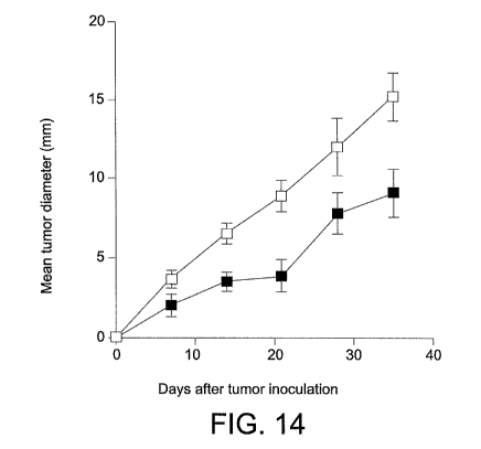
Figure 14 is a line graph showing showing the effect of intraperitoneal
injection of wild-type B7-DCIg on growth (mean tumor diameter in
millimeters) of P815 mastrocytoma murine tumor cells in syngeneic
immunocompetent (DBA/2) mice as a function of time (days). Mice were
injected intraperitonealy with 0.1 mg of control Ig (-^-) or wild-type B7-DCIg
(-m-) on day 3 and day 8.
DETAILED DESCRIPTION OF THE INVENTION
1. Definitions
As used herein the term "isolated" is meant to describe a compound
of interest (e.g., either a polynucleotide or a polypeptide) that is in an
environment different from that in which the compound naturally occurs e.g.
9
CA 02693707 2010-01-12
WO 2009/029342 PCT/US2008/069819
separated from its natural milieu such as by concentrating a peptide to a
concentration at which it is not found in nature. "Isolated" is meant to
include compounds that are within samples that are substantially enriched for
the compound of interest and/or in which the compound of interest is
partially or substantially purified.
As used herein, the term "polypeptide" refers to a chain of amino
acids of any length, regardless of modification (e.g., phospborylation or
glycosylation).
As used herein, a"costirnulatory polypeptide" is a polypeptide that,
upon interaction with a cell-surface molecule on T cells, enhances T cell
responses, enhances proliferation of T cells, enhances production and/or
secretion of cytokines by T cells, stimulates differentiation and effector
functions of T cells or promotes survival of T cells relative to T cells not
contacted with a costimulatory peptide.
As used herein, a "variant" polypeptide contains at least one amino
acid sequence alteration as compared to the amino acid sequence of the
corresponding wild-type polypeptide.
As used herein, an "amino acid sequence alteration" can be, for
example, a substitution, a deletion, or an insertion of one or more amino
acids.
As used herein, a"vector" is a replicon, such as a plasmid, phage, or
cosmid, into which another DNA segment may be inserted so as to bring
about the replication of the inserted segment. The vectors described herein
can be expression vectors.
As used herein, an "expression vector" is a vector that includes one or
more expression control sequences
As used herein, an "expression control sequence" is a DNA sequence
that controls and regulates the transcription and/or translation of another
DNA sequence.
As used herein, "operably linked" means incorporated into a gentic
construct so that expression control sequences effectively control expression
of a coding sequence of interest.
CA 02693707 2010-01-12
WO 2009/029342 PCT/US2008/069819
As used herein, a "fragment" of a polypeptide refers to any subset of
the polypeptide that is a shorter polypeptide of the full length protein.
Generally, fragments will be five or more amino acids in length.
As used herein, "valency" refers to the number of binding sites
available per molecule.
As used herein, "conservative" amino acid substitutions are
substitutions wherein the substituted amino acid has similar structural or
chemical properties.
As used herein, "non-conservative" amino acid substitutions are those
in which the charge, hydrophobicity, or bulk of the substituted amino acid is
significantly altered.
As used herein, "isolated nucleic acid" refers to a nucleic acid that is
separated from other nucleic acid molecules that are present in a mammalian
genome, including nucleic acids that normally flank one or both sides of the
nucleic acid in a mammalian genome (e.g., nucleic acids that encode non-
B7-DC proteins).
As used herein with respect to nucleic acids, the term "isolated"
includes any non-naturally-occurring nucleic acid sequence, since such non-
naturally-occurring sequences are not found in nature and do not have
immediately contiguous sequences in a naturally-occurring genome.
As used herein, the term "host cell" refers to prokaryotic and
eukaryotic cells into which a recombinant expression vector can be
introduced.
As used herein, "transformed" and "transfected" encompass the
introduction of a nucleic acid (e.g. a vector) into a cell by a number of
techniques known in the art.
As used herein, the term "antibody" is meant to include both intact
molecules as well as fragments thereof that include the antigen-binding site.
These include Fab and F(ab')2 fragments which lack the Fc fragment of an
intact antibody.
The terms "individual", "host", "subject", and "patient" are used
interchangeably herein, and refer to a mammal, including, but not limited to,
11
CA 02693707 2010-01-12
WO 2009/029342 PCT/US2008/069819
murines, simians, humans, mammalian farm animals, mammalian sport
animals, and mammalian pets.
As used herein the tezm "effective amount" or "therapeutically
effective amount" means a dosage sufficient to treat, inhibit, or alleviate
one
or more symptoms of an inflammatory response or autoimmune disease state
being treated or to otherwise provide a desired pharmacologic and/or
physiologic effect. The precise dosage will vary according to a variety of
factors such as subject-dependent variables (e.g., age, immune system health,
etc.), the disease, and the treatment being effected.
11. Compositions
A. Isolated B7-DC polypeptides
1. Variant B7-DC polypeptides
Isolated B7-DC polypeptides are disclosed herein. The B7-DC
polypeptide may be of any species of origin. In one embodiment, the B7-DC
polypeptide is from a mammalian species. In a preferred embodiment, the
137-DC polypeptide is of murine or human origin. The full-length, immature
amino acid sequence of mouse B7-DC (SEQ ID NO: 2) is depicted in Figure
2. The signal sequence of murine B7-DC contains the first 19 amino acids of
the full-length immature amino acid sequence. The full-length, immature
amino acid sequence of human B7-DC (SEQ ID NO: 1) is depicted in Figure
1. The signal sequence of human B7-DC contains the first 19 amino acids of
the full-length immature amino acid sequence. As used herein, the term
"polypeptide" refers to a chain of amino acids of any length, regardless of
modification (e.g., phosphorylation or glycosylation).
In one embodiment the variant B7-DC polypeptide has the same
activity, substantially the same activity, or different activity as wildtype
137-
DC. Substantially the same activity means it retains the ability to co-
stimulate T cells.
The polypeptides disclosed herein include variant B7-DC
polypeptides. As used herein, a "variant" polypeptide contains at least one
amino acid sequence alteration as compared to the amino acid sequence of
the corresponding wild-type polypeptide. An amino acid sequence alteration
12
CA 02693707 2010-01-12
WO 2009/029342 PCT/US2008/069819
can be, for example, a substitution, a deletion, or an insertion of one or
more
amino acids.
A variant B7-DC polypeptide can have any combination of amino
acid substitutions, deletions or insertions. In one embodiment, isolated B7-
DC variant polypeptides have an integer number of amino acid alterations
such that their amino acid sequence shares at least 60, 70, 80, 85, 90, 95,
97,
98, 99, 99.5 or 100% identity with an amino acid sequence of a wild type
B7-DC polypeptide. In a preferred embodiment, B7-DC variant
polypeptides have an amino acid sequence sharing at least 60, 70, 80, 85, 90,
95, 97, 98, 99, 99.5 or 100% identity with the amino acid sequence of a wild
type murine or wild type human B7-DC polypeptide.
Percent sequence identity can be calculated using computer programs
or direct sequence comparison. Preferred computer program methods to
determine identity between two sequences include, but are not limited to, the
GCG program package, FASTA, BLASTP, and TBLASTN (see, e.g., D. W.
Mount, 2001, Bioinformatics: Sequence and Genome Analysis, Cold Spring
Harbor Laboratory Press, Cold Spring Harbor, N.Y.). The BLASTP and
TBLASTN programs are publicly available from NCBI and other sources.
The well-known Smith Waterman algorithm may also be used to determine
identity.
Exemplary parameters for amino acid sequence comparison include
the following: 1) algorithm from Needleman and Wunsch (J. Mol. Biol.,
48:443-453 (1970)); 2) BLOSSUM62 comparison matrix from Hentikoff and
Hentikoff (Proc. Natl. Acad. Sci. U.S:A., 89:10915-10919 (1992)) 3) gap
penalty = 12; and 4) gap length penalty - 4. A program useful with these
parameters is publicly available as the "gap" program (Genetics Computer
Group, Madison, Wis.). The aforementioned parameters are the default
parameters for polypeptide comparisons (with no penalty for end gaps).
Alternatively, polypeptide sequence identity can be calculated using
the following equation: % identity =(the number of identical
residues)/(alignment length in amino acid residues)* 100. For this
13
CA 02693707 2010-01-12
WO 2009/029342 PCT/US2008/069819
calculation, alignment length includes internal gaps but does not include
terminal gaps.
Amino acid substitutions in B7-DC polypeptides may be
"conservative" or "non-conservative". As used herein, "conservative" amino
acid substitutions are substitutions wherein the substituted amino acid has
similar structural or chemical properties, and "non-conservative" amino acid
substitutions are those in which the charge, hydrophobicity, or bulk of the
substituted amino acid is significantly altered. Non-conservative
substitutions will differ more significantly in their effect on maintaining
(a)
the structure of the peptide backbone in the area of the substitution, for
example, as a sheet or helical conformation, (b) the charge or hydrophobicity
of the molecule at the target site, or (c) the bulk of the side chain.
Examples of conservative amino acid substitutions include those in
which the substitution is within one of the five following groups: 1) small
aliphatic, nonpolar or slightly polar residues (Ala, Ser, Thr, Pro, Gly); 2)
polar, negatively charged residues and their amides (Asp, Asn, Glu, Gln);
polar, positively charged residues (His, Arg, Lys); large aliphatic, nonpolar
residues (Met, Leu, Ile, Val, Cys); and large aromatic resides (Phe, Tyr,
Trp).
Examples of non-conservative amino acid substitutions are those where 1) a
hydrophilic residue, e.g., seryl or threonyl, is substituted for (or by) a
hydrophobic residue, e.g., leucyl, isoleucyl, phenylalanyl, valyl, or alanyl;
2)
a cysteine or proline is substituted for (or by) any other residue; 3) a
residue
having an electropositive side chain, e.g., lysyl, arginyl, or histidyl, is
substituted for (or by) an electronegative residue, e.g., glutamyl or
aspartyl;
or 4) a residue having a bulky side chain, e.g., phenylalanine, is substituted
for (or by) a residue that does not have a side chain, e.g., glycine.
B7 family molecules, including B7-DC are expressed at the cell
surface as homodimers with a membrane proximal constant IgC domain and
a membrane distal IgV domain. Receptors for these ligands share a common
extracellular IgV-like domain. Interactions of receptor-ligand pairs are
mediated predominantly through residues in the IgV domains of the ligands
and ;receptors. In general, IgV domains are described as having two sheets
14
CA 02693707 2010-01-12
WO 2009/029342 PCT/US2008/069819
that each contain a layer of (3-strands. These P-strands are referred to as
A',
B, C, C', C", D, E, F and G. In one embodiment the B7-DC variant
polypeptides contain amino acid alterations (i.e., substitutions, deletions or
insertions) within one or more of these 0-strands in any possible
combination. In another embodiment, B7-DC variants contain one or more
amino acid alterations (i.e., substitutions, deletions or insertions) within
the
A', C, C', C", D, E, F or GP-strands. In a preferred embodiment B7-DC
variants contain one or more atnino acid alterations in the GP-strand.
With respect to murine B7-DC or human B7-DC, a variant B7-DC
polypeptide can contain, without limitation, substitutions, deletions or
insertions at position 33 of the A' P-strand, positions 39 or 41 of the B
0-strand, positions 56 or 58 of the C 0-strand, positions 65 or 67 of the C'
P-strand, positions 71 or 72 of the C" 0-strand, position 84 of the D f3-
stra.nd,
position 88 of the E0-strand, positions 101, 103 or 105 of the F0-strand, or
positions 111, 113 or 116 of the G 0-strand.
In one embodiment, variant B7-DC polypeptides contain a
substitution at position 33 (e.g., a serine substitution for aspartic acid at
position 33), a substitution at position 39 (e.g., a tyrosine substitution for
serine at position 39), a substitution at position 41 (e.g., a serine
substitution
for glutamic acid at position 41), a substitution at position 56 (e.g., a
serine
substitution for arginine at position 56), a substitution at position 58
(e.g., a
tyrosine substitution for serine at position 58), a substitution at position
65
(e.g., a serine substitution for aspartic acid at position 65), a substitution
at
position 67 (e.g., a tyrosine substitution for serine at position 67), a
substitution at position 71 (e.g., a serine substitution for glutamic acid at
position 71), a substitution at position 72 (e.g., a serine substitution for
arginine at position 72), a substitution at position 84 (e.g., a serine
substitution for lysine at position 84), a substitution at position 88 (e.g.,
an
alanine substitution for histidine at position 88), a substitution at position
101
(e.g., a serine substitution for arginine at position 101), a substitution at
position 103 (e.g., an alanine substitution for leucine at position 103), a
substitution at position 105 (e.g., an alanine substitution for isoleucine at
CA 02693707 2010-01-12
WO 2009/029342 PCT/US2008/069819
position 105), a substitution at position 111 (e.g., a serine substitution for
aspartic acid at position 111), a substitution at position 113 (e.g., a serine
substitution for lysine at position 113), or a substitution at position 116
(e.g.,
a tyrosine substitution for threonine at position 116).
It is understood, however, that substitutions at the recited amino acid
positions can be made using any amino acid or amino acid analog. For
example, the substitutions at the recited positions can be made with any of
the naturally-occurring amino acids (e.g., alanine, aspartic acid, asparagine,
arginine, cysteine, glycine, glutamic acid, glutamine, histidine, leucine,
valine, isoleucine, lysine, methionine, proline, threonine, serine,
phenylalanine, tryptophan, or tyrosine).
While the substitutions described herein are with respect to mouse
and human B7-DC, it is noted that one of ordinary skill in the art could
readily make equivalent alterations in the corresponding polypeptides from
other species (e.g., rat, hamster, guinea pig, gerbil, rabbit, dog, cat,
horse,
pig, sheep, cow or non-human primate).
2. Properties of variant B7-DC polypeptides
The disclosed isolated B7-DC polypeptides are capable of
costimulating T cells. A "costimulatory polypeptide" is a polypeptide that,
upon interaction with a cell-surface molecule on a T cell, enhances T cell
responses, enhances proliferation of T cells, enhances production and/or
secretion of cytokines by T cells, stimulates differentiation and effector
functions of T cells or promotes survival of T cells relative to T cells not
contacted with a costimulatory peptide. The T cell response that results from
the interaction typically is greater than the response in the absence of the
costimulatory polypeptide. The response of the T cell in the absence of the
costimulatory polypeptide can be no response or can be a response
significantly lower than in the presence of the costimulatory polypeptide.
The response of the T cell can be an effector (e,g., CTL or antibody-
producing B cell) response, a helper response providing help for one or more
effector (e.g., CTL or antibody-producing B cell) responses, or a suppressive
response.
16
CA 02693707 2010-01-12
WO 2009/029342 PCT/US2008/069819
Variant B7-DC polypeptides disclosed herein have reduced binding
affinity for PD-1 as compared to the binding affinity of the corresponding
wild-type B7-DC polypeptide. The binding affinity of a variant typically is
reduced by at least 50 percent, 55 percent, 60 percent, 70 percent, 75
percent,
80 percent, 90 percent, 95 percent, 99 percent, or more than 99 percent as
compared to the binding affinity of the corresponding wild-type polypeptide.
Methods for measuring the binding affinity between two molecules
are well known in the art. Methods for measuring the binding affinity of B7-
DC variant polypeptides for 1'D-1 include, but are not limited to,
fluorescence activated cell sorting (FACS), surface plasmon resonance,
fluorescence anisotropy, affinity chromatography and affinity selection-mass
spectrometry.
ln addition, disclosed variant B7-DC polypeptides with reduced
binding affinity for PD-1 retain substantial costimulatory activity. For
example, a variant B7-DC polypeptide can have at least 20 percent, 25
percent, 30 percent, 40 percent, 50 percent, 60 percent, 75 percent, 90
percent, 100 percent, or more than 100 percent of the level of costimulatory
activity exhibited by the corresponding wild-type B7-DC polypeptide.
Methods for measuring costimulation of T cells are well known in the
art and include measurements of T cell proliferation and secretion of
cytokines, including, but not limited to,11-2,1L-4,1L-5,1L-6, IL-10,1L-13,
and IFN-y. Proliferation of T cells can be measured by a number of methods
including, but not limited to, cell counting, measuring DNA synthesis by
uptake of labeled nucleotides (such as [3H] TdR and BrdU) and measuring
metabolic activity with tetrazolium salts. Methods for measuring the
secretion of cytokines include, but are not limited to, ELISA.
3. Fragments of variant B7-DC polypeptides
The B7-DC polypeptides disclosed herein can be full-length
polypeptides, or can be a fragment of a full length B7-DC polypeptide. As
used herein, a fragment of B7-DC refers to any subset of the polypeptide that
is a shorter polypeptide of the full length protein.
17
CA 02693707 2010-01-12
WO 2009/029342 PCT/US2008/069819
In one embodiment, variant B7-DC polypeptide fragments are those
that retain the ability to costimulate T cells. A variant B7-DC polypeptide
that is a fragment of full-length B7-DC typically has at least 20 percent, 30
percent, 40 percent, 50 percent, 60 percent, 70 percent, 80 percent, 90
percent, 95 percent, 98 percent, 99 percent, 100 percent, or even more than
100 percent of the costimulatory activity of the full-length variant B7-DC
polypeptide.
Human and mouse B7-DC proteins contain a short intracytoplasmic
domain, a single transmembrane domain and an extracellular domain. The
extracellular domain contains two Ig domains; a membrane proximal IgC
domain and a membrane distal IgV domain. Useful fragments of variant B7-
DC polypeptides include soluble fragments. Soluble B7-DC fragments are
fragments of B7-DC that may be shed, secreted or otherwise extracted from
the producing cells. ln one embodiment, variant B7-DC polypeptide
fragments include the entire extracellular domain of B7-DC. The
extracellular domain of B7-DC includes amino acids from about 26 to about
amino acid 226 of murine or human B7-DC or costimulatory fragments
thereof. In another embodiment, variant B7-DC polypeptide fragments
include the IgC and IgV domains of B7-DC. In another embodiment, variant
B7-DC polypeptide fragments include the IgV domain of B7-DC.
In one embodiment, variant B7-DC polypeptide fragments may
contain a region of the polypeptide that is important for binding affinity for
PD-1. These polypeptide fragments may be useful to compete for binding to
PD-1 and to prevent native B7-DC from binding to PD-1. By competing for
binding to PD-1, these fragments may be useful to enhance an immune
response, as inhibiting interactions of B7-H1 and B7-DC with PD-1 may also
inhibit the suppression of imrnune responses that would otherwise occur. A
polypeptide fragment of mouse or human B7-DC that could competitively
bind to PD-1 can contain, for example, amino acids 67-71, 101-105, or 111-
113. The binding of B7-DC to PD-1 typically is inhibited by at least 50
percent, 60 percent, 70 percent, 75 percent, 80 percent, 90 percent, 95
18
CA 02693707 2010-01-12
WO 2009/029342 PCT/US2008/069819
percent, or more than 95 percent as compared to the level of binding of B7-
DC to PD-1 in the absence of the fragment.
4. Modified variant B7-DC p lypeptides
Variant B7-DC polypeptides may be modified by chemical moieties
that may be present in polypeptides in a normal cellular environment, for
example, phosphorylation, methylation, amidation, sulfation, acylation,
glycosylation, sumoylation and ubiquitylation. Variant B7-DC polypeptides
may also be modified with a label capable of providing a detectable signal,
either directly or indirectly, including, but not limited to, radioisotopes
and
fluorescent compounds.
Variant B7-DC polypeptides may also be modified by chemical
moieties that are not normally added to polypeptides in a cellular
environment. Such modifications may be introduced into the molecule by
reacting targeted amino acid residues of the polypeptide with an organic
derivatizing agent that is capable of reacting with selected side chains or
terminal residues. Another modification is cyclization of the protein.
Examples of chemical derivatives of the polypeptides include lysinyl
and amino terminal residues derivatized with succinic or other carboxylic
acid anhydrides. Derivatization with a cyclic carboxylic anhydride has the
effect of reversing the charge of the lysinyl residues. Other suitable
reagents
for derivatizing amino-containing residues include imidoesters such as
methyl picolinimidate; pyridoxal phosphate; pyridoxal; chloroborohydride;
trinitrobenzenesulfonic acid; 4-methylisourea; 2,4 pentanedione; and
transaminase-catalyzed reaction with glyoxylate. Carboxyl side groups,
aspartyl or glutamyl, may be selectively modified by reaction with
carbodiimides (R-N=C-N--R') such as 1-cyclohexyl-3-(2-morpholinyl-(4-
ethyl)carbodiimide or 1-ethyl-3-(4-azonia-4,4-dimethylpentyl) carbodiimide.
Furthermore, aspartyl and glutamyl residues can be converted to asparaginyl
and glutaminyl residues by reaction with ammonia. Polypeptides may also
include one or more D-amino acids that are substituted for one or more L-
amino acids.
19
CA 02693707 2010-01-12
WO 2009/029342 PCT/US2008/069819
5. Variant B7-DC fusion polypeptides
The variant B7-DC polypeptides disclosed herein may be coupled to
other polypeptides to form fusion proteins. Provided are variant B7-DC
fusion polypeptides having a first fusion partner comprising all or a part of
a
variant B7-DC protein fused (i) directly to a second polypeptide or, (ii)
optionally, fused to a linker peptide sequence that is fused to the second
polypeptide. The presence of the fusion partner can alter the solubility,
affinity and/or valency of the variant B7-DC polypeptide. As used herein,
"valency" refers to the number of binding sites available per molecule.
Variant B7-DC fusion proteins described herein include any combination of
amino acid alteration (i.e. substitution, deletion or insertion), fragment of
B7-
DC, and/or modification as described above. In one embodiment, variant
B7-DC fusion proteins include the extracellular domain of a B7-DC protein,
or a costimulatory fragment thereof, as the first binding partner. In another
embodiment, variant B7-DC fusion proteins include the IgV and IgC domain
of a B7-DC protein as the first binding partner. In another embodiment,
variant B7-DC fusion proteins include the IgV domain of a B7-DC protein as
the first binding partner.
The second polypeptide binding partner may be N-terminal or C-
terminal relative to the variant B7-DC polypeptide. In a preferred
embodiment, the second polypeptide is C-terminal to the variant B7-DC
polypeptide.
A large number of polypeptide sequences that are routinely used as
fusion protein binding partners are well known in the art. Examples of
useful polypeptide binding partners include, but are not limited to, green
fluorescent protein (GFP), glutathione S-transferase (GST), polyhistidine,
myc, hemaglutinin, FlagTM tag (Kodak, New Haven, CT), maltose E binding
protein and protein A. In one embodiment, the variant B7-DC fusion protein
is fused to one or more domains of an Ig heavy chain constant region,
preferably having an amino acid sequence corresponding to the hinge, ClU
and CH3 regions of a human immunoglobulin Cyl chain.
CA 02693707 2010-01-12
WO 2009/029342 PCT/US2008/069819
B. Isolated Nucleic Acid Molecules
Isolated nucleic acid sequences encodzng variant B7-DC polypeptides
are disclosed herein. As used herein, "isolated nucleic acid" refers to a
nucleic acid that is separated from other nucleic acid molecules that are
present in a mammalian genome, including nucleic acids that normally flank
one or both sides of the nuclcic acid in a mammalian genome (e.g., nucleic
acids that encode non-B7-DC proteins). The term "isolated" as used herein
with respect to nucleic acids also includes any non-naturally-occurring
nucleic acid sequence, since such non-naturally-occurring sequences are not
found in nature and do not have immediately contiguous sequences in a
naturally-occurring genome.
An isolated nucleic acid can be, for example, a DNA molecule,
provided one of the nucleic acid sequences normally found immediately
flanking that DNA molecule in a naturally-occurring genome is removed or
absent. Thus, an isolated nucleic acid includes, without limitation, a DNA
molecule that exists as a separate molecule independent of other sequences
(e.g., a chemically synthesized nucleic acid, or a cDNA or genomic DNA
fragment produced by PCR or restriction endonuclease treatment), as well as
recombinant DNA that is incorporated into a vector, an autonomously
replicating plasmid, a virus (e.g., a retrovirus, lentivirus, adenovirus, or
herpes virus), or into the genomic DNA of a prokaryote or euharyote. In
addition, an isolated nucleic acid can include an engineered nucleic acid such
as a recombinant DNA molecule that is part of a hybrid or fusion nucleic
acid. A nucleic acid existing among hundreds to millions of other nucleic
acids within, for example, a cDNA library or a genomic library, or a gel slice
containing a genomic DNA restriction digest, is not to be considered an
isolated nucleic acid.
Nucleic acids can be in sense or antisense orientation, or can be
complementary to a reference sequence encoding a B7-DC polypeptide.
Reference sequences include, for example, the nucleotide sequence ofhuman
B7-DC (SEQ ID NO: 3) set forth in Figure 3, which encodes full-length,
immature B7-DC having the amino acid sequence (SEQ ID NO: 1) set forth
21
CA 02693707 2010-01-12
WO 2009/029342 PCT/US2008/069819
in Figure 1 and the nucleotide sequence of murine B7-DC (SEQ ID NO: 4)
set forth in Figure 4, which encodes full-length, immature B7-DC having the
amino acid sequence (SEQ ID NO: 2) set forth in Figure 2.
Nucleic acids can be DNA, RNA, or nucleic acid analogs. Nucleic
acid analogs can be modified at the base moiety, sugar moiety, or phosphate
backbone. Such modification can improve, for example, stability,
hybridization, or solubility of the nucleic acid. Modifications at the base
moiety can include deoxyuridine for deoxythymidine, and 5-methyl-2'-
deoxycytidine or 5-bromo-2'-deoxycytidine for deoxycytidine.
Modifications of the sugar moiety can include modification of the 2'
hydroxyl of the ribose sugar to form 2'-O-methyl or 2'-O-allyl sugars. The
deoxyribose phosphate backbone can be modified to produce morpholino
nucleic acids, in which each base moiety is linked to a six membered,
morpholino ring, or peptide nucleic acids, in which the deoxyphosphate
backbone is replaced by a pseudopeptide backbone and the four bases are
retained. See, for example, Summerton and Weller (1997) Antisense Nucleic
Acid Drug Dev. 7:187-195; and Hyrup et al. (1996) Bioorgan. Med. Chem.
4:5-23. In addition, the deoxyphosphate backbone can be replaced with, for
example, a phosphorothioate or phosphorodithioate backbone, a
phosphoroamidite, or an alkyl pbosphotriester backbone.
C. Vectors and host ceXls
Nucleic acids, such as those described above, can be inserted into
vectors for expression in cells. As used herein, a "vector" is a replicon,
such
as a plasmid, phage, or cosmid, into which another DNA segment may be
inserted so as to bring about the replication of the inserted segment. Vectors
can be expression vectors. An "expression vector" is a vector that includes
one or more expression control sequences, and an "expression control
sequence" is a DNA sequence that controls and regulates the transcription
and/or translation of another DNA sequence.
Nucleic acids in vectors can be operably linked to one or more
expression control sequences. As used herein, "operably linked" means
incorporated into a genetic construct so that expression control sequences
22
CA 02693707 2010-01-12
WO 2009/029342 PCT/US2008/069819
effectively control expression of a coding sequence of interest. Examples of
expression control sequences include promoters, enhancers, and transcription
terminating regions. A promoter is an expression control sequence
composed of a region of a DNA molecule, typically within 100 nucleotides
upstream of the point at which transcription starts (generally near the
initiation site for RNA polymerase II). To bring a coding sequence under the
control of a promoter, it is necessary to position the translation initiation
site
of the translational reading frame of the polypeptide between one and about
fifty nucleotides downstream of the promoter. Enhancers provide expression
specificity in terms of time, location, and level. Unlike promoters, enhancers
can function when located at various distances from the transcription site.
An enhancer also can be located downstream from the transcription initiation
site. A coding sequence is "operably linked" and "under the control" of
expression control sequences in a cell when RNA polymerase is able to
transcribe the coding sequence into inRNA, which then can be translated into
the protein encoded by the coding sequence.
Suitable expression vectors include, without limitation, plasmids and
viral vectors derived from, for example, bacteriophage, baculoviruses,
tobacco mosaic virus, herpes viruses, cytomegalo virus, retroviruses,
vaccinia viruses, adenoviruses, and adeno-associated viruses. Numerous
vectors and expression systems are commercxally available from such
corporations as Novagen (Madison, WI), Clontech (Palo Alto, CA),
Stratagene (La Jolla, CA), and Invitrogen Life Technologies (Carlsbad, CA).
An expression vector can include a tag sequence. Tag sequences, are
typically expressed as a fusion with the encoded polypeptide. Such tags can
be inserted anywhere within the polypeptide including at either the carboxyl
or amino terminus. Examples of useful tags include, but are not limited to,
green fluorescent protein (GFP), glutathione S-transferase (GST),
polyhistidine, c-myc, hemagglutinin, F1agTM tag (Kodak, New Haven, CT),
maltose E binding protein and protein A. In one embodiment, the variant
B7-DC fusion protein is present in a vector containing nucleic acids that
encode one or more domains of an Ig heavy chain constant region, preferably
23
CA 02693707 2010-01-12
WO 2009/029342 PCT/US2008/069819
having an amino acid sequence corresponding to the hinge, Cu2 and Cn3
regions of a human immunoglobulin Cy1 chain.
Vectors containing mucleic acids to be expressed can be transferred
into host cells. The term "host cell" is intended to include prokaryotic and
eukaryotic cells into which a recombinant expression vector can be
introduced. As used herein, "transformed" and "transfected" encompass the
introduction of a nucleic acid molecule (e.g., a vector) into a cell by one of
a
number of techniques. Although not limited to a particular technique, a
number of these techniques are well established within the art. Prokaryotic
cells can be transformed with nucleic acids by, for example, electroporation
or calcium chloride mediated transformation. Nucleic acids can be
transfected into manunalian cells by techniques including, for example,
calcium phosphate co-precipitation, DEAE-dextran-mediated transfection,
lipofection, electroporation, or microinjection. Host cells (e.g., a
prokaryotic
cell or a eukaryotic cell such as a CHO cell) can be used to, for example,
produce the variant B7-DC polypeptides descirbed herein. In some
embodiments, a host cell (e.g., an antigen presenting cell) can be used to
express the variant B7-DC polypeptides disclosed herein for presentation to a
T cell.
D. Antibodies
Monoclonal antibodies (mAbs) and methods for their production and
use are described in Kohler and Milstein, Nature 256:495-497 (1975); U.S.
Pat. No. 4,376,110; Hartlow, E. et al., Antibodies: A Laboratory Manual,
Cold Spring Harbor Laboratory Press, Cold Spring Harbor, N.Y., 1988);
Monoclonal Antibodies and Hybridomas: A New Dimension in Biological
Analyses, Plenum Press, New York, N.Y. (1980); H. Zola et al., in
Monoclonal Hybridoma Antibodies: Techniques and Applications, CRC
Press, 1982)).
Immunoassay methods are described in Coligan, J. E. et al., eds.,
Current Protocols in Immunology, Wiley-Interscience, New York 1991 (or
current edition); Butt, W. R. (ed.) Practical Immunoassay: The State of the
Art, Dekker, N.Y., 1984; Bizollon, Ch. A., ed., Monoclonal Antibodies and
24
CA 02693707 2010-01-12
WO 2009/029342 PCT/US2008/069819
New Trends in Immunoassays, Elsevier, N.Y., 1984; Butler, J. E., ELISA
(Chapter 29), In: van Oss, C. J. et al., (eds),1MMUNOCHEMrSTRY,
Marcel Dekker, Inc., New York, 1994, pp. 759-803; Butler, J. E. (ed.),
Immunochemistry of Solid-Phase lrnmunoassay, CRC Press, Boca Raton,
1991; Weintraub, B., Principles of Radioimmunoassays, Seventh Training
Course on Radioligand Assay Techniques, The Endocrine Society, March,
1986; Work, T. S. et al., Laboratory Techniques and Biochemistry in
Molecular Biology, North Holland Publishing Company, NY, (1978)
(Chapter by Chard, T., "An Introduction to Radioimmune Assay and Related
Techniques").
Anti-idiotypic antibodies are described, for example, in Idiotypy in
Biology and Medicine, Academic Press, New York, 1984; Immunological
Reviews Volume 79, 1984; Immunological Reviews Volume 90, 1986; Curr.
Top. Microbiol., Immunol. Volume 119, 1985; Bona, C. et al., CRC Crit.
Rev.lmmunol., pp. 33-81 (1981); Jerme, N K, Ann. Immunol. 125C:373-
389 (1974); Jeme, N K, ln:ldiotypes--Antigens on the Inside, Westen-
Schnurr, I., ed., Editiones Roche, Basel, 1982, Urbain, J. et aL, Ann.
Immunol. 133D:179-(1982); Rajewsky, K. et al., Ann. Rev.lmmunol. 1:569-
607 (1983).
Monoclonal and polyclonal antibodies that are reactive with novel
epitopes of B7-DC that are absent from known B7 family proteins are
described berein. The antibodies may be xenogeneic, allogeneic, syngeneic,
or modified forms thereof, such as humanized or chimeric antibodies.
Antiidiotypic antibodies specific for the idiotype of an anti-B7-DC antibody
are also included. The term "antibody" is meant to include both intact
molecules as well as fragments thereof that include the antigen-binding site
and are capable of binding to a B7-DC epitope. These include, Fab and
F(ab')z fragments which lack the Fc fragment of an intact antibody, clear
more rapidly from the circulation, and may have less non-specific tissue
binding than an intact antibody (Wahl et al., J. Nuc. Med. 24:316-325
(1983)). Also included are Fv fragments (Hochman, J. et al. (1973)
Biochemistry 12:1130-1135; Sharon, J. et al.(1976) Biochemistry 15:1591-
CA 02693707 2010-01-12
WO 2009/029342 PCT/US2008/069819
1594). These various fragments are produced using conventional techniques
such as protease cleavage or chemical cleavage (see, e.g., Rousseaux et al.,
Meth. Enzymol., 121:663-69 (1986)).
Polyclonal antibodies are obtained as sera from immunized animals
such as rabbits, goats, rodents, etc. and may be used directly without further
treatment or may be subjected to conventional enrichment or purification
methods such as ammonium sulfate precipitation, ion exchange
chromatography, and affinity chromatography.
The immunogen may comprise the complete B7-DC protein, or
fragments or derivatives thereof. Preferred immunogens comprise all or a
part of the extracellular domain (ECD) ofhuman B7-DC, where these
residues contain the post-translation modifications, such as glycosylation,
found on the native B7-DC. Immunogens comprising the extracellular
domain are produced in a variety of ways known in the art, e.g., expression
of cloned genes using conventional recombinant methods, isolation from
cells of origin, cell populations expressing high levels of B7-DC, etc.
Monoclonal antibodies may be produced using conventional
hybridoma technology, such as the procedures introduced by Kohler and
Milstein (Nature, 256:495-97 (1975)), and modifications thereof (see above
references). An animal, preferably a mouse is primed by immunization with
an immunogen as above to elicit the desired antibody response in the primed
animal. B lymphocytes from the lymph nodes, spleens or peripheral blood of
a primed, animal are fused with myeloma cells, generally in the presence of a
fusion promoting agent such as polyethylene glycol (PEG). Any of a
number of murine mycloma cell lines are available for such use: the P3-
NS1/1-Ag4-1, P3-x63-k0Ag8.653, Sp2/0-Ag14, or HLI.-653 myeloma lines
(available from the ATCC, Rockville, Md.). Subsequent steps include
growth in selective medium so that unfused parental myeloma cells and
donor lymphocyte cells eventually die while only the hybridoma cells
survive. These are cloned and grown and their supematants screened for the
presence of antibody of the desired specificity, e.g. by immunoassay
26
CA 02693707 2010-01-12
WO 2009/029342 PCT/US2008/069819
techniques using B7-DC fusion proteins. Positive clones are subcloned, e.g.,
by limiting dilution, and the monoclonal antibodies are isolated.
Hybridomas produced according to these methods can be propagated
in vitro or in vivo (in ascites fluid) using techniques known in the art (see
generally Fink et al., Prog. Clin. Pathol., 9:121-33 (1984)). Generally, the
individual cell line is propagated in culture and the culture medium
containing high concentrations of a single monoclonal antibody can be
harvested by decantation, filtration, or centrifugation.
The antibody may be produced as a single chain antibody or scFv
instead of the normal multimeric structure. Single chain antibodies include
the hypervariable regions from an Ig of interest and recreate the antigen
binding site of the native Ig while being a fraction of the size o1'the intact
Ig
(Skerra, A. et al, (1988) Science, 240: 1038-1041; Pluckthun, A. et al. (1989)
Methods Enzymol. 178: 497-515; Winter, G. et al. (1991) Nature, 349: 293-
299). In a preferred embodiment, the antibody is produced using
conventional molecular biology techniques.
E. Immunogenic compositions
Vaccines require strong T cell response to eliminate cancer cells and
infected cells. Variant B7-DC variants described herein can be administered
as a component of a vaccine to provide a costimulatory signal to T cells.
Vaccines disclosed herein include antigens, a source of variant B7-DC
polypeptides and optionally adjuvants and targeting molecules. Sources of
variant B7-DC polypeptides include any variant B7-DC polypeptide, variant
B7-DC fusion proteins, nucleic acids encoding variant B7-DC polypeptides
or variant B7-DC fusion proteins, or host cells containing vectors that
express B7-DC polypeptides or variant B7-DC fusion proteins.
1. Antigens
Antigens can be peptides, proteins, polysaccharides, saccharides,
lipids, nucleic acids, or combinations thereof. The antigen can be derived
from a virus, bacterium, parasite, plant, protozoan, fungus, tissue or
transl'ormed cell such as a cancer or leukemic cell and can be a whole cell or
27
CA 02693707 2010-01-12
WO 2009/029342 PCT/US2008/069819
immunogenic component thereof, e.g., cell wall components or molecular
components thereof.
Suitable antigens are known in the art and are available from
commercial government and scientific sources. In one embodiment, the
antigens are whole inactivated or attenuated organisms. These organisms
may be infectious organisms, such as viruses, parasites and bacteria. These
organisms may also be tumor cells. The antigens may be purified or partially
purified polypeptides derived from tumors or viral or bacterial sources. The
antigens can be recombinant polypeptides produced by expressing DNA
encoding the polypeptide antigen in a heterologous expression system. The
antigens can be DNA encoding all or part of an antigenic protein. The DNA
may be in the form of vector DNA such as plasmid DNA.
Antigens may be provided as single antigens or may be provided in
combination. Antigens may also be provided as complex mixtures of
polypeptides or nucleic acids.
i. Viral antigens
A viral antigen can be isolated from any virus including, but not
limited to, a virus from any of the following viral families: Arenaviridae,
Arterivirus, Astroviridae, Baculoviridae, Badnavirus, Barnaviridae,
Birnaviridae, Bromoviridae, Bunyaviridae, Caliciviridae, Capillovirus,
Carlavirus, Caulimovirus, Circoviridae, Closterovirus, Comoviridae,
Coronaviridae (e.g., Coronavirus, such as severe acute respiratory syndrome
(SARS) virus), Corticoviridae, Cystoviridae, Deltavirus, Dianthovirus,
Enamovirus, Filoviridae (e.g., Marburg virus and Ebola virus (e.g., Zaire,
Reston, Ivory Coast, or Sudan strain)), Flaviviridae, (e.g., Hepatitis C
virus,
Dengue virus 1, Dengue virus 2, Dengue virus 3, and Dengue virus 4),
Hepadnaviridae, Herpesviridae (e.g., Human herpesvirus 1, 3, 4, 5, and 6,
and Cytomegalovirus), Hypoviridae, Iridoviridae, Leviviridae,
Lipothrixviridae, Microviridae, Orthomyxoviridae (e.g.,lnffluenzavirus A
and B and C), Papovaviridae, Paramyxoviridae (e.g., measles, mumps, and
human respiratory syncytial virus), Parvoviridae, Picornaviridae (e.g.,
poliovirus, rhinovirus, hepatovirus, and aphthovirus), Poxviridae (e.g.,
28
CA 02693707 2010-01-12
WO 2009/029342 PCT/US2008/069819
vaecinia and smallpox virus), Reoviridae (e.g., rotavirus), Retroviridae
(e.g.,
lentivirus, such as human immunodeficiency virus (HIV) I and HIV 2),
Rhabdoviridae (for example, rabies virus, measles virus, respiratory
syncytial virus, etc.), Togaviridae (for example, rubella virus, dengue virus,
etc.), and Totiviridae. Suitable viral antigens also include all or part of
Dengue protein M, Dengue protein E, Dengue DINS1, Dengue D1NS2, and
Dengue D1NS3.
Viral antigens may be derived from a particular strain such as a
papilloma virus, a herpes virus, i.e. herpes simplex 1 and 2; a hepatitis
virus,
for example, hepatitis A virus (HAV), hepatitis B virus (HBV), hepatitis C
virus (HCV), the delta hepatitis D virus (HDV), hepatitis E virus (HEV) and
hepatitis G virus (HGV), the tick-borne encephalitis viruses; parainfluenza,
varicella-zoster, cytomeglavirus, Epstein-Barr, rotavirus, rhinovirus,
adenovirus, coxsackieviruses, equine encephalitis, Japanese encephalitis,
yellow fever, Rift Valley fever,and lymphocytic choriomeningitis.
ii. Bacterial antigens
Bacterial antigens can originate from any bacteria including, but not
limited to, Actinomyces, Anabaena, Bacillus, Bacteroides, Bdellovibrio,
Bordetella, Borrelia, Campylobacter, Caulobacter, Chlamydia, Chlorobium,
Chromatium, Clostridium, Corynebacterium, Cytophaga, Deinococcus,
Escherichia, Francisella, Halobacterium, Heliobacter, Haemophilus,
Hemophilus influenza type B (HIB), Hyphomicrobium, Legionella,
Leptspirosis, Listeria, .Meningococcus A, B and C, Methanobacterium,
Micrococcus, Myobacterium, Mycoplasma, Myxococcus, Neisseria,
Nitrobacter, Oscillatoria, Prochloron, Proteus, Pseudomonas,
Phodospirillum, Rickettsia, Salmonella, Shigella, Spirillum, Spirochaeta,
Staphylococcus, Streptococcus, Streptomyces, Sulfolobus, Thermoplasma,
Thiobacillus, and Treponema, Vibrio, and Yersinia.
iii. Parasite antigens
Parasite antigens can be obtained from parasites such as, but not
limited to, an antigen derived from Cryptococcus neoformans, Histoplasma
capsulatum, Candida albicans, Candida tropicalis, Nocardia asteroides,
29
CA 02693707 2010-01-12
WO 2009/029342 PCT/US2008/069819
Rickettsia ricketsii, Rickettsia typhi, MycopZasma pneumoniae, Chlamydial
psittaci, Chlamydial trachomatis, Plasmodiumfalciparum, Trypanosoma
brucei, Entamoeba histolytica, Toxoplasma gondii, Trichomonas vaginalis
and Schistosoma mansoni. These include Sporozoan antigens, Plasmodian
antigens, such as all or part of a Circumsporozoite protein, a Sporozoite
surface protein, a liver stage antigen, an apical membrane associated protein,
or a Merozoite surface protein.
iv. Allergens and environmental antigens
The antigen can be an allergen or enviromnental antigen, such as, but
not limited to, an antigen derived from naturally occurring allergens such as
pollen allergens (tree-, herb, weed-, and grass pollen allergens), insect
allergens (inhalant, saliva and venom allergens), animal hair and dandruff
allergens, and food allergens. Important pollen allergens from trees, grasses
and herbs originate from the taxonomic orders of Fagales, Oleales, Pinales
and platanaceae including i.a. birch (Betula), alder (Alnus), hazel (Corylus),
hombeam (Carpinus) and olive (Olea), cedar (CryptomeriaandJuniperus),
Plane tree (Platanus), the order of Poales including i.e. grasses of the
genera
Lolium, Phleum, Poa, Cynodon, Dactylis, Holcus, Phalaris, Secale, and
Sorghum, the orders of Asterates and Urticales including i.a. herbs of the
genera Ambrosia, Artemisia, and Parietaria. Other allergen antigens that
may be used include allergens from house dust mites of the genus
Dermatophagoides and Euroglyphus, storage mite e.g Lepidoglyphys,
Glycyphagus and Tyrophagus, those from cockroaches, midges and fleas e.g.
Blatella, Periplaneta, Chironomus and Ctenocepphalides, those from
mammals such as cat, dog and horse, birds, venom allergens including such
originating from stinging or biting insects such as those from the taxonomic
order of Hymenoptera including bees (superfamily Apidae), wasps
(superfamily Vespidea), and ants (superfamily Formicoidae). Still other
allergen antigens that may be used include inhalation allergens from fungi
such as from the genera Alternaria and Cladosporium.
CA 02693707 2010-01-12
WO 2009/029342 PCT/US2008/069819
v. Tumor antigens
The antigen can be a tumor antigen, including a tumor-associated or
tumor-specific antigen, such as, but not limited to, alpha-actinin-4, Bcr-Abl
fusion protein, Casp-8, beta-catenin, cdc27, cdk4, cdkn2a, coa- 1, dek-can
fusion protein, EF2, ETV6-AML1 fusion protein, LDLR-
fucosyltransferaseAS fusion protein, HLA-A2, HLA-A11, hsp70-2,
KIAAO205, Mart2, Mum-1, 2, and 3, neo-PAP, myosin class I, OS-9, pml-
RARa fusion protein, PTPRK, K-ras, N-ras, Triosephosphate isomeras,
Bage-1, Gage 3,4,5,6,7, GnTV, Herv-K-mel, Lage-1, Mage-
A1,2,3,4,6,10,12, Mage-C2, NA-88, NY-Eso-1/Lage-2, SP17, SSX-2, and
TRP2-Int2, MelanA (MART-1), gp100 (Pmel 17), tyrosinase, TRP-1, TRP-2,
MAGE-1, MAGE-3, BAGE, GAGE-1, GAGE-2, p15(58), CEA, RAGE,
NY-ESO (LAGE), SCP-1, Hom/Mel-40, PRAME, p53, H-Ras, HER-2/neu,
BCR-ABL, E2A-PRL,1-14-RET,IGH-IGK, MYL-RAR, Epstein Barr virus
antigens, EBNA, human papillpmavirus (HPV) antigens E6 and E7, TSP-
180, MAGE-4, MAGE-5, MAGE-6, p185erbB2, p180erbB-3, c-met, nm-
23H1, PSA, TAG-72-4, CA 19-9, CA 72-4, CAM 17.1, NuMa, K-ras, P-
Catenin, CDK4, Murn-1, p16, TAGE, PSMA, PSCA, CT7, telomerase, 43-
9F, 5T4, 791 Tgp72, a-fetoprotein, 13HCG, BCA225, BTAA, CA 125, CA
15-3 (CA 27.29\BCAA), CA 195, CA 242, CA-50, CAM43, CD681KP 1,
CO-029, FGF-5, G250, Ga733 (EpCAM), HTgp-175, M344, MA-50, MG7-
Ag, MOV 18, NB\70K, NY-CO-1, RCAS 1, SDCCAGI6, TA-90 (Mac-2
binding protein\cyclophilin C-associated protein), TAAL6, TAG72, TLP,
and. TPS.
2. Targeting molecules
Of the main types of antigen-presenting cells (B cell, macrophages
and dendritic cells (DCs)), the DC is the most potent and is responsible for
initiating all antigen-specific immune responses. Thus, targeting DCs
provides the opportunity to enhance the delivery of antigen and antigen
responses.
Dendritic cells express a number of cell surface receptors that can
mediate the endocytosis of bound antigen. Targeting exogenous antigens to
31
CA 02693707 2010-01-12
WO 2009/029342 PCT/US2008/069819
internalizing surface molecules on systemically-distributed antigen
presenting cells facilitates uptake of antigens and thus overcomes a major
rate-limiting step in immunization and thus in vaccination.
Dendritic cell targeting molecules include monoclonal or polyclonal
antibodies or fragments thereof that recognize and bind to epitopes displayed
on the surface of dendritic cells. Dendritic cell targeting molecules also
include ligands which bind to a cell surface receptor on dendritic cells. One
such receptor, the lectin DEC-205, has been used in vitro and in mice to
boost both humoral (antibody-based) and cellular (CD8 T cell) responses by
2-4 orders of magnitude (Hawiger, et al., J. Exp. Med., 194(6):769-79
(2001); Bonifaz, et al., J. Exp. Med., 196(12):1627-38 (2002); Bonifaz, et
al.,
J. Exp. Med, 199(6):815-24 (2004)). In these experiments, antigens were
fused to an anti-DEC205 heavy chain and a recombinant antibody molecule
was used for immunization.
A variety of other endocytic receptors, including a mannose-specific
lectin (mannose receptor) and IgG Fc receptors, have also been targeted in
this way with similar enhancement of antigen presentation efficiency. Other
suitable receptors which may be targeted include, but are not limited to, DC-
SIGN, 33D1, SIGLEC-H, DCIR, CDI lc, heat shock protein receptors and
scavenger receptors.
Other receptors which may be targeted include the toll-like receptors
(TLRs). TLRs recognize and bind to pathogen-associated molecular patterns
(PAMPs). PAMPs target the TLR on the surface of the dendritic cell and
signals internally, thereby potentially increasing DC antigen uptake,
maturation and T-cell stimulatory capacity. PAMPs conjugated to the
particle surface or co-encapsulated include unmethylated CpG DNA
(bacterial), double-stranded RNA (viral), lipopolysacharride (bacterial),
peptidoglycan (bacterial), lipoarabinozna.rmin. (bacterial), zymosan (yeast),
mycoplasmal lipoproteins such as MALP-2 (bacterial), flagellin (bacterial)
poly(inosinic-cytidylic) acid (bacterial), lipoteichoic acid (bacterial) or
imidazoquinolines (synthetic).
32
CA 02693707 2010-01-12
WO 2009/029342 PCT/US2008/069819
3e Adjuvants
Optionally, the vaccines described herein may include adjuvants.
The adjuvant can be, but is not limited to, one or more of the following: oil
emulsions (e.g., Freund's adjuvant); saponin formulations; virosomes and
viral-like particles; bacterial and microbial derivatives; immunostimulatory
oligonucleotides; ADP-ribosylating toxins and detoxified derivatives; alum;
BCG; mineral-containing compositions (e.g., mineral salts, such as
aluminium salts and calcium salts, hydroxides, phosphates, sulfates, etc.);
bioadhesives andJor mucoadhesives; microparticles; liposomes;
polyoxyethylene ether and polyoxyethylene ester formulations;
polyphosphazene; muramyl peptides; imidazoquinolone compounds; and
surface active substances (e.g. lysolecithin, pluronic polyols, polyanions,
peptides, oil emulsions, keyhole limpet hemocyanin, and dinitrophenol).
Adjuvants may also include immunomodulators such as cytokines,
interleukins (e.g., IL-1, IL-2, IL-4,1.L-5, IL-6, IL-7, IL-12, etc.),
interferons
(e.g., interferon-.gamma.), macrophage colony stimulating factor, and tumor
necrosis factor. In addition to variant B7-DC polypeptides, other co-
stimulatory molecules, including other polypeptides of the B7 family, may
be administered. Such proteinaceous adjuvants may be provided as the full-
length polypeptide or an active fragment thereof, or in the form of DNA,
such as plasmid DNA.
F. Pharmaceutical compositions
Pharmaceutical compositions including variant B7-DC polypeptides,
variant B7-DC fusion proteins, nucleic acids encoding variant B7-DC
polypeptides and fusion proteins, and vectors containing the same are
provided. The pharmaceutical compositions may be for administration by
oral, parenteral (intramuscular, intraperitoneal, intravenous (IV) or
subcutaneous injection), transdertnal (either passively or using iontophoresis
or electroporation), transmucosal (nasal, vaginal, rectal, or sublingual)
routes
of administration or using bioerodible inserts and can be formulated in
dosage forms appropriate for each route of administration. In a preferred
embodiment, the peptides are administered in an aqueous solution,
33
CA 02693707 2010-01-12
WO 2009/029342 PCT/US2008/069819
particularly for parenteral injection. In general, pharmaceutical compositions
are provided including effective amounts of a variant B7-DC polypeptide,
fusion proteinor nucleic acid encoding the same, or derivative products, and
pharmaceutically acceptable diluents, preservatives, solubilizers,
emulsifiers,
adjuvants and/or carriers. Such compositions include diluents of various
buffer content (e.g., Tris-HC1, acetate, phosphate), pH and ionic strength;
additives such as detergents and solubilizing agents (e.g., TWEEN 20,
TWEEN 80, Polysorbate 80), anti-oxidants (e.g., ascorbic acid, sodium
metabisulfite), preservatives (e.g., Thimersol, benzyl alcohol) and bulking
substances (e.g., lactose, mannitol); incorporation of the material into
particulate preparations of polymeric compounds such as polylactic acid,
polyglycolic acid, etc. or into liposomes. Hylauronic acid may also be used.
Such compositions may influence the physical state, stability, rate of in vivo
release, and rate of in vivo clearance of the present proteins and
derivatives.
See, e.g., Remington's Pharmaceutical Sciences, 18th Ed. (1990, Mack
Publishing Co., Easton, Pa. 18042) pages 1435-1712. The compositions may
be prepared in liquid form, or may be in dried powder (e.g., lyophilized)
form.
1. Oral Delivery
Variant B7-DC polypeptides, fusion proteins and nucleic acids
encoding the same can be formulated for oral delivery. Oral solid dosage
forms are described generally in Remington's Pharmaceutical Sciences, 18th
Ed. 1990 (Mack Publishing Co. Easton Pa. 18042) at Chapter 89. Solid
dosage forms include tablets, capsules, pills, troches or lozenges, cachets,
pellets, powders, or granules. Also, liposomal or proteinoid encapsulation
may be used to formulate the present compositions (as, for example,
proteinoid microspheres reported in U.S. Pat. No. 4,925,673). Liposomal
encapsulation may be used and the liposomes may be derivatized with
various polymers (e.g., U.S. Pat. No. 5,013,556). A description of possible
solid dosage forms for the therapeutic is given by Marshall, K. In: Modem
Pharmaceutics Edited by G. S. Banker and C. T. Rhodes Chapter 10, 1979.
In general, the formulation will include the ABC transporter ligands (or
34
CA 02693707 2010-01-12
WO 2009/029342 PCT/US2008/069819
chemically modified forms thereof) and inert ingredients which allow for
protection against the stomach environment, and release of the biologically
active material in the intestine.
Another embodiment provides liquid dosage forms for oral
administration, including pharmaceutically acceptable emulsions, solutions,
suspensions, and syrups, which may contain other components including
inert diluents; adjuvants such as wetting agents, emulsifying and suspending
agents; and sweetening, flavoring, and perfuming agents.
The polypeptide antagonists may be chemically modified so that oral
delivery of the derivative is efficacious. Generally, the chemical
modification
contemplated is the attachment of at least one moiety to the component
molecule itself, where said moiety permits (a) inhibition of proteolysis; and
(b) uptake into the blood stream from the stomach or intestine. Also desired
is the increase in overall stability of the component or components and
incrcase in circulation time in the body. PEGylation is a preferred chemical
modification for pharmaceutical usage. Other moieties that may be used
include: propylene glycol, copolymers of ethylene glycol and propylene
glycol, carboxymethyl cellulose, dextran, polyvinyl alcohol, polyvinyl
pyrrolidone, polyproline, poly- 1,3-dioxolane and poiy-1,3,6-tioxocane [see,
e.g., Abuchowski and Davis (1981) "Soluble Polymer-Enzyme Adducts," in
Enzymes as Drugs. Hocenberg and Roberts, eds. (Wiley-Interscience: New
York, N.Y.) pp. 367-383; and Newmark, et al. (1982) J. Appl. Biochem.
4:185-1891.
For oral formuXations, the location of release may be the stomach, the
small intestine (the duodenum, the jejunem, or the ileum), or the large
intestine. One skilled in the art has available formulations which will not
dissolve in the stomach, yet will release the material in the duodenum or
elsewhere in the intestine. Preferably, the release will avoid the deleterious
effects of the stomach environment, either by protection of the peptide (or
derivative) or by release of the peptide (or derivative) beyond the stomach
environment, such as in the intestine.
CA 02693707 2010-01-12
WO 2009/029342 PCT/US2008/069819
To ensure full gastric resistance a coating impermeable to at least pH
5.0 is essential. Examples of the more common inert ingredients that are used
as enteric coatings are cellulose acetate trimellitate (CAT),
hydroxypropylmethylcellulose phthalate (HPMCP), HPMCP 50, HPMCP
55, polyvinyl acetate phthalate (PVAP), Eudragit L30D, Aquateric, cellulose
acetate phthalate (CAP), Eudragit L, Eudragit S, and Shellac. These coatings
may be used as mixed films.
A coating or mixture of coatings can also be used on tablets, which
are not intended for protection against the stomach. This can include sugar
coatings, or coatings which make the tablet easier to swallow. Capsules may
consist of a hard shell (such as gelatin) for delivery of dry therapeutic
(i.e.
powder), for liquid forms a soft gelatin shell may be used. The shell material
of cachets could be thick starch or other edible paper. For pills, lozenges,
molded tablets or tablet triturates, moist massing techniques can be used.
The variant B7-DC polypeptide, variant B7-DC fusion protein or
nucleic acid encoding the same (or derivative) can be included in the
forrnulation as fine multiparticulates in the form of granules or pellets of
particle size about 1 mm. The formulation of the material for capsule
administration could also be as a powder, lightly compressed plugs, or even
as tablets. These therapeutics could be prepared by compression.
Colorants and/or flavoring agents may also be included.l='or example,
the sH4 antagonist (or derivative) may be formula.ted (such as by liposome or
microsphere encapsulation) and then further contained within an edible
product, such as a refrigerated beverage containing colorants and flavoring
agents.
One may dilute or increase the volume of the variant B7-DC
polypeptide, variant B7-DC fiusion protein or nucleic acid encoding the same
(or derivative) with an inert materiai. These diluents could include
carbohydrates, especially mannitol, a-lactose, anhydrous lactose, cellulose,
sucrose, modified dextrans and starch. Certain inorganic salts may be also
be used as fillers including calcium triphosphate, m.agnesiuzn carbonate and
36
CA 02693707 2010-01-12
WO 2009/029342 PCT/US2008/069819
sodium chloride. Some commercially available diluents are Fast-Flo, Emdex,
STA-Rx 1500, Emcompress and Avicell.
Disintegrants may be included in the formulation of the therapeutic
into a solid dosage form. Materials used as disintegrates include but are not
limited to starch, including the commercial disintegrant based on starch,
Explotab. Sodium starch glycolate, Amberlite, sodium
carboxymethylcellulose, ultramylopectin, sodium alginate, gelatin, orange
peel, acid carboxymethyl cellulose, natural sponge and bentonite may all be
used. The disintegrants may also be insoluble cationic exchange resins.
Powdered gums may be used as disintegrants and as binders and can include
powdered gums such as agar, Karaya or tragacanth. Alginic acid and its
sodium salt are also useful as disintegrants.
Binders may be used to hold the variant B7-DC polypeptide, variant
B7-DC fusion protein or nucleic acid encoding the same (or derivative)
together to form a hard tablet and include materials from natural products
such as acacia, tragacanth, starch and gelatin. Others include methyl
cellulose (MC), ethyl cellulose (EC) and carboxymethyl cellulose (CMC).
Polyvinyl pyrrolidone (PVP) and hydroxypropylmethyl cellulose (HPMC)
could both be used in alcoholic solutions to granulate the peptide (or
derivative).
An antifrictional agent may be included in the formulation of the sH4
antagonist (or derivative) to prevent sticking during the formulation process.
Lubricants may be used as a layer between the peptide (or derivative) and the
die wall, and these can include but are not limited to; stearic acid including
its magnesium and calcium salts, polytetrafluoroethylene (PTFE), liquid
paraffin, vegetable oils and waxes. Soluble lubricants may also be used such
as sodium lauryl sulfate, magnesium lauryl sulfate, polyethylene glycol of
various molecular weights, Carbowax 4000 and 6000.
Glidants that might improve the flow properties of the drug during
formulation and to aid rearrangement during compression might be added.
The glidants may include starch, talc, pyrogenic silica and hydrated
silicoaluminate.
37
CA 02693707 2010-01-12
WO 2009/029342 PCT/US2008/069819
To aid dissolution of the peptide (or derivative) into the aqueous
environrn.ent a surfactant might be added as a wetting agent. Surfactants may
include anionic detergents such as sodium lauryl sulfate, dioctyl sodium
sulfosuccinate and dioctyl sodium sulfonate. Cationic detergents might be
used and could include benzalkonium chloride or benzethomium chloride.
The list of potential nonionic detergents that could be included in the
formulation as surfactants are lauromacrogol 400, polyoxyl 40 stearate,
polyoxyethylene hydrogenated castor oil 10, 50 and 60, glycerol
monostearate, polysorbate 20, 40, 60, 65 and 80, sucrose fatty acid ester,
methyl cellulose and carboxymethyl cellulose. These surfactants could be
present in the formulation of the protein or derivative either alone or as a
mixture in different ratios.
Additives which potentially enhance uptake of the variant B7-DC
polypeptide, variant B7-DC fusion protein or nucleic acid encoding the same
(or derivative) are for instance the fatty acids oleic acid, linoleic acid and
linolenic acid.
Controlled release oral formulations may be desirable. The variant
B7-DC polypeptide, variant B7-DC fusion protein or nucleic acid encoding
the same (or derivative) could be incorporated into an inert matrix which
permits release by either diffusion or leaching mechanisms, e.g., gums.
Slowly degenerating matrices may also be incorporated into the formulation.
Some enteric coatings also have a delayed release effect. Another form of a
controlled release is by a method based on the Oros therapeutic system (Alza
Corp.), i.e. the drug is enclosed in a semipermeable membrane which allows
water to enter and push drug out through a single small opening due to
osmotic effects.
Other coatings may be used for the formulation. These include a
variety of sugars which could be applied in a coating pan. The variant B7-
DC polypeptide, variant B7-DC fusion protein or nucleic acid encoding the
same (or derivative) could also be given in a film coated tablet and the
materials used in this instance are divided into 2 groups. The first are the
nonenteric materials and include methyl cellulose, ethyl cellulose,
38
CA 02693707 2010-01-12
WO 2009/029342 PCT/US2008/069819
hydroxyethyl cellulose, methylhydroxy-ethyl cellulose, hydroxypropyl
cellulose, hydroxypropyl-methyl cellulose, sodium carboxy-methyl cellulose,
providone and the polyethylene glycols. The second group consists of the
enteric materials that are commonly esters of phthalic acid.
A mix of materials might be used to provide the optimum film
coating. Film coating may be carried out in a pan coater or in a fluidized bed
or by compression coating.
2. Parenteral Delivery
Preparations according to this invention for parenteral administration
include sterile aqueous or non-aqueous solutions, suspensions, or emulsions.
Examples of non-aqueous solvents or vehicles are propylene glycol,
polyethylene glycol, vegetable oils, such as olive oil and corn oil, gelatin,
and injectable organic esters such as ethyl oleate. Such dosage forms may
also contain adjuvants such as preserving, wetting, emulsifying, and
dispersing agents. They may be sterilized by, for example, filtration through
a bacteria retaining filter, by incorporating sterilizing agents into the
compositions, by irradiating the compositions, or by heating the
compositions. They can also be manufactured using sterile water, or some
other sterile injectable mediurxa., immediately before use.
3. Mucous Membrane Delivery
Compositions for rectal or vaginal administration are preferably
suppositories which may contain, in addition to the active substance,
excipients such as cocoa butter or a suppository wax. Compositions for nasal
or sublingual administration are also prepared with standard excipients well
known in the art (see below).
4. Pulmonary Delivery
Also contemplated herein is pulmonary delivery of the sH4
antagonists (or derivatives thereof). The variant B7-DC polypeptide, variant
B7-DC fusion protein or nucleic acid encoding the same (or derivative) is
delivered to the lungs of a mammal while inhaling and traverses across the
lung epithelial lining to the blood stream (see, e.g., Adjei, et al. (1990)
Pharmaceutical Research 7:565-569; Adjei, et al. (1990) lnt. J.
39
CA 02693707 2010-01-12
WO 2009/029342 PCT/US2008/069819
Pharmaceutics 63:135-144 (leuprolide acetate); Braquet, et al. (1989) J.
Cardiovascular Pharmacology 13(sup5):143-146 (endothelin-1); Hubbard, et
al. (1989) Annals ofInternal Medicine, Vol. III, pp. 206-212 (al-
antitrypsin); Smith, et al. (1989) J. Clin. Invest. 84:1145-1146 (alpha-l-
proteinase); Oswein, et al. (1990) "Aerosolization of Proteins", Proceedings
of Symposium on Respiratory Drug Delivery II Keystone, Colorado
(recombinant human growth hormone); Debs, et al. (1988) J. Immunol.
140:3482-3488 (interferon-.garnma. and tumor necrosis factor alpha); and
U.S. Pat. No. 5,284,656 to Platz, et al. (granulocyte colony stimulating
factor). A method and composition for pulmonary delivery of drugs for
systemic effect is described in U.S. Pat. No. 5,451,569 to Wang, et al.
A wide range of mechanical devices designed for pulmonary delivery
of therapeutic products can be used, including but not limited to nebulizers,
metered dose inhalers, and powder inhalers, all of which are familiar to those
skilled in the art. Some specific examples of commercially available devices
suitable for the practice of this invention are the Ultravent nebulizer
(Mallinckrodt Inc., St. Louis, Mo.); the Acorn II nebulizer (Marquest
Medical Products, Englewood, Colo.); the Ventolin metered dose inhaler
(Glaxo Inc., Research Triangle Park, N.C.); and the Spinhaler powder inhaler
(Fisons Corp., Bedford, Mass.).
All such devices require the use of formulations suitable for the
dispensing of the variant B7-I.DC polypeptide, variant B7-DC fusion protein
or nucleic acid encoding the same (or derivative). Typically, each
formulation is specific to the type of device employed and may involve the
use of an appropriate propellant material, in addition to the usual diluents,
adjuvants and/or carriers useful in therapy. Also, the use of liposomes,
microcapsules or microspheres, inclusion complexes, or other types of
carriers is contemplated. Chemically modified peptides may also be prepared
in different forrnulatxons depending on the type of chemical modification or
the type of device employed.
p'ormulations suitable for use with a nebulizer, either jet or ultrasonic,
will typically comprise peptide (or derivative) dissolved in water at a
CA 02693707 2010-01-12
WO 2009/029342 PCT/US2008/069819
concentration of about 0.1 to 25 mg of biologically active protein per mL of
solution. The formulation may also include a buffer and a simple sugar (e.g.,
for protein stabilization and regulation of osmotic pressure). The nebulizer
formulation may also contain a surfactant, to reduce or prevent surface
induced aggregation of the peptide (or derivative) caused by atomization of
the solution in forming the aerosol.
Formulations for use with a metered-dose inhaler device will
generally include a finely divided powder containing the peptide (or
derivative) suspended in a propellant with the aid of a surfactant. The
propellant may be any conventional material employed for this purpose, such
as a chlorofluorocarbon, a hydrochlorofluorocarbon, a hydrofluorocarbon, or
a hydrocarbon, including trichlorofluoromethane, dichlorodifluoromethane,
dichlorotetrafluoroethanol, and 1, 1, 1,2-tetrafluoroethane, or combinations
thereof. Suitable surfactants include sorbitan trioleate and soya lecithin.
Oleic acid may also be useful as a surfactant.
Formulations for dispensing from a powder inhaler device will
include a finely divided dry powder containing peptide (or derivative) and
may also include a bulking agent, such as lactose, sorbitol, sucrose, or
mannitol in amounts which facilitate dispersal of the powder from the
device, e.g., 50 to 90% by weight of the formulation. The variant B7-DC
polypeptide, variant B7-DC fusion protein or nucleic acid encoding the same
(or derivative) should most advantageously be prepared in particulate fonn
with an average particle size of less than 10 znm (or microns), most
preferably 0.5 to 5 mm, for most effective delivery to the distal lung.
5. Polymeric Matrices
Both non-biodegradable and biodegradable matrices can be used for
delivery of variant B7-DC polypeptides, variant B7-DC fusion proteins or
nucleic acids encoding the same, although biodegradable matrices are
preferred. These may be natural or synthetic polymers, although synthetic
polymers are preferred due to the better characterization of degradation and
release profiles. The polymer is selected based on the period over which
release is desired. In some cases linear release may be most useful, although
41
CA 02693707 2010-01-12
WO 2009/029342 PCT/US2008/069819
in others a pulse release or "bulk release" may provide more effective
results.
The polymer may be in the form of a hydrogel (typically in absorbing up to
about 90% by weight of water), and can optionally be crosslinked with
multivalent ions or polymers.
Representative synthetic polymers that can be used for delivery
include polyamides, polycarbonates, polyalkylenes, polyalkylene glycols,
polyalkylene oxides, polyalkylene terepthalates, polyvinyl alcohols,
polyvinyl ethers, polyvinyl esters, polyvinyl halides, polyvinylpyrrolidone,
polyglycolides, polysiloxan.es, polyurethan.es and co-polymers thereof, alkyl
cellulose, hydroxyalkyl celluloses, cellulose ethers, cellulose esters, nitro
celluloses, polymers of acrylic and methacrylic esters, methyl cellulose,
ethyl
cellulose, hydroxypropyl cellulose, hydroxy-propyl methyl cellulose,
hydroxybutyl methylcellulose, cellulose acetate, cellulose propionate,
cellulose acetate butyrate, cellulose acetate phthalate, carboxylethyl
cellulose, cellulose triacetate, cellulose sulphate sodium salt, poly(methyl
methacrylate), poly(ethyl methacrylate), paly(butylmethacrylate),
poly(isobutyl methacrylate), poly(hexylmethacrylate), poly(isodecyl
methacrylate), poly(lauryl methacrylate), poly(phenyl methacrylate),
poly(methyl acrylate), poly(isopropyl acrylate), poly(isobutyl acrylate),
poly(octadecyl acrylate), polyethylene, polypropylene, poly(ethylene glycol),
poly(ethylene oxide), poly(ethylene terephthalate), poly(vinyl alcohols),
poly(vinyl acetate, poly vinyl chloride, polystyrene and
polyvinylpyrrolidone.
Examples of non-biodegradable polymers include ethylene vinyl
acetate, poly(meth)acrylic acid, polyamides, copolymers and mixtures
thereof. Examples of biodegradable polymers include synthetic polymers
such as polymers of lactic acid and glycolic acid, polyanhydrides,
poly(ortho)esters, polyurethanes, poly(butic acid), poly(valeric acid), and
poly (lactide-co-caprolactone), and natural polymers such as alginate and
other polysaccharides including dextran and cellulose, collagen, chemical
derivatives thereof (substitutions, additions of chemical groups, for example,
alkyl, alkylene, hydroxylations, oxidations, and other modifications routinely
42
CA 02693707 2010-01-12
WO 2009/029342 PCT/US2008/069819
made by those skilled in the art), albumin and other hydrophilic proteins,
zein and other prolamines and hydrophobic proteins, copolymers and
mixtures thereof. In general, these materials degrade either by enzymatic
hydrolysis or exposure to water in vivo, by surface or bulk erosion.
Bioadhesive polymers of particular interest include bioerodible
hydrogels described by H. S. Sawhney, C. P. Pathak and J. A. Hubell in
Macromolecules, 1993, 26, 581-587, polyhyaluronic acids, casein, gelatin,
glutin, polyanhydrides, polyacrylic acid, alginate, chitosan, poly(methyl
methacrylates), poly(ethyl methacrylates), poly(butylmethaerylate), poly
(isobutyl methacrylate), poly (hexylmethacrylate), poly (isodecyl
methacryl_ate), poly (lauryl methacrylate), poly (phenyl methacrylate),
poly(methyl acrylate), poly(isopropyl acrylate), poly(isobutyl acrylate), and
poly(octadecyl acrylate).
The matrix can be in the form of microparticles such as microspheres,
where peptides are dispersed within a solid polymeric matrix or
microcapsules, where the core is of a different material than the polymeric
shell, and the peptide is dispersed or suspended in the core, which may be
liquid or solid in nature. Unless specifically defined herein, microparticles,
microspheres, and microcapsules are used interchangeably. Alternatively,
the polymer may be cast as a thin slab or film, ranging from nanometers to
four centimeters, a powder produced by grinding or other standard
techniques, or even a gel such as a hydrogel.
The matrices can be formed by solvent evaporation, spray drying,
solvent extraction and other methods known to those skilled in the art.
Bioerodible microspheres can be prepared using any of the methods
developed for making microspheres for drug delivery, for example, as
described by Mathiowitz and Langer, J. Controlled Release 5,13-22 (1987);
Mathiowitz, et al., Reactive Polymers 6, 275-283 (1987); and Mathiowitz, et
al., J. Appl. Polymer Sci. 35, 755-774 (1988). The selection of the method
depends on the polymer selection, the size, external morphology, and
crystallinity that is desired, as described, for example, by Mathiowitz, et
al.,
Scanning Microscopy 4,329-340 (1990); Mathiowitz, et al., J. Appl. Polymer
43
CA 02693707 2010-01-12
WO 2009/029342 PCT/US2008/069819
Sci. 45, 125-134 (1992); and Benita, et al., J.1'hann. Sci. 73, 1721-1724
(1984). In solvent evaporation, described for example, in Mathiowitz, et al.,
(1990), Benita, and U.S. Patent No. 4,272,398 to Jaffe, the polymer is
dissolved in a volatile organic solvent. The anagonist either in soluble form
or dispersed as fine particles, is added to the polymer solution, and the
mixture is suspended in an aqueous phase that contains a surface active agent
such as poly(vinyl alcohol). The resulting emulsion is stirred until most of
the organic solvent evaporates, leaving solid microspheres. In general, the
polymer can be dissolved in methylene chloride. Microspheres with
different sizes (1-1000 microns) and morphologies can be obtained by this
method which is useful for relatively stable polymers such as polyesters and
polystyrene. However, labile polymers such as polyanhydrides may degrade
due to exposure to water. For these polymers, hot melt encapsulation and
solvent removal may be preferred.
In hot melt encapsulation, the polymer is first melted and then mixed
with the solid particles of variant B7-DC polypeptide, variant B7-DC fusion
protein or nucleic acid encoding the same. The mixture is suspended in a
non-miscible solvent such as silicon oil and, with continuous stirring, heated
to 5 C above the melting point of the polymer. Once the emulsion is
stabilized, it is cooled until the polymer particles solidify. The resulting
microspheres are washed by decantation with petroleum ether to give a free-
flowing powder. Microspheres with diameters between one and 1000
microns can be obtained with this method. The external surface of spheres
prepared with this technique is usually smooth and dense. This procedure is
useful with water labile polymers, but is limited to use with polymers with
molecular weights between 1000 and 50000. Solvent removal was primarily
designed for use with polyanhydrides. In this method, the variant B7-DC
polypeptide, variant B7-DC fusion protein or nucleic acid encoding the same
is dispersed or dissolved in a solution of a selected polymer in a volatile
organic solvent like methylene chloride. The mixture is then suspended in
oil, such as silicon oil, by stirring, to form an emulsion. Within 24 hours,
the
solvent diffuses into the oil phase and the emulsion droplets harden into
solid
44
CA 02693707 2010-01-12
WO 2009/029342 PCT/US2008/069819
polymer microspheres. Unlike solvent evaporation, this method can be used
to make microspheres from polymers with high melting points and a wide
range of molecular weights. Microspheres having a diameter between one
and 300 microns can be obtained with this procedure. The external
morphology of the spheres is highly dependent on the type of polymer used.
In spray drying, the polymer is dissolved in methylene chloride (0.04 g/ml).
A known amount of active drug is suspended (if insoluble) or co-dissolved
(if soluble) in the polymer solution. The solution or the dispersion is then
spray-dried. Double walled microspheres can be prepared according to U.S.
Pat. No. 4,861,627 to Mathiowitz.
Hydrogel microspheres made of gel-type polymers such as alginate or
polyphosphazines or other dicarboxylic polymers can be prepared by
dissolving the polymer in an aqueous solution, suspending the material to be
incorporated into the mixture, and extruding the polymer mixture through a
microdroplet forming device, equipped with a nitrogen gas jet. The resulting
microspheres fall into a slowly stirring, ionic hardening bath, as described,
for example, by Salib, et al., Pharmazeutische Industrie 40-1 lA, 1230
(1978). Chitosan microspheres can be prepared by dissolving the polymer in
acidic solution and crosslinking with tripolyphosphate. For example,
carboxymethylcellulose (CMC) microsphere are prepared by dissolving the
polymer in an acid solution and precipitating the microspheres with lead
ions. Alginate/polyethylene imide (PEI) can be prepared to reduce the
amount of carboxyl groups on the alginate microcapsules.
Other delivery systems including films, coatings, pellets, slabs, and
devices can be fabricated using solvent or melt casting, and extrusion, as
well as standard methods for making composites. The polymer can be
produced by first mixing monomers and peptides as described by Sawhney,
et al., and polymerizing the monomers with UV light. The polymerization
can be carried out in vitro as well as in vivo.
CA 02693707 2010-01-12
WO 2009/029342 PCT/US2008/069819
11. Methods of manufacture
A. Methods for producing variant B7-DC polypeptides
Isolated variant 137-DC polypeptides can be obtained by, for example,
chemical synthesis or by recombinant production in a host cell. To
recombinantly produce a costimulatory polypeptide, a nucleic acid
containing a nucleotide sequence encoding the polypeptide can be used to
transfazm, transduce, or transfect a bacterial or eukaryotic host cell (e.g.,
an
insect, yeast, or mammalian cell). In general, nucleic acid constructs include
a regulatory sequence operably linked to a nucleotide sequence encoding a
costimulatory polypeptide. Regulatory sequences (also referred to herein as
expression control sequences) typically do not encode a gene product, but
instead affect the expression of the nucleic acid sequences to which they are
operably linked.
Useful prokaryotic and eukaryotic systems for expressing and
producing polypeptides are well know in the art include, for example,
Escherichia c li strains such as BL-21, and cultured mammalian cells such
as CHO cells.
In eukaryotic host cells, a number of viral-based expression systems
can be utilized to express variant B7-DC polypeptides. Viral based
expression systems are well known in the art and include, but are not limited
to, baculoviral, SV4O, retroviral, or vaccinia based viral vectors.
Mammalian cell lines that stably express variant costimulatory
polypeptides can be produced using expression vectors with appropriate
control elements and a selectable marker. For example, the eukaryotic
expression vectors pCR3.1 (Invitrogen Life Technologies) and p91023(B)
(see Wong et al. (1985) Science 228:810-815) are suitable for expression of
variant costimulatory polypeptides in, for example, Chinese hamster ovary
(CHO) cells, COS-1 cells, human embryonic kidney 293 cells, N1H3T3 cells,
BHK21 cells, MDCK cells, and human vascular endothelial cells (HUVEC).
Following introduction of an expression vector by clectroporation,
lipofection, calcium phosphate, or calcium chloride co-precipitation, DEAE
dextran, or other suitable transfection method, stable cell lines can be
4G
CA 02693707 2010-01-12
WO 2009/029342 PCT/US2008/069819
selected (e.g., by antibiotic resistance to G418, kanamycin, or hygroinycin).
The transfected cells can be cultured such that the polypeptide of interest is
expressed, and the polypeptide can be recovered from, for example, the cell
culture supernatant or from lysed cells. Alternatively, a variant B7-DC
polypeptide can be produced by (a) ligating amplified sequences into a
mammalian expression vector such as pcDNA3 (Invitrogen Life
Technologies), and (b) transcribing and translating in vitro using wheat germ
extract or rabbit reticulocyte lysate.
Variant costimulatory polypeptides can be isolated using, for
example, chromatographic methods such as DEAE ion exchange, gel
filtration, and hydroxylapatite chromatography. For example, a
costimulatory polypeptide in a cell culture supematant or a cytoplasmic
extract can be isolated using a protein G column. In some embodiments,
variant costimulatory polypeptides can be "engineered" to contain an amino
acid sequence that allows the polypeptides to be captured onto an affinity
matrix. For example, a tag such as c-myc, hemagglutinin, polyhistidine, or
FIagTM (Kodak) can be used to aid polypeptide purification. Such tags can
be inserted anywhere within the polypeptide, including at either the carboxyl
or amino terminus. Other fusions that can be useful include enzymes that aid
in the detection of the polypeptide, such as alkaline phosphatase.
Immunoaffinity chromaatography also can be used to purify costimulatory
polypeptides.
B. Methods for producing isolated nucleic acid molecules
Isolated nucleic acid molecules can be produced by standard
techniques, including, without limitation, common molecular cloning and
chemical nucleic acid synthesis techniques. For example, polymerase chain
reaction (PCR) techniques can be used to obtain an isolated nucleic acid
encoding a variant costimulatory polypeptide. PCR is a technique in which
target nucleic acids are enzymatically amplified. Typically, sequence
information from the ends of the region of interest or beyond can be
employed to design oligonucleotide primers that are identical in sequence to
opposite strands of the template to be amplified. PCR can be used to amplify
47
CA 02693707 2010-01-12
WO 2009/029342 PCT/US2008/069819
specific sequences from DNA as well as RNA, including sequences from
total genomic DNA or total cellular RNA. Primers typically are 14 to 40
nucleotides in length, but can range from 10 nucleotides to hundreds of
nucleotides in length. General PCR techniques are described, for example in
PCR Primer: A Laborato Manual, ed. by Dieffenbach and Dveksler, Cold
Spring Harbor Laboratory Press, 1995. When using RNA as a source of
template, reverse transcriptase can be used to synthesize a complementary
DNA (eDNA) strand. Ligase chain reaction, strand displacement
amplification, self-sustained sequence replication or nucleic acid sequence-
based amplification also can be used to obtain isolated nucleic acids. See,
for example, Lewis (1992) Genetic Engineering News 12:1; Guatelli et al.
(1990) Proc. Natl. Acad. Sci. USA 87:1874-1878; and Weiss (1991) Science
254:1292-1293.
Isolated nucleic acids can be chemically synthesized, either as a
single nucleic acid molecule or as a series of oligonucleotides (e.g., using
phosphoramidite technology for automated DNA synthesis in the 3' to 5'
direction). For example, one or more pairs of long oligonucleotides (e.g.,
>100 nucleotides) can be synthesized that contain the desired sequence, with
each pair containing a short segment of complementarity (e.g., about 15
nucleotides) such that a duplex is formed when the oligonucleotide pair is
annealed. DNA polymerase can be used to extend the oligonucleotides,
resulting in a single, double-stranded nucleic acid molecule per
oligonucleotide pair, which then can be ligated into a vector. Isolated
nucleic acids can also obtained by mutagenesis. B7-DC encoding nucleic
acids can be mutated using standard techniques, including oligonucleotide-
directed mutagenesis and/or site-directed mutagenesis through PCR. See,
Short Protocols in Molecular Biology. Chapter 8, Green Publishing
Associates and John Wiley & Sons, edited by Ausubel et al, 1992. Examples
of amino acid positions that can be modified include those described herein.
48
CA 02693707 2010-01-12
WO 2009/029342 PCT/US2008/069819
III. Methods of Use
A. Costimulation of T cells
Variant B7-DC polypeptides, variant B7-DC fusion proteins, nucleic
acids encoding variant B7-DC polypeptides or B7-DC fusion proteins, or
cells expressing variant B7-DC polypeptides can be used to costimulate T
cells (i.e., increase antigen-specific proliferation of T cells, enhance
cytokine
production by T cells, stimulate differentiation ad effector functions of T
cells and/or promote T cell survival).
B7-DC can bind to PD-1 (programmed cell death-1), a CD28
homolog with an immunoreceptor tyrosine-based inhibitory motifinits
cytoplasmic domain (Ishida et al. (1992) EMBO J. 11:3887-3895). PD-1 is
expressed on a subset of thymocytes and is up-regulated on T cells, B cells,
and myeloid cells after their activation (Agata et al. (1996) Int. Tmmunol.
8:765-772). PD-1 appears to be a negative regulator of immune responses in
vivo. For example, PD-1"- mice in the C57BL/6 background slowly
developed a lupus-like glomerulonephritis and progressive arthritis
(Nishimura et al. (1999) Immunity 11:141-151). Additionally, PD-1-/- mice
in the BALB/c background rapidly developed a fatal autoimmune dilated
cardiomyopathy (Nishimura et al. (2001) Science 291:319-322). Evidence
also indicates, however, that B7-DC can function to costimulate a T cell
response. In the presence of suboptimal TCR signals, B7-DC can stimulate
increased proliferation and production of cytokines in vitro. Thus, B7-DC
appears to also bind to T cell receptors other than PD-l. The experiments
described in the Examples below indicate that the costimulatory activity of
B7-DC, and the variants of B7-DC described herein, is not mediated by the
PD-1 receptor.
The B7-DC variants described herein demonstrate reduced binding to
PD-1 relative to wild type B7-DC, yet retain the ability to costimulate T
cells. Thus, the B7-DC variants described herein retain their ability to
costimulate T cells and have a reduced ability to suppress T cell activation
by
binding to PD-1. These B7-DC variants are therefore advantageous over
wild type B7-DC for costimulating T cells. Methods for using a variant B7-
49
CA 02693707 2010-01-12
WO 2009/029342 PCT/US2008/069819
DC polypeptides and variant B7-DC fusion proteins with reduced affinity for
PD-1 to stimulate a T cell response are disclosed herein. The methods can
include contacting a T cell with a isolated variant costimulatory polypeptide.
The contacting can be in vitro, ex vivo, or in vivo (e.g., in a mammal such as
a mouse, rat, rabbit, dog, cow, pig, non-human primate, or a human).
The contacting can occur before, during, or after activation of the T
cell. Typically, contacting of the T cell with variant costimulatory
polypeptide can be at substantially the same time as activation. Activation
can be, for example, by exposing the T cell to an antibody that binds to the T
cell receptor (TCR) or one of the polypeptides of the CD3 complex that is
physically associated with the TCR. Alternatively, a T cell can be exposed
to either an alloantigen (e.g., a MHC alloantigen) on, for example, an APC
[e.g., an interdigitating dendritic cell (referred to herein as a dendritic
cell), a
macrophage, a monocyte, or a B cell] or an antigenic peptide produced by
processing of a protein antigen by any of the above APC and presented to the
T cell by MHC molecules on the surface of the APC. The T cell can be a
CD4+ T cell or a CD8+ T cell.
In some embodiments, a isolated variant costimulatory polypeptide
can be administered directly to a T cell. Alternatively, an APC such as a
macrophage, monocyte, interdigitating dendritic cell (referred to herein as a
dendritic cell), or B cell can be transformed, transduced, or transfected with
a
nucleic acid containing a nucleotide sequence that encodes a variant
costimulatory polypeptide, and the T cell can be contacted by the
transformed, transduced, or transfected APC. The transformed, transduced,
or transfected cell can be a cell, or a progeny of a cell that, prior to being
transformed, transduced, or transfected, was obtained from the subject to
which it is administered, or from another subject (e.g., another subject of
the
same species).
The variant 137-DC polypeptide can be any of those described herein,
including any of the disclosed a-mino acid alterations, polypeptide fragments,
fusion proteins and combinations thereof.
CA 02693707 2010-01-12
WO 2009/029342 PCT/US2008/069819
If the activation is in vitro, the variant B7-DC molecule can be bound
to the floor of a relevant culture vessel, e.g. a well of a plastic microtiter
plate.
In vitro application of the isolated variant costimulatory polypeptides
can be useful, for example, in basic scientific studies of immune mechanisms
or for production of activated T cells for use in studies of T cell function
or,
for example, passive immunotherapy. Furthermore, variant B7-DC
polypeptides can be added to in vitro assays (e.g., T cell proliferation
assays)
designed to test for immunity to an antigen of interest in a subject from
which the T cells were obtained. Addition of variant costimulatory
polypeptides to such assays would be expected to result in a more potent, and
therefore more readily detectable, in vitro response. Moreover, a variant B7-
DC polypeptide, or an APC transformed, transfected, or transduced with a
nucleic acid encoding such a polypeptide, can be used: (a) as a positive
control in an assay to test for co-stimulatory activity in other molecules; or
(b) in screening assays for compounds useful in inhibiting T costimulation
(e.g., compounds potentially useful for treating autoimmune diseases or
organ graft rejection).
B. Therapeutic uses of B7-DC variants
1. Conditions to be treated
The variant B7-DC polypeptides provided herein are generally useful
in vivo and ex vivo as immune response-stimulating therapeutics. In general,
the compositions described herein are useful for treating a subject having or
being predisposed to any disease or disorder to which the subject's immune
system mounts an immune response. The ability of variant B7-DC
polypeptides to costimulate T cells makes the disclosed compositions useful
to stimulate or enhance immune responses involving T cells. Thus, in a
preferred embodiment, the type of disease to be treated or prevented is a
malignant tumor or a chronic infectious disease caused by a bacterium, virus,
protozoan, helminth, or other microbial pathogen that enters intracellularly
and is attacked, i.e., by cytotoxic T lymphocytes. Costimulation of T cells
51
CA 02693707 2010-01-12
WO 2009/029342 PCT/US2008/069819
using the variant B7-DC compositions described herein is also advantageous
to treat or prevent conditions characterized by immunosuppression.
i. Viral infections
Because viral infections are cleared primarily by T-cells, an increase
in T-cell activity would be therapeutically useful in situations where more
rapid or thorough clearance of an infective viral agent would be beneficial to
an animal or human subject. Thus, variant B7-DC polypeptides and variant
B7-DC fusion proteins can be administered for the treatment of local or
systemic viral infections, including, but not limited to, immunodeficiency
(e.g., HIV), papilloma (e.g., HPV), herpes (e.g., HSV), encephalitis,
influenza (e.g., human influenza virus A), and common cold (e.g., human
rhinovirus) viral infections. For example, pharmaceutical formulations of
B7-DC compositions factors can be administered topically to treat viral skin
diseases such as herpes lesions or shingles, or genital warts. Pharmaceutical
formulations of variant B7-DC compositions can also be administered to
treat systemic viral diseases, including, but not limited to, AIDS, influenza,
the common cold, or encephalitis.
ii. Cancer
Variant B7-DC polypeptides, variant B7-DC fusion proteins and
nucleic acids encoding the same may be useful in the induction of tumor
immunity. For example, tumor cells, including, but not limited to, sarcoma,
melanoma, lymphoma, leukemia, neuroblastoma, or carcinoma cells, can be
engineered to carry a nucleic acid encoding a variant B7-DC polypeptide or
variant B7-DC fusion protein as described herein, and then administered to a
subject to traverse tumor-specific tolerance in the subject. Notably, ectopic
expression of 137-I in B7 negative murine tumor cells has been shown to
induce T-cell mediated specific immunity accompanied by tumor rejection
and prolonged protection to tumor challenge in mice (L. Chen et al., supra; S.
Townsend et al., supra; S. Baskar et al., supra). Tumor or cancer cell gene
therapy treatments utilizing 137-related factors may be modeled on animal
experiments (see K. Dunussi-Joannopoulos et al. (1997) J. Pediatr. Hematol.
Oncol. 19:356-340; K. Hiroishi et al. (1999) Gene Ther. 6:1988-1994; B. K.
52
CA 02693707 2010-01-12
WO 2009/029342 PCT/US2008/069819
Martin et aI. (1999) J. Immunol. 162:6663-6670; M. Kuiper et a1. (2000)
Adv. Exp. Med. Biol. 465:381-390), or human phase I trial experiments (H.
L. Kaufman et al. (2000) Hum. Gene Ther. 11:1065-1082), which use B7-1
or B7-2 for gene transfer therapy. It will be understood that such methods
may be adapted for use with various tumor or cancer cells. Additionally,
tumor immunity may be achieved by administration of variant B7-DC
polypeptides and variant B7-DC fusion proteins that directly stimulates the
immune cells.
Malignant tumors which may be treated are classified herein
according to the embryonic origin of the tissue from which the tumor is
derived. Carcinomas are tumors arising from endodermal or ectodermal
tissues such as skin or the epithelial lining of internal organs and glands.
Sarcomas, which arise less frequently, are derived from mesodermal
connective tissues such as bone, fat, and cartilage. The leukemias and
lymphomas are malignant tumors of hematopoietic cells of the bone marrow.
Leukemias proliferate as single cells, whereas lymphomas tend to grow as
tumor masses. Malignant tumors may show up at numerous organs or
tissues of the body to establish a cancer.
The types of cancer that can be treated in with the provided
compositions and methods include, but are not limited to, the following:
bladder, brain, breast, cervical, colo-rectal, esophageal, kidney, liver,
lung,
nasopharangeal, pancreatic, prostate, skin, stomach, uterine, and the like.
Administration is not limited to the treatment of an existing tumor or
infectious disease but can also be used to prevent or lower the risk of
developing such diseases in an individual, i.e., for prophylactic use.
Potential candidates for prophylactic vaccination include individuals with a
high risk of developing cancer, i.e., with a personal or familial history of
certain types of cancer.
iii. Immunosuppressed conditions
Variant B7-DC polypeptides and variant B7-DC fusion proteins can
be used for treatment of disease conditions characterized by
immunosuppression, including, but not limited to, AIDS or AIDS-related
53
CA 02693707 2010-01-12
WO 2009/029342 PCT/US2008/069819
complex, other virally or environmentally-induced conditions, and certain
congenital immune deficiencies. Variant B7-DC polypeptides and variant
B7-DC fusion proteins can also be employed to increase immune function
that has been impaired by the use of radiotherapy of immunosuppressive
drugs (e.g., certain chemotherapeutic agents), and therefore can be
particularly useful when given in conjunction with such drugs or
radiotherapy.
2. Use of B7-DC variants in vaccines
Variant B7-DC polypeptides, variant 137-DC fusion proteins, and/or
nucleic acids encoding the same may be administered alone or in
combination with any other suitable treatment. In one embodiment, variant
B7-DC polypeptides, variant B7-DC fusion proteins, and/or nucleic acids
encoding the same may be administered in conjunction with, or as a
component of, a vaccine composition. Suitable components of vaccine
compositions are described above. Variant B7-DC compositions described
herein can be administered prior to, concurrently with, or after the
administration of a vaccine. In one embodiment the variant B7-DC
composition is administered at the same time as adminzstration of a vaccine.
The variant B7-DC compositions described herein may be
administered in conjunction with prophylactic vaccines, which confer
resistance in a subject to subsequent exposure to infectious agents, or in
conjunction with therapeutic vaccines, which can be used to initiate or
enhance a subject's immune response to a pre-existing antigen, such as a
tumor antigen in a subject with cancer, or a viral antigen in a subject
infected.
with a virus.
The desired outcome of a prophylactic, therapeutic or de-sensitized
immune response may vary according to the disease, according to principles
well known in the art. For example, an immune response against an
infectious agent may completely prevent colonization and replication of an
infectious agent, affecting "sterile immunity" and the absence of any disease
symptoms. However, a vaccine against infectious agents may be considered
effective if it reduces the number, severity or duration of symptoms; if it
54
CA 02693707 2010-01-12
WO 2009/029342 PCT/US2008/069819
reduces the number of individuals in a population with symptoms; or reduces
the transmission of an infectious agent. Similarly, immune responses against
cancer, allergens or infectious agents may completely treat a disease, may
alleviate symptoms, or may be one facet in an overall therapeutic
intervention against a disease. For example, the stimulation of an immune
response against a cancer may be coupled with surgical, chemotherapeutic,
radiologic, hormonal and other inununologic approaches in order to affect
treatment.
C. Methods of administration of variant B7-DC polypeptides
In some in vivo approaches, a variant B7-DC polypeptide or variant
B7-DC fusion protein itself is administered to a subject in a therapeutically
effective amount. Typically, the polypeptides can be suspended in a
pharmaceutically-acceptable carrier. Pharmaceutically acceptable carriers
are biologically compatible vehicles (e.g., physiological saline) that are
suitable for administration to a human. A therapeutically effective amount is
an amount of a variant costimulatory polypeptide that is capable of
producing a medically desirable result (e.g., an enhanced T cell response) in
a treated animal. Variant B7-DC polypeptides and B7-DC fusion proteins
can be administered orally or by intravenous infusion, or injected
subcutaneously, intramuscularly, intraperitoneally, intrarectally,
intravaginally, intranasally, intragastrically, intratracheally, or
intrapulmonarily. The variant costimulatory polypeptides can be delivered
directly to an appropriate lymphoid tissue (e.g., spleen, lymph node, or
mucosal-associated lymphoid tissue).
D. Methods of administration of nucleic acids and cells
Nucleic acids encoding variant B7-DC polypeptides or fusion
proteins can be administered to subjects in need thereof. Nucleic delivery
involves introduction of "foreign" nucleic acids into a cell and ultimately,
into a live animal. Several general strategies for gene therapy have been
studied and have been reviewed extensively (Yang, N-S., Crit. Rev.
Biotechn.ol. 12:335-356 (1992); Anderson, W. F., Science 256:808-813
(1992); Miller, A. S., Nature 357:455-460 (1992); Crystal, R. G., Amer. J.
CA 02693707 2010-01-12
WO 2009/029342 PCT/US2008/069819
Med. 92(suppl 6A):44S-52S (1992); Zwiebel, J. A. et al., Ann. N.Y. Acad.
Sci. 618:394-404 (1991); McLachlin, J. R. et al., Prog. Nuc. Acid Res.
Molec. Biol. 38:91-135 (1990); Kohn, D. B. et al., Cancer Invest. 7:179-192
(1989).
One approach includes nucleic acid transfer into primary cells in
culture followed by autologous transplantation of the ex vivo transformed
cells into the host, either systemically or into a particular organ or tissue.
In
one embodiment, vectors containing nucleic acids encoding variant B7-DC
polypeptides are transfected into cells that are administered to a subject in
need thereof. In a preferred embodiment the cells containing the vectors
containing nucleic acids encoding variant B7-DC polypeptides are antigen
presenting cells.
Ex vivo methods can include,l'or example, the steps of harvesting
cells from a subject, culturing the cells, transducing them with an expression
vector, and maintaining the cells under conditions suitable for expression of
the variant costimulatory polypeptides provided herein. These methods are
known in the art of molecular biology. The transduction step can be
accomplished by any standard means used for ex vivo gene therapy,
including, for example, calcium phosphate, lipofection, electroporation, viral
infection, and biolistic gene transl'er. Alternatively, liposomes or polymeric
microparticles can be used. Cells that have been successfully transduced
then can be selected, for example, for expression of the coding sequence or
of a drug resistance gene. The cells then can be lethally irradiated (if
desired) and injected or implanted into the subject.
In some ex vivo methods, peripheral blood mononuclear cells
(PBMC) can be withdrawn from a patient or a suitable donor and exposed ex
vivo to an activating stimulus (see above) and a variant costimulatory
polypeptide (whether in soluble form or attached to a sold support by
standard methodologies). The PBMC containing highly activated T cells
then can be introduced into the same or a different patient.
An alternative ex vivo strategy can involve transfecting or transducing
cells obtained from a subject with a nucleic acid encoding a variant B7-DC
56
CA 02693707 2010-01-12
WO 2009/029342 PCT/US2008/069819
polypeptide. The transfected or transduced cells then can be returned to the
subject. While such cells typically would be hemopoietic cells (e.g., bone
marrow cells, macrophages, monocytes, dendritic cells, or B cells), 20 they
can also be any of a wide range of types including, without limitation,
fibroblasts, epithelial cells, endothelial cells, keratinocytes, or muscle
cells in
which they act as a source of the variant B7-DC polypeptide for as long as
they survive in the subject. The use of hemopoietic cells, including the
above APC, can be particularly useful, as such cells typically are expected to
home to, among others, lymphoid tissue (e.g., lymph nodes or spleen) and
thus the variant B7-DC polypeptide can be produced in high concentration at
the site where its effect (i.e., enhancement of an immune response) is
exerted. In addition, if APC are used, the APC expressing the exogenous
variant costimulatory molecule can be the same APC that present an
alloantigen or antigenic peptide to the relevant T cell. The variant B7-DC
can be secreted by the APC or expressed on its surface.
Nucleic acid therapy can be accomplished by direct transfer of a
functionally active DNA into mammalian somatic tissue or organ in vivo. For
example, nucleic acids encoding B7-DC variant polypeptides can be
administered directly to lymphoid tissues. Alternatively, lymphoid tissue
specific targeting can be achieved using lymphoid tissuc-specific
transcriptional regulatory elements (TREs) such as a B lymphocyte-, T
lymphocyte-, or dendritic cell-specific TRE. Lymphoid tissue specific TREs
include, for example, those known in the art [see, e.g., Thompson et al.
(1992) Mol. Cell. Biol. 12:1043-1053; Todd et al. (1993) J. Exp. Med.
177:1663-1674; and Penix et al. (1993) J. Exp. Med. 178:1483-1496].
DNA transfer can be achieved using a number of approaches
described below. These systems can be tested for successful expression in
vitro by use of a selectable marker (e.g., G418 resistance) to select
transfected clones expressing the DNA, followed by detection of the
presence of the B7-H4 expression product (after treatment with the inducer
in the case of an inducible system) using an antibody to the product in an
appropriate immunoassay. Efficiency of the procedure, including DNA
57
CA 02693707 2010-01-12
WO 2009/029342 PCT/US2008/069819
uptake, plasmid integration and stability of integrated plasmids, can be
improved by linearizing the plasmid DNA using known methods, and co-
transfection using high molecular weight mammalian DNA as a"ca.rz-ier".
Exarnples of successful "gene transfer" reported in the art include: (a)
direct injection of plasmid DNA into mouse muscle tissues, which led to
expression of marker genes for an indefinite period of time (Wolff, J. A. et
al., Science 247:1465 (1990); Acsadi, G. et al., The New Biologist 3:71
(1991)); (b) retroviral vectors are effective for in vivo and in situ
infection of
blood vessel tissues; (c) portal vein injection and direct injection of
retrovirus
preparations into liver effected gene transfer and expression in vivo
(Horzaglou, M. et al., J. Bio1. Chem. 265:17285 (1990); Koleko, M. et al.,
Human Gene Therapy 2:27 (1991); Ferry, N. et al., Proc. Natl. Acad. Sci.
USA 88:8387 (1991)); (d) intratracheal infia.sion of recombinant adenovirus
into lung tissues was effective for in vivo transfer and prolonged expression
of foreign genes in lung respiratory epithelium (Rosenfeld, M. A. et al.,
Science 252:431 (1991); (e) Herpes simplex virus vectors achieved in vivo
gene transfer into brain tissue (Ahmad, F. et al., eds, Miami Short Reports--
Advances in Gene Technology: The Molecular Biology of Human Genetic
Disease, Vol 1, Boerringer Manneheim Biochemicals, USA, 1991).
Retroviral-mediated human therapy utilizes amphotrophic,
replication-deficient retrovirus systems (Temin, H. M., Human Gene
Therapy 1:111 (1990); Temin et al., U.S. Pat. No. 4,980,289; Temin et al.,
U.S. Pat. No. 4,650,764; Temin et al., U.S. Pat. No. No. 5,124,263; Wills, J.
W. U.S. Pat. No. 5,175,099; Miller, A. D., U.S. Pat. No. 4,861,719). Such
vectors have been used to introduce functional DNA into human cells or
tissues, for example, the adenosine deaminase gene into lymphocytes, the
NPT-II gene and the gene for tumor necrosis factor into tumor infiltrating
lymphocytes. Retrovirus-mediated gene delivery generally requires target
cell proliferation for gene transfer (Miller, D. G. et al., Mol. Cell. Biol.
10:4239 (1990). This condition is met by certain of the preferred target cells
into which the present DNA molecules are to be introduced, i.e., actively
growing tumor cells. Gene therapy of cystic fibrosis using transfection by
58
CA 02693707 2010-01-12
WO 2009/029342 PCT/US2008/069819
plasmids using any of a number of methods and by retroviral vectors has
been described by Collins et al., U.S. Pat. No. 5,240,846.
Nucleic acid molecules encoding variant B7-DC polypeptides or
fusion proteins may be packaged into retrovirus vectors using packaging cell
lines that produce replication-defective retroviruses, as is well-known in the
art (see, for example, Cone, R. D. et al., Proc. Natl. Acad. Sci. USA 81:6349-
6353 (1984); Mann, R. F. et al., Cell 33:153-159 (1983); Miller, A. D. et al.,
Molec. Cell. Biol. 5:431-437 (1985), Sorge, J., et al., Molec. Cell. Biol,
4:1730-1737 (1984); Hock, R. A. et al., Nature 320:257 (1986); Miller, A. D.
et al., Molec. Cell. Biol. 6:2895-2902 (1986). Newer packaging cell lines
which are efficient and safe for gene transfer have also been described (Bank
et al., U.S. Pat. No. 5,278,056).
This approach can be utilized in a site specific manner to deliver the
retroviral vector to the tissue or organ of choice. Thus, for example, a
catheter delivery system can be used (Nabel, E G et al., Science 244:1342
(1989)). Such methods, using either a retroviral vector or a liposome vector,
are particularly useful to deliver the nucleic acid to be expressed to a blood
vessel wall, or into the blood circulation of a tumor.
Other virus vectors may also be used, including recombinant
adenoviruses (Horowitz, M. S., In: Virology, Fields, B N et al., eds, Raven
Press, New York, 1990, p. 1679; Berkner, K. L., Biotechniques 6:616
9191988), Strauss, S. E., In: The Adenoviruses, Ginsberg, H S, ed., Plenum
Press, New York, 1984, chapter 11), herpes simplex virus (HSV) for neuron-
specific delivery and persistence. Advantages of adenovirus vectors for
human gene therapy include the fact that recombination is rare, no human
malignancies are known to be associated with such viruses, the adenovirus
genome is double stranded DNA which can be manipulated to accept foreign
genes of up to 7.5 kb in size, and live adenovirus is a safe human vaccine
organism. Adeno-associated virus is also useful for human therapy
(Samulski, R. J. et al., EMBO J. 10:3941 (1991).
Another vector which can express the disclosed DNA molecule and is
useful in the present therapeutic setting, particularly in humans, is vaccinia
59
CA 02693707 2010-01-12
WO 2009/029342 PCT/US2008/069819
virus, which can be rendered non-replicating (U.S. Pat. Nos. 5,225,336;
5,204,243; 5,155,020; 4,769,330; Sutter, G et al., Proc. Natl. Acad. Sci. USA
(1992) 89:10847-10851; Fuerst, T. R. et al., Proc. Natl. Acad. Sci. USA
(1989) 86:2549-2553; Falkner F. G. et al.; Nucl. Acids Res (1987) 15:7192;
Chakrabarti, S et al., Molec. Cell. Biol. (1985) 5:3403-3409). Descriptions of
recombinant vaccinia viruses and other viruses containing heterologous
DNA and their uses in immunization and DNA therapy are reviewed in:
Moss, B., Cuzx. Opin. Genet. Dev. (1993) 3:86-90; Moss, B. Biotechnology
(1992) 20: 345-362; Moss, B., Curr Top Microbiol hnmunol (1992) 158:25-
38; Moss, B., Science (1991) 252:1662-1667; Piccini, A et al., Adv. Virus
Res. (1988) 34:43-64; Moss, B. et a.L, Gene Amplif Anal (1983) 3:201-213.
In addition to naked DNA or RNA, or viral vectors, engineered
bacteria may be used as vectors. A number of bacterial strains including
Salmonella, BCG and Listeria monocytogenes (LM) (Hoiseth & Stocker,
Nature 291, 238-239 (1981); Poirier, T P et al. J. Exp. Med. 168, 25-32
(1988); (Sadoff, J. C., et al., Science 240, 336-338 (1988); Stover, C. K., et
al., Nature 351, 456-460 (1991); Aldovini, A. et al., Nature 351, 479-482
(1991); Schafer, R., et al., J. Immunol. 149, 53-59 (1992); Ikonomidis, G. et
a1., J. Exp. Med. 180, 2209-2218 (1994)). These organisms display two
promising characteristics for use as vaccine vectors: (1) enteric routes of
infection, providing the possibility of oral vaccine delivery; and (2)
infection
of monocytes/macrophages thereby targeting antigens to professional APCs.
In addition to virus-mediated gene transfer in vivo, physical means
well-known in the art can be used for direct transfer of DNA, including
administration of plasmid DNA (Wolff et a1., 1990, supra) and particle-
bombardment mediated gene transfer (Yang, N. -S., et al., Proc. Natl. Acad.
Sci. USA 87:9568 (1990); Williams, R. S. et al., Proc. Natl. Acad. Sci. USA
88:2726 (1991); Zelenin, A. V. et al., FEBS Lett. 280:94 (1991); Zelenin, A.
V. et al., FEBS Lett. 244:65 (1989); Johnston, S. A. et al., In Vitro Cell.
Dev.
Biol. 27:11 (1991)). Furthermore, electroporation, a well-known means to
transfer genes into cell in vitro, can be used to transfer DNA molecules to
CA 02693707 2010-01-12
WO 2009/029342 PCT/US2008/069819
tissues in vivo (Titomirov, A. V. et al., Biochim. Biophys. Acta 1088:131
((1991)).
"Carrier mediated gene transfer" has also been described (Wu, C. H.
et al., J. Biol. Chem. 264:16985 (1989); Wu, G. Y. et al., J. Biol. Chem.
263:14621 (1988); Soriano, P. et al., Proc. Natl. Acad. Sci. USA 80:7128
(1983); Wang, C-Y. et a1.,1'roc. Natl. Acad. Sci. USA 84:7851 (1982);
Wilson, J. M. et al., J. Biol. Chem. 267:963 (1992)). Preferred carriers are
targeted liposomes (Nicolau, C. et al., Proc. Nat1. Acad. Sci. USA 80:1068
(1983); Soriano et al., supra) such as immunoliposomes, which can
incorporate acylated mAbs into the lipid bilayer (Wang et al., supra).
Polycations such as asialoglycoprotein/polylysine (Wu et al., 1989, supra)
may be used, where the conjugate includes a molecule which recognizes the
target tissue (e.g., asialoorosomucoid for liver) and a DNA binding
compound to bind to the DNA to be transfected. Polylysine is an example of
a DNA binding molecule which binds DNA without damaging it. This
conjugate is then complexed with plasmid DNA.
Plasmid DNA used for transfection or microinjection may be
prepared using methods well-known in the art, for example using the
Quiagen procedure (Quiagen), followed by DNA purification using known
methods, such as the methods exemplified herein.
E. Dosages
For variant B7-DC polypeptides, variant B7-DC fusion proteins,
nucleic acids encoding B7-DC polypeptides and variant B7-DC fusion
proteins, as further studies are conducted, information will emerge regarding
appropriate dosage levels for treatment of various conditions in various
patients, and the ordinary skilled worker, considering the therapeutic
context,
age, and gcneral health of the recipient, will be able to ascertain proper
dosing. The selected dosage depends upon the desired therapeutic effect, on
the route of administration, and on the duration of the treatment desired.
Generally dosage levels of 0.001 to 10 mg/kg of body weight daily are
administered to rnammals. Generally, for intravenous injection or infusion,
dosage may be lower.
61
CA 02693707 2010-01-12
WO 2009/029342 PCT/US2008/069819
Examples
The present invention may be further understood by reference to the
following non-limiting examples.
Example 1. Molecular modeling of B7-DC and sequence analysis in
three dimensions
Materials and Methods:
Molecular models of the Ig V-type domains of human 137-H1 (hB7-
Hl), mouse B7-H1 (.mB7-H1), human B7-DC (hB7-DC), and mouse B7-DC
(mB7-DC) were generated by homology (or comparative) modeling based on
X-ray coordinates of human CD80 and CD86, as seen in the structures of the
CD80/CTLA-4 and CD86/CTLA-4 complexes. First, the V-domains of
CD80 and CD86 were optimally superimposed, and sequences of B7 family
members were aligned based on this superimposition. The superimposition
and initial aligmnents were carried out using the sequence-structure
alignment function of MOE (Molecular Operating Environment, Chemical
Computing Group, Montreal, Quebec, Canada). The alignment was then
manually adjusted to match Ig consensus positions and to map other
conserved hydrophobic residues in the target sequences to core positions in
the X-ray structures. Corresponding residues in the aligned sequences thus
were predicted to have roughly equivalent spatial positions. Taking this kind
of structural infornnation into account typically is a more reliable alignment
criterion than sequence identity alone if the identity is low, as in this
case. In
the aligned region, the average identity of the compared B7 sequences
relative to the two structural templates, CD80 and CD86, was only
approximately 16%. The final version of the structure-oriented sequence
alignment, which provided the basis for model building, is shown in Figure
5. Following the alignment, core regions of the four models were
automatically assembled with MOE from the structural templates, and
insertions and deletions in loop regions were modeled by applying a segment
matching procedure (Levitt, J. Mol. Biol., 226:507-533 (1992); and
Fechteler, et al., J. Mpl. Biol., 253:114-131 (1995)). Side chain replacements
were carried out using preferred rotamer conformations seen in high-
62
CA 02693707 2010-01-12
WO 2009/029342 PCT/US2008/069819
resolution protein databank structures (Ponder and Richards, J. Ml. Biol.,
193:775-791 (1987); and Berman, et al., Nucl. Acids Res., 28:235-242
(2000)). In each case, twenty intermediate models were generated, average
coordinates were calculated, and the resulting structures were energy
minimized using a protein force field (Engh and Huber, Ada Cryst.,
A47:392-400 (1991)) until intramolecular contacts and stereochemistry of
each model were reasonable. Graphical analysis of the models, including
calculation of solvent-accessible surfaces (Connolly, J. Appl. Cryst., 16:548-
558 (1983)) and residue mapping studies were carried out with Insightll
(Accelrys, San Diego, California).
Results:
The V-regions in CD80 and CD86 share only limited sequence
identity (approximately 20%), but their three-dimensional structures are very
sirriilar as revealed by independent crystallographic studies. Many core or Ig
superfamily consensus residue positions seen in CD80/CD86 also are
conserved or conservatively replaced in other B7 family members, including
B7-H 1 and B7-DC (Figure 5).
Molecular models of mouse and human B7-H1 and B7-DC molecules
were constructed. These models revealed that in the V-regions, B7-H1 and
B7-DC share more sequence identity than average across the B7 family -
approximately 34%. Since both B7-H1 and B7-DC bind PD-1, residue
conservation could be significant for formation of the receptor binding
structure. Therefore, the models were used to compare the putative
distribution of conserved residues that are exposed on the protein surface. A
side-by-side comparison of these molecular models revealed significant
conservation of surface residues on the BED faces of B7-H 1 and B7-DC,
more so in the human than the mouse proteins. In contrast, the opposite
A'GFCC'C" faces did not display significant residue conservation. This
result was somewhat unexpected because the corresponding A'GFCC'C"
faces of both CD80 and CD86 contain the CD28/CTLA-4 binding sites.
63
CA 02693707 2010-01-12
WO 2009/029342 PCT/US2008/069819
Example 2. Mutagenesis analysis of receptor binding sites
Materials and Methods:
Mice and cell lines:
Female C57BL/6 (B6) mice were purchased from the National
Cancer Institute (Frederick, MD). PD-1-deficient (PD-1 "l") mice were
generated as described previously (Nishimura, et al., lnt. Immunol., 10:1563-
1572 (1998)). Stably transfected Chinese hamster ovary (CHO) cell clones
secreting fusion proteins were maintained in CHO-SF Il medium (Invitrogen
Life Technologies) supplemented with 1% dialyzed fetal bovine serum
(FBS; HyClone, Logan, UT). Lymphocytes and COS cells were grown in
Dulbecco's modified Eagle medium (DMEM; Invitrogen Life Technologies)
supplemented with 10% FBS, 25 mM HEPES, 2mM L-glutamine, 1 mM
sodium pyruvate, 1%MEM nonessential amino acids, 100 U/ml penicillin G,
and 100 g/mi streptomycin sulfate.
Site-directed Mutagenesis:
All variants ofB7-DCIg were constructed using a two-step PCR
technique using B7-DCIg cDNA as a template. Overlapping oligonucleotide
primers were synthesized to encode the desired mutations, and two flanking
5' and 3' primers were designed to contain EcoR T and Bgl 1'I restriction
sites, respectively. Appropriate regions of the cDNAs initially were
amplified using the corresponding overlapping and flanking primers. Using
the flanking 5' and 3' primers, fragments with overlapping sequences were
fused together and amplified. PCR products were digested with EcoR I and
Bgl II and ligated into EcoR I/Bgl Ir digested pHig vectors. To verify that
the desired mutations were introduced, each variant was sequenced using an
ABI Prism 310 Genetic Analyzer. Plasmids were transfected into COS cells,
and serum-free supernatants were harvested and used for in vitro binding
assays or isolated on a protein G column for BTAcore analysis and functional
assays.
Ig Fusion Proteins:
Fusion proteins containing the extracellular domain of mouse PD-1
linked to the Fc portion of mouse IgG2a (PD-1 Ig) were produced in stably
64
CA 02693707 2010-01-12
WO 2009/029342 PCT/US2008/069819
transfected CHO cells and isolated by protein G affinity column as described
previously (Wand, et al. supra). Total RNA was isolated from mouse spleen
cells and B7-DC cDNA was obtained by reverse-transcription PCR. Murine
B7-DCIg was prepared by transiently transfecting COS cells with a plasmid
containing a chimeric eDNA that included the extracellular domain of mouse
B7-DC linked in frame to the CH2-CH3 portion of human IgGI. Human B7-
DCIg was prepared by transiently transfecting COS cells with a plasmid
containing a chimeric eDNA that included the extracellular domain of
human B7-DC linked in frame to the CH2-CH3 portion of human IgGI. The
transfected COS cells were cultured in serum-free DMEM, and concentrated
supernatants were used as sources of Ig fusion proteins for initial binding
assays. The Ig proteins were further isolated on a protein G column for
BIAcore analysis and functional assays as described previously (Wand, et al.
supra).
ELISA:
A sandwich ELISA specific for B7-DCIg was established. Microtiter
plates were coated with 2 fig/mI goat anti-human IgG (Sigma, St. Louis,
MO) overnight at 4 C. Wells were blocked for 1 hour with blocking buffer
(10% FBS in PBS) and washed with PBS containing 0.05% Tween 20 (PBS-
Tween). COS cell culture supernatants were added and incubated for 2 hours
at room temperature. Known concentrations of isolated B7-DCIg also were
added to separate wells on each plate for generation of a standard curve.
After extensive washing, horseradish peroxidase (HRP)-conjugated goat
anti-human IgG (TAGO, Inc., Burlingame, CA) diluted 1:2000 was added
and subsequently developed with TMB substrate before stopping the reaction
by the addition of 0.5 M H2S04. Absorbance was measured at 405 mm on a
microtiter plate reader. Concentrations of variant fusion proteins were
determined by comparison with the linear range of a standard curve of B7-
DClg. Data from triplicate wells were collected, and the standard deviations
from the mean were <10%. Experiments were repeated at least three times.
The ability of mutant and wild type B7-DCIg fusion polypeptides to
bind PD-1 was measured using a capture ELISA assay. Recombinant PD-
CA 02693707 2010-01-12
WO 2009/029342 PCT/US2008/069819
1Ig fusion proteins were coated on microtiter plates at 5 g/ml overnight at
4 C. The plates were blocked and washed, and COS cell culture media was
added and incubated for 2 hours at room temperature. After extensive
washing, HRP-conjugated goat anti-human IgG was added, followed by
TMB substrate and measurement of absorbance at 405 mm.
Flow Cytometry:
Human embryonal kidney 293 cells were transfected with a PD-I
GFP vector, which was constructed by fusing GFP (green fluorescent protein
eDNA) in frame to the C terminal end of a full-length mouse PD-1 cDNA.
The cells were harvested 24 hours after transfection and incubated in FACS
(fluorescence activated cell sorting) buffer (PBS, 3% FBS, 0.02% NaN3)
with equal amounts of fusion proteins, which had been titrated using wild
type B7-DCIg in COS cell culture media on ice for 45 minutes. An
unrelated fusion protein containing human Ig was used as a negative control.
The cells were washed, further incubated with fluorescein isothiocyanate
(PE)-conjugated goat anti-human IgG (BioSource, Camarillo, CA), and
analyzed on a FACScaliber (Becton Dickinson, Mountain View, CA) with
Cell Quest software (Becton Dickinson). GFP-positive cells were gated by
FL1.
Surface Plasmon Resonance Analysis:
The affinity of isolated wild type and variant B7-DC polypeptides
was analyzed on a BTAcoreTM 3000 instrument (Biacore AB, Uppsala,
Sweden). All reagents except fusion proteins were purchased, pre-filtered,
and degassed from BlAcore. All experiments were performed at 25 C using
0.1 M HEPES, 0.15 M NaCI (pH 7.4) as a running buffer. Briefly, PD-1Ig
was first immobilized onto a CM5 sensor chip (BlAcore) by amine coupling
according to the BlAcore protocol. A flow cell of the CM5 chip was
derivatized through injection of a 1:1 EDC:NHS [Nethyl-N'-
(diethylaminopropyl) carbodiimide:llj hydroxysuccinimide] mixture for
seven minutes, followed by injection of 20 [tg/ml of PD-1Ig at 10 [t 1 /min
diluted in 10 mM sodium acetate (pH 4.5). The PD-1Ig was immobilized at
2000 RUs. This was followed by blocking the remaining activated carboxyl
66
CA 02693707 2010-01-12
WO 2009/029342 PCT/US2008/069819
groups with 1 M ethanolamine (pH 8.5). A control flow cell was prepared in
a similar fashion as above, substituting running buffer alone in place of PD-
1 Ig. The fusion proteins were diluted in running buffer in a concentration
series of 3.75, 7.5,15, 30, and 60 [tg/ml. The proteins were injected at a
flow
rate of 20 llmin for 3 minutes, and buffer was allowed to flow over the
surface for 5 minutes for dissociation data. The flow cells were regenerated
with a single 30-second pulse of 10 mM NaOH. Data analysis was
performed using BIAevaluation software package 3.1 (BlAcore).
Results:
With the aid of the molecular models, the V-domain of B7-DC was
scanned for important residues. Conserved and nonTconserved residues on
both the BED and A'GFCC'C" faces were selected for site-specific
mutagenesis. Residues in the mouse molecules were mutated to enable
subsequent functional studies of selected mutant proteins. The binding
characteristics of the resulting variant polypeptides were assessed by
specific
ELISA and FACS analysis for binding to PD-1. A total of 17 mB7-DC
variants were prepared and tested. The results are summarized in Table 1.
Particular residues within mB7-DC were only considered t.o be important for
ligand-receptor interactions if their mutation caused at least a 50% loss of
binding by FACS, or at least an order of magnitude loss by ELISA.
Mutation of about half of these residues significantly abolished
binding to mPD-1. In particular, mB7-DC residues E71,1105, D111, and
K113 were identified as important for binding to mPDI. Mutation of residue
S58 in mB7-DC increased binding to mPD-1 as determined by ELISA.
Thus, this residue must at least be proximal to the receptor-ligand interface
and have not only some tolerance for substitution but also potential
optimization of binding interactions.
Variants of human B7-DC were also tested for binding to PD-1 using
ELISA and FACS analysis. Mutation of hB7-DC residues K113 and D111
were identified as important for binding to PD-1. FACS analysis results are
shown in Figure 7.
67
CA 02693707 2010-01-12
WO 2009/029342 PCT/US2008/069819
Table I: Summ of amino acid substitutions and bindin characteristics of
mouse B7-DC mutants
substi.tuti~llsb` PD-1 binding
Nucleic Ani:mc~ ELISA
Mutan& Sites acidsW acid FA~~~ C/o}d
B7-DC ++++ 100
D33S A.# strand GAC-3AGC D-+S + + ++ 30
S39Y B strand AGdC--4'T`AC S-4Y ++++ 60
E41S- B strand GAG--3AGC; E-45 -1--1-++ 100
R56S C strand .AGA---~"TCT R-->S +++/++ 5
S5$Y C strwid .AGT-4 ~AC, S-4Y + + + + 170
D65S C~ strand GAT-->AGC D-~S ++++ 100
S67Y C'strand T'CT-4'l `A.C S-4Y +++/++ 3
B71S C" straiid GAA--4ACC B-~S +++/++ 2
R72S U I straiid AGA-4AGC P,--->S ++++ 60
K84S D strand AAG-4AG~ K-4S +++/++++ 13
H-88A E straaid CAC--~GCC H--4.A. +++d'-t-+ ++ 20
R101S F strand CGT-4AGC P..--*S ++-1-~ 7
L103A F stratxd CTG-3GCC L-AA +++ 25
1105A F s.trand. ATCC-*GCC 1--A ++ 0.5
D1: 11:S G strand GAC-3AG~ D --- >S ++ 0.3
K113s G strand AAG---3AGC K-->S --/-i" <0.1.
T"~ ~ 6Y G strwid. ACG-+TAC T-4Y + + +/++ + + 20
The PD-1 binding sites mapped to equivalent regions on the opposite
A'Gp'CC'C" face. Mapping of the binding site regions revealed that residues
whose mutation negatively (or positively) affected PD-1 binding could form
coherent surfaces in both ligands. The proximity of important residues and
some residues not important for binding again suggested that the observed
effects were specific, and were not a consequence of global structural
changes. Comparison of important residue positions confirmed that the
location of the putative binding sites in mB7-DC closely corresponded to the
CD28/CTLA-4 binding sites in CD86 and CD80.
68
CA 02693707 2010-01-12
WO 2009/029342 PCT/US2008/069819
Surface plasmon resonance analysis of binding of wild type and
mutant proteins to PD-1 was largely consistent with the results from the
FACS and ELISA analyses. The wild type B7-DC protein had an Rma, of
227 RU, and the B7-DC variant K113 did not bind to PD-1 at all (Figure 6).
These data demonstrated that wild type B7-DC had a greater steady state
affinity for PD-1 than mutant K113. The B7-DC KI 13 variant showed
slower or no on- and off-rates, as compared to wild type T.37-DC.
Example 3. Costimulatory function of B7-DC variants
Materials and Methods:
T cell proliferataan and cytokine assays:
T cells from wild type B6 mice or PD-1-~' mice were isolated using
nylon wool columns (Robbins Scientific Co, Sunnyvale, CA) as described
previously (Wang, et al. supra). The enriched T cells were cultured at 3 x
105 cells per well in flat-bottomed 96-well microplates that were pre-coated
with anti-CD3 mAb (clone 145-2C11, Pharmingen, San Diego, CA) in the
presence of 5~ig/ml of fusion or control polypeptides. Proliferation of T
cells was determined by incorporation of 1~tCi/well 3H-TdR during the last
12 hours of the 3-day culture. 3 H-TdR incorporation was counted using a
MicroBeta Trilux liquid scintillation counter (Wallac, Finland). To detect
cytokine, culture supernatants were collected at various time points, and the
concentration of IFN-,( was measured by sandwich ELISA following the
manufacturer's instructions (Pharmingen).
Results:
The costimulatory potential of selected variants also was tested. B7-
DC variants Ki 13 and D1 I1 were selected for analysis. Both K113 and
D111 had minimal binding to PD-1 in both FACS and ELISA assays (Table
1). As shown in Figures 8A and 8B, these variants were still able to
costimulate T cell proliferation and IFN-y production in comparison with
wild type B7-DC.
Although B7-DC might costimulate T cell growth through a yet
unknown receptor, these findings could be interpreted as an integrated
stimulatory effect of unidentified costimulatory receptor(s) and PD-1.
69
CA 02693707 2010-01-12
WO 2009/029342 PCT/US2008/069819
Therefore, the costimulatory effects of B7-DC variants were tested in PD-1
deficient T cells. Wild type and variant B7-DC polypeptides costimulated
proliferation of PD-1 T1" T cells as well as or better than PD-1 "' cells
(Figure
9 as compared with Figure 8A). Thus, these observations strongly suggest
that B7-DC costirnulates T cell growth through a non-PD-1 receptor.
Example 4. Effect of wild-type and variant B7-DC on growth of tumor
cells in syngeneic immunocompetent and immunocompromised mice.
The Examples above indicate that B7-DC mutants, which lose
binding to inhibitory receptor program death-1 (PD-1), retain costimulatory
function for T cells. These results indicate that B7-DC mutants could be
applied to enhance antitumor immune responses. Therefore, the capacity of
B7-DC and B7-DC variants to stimulate antiturnor immunity in whole
animals was examined. The plasmid K113S B7-DC, which contains cDNA
encoding a point mutation was transfected into a murine tumor line EG7 by
electroporation. EG7 cell lines which stably express K113S B7-DC were
established. Expression of B7-DC on the cell surface of a subline of EG7
transfectants, EG7/K113S, was demonstrated by flow cytometry analysis
using specific monoclonal antibody to 117-DC. EG7 cells were also
transfected with either parent plasmid (mock) as negative control or the
plasmid containing wild type B7-DC (EG7/Wt B7-DC) as an additional
control. Mock EG7 transfected cells do not express detectable B7-DC while
wt B7-DC stable transfectants express a comparable level of B7-DC as cells
stably transfected with K113 S B7-DC.
Subcutaneous inoculation of EG7/K113S cells into 10 syngeneic
immunocompetent C57BL/6 mice induced transient growth of tumors as
nodules. However, these tumors regressed rapidly. At day 20, all tumors
disappeared completely. In contrast, inoculation of mock EG7 line induced
progressively growing tumors and eventually killed the mice in 30-40 days.
Inoculation of mice with cells of the Wt B7-DC line also induced
progressively growing tumors. However, their growth was much slower than
mock EG71ine (Figure 10).
CA 02693707 2010-01-12
WO 2009/029342 PCT/US2008/069819
While these results suggest that expression of K113S B7-DC on
tumor cells induces regression of tumor in immune competent mice, it is
unclear whether this effect is mediated by immune system. To confirm this,
Kl 13S B7-DC cells were introduced into immune deficient nude mice
subcutaneously. The results demonstrate that EG7/K113S cells has a similar
growth rate as cells o1'the mock line and Wt B7-DC line, and tumors did not
regress (Figure 11). These results thus demonstrate that tumor regression is
mediated by the immune system and the expression of K113 S B7-DC
stimulates potent immune responses.
To demonstrate that stimulation of antitumor immunity by K 113 S
B7-DC is not limited to EG7 tumor cells, similar experiment were perfozrned
using stably transfected murine P815 mastacytoma cells. K113S B7-DC,
parental plasmid, as well as the plasmid containing Wt B7-DC were also
transfected to establish control cell lines. Sublines of tumor cells
expressing
comparable levels of B7-DC were selected after flow cytometry analysis
using specific antibody as described above. Similar to the EG7/K11 S tumor
line, subcutaneous inoculation o1'P815/K113S cells into 10 syngeneic
immunocompetent DBA/2 mice induced transient growth, and all tumors
regressed rapidly. Inoculation of mock-transfected P815 cells induced
progressively growing tumors, while injection of P815/Wt B7-DC cells also
induced progressively growing tumors. However, their growth was much
slower than mock P815 line (Figure 12). Inoculation of these tumor lines
into immune deficient nu/nu mice induced growth of tumors at a similar rate
(Figure 13), supporting that immune system plays a role in growth resistance
of B7-DC transfectant.
Example S. Therapeutic effect of B7-DClg on tumor growth in miee
To determine therapeutic effect of B7-DC protein on tumor growth,
mice with established P815 tumors in immune competent mice were treated
with B7-DCIg fusion proteins. Inoculation of wt P815 cells into mice
induced progressive growing tumors. Injection of mice with 0.1 mg of B7-
DC1g intraperitoneally at day 3 and 8 inhibited the growth of the tumors
(Figure 14).
71
CA 02693707 2010-01-12
WO 2009/029342 PCT/US2008/069819
Unless defined otherwise, all technical and scientific terms used herein
have the same meanings as commonly understood by one of skill in the art to
which the disclosed invention belongs.
Those skilled in the art will recognize, or be able to ascertain using no
more than routine experimentation, many equivalents to the specific
embodiments of the invention described herein. Such equivalents are
intended to be encompassed by the following claims.
72