Note: Descriptions are shown in the official language in which they were submitted.
CA 02694035 2010-01-19
WO 2009/027864 PCT/1B2008/052510
TITLE OF INVENTION
GASTRO-ESOPHAGEAL REFLUX CONTROL SYSTEM AND PUMP
FIELD OF INVENTION
The present invention relates to a system for preventing gastro-esophageal
reflux by regulating or counterbalancing stomach pressure generated during and
in
between episodes of gastric-enteral feeding of a patient.
BACKGROUND
Spontaneous release of gastric pressure is often associated with reflux,
which is the transport of stomach contents to the pharynx. Castro-esophageal
"reflux fluid" as used herein includes any gas, any liquid, any partially
solid and
liquid substance or any material that can be expelled from the stomach into
the
patient's pharynx. Fluids that commonly accumulate in the stomach of a tube-
fed
patient include the tube-feeding formula, swallowed saliva (more than about
0.8
L/day), gastric secretion (about 1.5 L/day), and regurgitated small bowel
secretion
(about 2.7 to 3.7 L/day) into the stomach. Castro-esophageal reflux (GER)
often
appears as an intermittent more or less massive, bolus-like regurgitation of
stomach contents, but also can manifest as a continuous, silent ascension and
descension of liquid and solid material between the gastrointestinal tract and
the
pharyngeal tract. GER alongside of gastric feeding and decompression tubes in
intubated patients, both ventilated and spontaneously breathing, is a common
problem in ICU therapy, being associated with a high infection relevance.
Especially under so called intra-gastric or intra-duodenal feeding, the
incidence of reflux of stomach contents into the pharynx of the patient is
increased.
Gastric, duodenal or enteral feeding is a form of hyper-alimentation and
metabolic
support in which nutrient formulas or medicaments are delivered directly to
the
gastrointestinal tract, either the stomach or the duodenum. In the majority of
cases, nutrient administration is accomplished through use of a tube based
device
or system, delivering the nutrient through the patient's pharynx and esophagus
directly into the stomach, the duodenum or small intestinum (jejunum), often
referred to as so-called enteral feeding. Certain enteral feeding devices
include
pumps that deliver feeding fluid to the patient. Other enteral feeding devices
rely
upon gravity to move the feeding fluid from a container (suspended above
patient
level) to the patient.
1
CA 02694035 2013-09-04
Enteral tubes for providing food and medication to a patient have been used
in medical settings for many years. Examples of enteral feeding devices are
described in U.S. Patent Nos. 4,666,433; 4,701,163; 4,798,592; and 4,685,901.
In
critical care therapy, gastric (enteral) feeding is ususally performed via so
called
naso-gastric decompression catheters (NG-tubes), which are primarily used to
release pressure building up in the stomach of a patient. Excessive gastric
pressure may result from the accumulation of liquid intestinal secretions,
feeding
solution applied into the stomach or duodenum, abdominal motility, body
movement or positioning of the patient, or through normal formation of gas.
For
decompression of gastric pressure and drainage of gastric contents, such
patients
may be intubated with so called naso-gastric or oro-gastric tubes or probes.
An
example of one such stomach probe is described in German Utility Model
Application No. 202006002832.3. Another is described in U.S. Patent No.
6,551,272 B2.
Because solids and/or higher viscosity liquid secretions frequently obstruct
the drainage lumen of a stomach probe, in many cases stomach probes
insufficiently decompress the stomach. The insufficient decompression of the
stomach permits reflux of fluids through the esophageal lumen alongside the NG
tube. Further, instead of preventing GER, the literature describes the trans-
esophageal passage of the rigid decompression tube shaft as itself impairing
the
seal efficacy of the esophagus and its sphincters by partially opening the
sphincters and thus facilitating the ascension of secretions from the stomach
into
the pharynx alongside the tube shaft. Studies have shown that while GER occurs
in about 15% of supine positioned patients without NG tubes, the prevalence of
GER in supine positioned patients with NG tubes may increase to about 80% of
cases.
Moreover, GER occurs in critically ill patients even in the absence of
nasogastric (NG) tubes and enteral delivery of feeding solutions. Up to 30% of
patients who are kept in the supine position are estimated to have GER.
The free communication of secretions between pharynx and stomach often
results in a state of continuous ascension and decension of high volumes of
2
CA 02694035 2010-01-19
WO 2009/027864 PCT/1B2008/052510
colonized fluids, which may be on the order of several hundred milliliters per
day or
even on the order of liters per day. Typically, after about 4 to 6 days of
mechanical
ventilation, a mixed bacterial flora becomes established and populates the
upper
Cl-tract as well as the entity of the pharyngeal, i.e., cranio-facial
cavities. Such
colonized material may pool in predisposed spaces such as the maxillary or
sphenoidal sinuses, representing a most relevant source for bacteria inducing
so
called ventilator-associated pneumonia (VAP) as well as an origin for the
septic
spread of bacterial pathogens.
The free communication between the pharyngeal and gastro-intestinal
compartment also impairs gastric delivery of enteral feeding solutions, which
frequently becomes a problem in administering sufficient calories in the
natural
way via the upper Cl-tract, and may require expensive and complication
associated par-enteral feeding. In many cases, one can observe that feeding
solution runs out of the patient's oral and nasal openings, implying that the
reflux
volume has been high and that all cranio-facial surfaces have been covered
with a
layer of bacteria feeding nutrients, supporting a major reservoir of
pathogenic
bacteria, especially in the etiology of VAP.
Preventive strategies against reflux of gastro-esophageal contents were
essentially medicinal/antibiotic based, as for example so-called selective
digestive
decontamination (SDD) of the pharynx and the stomach by application of
topical,
non-resorbable antibiotics. Additionally, oral care procedures are being
performed
on most ICU wards, whereby the oro-pharyngeal cavity is cleaned by a swab or a
brush, applying a small volume of water or cleaning solution into the oro-
pharynx.
Further, medication has been administered to long term ventilated patients,
preventing bacterial colonization of the stomach by keeping the stomach pH
within
an acidic, antiseptic range.
Perhaps the most frequently practiced and probably most efficient
preventive measure against reflux of gastro-esophageal contents has been to
elevate the patient's upper body into a semi-recumbent position, thereby
reducing
the ascension of colonized gastric material into the pharynx. At least two
studies
have shown a reduction of GER when critically ill patients are kept in the
semi-
recumbent position. Thus, patients undergoing mechanical ventilation are
usually
put in a supine or a semi-recumbent body position.
3
CA 02694035 2010-01-19
WO 2009/027864 PCT/1B2008/052510
When gastrointestinal motility is normal, secretions and ingested fluids are
propelled forward by the upper gastro-intestinal tract with little difficulty.
Significant
gastrointestinal dysmotility, ranging from moderate delay in gastric emptying
to
marked gastric paresis, has been described in patients with a variety of
clinical
conditions such as burns, sepsis, trauma, surgery, and shock. GER frequently
can
be observed during tracheal intubation and mechanical ventilation, where
sphincter
function and gastric motility may be impaired as a side effect of the analog-
sedating medication applied, and an extended period of demobilization of the
patient in supine position. In order to prevent reflux under gastric feeding,
respectively to support gastric and duodenal motility and emptying, ICU
clinicians
administer special drugs like e.g. metoclopramid.
When the combination of feeding solution blended with gastro-intestinal fluid
can freely communicate between the upper GI tract and the entity comprised of
all
the cranio-facial spaces connected to the patient's pharynx, the patient can
suffer
severe consequences in several regards:
- First, feeding solution is lost, and necessary calories cannot be
administered
successfully, resulting in the need for costly prolonged par-enteral patient
feeding.
- Second, the mucosal surfaces of the cranio-facial cavities are getting
covered
intermittently with nutrients contained in the feeding solution, providing
ideal
growth conditions for bacteria, increasing the risk of colonization with
bacteria
relevant for the development ventilator associated pneumonia (VAP). Pharyngeal
secretions, descending via the tracheal tube cuff to the distal airways are
known to
be a major cause of pulmonary infections in the intubated and ventilated
patient.
- Third, feeding solution, which is pooling in the remote cranio-facial
cavities as the
naso-pharynx and the para-nasal sinuses, cannot be removed by state of the art
care techniques, may turn into a purulent state and become a permanent source
for VAP pathogens or bacteria causing septic complications, by so called
translocation of the bacteria through the inflamed mucosa from the purulent
pool
into the blood stream.
The measurement of esophageal and gastric pressures with balloon-tipped
catheters has been employed with great success over the past half century to
delineate the physiology of the respiratory system. The determination of so
called
trans-diaphragmatic pressure, which is usually detected by sensing the
pressure
4
CA 02694035 2010-01-19
WO 2009/027864 PCT/1B2008/052510
gradient between a balloon element disposed in the esophagus and a balloon
element disposed in the stomach or intestine, has led to the development of
according measuring probes and pressure sensing hardware, whereby the
balloons are small in dimension and incapable of effecting an esophageal seal
function. The related hardware is set for pressure detection exclusively and
cannot actively regulate a seal pressure gradient.
In recent years there have been clinical attempts to effect an esophageal
balloon seal against gastric material ascending from the stomach into the
pharynx,
using probe material designed for esophageal bleeding intervention (Sengstaken
Blakemore tubes). Orozco et al. (details) were able to show a significant
reduction
of gastro-esophageal reflux. However, the structures of the esophageal wall
react
extremely sensitively to persistent pressure or organ wall distension. Thus,
such
conventional blocking techniques, in which the hull of a sealing bladder
structure is
placed under tension, are not, or only with limitations, desirable in the case
of the
esophagus. Due to the potential esophageal trauma risk, the application period
of
the stationary pressured balloon was limited to 8 hours.
A stomach probe such as described in German Utility Model Application No.
202006002832.3 has an esophageal bladder and enteral feeding tube that are
integrated such that the feeding tube sits at or near the center of the
bladder when
used in a patient. The feeding tube has a thin-walled bladder associated with
the
feeding lumen. Around the feeding lumen is either one or a plurality of
ferrules that
are used to conduct air or other gas along the length of the bladder. A
stomach
probe of this type has a lumen that is located on the delivery cannula in the
region
of the inflatable bladder, which arrangement guarantees a rapid equalization
of
volume between sections or partial volumes of the inflatable bladder. The
lumen is
arranged so that a channel is formed between the lumen and the delivery
cannula,
which is connected to the interior of the inflatable bladder via a number of
openings, and which is arranged on the lumen. The interior of the inflatable
bladder is connected to means for producing pressure in the inflatable bladder
via
the channel formed between the delivery cannula and the lumen. The lumen is
thereby kept open by stent-like devices or spacers between an outer and an
inner
wall of the probe or the delivery cannula of the stomach probe. However, a
5
CA 02694035 2010-01-19
WO 2009/027864 PCT/1B2008/052510
stomach probe of this type is therefore much more complicated to produce than
conventional stomach probes without a lumen, for example.
SUMMARY OF THE INVENTION
According to the present disclosure, a pressure gradient based esophageal
seal is provided that is optionally self-adjusting to continuously changing
seal
pressure requirements as well as to long-term organ compatible and atraumatic
intra-esophageal bladder placement.
The present disclosure rectifies the disadvantages associated with
conventional gastric or duodenal decompression and feeding catheters. The
present disclosure includes a decompression or feeding probe that enables a
clinician to close off or seal a patient's esophagus over extended periods
well in
excess of eight consecutive hours, without causing patient irritation and
without
causing deleterious effects on the esophageal structures. By interrupting the
free
communication between the gastro-intestinal tract and the upper respiratory
tract,
gastro esophageal reflux of stomach contents into the pharynx can be reduced.
Thus, the efficacy of gastro-duodenal application of feeding solution can be
improved, and the amount of bacterial colonization of the pharynx and the
adjunct
cranio-facial cavities can be lowered.
In one aspect of the disclosure, a pressure sensor element placed inside
the stomach continuously senses intra-gastric pressure and reports to a
control
device/unit that accordingly regulates the filling pressure of an esophageal
placed
organ sealing bladder. In one mode, the control device/unit regulates the
filling
pressure of the esophageal placed organ sealing bladder according to a
pressure
that is manually set at a predetermined constant pressure. This is the
manually
set and operated stationary mode. In another mode, the control device/unit
regulates the filling pressure of the esophageal placed organ sealing bladder
according to a pressure that is constantly changing and that is the pressure
measured by a second pressure sensor placed in the esophagus. This is the self-
regulated or dynamical mode. Each mode enables the setting of a user
determined continuous seal pressure gradient by which the pressure in the
esophageal seal bladder exceeds the intra-gastric pressure, thereby effecting
a
pressure gradient that serves a reflux-preventive esophageal seal function
against
6
CA 02694035 2010-01-19
WO 2009/027864 PCT/1B2008/052510
gastro-intestinal contents ascending from the stomach past the esophageal seal
bladder.
The control device/unit can be connected or integrated into a feeding pump
that delivers the feeding solution to the patient. Such integration enables
the
above described regulation of a pressure gradient-based esophageal seal
function,
preventing especially the ascension and loss of pharyngeal feeding fluid into
the
pharynx, as well as creating a pressure gradient between the stomach and the
duodenum, facilitating the spontaneous emptying of the stomach and intestinal
directed flow of feeding solution. The combination of seal pressure control
device
and feeding pump provides the ideal tool for the user not only for improving
the
efficacy of enteral feeding, but also, reducing the amount of GER in the
periods
intermittent of gastric feeding, thus having a preventive effect on the
development
of VAP. Further, the feeding pump unit can integrate special control
algorithms
that improve the intestinally directed uptake of feeding solution and reduce
potential traumatic effects of a permanently exposed seal force on the
pressure
sensitive esophageal structures.
Additionally, a particular oro/naso-gastric/duodenal catheter design for
combined use with the above described control device or control device/pump
combination is described. The catheter can be provided with a lumen, which is
located between the delivery cannula and an inflatable tampooning esophageal
bladder and which is connected to the interior of the inflatable bladder. The
catheter can be produced by a relatively simple technique, and at the same
time
guarantees adequate volume equalization between the partial volumes of the
inflatable bladder. The catheter desirably includes: a tube having at least a
double
lumen, a gastric pressure sensor element and an esophageal tampon bladder,
whereby the gastric pressure sensor and the tampooning esophageal bladder are
connected to a pressure sensing and regulating control-device. The esophageal
bladder can be pre-shaped to a residually dimensioned preformed diameter that
includes a plurality of pleats that can intermesh with the mucosal folding of
a
patient's esophagus. In this way, in order to effect a sufficient seal of an
expanding esophageal lumen, the pleated wall of the tampon bladder need not be
stretched by increasing the internal pressure, but rather merely unfolds at
the
same pressure and can therefore resize itself sufficiently to cover the
physiologic
7
CA 02694035 2013-09-04
axially directed folding of the esophageal mucosa at the lowest possible
filling
pressure. This unfolding mechanism essentially effects a tamponade of the
remaining open lumen in the esophagus, instead of creating a pressure
intensive
organ blockage, as effected by conventional compliant, expandable bladder
materials. Further, the tampon carrying segment of the catheter shaft may be
equipped with a special shaft profile, enabling the esophageal placed tampon
to
withstand peristaltic contractions by performing an intra-tampon volume shift
of the
applied filling medium from the portion distal of the peristaltic contraction
into the
portion proximal and already released of the peristaltic contraction.
In another aspect, the present invention relates to a method or process for
effectively reducing gastric reflux into a patient's esophagus. The method
involves: providing an enteral feeding tube having at least a double lumen, an
esophageal seal bladder and a gastric pressure sensor element (e.g., gastric
balloon); inserting said enteral feeding tube into said patient's upper
alimentary
canal, to position said gastric balloon in said patient's stomach and said
esophageal bladder in said patient's esophagus; receiving from the gastric
pressure sensor element an intra-gastric pressure signal that can be averaged
using a filter algorithm; setting of a user determined gradient value that is
continuously added to the sensed actual gastric pressure, thereby defining a
relative level of esophageal pressure that should be applied to seal the
esophagus
from gastro-pharyngeal reflux, respectively enabling the built-up of a
pressure
gradient directed from the stomach towards the duodenum, facilitating the
emptying of the stomach contents into the distal digestive tract.
In one aspect, there is provided an anti-gastro-esophageal reflux device for
use during enteral feeding, the device comprising: a pressure-regulating unit;
a
tube having a double lumen, a gastric balloon, and an esophageal bladder, said
gastric balloon being connected by a first conduit to said pressure-regulating
unit
and configured to be disposed in the patient's stomach to sense the gastric
pressure therein, said esophageal bladder being connected by a second conduit
to
said pressure-regulating unit, said esophageal bladder having a compressible
volume and an outer surface with a plurality of pleats that are configured to
intermesh with a patient's esophagus wall structures, and said pressure-
regulating
unit being configured to maintain a pressure within said esophageal bladder at
a
8
CA 02694035 2013-09-04
level greater than the gastric pressure exerted on said gastric balloon when
the
anti-gastro-esophageal reflux device is in use.
In another aspect, there is provided an anti-gastro-esophageal reflux device
for use during enteral feeding, the device comprising: a tube having a double
lumen; a gastric pressure sensor configured to be disposed in the patient's
stomach to sense the gastric pressure therein and configured for monitoring
gastric pressure when enteral feeding is in process; an esophageal bladder
having
a compressible volume and an outer surface with a plurality of pleats that are
configured to intermesh with a patient's esophagus wall structures; a control
device that is connected via a first conduit to said esophageal bladder and
configured to regulate fluid pressure within said esophageal bladder, said
gastric
pressure sensor being connected in communication with said control device,
said
control device including a filter algorithm configured to provide an averaged
signal
from signals received from said gastric pressure sensor, said control device
being
configured to add a pre-set gradient value to said averaged signal to define a
relative level of esophageal seal pressure, and said control device being
configured to maintain said relative level of esophageal seal pressure within
said
esophageal bladder when the anti-gastro-esophageal reflux device is in use.
In another aspect, there is provided an enteral-feeding device comprising:
an automatable feeding pump; a control device having a feedback sensor for
sensing a pressure gradient between the pressure in a patient's stomach and
the
pressure in a patient's esophagus, said control device being configured for
controlling and monitoring the pump's feeding rate to the patient as a
function of
said pressure gradient; a pressure-regulating unit; a tube having a double
lumen, a
gastric balloon, and an esophageal bladder, said gastric balloon being
connected
by a first conduit to said pressure-regulating unit and configured to be
disposed in
the patient's stomach to sense the gastric pressure therein, said esophageal
bladder being connected by a second conduit to said pressure-regulating unit,
said
esophageal bladder having a compressible volume and an outer surface with a
plurality of pleats that are configured to intermesh with a patient's
esophagus wall
structures, and said pressure-regulating unit being configured to maintain a
pressure within said esophageal bladder at a level greater than the gastric
8a
CA 02694035 2013-09-04
pressure exerted on said gastric balloon when the anti-gastro-esophageal
reflux
device is in use.
Other features and advantages of the present system and individual devices
or components will become evident from the following detailed description. It
is
understood that both the foregoing general description and the following
detailed
description and examples are merely representative of the invention, and are
intended to provide an overview for understanding the invention as claimed.
BRIEF DESCRIPTIONS OF DRAWINGS
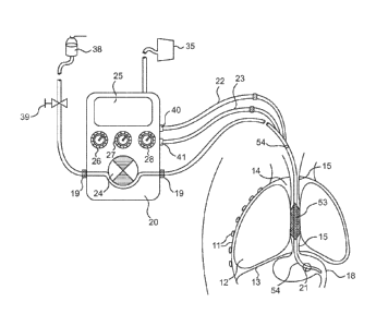
Fig. 1 is a general schematic representation of an embodiment of the
present invention as inserted in a silhouette outline of a patient's head,
torso and
upper abdomen with a diagram of a pump system according to an embodiment of
the present invention.
20
30
8b
CA 02694035 2010-01-19
WO 2009/027864
PCT/1B2008/052510
Fig. 2 is partial cut-away illustration of an embodiment of the esophageal
bladder device and feeding tube according to an embodiment of the present
invention.
Fig. 3 is a cross-sectional view of the device shown in Fig. 2, along line II-
11,
as it may sit in the esophagus.
Fig. 4 shows a perspective view of a shaped body shown in Figs. 2 and 3,
according to a first embodiment.
Fig. 5 shows a perspective view of a delivery cannula.
Fig. 6 shows a perspective view of a disclosed shaped body according to a
second embodiment.
Fig. 7 shows a perspective view of a disclosed shaped body according to a
third embodiment.
Fig. 8 shows a schematic view of an alternative design for the ferrule.
Figs. 9 and 10 show variations of the design of Fig. 8.
DETAILED DESCRIPTIONS OF ILLUSTRATIVE EMBODIMENTS OF THE
INVENTION
The present invention describes a device and method, which effects a static
or dynamical, low irritating, long-term organ compatible and stationary seal
function within the esophagus, intending to interrupt the above described free
communication of secretions and gastric material between the upper respiratory
tract and the gastro-intestinal tract.
Referring to Fig. 1, which schematically illustrates a cross-section of part
of
a patient's torso, the patient's chest cavity wall 11, lungs 12, diaphragm 13,
intra-
thoracic space 14, esophagus 15, and stomach 18 are depicted. Also depicted in
Fig. 1 is a presently preferred embodiment of an anti-gastro-esophageal reflux
device for use during enteral feeding as it may operate in situ in a patient's
thorax
in combination with a feeding pump function/unit. As schematically illustrated
in
Fig. 1, an embodiment of a seal system includes a combination of a gastric
tube 54
inserted through the nasal or oral cavity, passing through the esophagus 15,
and
terminating in the stomach 18. The oro/naso-gastric tube 54 has a pressure
sensing balloon 21, which alternatively can be provided by an electronic
pressure
sensing element 21, situated near the end of the tube's tip that is situated
in the
stomach 18. This gastric balloon/sensor 21 is connected to a respective
9
CA 02694035 2010-01-19
WO 2009/027864 PCT/1B2008/052510
filling/communication line 23. Proximal of the gastric sensor balloon 21 is
situated
an esophageal sealing bladder 53 with a filling line 22 along or integrated in
the
shaft of the naso-gastric tube 54.
As schematically shown in Fig. 1, in accordance with a presently preferred
embodiment of the invention, a decompression/feeding tube 54 can be specially
designed for combined use with a sensing and regulating device 20, which is
configured to receive signals from one or more pressure sensors and is
configured
to regulate the seal force in the esophagus 15 according to the sensed
pressure(s). As schematically shown in Fig. 1, the control device 20 can be
integrated with a feeding pump 24, such as a roller pump 24, or similar
mechanism
used in gastric feeding pumps for delivering feeding solution from a reservoir
38
via a tube segment 19 to the patient's stomach 18. The combination provides
the
benefit of a regulated reflux-preventive esophageal seal 53, especially suited
for
the requirements of enteral feeding of a critically ill patient.
The control device 20, which desirably is configured to receive and process
signals from pressure sensor 21 and to regulate the seal force exerted by the
bladder 53 on the wall 16 of the esophagus 15, desirably can include
mechanical
pump/pumps, pressure transducers, analog-digital-converters, and a
logical/control
unit such as a programmable logic controller and/or a programmable
microprocessor.
The control device 20 desirably can be configured to continuously monitor
and optionally display the actual intra-gastric pressure sensed by sensor 21
and to
regulate the inflation pressure of the esophageal seal bladder 53 so as to
ensure a
user determined pressure gradient (AP) between the sealing esophageal bladder
53 and the pressure inside the stomach 18 to seal against. As schematically
shown in Fig. 1, the control device 20 or regulator mechanism can be provided
with a display 25 for feedback from sensors and other parameters. The display
25
can be configured to provide a visual display of the user determined pressure
gradient between esophageal and gastric pressure (AP), the actual and desired
volume/unit time (V/h) of nutrient to be fed to the patient, the esophageal
pressure
(Pesophagus) sensed by the seal bladder 53, and the gastric pressure
(Pgastric) sensed
by the gastric sensor 21.
CA 02694035 2010-01-19
WO 2009/027864 PCT/1B2008/052510
The control device 20 or regulator mechanism can be provided with manual
controls for regulating the rate at which feeding solution is supplied to the
patient
and other parameters. As schematically shown in Fig. 1, the control device 20
can
be provided with a manual input mechanism 26 option that enables the user to
set
the magnitude of the desired pressure gradient AP. As schematically shown in
Fig. 1, the control device 20 can be provided with a manual input mechanism 27
for controlling the volume of nutrient to be fed to the patient, a manual
input
mechanism 28 for controlling the delivery time during which nutrient is to be
fed to
the patient, and a manual input mechanism 39 for controlling the connection of
the
system to a feeding container 38 that contains the feeding solution.
By continuously adding the user determined seal pressure gradient (AP) to
the actual intra-gastric pressure detected by sensor 21, the force exerted by
the
esophageal seal 53 against the esophageal tissue 16 can be continuously
reduced
to the required minimum and thus reduce accordingly the likelihood of pressure
induced trauma that otherwise might be caused by continuous, inappropriately
high seal pressures. If the level of intra-gastric pressure is relatively low,
then the
esophageal seal force and trans-murally effected force is commensurately
relatively low. If the level of gastric pressure increases, then the
esophageal seal
pressure only is increased by a gradient (AP), which can be determined by the
user as being sufficient for reflux prevention. Stationary, high seal pressure
gradients that exceed the actually required seal force thus can be prevented.
Alternative to a continuous adjustment of esophageal seal pressure to
actual intra-gastric pressure, the addition of the user determined seal
pressure
gradient (AP) to the actual intra-gastric pressure can be performed
intermittently
within time intervals that can be pre-set or fixed by the user in the control
device 20
as by a manual input mechanism 28 for controlling the time interval for
feeding
nutrient to the patient or determined by a manual mode, whereby the user
determines the addition of the seal gradient (AP) to the gastric pressure by
e.g.
manually entering the desired seal gradient AP, which remains effective till
the
manual adjustment is repeated.
Integrated into or connected to a feeding pump 24, the control device 20 for
regulating the esophageal seal force can be configured to actively keep the
seal
pressure of the esophageal bladder 53 in dynamic accordance with the actual
11
CA 02694035 2013-09-04
intra-gastric pressures reached under ongoing and post-gastric feeding, so
that a
seal-sufficient pressure gradient (AP) between intra-esophageal pressure and
the
intra-gastric stomach pressure can be continuously maintained. The control
device 20 can be configured to control the feeding pump unit 24 to further
control
the relative feeding rate to a patient as a function of the gastric pressure
sensed
through the gastric pressure sensor 21, thereby preventing critical esophageal
seal
forces from being reached and feeding the nutrient under optimal pressure
conditions and/or during optimal feeding periods.
Algorithmic control
Analogous to a ventilation control technique, such as described in U.S.
Patent No. 7,040,321 B2, the present enteral feeding system also desirably can
use an algorithmic control for controlling the feeding pump. A possible
example of
such an algorithmic control could include the following. After placement of a
gastric probe 21 and activation of the system, the control device 20 can be
configured to pump a defined volume of filling fluid via filling line 23 into
the gastric
balloon 21 to fill the balloon, which is preferably smaller than the volume of
the
gastric balloon 21 in its freely inflated pre-shaped state. As schematically
shown in
Fig. 1, the control device 20 can be configured to operate a pump 41 connected
via filling line 23 to the gastric pressure sensing balloon 21 to fill the
balloon 21.
By inflating the gastric sensor balloon 21 partially, it remains in a floppy
non-
extended state, being able to respond to slightest changes of intra-gastric,
i.e.,
intra-abdominal pressure. Once the pressure within the balloon 21 reaches a
stable reading of the intra-gastric pressure (i.e., a mean pressure level
derived
through an averaging process), the control device 20 can be configured to
operate
a pump 40 connected via filling line 22 to apply the esophageal seal pressure
to
the esophageal seal tamponade 53 via filling line 22. The esophageal seal
pressure desirably can be regulated by the control device 20 on the basis of a
predetermined AP value that can be preset in the software of the control
device 20
and can be manually adjusted by a user via the input mechanism 26. The
esophageal seal pressure calculates as the gastric pressure (measured by the
gastric sensor 21) plus the AP value.
12
CA 02694035 2010-01-19
WO 2009/027864 PCT/1B2008/052510
As schematically shown in Fig. 1, the filling fluid for the sensing balloon 21
and the esophageal seal tamponade 53 can be supplied from a fluid reservoir
35,
which can hold a liquid or a gas, at room conditions or under pressure as the
case
may be.
Due to the particular membrane characteristics of the foil of the sealing
esophageal bladder 53, a hydrostatic pressure gradient of about 10 cm to about
20
cm of water above the actual gastric pressure is considered desirable to
produce a
reliable seal against passive reflux of gastric contents. Typically, a
hydrostatic AP
pressure of up to about 10 cm is employed.
As schematically shown in Fig. 1, the actual esophageal seal pressure to be
maintained in the esophageal seal bladder 53 can be constantly determined and
adjusted by the control device 20 that operates a pump 40 connected via
filling line
22. The control device 20 desirably is configured to derive this seal pressure
from
the actual intra-gastric pressure detected by the gastric balloon/electronic
sensor
21and the seal pressure gradient AP that has been set by the user via manual
input mechanism 26. In order not to exceed a pressure level in the esophageal
seal 53 that may cause tissue infarction and possibly cause ulcers, the
control
software employed by the control device 20 can be configured to contain a
preset
value P
esophagus-max defining a maximum seal pressure not to be exceeded by the
esophageal seal bladder 53.
The control device 20 desirably can be configured to permit the user to
enter via input mechanism 27 a desired volume of feeding solution to be
administered over a certain time period, whereby the duration of the delivery
interval of the volume of the feeding solution to the patient can be
separately
defined or entered by the user via manual input mechanism 28 as another of a
predefined set of parameters. The control device 20 can be configured to
calculate a constant flow rate that is able to deliver the desired volume of
feeding
solution over the desired delivery period. The control device 20 desirably can
be
configured to operate the patient's nutrient feeding pump according to several
modes, including the following examples.
- operation under constant flow:
This mode of operation calls for continuous adjustment of esophageal seal
pressure according to a user defaulted seal pressure gradient, following
operation
13
CA 02694035 2010-01-19
WO 2009/027864 PCT/1B2008/052510
of the feeding solution pump according to a machine calculated linear feeding
rate,
which is calculated to be able to deliver the desired volume of feeding
solution over
a desired time interval, automatically stopping of the feeding pump function
when
Pesophagus-max is reached, pausing of the feeding pump function till
Pesophagus has
dropped below Pesophagus-max., continuation of the feeding pump function
according
to the initially calculated feeding rate, till delivery of the desired total
fluid volume of
the feeding solution has been accomplished.
- operation under dynamically adjusting flow ¨ delivery volume
oriented:
This mode of operation calls for continuous adjustment of esophageal seal
pressure to try to maintain a user-preselected defaulted seal pressure
gradient A
Pgastric. The control device 20 is configured to perform a continuous or
intermittent
determination of A Pgastric over At (control software defined time intervals,
e.g., 3
minutes before and after the actual pressure value determination), linear
extrapolation of A Pgastric over At, in case the slope of extrapolated
pressure curve
Pgastric reaches P
= esophagus-max within At (or several At periods, or the total user
determined delivery period), a reduction of the feeding solution flow rate is
figured
and executed by the control algorithm, which is configured to lower the slope
of the
extrapolation sufficiently so as not to exceed P
= esophagus-max within At (or several At
periods, or the total user determined delivery period), dynamical extension of
the
feeding period till the desired total volume of feeding solution has been
delivered.
- operation under dynamically adjusting flow ¨ delivery time
optimized:
This mode of operation calls for continuous adjustment of esophageal seal
pressure according to a user-preselected defaulted seal pressure gradient,
continuous or intermittent determination of A Pgastric over At (control
software
defined time intervals, e.g. 3 minutes before and after the actual pressure
value
determination), linear extrapolation of slope (see above), if extrapolated
pressure
curve of P
= gastric does not reach P
= esophagus-max within At (or several At periods, or the
total user determined delivery period), successive increase of flow rate to
reach or
nearly reach P
= esophagus-max within At (or several At periods, or the total user
determined delivery period). Automatic stopping of the feeding pump function
is
effected when P
= esophagus-max is reached, the feeding pump function is paused till
14
CA 02694035 2010-01-19 .
WO 2009/027864 PCT/1B2008/052510
Pesophagus has dropped below P
= esophagus-max , the feeding pump function is resumed
according to the prior calculated feeding rate of the feeding solution, till
delivery of
the desired total fluid volume of the feeding solution has been accomplished.
-operation under dynamically adjusting flow ¨ delivery time optimized
and delivery volume oriented:
This mode of operation calls for operating according to the delivery time
optimized mode as described above utill P
= esophagus-max is reached, then changing to
the delivery volume oriented mode as described above.
Gravity-operated feeding control:
The feeding solution can be supplied using gravity instead of by a
mechanical pump. When the feeding process is gravity driven, the process can
be
controlled by an electronic occlusion element (not shown) that interrupts or
gradually controls the flow and amount of the delivered feeding solution. A
dripping chamber (not shown) can be integrated into a feeding line 19, and an
optical detection device (not shown) can be used to detect and count the
number
of drops of feeding solution entering such chamber in order that the flow and
volume of feeding solution can be detected and used to control the occlusion
element. Thus, the above suggested control algorithms can be used in a manner
similar to the computer program-assisted control described above.
ITP as a parameter
By inflation of the esophageal bladder 53, the gastric probe 54 that can be
introduced into the esophagus 15 is placed against the surface of the wall 16
of the
esophagus 15, which in its middle portion and even better in its lower third
transmits the pressure course inside the thorax through the wall 16 of the
esophagus 15 (transmurally) to the esophageal placed bladder 53 of the gastric
probe 54. The inter-transmural pressure (ITP) that is transmurally transmitted
through the wall 16 of the esophagus 15 is detected by this bladder 53 and
becomes a measured value that can be used as a control signal indicative of
the
pressure inside the esophagus 15 and that can enable the user to detect and
monitor chest movement activity of the patient.
Probe design requirements:
The outer diameter of the delivery cannula 54 is advantageously between
about 3 mm and about 6 mm, and especially between about 4 and about 5 mm. In
CA 02694035 2010-01-19
WO 2009/027864 PCT/1B2008/052510
the interior of the delivery cannula 54, in addition to a nutrient channel 61,
through
which liquid nutrients are delivered to the patient, there is a delivery
channel 62,
via which the inflatable bladder 53 can be filled with a fluid, whether
gaseous or
liquid.
The performance of the device and the method, to prevent gastric content
from ascending into a patient's pharynx via the esophagus 15, further depends
on
the specific design and a particular performance of the esophageal sealing
bladder
53. To prevent pressure-induced esophageal lesions, the present invention
describes a low-pressure bladder tamponade /occlusion of the esophageal organ
lumen. Next to the prevention of pressure induced esophageal lesions, the
esophageal sealing bladder 53 must be configured to meet the requirements of
permanent placement inside the esophagus' highly dynamic structure that is
constantly in movement and changing cross-sectional mucosal folding and shape.
On account of these difficulties, the search for a simple designed intra-
esophageal
bladder seal, which is atraumatic, not irritating, withstanding peristaltic
movement,
and effecting a sufficient mechanical separation of airway and digestive
tract, could
not until now be satisfactorily resolved. The functional features of the
invented
bladder equipped decompression probe described in the invention meet such
requirements.
Residual bladder
The diameter of the inflatable bladder 53 in a freely unfolded condition is
between about 20 mm and about 50 mm. A diameter of about 30 mm to about 40
mm is particularly desirable for the diameter of the inflatable bladder 53 in
a freely
unfolded condition. The tampooning bladder 53, when freely inflated to its
full pre-
shaped dimension, has a larger diameter than that of the expected distended
esophagus 15. Hence, as schematically shown in Fig. 3, the sealing bladder 53
includes a residual volume 58 that is able to engage with the ridges and
pleated
lining of the esophagus without separating from contact with the pre-shaped,
undistended dimensions of the esophageal wall 16. As schematically shown in
Fig. 3, the residual diameter of the tampon bladder 53 further creates a
number of
reserve interpleatings 43 along its surface in order to ensure that the
pleated
lumen of the esophagus can be securely covered by the bladder hull over its
entire
circumference without having to distend or stretch the bladder material in
order to
16
CA 02694035 2010-01-19
WO 2009/027864 PCT/1B2008/052510
effect an organ lumen obstruction. Due to the prevention of any stretch of the
bladder hull, the pressures inside the bladder 53 needed to effect the desired
sealing therefore can be kept low, in the ideal case only slightly exceeding
intra-
luminal organ pressure by a few millibars (cm H20), enabling a fluid seal at
filling
pressures that can be kept below perfusion relevant trans-mural forces, and
enabling the user to set the barometrically measured pressure inside the
bladder
53 equal to such effected trans-mural forces.
Bladder thickness
In order to meet the various design requirements on an atraumatic sealing
intra-esophageal bladder 53 in the best possible way, the bladder 53 ideally
is
preferably made from microthin-walled, easily pliable plastic film with a wall
thickness of less than or equal to about 0.03 mm. The seal bladder 53 is
subjected
to a fill pressure of less than or equal to 30 mbar, being set ideally within
a
pressure range of about 10 mbar to about 20 mbar, which are pressures that are
known to be non-critical for tissue perfusion, and granting a sufficient
degree of
compatibility to the motility of the esophagus 15. The bladder 53 can be made
of
blow-moulded, foil-welded, or dipped material. The bladder 53 can be made from
polyurethanes, polyethylenes, silcone, natural and synthetic rubbers,
polyvinylchloride, or other materials offering adequate pliability and
stability in the
required foil thickness range.
Bladder length:
The membrane forming the esophageal bladder 53 is ideally sized to cover
the entire length of the esophagus. The bladder body preferably is sized so
that it
can extend between the upper and the lower esophageal sphincter. In most
embodiments, the tampon-bladder 53 usually has a length of about 6 cm to about
15 cm, desirably about 6 cm to about 9 cm.
Adjacent organs
Further, the invention considers immediately adjoining structures such as
the great vessels, the accompanying nerves, the trachea and main bronchi, the
lungs 12 themselves and, not least, the heart, particularly the left atrium.
In
contrast to conventional blocking techniques, the invented reflux-sealing
esophageal probe does not endanger such structures due to perfusion or tissue
critical pressures effected by the permanent pressurized bladder seal element
53.
17
CA 02694035 2010-01-19
WO 2009/027864 PCT/1B2008/052510
Filling media
Different fluids may be used as the medium for filling the esophageal seal
bladder 53, depending on the application. A presently preferred bladder
filling
medium, which is distinguished by compressibility as well as a certain
adaptability
of its own to the fluctuations mentioned below is, for instance, a gaseous
one. Air
is a presently preferred gas to provide the fluid medium for filling the
esophageal
sealing bladder 53, and gas mixtures can be used. However, a liquid medium for
filling of the esophageal seal bladder 53 is possible and viscous liquids,
water, or
gas/liquid mixtures such as air and water, can be used.
Shift of the bladder filling medium during peristaltic, lengthwise
directed contraction of the esophagus (swallowing):
Desirably, the invented probe 50 can be equipped with a special
mechanism, which permits an intra-bladder shift of the bladder filling medium
within the esophageal sealing bladder 53, giving the device the required
ability to
remain stationary in the location where it is placed and preventing a
transport of
the bladder equipped probe 50 towards the stomach and/or preventing patient
irritating pressure peaks (bolus sensations) being generated in the esophagus
by
the filling medium accumulating in the lower portion of the seal bladder 53,
below
the peristaltic contraction wave. As schematically shown in Fig. 3, within the
segment of the probe 50 that carries the bladder 53, the device can include a
second lumen 62 that is disposed next to the drainage or decompression lumen
61. As schematically shown in Fig. 2, the drainage lumen 61 can be arranged
relative to the second lumen 62 in a manner such that a channel 55 is formed
between the interior 58 of the bladder 53 and the second lumen 62. The second
lumen 62 can be positioned relative to the interior 58 of the bladder 53 by
means
of dividing fixtures or baffle-like structures that bridge the passageway
defining the
channel 55.
As schematically shown in Fig. 2, a conduit 52 that is disposed around the
feeding tube 54 can be configured to channel the air or other gaseous medium
filling the esophageal bladder 53 so as to be redistributed with each wave of
a
peristaltic contraction from the bladder portion 60 that is disposed below the
peristaltic wave into the bladder portion 59 that is disposed above and
already
released from the peristaltic wave. In this way, an intra-bladder shift of the
filling
18
CA 02694035 2010-01-19
WO 2009/027864 PCT/1B2008/052510
medium is effected to accommodate the peristaltic wave imposed on the
esophagus. As shown in accompanying Figs. 4-7 for example, the described
particular tube shaft profile within the bladder carrying tube segment
facilitates the
volume shift that prevents undesired pressure increases in the tamponade 53,
pressure increases that otherwise could pose a painful irritation of the
patient.
The inner cavity 58 of the tampon-bladder 53 may be filled with a medium,
through a delivery channel 55 lying between the delivery lumen 62 and the
inner
cavity 58 of the tampon-bladder 53, from a filling line 22 connected to the
channel
55 via the delivery lumen 62. As schematically shown in Fig. 1, simply
operated
examples of such a filling device are a reservoir or equalizing vessel 35,
particularly one situated outside the patient and connected via filling line
22. A
supply of the filling medium sufficient to fill the inner cavity of the tampon-
bladder
53, and in addition to allow for the abovementioned functional fluctuations of
the
lumen and the tonus of the esophageal wall 16 through further outflow or
intake of
the medium by expansion and collapse of the tampon-bladder 53, is kept in the
reservoir or equalizing vessel 35.
In this connection it could be seen as an additional advantage for the
bladder filling medium to be actively led into the inner cavity 58 of the
tampon-
bladder 53 or withdrawn from the inner cavity through the channel 55. Such
active
supply and withdrawal desirably can take place through a pump 40 that is
operated
by the control device and that is regulated preferably to compensate for any
extensive pressure-passive fluctuations in the tampon-bladder 53.
Stomach probe, volume shift mechanism, advanced profiles:
Fig. 2 illustrates the basic construction of an embodiment of an anti-gastric
reflux esophageal-stomach probe 50 according to the present invention. A
shaped, conduit body 52 is superimposed around and over a delivery cannula 54
in the region of an inflatable bladder 53. The conduit body 52 encloses a
lumen 55
in its interior. The lumen 55 also is shown in the view of Fig. 3, which
represents
the cross section II-II through the stomach probe shown in Fig. 2. In this
example
of the embodiment, the lumen 55 is located between the delivery cannula 54 and
the surface 56 of the conduit body 52.
As can be seen in Fig. 2, several openings 57 defined through the surface
56 of the shaped body 52 and desirably are distributed over the entire surface
56
19
CA 02694035 2010-01-19
WO 2009/027864 PCT/1B2008/052510
of the shaped body 52. The lumen 55 is connected to the interior 58 of the
inflatable bladder 53 via the openings 57. This means that the openings 57 are
configured and disposed to permit volume or fluid exchange between the lumen
55
and the interior 58 of the inflatable bladder 53.
Fig. 4 shows an enlarged image of the disclosed shaped body 52 as shown
in the first embodiment of the invention shown in Fig. 2 wherein the shaped
body
52 has an almost cylindrical external shape. The number and shape of the
openings 57 defined through the surface 56 of the shaped body 52 may vary,
depending on the end use. In addition to the approximately round or oval
openings 57 shown in Figs. 2 and 4 for example, the openings 57 may also be
elongated, for example. The shape or contour of the openings 57 may vary from
being a largely round or oval cross-sectional profile, to triangular,
quadrangular or
polygonal shaped openings 57. Nor must the openings 57 be distributed more or
less evenly over the surface 56 of the conduit body 52 as in the embodiment
shown in Figs. 2 and 4. Alternatively, the openings 57 may also be distributed
unevenly. In this case, it is important that the shape and arrangement of the
openings 57 permit adequate volume exchange between two sections, 59 and 60,
of the inflatable bladder 53. The number of openings 57 may vary from one to
any
number of individual openings, e.g. 100 or 1000 openings. The number of
openings 57 is restricted only by the area of the surface 56 of the conduit
body 52
and the shape of the openings 57.
In one embodiment of the invention, the cross section of the shaped body
52 may have several wall sections 64. As shown in Fig. 4 for example, several
wall sections 64 extend radially from the cylindrical surface 56 of the shaped
body
52 into the interior of the shaped body 52. The free, front ends 65 of the
wall
sections 64 define a diameter, which corresponds approximately to the outer
diameter of the delivery cannula 54 and which are supported at the delivery
cannula 54 of the probe 50 and, together with it, define at least one section
66 of
the lumen 55. As shown in Fig. 3 for example, when the shaped body 52 is
located on the delivery cannula 54, the front ends 65 of the wall sections 64
rest on
the delivery cannula 54. The wall sections 64 may extend in a roughly star-
shaped
configuration into the interior of the shaped body 52. This arrangement
CA 02694035 2010-01-19
WO 2009/027864 PCT/1B2008/052510
guarantees an approximately even distribution of the wall sections 64 and in
turn
guarantees secure support and retention of the shaped body 52.
As shown in Fig. 3, together with the delivery cannula 54, the lumen 55
inside the shaped body 52 can be divided into separate lumen sections 66. A
single lumen section 66 is delimited by two wall sections 64, the portion of
the
surface of the shaped body 56 which lies between the two wall sections 13 and
the
portion of the surface of the delivery cannula 54 which is located between the
contact surfaces of the front ends 65 of the wall sections 64. In this example
of the
embodiment shown in Figs. 2, 3 and 4, the shaped body 52 has eight wall
sections
64, which all extend in a finger-like manner by roughly the same amount into
the
shaped body 52. These wall sections 64 can form a passageway with their front
ends 65, whose dimensions correspond approximately to those of the delivery
cannula 54. The shaped body 52 can therefore be mounted easily onto the
delivery cannula 54.
In other embodiments of the invention, the number of wall sections 64,
however, may vary arbitrarily, and thus influence the shape of the lumen 55 or
the
individual lumen sections 66. The depth to which the wall sections 64
penetrate
into the interior of the shaped body 52 also may vary, and this depth
determines
the position of the shaped body 52 in relation to the delivery cannula 54.
Depending on the particular application, the longitudinal axis 36 of the
shaped
body 52 may also be displaced in relation to the longitudinal axis 37 of the
delivery
cannula 54. This means that the shaped body 52 need not necessarily sit more
or
less concentrically on the delivery cannula 54 as in the embodiment shown in
Figs.
2, 3 and 4 where the longitudinal axis 36 of the shaped body 52 coincides with
the
longitudinal axis 37 of the delivery cannula 54.
In the region of the axial front side of the shaped body 52, the lumen 55
may expediently be connected to a delivery channel 62, via which the
inflatable
= bladder 53 can be filled with a fluid. In this embodiment shown in Figs.
2 and 3,
the delivery channel 62 for the filling fluid extends, at least in parts, into
the conduit
body 52 and has at least one access opening 51, which connects the delivery
channel 62 to the lumen 55 and joins the lumen 55 with the interior 58 of the
inflatable bladder 53. The access opening 51 guarantees good volume
equalization between the sections of the inflatable bladder 53, and can also
be
21
CA 02694035 2010-01-19
WO 2009/027864 PCT/1B2008/052510
produced using simple techniques. The access opening 51 may extend over
roughly the entire length of the shaped body 52. As schematically shown in
Figs. 2
and 3 for example, the shaped body 52 may have at least one access opening 51
which extends in roughly the longitudinal direction of the shaped body 52 over
at
least 50 to 60%, preferably over up to 70%, and especially over up to 80%, of
the
total length of the shaped body. This arrangement can be produced using simple
techniques and simplifies the construction of the stomach probe 50, since the
inflatable bladder 53 can be filled directly via the lumen 55 with which it is
connected.
In embodiment shown in Figs. 2 and 3, the access opening 51 runs radially
in relation to the shaped body 52. The access opening 51 of the delivery
channel
62 need not necessarily run radially, but may also run in the region around
the
axial front surface of the shaped body 52 rather than axially to the shaped
body 52.
In other embodiments of the disclosed stomach probe 50, the delivery channel
62
also may run along the outside of the delivery cannula 54. As shown in Fig. 5,
the
delivery channel 62 may, for example, be located, at least partly, in an
indentation
63, which runs along the delivery cannula 54.
Figs . 6 through 10 show perspective views of further embodiments of the
disclosed shaped body 52. Figs. 6 and 7 show second and third embodiments of
the disclosed shaped body 52. The reference numbers used in Figs. 2 through 5
refer to the same components as those in Figs. 6 and 7.
As shown in Figs. 6 and 7, each shaped body 52 can have a central,
roughly tubular structure 68, with a roughly circular transverse cross
section. The
inner diameter of the shaped body 52, as well as the contact surface between
the
shaped body 52 and the delivery cannula 54, are formed by the tubular
structure
68. As shown in Fig. 7, the shape of the inner cover surface 69 roughly
corresponds to the shape of the surface of the delivery cannula 54. As shown
in
Figs. 6 and 7, several wall sections 70 extend radially outwards from the
central,
tubular structure 68. At the outermost end 71 of each wall section 70 lying
opposite to the central, tubular structure 68 is a surface 72, which runs
roughly
transversely to the wall section 70.
In the embodiment of Fig. 6, the shaped body 52 has four wall sections 70
arranged roughly in a circle. The wall sections 70, together with the
associated
22
CA 02694035 2010-01-19
WO 2009/027864 PCT/1B2008/052510
surfaces 72, form an approximately T-shaped profile in the cross section. This
T-
shaped profile can be produced easily, and provides a lumen 55 of sufficient
size,
as well as a good contact surface for the inflatable bladder 53. In the
embodiment
of Fig. 7, the shaped body 52 has five wall sections 70 arranged in an
approximate
star-shaped configuration around the tubular structure 68. In the embodiment
of
Fig. 7, the wall sections 70, together with their respective transverse
surfaces 72,
form a roughly L-shaped profile in cross section. This L-shaped profile can
also be
produced using simple techniques, and provides for a lumen and contact surface
that permits rapid volume exchange between the sections of the inflatable
bladder
53.
The T- and L-shaped profiles of the shaped bodies 52 shown in FIGS. 6 and
7 are located at such a distance from each other, or are dimensioned in such a
way, that the transverse surfaces 72 of two adjacent T- or L-shaped profiles
are at
a distance from each other. This means that every two of the transverse
surfaces
72, which define the surface 56 of the shaped body 52, define an opening 73 or
slit, which runs along the length of the shaped body 52. In these examples of
the
embodiment shown in FIGS. 6 and 7, the lumen 55, which is located here between
the transverse surfaces 72 and the tubular structure 68, is divided by the T-
shaped
profiles or L-shaped profiles into separate lumen sections 66. The shape of an
individual lumen section 66 is thereby determined by in each case two adjacent
T-
shaped profiles or L-shaped profiles and the portion of the surface 56 of the
tubular
structure 68 enclosed by them. The number of wall sections 70 may be varied,
depending on the end use. If this end use changes, the shape and the number of
lumen sections 66 and openings 73 in the surface 56 of the shaped body 52 also
desirably change.
In a further embodiment of the invention, the wall sections 70 may also be
arranged unevenly around the tubular structure 68, unlike the examples shown
here. The transverse surfaces 72 at the ends 71 of the wall sections 70 also
can
be dispensed with in some embodiments. In this case, the surface 56 of the
shaped body 52 is determined by the ends 71 of the wall sections 70. The
number
of wall sections 70 may be increased accordingly, and there may be between
about 5 and about 15 wall sections 70, for example.
23
CA 02694035 2010-01-19
WO 2009/027864 PCT/1B2008/052510
The abovementioned first through fourth embodiments of the disclosed
shaped body 52 in Figs. 2 ¨ 7 also can be twisted, rather like a screw, and
thus
can be shaped like a coil.
FIG. 8 shows a further embodiment of the disclosed shaped body 52 in the
form of a spiral that is formed as a coil 74. The inner diameter of the coil
74
corresponds approximately to the outer diameter of the delivery cannula 54. In
this
embodiment, the lumen 55 also has a spiral shape. In use, that is when the
shaped body 52 is located on the delivery cannula 54, as shown in FIG. 8, the
coil
74 is defined by a plurality of consecutive windings 77. Each winding 77 of
the coil
74 helically wraps once completely around the delivery cannula 54. As shown in
FIG. 8, an opening 33, which runs spirally around the delivery cannula 54, is
defined between the individual windings 77 of the coil 74 and encloses the
lumen
55. The thickness of the coil 74 determines the height of the lumen 55. The
coil
74 may have a roughly circular cross section. However, alternatively, the
cross
section of the coil 74 may have an oval shape or angular shape.
With the coil 74 of the shaped body 52 shown in FIG. 8, the inner diameter
of the shaped body 52 is determined by the inner diameter of the coil 74. The
contact surface between the shaped body 52 and the delivery cannula 54
corresponds, in this case, to the spiral installation line or surface of the
individual
windings 77 of the coil 74. Whether it is in the form of a line or a planar
configuration will be determined by the cross section of the coil 74.
In addition to single or interconnected coils, a pipe-like or tubular
structure
also can be applied. As shown in Fig. 8 by a line consisting of a sequence of
dots
and dashes, pipe-like or tubular structure can have openings. The external
shape
of this type of shaped body 52 would then be similar to the shaped body shown
in
Fig. 2.
In a further embodiment, the lumen 55 may also be defined by several coils,
for example two coils, which are roughly concentrically disposed so that the
one is
on top of, i.e., surrounding, the other. In this case, the two coils may have
the
same gradient or different gradients. The coils also can be superimposed so
that
each one runs in opposite direction to the other one. In this case, the lumen
55 is
defined by the intermediate space between the individual windings of the
relevant
coil, i.e., by the overlapped sections of these intermediate spaces.
24
CA 02694035 2010-01-19
WO 2009/027864 PCT/1B2008/052510
Fig. 9 shows another embodiment of the disclosed shaped body 52 that is
pipe-like or tubular in shape and has a net-like construction 25. The inner
diameter of the shaped body 52 corresponds approximately to the outer diameter
of the delivery cannula 54. As shown in Fig. 9, the net-like construction 75,
the
inner diameter of the shaped body 52 and the contact surface between the
shaped
body 52 and the delivery cannula 54 are determined by the individual
connecting
pieces 78 of the net-like construction 75. In this embodiment shown in Fig. 9,
the
lumen 55 is located within the mesh or openings 76 of the net-like
construction 75,
which are at least partly connected to each other, and thus permit volume
exchange between the individual openings 76 of the net-like construction 75.
In a further embodiment of the invention, the shaped body 52 may also
comprise several layers of the net-like construction 75, as Fig . 10 shows.
These
layers of the net-like construction 75 are arranged roughly concentrically in
relation
to each other, whereby the inner diameter of the innermost layer corresponds
approximately to the outer diameter of the delivery cannula 54. In this
embodiment, the lumen 55 is defined by the holes 76 in the net-like
construction
75, which overlap at least in parts. This means that the overlapping holes 76
of the
individual layers of the net-like construction 75 form channels or individual
lumen
sections 66. When the shaped body 52 is in the state it is in during use,
i.e., when
the shaped body 52 is located on the delivery cannula 54, at least part of the
lumen section 66 extends at least in sections along the delivery cannula 54,
and
thus permits volume exchange between the individual sections of the inflatable
bladder 53. This net-like construction can be produced efficiently and can be
premounted onto the coil, and so can simplify assembly.
The dimensions of the different embodiments of the shaped body 52
described here may vary, depending on the end use. In practice, however, an
approximate length of about 6 cm to about 12 cm, and especially a length of
about
6 cm to about 9 cm, has proved to be particularly advantageous for the shaped
body 52. They provide a sufficiently large contact surface for the inflatable
bladder
53. At the same time, an adequate volume exchange between all the sections of
the inflatable bladder 53 is guaranteed. The outer diameter of the shaped body
52
also depends on the end use, as well as on the dimensions of the delivery
cannula
54 and the inflatable bladder 53, and is advantageously in the region of
between
CA 02694035 2010-01-19
WO 2009/027864 PCT/1B2008/052510
about 7 mm and about 12 mm, and especially between about 6 mm and about 8
mm. These dimensions guarantee good volume exchange between the sections
of the inflatable bladder 53. However, for special end uses, the dimensions of
the
shaped body 52 may deviate from the abovementioned dimensions.
The inflatable bladder 53 is filled with a fluid, e.g., water, via the
delivery
channel 62, whereby the fluid flows through the access opening 51 of the
delivery
channel 62 into the lumen 55 of the shaped body 52. The fluid flows into the
interior 58 of the inflatable bladder 53 through the openings 57, 73, 76 and
33 of
the shaped body 52. As the inflatable bladder 53 fills with the fluid, the
inflatable
bladder 53 expands until at least a portion of its exterior surface lies
almost
completely against an uninterrupted annular portion of the wall 16 of the
esophagus 15, as can be seen in Fig. 3. This enables the esophagus to largely
be
sealed off from liquids or solid substances, which tend to move up from the
region
of the stomach 18 towards the pharyngeal cavity, and thus to keep the windpipe
free from harmful substances.
The swallowing motions made by the patient who has been fitted with the
disclosed stomach probe 50 cause the muscles to contract along the wall 16 of
the
esophagus 15. These muscles create one or usually several annular
constrictions
in the esophagus 15, which are propagated along the esophagus 15 from the
larynx region towards the stomach 18.
In order to illustrate the functions of the shaped body 52, the movement of a
single, annular constriction will now be examined. In the area around the
inflatable
bladder 53, the annular constriction in the esophagus causes a partial
reduction in
the outer diameter of the inflatable bladder 53, i.e., a local narrowing 31 of
the
inflatable bladder 53 occurs, which is shown in Fig. 2 as a dashed line. This
narrowing 31 divides the inflatable bladder 53 into two sections, 59 and 60.
While
the esophageal constriction is imposed as a wave that moves along the
inflatable
bladder 53 as when swallowing occurs, the dimensions of the individual
sections,
59 and 60, change. In this case employing the probe 50 of the present
invention,
however, the volume of fluid that can be contained in the relevant sections,
59 and
60, of the inflatable bladder 53, also changes. The disclosed shaped body 52
provides a lumen 55, which permits rapid volume exchange between the
individual
sections, 59 and 60, of the inflatable bladder 53. The surface 56 of the
disclosed
26
CA 02694035 2010-01-19
WO 2009/027864 PCT/1B2008/052510
shaped body 52 provides, if necessary, a relatively rigid contact surface for
the
constricted wall section 31 of the inflatable bladder 53. The lumen 55 is
therefore
kept free of these external influences, and is available entirely for volume
exchange. As schematically shown in Fig. 2, while the constriction 31 moves
along the inflatable bladder 53 in the direction of arrow 30, the fluid is
forced out of
the interior 58 of the second section 60 of the inflatable bladder 53 via the
openings 57 beneath the second section 60 of the inflatable bladder 53, and
the
fluid is forced into the interior 58 of the first section 59 of the inflatable
bladder 53
via the openings 57 beneath the first section 59 of the inflatable bladder 53.
A stomach probe of the type disclosed in German Utility Model Application
No. 202006002832.3 has been improved in the present disclosure. In accordance
with the present invention, the lumen 55, which is located between the
delivery
cannula 54 and the inflatable esophageal seal 53 and which is connected to the
interior 58 of the inflatable esophageal seal 53, can be produced by a
relatively
simple technique, and at the same time guarantees adequate volume equalization
between the partial volumes of the inflatable esophageal seal 53.
The separate shaped body 52 of the stomach probe 50 can be produced by
a simple technique, since it can be prefabricated as a separate component. The
shaped body 52 described above is preferably made from plastic and is produced
desirably by an extrusion process. This manufacturing process enables the
shaped body 52 to be produced by a relatively simple and quick technique.
Alternatively, the shaped body 52 may be produced by casting or injection
molding.
In principle, the materials used for the shaped body 52 are ones that can
deform easily to suit the human body, i.e., they do not injure the patient
whilst
being inserted or during long-term use of the probe, but they are rigid enough
to
provide a non-collapsible shape when peristalsis occurs over the shaped body
52.
Advantageous materials are, for example, PVC, FUR, blends of PVC and FUR,
blends of FUR and polyamides, and/or silicones. These materials guarantee good
compatibility with the tissue of the patient. These materials can be shaped
easily
and thus reduce the risk of injury during introduction of the stomach probe 50
into
the patient, yet these materials are stable enough to maintain the lumen 55
during
peristalsis.
27
CA 02694035 2010-01-19
WO 2009/027864
PCT/1B2008/052510
During assembly of the stomach probe 50, the separate shaped body 52
desirably can be mounted as a finished component on the delivery cannula 54,
and attached to the delivery cannula 54. Applying the shaped body 52 to the
delivery cannula 54 determines the shape of the lumen 55 at the same time,
which
ensures that there is sufficiently rapid volume exchange between the sections
of
the inflatable esophageal seal 53. This configuration simplifies assembly of
the
stomach probe 50, since the number of individual processing stages needed to
produce the lumen 55 can be reduced. Such simplified assembly results in a
potential for reducing both time and costs when producing the stomach probe
50.
The shaped body 52 may have a tubular structure, whose internal shape
corresponds roughly to the external shape of the delivery cannula 54. The
tubular
structure enables the shaped body 52 to be attached roughly concentrically to
the
delivery cannula 54. These complementary shapes simplify the assembly process
for the disclosed stomach probe 50, as the shaped body 52 desirably can be
applied to the delivery cannula 54 by means of a sliding process. Since the
inner
diameter of the relevant shaped body 52 corresponds approximately to the outer
diameter of the delivery cannula 54, or is at least slightly smaller than the
outer
diameter of the delivery cannula 54, a slight press-fitting effect occurs
during
mounting of the shaped body 52 onto the delivery cannula 54. The resulting
static
friction fixes the shaped body 52 radially and axially onto the delivery
cannula 54
and guarantees axial and/or radial fixing of the shaped body 52 on the
delivery
cannula 54 of the stomach probe 50.
Alternatively, the shaped body 52 may also be fixed onto the delivery
cannula 54 by means of adhesion, e.g., by applying an adhesive at least on
part of
the contact surface between the shaped body 52 and the delivery cannula 54.
Alternatively, the shaped body 52 may also be fixed by material-bonding
whereby,
for example, at least part of the contact surface between the shaped body 52
and
the delivery cannula 54 is treated with a solvent. Solvent etching of the
shaped
body 52, and/or the delivery cannula 54, at least in part, guarantees good
bonding
of the two components. In principle, any possible combination of the above-
mentioned fixing techniques are feasible as a means of attaching the shaped
body
52 onto the delivery cannula 54.
28
CA 02694035 2010-01-19
WO 2009/027864 PCT/1B2008/052510
The final, assembled stomach probe 50 desirably can be used for treating
comatose patients, for example, who are unable to feed themselves. In this
application, the disclosed stomach probe 50, i.e., the delivery cannula 54 of
the
stomach probe 50, is inserted into the patient's esophagus, whereby the
section of
the stomach probe 50 that is fitted with the inflatable bladder 53 is located
above
the entrance to the stomach 18 in the esophagus 15. The presently preferred
length of the shaped body 52 of approximately about 6 cm to about 9 cm ensures
that the shaped body 52 fits well in the section between the upper and lower
sphincter of the esophagus.
To improve orientation, the stomach probe 50 may be fitted with at least one
radiopaque marker, such as a metal ring 67. The radiopaque marker 67 makes it
possible to check that the probe 50 is in the correct position by means of an
X-ray
image. The marker 67 facilitates positioning of the probe 50 in the patient
and acts
as a reference point to orientating organs, such as the diaphragm and/or
thyroid,
on the X-ray image of the thorax. As shown in FIG. 2, more than one marker 67
may be employed. These radiopaque markers 67 may be placed at the shaped
body 52, the delivery cannula 54 and/or the inflatable bladder 53.
29