Note: Descriptions are shown in the official language in which they were submitted.
CA 02694072 2010-01-19
Attorney Docket No.: STMCNZ00200
REMOVABLE DEVICE AND-METHOD FOR TISSUE DISRIIPTION -
CROSS-REFERENCE TO RELATED APPLICATTONS
[0001] This is a continuation-in-part of U.S. Patent Application Serial No.
10/454,846 filed June 4, 2003, which claims priority to U.S. Provisional
Patent
Application Serial No. 60/384,998 filed June 4, 2002, each of which is
incorporated
herein by reference in its entirety.
BACKGROUND OF THE INVENTION
i. Field of the Invention
[0002] The invention related to a device and method for extra.ction-of tissue
from
an enclosed body cavity.
ii. State of the Related Art
[0003] Bone Marrow is a rich source of pluripotent hematopoietic stem cells
from
which red blood cells, white blood cells, and platelets are formed. Bone
marrow also.
contains additional populations of inesenchymal stem cells and other stem and
progenitor
cells which have the potential to repair and regenerate other tissues.
[0004] Since the early 1970's bone marrow and hematopoietic stem cell
transplantation has been used to treat patients with a wide variety of
disorders, including
but not limited to cancer, genetic and autoimmune diseases. Currently over
60,000
transplants for a variety of indications are performed worldwide each year.
[00051 In autologous transplants, the patient has their own bone marrow
collected
prior to receiving high dose chemotherapy. Following high dose, myeloablative
chemotherapy, which kills the majority of the patients' marrow stem cells, the
stored
autologous marrow or hematopoietic stem cells purified or enriched from the
marrow are
infused, and serves to improve the patient's hematolymphoid system.
[0006] In allogeneic transplants bone marrow, or other sources of
hematopoietic
stem cells derived from a full or partially human leukocyte antigen (HLA)
matched
sibling, parent or unrelated donor is infused into the recipient patient and
following
1
CA 02694072 2010-01-19
Attorney Docket No.: STMCNZ00200
engraftment, serves to reconstitute the recipients hematopoietic system with
cells derived
from the donor.
[00071 Following myeloablative or non-myeloablative conditioning of a patient
with chemotherapy and/or radiation therapy, the marrow is regenerated through
the
administration and engraflrnent of hematopoietic stem cells contained in the
donor bone
marrow.
[0008] In addition to hematopoietic stem cells and hematopoietic progenitors,
bone marrow contains mesenchymal and other stem cell populations thought to
have the
ability to differentiate into muscle, myocardium, vasculature and neural
tissues and
possibly some organ tissues such as liver and pancreas. Research in
preclinical animal
studies and clinical trials suggest that bone marrow or some portion of the
cells contained
within marrow can regenerate tissues other than the hematopoietic system. This
includes
the ability for cells contained within the marrow to regenerate or fa.cilitate
repair of
myocardial tissue following a myocardial infarction, and in the setting of
congestive heart
failure as evident by improved cardiac function and patient survival.
[0009] Bone marrow derived stem cells also show evidence for their ability to
regenerate damaged liver and hepatic cells and portions of the nervous system
including
spinal cord. Additional organ systems including kidney and pancreas show
benefit from
bone marrow derived cells. Use of bone marrow and the stem cells contained
within bone
marrow may be of increasing clinical utility in the fature treatment of
patients.
Furthermore a patient's own marrow has multiple applications in orthopedic
procedures,
including but not limited to spinal fusions, treatment of non-union fractures,
osteonecrosis, and tissue engineering.
[0010] Stem cells utilized in transplantation are usually collected using one
of
two methods. In a first method known as a bone marrow harvest, bone marrow is
directly
accessed in and removed from the patient usually by multiple aspirations of
marrow from
the posterior ileac crest. The bone marrow harvest procedure is often
performed in the
operating room.
[0011] To perform a harvest of 500-1500 milliliters of marrow, multiple
separate
entries into the marrow cavity are required to in order to remove a sufficient
amount of
bone marrow. A bone marrow aspiration needle, such as a sharp metal trocar, is
placed
2
CA 02694072 2010-01-19
Attorney Docket No.: STMCNZ00200
into the marrow space through the soft tissue and the outer cortex of the
ileac crest. The
aspiration needle enters less than 2 cm into the marrow cavity. Negative
pressure is
applied through the hollow harvest needle, usually by the operator pulling on
an attached
syringe into which 5-10 ml of marrow is aspirated. The needle and syringe are
then
removed.
[0012] After removing the collected marrow, the aspiration needle accesses a
separate location on the ileac bone for another aspiration. This method of
inserting the
needle into the bone, removing the marrow, and removing the needle from the
bone is
performed on the order of 100-200 separate entries for an average patient to
remove a
volume of bone marrow required for transplantation.
[0013] Each puncture and entry into the marrow cavity accesses only a limited
area of the marrow space, and the majority of practitioners only remove 5-10
milliliters
of marrow with each marrow penetration. Pulling more marrow from a single
marrow
entry site otherwise results in a collected sample highly diluted by
peripheral blood.
[0014] The bone marrow harvest procedure requires general anesthesia because
the ileac crest is penetrated 100-300 times with a sharp bone marrow trocar.
Local
anesthesia is generally not possible given the large surface area and number
of bone
punctures required.
[0015] The donor needs time to recover from general anesthesia, and frequently
suffers from days of sore throat, a result of the endotracheal intubation tube
placed in the
operating room.
[0016] Pre-operative preparation, the harvest procedure, recovery from
anesthesia, and an overnight observation stay in the hospital following the
procedure
requires considerable time on behalf of the donor and the physician, and
similarly
additional expense. The cost of the procedure is often $10,000 to $15,000,
which
includes costs for operating room time, anesthesia supplies and professional
fees, and
post-operative care and recovery.
[0017] In addition to general operating room staff, the traditional bone
marrow
harvest procedure requires two transplant physicians. Each physician aspirates
marrow
from the left or right side of the ileac crest. The procedure itself usually
takes
approximately one and half hours for each operating physician.
3
CA 02694072 2010-01-19
Attorney Docket No.: STMCNZ00200
[0018] Many donors experience significant pain at the site of the multiple
bone
punctures which persists for days to weeks,
[0019] Traditional bone marrow aspiration inaurs a significant degree of
contamination with peripheral blood. Peripheral blood contains high numbers of
mature
T-cells unlike pure bone marrow. T-cells contribute to the clinical phenomenon
termed
Graft vs. Host Disease (GVHD), in both acute and chronic forms following
transplant in
which donor T-cells present in the transplant graft react against the
recipient (host)
tissues. GVHD incurs a high degree of morbidity and mortality in allogeneic
transplants
recipients.
[0020] In a second method to collect stem cells for transplantation,
mononuclear
cells are removed from the donor's pexipheral blood. The peripheral blood
contains a
fraction of hematopoietic stem cells as well as other populations of cells
including high
numbers of T-cells. In this procedure peripheral blood stem cells are
collected by
apheresis following donor treatment with either chemotherapy - usually
cyclophosphamide - or with the cytokine Granulocyte Colony Stimulating Factor
(GCSF). Treatment with cyclophosphamide or GCSF functions to mobilize and
increase
the numbers of hematopoietic stem cells circulating in the blood.
[0021] This collection method can be slow and time consuming. It requires the
donor to first undergo five or more days of daily subcutaneous injections with
high doses
of the cytokine GCSF prior to the collection. These daily injections can be
uncomfortable and painful and bone pain is a common side effect. Peripheral
blood stem
cells can not be obtained without this seven-plus day lead time.
[0022] Each day.of apheresis costs approximately $3,000 including but not
limited to the cost of the apheresis machine, nursing, disposable supplies and
product
.25 processing. The patient often has to come back on multiple days in order
to obtain an
adequate number of stem cells. Costs for the GCSF drug alone approximate
$6,000-
$10,000 depending upon the weight of the patient.
[0023] Given the multiple days required to collect adequate numbers of
hematopoietic stem cells, individual bags of peripheral blood product must
processed and
frozen separately. These bags are then thawed, and given back to the recipient
patient at
4
CA 02694072 2010-01-19
Attorney Docket No.: STMCNZ00200
the time of transplant. The volume, and chemicals contained in the product
freezing -
media can cause some complications, such as mild side effects, at the time of,
infusion.
[0024] Accordingly, there is a need for a minimally invasive, less expensive,
time-efficient bone marrow harvest procedure with minimal complications which
does
not require general anesthesia, offers fast recovery time, and does not cause
significant
pain to the bone marrow donor.
SUMMARY OF THE INVENTION
[0025] Devices and methods for manipulation and extraction of body tissue from
an enclosed body cavity are disclosed. The device can have a hollow
introduction or
entry cannula that can have a trocar. The introduction cannula and a core
element can
penetrate body tissue, such as the marrow space contained within the ileac. A
flexible
aspiration cannula can then be inserted through the introduction cannula into
body tissue
and can be advanced through the body cavity.
[0026] Within the aspiration cannula there may be a stylet (e.g., an
aspiration
stylet). The stylet can aid in advancing the cannula through the cavity. The
stylet can be
removed to facilitate extraction of body tissue through the aspiration
cannula.
[0027] The aspiration cannula can have inlet openings near the distal tip
through
which tissue is aspirated. At the proximal end of the aspiration cannula a
negative
pressure (i.e., suction) source can provide controlled negative pressure, for
example, to
increase the aspiration of tissue through the aspiiation cannula into a
collection reservoir.
The aspiration cannula can be withdrawn and positioned for multiple entries
through the
same tissue entry point, for example, followiag different paths through the
tissue space
for subsequent aspiration of more tissue.
[00281 A device or apparatus that can disrupt and aspirate bone marrow and/or
other tissue rapidly and for large volumes of cancellous bone (i.e., marrow)
from a target
bone such as the ileac, femur, humerus, other bone, or combinations thereof is
disclosed.
The target bone can be in vivo or in vitro.
[0029] The apparatus can include a lumen adapted to receive an elongated
aspiration cannula. Following entry through the bone wall, the aspiration
cannula may be
controlled to move in a linear or non-linear fashion within the marrow cavity.
The
5
CA 02694072 2010-01-19
Attorney Docket No.: STMCNZ00200
aspiration cannula, for example while moving non-linearly, can access a
majority of the -
bone marrow space through a single point of entry. Suction may be optionally
applied to
the aspiration cannula while accessing the marrow space to increase the
harvest of the
bone marrow or other aspiratable substances. The apparatus can also optionally
check for
a threshold amount of aspiratable substance obtained. Additionally, a
controller in the
apparatus can adjust the aspiration cannula or signal for the operator to
adjust the
aspiration cannula to enable further harvesting from the same bone wall entry
point, or in
the alternate from an alternative bone wall entry point.
[0030] As the devices and methods can access large volumes of marrow with
each catheter insertion, the devices and methods can be moved to directly
contact more of
the marrow space and aspirate a more concentrated, less diluted-aspirant. The
aspirated
bone marrow can be more concentrated in stem cells, for example, because the
device can
penetrate the pelvic cavity more broadly and thus the extracted material can
be less
diluted with blood drawn into the void created by the extraction. The
decreased numbers
of contaminating T-cells can lead to less Graft vs. Host Disease (GVHD) in
allogeneic
bone marrow recipients. Less total volume of bone marrow can be removed (e.g.,
as it is
more concentrated).
[0031] As mentioned, the harvest (i.e., aspiration, extraction) performed with
the
devices and methods disclosed herein can utilize one access point into the
marrow cavity
on one or both sides of the body to remove a minimal total volume of material
that is
highly concentrated. A marrow access site can be the anterior ileac crest
access site
which can be easy to locate and access on a broad array of patients (from thin
to obese)
and utilizing this access site can also reduce harvest time.
[0032] The method described herein can be performed by a single operator with
no operating room time, reduced support personnel, no anesthesiologist, and
can also be
performed with no significant lead or preparative time. The method can be
performed on,
among others, critically ill subjects, or bone marrow donors who could not
easily tolerate
multiple surface puncture wounds for rapidly obtaining marrow and/or stem
cells derived
from marrow for use in immediate or long-term follow-on therapeutic
interventions.
Furthermore, the devices and methods disclosed herein can aspirate bone
marrow,
6
CA 02694072 2010-01-19
Attorney Docket No.: STMCNZ00200
remove fat, aspirate blood and muscle, or combinations thereof, through a
single skin
(and bone, where applicable) puncture site into the tissue space (e.g., marrow
cavity).
[0033] The device and method disclosed herein can also control the
directionality
of the cannula enough within the marrow cavity such that the device can access
a
majority of bone marrow space in a single bone or marrow cavity in vivo
through a single
point of entry. Alternatively, the device and method can access multiple
diagnostic
samples of bone marrow from disparate sites within a single marrow cavity. The
device
and method can also have aspiration suction controlled to aspirate bone marrow
or fat, for
example.
[0034] The device can have an elongated cannula having a flexible length, a
hollow channel, a cannula first end and a cannula second end. The cannula
first end can
be open to provide fluid communication between the hollow channel and the
outside of
the cannula. Additionally, the device can include a motor which is rotatably
connected to
the cannula. The cannula may additionally include a tissue disruptor which is
attached to
or integral with the cannula, e.g., a whisk having a first end and a second
end where the
first end can be fixed to the cannula such that the whisk extends from the
cannula. The
second end can also be fixed to the cannula such that the whisk is configured
in a semi-
circular or closed loop configuration. The cannula can additionally include a
second
whisk extending from the cannula end.
[0035] The whisk can be configured to be rigid enough to substantially disrupt
a
first portion of a cancellous bone matrix when rotated by the motor to make
the first
portion removable from a surrounding portion of cancellous bone matrix while
remaining
flexible enough so as to inhibit or prevent its puncturing through cortical
bone
surrounding the body cavity.
[0036] To rotate the cannula and whisk, the motor can be configured to rotate
the
cannula at least at one operating speed from about 30 rpm to about 160 rpm.
The device
can include a mechanical transmission between the motor and the cannula to
transmit the
torque. A rotation-limiting resistor or slip-clutch in mechanical
communication with the
motor and the cannula may additionally be included to further control or limit
the rotation
of the cannula and whisk within the body space.
7
CA 02694072 2010-01-19
Attorney Docket No.: STMCNZ00200
[0037] A pump in fluid communication with the hollow channel through the
cannula may include an aspirant reservoir such that the hollow channel is in
fluid
communication with the reservoir. Additionally, an aspirant filter in fluid
communication with the hollow chaimel and the aspirant reservoir may also be
included
to filter out undesirable material or debris. An irrigant reservoir holding a
fluid, e.g.,
saline solution, may additionally be included for providing irrigation fluid
which may be
optionally perfused into the body space to facilitate tissue removal.
[0038] One method for removing cancellous bone or bone marrow from an in
vivo bone in a subject may entail inserting the tissue disruptor into a first
section of the
cancellous bone or bone marrow into a patient by inserting a flexible hollow
shatt into
the first section of the cancellous bone. Prior to or upon insertion into the
cancellous
bone or bone marrow, the cannula and tissue disruptor may be rotated withi.n
the patient
to disrupt the tissue matrix prior to or while optionally aspirating the
disrupted portion of
first section of cancellous bone or bone marrow into the cannula.
[0039] The method can also include re-positioning the flexible shaft into a
second
section of the cancellous bone and aspirating the disrupted tissue. The re-
positioning of
the flexible shaft into the second section of the cancellous bone can include
completely or
partially removing the tissue disruptor, e.g., a whisk-like disruptor, from
the body cavity.
[0040] Additionally, the method can include irrigating the tissue matrix with
a
solution that can have saline, anesthetic, analgesic, anti-inflammatory,
osteogenic powder
or slurry, or combinations thereof. The method can fiuther include f ltering
the aspirated
disrupted portion.
BRIEF DESCRIPTION OF THE DRAWINGS
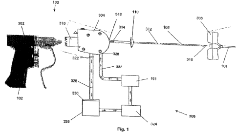
[0041] Figure 1 illustrates an exploded, partially schematic, view of a
variation of
the device for tissue disruption and aspiration.
[0042] Figure 2 illustrates an assembled, partially schematic view of a
variation
of the device for tissue disruption and aspiration.
[0043] Figure 3 illustrates an exploded, partially schematic, view of a
variation of
the device for tissue disruption and aspiration.
8
CA 02694072 2010-01-19
Attorney Docket No.: STMCN7,00200
[0044] Figure 4 illustrates an assembled, partially schematic view of a
variation
of the device for tissue disruption and aspiration.
[0045] Figure 5 is a see-through view of a variation of the entry cannula with
the
core element.
[0046] Figure 6 is a see-through view of a variation of the aspiration
cannula.
[0047] Figure 7 illustrates a variation of the aspiration cannula with one or
more
steering wires.
[0048] Figures 8a-8c illustrate variations of the perforated wall and cross-
section
of the aspiration cannula.
[0049] Figure 9 illustrates a variation of the universal joint of the
aspiration
cannula.
[0050] Figure 10 illustrates a variation of the squash plate of the aspiration
cannula.
[0051] Figure 11 a and 1 lb illustrate variations of the preset degree of
curvature of
the aspiration cannula.
[0052] Figure 12 illustrates a variation of the groove cup.
[0053] Figures 13-18 illustrate variations of the distal tip.
[0054] Figure 19 illustrates a variation of inlet openings near the distal tip
of the
aspiration cannula.
[0055] Figures 20, 22, 25, and 27 are side views of variations of the distal
tip of
the aspiration cannula.
[0056] Figure 21 is a front view of a variation of the distal tip of Figure
20.
[00571 Figures 23 and 24 are front views of variations of the distal tip of
Figure
22.
[0058] Figure 26 is a front view of a variation of the distal tip of Figure
25.
[0059] Figure 28 is a front view of a variation of the distal tip of Figure
27.
[0060] Figure 29 is a front view of a variation of the distal tip.
[0061] Figure 30 illustrates a variation of the ports on the aspiration
cannula.
[0062] Figure 31 illustrates a variation of a method for using the tissue
disruption
and aspiration device.
9
CA 02694072 2010-01-19
Attorney Docket No.: STMCNZ00200
[0063] Figure 32 illustrates a variation of a method for entry on one side
ofthe
body with multiple aspiration paths.
[0064] Figure 33 illustrates a variation of a method for harvesting bone
marrow
through one bone entry point.
[00651 Figure 34 illustrates a variation of a method for harvesting bone
marrow
using several bone punctures and separate volume aspirations.
[0066] Figures 35-39 illustrate a variation of a method for using the tissue
disruption and aspiration device.
[0067] Figure 40 illustrates a variation of a method for rapid aspiration and
collection of body tissue from within an enclosed body space.
[0068] Figure 41 illustrates a variation of a method for entry on one side of
the
body with multiple aspiration paths.
DETAILED DESCRIPTION OF THE INVENTION
[0069] Figure 1 illustrates a tissue disruption and aspiration device 100 that
can
aspirate and collect body tissue from within an enclosed body space in vivo or
in vitro
(also referred to as "aspiration device"). The aspiration device can have a
drill 302, a
connector and aspiration assembly 304, an aspiration cannula 105, an access
trocar 306,
and one or more fluid circuits 308.
[0070] The aspiration cannula 105 can attach to the connector 304 and/or drill
302 for ease of holding and operation such that the aspiration cannula 105 is
in
mechanical communication with the dril1302. The aspiration cannula 105 can be
configured to be flexible or rigid and it may also include indentations,
ridges, rings,
visualization markers 312, or combinations thereof, for example to alter the
flexibility of
the aspiration cannula 105 along the entire length or a portion of the length
of the .
aspiration cannula 105. The visualization markers 312 can be optionally radio-
opaque
and/or echogenic.
[0071] The aspiration cannula 105 may further include a rotational interface
314
configured to rotationally attach or couple to the connector 304 and/or the
drill 302 for
transmitting the rotational torque from the drill 302 to the cannula 105.
CA 02694072 2010-01-19
Attorney Docket No.: STMCNZ00200
[0072] The aspiration cannula 105 can further include a guard and/or a=squash
plate 110 to prevent over-insertion of the aspiration cannula into the
connector 304 and/or
the drill 302. The guard can non-rotationally attach to the connector 304
and/or the drill
302 such that during use, the guard can remain rotationally constant. The
guard may
further cover a gap between the aspirant cannula 105 and the connector 304
and/or drill
302, for example, to prevent the operator from pinching his/her hands in the
device 100
while the aspirant cannula 105 is rotating.
[0073] The aspirant cannula 105 can fur[her include one or more control wires
along the length of the aspirant cannula 105 (e.g., see Figure 10). The squash
plate 110
10. can be attached to the control wires such that the squash plate 110 can be
manipulated by
hand and/or by the connector 304 and/or by the drill 302 to steer, bend, flex,
or
combinations thereof, the distal end of the aspiration cannula 105.
[0074] The distal end of the aspiration cannula 105 can have a tissue
disruptor,
e.g., a whisk 310, which may be fixed, coupled, or otherwise integrated with
the distal
end of the aspiration cannula 105, as described in further detail below. The
aspiration
cannula 105 can facilitate aspiration and/or irrigation by defining one, two,
or more
lumens, for aspirating concurrently or subsequently to irrigating.
[0075] To provide an initial entry pathway into and through the cortical bone
and
into the medullary cavity, an access trocar 306 may be used which has an entry
cannula
101 which defines an entry cannula channel that can pass through the length of
the access
trocar 306. The access trocar 306 can have one or more handles extending
laterally and
the entry cannula 101 can be configured to drive through cortical bone. Once
the trocar
306 has been inserted and desirably positioned within the cortical bone
creating an entry
point, the aspiration cannula 105 may be passed through the entry cannula
channel 101
and into the tissue matrix; accordingly, the channel 101 has a diameter which
can
reasonably accommodate the outer diameter of the aspiration cannula 105.
[0076] The connector and aspiration assembly 304 can have a drill interface
316
which mechanically couples the drill 302 and the connector 304 to one another
via a
removable interface which allows the drill interface 316 to couple and de-
couple from the
dri11302 itself. The connector and aspiration assembly 304 and!or the drill
302 can
additionally include a mechanical transmission, for example, to increase
and/or decrease
11
CA 02694072 2010-01-19
Attorney Docket No.: STMCNZ00200
the transmitted torque or speed from the dri11302 to the cannula 105: The
connector and
aspiration assembly 304 and/or the drill 302 can further include a govern.or,
for example,
to limit the rotational speed of the drill 302 transmitted to the aspiration
cannula 105.
Such a governor can be configured as a resistor, slip-clutch, etc., or
combinations thereof.
The maximum rotational speed of the aspiration cannula 105 can be from about
30 rpm to
about 160 rpm, for example about 120 rpm.
[0077] The connector and aspiration assembly 304 can be further configured to
direct and/or control aspiration and/or irrigation between the fluid circuit
308 and the first
and/or second lumen of the aspiration cannula 105. The connector and
aspiration
assembly 304 can removably attach to the aspiration cannula 105 at a cannula
port 318
and the connector and aspiration assembly 304 can further include an
irrigation port 320
and/or aspiration port 322, each of which can be configured to be removably
attached to
fluid lines. The connector and aspiration assembly 304 can be configured to
place the
irrigation port 320 in fluid communica.tion with a lumen in the aspiration
cannula 105, for
example a first lumen. The connector and aspiration assembly 304 can be
further
configured to place the aspiration port 322 in fluid communication with a
lumen in the
aspiration cannula 105, for example a second lumen, or the same lumen the
irrigation port
320 is in fluid communication with.
[0078] The fluid circuit 308 can further include a pump 324 which is in fluid
communication with an irrigant reservoir 161 and/or an aspirant reservoir 326.
The
irrigant reservoir 161 can have an irrigant, for example, saline solution. The
pump 324
can deliver positive fluid pressure, as shown by arrows, to the irrigant
reservoir 161 while
also providing negative fluid pressure (i.e., suction), as shown by arrows, to
the aspirant
reservoir 326. The pump 324 can also be configured to reverse direction, i.e.,
providing
negative pressure to the irrigant reservoir 161, and positive fluid pressure
to the aspirant
reservoir 326, for example, durnig cleaning to backwash the fluid system or to
perfuse
fluid into the tissue matrix to facilitate aspiration of the disrupted tissue.
In this case, the
irrigant perfusion rate can be, for example, from about 1 to 2 cc/min to about
30 cc/hnin.
[0079] An optional first aspiration filter 328 can be positioned in the flow
between the aspiration port 322 and the aspirant reservoir 326 while an
additional
optional second aspiration filter 330 can be positioned in the aspirant
reservoir 326, e.g.,
12
CA 02694072 2010-01-19
Attorney Docket No.: STMCNZ00200
near the inlet port. An optional irrigation filter 332 can also be positioned
between the
irrigant reservoir 161 and the irrigation port 320. The first aspiration
filter 328 and/or the
second aspiration filter 330 can have pore sizes about 10 m. While filters
are shown
positioned within the fluid lines or reservoirs, filters may alternatively be
positioned
within the cannula 105 itself, e.g., near or at the distal tip, for filtering
out undesirable
debris during aspiration such that the debris is prevented from passing
through the
cannula 105 and/or connector and aspiration assembly 304.
[0080] The dri11302, having a handle 102 and controls 103, can include any
number of drills which are available for surgical purposes as interface 316
may be
configured with a standard interface to couple and de-couple from any
conventional drill
interface. Examples of such drills 302 may include, for example, drills from
DePuy
Mitek, Inc. (Raynham, MA), Aesculap, Inc. (Center Valley, PA), Universal
Driver or
C.O.R.E. Micro Drill, Impaction Drill, Universal Series Drill (e.g., UHT
Drill, U Drill),
or Saber Drill commercially available from Stryker Corp. (Kalamazoo, MI),
etc..
[0081] Figure 2 illustrates another variation showing the aspirant reservoir
326
and the irrigant reservoir 161 integrated and/or attached to one another. As
further
shown, dri11302 is engaged to connector and aspiration assembly 304.
[00821 Figure 3 illustrates another variation where the fluid circuit 308 can
have
separated irrigation and aspiration fluid flow sub-circuits. The irrigation
sub-circuit can
have an irrigation pump 324 while the aspiration sub-circuit can have an
aspiration pump
324a separated from the irrigation pump 324b.
[0083] Figure 4 illustrates an optional steering control 140 for steering,
guiding,
advancing, and/or retracting aspiration cannula 105 while aspiration cannula
105 in
outside and/or inside bone marrow space (or other body tissue area). The
aspiration
cannula 105 can have one or inore steering wires 107 (as shown in Figure 10).
The
activation of the steering control 140 can contract or pull one or more of the
steering
wires 107. Contracting or pulling the steering wires 107 can result in the
bending,
curvature, and/or changing of direction of the aspiration cannula 105.
[0084] The steering control 140 can have a manual control, such as a handle,
which can be moved to steer or manipulate the aspiration cannula 105. For
example,
forward movement of device 100 can advance the aspiration cannula 105 while
backward
13
CA 02694072 2010-01-19
Attorney Docket No.: STMCNZ00200
movement of the device 100 can withdraw the aspiration cannula 105. Movement
of the
steering control 140 handle to different sides (e.g., to the left, right, up
or down) curves or
bends the aspiration cannula 105 to the corresponding side (e.g., to the left,
right, up or
down). The steering control 140 can have a powered control, such as a multi-
way thumb-
stick or one or more buttons for steering and/or advancing and retracting
aspiration
cannula 105 (shown in Figure 4).
[0085] Figure 5 illustrates entry cannula 101 with a core element 104. The
entry
cannula 101 comprises a needle with hollow central lumen accommodating a core
element 104 for initial insertion into a bone marrow cavity or body tissue,
for example
through the anterior ileac crest, posterior ileac crest, lateral trocanter of
the femur, or
other location for example, for aspiration of bone marrow, fat, or other body
tissue. The
aspiration cannula 105 can enter the body tissue through the central lumen of
the entry
cannula 101, for example when the core element 104 is removed from the entry
cannula
101.
[0086] The core element 104 comprises a rod, trocar or other element for
breaking or piercing through the bone wall or other tissue boundary and
creating an
entryway for subsequent aspiration. The entry cannula 101 can be strong
enough, or may
not be strong enough, to break or pierce through the bone wall (e.g., cortical
bone)
without the help of core element 104.
[0087] An entry site in the bone wall can be created using a tool other than
entry
cannula 101 and/or core element 104, such as by a separate trocar or other
sharp tool for
breaking or piercing the bone wall. The aspiration cannula 105 can enter the
bone
through the break or piece in the bone wall (or other tissue area) for
example, for the
entry of aspiration cannula 105.
[00881 Once an entryway or entry site is created in the bone marrow and the
entry
cannula 101 can enter the bone marrow (or other body tissue intended for
aspiration), the
core element 104 can be removed leaving a hollow entryway or entry lumen with
access
to the medullary cavity.
[0089] Figure 6 illustrates that the aspiration cannula 105 can translate
through a
hollow channel of the entry cannula 101. The aspiration cannula 105 can pass
through
the bone wall, or other tissue surface, and enter into the marrow or other
tissue space.
14
CA 02694072 2010-01-19
Attorney Docket No.: STMCNZ00200
The aspiration cannula 105 can be flexible, for example flexing and curving to
follow the
bone marrow cavity or other tissue area. The aspiration cannula 105 can have a
length of
about 15 cm (6 in.) to about 41 cm (16 in.). The size of the aspiration
cannula 105 can be
selected for the size and anatomy of the patient andlor the bone marrow cavity
or other
body tissue area intended for harvest.
[0090] The aspiration cannula 105 optionally comprises a stylet 106 (e.g., an
aspiration stylet). When inserted into the aspiration cannula 105, the
aspiration stylet 106
can increase the structural strength of the aspiration cannula 105. The
aspiration stylet
106 can transmit force to aid in advancing the aspiration cannula 105 through
the marrow
space or other tissue area. The marrow space can be the intramedullary bone
marrow
space of the ileac or femur bone. The aspiration stylet 106 can be straight or
have a
curvature prior to and following entry into body cavity through the entry
cannula 101.
The aspiration stylet 106 can be removed from the aspiration cannula 105 to
allow
aspiration of marrow (or other body tissue) through aspiration cannula 105.
The
aspiration stylet 106 can remove and/or disrupt tissue blockages within the
aspiration
cannula 105. The tissue blockages can be made from bone fragments, fat,
coagulation,
blood clots, other substances, or combinations thereof.
j00911 Figure 7 illustrates that the aspiration cannula 105 can be steerable
and
directable. The aspiration cannula 105 can be equipped with one or more
steering wires
107. Contraction or pulling of a steering wire 107 by an operator can flex
(e.g., curve)
the aspiration cannula 105 according to the direction and/or location of
contracted or
pulled steering wire 107.
[00921 Figure 8 illustrates that the aspiration cannula 105 can be rigid or
flexible.
The aspiration cannula 105 can have grooves, slots or perforations 108 on the
wall of
aspiration cannula 105, as shown in Figure 8a. The perforations 108 allowing
for
curvature and increased lateral flexibility, and/or oval cross-section for
limiting axes of
curvature. The aspiration cannula 105 can have or be made from material with
shape-
memory, for example a shape memory alloy (such as Nitinol), a shape memory
plastic, or
other metallic or non-metallic material with shape-memory, for example
resulting in a
curved profile of aspiration cannula 105, for providing directionality to
aspiration cannula
105 upon aspiration cannula's 105 entry into the body tissue or body cavity.
CA 02694072 2010-01-19
Attorney Docket No.: STMCNZ00200
Alternatively, the cross-sectional profile of the cannula 105 may be varied as
well from a
circular profile, as shown in Figure 8b, to an elliptical profile, as shown in
Figure 8c, to
alter the flexibility characteristics.
[0093] Figure 9 illustrates that the aspiration cannula 105 can have a
universal
joint 109. The universal joint can be a pivot point. The universal joint can
allow the
contraction or pulling of steering wires 107 to result in steering and/or
change of
direction of aspiration cannula 105.
[0094] Figure 10 illustrates that the aspiration cannula 105 can have a squash
plate 110, as mentioned above, allowing the contraction or pulling of steering
wires 107
to result in steering and/or change of direction of aspiration cannula 105.
[0095] Figures 11 a and l lb illustrate the aspiration cannula 105 in a
configuration advanced out of the entry cannulas 101, as shown by arrow. The
aspiration
cannula 105 can have a preset degree of curvature such that after passing
through the
entry cannula 101 and into the bone cavity, the aspiration cannula 105 can
assume a
curvature according to the preset curvature, thereby assisting its direction
when
advancing within the cavity.
[00961 Figure 12 illustrates that a groove cup 120 can guide the aspiration
cannula 105, for example, through the bone surface 342 and into bone marrow
340 or
other body tissue. The aspiration cannula 105 can be attached and/or slidably
attach to an
aspiration cannula entry 346 with a groove dial 122. Groove cup 120 comprises
one or
more grooves 121, a groove 121 providing directional entry of aspiration
cannula 105
into bone marrow 340. The placement of the aspiration cannula 105 into an
appropriate
groove 121 allows entry of the aspiration cannula 105 into the bone marrow
with
directionality according to selected groove 121. The groove cup 120 can have a
groove
dial 122 for convenient selection of groove 121 and guiding of aspiration
cannula 105
through selected groove 121 and into the bone marrow space. Various possible
paths of
the groove cup are shown by arrows, 344.
[0097] Figure 13 illustrates that the device 100 can have a distal tip 130 at
the
distal end of aspiration cannula 105 or at the distal end of optional
aspiration stylet 106,
for advancing through the bone marrow cavity (or other body tissue).
16
CA 02694072 2010-01-19
Attorney Docket No.: STMCNZ00200
[0098] Figure 14 illustrates that the distal tip 130 can have a sharp tip 131.
Figure
15 illustrates that the distal tip 130 can have a transducer 133, such as a
sonication
device. The transducer can disrupt tissue, for example for penetratiag and/or
advancing
through the cortical bone, cancellous bone (i.e., marrow) or other body
tissue.
[0099] Figure 16 illustrates that the distal tip can have a rotating drill tip
132.
The rotating drill tip can be manual or motor-powered, for example powered by
an
electric motor 162 as shown in Figure 4, with the motor 106 using power from
batteries
163 or from an outside electrical source. The device 100 can have a variable
speed
controllable motor and/or reversible drill tip.
[0100] Figure 17 illustrates that the distal tip 130 can have an ultrasound
transducer or other navigation element 134= for providing navigation and/or
visual
guidance within bone marrow space (or other body tissue) to assist steering of
aspiration
cannula 105, such as providing feedback indicating proximity of distal tip 130
or
aspiration cannula 105 to bone wall (or to other tissue boundary).
[0101] Figure 18 illustrates that the distal tip 130 can be modified to have a
rounded blunt tip 135. The distal tip 130 can be configured to not punctare
out of the
body space or cavity. For example the distal tip 130 can be dulled or softened
to not pass
through cortical bone during normal use. Upon encountering a wall or boundary
(e.g.,
cortical bone) while the distal tip 130 is under pressure, the distal tip 130
can be
configured to instead move sideway along a wall or boundary (e.g., cortical
bone) upon
encountering such a wall or boundary.
[01021 The device 100 can have radio-opaque and/or radio-transparent and/or
echogenic markers or other materials. For example, the device 100 can be used
with an
imaging device, such as an X-ray or ultrasound device, for visual location of
the
aspiration cannula 105. The aspiration cannula 105 and/or other elements of
the device
100 can be radio-transparent, and the aspiration cannula 105 can have a radio-
opaque
visual marker, such as a strip with visual distance markings showing how far
aspiration
cannula 105 has advanced into bone marrow space or other body tissue area,
along the
length of aspiration cannula 105.
[0103] Figure 19 illustrates that the aspiration cannula 105 can have one or
more
inlet openings 150 near the distal tip 130. Marrow or other tissues can be
aspirated by the
17
CA 02694072 2010-01-19
Attorney Docket No.: STMCNZ00200
application of negative pressure through the inlet openings 150. A negative
pressure
element, such as the aspiration pump, can be placed in fluid communication
with the
proximal end of aspiration cannula 105. The negative pressure element can
apply a
negative pressure resulting in aspiration (i.e., suction) of bone marrow or
other body
tissue into the aspirant reservoir. The negative pressure element can have a
syringe. The
negative pressure element can have a powered device. The negative pressure
element
powered device can be a wall-mounted continuous negative pressure device or
other
powered device for providing controlled negative pressure. The handle 102 can
have a
trigger element 103 (see Figures 1-4) that can control the aspiration negative
pressure or
degree of suction, for example by controlling a pressure gate for allowing a
desired
degree of negative pressure.
[0104] The aspiration device 100 can have a pain attenuating device for
dampening pain and/or sensation during the aspiration procedure. The
aspiration cannula
105 can have one or more elements for providing electrical nerve stimulation
to the tissue
harvest area. The electrical nerve stimulation can be configured to attenuate
pain, for
example, as shown in U.S. Pat. No. 6,159,163, Strauss et ai, May/1998, which
is
incorporated herein in its entirety.
[0105] The inside wall of the entry cannula 101 and/or the aspiration cannula
105
can have an anticoagulant material such as heparin. The inside wall of the
entry cannula
101 and/or the aspiration cannula 105 can be coated or otherwise lined. The
anticoagulant can be configured to prevent blood and/or marrow from
coagulating, for
example to minimize hindering aspiration of marrow or body tissue. The entry
cannula
101 and/or the aspiration cannula 105 can be flushed with anticoagulant
solution to
prevent and/or dissolve clots.
[0106] Figures 20 and 21 illustrate additional variations of the aspiration
cannula
105 incorporating a tissue disruptor end effector configured in this variation
as a whisk
310, as mentioned above. The whisk 310 can have a whisk first end 314a and a
whisk
second end 314b which can be attached to, or integral with, the distal end of
the
aspiration cannula 105. While the whisk 310 is illustrated as having a semi-
circular or
looped configuration, it may be configured in any number of shapes so long as
clearance
between the whisk 310 and cannula opening 350 is provided to allow for entry
of the
18
CA 02694072 2010-01-19
Attorney Docket No.: STMCNZ00200
disrupted tissue therethrough. Tl2e whisk 310 can be resilient or deformable
or
alternatively flexible or rigid. The whisk 310 is also preferably rigid enough
to disrupt
cancellous bone yet flexible enough so as to not penetrate cortical bone
during normal
use.
[0107] Figure 22 illustrates another variation with the cannula 105 utilizing
two
or more whisks 310a and 310b. The first and second ends of the whisks 310a,
310b can
be attached to and/or integral with the distal end of the cannula 105. Figure
23 illustrates
an end view of a variation where that the first whisk 310a can be non-
integral,
unattached, or unconnected from the second whisk 310b while Figure 24
illustrates
likewise illustrates an end view of another variation where the first whisk
310a can be
integral, coupled, or otherwise attached with the second whisk 310b.
[0108] Figures 25 and 26 illustrate side and end views, respectively, of yet.
another variation where the whisk 310 can have a whisk second end 314b that is
not
attached to, or integral with, the aspiration cannula 105. Instead, the whisk
310 can have
a helical or generally conical configuration where the second end 314b extends
distally
from cannula 105.
[0109] Figures 27 and 28 illustrate side and end views, respectively, of yet
another variation in which a single whisk 310 can have a helical configaration
where its
first and second ends 314a and 314b can be integral with or attached to the
distal end of
the aspiration cannula 104. Figure 29 illustrates a similar variation where
the whisk 310
has a configuration similar to multiple oppositely-directed conical helixes.
[0110] Figure 30 illustrates that the aspiration cannula 105 can have one or
more
additional ports 160 through which material or liquid (such as the
anticoagulant described
above) can be administered. The ports in the aspiration cannula 105 can be
ports through
25_ which a stylet can be passed into the aspiration cannula for unblocking or
removing any
blood or tissue clots which may occur. As shown in Figures 1 through 4, the
aspiration
device 100 can have an irrigant reservoir 161 for materials or liquids (such
as
anticoagulant described above) for administration, as shown in Figure 9.
[0111] Figure 31 illustrates a method of access where the access trocar 306
can be
inserted percutaneously, as shown by the arrow, through the subject's skin 360
and into
the target site, such as the ileac crest 362. With the trocar 306 desirable
positioned
19
CA 02694072 2010-01-19
Attorney Docket No.: STMCNZ00200
through the ileac crest 362 and providing a entry port, the aspiration cannula
105- can then
be introduced through the entry cannula 101, through the access trocar 362,
and directly
into the ileac crest 362.
[0112] Figure 32 illustrates that the length and/or diameter and/or
flexibility
and/or curvature of entry cannula 101 and/or aspiration cannula 105 can be
chosen to
accommodate different anatomies (e.g., different ages, bone sizes, amount of
body fat,
and other anatomical factors) and for the harvest of a range of body tissues,
such as bone
marrow, fat (e.g., liposuction), fluid in the abdomen of a patient (e.g.,
liver disease
symptoms), or minimally invasive removal of a soft tissue mass such as a
tumor. For
example, a child may require a shorter, more flexible aspiration cannula 105.
As another
example, aspiration of bone through the lateral trocanter of the femur, or via
the anterior
ileac crest may require a shorter entry cannula 101 and/or aspiration cannula
105 than
aspiration of bone marrow through the posterior ileac crest which may have
more soft
tissue above the bone. Figure 32 shows various paths 364 that can be taken by
the
aspiration catheter 105 during the procedure through a single entry site 366
through the
cortical bone.
[0113] Figure 33 illustrates that a single operator can harvest marrow 340,
fat or
other tissue through a single bone entry point in the cortical bone 368.
Figure 34
illustrates that one operator can harvest marrow, fat or other tissue tbrough
several dozen
to hundreds of (bone) punctures and separate aspirations with one or more
aspiration
cannulas 105.
[0114] Figure 35 illustrates a detail view illustrating the aspiration cannula
105
being translated, as shown by the arrow, through an entry port 370 cut into
the cortical
bone 368 by the access trocar 306 oranother tool. Figure 36 illustrates that
once the
whisk 310 is positioned wholly or at least partially within the cancellous
bone 340, the
aspiration cannula 105 can be rotated via the dri11302, as indicated by the
arrows. As the
matrix of the cancellous bone 340 around the whisk 310 is disrupted, as shown
in
disruption zone 372 illustrated in Figure 37, the pump 324 can be activated
(e.g., by a
trigger or switch on the handle of the drill 302 or on the connector 304). The
pump 324
can force, as shown by arrow 374, saline solution under pressure through one
of the
lumens 350 of the aspiration cannula 105. The irrigant can then mix under
pressure with
CA 02694072 2010-01-19
Attorney Docket No.: STMCNZ00200
the disrupted cancellous bone. The pump 324 can produce suction, as shown by
arrow
376, in one of the lumens 350 of the aspiration cannula 105 to remove the
disrupted zone
372 of cancellous bone 340, as well as bone between the disrupted zone 372 and
the
lumen 350 tbrough which the suction is delivered. The cannula 105 may be
advanced
distally along a first path into the cancellous bone 340 while rotating the
cannula 105
and/or aspirating and/or perfusing.
[01151 Figure 38 illustrates that the aspiration cannula 105 can be adjusted
and
repositioned, as shown by arrows 378. The adjustment and reposition can be
concurrent
with rotation of the aspiration cannula 105, for example to disrupt additional
cancellous
bone 340, or the adjustment and repositioning can occur without rotating the
aspiration
cannula 105. The aspiration cannula- 105 can be repositioned through the same
entry port
370 through the cortical bone 368.
[01161 Figure 39 illustrates that in the repositioned configuration, the whisk
310
can be surrounded by the cancellous bone 340 and the method shown in Figures
36
though 39 and descrYbed above can be repeated. The aspiration cannula 105 can
be
rotating throughout the method or the rotation can be stopped during
repositioning.
[0117] Figure 40 illustrates a method for rapid aspiration and collection of
body
tissue from within an enclosed body space. After providing 200 an entry into
the marrow
using entry cannula 101 (and/or using core element 104, in which case the core
element
104 of the entry cannula 101 is removed after providing the entry), a hollow
entry lumen
is left with access to the medullary cavity. Next, aspiration cannula 105 is
placed 201
through the hollow entry cannula 101 and introduced into the marrow space. The
aspiration cannula 105 is then manipulated 202 (using steering control 140) to
move and
follow the bone marrow cavity, assisted by the distal tip 130 the aspiration
cannula.
[01181 Figure 41 illustrates that the aspiration cannula 105 will have a
degree of
flexibility and/or curvature allowing the aspiration cannula 105 to follow the
cavity (e.g.,
the intramedullary bone marrow space of the ileac or femur bone). The
aspiration
cannula can have an ultrasound transducer device at the distal tip 130 of the
aspiration
cannula 105, for example to visualize the cavity (e.g., define the width of
the cavity).
[0119] Once the aspiration catheter 105 is fully introduced into the body
cavity,
negative pressure can be initiated 203, using a syringe or a powered negative
pressure
21
CA 02694072 2010-01-19
Attorney Docket No.: STMCNZ00200
device (e.g., the pump). As bone marrow is aspirated, the aspiration cannula
105 can be
slowly withdrawn 204, with aspiration continuing as the aspiration cannula 105
is
withdrawn. If 205 sufficient amount of bone marrow is aspirated 205, the
aspiration
process is complete 206. Otherwise 207, after withdrawal of aspiration cannula
105, the
curvature and/or directionality of the aspiration cannula 105 can be adjusted
208, and the
aspiration cannula 105 can be redirected through the entry into the bone
marrow space
and manipulated to follow a different path through the space and aspirating
more bone
marrow. This process can be repeated for example 3-4 times, resulting in its
aspiration of
bone marrow from the majority of the bore marrow space (for example the ileac
crest).
This process can be repeated on both sides of the body as needed (e.g., Figure
32 shows
an entry site on one side of the body with multiple aspiration paths).
[0120] Stexn cells may be utilized to regenerate or improve function of
damaged
myocardium following a myocardial infarction, and may be usefal in treating
and
preventing congestive heart failure. For example; a patient who has recently
been
diagnosed with a significant myocardial infarction and is brought to the
catheterization
suite, where interventional cardiologists perform angioplasty to open up a
blocked
coronary artery. Before, during or after the angioplasty procedure, a
significant volume
of bone marrow would be harvested. The bone marrow could be rapidly processed
to
enrich for hematopoietic stem cells or other populations or fraction of cells
contained
within bone marrow. These cells would then be delivered via catheter of other
delivery
device to the region of the heart which has undergone infarction and injury or
death
secondary to acute cardiac ischemia or other acute or chronic insults to the
myocardial
tissue. The delivered bone marrow or stem cell component contributes to
regeneration of
the myocardium or otherwise acts to improve cardiac function in the area of
the infarct
and leads to improved cardiac function and patient functional status and
mortality.
Optionally, marrow could be harvested separately from the initial cardiac
catheterization
procedure (for example 7 days after the MI, and in a separate procedure, stem
cells or
marrow enriched for stem cells could be delivered by any number of delivery
mechanisms, for example by intracoronary or intramuscular injection. Use of a
minimally
invasive harvest device 100 would facilitate ease of harvest in patients who
may be
critically ill and not able to easily tolerate traditional marrow harvest
procedures. In
22
CA 02694072 2010-01-19
Attorney Docket No.: STMCNZ00200
addition, minimally invasive harvesting of marrow has a role in intraoperative
bone
marrow harvesting for orthopedic applications.
[0121] As described above, there is the option of utilizing one or more
aspiration
cannulae 105 with preset or modifiable degrees of curvature and/or length
and/or
diameter and/or flexibility to adapt to different individual patients' anatomy
and degree of
ileac or other bone anatomy. Aspirated bone marrow can go directly into a bone
marrow
reservoir (e.g., the aspirant reservoir) or container through a closed system
for initial
storage and/or follow-on manipulation, such as filtering, stem cell
enrichment, or other
follow-on manipulation or treatment of bone marrow.
[01221 The apparatus and method shown herein provide many advantages for
rapid aspiration and collection of body tissue from within an enclosed space.
The
directional control of the aspiration cannula by the operator enables the
cannula to
directly contact more of the marrow space and thereby aspirate a bone marrow
that is
more concentrated with stem cells than that available in the prior art. In
addition, the
harvest performed with the apparatus shown herein proceeds faster than prior
art
harvesting with a trocar since only one access point is required on each side
of the body
and less total volume of material is extracted. Finally, the procedure
outlined above
requires less time and reduced support personnel, thereby reducing costs for a
procedure
for harvesting bone marrow and/or tissue.
[01231 It is apparent to one skilled in the art that various changes and
modifications can be made to this disclosure, and equivalents employed,
without
departing from the spirit and scope of the invention. Elements shown with any
variation
are exemplary for the specific variation and can be used on or in combination
with any
other variation within this disclosure.
23