Note: Descriptions are shown in the official language in which they were submitted.
CA 02694169 2010-01-21
WO 2009/018309 PCT/US2008/071541
PATHOGEN INACTIVATION OF WHOLE BLOOD
CROSS REFERENCE TO RELATED APPLICATIONS
This application claims benefit under 35 U.S.C. 119(e) of United States
Provisional Application No. 60/953374, filed August 1, 2007 and of United
States
Application No. 12/182280, filed July 30, 2008.
BACKGROUND
Contamination of blood supplies with infectious microorganisms such as HIV,
hepatitis and other viruses and bacteria presents a serious health hazard for
those who
must receive transfusions of whole blood or administration of various blood
components such as platelets, red cells, plasma, Factor VIII, plasminogen,
fibronectin,
anti-thrombin III, cryoprecipitate, human plasma protein fraction, albumin,
immune
serum globulin, prothrombin complex, plasma growth hormones, and other
components isolated from blood. Blood screening procedures may miss
contaminants, and sterilization procedures which do not damage cellular blood
components but effectively inactivate all infectious viruses and other
microorganisms
have only recently been developed.
Photosensitizers, or compounds which absorb light of a defmed wavelength
and transfer the absorbed energy to an electron acceptor may be a solution to
the
above problems. Photosensitizers may be used to inactivate infectious
microorganisms or other undesirable elements such as white blood cells which
may be
contaminating a blood product, without damaging the desirable components of
blood.
There are many photosensitizer compounds known in the art to be useful for
inactivating undesirable elements. Examples of such photosensitizers include
porphyrins, psoralens, dyes such as neutral red, methylene blue, acridine,
toluidines,
flavine (acriflavine hydrochloride) and phenothiazine derivatives, coumarins,
quinolones, quinones, anthroquinones and endogenous photosensitizers such as
riboflavin.
1
CA 02694169 2010-01-21
WO 2009/018309 PCT/US2008/071541
Whole blood collected from volunteer donors for transfusion recipients is
typically separated into platelets, plasma and red blood cells using various
known
methods. If a photosensitizer is used to inactivate pathogens in blood, whole
blood is
usually separated into its components before each component is subjected to a
pathogen inactivation procedure. This is because the red blood cell component
of
whole blood absorbs a large portion of the light needed to activate certain
photosensitizers, increasing the chance of any pathogens which may be present
not
getting inactivated. To deliver light to the whole blood in the amount
necessary to
inactivate pathogens in the presence of red blood cells would be high enough
to cause
damage to the other components in the whole blood. It is to this problem of
pathogen
reducing whole blood before it is separated into components that the present
invention
is directed.
BRIEF SUMMARY OF THE INVENTION
This invention is directed toward a method of pathogen inactivating whole
blood. The steps include collecting whole blood from a donor into a bag;
illuminating
the whole blood with light at a sufficient energy so that an alloxazine
photosensitizer
present in the whole blood may be photolyzed to inactivate any pathogens which
may
be present in the whole blood; and storing the pathogen inactivated whole
blood. The
invention also includes a method of separating the pathogen inactivated whole
blood
into components.
BRIEF DESCRIPTION OF THE DRAWINGS
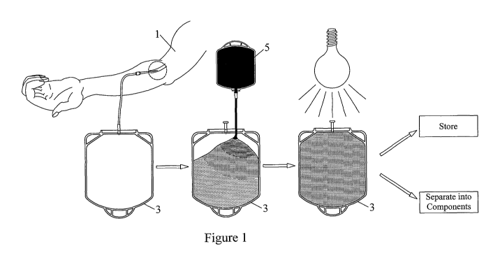
FIG. 1 is a schematic view of a set of bags and a flow diagram of a process of
the
present invention.
FIG. 2 is a schematic view of another set of bags and another process of the
present
invention.
FIG. 3 is a schematic view of a blood component expresser which may be used
with
the present invention.
FIG. 4 is a cross-sectional view of a whole blood separation apparatus which
may be
used with the present invention.
2
CA 02694169 2010-01-21
WO 2009/018309 PCT/US2008/071541
FIG. 5 is a schematic view and partial cross-section of a set of separation
and
collection bags designed for cooperating with the automated whole blood
separation
apparatus of FIG. 4.
FIG. 6 is a flow diagram of a process for separating pathogen inactivated
whole blood
into components using the apparatus of FIG. 4.
FIG. 7 is a schematic view of another set of separation and collection bags
designed
for cooperating with another automated whole blood separation apparatus.
FIG. 8 is a cross-sectional view of another whole blood separation apparatus
which
may be used with the present invention.
FIG. 9 is a top view of the rotor of the separation apparatus of FIG. 8.
FIG. 10 is a flow diagram of a process for separating pathogen inactivated
whole
blood into components using the apparatus of FIG. 8.
FIG. 1 lA and 11B are graphs of the log reduction of both enveloped and non-
enveloped viruses as a function of illumination energy.
FIG. 12 is a graph of hemolysis during refrigerated storage of treated red
blood cells
as a function of illumination energy.
FIG. 13 is a graph of ATP levels during refrigerated storage of treated red
blood cells
as a function of illumination energy.
FIG. 14 is a graph of mean osmotic fragility of treated red blood cells during
refrigerated storage as a function of illumination energy.
FIG. 15 is a graph of potassium levels in treated and untreated whole blood
over 5
days of storage as whole blood at room temperature as a function of
illumination
energy.
FIG. 16A and 16B are graphs of plasma quality during frozen storage as a
function of
illumination energy.
FIG. 17 is a table of measures of platelet quality for both treated and
untreated
platelets as a function of illumination energy.
DETAILED DESCRIPTION
A "photosensitizer" useful in this invention is defined as any compound which
absorbs radiation at one or more defined wavelengths and subsequently utilizes
the
absorbed energy to carry out a chemical process.
3
CA 02694169 2010-01-21
WO 2009/018309 PCT/US2008/071541
Endogenous photosensitizers may be used in this invention. The term
"endogenous" means naturally found in a human or mammalian body, either as a
result of synthesis by the body or because of ingestion as an essential
foodstuff (e.g.
vitamins) or formation of metabolites and/or byproducts in vivo. When
endogenous
photosensitizers are used, particularly when such photosensitizers are not
inherently
toxic or do not yield toxic photoproducts after photoradiation, no removal or
purification step is required after decontamination, and the decontaminated
product
can be directly administered to a patiet.
Examples of such endogenous photosensitizers which may be used in this
invention are alloxazines such as 7,8-dimethyl-10-ribityl isoalloxazine
(riboflavin),
7,8,10-trimethylisoalloxazine (lumiflavin), 7,8-dimethylalloxazine
(lumichrome),
isoalloxazine-adenine dinucleotide (flavin adenine dinucleotide [FAD]) and
alloxazine mononucleotide (also known as flavin mononucleotide [FMN] and
riboflavin-5-phosphate). The term "alloxazine" includes isoalloxazines.
Use of endogenous isoalloxazines as photosensitizers to inactivate blood and
blood components are described in United States Patent Nos. 6, 258,577 and
6,277,337 both issued to Goodrich et al., and herein incorporated by reference
to the
amount not inconsistent.
The amount of photosensitizer to be mixed with the whole blood to be
inactivated will be an amount sufficient to adequately inactivate any pathogen-
associated nucleic acids which may be present in the fluid, but less than a
toxic (to the
blood components) or insoluble amount. A pathogen may be defined as any
undesirable element found in blood, such as bacteria, virus and white blood
cells.
If riboflavin is used as the photosensitizer, it may be added to the whole
blood
at a final concentration of between about 50-500 M. Pathogen-associated
nucleic
acid includes any undesirable nucleic acid such as nucleic acid contained in
white
blood cells, bacteria or viruses. Nucleic acids include either
deoxyribonucleic acid
(DNA), ribonucleic acid (RNA) or both.
4
CA 02694169 2010-01-21
WO 2009/018309 PCT/US2008/071541
The whole blood to which the photosensitizer has been added is exposed to
light of the appropriate wavelength to activate the photosensitizer and to
substantially
inactivate and cause permanent damage to the pathogen-associated nucleic
acids.
Substantially permanent damage means that the nucleic acids will not undergo
self-
repair or replication during storage or upon infusion into a donor, while
maintaining
the antigenic potential of the pathogen to be removed by the recipient's
immune
system.
It should be noted that in the drawings, like elements are represented by like
numerals.
As shown in FIG. 1, whole blood to be pathogen inactivated may be collected
from a donor by any method known in the art. Typically, a unit of whole blood
450 mL) is collected from a donor 1 into a whole blood collection bag 3. The
collection bag may be a standard blood collection bag (shown in FIG. 1), or
may be a
round bag such as bag 11 shown in FIG. 5. Once the whole blood is collected,
the
blood can be transferred to a separate illumination bag (see FIG. 2), or can
be
illuminated in the collection bag 3, depending upon the material of the
collection bag.
If illumination is to take place in the collection bag, the collection bag
must be at least
light permeable and of a size that permits mixing of the whole blood and
photosensitizer during illumination. 35 mL of 500 M riboflavin contained in a
bag 5
is added to the whole blood in bag 3, and the whole blood + riboflavin in bag
3 is
illuminated with between 22-110 J/mLRBc of radiation. After illumination, the
inactivated whole blood can either be stored for later use or can be separated
into
desired components which may either be used immediately or stored for later
use.
The pathogen inactivated whole blood may be stored for less than one half
hour, or
may be stored for a time period up to the point the blood is no longer viable.
With the
present invention, whole blood does not need to be leukoreduced before the
addition
of photosensitizer, illumination and subsequent pathogen inactivation, nor
does the
whole blood or separated pathogen reduced blood components need to be
leukoreduced at any time before infusion into a patient.
In another embodiment shown in FIG. 2, whole blood is collected in a whole
blood collection bag 2 and transferred to an illumination bag 4. Bag 2 is
subsequently
CA 02694169 2010-01-21
WO 2009/018309 PCT/US2008/071541
removed from the remaining tubing set. Photosensitizer 5 is then added to the
whole
blood in the illumination bag 4 and the bag is illuminated in an illuminator
6. After
illumination, the pathogen inactivated whole blood may be transferred to an
integrally
attached or pre-connected storage bag 8.
If it is desired to separate the inactivated whole blood into various blood
components, the inactivated whole blood can be separated manually or using an
automated whole blood separator.
Whole blood is most commonly separated into components manually. After
whole blood is collected from a patient, the whole blood is processed in a
laboratory.
In the processing laboratory, a technician places the bags of whole blood into
a large,
swinging bucket centrifuge, which must be carefully balanced as the bags are
loaded.
The centrifuge is started and the bags are spun at a high rate of speed. In
the first
centrifugation, the red cells, which are the densest component, are forced to
the
bottom of the bag while the platelet-rich plasma, which is lighter, rises to
the top.
The technician next places each bag in an expresser 80 (see FIG. 3) consisting
of two rigid plates 81, 82 that are joined by a spring loaded hinge 84. One of
the
plates is fixed and the other is moveable. The blood bag 86 is positioned
between the
two plates and the spring catch released causing the moveable plate to press
against
the bag. A port 87 on the top of the bag is then opened and the platelet-rich
plasma is
expressed into a pre-connected, empty bag 88. When the technician observes
that red
cells are about to reach the outlet port, the expression is stopped and the
tubing
clamped.
If platelets are to be separated, the bags containing the platelet rich plasma
are
returned to the centrifuge, the rotor is again balanced and a second spin
begins, this
time at a higher speed. This spin forces the platelets to the bottom of the
bag and
allows the lighter plasma to rise to the top. The expression process described
above is
then repeated so that the platelets can be diverted to a separate bag foN
storage.
6
CA 02694169 2010-01-21
WO 2009/018309 PCT/US2008/071541
Whole blood may also be separated into components using an automated
whole blood separator. Whole blood separation can be performed to obtain
either two
component products, for example plasma and red blood cells (RBCs), or to
obtain
three (or more) component products, for example plasma, RBCs and either a
buffy
coat or a platelet product. The present system and method may be desirable
particularly when pathogen reduced whole blood is to be separated into
components
in a completely sterile manner.
FIG. 4 shows an embodiment of an apparatus for separating a volume of
composite liquid by centrifugation. The apparatus comprises a centrifuge
adapted for
receiving the separation bags shown in FIG. 5 and a component transferring
means for
causing the transfer of separated components into the satellite bags.
The centrifuge comprises a rotor that is supported by a bearing assembly 30
allowing the rotor to rotate about a vertical central axis (not shown). The
rotor
includes a cylindrical rotor shaft 32, 33; a central compartment 34 for
containing
satellite bags, which is connected to the rotor shaft 32, 33; a support member
(not
shown in FIG. 4) for supporting at least one satellite bag in a determined
position
within the central compartment 34; and a circular turntable 35 for supporting
a
separation bag, which is connected to compartment 34.
The rotor shaft comprises a first upper portion 32 and a second lower portion
33. The upper portion 32 of the shaft extends in part through the bearing
assembly
30. A pulley 36 is connected to the lower end of the upper portion 32 of the
shaft.
The centrifuge further comprises a motor 40 coupled to the rotor by a belt 41
engaged in a groove of the pulley 36 so as to rotate the rotor about a central
vertical
axis.
The separation apparatus further comprises a first, second and third pinch
valve members (not shown) that are mounted on the rotor for selectively
blocking or
allowing a flow of liquid through a flexible plastic tube, and selectively
sealing and
cutting a plastic tube.
7
CA 02694169 2010-01-21
WO 2009/018309 PCT/US2008/071541
The turntable 35 comprises a central frusto-conical portion 46, the upper,
smaller edge of which is connected to the rim of the compartment 34, an
annular flat
portion 47 connected to the lower, larger edge of the frusto-conical portion
46, and an
outer cylindrical flange 48 extending upwards from the outer periphery of the
annular
portion 47. The turntable 35 further comprises a vaulted circular lid 49 that
is secured
to the flange 48 by a hinge so as to pivot between an open and a closed
position. The
lid 49 is fitted with a lock 51 by which it can be blocked in the closed
position. The
lid 49 comprises a large cut-out in its upper part that gives access to the
central
compartment 34 of the rotor. The lid 49 has an annular interior surface that
is so
shaped that, when the lid 49 is in the closed position, it defines with the
frusto-conical
portion 46 and the annular flat portion 47 of the turntable 38 a frusto-
conical annular
compartment 53 having a radial cross-section that has substantially the shape
of a
parallelogram. The frusto-conical annular compartment 53, later the
"separation
compartment", is intended for containing the separation bag 11.
The component transferring means comprises a squeezing system for
squeezing the separation bag within the separation compartment 53 and causing
the
transfer of separated components into the satellite bags. The squeezing system
comprises a flexible annular diaphragm 54 that is so shaped as to line the
frusto-
conical portion 46 and the annular flat portion 47 of the turntable 35, to
which it is
secured along its smaller and larger circular edges. The squeezing system
further
comprises a hydraulic pumping station 60 for pumping a hydraulic liquid in and
out
an expandable hydraulic chamber defined between the flexible diaphragm 54 and
the
turntable 35, via a duct 37 extending through the rotor from the lower end of
the
lower portion 33 of the rotor shaft to the turntable 35. The pumping station
60
comprises a piston pump having a piston 61 movable in a hydraulic cylinder 62
fluidly connected via a rotary fluid coupling 38 to the rotor duct 37. The
piston 61 is
actuated by a stepper motor 63 that moves a lead screw 64 linked to the piston
rod.
The hydraulic cylinder 62 is also connected to a hydraulic liquid reservoir 65
having
an access controlled by a valve 66 for selectively allowing the introduction
or the
withdrawal of hydraulic liquid into and from a hydraulic circuit including the
hydraulic cylinder 62, the rotor duct 37 and the expandable hydraulic chamber.
A
pressure gauge 67 is connected to the hydraulic circuit for measuring the
hydraulic
pressure therein.
8
CA 02694169 2010-01-21
WO 2009/018309 PCT/US2008/071541
The separation apparatus further comprises a controller 70 including a control
unit (microprocessor) and a memory for providing the microprocessor with
information and programmed instructions relative to various separation
protocols and
to the operation of the apparatus in accordance with such separation
protocols. In
particular, the microprocessor is programmed for receiving information
relative to the
centrifugation speed(s) at which the rotor is to be rotated during the various
stages of
a separation process, and information relative to the various transfer flow
rates at
which separated components are to be transferred from the separation bag 11
into the
satellite bags 12, 14. The information relative to the various transfer flow
rates can be
expressed, for example, as hydraulic liquid flow rates in the hydraulic
circuit, or as
rotation speeds of the stepper motor 63 of the hydraulic pumping station 60.
The
microprocessor is further programmed for receiving, directly or through the
memory,
information from the pressure gauge 67 and from the photocells (not shown) and
for
controlling the centrifuge motor 40, the stepper motor 63, and the pinch valve
members so as to cause the separation apparatus to operate along a selected
separation
protocol.
A variety of alternative sets 10 of containers which may be used with the
system/machine of the present invention are shown in FIG. 5. A separation
container
11 may be a part of the bag set wherein in an embodiment, the separation
container 11
is annular and/or of a ring shape. In some embodiments this may be flat, or it
may be
a somewhat frusto-conical separation container 11 and may be of a flexible
plastic
material, which in some instances, may be of the same or a similar type as
used in
conventional blood or blood component or other biological fluid bags. If bag
11 is to
be illuminated, it must also be at least light permeable.
As shown in the substantially schematic embodiment of FIG. 5, a first
component collection container 12 may be connected by a tube 13 to the
separation
container 11, and a second component collection container 14 may similarly be
connected to the separation container 11 by a second tube 15. Both such
connections
may be at the inner circumference of ring bag 11 or though not shown, either
or both
could be connected to the outer circumference or at any desired radial
location
therebetween. The component collection containers 12, 14 may be shaped in any
of a
9
CA 02694169 2010-01-21
WO 2009/018309 PCT/US2008/071541
variety of ways and/or formed of any of a variety of materials, though they
may in
some embodiments be, as shown, substantially rectangular bags of flexible
plastic
sheet material of substantially conventional type, the plastic sheet material
being
preferably selected with a view to the type of cells or blood component
products
which may be chosen to be stored in the respective container. In the two
component
(2C) set 10 of FIG. 4, these two component collection bags 12, 14 are the only
end
component bags; however, in the three component (3C) set, a third component
collection bag (not shown) may also be connected by a third tubing line, to
the ring
bag 11. In pathogen inactivated whole blood (WB) separation, the first
collection bag
12 may be adapted to receive plasma, the second collection bag 14 adapted to
receive
RBCs and the third collection bag (in the 3C set), adapted to receive
platelets.
In the separation of pathogen inactivated whole blood and the preparation of
pathogen inactivated blood components, the bags may all be initially empty, or
one or
more of the finished component collection container or product bags, e.g., the
second
component container 14 (in FIG. 5) may be initially filled with a certain
amount of an
additive or storage fluid or liquid 16 for the component to be disposed
therein, e.g.,
red blood cells. Examples of such a fluid may include storage solutions such
as SAG,
SAG-M, AS-1, AS-3, or AS-5.
As an alternative, the storage or additive solution may be predisposed in an
optional separate bag, see, for example, satellite bag 26 in FIG. 5, which
would be
connected to or connectable with bag 14 via an additive solution tube
271eading from
bag 14, and a connecting tube 28. An optional sterile barrier or filter 29
represented
schematically on line 28 may also be included. The additive or storage
solution 16
may then be passed from such a satellite container 26 to container 14 via
lines 28, 27.
In some embodiments, the solution bag 26 may be pre-connected to bag 14, i.e.,
during the manufacturing process of the set 10, or as an alternative, the
additive
solution bag 26 may be later connected or docked via sterile docking or spike
connection and thus not be previously stored within or as part of set 10, but
instead
added at a different time, before or after blood component
separation/processing. The
component container 14 may in such a case then be temporarily sealed by, for
instance, a frangible or a breaking pin 17, or other sealing means such as a
peelable or
pressure rupturable seal, to keep the solution sealed therein until its use
may be
CA 02694169 2010-01-21
WO 2009/018309 PCT/US2008/071541
desired, i.e., until loaded in the centrifuge and ready to receive a component
product
such as RBCs.
With the other component bag(s), a storage or an additive solution may
similarly be pre-disposed in or adapted to be added to the bag(s) for the
benefit of the
component product to be later added thereto. If platelets are to be collected,
the third
component collection container may contain a storage solution for platelets
such as
PAS, PAS IIIM, or Composol.
In an embodiment such as the one illustrated, the separation container 11 may
be provided with a connection tube 19 which may be connected by sterile
docking 23
to a source of pathogen inactivated whole blood 20. In another embodiment,
whole
blood may be collected and pathogen inactivated in separation container 11, in
accordance with the principles described in FIGS. 1 and 2 above. In this
embodiment,
there would be no need for bag 20 and tubing line 19.
As shown in FIG. 6 in a first step 121 of the general process 110, the
pathogen
inactivated whole blood is supplied to the separation container/bag 11. Then,
in a
second general step 122, the pathogen inactivated whole blood is spun and the
component parts thereby separated. Next, as shown in box 123, a first
component
product is moved or expressed out of the separation container 11 to a first
component
container 12. The second component product is also moved or expressed out of
the
separation container 11 to its second component container 14. This is depicted
by box
124 in the process diagram 110. Lastly, the first and/or second component
containers
12, 14 are closed off by valving, sealing and/or cutting the inlets, e.g.,
tubing lines,
thereto. This is depicted by/in box 125. Note, as a general concept, the
third, fourth
and fifth steps 123, 124, and 125 may occur independently and/or after a
decrease in
rotation speed of the centrifuge and separation of the second step 122, or
more
generally here, the rotation/centrifugation of step 122 continues throughout
the
performance of the other steps 123, 124 and/or 125 and any alternatives and/or
intermediary steps thereto. Thus, the rotation/centrifugation and separation
step 122
will most often here, cease usually only after completion of steps 123, 124
and/or 125
and any intermediaries and/or alternatives thereto. Cessations of the second
step 122
would then constitute the end of the usual process (note, unloading and/or
other
11
CA 02694169 2010-01-21
WO 2009/018309 PCT/US2008/071541
administrative-type handling processes, marking, labeling, storing and the
like post
centrifugation process steps, if performed post-processing, notwithstanding).
Note, an
alternative, optional process line 123a is also shown (in dashed lines) in
FIG. 5 to
emphasize the alternative that a valving, sealing and/or cutting step 125 may
be
performed relative to the first component container prior to or during the
fourth step
124 and, in any event, prior to and separate from the valving, sealing and/or
cutting
step for the second product container.
If a third product is collected, an intermediate step 126 may be used for the
third product movement or expression from the separation container to the
third
product container. Note, an alternative, optional process line 124a is also
shown (in
dashed lines) to emphasize the alternative that a valving, sealing and/or
cutting step
125 may be performed relative to the second component container prior to or
during
the intermediate optional step 126 and, in any event, prior to and separate
from the
valving, sealing and/or cutting step for the third product container.
FIG. 7 shows another example of a set of bags adapted for another
system/machine which may be used in centrifugal separation of pathogen
inactivated
whole blood into component products. This bag set comprises a flexible
separation
bag 1000 and three flexible satellite bags 200, 300, 150 connected thereto.
The separation bag 1000 may be used as a whole blood collection bag, a
pathogen inactivation bag and a bag for separating the pathogen inactivated
whole
blood into components. The separation bag 1000 is flat and generally
rectangular. It
is made of two rectangular sheets of plastic material that are welded together
so as to
defme therebetween an interior space having a main rectangular portion
connected to
a triangular top downstream portion. A first tube 400 is connected to the tip
of the
triangular portion, and second and third tubes 500, 600 are connected to
either lateral
edges of the triangular portion, respectively. The proximal ends of the three
tubes
400, 500, 600 are embedded between the two sheets of plastic material so as to
be
parallel. The separation bag 1000 further comprises a hole 800 in each of its
corners
that are adjacent to the three tubes 400, 500, 600. The holes 800 are used to
secure
the separation bag to a separation compartment.
12
CA 02694169 2010-01-21
WO 2009/018309 PCT/US2008/071541
A volume of anticoagulant (typically about 63 ml for a blood donation of
about 450 ml) is initially added to the separation bag, and the first and
third tubes 400,
600 are fitted at their proximal end with a breakable stopper 90, 100
respectively,
blocking a liquid flow therethrough.
The second tube 500 is a collection tube having a needle 120 connected to its
distal end. At the beginning of a blood donation, the needle 120 is inserted
in the vein
of a donor and blood flows into the collection (separation) bag 1000. After a
desired
volume of blood has been collected in the collection (separation) bag 1000,
the
collection tube 500 is sealed and cut. Photosensitizer may be initially added
to bag
1000 before the whole blood is added, or may be added after the whole blood is
added
through tubing 500. It may also be added through a separate tube (not shown).
The first satellite bag 200 is intended for receiving a plasma component. It
is
flat and substantially rectangular. It is connected to the distal end of the
first tube
400.
The second satellite bag 300 is intended for receiving a red blood cell
component. It is flat and substantially rectangular. It is connected to the
distal end of
the third tube 600. The second satellite bag 300 may contain a volume of
storage
solution for storage of red blood cells, and the third tube 600 is fitted at
its distal end
with a breakable stopper 140 blocking liquid flow therethrough.
The third satellite bag 150 is intended to receive a platelet component. Like
the first and second satellite bags 200, 300, the third satellite bag 150 is
flat and
substantially rectangular.
The bag set also contains a T-shaped three-way connector 160 having its leg
connected by the first tube 400 to the separation bag 1000, a first arm
connected by a
fourth tube 170 to the first satellite bag 200 (plasma component bag), and a
second
arm connected by a fifth tube 180 to the third satellite bag 150 (platelet
component
bag).
An apparatus for simultaneously separating by centrifugation four discrete
13
CA 02694169 2010-01-21
WO 2009/018309 PCT/US2008/071541
volumes of pathogen inactivated whole blood may be used with the bag set of
FIG. 7.
The apparatus includes a centrifuge adapted to receive the four bag sets shown
in FIG.
7, with the four discrete volumes of a composite liquid contained in the four
separation bags; a component transferring system for transferring at least one
separated component from each separation bag into a satellite bag connected
thereto;
a first balancing system for initially balancing the rotor when the weights of
the four
separation bags are different; and a second balancing system for balancing the
rotor
when the weights of the separated components transferred into the satellite
bags cause
an unbalance of the rotor.
As shown in FIG. 8, the centrifuge comprises a rotor that is supported by a
bearing assembly 3000 allowing the rotor to rotate around a rotation axis 310.
The
rotor includes a cylindrical rotor shaft 320 to which a pulley 330 is
connected; a
system comprising a central cylindrical container 340 for containing satellite
bags,
which is connected to the rotor shaft 320 at the upper end thereof so that the
longitudinal axis of the rotor shaft 320 and the longitudinal axis of the
container 340
coincide with the rotation axis 310, and a frusto-conical turntable 350
connected to
the upper part of the central container 340 so that its central axis coincides
with the
rotation axis 310. The frusto-conical turntable 350 flares underneath the
opening of
the container 340. Four identical separation cells 4000 are mounted on the
turntable
350 so as to form a symmetrical arrangement with respect to the rotation axis
310.
The centrifuge further comprises a motor 360 coupled to the rotor by a belt
370 engaged in a groove of the pulley 330 so as to rotate the rotor about the
rotation
axis 310.
Each separation cel14000 comprises a container 410 having the general shape
of a rectangular parallelepiped. The separation cells 4000 are mounted on the
turntable 350 so that their respective median longitudinal axes 420 intersect
the
rotation axis 310, so that they are located substantially at the same distance
from the
rotation axis 310, and so that the angles between their median longitudinal
axes 420
are substantially the same (i.e. 90 degrees). The exact position of the
separation cells
4000 on the turntable 350 is adjusted so that the weight on the turntable is
equally
distributed when the separation cells 4000 are empty, i.e. so that the rotor
is balanced.
14
CA 02694169 2010-01-21
WO 2009/018309 PCT/US2008/071541
It results from the arrangement of the separating cells 4000 on the turntable
350 that
the separating cells 4000 are inclined with respect to the rotation axis 310
of an acute
angle equal to the angle of the frustum of a cone that geometrically defines
the
turntable 350.
Each container 410 comprises a cavity 430 that is so shaped and dimensioned
as to loosely accommodate a separation bag 1000 full of liquid, of the type
shown in
FIG. 4. The cavity 430 (which will be referred to later also as the
"separation
compartment") is defined by a bottom wall that is the farthest to the rotation
axis 310,
a lower wall that is the closest to the turntable 350, an upper wall opposite
to the
lower wall, and two lateral walls. The cavity 430 comprises a main part,
extending
from the bottom wall, which has substantially the shape of a rectangular
parallelepiped with rounded angles, and an upper part, which has substantially
the
shape of a prism having convergent triangular bases. In other words, the upper
part of
the cavity 430 is defined by two couples of opposite walls converging towards
the
central median axis 420 of the cavity 430.
One interest of this design is to cause a radial dilatation of the thin layer
of a
minor component of whole blood (e.g. the platelets) after separation by
centrifugation,
and makes it more easily detectable in the upper part of a separation bag. As
shown
in FIG. 8, the two couples of opposite walls of the upper part of the
separation cell
4000 converge towards three cylindrical parallel channels 440, 450, 460,
opening at
the top of the container 410, and in which, when a separation bag 1000 is set
in the
container 410, the three tubes 400, 500, 600 extend.
The container 410 also comprises a hinged lateral lid (not shown), which is
comprised of an upper portion of the external wall of the container 410, i.e.
the wall
that is opposite to the turntable 350. The lid is so dimensioned as to allow,
when
open, an easy loading of a separation bag 1000 full of liquid into the
separation cell
4000. The container 410 comprises a fast locking means (not shown) by which
the lid
can be locked to the remaining part of the container 410.
The container 410 also comprises a securing means for securing a separation
bag 1000 within the separation cell 4000. The bag securing means comprises two
CA 02694169 2010-01-21
WO 2009/018309 PCT/US2008/071541
pins (not shown) protruding on the internal surface of the lid, close to the
top of
separation cel14000, and two corresponding recesses in the upper part of the
container
410. The two pins are so spaced apart and dimensioned as to fit into the two
holes
800 in the upper corner of a separation bag 1000.
The separation apparatus further comprises a component transferring means
for transferring at least one separated component from each separation bag
into a
satellite bag connected thereto. The component transferring means comprises a
squeezing system for squeezing the separation bags 1000 within the separation
compartments 430 and causing the transfer of separated components into
satellite
bags 200, 300, 150.
The squeezing system comprises a flexible diaphragm 500 that is secured to
each container 410 so as to define an expandable chamber 510 in the cavity
thereof.
More specifically, the diaphragm 500 is dimensioned so as to line the bottom
wall of
the cavity 430 and a large portion of the lower wall of the cavity 430, which
is the
closest to the turntable 350.
The squeezing system further comprises a peripheral circular manifold 520
that forms a ring within the turntable 350 extending close to the periphery of
the
turntable 350. Each expansion chamber 510 is connected to the manifold 520 by
a
supply channel 530 that extends through the wall of the respective container
410,
close to the bottom thereof.
The squeezing system further comprises a hydraulic pumping station 6000 for
pumping a hydraulic liquid in and out the expandable chambers 510 within the
separation cells 4000. The hydraulic liquid is selected so as to have a
density slightly
higher than the density of the more dense of the components in the composite
liquid
to be separated (e.g. the red blood cells, when the composite liquid is
blood). As a
result, during centrifugation, the hydraulic liquid within the expandable
chambers
510, whatever the volume thereof, will generally remain in the most external
part of
the separation cells 4000. The pumping station 6000 is connected to the
expandable
chambers 510, through a rotary seal 690, by a duct 560 that extends through
the rotor
shaft 320, the bottom and lateral wall of the central container 340, and, from
the rim
16
CA 02694169 2010-01-21
WO 2009/018309 PCT/US2008/071541
of the central container 340, radially through the turntable 350 where it
connects to
the manifold 520.
The pumping station 6000 comprises a piston pump having a piston 610
movable in a hydraulic cylinder 620 fluidly connected via a rotary fluid
coupling to
the rotor duct 540. The piston 610 is actuated by a stepper motor 640 that
moves a
lead screw 650 linked to the piston rod. The hydraulic cylinder 620 is also
connected
to a hydraulic liquid reservoir 660 having an access controlled by a valve 670
for
selectively allowing the introduction or the withdrawal of hydraulic liquid
into and
from a hydraulic circuit including the hydraulic cylinder 620, the rotor duct
560 and
the expandable hydraulic chambers 510. A pressure gauge 680 is connected to
the
hydraulic circuit for measuring the hydraulic pressure therein.
The separation apparatus further comprises four pairs of first and second
pinch
valve members 700, 710 that are mounted on the rotor around the opening of the
central container 340. Each pair of pinch valve members 700, 710 faces one
separation cell 4000, with which it is associated. The pinch valve members
700, 710
are designed for selectively blocking or allowing a flow of liquid through a
flexible
plastic tube, and selectively sealing and cutting a plastic tube. Each pinch
valve
member 700, 710 comprises an elongated cylindrical body and a head having a
groove 720 that is defmed by a stationary upper jaw and a lower jaw movable
between an open and a closed position. The groove 720 is so dimensioned that
one of
the tubes 400, 170, 180 of the bag set shown in FIG. 7 can be snuggly engaged
therein
when the lower jaw is in the open position. The elongated body contains a
mechanism for moving the lower jaw and it is connected to a radio frequency
generator that supplies the energy necessary for sealing and cutting a plastic
tube.
The pinch valve members 700, 710 are mounted inside the central container 340,
adjacent the interior surface thereof, so that their longitudinal axes are
parallel to the
rotation axis 310 and their heads protrude above the rim of the container 340.
Electric
power is supplied to the pinch valve members 700, 710 through a slip ring
array that
is mounted around a lower portion of the rotor shaft 320.
The separation apparatus further comprises a first balancing means for
initially
balancing the rotor when the weights of the four separation bags 1000
contained in the
17
CA 02694169 2010-01-21
WO 2009/018309 PCT/US2008/071541
separation cells 4000 are different. The first balancing means substantially
comprises
the same structural elements as the elements of the component transferring
means
described above, namely: four expandable hydraulic chambers 510 interconnected
by
a peripheral circular manifold 520, and a hydraulic liquid pumping station
6000 for
pumping hydraulic liquid into the hydraulic chambers 510 through a rotor duct
560,
which is connected to the circular manifold 520. In order to initially balance
the
rotor, whose four separation cells 4000 contain four discrete volumes of a
composite
liquid that may not have the same weight (because the four volumes may be not
equal,
and/or the density of the liquid may slightly differ from one volume to the
other one),
the pumping station 6000 is controlled so as to pump into the interconnected
hydraulic chambers 510, at the onset of a separation process, a predetermined
volume
of hydraulic liquid that is so selected as to balance the rotor in the most
unbalanced
situation. For pathogen inactivated whole blood, the determination of this
balancing
volume takes into account the maximum difference in volume between two blood
donations, and the maximum difference in hematocrit (i.e. in density) between
two
blood donations. Under centrifugation forces, the hydraulic liquid will
distribute
unevenly in the four separation cells 4000 depending on the difference in
weight of
the separation bags 1000, and balance the rotor. In order to get an optimal
initial
balancing, the volume of the cavity 430 of the separation cells 4000 should be
selected so that the cavities 430, whatever the volume of the separation bags
1000
contained therein, are not full after the determined amount of hydraulic
liquid has
been pumped into the interconnected expansion chambers 510.
The separation apparatus further comprises a second balancing means, for
balancing the rotor when the weights of the components transferred into the
satellite
bags 200, 300, 150 in the central container 340 are different. For example,
when two
blood donations have the same hematocrit and different volumes, the volumes of
plasma extracted from each donation are different, and the same is true when
two
blood donations have the same volume and different hematocrit.
As shown in FIG. 9, the second balancing means comprises four flexible
rectangular pouches 810, 820, 830, 840 that are interconnected by four tube
sections
(not shown), each tube section connecting two adjacent pouches by the bottom
thereo The pouches 810, 820, 830, 840 contain a volume of balancing liquid
having
18
CA 02694169 2010-01-21
WO 2009/018309 PCT/US2008/071541
a density close to the density of the composite liquid. The volume of
balancing liquid
is so selected as to balance the rotor in the most unbalanced situation. The
four
pouches 810, 820, 830, 840 are so dimensioned as to line the inner surface of
the
central container 340 and to have an internal volume that is larger than the
volume of
balancing liquid so that the balancing liquid can freely expand in any of the
pouches
810, 820, 830, 840. In operation, if, for example, four satellite bags 200
respectively
adjacent to the four pouches 810, 820, 830, 840 receive different volumes of a
plasma
component, the four satellite bags 200 will press unevenly, under
centrifugation
forces, against the four pouches 810, 820, 830, 840, which will result in the
balancing
liquid becoming unevenly distributed in the four pouches 810, 820, 830, 840
and
compensating for the difference in weight in the satellite bags 200.
The separation apparatus further comprises a controller 900 including a
control unit (e.g. a microprocessor) and a memory unit for providing the
microprocessor with information and programmed instructions relative to
various
separation protocols (e.g. a protocol for the separation of a plasma component
and a
blood cell component, or a protocol for the separation of a plasma component,
a
platelet component, and a red blood cell component) and to the operation of
the
apparatus in accordance with such separation protocols. In particular, the
microprocessor is programmed for receiving information relative to the
centrifugation
speed(s) at which the rotor is to be rotated during the various stages of a
separation
process (e.g. stage of component separation, stage of a plasma component
expression,
stage of suspension of platelets in a plasma fraction, stage of a platelet
component
expression, etc), and information relative to the various transfer flow rates
at which
separated components are to be transferred from the separation bag 1000 into
the
satellite bags 200, 300, 150. The information relative to the various transfer
flow
rates can be expressed, for example, as hydraulic liquid flow rates in the
hydraulic
circuit, or as rotation speeds of the stepper motor 640 of the hydraulic
pumping
station 6000. The microprocessor is further programmed for receiving, directly
or
through the memory, information from the pressure gauge 680 and from the four
pairs
of photocells 730, 740 and for controlling the centrifuge motor 360, the
stepper motor
640 of the pumping station 6000, and the four pairs of pinch valve members
700, 710
so as to cause the separation apparatus to operate along a selected separation
protocol.
19
CA 02694169 2010-01-21
WO 2009/018309 PCT/US2008/071541
According to the separation protocol shown in FIG. 10, four discrete volumes
of pathogen reduced blood are separated into a plasma component, a first cell
component comprising platelets, white blood cells, some red blood cells and a
small
volume of plasma (later the "buffy coat" component) and a second cell
component
mainly comprising red blood cells. Each volume of blood is contained in a
separation
bag 1000 of a bag set represented in FIG. 7, in which it has previously been
collected
from a donor using the collection tube 500. After the blood collection, the
collection
tube 500 has been sealed and cut close to the separation bag. Typically, the
volumes
of blood are not the same in the four separation bags 1000, and the hematocrit
varies
from one separation bag 1000 to another one. Consequently, the separation bags
1000
have slightly different weights.
As shown in FIG. 10, the first stage of the separation procedure begins by
loading the four bag sets into the four separation cells 4000 on the rotor.
In the second stage, the rotor is balanced in order to compensate for the
difference in weights of the separation bags.
In the third stage, the pathogen inactivated whole blood within the separation
bag 1000 is sedimented to a desired level.
In the fourth stage the plasma component is transferred into the plasma
component bag 200.
In the fifth stage the platelet component is transferred into the platelet
component bag 150.
In the sixth stage the centrifugation process is ended.
When the fifth stage is completed, the red blood cells are transferred from
separation bag 1000 into the red blood cell component bag 300.
Pathogen inactivated whole blood may also be separated into blood components
using the whole blood separator described in US patent 6,910,998.
CA 02694169 2010-01-21
WO 2009/018309 PCT/US2008/071541
In any of the whole blood separation processes described above, no prior
leukoreduction of the pathogen inactivated whole blood before separation into
individual components is necessary. Nor is it necessary to leukoreduce the red
blood
cells after separation. Pathogen inactivation of the whole blood using
riboflavin and
light functionally inactivates the white blood cells in the whole blood. This
is shown
in Example 1 below. A pathogen inactivation procedure is particularly
important
when buffy coats containing white blood cells are collected.
Methods
For the control units, whole blood is processed manually, centrifuged using a
soft spin, the platelet rich plasma (PRP) supernatant expressed, and a full
volume
(approximately 100 mL) of AS-3 additive solution added to the separated RBCs
for
storage. A platelet concentrate is made from the PRP and stored at 22 C in a
Helmer
incubator for 1 day and 5 days prior to sampling for Day 1 and Day 5 platelet
quality
measurements. The remaining plasma is stored frozen for 28 days, and protein
quality assessed for Day 0 and Day 28 samples. The plasma, platelet
concentrates and
RBCs for the controls undergo the same testing as the treated units.
For the treated units, 35 mL of riboflavin is added to 470 10 mL of whole
blood in a 1 L ELP bag and illuminated at 22, 33, 44, 80 and 110 J/mLRBo in an
illuminator (Mirasol Whole Blood Illuminator R5Øwb.12, available from
CaridianBCT, Inc., Lakewood, CO). A sample is removed pre-illumination to
measure in vitro plasma quality. After illumination, the whole blood is
transferred to
a UBB bag, centrifuged using a soft spin, the PRP/riboflavin supematant
expressed,
and a fall volume bag of AS-3 additive solution (approximately 100 mL) is
added to
the RBCs for storage. A platelet component and plasma component were made from
the PRP as described above. The platelet component is stored at 22 C in a
Helmer
incubator prior to sampling for Day 1 and Day 5 platelet quality measurements.
21
CA 02694169 2010-01-21
WO 2009/018309 PCT/US2008/071541
Platelet quality is assessed with measurements of pH, swirl, lactate
production
rate and glucose consumption rate on Days 1 and 5.
Plasma quality is assessed with measurements of fibrinogen, total protein, and
Factors V, VIII and XI on Day 0 and Day 28 of frozen storage.
RBC quality is monitored through Day 42 of storage at 4-C to assess hemolysis,
osmotic fragility and ATP release. Samples were removed for Day 0 sampling
with
subsequent sampling occurring on Days 28, 35 and 42. Red blood cell quality
was
also assessed without separation of red blood cells from the whole blood.
Treated
whole blood was stored at room temperature and percent hemolysis and potassium
concentration ([K+]) were measured.
Example 1
Transfusion of blood products containing white blood cells (WBC) can result
in the induction of immune responses that can negatively impact the
transfusion
recipient. These immunological consequences can include transfusion-associated
graft-versus-host disease (TA-GvHD) and production of cytokines and
alloantibodies.
TA-GvHD, a donor-anti-recipient response, is almost always fatal and is
mediated by
proliferating T lymphocytes of the donor. The standard approach to inactivate
leukocytes and prevent TA-GvHD has been to expose blood products to y-
irradiation.
In the following assays, non-leukoreduced units of fresh (< 8 hours from
collection) whole blood were treated at energies of 22, 33 and 44 J/mLRBe. For
treated cells, riboflavin was added to the whole blood before illumination.
After
illumination, leukocytes were isolated from the whole blood units and the
functionality of white blood cells ()MBCs) was assessed for: (1) exhibiting
cell
activation (CD69 expression) in response to PMA (Phorbol 12-myristate 13-
acetate),
(2) WBC proliferation in response to PHA (Phytohemagglutinin), to allogeneic
stimulating cells, and to CD3/CD28 stimulation, (3) antigen presentation to
allogeneic
22
CA 02694169 2010-01-21
WO 2009/018309 PCT/US2008/071541
responder cells, and (4) the ability of WBCs to produce cytokines in response
to LPS
(lipopolysaccharide) or CD3/CD28 antibodies.
1.) The ability of WBCs to be activated by PMA and express CD69
CD69 is an early activation marker on T cells and can be visualized by flow
cytometry using anti-CD69 antibodies. Within 4 hrs of T-cell activation, CD69
is
detectable and stays upregulated as long as the cell is in an activated state.
As shown
in Graph 1, treatment with riboflavin and light inhibited expression of CD69
on T
cells after PMA activation at all energies tested.
CD69 expression
80
60
T
e 50
30
10
0
^i.ntreated 1322 J*nI RBC o33 J/ml RBC 44 J/rnl RBC I
Graph 1: PMA induced CD69 upregulation on T cells after treatment of whole
blood
2.) WBC proliferation in response to PHA and anti-CD3/CD28
The ability of treated WBCs to proliferate was analyzed by thymidine
incorporation after 3 days of incubation. As shown in Graph 2, exposure to PHA
(A)
or to plate-bound anti-CD3 plus anti-CD28 antibodies (B) induced proliferation
in
untreated WBCs. Treated WBCs showed no detectable induced proliferation at 33
J/rnlRBo and above when exposed to these mitogens.
23
CA 02694169 2010-01-21
WO 2009/018309 PCT/US2008/071541
A PHA proliferation
1000000
~ 100000
c
10000
0 1000
100
U_
B 10
L
F
1
0 22 33 44
energy [J/ml RBC]
-X- medium control X~ donor #1 -~-donor #2 +donor #3 -X- donor #4 -6 donor #5 -
1- donor #6
B CD3128 proliferation
-ff 1000000
Cl- 100000
0 10000
E ~m 1000
a. 100
1
0 22 33 44
energy [J/ml RBC]
--Ã-mediumcontrol --*- donor #1 tdonor#2 0-donor#3 -X- donor #4 8-donor#5 --I--
donor#6
Graph 2: Effect of treatment on proliferative response to PHA (graph A) or
anti-CD8/CD29 (graph B)
3.) WBC proliferation in response to allogeneic stimulators and antigen
presentation to allogeneic responder cells
WBCs in blood products are able to present antigen to recipient cells and
induce proliferation and allo-antibody formation. Treated WBCs were evaluated
in
Mixed Lymphocyte culture (MLC) both as responder cells (proliferate in
response to
stimulation) and as stimulators (promote proliferation of responder WBCs).
Treated
WBCs tested as responder cells in the MLC were not able to proliferate in
response to
allogeneic stimulator cells (Graph 3A), but untreated WBCs were. The amount of
proliferation detected in a MLC depends on the stimulator-responder
combination,
and thus is donor dependent. Allogeneic stimulator cells were treated with
mitomycin
C (a mitotic spindle poison) to prevent proliferation of the stimulator cells
in culture
with untreated and treated responder cells.
24
CA 02694169 2010-01-21
WO 2009/018309 PCT/US2008/071541
Treated WBCs (stimulators) were analyzed for their ability to induce
proliferation of allogeneic responder cells (Graph 3B) in the MLC. No
proliferation
by allogeneic responder cells could be detected, indicating that treatment
with
riboflavin and light inhibits antigen presentation of WBCs.
In summary, untreated WBCs proliferated in response to mitogens (PHA),
surface receptor crosslinking by antibodies (anti-CD3/CD28) and allogeneic
stimulator cells. In contrast, treatment of WBCs with riboflavin + UV light at
all
energies tested inhibited proliferation in response to any of these stimuli,
showing that
antigen specific as well as unspecific induction of proliferation is blocked
due to
treatment. Treated WBCs did not present antigen or induce proliferation in
allogeneic
responder cells, while untreated WBCs did.
Allorecognotion: treated responders
100000
Thymidine corporation [cpm]
10000
1000 =
~------ _
- -.; - - _ -
100 1
0 22 33 44
energy [J/m[ RBC]
donor #1 -x- donor #2 donor #3 donor #4 - donor #5 donor #6 x medium control
B Allostimulation: treated stimulators
100000_
Thymidine ii icorporation [cpm]
10000
T
1000
............... 100
0 22 33 44
energy [J/mi RBC]
donor #1 -=- donor #2 ~ donor #3 - donor #4 - donor #5 donor #6
Graph 3: Effect of treatment on allogeneic stimulator or responder cells
versus untreated. Dashed line
represents proliferation rate of cells in the absence of stimulator cells.
CA 02694169 2010-01-21
WO 2009/018309 PCT/US2008/071541
Levels of inhibition of proliferation due to treatment are shown in Table 1,
comparing stimulated control cells to stimulated treated cells. The
proliferative
response was decreased 93-99%. The proliferation of treated cells is down to
detection limits of the assay, since levels of proliferation of stimulated
treated cells
are as low as proliferation levels detected in cells cultured in PBS. A
comparison of
stimulated control cells to PBS cultured control cells shows a decrease in
proliferation
of 99% (data not shown).
% inhibition
22 J/m1RBc 33 J/mlRBc 44 J/ml"c
PHA 98+1 99+1 99+0
CD3/28 93 + 2 99 + 0 99 + 0
Allorecognition: treated responders 95 + 3 95 2 96 + 2
Allostimulation: treated stimulators 93 3 96 + 2 95 + 2
Table 1: Levels of inhibition
4.) Ability of WBCs to produce cytokines in response to anti-CD3/CD28 or LPS.
Another measure of functionality of WBCs is to measure cytokine production
in response to LPS (Lipopolysaccharide) or anti-CD3/CD28 antibodies.
Stimulation
with anti-CD3/28 induces cytokine production in T cells (TH1/TH2 cytokines).
LPS
activates monocytes and macrophages leading to the release of inflammatory
cytokines. Cytokines were detected using CBA ((Cytometric Bead Assay) (kits
purchased from BD Biosciences, PharMingen). A solution with standards is
provided
in the kit. Based on the values obtained for the standard curve a computer
program
determines a linear regression and the results of the individual samples. The
limit of
detection of these CBA assays is approximately 5-10 pg/ml.
As shown in Table 2A, the induction of TH1/TH2 cytokines is higher with
anti-CD3/28 stimulation (Table 2A) than with LPS (Table 2B). Treatment
significantly reduced TH1/TH2 cytokine production induced by anti-CD3/28
stimulation to levels comparable to the medium control of treated or untreated
cells at
all energies tested. When exposed to anti-CD3/28 antibodies, IL-2, TNF-a and
IFN-
y production was not reduced to medium control levels at 33 J/mlRBc and above,
but
26
CA 02694169 2010-01-21
WO 2009/018309 PCT/US2008/071541
compared to cytokine levels produced by untreated cells, the level of cytokine
production was inhibited by >90%. TH1/TH2 cytokine levels induced by LPS were
reduced to medium control levels after treatment at 44 J/mlRBc.
Inflammatory cytokines are induced by LPS as well as anti-CD3/28
stimulation. Treatment significantly reduced inflammatory cytokine production
in
response to anti-CD3/28 antibodies. High levels of IL-8 in the medium control
represent stored cytokines, rather than produced cytokines. Treatment also
reduced
the levels of IL-8 in medium control cells. Inflammatory cytokine production
in
response to LPS was reduced with treatment, but not to medium control levels
as seen
with anti-CD3/28 stimulation.
Cytokine production in response to anti-CD3/anti-CD28 antibodies was
blocked >90% at all energies tested, with the exception of IL-4 and IL-8 at 22
J/mlRBo
(see Table 2A). Inhibition of cytokine production in response to LPS was below
90%
at 33 J/mlRBc for the following cytokines: IL-5 and IL-2. IL-5 and IL-2 are
produced
at very low levels in untreated cells in response to LPS and are reduced to
levels of
detection after treatment. TNF-a and IL- 10 were measured using the CBA kits
for
inflammatory and TH1/TH2 cytokines.
Standard deviation values were high in samples after LPS stimulation,
compared to values obtained after anti-CD3/28 stimulation. Anti-CD3/28
stimulation
specifically activates T lymphocytes through the T cell receptor. In contrast,
LPS is a
major component of the outer membrane of Gram-negative bacteria and promotes
the
secretion of cytokines in many cell types, mainly macrophages. This endotoxin
function of LPS triggering a polyclonal response may explain the observed
variability
between donors.
A % inhibition
anti-CD3/28 22 J/mIRBC 33 J/mIRBC 44 J/mIRec
IL-12 p70 98 + 4 99+ 2 97 4
TNFa 90 + 12 100+0 100+ 0
Inflammatory
Cytokines IL-10 99 + 1 100+ 0 100+ 0
IL-6 99 + 2 100+ 0 100+ 0
IL-1 R 98 + 2 100+ 0 100+ 0
27
CA 02694169 2010-01-21
WO 2009/018309 PCT/US2008/071541
IL-8 89 + 5 98+ 2 100+ 0
IFN-y 99+1 100+0 100+0
TNF a 92 + 15 99+ 1 100+ 0
THI/TH2 IL-5 94 + 3 99+ 1 99+ 1
cytokines
IL-4 84 + 11 95+ 3 97+ 2
IL-2 91 +7 99+1 100+0
IL-10 99 + 1 100+ 0 100+ 0
B % inhibition
LPS 22 J/mIRBC 33 JImIRBC 44 J/mIRBc
IL-12 p70 NA NA NA
TNFa 54 + 35 92+ 8 98 + 2
Inflammatory IL-10 63 21 94 5 99 1
Cytokines IL-6 84 + 8 96 + 3 99 + 1
- - -
68+19 91 +8 98+3
IL-1(3 - - -
85+8 96+4 99+1
IL-8 - - -
IFN- y 63 + 71 93+ 8 93 + 7
TNFa 45+53 94+5 94+8
TH1/TH2 IL-10 66 + 21 94+ 4 99 + 1
cytokines IL-5 89 + 2 89+ 5 91 + 6
IL-4 59 + 65 90+ 4 90 + 4
IL-2 63 + 54 88+ 5 88 + 4
Table 2: Inhibition of cytokine production after treatment
In summary, WBC proliferation in response to all tested stimuli and antigen
presentation to allogeneic responder cells was inhibited >90% at all energies
tested.
Cytokine production in response to anti-CD3/anti-CD28 antibodies was blocked
>90% at all energies tested, with the exception of IL-4 and IL-8 at 22
J/mlRBc, that
were inhibited by 84% and 89% respectively.
Example 2
To test whether pathogen inactivation of whole blood was effective in
inactivating viruses which may be present in the whole blood, both non-
enveloped
and enveloped model viruses were tested. Hepatitis A(HAV), canine parvovirus
(CPV), vesicular stomatitis virus (VSV) and infectious bovine rhinotracheitis
virus
(IBR) were the viruses used.
28
CA 02694169 2010-01-21
WO 2009/018309 PCT/US2008/071541
As can be seen in FIG. 10, log reduction of both non-enveloped (FIG. 10A)
and enveloped (FIG. 10B) viruses increases linearly with respect to energy.
Example 3
To test whether pathogen inactivation of whole blood was effective in
inactivating clinically relevant levels of bacteria which may be present in
the whole
blood, low titer bacteria studies were done. After pathogen inactivation of
whole
blood, the whole blood was separated into a red blood cell (RBC) component and
platelet rich plasma (PRP) component. The results are shown in Table 3 below.
A +
symbol means that some of the replicates of the cultures in the panel grew in
under 5
days. A - symbol means that none of the replicates of the cultures in the
panel grew
in under 5 days.
Bacteria detected (+) or not detected (-) after treatment
44 J/mLRBC 80 J/mLp~Bc 110 J/mLRBC
(n=8) (n=3) (n=3)
RBC PRP RBC PRP RBC PRP
not not +/ a +/_a + -
S. epidermidis measured measured
not not - - - -
Y. enterocolitica measured measured
+/- +/- - - - -
S. liquefaciens
A. baumannii
not not +/-
S. pyogenes measured measured
a1 of 2 replicates negative
b2 of 3 replicates negative
7 of 8 replicates negative
d3 of 8 replicates negative
el of 8 replicates negative
Table 3: Low bacteria titer studies
As seen in Table 3, only S. epidermidis grew in red blood cells illuminated at
110 J/mLRBo.
29
CA 02694169 2010-01-21
WO 2009/018309 PCT/US2008/071541
Example 4
Whole blood was illuminated at 20, 33, 44, 60, 80 and 110 J/mLRBc. The
separated red blood cell component was stored at 4-C up to 42 days in AS-3.
The
percentage of red blood cell hemolysis during storage was measured after 28,
35 and
42 days of storage.
As shown in FIG. 11, whole blood illuminated at 110 J/mLRBo had the greatest
amount of hemolysis at 42 days of storage. Illumination at the other energies
did not
produce significant differences in hemolysis over 42 days of storage.
Example 5
ATP level or concentration was measured in red blood cells separated from
pathogen inactivated whole blood. ATP concentration is a measure of the amount
of
ATP present in the cells at a given time.
As shown in FIG. 12, increased energy levels decrease the total concentration
of ATP in the cells.
Example 6
Osmotic fragility during storage of red blood cells separated from pathogen
inactivated whole blood was measured. The normal red blood cell is a
relatively
impermeable biconcave disc which maintains osmotic equilibrium with the
surrounding medium. As the surrounding medium becomes hypotonic, fluid will be
taken into the cell to maintain stability. Eventually under very hypotonic
conditions
the cell will fill to capacity and rupture. Red blood cells with damaged
membranes
have a decreased capacity to expand, and will rupture in mildly hypotonic
conditions
that fail to lyse normal red cells. They thus exhibit increased osmotic
fragility. Mean
osmotic fragility (MOF) is a measure of red blood cell membrane fragility. The
higher the MOF, the more fragile the red blood cells are. MOF is the
concentration of
NaCI where 50% of the red blood cells hemolyze.
As can be seen in FIG. 13, treated red blood cells separated from illuminated
whole blood which was then stored for 42 days were on average slightly more
fragile
than untreated cells.
CA 02694169 2010-01-21
WO 2009/018309 PCT/US2008/071541
Example 7
Measurement of potassium concentration in stored red blood cells is another
measure of red blood cell viability. Potassium leaks out of the red blood
cells when
the potassium pump in the red blood cell membrane is not working correctly.
Damage to the potassium pump may occur during a pathogen inactivation
procedure.
The potassium concentration of whole blood (not separated blood
components) stored at room temperature over 5 days was measured. As shown in
FIG. 14, the effects of energy on red blood cell integrity increase as the
energy
increases.
Example 8
Plasma was separated from pathogen inactivated whole blood and the quality
of the plasma proteins was measured on day 0 and day 28.
As seen in FIG. 15A and 15B, percent activity or concentration of plasma
proteins appears to decrease in treated plasma at 110 J/mLRBo, as compared to
untreated plasma and treated plasma at lower energies. This occurs at both day
0 and
day 28.
Example 9
Platelets were separated from pathogen inactivated whole blood and markers
of platelet quality were measured. The results are shown in Fig. 16. While pH
remains the same over 5 days of storage for both treated and untreated cells,
oxygen
consumption appears to increase in treated platelets over 5 days of storage at
higher
energy levels, as does lactate production, while carbon dioxide production and
glucose consumption appears to decrease at higher energy levels over 5 days of
storage.
From the results above, blood components separated from pathogen
inactivated whole blood appear to be viable even when illuminated at a variety
of
energy levels.
31