Note: Descriptions are shown in the official language in which they were submitted.
CA 02694296 2010-03-01
77402-9D
-1-
This is a divisional of Canadian Application No. 2,195,446
filed July 20, 1995.
GROWTH CONTROL FOR CELLS
ENCAPSULATED WITHIN BIOARTIFICIAL ORGANS.
Field of the Invention
This invention relates to methods and
compositions"for controlling growth of cells'
encapsulated in a bioartificial organ.
Background of the Invention
Bioartificial organs "BAO" are devices which
contain living cells and are designed to provide a
needed metabolic function to a host.
The cells encapsulated in BAOs supply one or
more biologically active molecules to the host that may
be used to prevent or treat many clinical conditions,
deficiencies, and disease states.
For example, BAOs containing insulin
secreting cells may be used to treat diabetes.
Similarly other diseases such as hypoparathyroidism and
anemia may be treated by using cells which secrete
parathyroid hormone and erythropoietin, respectively.
Bioartificial orgAns may also be used to
supply biologically active molecules for the treatment
.or prevention of neurodegenerative conditions such as
Huntington's disease, Parkinson's disease, Alzheimer's
disease, and.Acquired Immune Deficiency Syndrome-
related dementia. Additionally,, lymphokines and
cytokines may also be supplied by BAOs to modulate the
CA 02694296 2010-03-01
~ . _ ..
96/02" PCT/US95/09281
- 2 -
host immune system. Other biologically active
molecules which may_be provided by bioartificial organs
include, catecholamines, endorphins, enkephalins, and
other opioid or non-opioid peptides that are useful for
treating pain. Enzymatic deficiencies may also be
treated by using BAOs. Alternatively, the biologically
active molecule may remove or eliminate deleterious
molecules from the host. For example, a BAO may
contain cells which produce a biologically active
molecule that can be used to "scavenge" cholesterol
from a host.
Various "macrocapsule" BAOs are known. See,
e.g., Aebischer (UnitedStates Patent 5,158,881),
Dionne et al. (WO 92/03327), Mandel et al.
(WO 91/00119), Aebischer (WO 93/00128). BAOs also
include extravascular diffusion chambers, intravascular
diffusion chambers, intravascular ultrafiltration
chambers, and microcapsules.. See,e.g., Lim et al.,
Science 210:908-910 (1980); Sun, A.M., Methods in
Enzymology 137: 575-579 (1988); Dunleavy et al. (WO
93/03901) and Chick et al. (United States Patent
5,002,661).
Because the cells encapsulated in the BAO
provide the needed metabolic function, it is desirable
that those cells optimally supply the biologically
active molecule that effects that function. Typically,
differentiated, non-dividing cells may be preferred
over dividing cells for use in BAOs because they allow
for the optimal production of the desired biologically
active molecule. For example, many differentiated,
non-dividing cells produce a greater quantity of a
desired therapeutic protein than dividing cells because
the expression of differentiation specific genes and
cell division are thought to be antagonistic processes.
Wollheim, "Establishment and Culture of Insulin-
CA 02694296 2010-03-01
WO 96/02646 PCT/IJS951iN281
- 3 -
Secreting B Cell Lines," Methods in Enzymology, 192,
p. 223-235 (1990). Cellular replication capacity
decreases as cells differentiate. In many cases,
proliferation and differentiation are mutually
exclusive. Gonos, "Oncogenes in Cellular
Immortalisation and Differentiation," 13, Anticancer
Research, p. 1117 (1993).
The use of differentiated tissue is
advantageous because the functional properties of
tissue desired for incorporation into a BAO have most
often been defined by the properties of differentiated
tissue in vivo. Another advantage to the use of
differentiated, non-dividing cells is that the cell
number within the BAO will remain relatively constant.
This, in turn, leads to more predictable results and
stable dosage for the recipient host. Additionally,
differentiated cells are better suited for use in BAOs
which encapsulate more than one cell type secreting
biologically active molecules. In such BAOs, if
dividing cells are used, different cell types may grow
at different rates, resulting in the overgrowth of one
cell type. By using differentiated, non-dividing
cells, the relative proportions of two or-more
synergistic cell types can be more readily controlled.
Although in many instances the use of
differentiated cells is advantageous, there have been
various problems associated with utilizing
differentiated cells directly isolated from mammals.
First, there is the potential contamination
of the isolated tissue which may require that the
tissue taken from each animal be subjected to costly
and time-consuming testing to assure that it is
pathogen-free.
Second, tissue can be damaged during
isolation due to the use of mechanical or enzymatic
CA 02694296 2010-03-01
WO 96/02646 PCT/US95109281
- 4 -
isolation procedures in the isolation process. The
mechanical manipulations are not always easily
standardized, resulting in variability between
isolations.
Third, ischemia may occur during isolation
causing tissue damage.
Fourth, reproducible yields may be difficult
because of variations in tissue donors. For example,
the age, sex, health, hormonal status of the source
animal can affect the yield and quality of the tissue
of interest.
Fifth, some:times there is not enough source
tissue to meet the proj:ected demand for the BAO. This
occurs for example, in a case where the source tissue
comes from a small sized organ or where the ultimate
need for tissue amounts is high. If the source of the
isolated tissue is human, there is frequently a severe
shortage of donor tissue.
Sixth, in some cases, it is desirable to
genetically modify the cells used in the BAO. Non-
dividing tissue to date has been difficult to
genetically modify in vitro and the yields and
properties of the modified cells may be uncertain.
Thus, because of the foregoing problems, while the use
of differentiated, non-dividing cells is desirable, a
need exists for a method of producing and maintaining
differentiated, non-dividing cells for encapsulation in
BAOs.
Because of these problems, dividing cells and
cell lines have been favored for use within BAOs to
provide the needed biological function. One important
advantage in using dividing cells is that such cells
may be grown to large numbers in vitro and screened for
pathogens and banked. This allows an almost unlimited
supply of tissue for'lower production costs. Selection
CA 02694296 2010-03-01
WO 96/02646 PCT/US9~r"u9281
- 5 -
schemes such as cell sorting or cloning may be applied
to the cell bank to develop subpopulations with
improved characteristics. Additionally, dividing cells
and cell lines are more amenable to genetic engineering
than differentiated, non-dividing cells. The ability
to introduce heterologous recombinant DNA allows many
new possibilities for the alteration of the function or
phenotype of cells to be encapsulated in the BAO. This
in turn provides for a greater diversity of therapeutic
uses for BAOs.
However, as discussed supra, the
disadvantages in encapsulating continuously dividing
cells in a BAO include-poor regulation of cell numbers
in the device that may result in less predictability in
production of the desired biologically active molecule.
While in most cases it may be desirable to
limit or minimize cell growth within the BAO, in other
cases, e.g., where the BAO is implanted in a "hostile"
environment, it may be desirable to allow the cells to
proliferate slowly to maintain cell numbers in the BAO.
There is another problem associated with
encapsulating cells in general. A variety of cell
types have cell adherent properties such that cells
tend to adhere to each other and form dense
agglomerations or aggregates, especially if there is no
adequate substrate available for the cells. Such cell
clusters may develop central necrotic regions due to
the relative=inaccessibility of nutrients and oxygen to
cells embedded in the core, or due to the build up of
toxic products within the core. The necrotic tissue
may also release excess cellular proteins which
unnecessarily flood the host with xeno-proteins or
other factors which are detrimental to the surviving
cells, e.g., factors which elicit a macrophage or other
immune response. This problem may be exace'rbated when
CA 02694296 2010-03-01
wO 96102646 PCT/LTS95/09281
- 6 -
cells are encapsulated in a BAO with a semipermeable
membrane jacket because of diffusional constraints
across the membrane. Often less oxygen and fewer host
supplied nutrients are available within the BAO. In
addition, waste products may accumulate in the BAO.
These dense cellular masses can form slowly
into dense colonies of cell growth or form rapidly,
upon the reassociation of freshly-dispersed cells or
tissue mediated by cell-surface adhesion proteins.
Cells or tissues with a high metabolic activity may be
particularly susceptible to the effects of oxygen or
nutrient depr.ivation;.::and die shortly after becoming
embedded in the center.o:f-a large_cell cluster. Many
endocrine tissues, which.normally are sustained by
dense capillary beds, exhibit this behavior; islets of
Langerhans appear to be particularly sensitive when
encapsulated.
There is a need to have a method and
composition for controlling the growth of encapsulated
cells which..combines the various advantages of both
proliferating cells and differentiated, non-dividing
cells. The present invention provides methods and
compositions whereby cells can be proliferated and
expanded'indefinitely in vitro and where the balance
between proliferati.on and differentiation can be
controlled when the cells are encapsulated within the
BAO so that.the device performs in the desired manner.
This invention thus allows regulation of'the cell
number within the BAO and may therefore provide
improved regulation of the output level of the capsule.
This invention also provides methods for controlling
the growth of cells by controlling cell location within
the BAO, thereby reducing the formation of undesirable
necrotic cell cores in the BAO. Controlling the cell
number and cell iocation within the BAO also provides
I i CA 02694296 2010-03-01
WO 96102646 PCT/OS9516y121
_ 7 _
the advantage of facilitating optimization of the BAO
membrane and other device paramaters to the particular
encapsulated cell type. This is because the required
device characteristics are more readily determined for
a fixed cell population than for a dividing cell
population in the BAO. Additionally, long term
delivery of biologically active molecules can be
achieved.
Summary of the Invention
The present invention addresses the foregoing
problems by providing methods and compositions for
controlling the distribution of cells (i.e. cell number
or cell location in the BAO, or both) when encapsulated
in a BAO. The methods and compositions of this
T5 invention include (1) methods and compositions for
modification of the cells that are encapsulated within
the BAO and (2) methods and.compositions for modifying
the growth surfaces within the BAO.
Methods and compositions for cellular
manipulation include genetic alteration of the cells
with a gene which encodes a product that influences
ceil proliferation or differentiation. The treatment
may comprise'providing a chemical compound or growth
factor which inhibits proliferation or induces
differentiation. Alternatively, the treatment may
_ comprise removing from the growth medium a chemical
compound or growth factor which stimulates
proliferation or inhibits differentiation. The
treatment may be before or after encapsulation in the
BAO, preferably before encapsulation. Additionally,
cell proliferation may be controlled by irradiation.
Methods and compositions for growth surface
modification include coating at least one growth
surface within the BAO with one or more extracellular
CA 02694296 2010-03-01 =
WO 96/02646 PCT/1JS95109281
- 8 -
matrix molecules ("ECM"). The ECMs may be coated
directly onto the luminal surface or any inner support
within the BAO, or onto microsphere carriers
("microcarriers"). Cells or cell-seeded microcarriers
may additionally be suspended in a matrix material that
physically inhibits cell proliferation. Further, the
matrix material may be derivatized with chemical or
peptide derivatives.
In addition, a growth surface of the BAO can
be modified by chemical treatment to inhibit cell
attachment or to enhance cell attachment to the BAO's
luminal surface. Further, the growth surface can be
modified by addition of an inert scaffold prior to cell
loading. The scaffold physically inhibits cell
outgrowth and provides additional sites for cell
attachment. It is to be understood that the various
methods and compositions for cell modification and for
growth surface modification are not mutually exclusive
and may be used in combination.
Brief Description of The Drawings
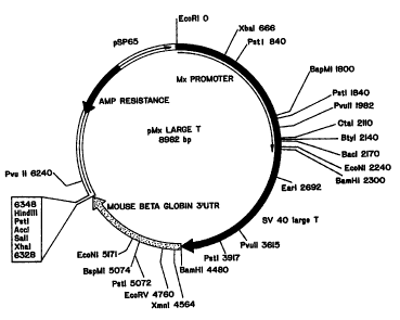
Figure 1 depicts the plasmid map of a
construct containing a 2.3 kb fragment of the murine
Mxl promoter fused to SV40 early region, followed by a
BamHl-Xbal fragment from mouse beta globin 3'
untranslated region.
Figure 2 shows NGF secretion (ng/ml/24 h)
after 4, 11 and 25 days from BHK cells encapsulated in
control, underivatized membranes (shown as "0%" in
legend) or 1% or 5% PEO-PDMS derivatized membranes
(shown as "1t" and "5%", respectively, in legend).
Cells were encapsulated with no matrix (shown as "no
mat" in legend), a Vitrogen"' matrix (shown as "vit" in
legend), or an agarose matrix (shown as "agse" in
legend).
CA 02694296 2010-03-01
;: . =
wo 9b1o2646 rcrIoS951T"291
- 9 -
Figure 3 shows NGF release from BHK cells
grown on CultiSphers" in the absence of an agarose
matrix (legend: n-mat-008, 0709-n-m) or in the
presence of an agarose matrix (legend: agaro-008,
agaro-0709).
Figure 4 shows release of catecholamines from
PC12A cells at 1, 14 and 28 days after encapsulation in
BAOs having a inert PHEMA scaffold. Panel A shows
basal catecholamine release; Panel B shows K+ -evoked
catecholamine release. The abbreviations L-dopa, NEPI,
epi, DOPAC, DA and HVA in the legend represent L-dopa,
norepinephrine, epinephrine, dopac, dopamine, and
homovanillic acid, respectively.
Figure 5 shows release of catecholamines from
PC12A cells at 1, 14 and 28 days after encapsulation in
BAOs having a inert PHEMA/MMA scaffold. Panel A shows
basal catecholamine release; Panel B shows K+ -evoked
catecholamine release. The abbreviations L-dopa, NEPI,
epi, DOPAC, DA and HVA in the legend represent L-dopa,
norepinephrine, epinephrine, dopac, dopamine, and
homovanillic acid, respectively.
Figure 6 shows release of L-dopa from
SV40/Df34-NGF cells grown on Cultisphers"' in the
presence of an alginate matrix (legend: CS/AL) or in
the presence of an agarose-matrix (legend: CS/AG) at
2, 20, 40 and 80 days after encapsulation in BAOs.
Detailed Description of the Invention
Definitions
As used herein, a "bioartificial organ" or
"BAO" is a device which may be designed for
implantation into a host or which may be made to
function extracorporeally and either be permanently or
removably attached to a host. A BAO contains cells or
living tissues which produce a biologically active
CA 02694296 2010-03-01 W696102646 PCTIUS95/09281
- 10 -
molecule that has a therapeutic effect on the host.
The BAO, upon implantation in a host recipient, should
be biocompatible. Accordingly, the BAO should not
elicit a detrimental host response sufficient to render
it inoperable or not therapeutically useful. Such
inoperability may occur, for example, by formation of a
fibrotic structure around the capsule limiting
diffusion of nutrients to the cells therein.
Detrimental effects may also include rejection of the
capsule or release of toxic or pyrogenic compounds
(e.g. synthetic polymer by-products) from the BAO to
surrounding host tissue.
BAOs comprising encapsulated cells.may be
constructed with immunoisolatory properties which
hinder elements of the host immune system from entering
the organ, thereby protecting the cells contained
withiri the bioartificial organ from detrimental immune
destruction. The use of a BAO increases the diversity
of cell types that can be employed in therapy. In
implanted BAOs, the devices, which may or may not be
immunoisolatory, usually contain the cells or tissues
producing a selected product within a semi-permeable
physical barrier which will allow diffusion of
nutrients, waste materials, and secreted products into
surrounding host tissue and retain the contained cells,
but minimize the deleterious effects of the cellular
and molecular effectors of immunological rejection.
Immunoisolatory properties, however, may not be
necessary in all cases (e.g., if the cells are
autologous or syngeneic to the host).
A "biologically active molecule" is one which
(a) may function within the cell in which it is made or
(b) may be expressed on the cell surface and affect the
cell's interactions with other cells or biologically
active molecules (e.g., a neurotransmitter receptor or
CA 02694296 2010-03-01
WO 96/02646 PCT/US9S0281
- 11 -
cell adhesion molecule), or (c) may be released or
secreted from the cell in which it is made and exert
its effect on a separate target cell or target molecule
in the host (e.g., a neurotransmitter, hormone, growth
factor, or cytokine).
As used herein, unless otherwise specified,
the term "cells" means cells in any form, including but
not limited to cells retained in tissue, cell clusters,
and individually isolated cells. The cells used in
this invention produce at least one biologically active
molecule.
Control of cell distribution within the BAO
refers to control of the cell number in the BAO,
control of the spatial location of cells within the
BAO, or both.
A wide variety of cells may be used in this
invention. These include well known, publicly
available immortalized cell.lines as well as dividing
primary cell cultures. Examples of publicly available
cell lines suitable for the practice of this invention
include, L-6 cells, MDCK cells, LLC-PK cells, B-CH3
cells, C2 cells, by hamster kidney (BHK), Chinese
hamster ovary (CHO), mouse fibroblast (L-M), NIH Swiss
mouse embryo (NIH/3T3), African green monkey cell lines
(including COS-a, COS-1, COS-6, COS-7, BSC-1, BSC-40,
BMT-10 and Vero), rat adrenal pheochromocytoma (PC12),
rat glial tumor cells (C6), RAJI (human lymphoma)
cells, MOPC-31C mouse plasmacytoma cells, MN9D cells,
MN9H cells, ripTAg transgenic mouse derived cells,
SCT-l, B-TC cells, Hep-G2 cells, AT-T20 cells, beta-
cell lines such as NIT cells or RIN cells, Ntera-2
cells (Pleasure et al.; Journ. Neuroscience, 12,
pp. 1802-15 (1992)) and human astrocyte cell lines such
as U-373 and U-937.
CA 02694296 2010-03-01
WO 96/02646 PCT/US95/09281
- 12 -
Primary cells that may be used include, bFGF-
responsive neural stem/progenitor cells derived from
the CNS of mammals (Richards et al., PNAS 89, pp. 8591-
8595 (1992); Ray et al., PNAS 90, pp. 3602-3606
(1993)), primary fibroblasts, Schwann cells (WO
92/03536), astrocytes, oligodendrocytes and their
precursors, myoblasts, and adrenal chromaffin cells.
For example, one such myoblast cell line is the C2C12
cell line.
Cells can also be chosen depending on the
particular method of growth control and differentiation
to be used. For example, stem cells can easily be used
with the methods which induce differentiation by
introducing a chemical substance. Generally, stem
cells are undifferentiated cells which in vivo are
normally quiescent but are capable of proliferation and
capable of giving rise to more stem cells having the
ability to generate a large number of progenitor cells
that can in turn give rise to differentiated or
differentiatable daughter cells. Stem cells represent
a class of cells which may readily be expanded in
culturc, and whose progeny may be terminally
differentiated by the administration of a specific
= growth factor. See, e.g., Weiss et al. (PCT/CA
92/00283).
Myoblasts are one type of cell that may be
encapsulated in a BAO according to this invention.
Myoblasts are muscle precursor cells originally derived
from mesodermal stem cell populations. A number of
myoblast cell lines are available which can undergo
differentiation in culture, e.g., L-6 and B-CH3 cells.
Primary myoblasts can be readily isolated from tissue
taken from an autopsy or a biopsy, and can be purified
and expanded. Myoblasts proliferate and fuse together
to form differentiated, multi-nucleated myotubes.
CA 02694296 2010-03-01
WO 96102646 PCT/OS95ivy281
- 13 -
Myotubes no longer divide, but continue to produce
muscle proteins. While proliferating, myoblasts may
readily be genetically engineered to produce
therapeutic molecules. Methods are known for
introducing one or more genes into myoblasts to produce
the desired biologically active.molecules. Myoblasts
are capable of migrating, fusing into pre-existing
fibers, and serving as carriers for the introduced
gene(s). Verma et al. (WO 94/01129); Blau, et al.,
TIG, 9, pp. 269-74 (1993); WO 93/03768; WO 90/15863.
The engineered cells may then be encapsulated and
allowed to differentiate.in the BAO or the
differentiated cells may themselves be encapsulated.
The choice of cells also depends upon the
intended application. The cells within the BAO may be
chosen for secretion of a neurotransmitter. Such
neurotransmitters include dopamine, gamma aminobutyric
acid (GABA), serotonin, acetylcholine, noradrenaline,
epinephrine, glutamic acid, and other peptide neuro-
transmitters. Cells can also be employed which
synthesize and secrete agonists, analogs, derivatives
or fragments of neurotransmitters which-are active,
including, for example, cells which secrete
bromocriptine, a dopamine agonist, and cells which
secrete L-dopa, a dopamine precursor.
The cells can be chosen for their secretion
of hormones, cytokines, growth factors, trophic
factors, angiogenesis factors, antibodies, blood
coagulation factors, lymphokines, enzymes, and other
therapeutic agents or agonists, precursors, active
analogs, or active fragments thereof. These include
enkephalins, catecholamines, endorphins, dynorphin,
insulin, factor VIII, erythropoietin, Substance P,
nerve growth factor (NGF), Glial cell line-derived
Neurotrophic Factor (GDNF); platelet-derived growth
. CA 02694296 2010-03-01
WO 96/02646 PCT/[1S95/09281
- 14 -
factor (PDGF), epidermal growth factor (EGF), brain-
derived neurotrophic factor (BDNF), neurotrophin-3
(NT-3), neurotrophin-4/5; CDF/LIF, bFGF, aFGF, an array
of other fibroblast growth factors, ciliary
neurotrophic factor (CNTF), and interleukins.
It should be understood from the foregoing
that the cells useful in the methods of this invention
include untransformed cells that secrete the desired
biologically active molecule(s), or cells that can be
transformed to do so. .
The genes encoding numerous biologically
active molecules have been cloned and their nucleotide
sequences published. Many of those genes are publicly
available from depositories such as the American Type
Culture Collection (ATCC) or various commercial
sources. Genes encoding the biologically active
molecules useful in this invention that are not
publicly available may be obtained using standard
recombinant DNA methods such as PCR amplification,
genomic and cDNA library screening with oligonucleotide
probes from any published sequences. Any of the known
genes coding for biologically active molecules may be
employed in the methods of this invention. See, e.g.,
United States Patent 5,049,493; Gage et al., United
States Patent 5,082,670; and United States Patent
5,167,762.
A gene of interest (i.e., a gene:that encodes
a suitable biologically active molecule) can be
inserted into a cloning site of a suitable expression
vector by using standard techniques. These techniques
are well known to those skilled in the art.
The expression vector containing the gene of
interest may then be used to.transfect the cell line to
be used in the methods of this invention. Standard
transfection techniques such as calcium phosphate co-
CA 02694296 2010-03-01
WO 96/02646 PCT/US9SIOy281
- 15 -
precipitation, DEAE-dextran transfection, lipid-
mediated methods, or electroporation may be utilized.
Methods are provided herein to control the
growth of dividing cells, whereby the balance between
proliferation and differentiation can be controlled to
provide a supply of differentiated, non-dividing
encapsulated cells within the BAO. Methods are also
provided to control the growth of both dividing and
non-dividing cells, whereby cell distribution and cell
number within the BAO.are controlled, resulting in
reduced formation of necrotic cell cores and reduced
cellular debris.
Control of Proliferation and
Differentiation By Genetic Engineering
Methods and compositions are herein provided
for controlling cell growth by genetic alteration of
cells with a gene encoding a product that influences
cell proliferation or differentiation.
According to one aspect of this invention,
conditionally immortalized cell lines are used to
achieve growth control in the BAO. Primary cells are
transformed with a gene encoding a proliferation-
promoting product. The proliferation-promoting gene is
operatively linked to a regulatable promoter. The
techniques described by Land et al., Nature, 304,
pp. 596-602 (1983) or Cepko, Neuron, 1, pp. 345-53
(1988) for producing immortalized cells can be
routinely modified to produce conditionally-
immortalized cells.
According to this method, cell proliferation
(i.e., mitosis) can be inhibited or arrested by
decreased expression of a proliferation-promoting gene,
such as an oncogene (e.g., c-myc, v-mos, v-Ha-ras, SV40
T-antigen,"El-A from adenoviruses). Reduced expression
of the oncogene is achieved by downregulation,
CA 02694296 2010-03-01
WO 96/02646 PCT/US95/09281
- 16 -
repression or inactivation of the promoter driving
oncogene expression when the BAO is implanted in vivo
in a host. Upregulation, activation or derepression of
the regulatable promoter in vitro results in expression
of the proliferation-promoting gene, thereby permitting
cell proliferation in vitro. Suitable promoters are
those which can be downregulated in vivo, including,
e.g., glucocorticoid responsive promoters, such as PNMT
(Hammang et al., Neuroprotocols, 3, pp. 176-83 (1993)
and interferon ("IFN")-responsive promoters, such as
Mxl (Hug et al., Mol. Cell. Biol., 8, pp. 3065-79
(1988); Arnheiter et al., Cell, 52, pp. 51-61 (1990)),
retroviral long terminal repeat promoters, tetracycline
responsive promoters, e.g., the lac promoter, and
insulin-responsive promoters. See also, McDonnell et
al. WO 93/23431. It will be appreciated that choice of
promoter will depend upon the intended implantation
site. Thus, e.g., glucocorticoid or IFN-responsive
promoters are useful for implantation in the brain
according to this method, since the levels of
glucocorticoid and/or IFN are very low in the brain.
Thus, these promoters would not be expected to direct
significant levels of expression of the oncogene upon
implantation of the BAO in the brain.
In one embodiment, conditionally-immortalized
cells are generated by operatively linking an oncogene
to a regulatable promoter. The promoter is activated
or upregulated in the presence of'a binding protein.
Production of the binding protein can be regulated by
operatively linking the gene encoding the binding
protein to a tetracycline responsive promoter.
For example, one embodiment contemplates a
transformed cell containing.a constitutive promoter
driving tet repressor expression. The cell
additionally contains a heterologous gene operatively
CA 02694296 2010-03-01
WO 96A2646 PCT/US9"281
- 17 -
linked to the CMV-IE promoter. If the CMV-IE promoter
is flanked with tet operator sequences, expression from
this promoter can be turned off by the tet repressor.
In the presence of tet, transcription occurs because
tet binds with the tet repressor allowing other
transcription factors to bind the CMV-IE promoter.
According to this embodiment, the oncogene is only
expressed when tetracycline is present. Thus, cells
can be proliferated in vitro in the presence of
tetracycline.
Several days prior to implantation,
tetracycline can be removed to reduce transgene
expression,and thus correspondingly reduce or halt
cell proliferation in the BAO.
15- In a specific embodiment using conditionally
immortal-ized -cells, growth control is achieved using
the Mxl promoter. The Mxl gene encodes a protein which
confers resistance to influenza A and B. The Mxl gene
is tightly regulated by its promoter. In the absence
of interferon ("IFN"), the gene is not expressed and
the gene is inducible in the presence of IFNa and IFNB.
Arnheiter et al., Cell, 52, pp. 51-61 (1990) reported
the generation of Mxl transgenic mice that exhibited
interferon inducible expression of the transgene in
several tissues. The SV40 large T-antigen is capable
of transforming and immortalizing cells derived from a
number of tissues.
In one embodiment, the mouse Mxl promoter can
be fused with the SV40 early region and the chimeric
gene used to generate transgenic mice. The tight
regulation afforded by the Mxl promoter elements allows
one to control oncogene-expression in tissues or in
cell cultures prepared from-the transgenic animals,
thereby allowing creation of conditionally-immortalized
cell lines.
CA 02694296 2010-03-01 77402-9
- 18 -
In the presence of IFNa or IFNB, the cell
lines produced in this manner can be expanded
arithmetically as with most other cell lines. Cell
division can be halted by removal of IFNa or IFNB,
either'before or after encapsulation. In a preferred
embodiment, neural stem cells (neurospheres) can be
prepared from transgenic mice containing the Mxl-SV40
T-antige.,.n construct using the method of Weiss
(W0 93/01275). The conditionally immortalized neural stem
cell line so obtained can then be encapsulated and
implanted in vivo in a host.
Additionally, if desired, the conditionally
immortalized neural stem cell line can be further
genetically modified to release any of a number of
growth factors or neurotransmitter molecules, according
to standard techniques. Other IFN-responsive promoters
may also be useful in this embodiment. These promoters
include metallothionein, H-2Kb, H-2Dd, H-2Ld, HLA-A3,
HLA=DRa, an HLA class I gene, 202, 56K, 6-16, IP-10,
ISG15, ISG54, and 2',5'-oligo(A) synthetase. See, Hug
et al., Mol. Cell. Biol., 8, pp. 3065-79 (1988).
This embodiment is particularly suited for
cells to be encapsulated in BAOs for implantation in
the brain. Circulating levels of IFNa and IFNB in the
brain. are sufficiently low that transcriptional
activity driven by the Mxl promoter is insufficient to
result in cell proliferation. In the founder
transgenic animals, the expression of T-antigen could
be induced in several tissues, but the natural
3-0 exprestion of the oncogene was seen only in the thymus.
However, thymic expression of the oncogene is a
relatively common phenomenon in transgenic animals
- expressing the SV40 early region. Thus, in the absence
of significant oncogene expression, the cells can be
kept in a near quiescent state in vivo.
I . = , i . = i
CA 02694296 2010-03-01
WO 96/02646 PGT/US9SIu1'L81
- 19 -
Another embodiment makes use of the
observation that in traditional retroviral infection
techniques to genetically engineer cells for use in
vivo, retroviral promoters, e.g., the long terminal
repeat ("LTR") promoter, are used. See, e.g., Gage
et al. (United States Patent 5,082,670). The
expression of genes driven by these promoters is
typically downregulated in vivo. It is thought that
this downregulation is mediated by circulating
cytokines. This invention makes use of this normally
detrimental downregulation of retroviral genes to stop
or decrease cellular proliferation when cells are
encapsulated within the BAO and implanted in vivo. In
this instance, an immortalizing gene (oncogene) is
driven from the LTR. This gene will "immortalize" the
cells while they are maintained and expanded in vitro.
Following implantation, in the presence of cytokines,
the "immortalizing" oncogene.-is downregulated,
proliferation decreases or stops and the cells may
become quiescent within the device.
According to this embodiment conditionally-
immortalized cells may be:produced by retroviral
infection or DNA transfection with cDNA containing an
oncogene (e.g. c-myc, v-mos, v-Ha-ras, SV40 T-antigen,
El-A from adenoviruses) operatively linked to a
retroviral promoter, e.g., the LTR promoter. We prefer
Moloney murine-leukemia virus (MLV), Rous sarcoma virus
(RSV), and mouse mammary tumor virus (MMTV) promoter
sequences.
These transformed cells will normally express
the oncogene in vitro. Successfully transformed cells
will be grown in culture using established culture
techniques. LTR-transgene expression can be stimulated
by the addition of dexamethasone or epidermal growth
factor to shorten the amount of time'needed to culture
CA 02694296 2010-03-01
W6 96102646 PCT/US95/09281
- 20 -
the transformed cells. By exposing the cells to
cytokines, e.g., gamma-interferon (IFN-ry), TNF-a and
transforming growth factor-B (TGFB), preferably several
days prior to encapsulation and implantation, mitosis
can be reduced by hindering LTR-driven transgene
expre5sion. Schinstine and Gage, Molecular and
Cellular Approaches to the Treatment of Neurological
Disease, 71, ed. Waxman, S.G. (1993); Seliger et al.,
J. Immunol., 141, pp. 2138-44 (1988); Seliger et al.,
J. Virolocxv, 61, pp. 2567-72 (1987); Seliger et al., J.
Virolocry, 62, pp. 619-21 (1988).
Any suitable-cell can be conditionally
immortalized according=to the above methods:. One of
ordinary skill in the art can determine the suitability
of a given cell type for conditional immmortalization
by screening methods well known in the art, including
according to the methods provided herein.
Methods are provided herein for growth
control of immortalized cell lines or other
continuously proliferating cells by transforming these
cells to include tumor suppressor genes, e.g., the p53
gene or RB gene, to halt or reduce proliferation.
Tumor suppressor genes, or anti-oncogenes, are believed
to be growth-constraining genes. See, e.g., Weinberg,
Neuron, 11, pp. 191-96 (1993). For example, a wild-
type p53-activated fragment 1(WAF1) can suppress tumor
cell growth in culture. It is theorized that genes
induced by the p53'protein may mediate its biological
role as a tumor suppressor. El-Deiry et al., "WAF1, a
Potential Mediator of p53 Tumor Suppression," Cell, 75,
pp. 817-825 (1993). The WAF1 gene is. also referred to
as the CIPi gene. Other p53-mediated growth arresting
genes include GADD45 and GADD153 (or CHOP). See Ron
Proc. Natl. Acad. Sci. USA, 91, pp. 1985-86 (1994).
CA 02694296 2010-03-01 = =
WO 96/02646 PCT/US95iuy281
- 21 -
The standard techniques for transforming cells with
heterologous DNA discussed above can be used here.
According to one embodiment, immortalized
cells or continuously proliferating cells are
transformed with a tumor suppressor gene operatively
linked to a regulatable promoter. Use of a suitable
regulatable or inducible promoter allows expression of
the transgene to be downregulated or "turned off" when
the transformed cells are cultured in vitro, thus
permitting expansion. Upon encapsulation and
implantation, the promoter is "induced," or
upregulated, and expression of the tumor suppressor
gene occurs, resulting in reduced or halted cell
proliferation.
The tyrosine hydroxylase and erythropoeitin
promoters may be useful in this aspect of the
invention. These promoters are typically
"downregulated" under high 02 conditions, such as those
encountered in vitro, but are "upregulated" under low
02 conditions, like those that cells encounter upon
encapsulation in a BAO and implantation in a host.
In addition, suitable coupled or
derepressible promoter systems may be used to achieve
the desired regulation of the proliferation-suppressing
gene. One suitable system, e.g., involves use of the
AP1 promoter and the lac operator/PGK1 promoter system
described by Hannan et al., Gene, 130, pp. 233-39
(1993). The AP1 promoter is operatively linked to the
lac repressor gene. The lac0 (lac operator) and
3-phosphoglycerate kinase (PGK1) promoter is
operatively linked to the proliferation-suppressing
gene. Addition of exogenous phorbol ester in vitro
induces the AP1 promoter, resulting in expression of
the lac repressor protein. In the presence of
repressor protein, the lac0-PGK1 promoter construct is
CA 02694296 2010-03-01 =
~ WO'96102646 PCTIUS9S/09281
- 22 -
repressed, and no expression of the proliferation-
suppressing gene occurs. In the absence of phorbol
ester in vivo, no repressor protein is expressed, the
lacO-PGK1 promoter is derepressed, and the
proliferation-suppressing gene is expressed.
According to one method, a suitable cell is
transformed with a gene encoding a differentiation-
inducing product. This.differentiation-inducing gene
is operatively linked to a regulatable promoter.
According to this method, the differentiation-inducing
gene would be expressed upon encapsulation and in vivo
implantation in a host. However, expression can be
arrested or inhibited in vitro by appropriate
downregulation, repression or inactivation of the
regulatable promoter, thus allowing expansion of a
desired cell or cell line in vitro. This method can be
used with dividing cells, or primary cells that have
been immortalized. High mobility group chromosomal
protein 14, "HMG," is one example of a gene involved in
regulating differentiation of cells. Any suitable
promoter that is upregulated in vivo but which can be
"turned off" or downregulated in vitro can be used in
this embodiment, as discussed supra for use with
proliferation-arresting genes. In addition, any
suitable derepressible promoter system can be used, as
discussed su ra, for the regulation of tumor suppressor
gene expression.
Another method of growth control uses
antisense RNA or DNA, or their derivatives. Antisense
RNA or DNA is a single-stranded nucleic acid which is
complementary to the coding strand of a gene or to the
"coding" mRNA produced from transcription of that gene.
If the antisense RNA is present in the cell at the same
time as the mRNA, the antisense RNA hybridizes to the
mRNA forming a double strand which then cannot be
CA 02694296 2010-03-01
WO 96102646 PCr/US9.%.,181
- 23 -
translated by ribosomes to make protein. Antisense RNA
can be administered to cells either via microinjection
or bulk addition to culture medium. The preferred
method of the instant invention is to transfect target
cells with eukaryotic expression vectors. Neckers
et al., "Antisense Technology: Biological Utility And
Practical Considerations", Am. J. Physiol., 265 (LunQ
Cell. Mol. Physiol., 9), pp. Ll-L12 (1993).
According to this embodiment, an antisense
gene encoding antisense RNA to either a proliferation-
inducing gene or a tumor suppressor gene can be
operatively linked to an ilnducible promoter. When the
promoter is induced, antisense RNA is produced. If the
transformed cells contain a proliferation-inducing
gene, according to this embodiment, antisense RNA
production would be halted or downregul.ated in vitro to
allow for cell expansion, and upregulated in vivo, to
achieve cessation or reduction of proliferation.
Alternatively, if the transformed cells
contain a tumor suppressor gene, antisense RNA
production would be upregulated in vitro and
downregulated in vivo to achieve the desired growth
control.
In addition, antisense technology could be
used to construct any antisense gene to a gene encoding
a product essential for proliferation or
differentiation. Appropriate induction of the
expression of the antisense gene would allow one of
skill in the art to achieve the desired growth control
of encapsulated cells according to this invention.
It is preferred to use a regulatable
promoter/gene construct that can be manipulated in vivo
in the event that it becomes_necessary or desirable to
induce further cell proliferation in vivo. For
example, in the Mxl/SV40 construct'discussed supra, IFN
. CA 02694296 2010-03-01 =
~77402-9
- 24 -
can be added locally or systemically to induce oncogene
expression. An increase in cell division in vivo in
the BAO may be desirable to increase cell number to
replace dead cells in the BAO, or to achieve increased
output of the desired biologically active molecule.from
the BAO.
Control of Growth and Differentiation
by Use of Chemical Compounds
According to another method of this
invention, cells may be exposed to a treatment which
inhibits proliferation or induces differentiation. In
some methods, the treatment comprises providing a
chemical compound or growth factor. In other methods,
the treatment comprises removing a chemical compound or
growth factor from the growth medium. The treatment
may be before or after encapsulation in the BAO,
preferably before encapsulation.
The protein or chemical compound used depends
on the cell type and the desired effect. One of
ordinary skill in the art could screen a given cell
type for its responsiveness to.a selected compound or
protein, with routine techniques.
In one method, cell.distribution is
controlled by a treatment that comprises removing a
-proliferation-inducing chemical compound or growth
factor from the cell growth medium. In one embodiment,
growth factors, such as epidermal growth factor
.("EGF"), transforming growth factor a ("TGF-a"),
amphiregulin, or any other suitable agent, can be used
to induce proliferation of stem or progenitor cells,
including cells from embryonic sympathetic ganglia, and
immortalized progeõpitor cells, preferably neural stem
cells (Weiss, WO 93/01275). This allows
maintenance and.expansion of a supply of neuronal
precursor cells in vitro. When encapsulated in the
CA 02694296 2010-03-01 ~ =
WO 96102646 PCT10S95...A81
- 25 -
absence of these proliferation-inducing growth factors,
the neuronal precursor cells cease dividing and
differentiate.
The neuronal precursor cells may be further
induced to differentiate by treatment with, e.g.,
phorbol ester, or growth on a fixed substrate,
including ionically charged surfaces such as poly-L-
lysine and poly-L-ornithine and the like.
Differentiation may also be induced by treatment with a
member of the FGF family in combination with at least 1
member of either the ciliary neurotrophic factor (CNTF)
or nerve growth factor (NGF) family of factors as
described in Ip et al. (WO 94/03199).
In another embodiment, a multilineage growth
factor produced in the stroma, also termed "mast cell
growth factor," "stem cell factor," "c-kit-ligand," or
"Steel factor," can be used to induce proliferation of
hematopoietic stem cells. To maintain a supply of
dividing cells in vitro, hematopoietic stem cells are
cultured in the presence of mast cell growth factor.
To arrest or reduce proliferation, the mast cell growth
factor is removed from the culture medium. This can be
done before or after encapsulation, preferably before
encapsulation.
Examples of other multilineage growth factors
that promote proliferation include interleukin-3 and
granulocyte-macrophage colony-stimulating factor. Mast
cell growth factor can also affect cell growth in
combination with other multilineage growth factors, or
lineage specific growth factors, e.g., erythropoietin.
For example, mast cell growth factor is thought to act
synergistically with IL-3 in inducing proliferation and
differentiation pf highly enriched murine hematopoietic
stem cells. Galli et al., "The Biology of Stem Cell
Factor, a New Hematopoietic Growth Factor'Involved in
CA 02694296 2010-03-01
W0 96/02646 PCT/OS95/09281*
- 26 -
Stem Cell Regulation," Int. J. Clin. Lab. Res., 23,
pp. 70-77 (1993).
In another method of this invention, control
of cell distribution in the BAO may be achieved by
providing a chemical compound or growth factor which
inhibits cell proliferation or induces differentiation.
Any suitable proliferation-inhibiting or
differentiation-inducing compound may be used according
to this method.
It will be appreciated that different cell
types may respond differently to various chemical
compounds. One of ordinary skill in the art can
routinely screen a particular compound to determine its
effectiveness in affecting proliferation or
differentiation of a given cell type.
In one embodiment, cytokines, including,
e.g., transfo"rming growth factor 131 (TGF131), may be
used to arrest or inhibit cell proliferation or to
induce cell differentiation. For example, decreased
proliferation and enhanced differentiation in BHK cells
can-be achieved by exposure to TGF81 and ascorbate.
Similarly, TGF81 can be used to induce differentiation
in fibroblast cells and also as a growth inhibitor of
keratinocytes and endothelial cells. Phillips et al.,
"Ascorbic Acid and Transforming Growth Factor-81
Increase Collagen Biosynthesis via Different
Mechanisms: Coordinate Regulation of Proacl(I) and
Proal(III) Collagens," Archives of Biochemistry and
Biophysics, 295, pp. 397-403 (1992).
In another embodiment, TGFB1, serotonin, or
FGF may be used to control the growth of neuroendocrine
cells. The growth of neuroendocrine cells can be
regulated by their own products in an autocrine
fashion. TGFB1 is an autocrine growth-inhibitory
factor for human pancreatic,carcinoid cells (BON),
CA 02694296 2010-03-01 =
~ . .
WO 96102646 PCT/US9Si~i>181
- 27 -
while FGF and serotonin are autocrine growth-
stimulatory factors. The inhibitory effect of TGFtii on
the growth of BON cells can be reversed by addition of
serotonin. Townsend Jr. et al., "Studies of Growth
Regulation in a Neuroendocrine Cell Line," Acta
Oncologica, 32, pp. 125-130 (1993).
A variety of other chemicals may also be used
according to the methods of this invention to arrest or
inhibit proliferation or induce differentiation of
cells. These chemicals include mitomycin-C, 5-bromo-
deoxyuridine (BrdU), prostaglandin El (PGE,), dibutyryl
cAMP, 1-B-D-arabinofuranosyl cytosine (Ara-C).,
nicotinamide, and heparin. Mitomycin may be
particularly suited for controlling proliferation of
encapsulated t3HC cell lines. See, e.g., Radvanyi
et al., Mol. Celi. B-i-oi., 13, pp. 4223-27 (199-3).
Sometimes a combination of chemicals can be
used. Human neuroblastoma cells IMR-32 may be induced
to differentiate in vitro when treated with mitomycin C
and BrdU or PGE1 and dibutyryl cAMP (dbcAMP). Gash
et al., "Amitotic Neuroblastoma Cells Used for Neural
Implants in Mon=keys," Science, 233, pp. 1420-22 (1986).
Serial pretreatments of human embryonal
rhabdomyosarcoma cell line with Ara-C results in marked
growth inhibition in vitro, loss of tumorigenicity in
vivo, and a more differentiated phenotype even
following removal of the compound.. Crouch et al.,
"Ara-C Treatment Leads to Differentiation and Reverses
the Transformed Phenotype in a Human Rhabdomyosarcoma
Cell Line," Exaerimental Cell Research, 204, pp. 210-
16 (1993). Nicotinamide (NIC) is thought to induce
differentiation and maturation of human fetal
pancreatic islet cells. Otonkoski et al.,
"Nicotinamide Is a Potent Inducer of Endocrine
CA 02694296 2010-03-01.
77402-9
,. .
- 28 -
Differentiation in Cultured Human Fetal Pancreatic
Cells," J. Clin. Invest., 92, pp. 1459-66 (1993).
The addition of dbcAMP has also been reported
to influence the differentiation of developing tissues.
For example, dbcAMP is thought to modulate the
differentiation of.astrocyte precursors, induce neurite
formation in PC12 cells, and stimulate schwann cell
proliferation. Baron-Van Evercooren et al., "Schwann
Cell Differentiation in vitro: Extracellular Matrix
lo Deposition.and Interaction," Dev. Neurosci., 8,
pp. 182-96 (1986). Similarly, differentiation of
Schwann cells can be induced by exposure to ascorbate.
Ibid.
Further, sialoglycopeptide ("SGP") molecules
may be used to inhibit or arrest cell proliferation.
For example, an 18 kDa cell surface sialoglycopeptide
isolated from i r_tact bovine cerebral cortex cells
arrested proliferation of exponentially growing'Swiss
3T3 cells. See, e.g., Toole-Simms et al., Jour. Cell.
Physiol., 147, pp. 292-97 (1991); Fattaey et al.,- E.
Cell. Res., 194, pp. 62-68 (1991). Numerous
transformed and untransformed cell types have been
shown to be sensitive to some SGPs. These cells
include epithelial-like and fibroblast cells from'a
broad spectrum of vertebrate and invertebrate species.
See, e.g., Fattaey et al., Jour.. Cell. Phvsiol., 139,
pp. 269-74 (1989).
It will be appreciated that some of the
foregoing treatments may only have a transient effect
on proliferation and differentiation. In such cases it
may be desireable to provide a continuously replenished
supply of the.compound or growth factor to the
encapsulated cell when implanted in vivo in the host.
This can be accomplished by use of a bioerodable
polymer non=cellular source of the growth factor or
CA 02694296 2010-03-01
WO 96/02646 PCT/US9_S,.;w281
- 29 -
compound, or by co-encapsulating a cellular source of
the growth factor or compound, or any other suitable
means. See, e.g., United States 5,106,627 and
5,156,844.
Control of Growth By Irradiation
Cell proliferation can also be controlled
through exposure of cells to a suitable dose of
irradiation, e.g., x-rays, ultraviolet (UV) radiation,
and the like. When cells are subjected to irradiation,
their progression through the cell cycle may be
arrested. The critical dose rate, or minimum dose rate
can be determined for a chosen cell type using methods
known in the art. See, e.g., Stanley and Lee, Radiat.
Res., 133, pp. 163-9 (1993); Mitchell et al., Radiat.
Res., 79, pp. 537-51 (1979). For example, normal human
epidermal keratinocytes irradiated with 5 and 10 mJ/cm2
ultraviolet B(UVB) radiation showed a significant (up
to 78%) decrease in proliferation 3 to 5 days post-
irradiation. Prystowsky et al., J. Invest. Dermatol.,
101, pp. 54-58 (1993). Yi et al., RadiationResearch,
133, pp. 163-69 (1993) provide a method for calculating
the lowest dosage required to stop cell proliferation
by exposure to x-rays..
Control of Growth and Differentiation
By Use of Extracellular Matrix Molecules
Methods are provided herein for the control
of cell distribution in a BAO by modification of a
growth surface with a growth controlling extracellular
matrix ("ECM") (or components thereof) alone or in
combination with a growth controlling physical matrix
or other growth regulating substances.
In living tissue, the ECM is formed from a
variety of proteins and polysaccharides which are
secreted by cells and assembled into a network in
CA 02694296 2010-03-01
WO`96/02646 PC7/US95/09281
- 30 -
proximity to the cells that secreted them. ECM
molecules include glycosaminoglycans and proteoglycans,
such as chrondroitin sulfate, fibronectin, heparin
sulfate, hyaluron, dermatan sulfate, keratin sulfate,
laminin, collagen, heparan sulfate proteoglycan (HSPG)
and elastin. In particular, collagen is a major
component of ECM in vivo. ECM molecules are known to
cause decreased cell proliferation and increased cell
differentiation. In addit~on, acellular ECM when used
in the methods of this invention may influence the
spatial location of cells encapsulated in the BAO.
ECM may be obtained by culturing cells known
to deposit ECM, including cells of mesenchymal or
astrocyte origin. Schwann cells can be induced to
synthesize ECM when treated with ascorbate and cAMP.
These ECM components resemble a precursor form of the
basement membrane which support Schwann cell
proliferation. Furthermore, naturally produced ECM
from endothelial cells and a reconstituted basement
membrane gel from Engelbreth Holm-Swarm tumor cells
(EHS) supports the growth and differentiation of
various epithelial and endothelial cells. Baron-Van
Evercooren et al., "Schwann Cell Differentiation in
vitro: Extracellular Matrix Deposition and
Interaction," Dev. Neurosci., 8, pp. 182-96 (1986).
In one embodiment, growth control is achieved
by coating a growth surface in the BAO with ECM (or its
growth controlling components). We prefer seeding the
growth surface in the BAO with cells that produce ECM,
and culturing the cells until confluent. The cells are
then treated with detergent and NH4OH. The resulting
BAO, with acellular ECM coated on a growth surface, is
then used to encapsulate cells that produce the desired
biologically active molecule.
CA 02694296 2010-03-01 WO 96/02646 PCT/US95,;,>181
- 31 -
In another embodiment, ECM is prepared
substantially in the same manner in vitro, lyophilized,
fragmented and mixed with cells as a suspension. The
cell/ECM fragments are then co-loaded into the BAO.
Cells grown in presence of some ECM molecules
show decreased proliferation and increased
differentiation compared to cells grown in conventional
monolayer culture. For example, adrenbcortical cells,
known to synthesize certain steroid hormones such as
aldosterone, exhibit decreased proliferation when grown
in vitro in the presence of collagen gel. Fujiyama
et al., "Influence of Extracellular Matrix on the
Proliferation and Differentiation of Adrenocortical
Cells in Culture," Path. Res..Pract., 189, pp. 12051-
14 (1993).
Schwann cells may also exhibit decreased
proliferation and increased differentiation when
cultured in the presence of collagen.
Endocrine cells are also known to
differentiate in vitro when grown on surfaces coated
with a combination of type IV collagen and HSPG. Type
IV collagen is necessary for cell adhesion and the HSPG
induces differentiation. de Bruine et al.,
"Extracellular Matrix Components Induce Endocrine
Differentiation In Vitro in NCI-H716 Cells," American
Journal of Patholoctv, 142, pp. 773-782 (1993).
Various growth factors or chemical compounds,
including those discussed supra, may be added to the ECM components to further
control the growth and
differentiation of cells. Growth factors may be
administered to the cells in vitro prior to
implantation or to the cel.ls.in vivo, or both. See,
e.g., United States Patents 5,156,844 and 5,106,627,
which refer to methods for delivering growth factors
using either a co-encapsulated cellular or non-
CA 02694296 2010-03-01 =
W096/02646 PGTIOS95/09281
- 32 -
cellular source of the growth factor. In addition, the
ECM molecules may be derivatized with growth
controlling peptides according to known techniques.
For example, transforming growth factor-B,
which modulates cell growth on its own, and which
reversibly binds to certain ECM molecules (e.g.
decorin), can be added to ECM to potentiate the growth-
inhibiting effects of ECM molecules.
Likewise, heparin has also been shown to
prevent the growth of both untransformed cells and
transformed cell lines. Matuoka et al., Cell Structure
and Function, 9, p. 357 (1984).
Basic fibroblast growth factor (bFGF) has
also been reported to enhance endocrine cell
differentiation when added along with ECM components.
See, de Bruine et al., "Extracellular Matrix Components
Induce Endocrine Differentiation In Vitro in NCI-H716
Cells," American Journal of Pathology, 142, pp. 773-
782 (1993).
Growth factors may exhibit different effects
on cells when combined with different components of
ECM. For example, fibroblast growth factor (FGF) has
been shown to be an effective differentiating factor
and a weak mitogen for chromaffin cells grown on
laminin. However, when FGF is added to chromaffin
cells grown on collagen, FGF is a weak differentiation
factor and a strong mitogen. This behavior has also
been shown for the cyclic AMP analogue 8-(4-
chlorophenylthio) cyclic AMP. Chu et al.,
Neuroscience, 95, pp. 43-54 (1994).
Table 1 is a partial list of ECM molecules
growth factors and chemical compounds known to
influence proliferation and differentiation in
particular cell types.
. . .. . I 1 .i . . i CA 02694296 2010-03-01
WO 96/02646 PCT/US951111181
- 33 -
Table 1: ECM MOLECULES, GROWTH FACTORS AND
CHEMICAL COMPOUNDS INFLUENCING PROLIFERATION OR DIFFERENTIATION
Differentiation Inducer/
Cell Tyae Growth Inhibitor Proliferation Promoter
Schwann ascorbate; collagen (VitrogenTM); TGF-[i; dbcAMP
Cultisphers/agarose
PC12 NGF; dbcAMP; SGP
Fibroblasts TGF-6-1; Cultisphers/agarose; Vitrogen"'
ascorbate; SGP
Myoblasts collagen; ascorbate
Neural stem laminin; Peptite. 2000; Culti- EGF; bFGF; TGF-a;
sphers/Peptite 2000; phorbol ester; amphiregulin
heparin; FGF and (CNTF or NGF)
Human embryonal Ara-C
rhabdomyosarcoma
cell line
Human fetal pancreatic Nicotinamide (NIC)
islet cells
Astroblasts dbcAMP
Swiss 3T3 SGP
Adrenocortical Collagen
Endocrine Type IV Collagen + HSPG; bFGF +
ECM components
Chromaffin FGF + laminin; 8-(4-chloro- FGF + collagen; 8-(4-
phenylthio)cyclic AMP + laminin chlorophenylthio)cyclic
AMP + collagen
Hematopoietic stem Mast cell Growth Factor
cells
BHK TGFB-l + Ascorbate; ECM from E15
rat meningeal cells
Keratinocytes TGFf3-1
Endothelial cells' TGFB-l
Neuroendocnne TGFIi-1 TGFt3-1 + Ascorbate;
(human pancreatic Serotonin; FGF
caranoid cells (BON))
Human neuroblastoma Mitomycin C + BrdU; PGEI +
Cell line IMR-32 dbcAMP; SGP
SCT-l. Collagen; Ascorbate
The growth surfaces within the BAO include
the luminal surfaces of the BAO, and additionally
CA 02694296 2010-03-01
rv0 96/02646 PCT/US95/09281F
34 -
include other growth surfaces, such as an inner
support, that may be encapsulated within the BAO.
Microcarriers may provide a surface for cell
growth. Use of microcarriers can allow a greater
number of cells to be encapsulated and evenly
distributed within the BAO, especially for cells that
become growth contact inhibited. Several types of
microcarriers are commercially available, including
Cytodex (Sigma, St. Louis, MO) dextran microcarriers,
and CultiSpher"' (HyClone Labs, Logan, UT) macroporous
gelatin microcarriers and glass microcarriers. These
microcarriers are often used for the culture of
anchorage dependent cells. Cell lines which have been
shown to grow on macroporous gelatin microcarriers
include OBHK, BHK-21, L-929, CHO-Kl, rCHO, MDCK, V79,
F9, HeLa, and MDBK. Microcarriers may also be made of
or coated with other ECM molecules (such as FACT"'
collagen coated microcarriers (Solo Hill Labs, Ann
Arbor, MI)), or acellular ECM, substantially as
described above.
In one preferred embodiment cells producing
the desired biologically active molecules can be seeded
onto the ECM coated microcarrier surfaces and cultured
on the microcarriers in vitro, prior to encapsulation
and implantation. Cherksey (WO 93/14790) refers to the
culturing of cells on glass or plastic microbeads and
subsequent implantation of the microbeads into the
brain of a recipient.
In another embodiment accordingto this
invention, cells seeded on microcarriers may be
suspended in the presence of a suitable growth-
inhibiting matrix and then encapsulated in the BAO.
Such matrix material (e.g., agarose or agar for
fibroblasts; collagen for adrenocortical cells)
physically inhibits further cell outgrowth. Such
CA 02694296 2010-03-01
-77402-9
- 35 -
hydrogel matrices are described in, e.g., Dionne WO
92/19195.
According to another aspect of this
invention, agarose may also be used as a substitute for
ECM by derivatization with peptide sequences to affect
cell attachment to the matrix. For example, agarose
hydrogels may be derivatized with.peptide sequences of
laminin or fibronectin.
In this method, cells are suspended in 3-D
.10 matrices composed of agarose derivatized with a peptide
sequence that recognizes a cell surface receptcr
molecule involved in cell adhesion. Several peptide
sequences have been shown (in 2-p) to promote cell
adhesion. See, e.g., Pierschbacher et al., Science,
309, pp. 30-33 (1984); Graf et al., Biochemistry, 26,
pp. 6896-900 (1987); Smallheiser et al., Dev. Brain
Res., 12, pp. 136-40 (1984); Jucker et al., J.
Neurosci. Res., 28, pp. 507-17 (1991). The derivatized
agarose matrices of this invention allow presentation
of the appropriate molecular cues for cell adhesion in
3-D. The agarose concentration is preferably 1.25% w/v
or less, most preferably about 1.0%. We prefer RGD-
containing sequences (i.e. ArgGlyAsp; AA2-AA4 of SEQ ID
NO:2), YIGSR-containing sequences (TyrIleGlySerArg;
AA5-AA9 of SEQ ID NO:1), IItVAV-containing sequences
(IleLysValAlaVal; AA11-AA15 of SEQ ID NO:3), and the
like. Derivatization can be achieved using a bi-
functional coupling agent, such as 111,.
carbonyldiimidazole or any other suitable method.
One particular advantage of using agarose
instead of ECM components is that naturally occurring
ECM components may be enzymatically degraded over time
in vivo while agarose is not as readily,degraded. The
use of agarose is also advantageous because it is a
defined product unlike materials like Matrigel , which
CA 02694296 2010-03-01
77402-9
- 36 -
is derived from a tumor cell line and therefore an
undefined mixture. Specifically, it has been shown
that Matrigel contains=bFGF, a potent mitogen for many
cell types. Agarose is a clear, thermoreversible
hydrogel made of polysaccharides. In addition to
physically restricting cell outgrowth, agarose itself
may inhibit proliferation and induce differentiation.
See, e.g., Aulthouse, in "Expression of the Human
Chondrocyte Phenotype In Vitro," In Vitro Cellular &
Developmental Biology, 25, pp. 659-668 (1989).
Agarose can be chemically modified by
derivatives, e.g., PEO-PDMS, to further inhibit cell
outgrowth, preferably without toxic effects to the
cells.
It will be appreciated that different cell
types may exhibit different responsiveness to a given
ÃCa'-S rualecula, or to acellular ECM from a particular
source. See, e.g., End and Engel, "Multidomain
Proteins Of The Extracellular Matrix And Cellular.
Growth", pp. 79-129, in Receptors For Extracellular
Matrix, [Eds] McDonald and Mecham, Academic Pree, New
York (1991). One of ordinary skill can readily screen a
cell type to determine its responsiveness to an ECM molecule
or to acellular ECM from a specific source, to determine its
effectiveness in controlling cell distribution.
Growth Control by:Growth
Surface Modification in the BAO
Methods are provided herein for cell growth
control in a BAO by chemically modifying growth
surfaces to control cell number and cell location
within the BAO. Growth surfaces within the
bioartificial organ can be modified to control cell
attachment to the growth surface. The growth surface
within the BAO can be the"luminal surface of the BAO,
,
CA 02694296 2010-03-01
WO 96/02646 PCT/US957v9281
- 37 -
or an internal membrane, microcarrier or inner support
placed inside the BAO. With the microcarrier and inner
support embodiments, cells can be cultured on these
structures in vitro and subsequently encapsulated in
the BAO for implantation.
The BAO membrane may be modified by a number
of different known methods, including chemical
modification, to produce carboxylic acid groups, amine
groups, or hydroxyl groups or other reactive functional
groups, or it can be modified by absorption. These
reactive functional groups, otherwise not present on
the polymer backbone, can subsequently be used as sites
for further derivatization.
In one embodiment, the luminal.surface of the
BAO is modified to promote cellular attachment thereto.
Controlled cell attachment to the luminal surface may
be useful in enhancing cell survival. By attaching the
cells preferentially to the membrane, an even
distribution of cells inside the capsule can be
achieved with fewer cells than that are used in
immobilization techniques using a hydrogel suspension.
The use of fewer cells results in a lesser amount of
cellular debris. Another benefit is the enhanced
diffusion of nutrients to the cells because the cells
are in close contact with the membrane. If the
membrane modification is used without a matrix material
within the capsule, complications of transport through
the gel and adsorption of proteins or cell products to
the matrix material can also be avoided. Cellular
attachment may be promoted by treatment of the BAO
luminal surface with poly(d-lysine) of various
molecular weights. The poly(d-lysine) can be adsorbed
onto the BAO luminal surface from a pH 11 buffered
solution. We prefer poly(d-lysine) of about 67,000
g/mole.
CA 02694296 2010-03-01
W696/02646 PCT/US95/09281
- 38 -
In addition, peptide derivatives, e.g., RGD
containing sequences (ArgGlyAsp; AA2-AA4 of SEQ ID
NO:2), YIGSR-containing sequences (TyrIleGlySerArg;
AA5-AA9 of SEQ ID NO: 1) , including CDPGYIGSR
5(CysAspProGlyTyrlleGlySerArg; SEQ ID NO:1), as well as
IKVAV containing sequences (IleLysValAlaVal; AAii-AA15
of SEQ ID NO:3) (preferably CysSerArgAlaArgLysGlnAlaAla
SerIleLysValAlaValSerAlaAspArg (SEQ ID NO:3)), have
been found to be particularly useful in promoting
cellular attachment. For example, RGD (ArgGlyAsp;
AA2-AA4 of SEQ ID NO:2), the most common of these
peptides can be chemically attached to the BAO
membrane, using known techniques. Some RGD (ArgGlyAsp;
AA2-AA4 of SEQ ID NO:2) containing molecules are
commercially available -- e.g., PepTite-20001" (Telios).
In another embodiment, the BAO membrane can
be modified to inhibit cell attachment through
adsorption of, e.g., PEO-PDMS or poly(d-lysine)-
alginate. We prefer PEO-PDMS modification,
particularly if the growth surface is porous. This is
because PEO-PDMS will tend to diffuse through the pores
and adsorb to the surface as it passes through the
pores through hydrophobic-hydrophobic bonding. In
particular, low molecular weight (600-3000 g/mole) PEO-
PDMS is preferred.
This embodiment is particularly useful when
cells are grown on microcarriers and encapsulated in
the BAO. In this manner, an even cell distribution may
be achieved, cell number may be controlled, and cell
adhesion may be limited to the microcarrier.
In addition, compounds promoting and
inhibiting cell attachment can be used in combination.
For example, the luminal surface of the BAO can be
treated with compounds inhibiting cell attachment, and
cell-carrying microspheres, or the matrix surrounding
CA 02694296 2010-03-01 WO 96102646 PCT/uS95ivy381
- 39 -
the cells (if used), may be treated with compounds
promotimg cell attachment.
In another embodiment, the interior of the
BAO may be altered by providing an inert scaffold
within the BAO prior to loading cells. This scaffold
pravides a structure for adhering and evenly
distributing cells within the capsule. Compounds
useful in the preparation of an inert scaffold include,
poly(hydroxyethyl methacrylate) ("PHEMA") and
poly(hydroxyethyl methacrylate-co-methyl methacrylate)
("PHEMA/1KA"). Furthermore, the scaffold may be
derivatized with various chemicals or proteins,
including those discussed. supra, to further control
.growth and differentiation. According to this method,
solutions of a suitable scaffold material are
precipitated in the. BAO for the desired scaffold.
Another embodiment contemplates culturing
cells on a member which will serve as an internal
support. The internal support may be made of any
substantially biocompatible material such as titanium
or a suitable polymer.. The.support can be in the form
of a strut .or may be desiqned to also function as a
scaffold, by providing a large amount of surface area
for cell growth. One example of such a scaffold
material.is a non-woven polyester fabric (NWPF)
(Reemay, Tennessee). There are numerous types of NWPF,
varying in tightness of weave and thickness of the
sheet. Such tedhnique allows precise control over
number of cells in a BAO, as well as the ability to
qualify the cells/scaffold prior to insertion in the
BAO'. Further, differentiation of cells cultured on
such a material (external to the device) could be
accomplished prior to insertion of the material into
the device. Such a scaffold could be modified, for
example, with cell adhesion peptide.s,.to induce
CA 02694296 2010-03-01
;. .
77402-9
40 -
cellular differentiation. Additionally, the material
adds strength to the BAO. The fabrication of BAOs
containing an inner support is described in US Patent Nos. 5,653,688,
5,713,887, 6,264,941, 5,639,275, 5,932,460, and 6,123,700.
The BAOs useful in this irivention typically
have at least one semipermeable outer surface membrane
or jacket surrounding a cell-containinq core. The
jacket permits the diffusion of nutrients, biologically
active molecules and other selected products through
the BAO. The BAO is biocompatible, and preferably
immunoisolatory. The core contains isolated cells,
either suspended in a,liquid medium or immobilized
within a hydrogel matrix._
It is to be understood that the foregoing
methods and compositions for controlling the
distribution of cells within a BAO are not exclusive.
It may be desireable to use several of the methods and
compositions in combination to achieve the desired
growth control.
For example, it may be desirable to produce
cells that have been genetically modified to include a
growth controlling gene according to the methods of
this invention, grow those.cell on ECM microcarriers,
and encapsulate the cell/microcarrier clusters in a
BAO in which one or more growth surfaces have been
inodified to control cell distribution.
The encapsulating membrane of the BAO may be
made of a material which is the same as that.of the
core, or it may be made of a different material. In
either case, a surrounding or peripheral membrane
region of the BAO which is permselective and
biocompatible will be formed. The membrane may also be
constructed to be immunoisolatory, if desired.
The choice of materials used to construct.the
BAO is determined by a number of factors and is
CA 02694296 2010-03-01 = 77402-9 r .. ..
- 41 -
described in detail in Dionne WO 92/19195. Briefly,
various polymers and polymer blends can be used to
manufacture the capsule jacket. Polymeric membranes
forming the BAO and the growth surfaces therein may
include polyacrylates (including acrylic copolymers),
polyvinyl.idenes, pol'yvinyl chloride copolymers,
polyurethanes, polystyrenes, polyamides, cellulose
acetates, cellulose nitrates, polysulfones,
polyphosphazenes, polyacrylonitriles,
poly(acrylonitrile/covinyl chloride), as well as
derivatives, copolymers and miXtures thereof.
BAOs may be formed by any suitable method
known in-the art. One such method involves coextrusion
of a polymeri-c casting solution and a coagulant which
can include biological tissue fragments, organelles, or
suspensions of cells and/or other therapeutic agents,
as described in Dionne, WO 92/19195 and United States
Patents 5,158,881, 5,283,187 and 5,284,761.
The jacket may have a single skin (Type 1,
2), or a double skin (Type 4). A single-skinned hollow
fiber may be produced by quenching only one of the
surfaces of the polymer solution as it is co-extruded.
A double-skinned hollow fiber may be produced by
quenching both surfaces of the polymer solution as it
is co-extruded. Typically, a greater percentage of the
outer surface of Type 1 hollow fibers is occupied by
macropores compared to Type 4 hollow-fibers.. Type 2
hollow fibers are intermediate.
Numerous capsule configurations, such as
Cylindrical, disk-shaped or. spherical are possible.
The jacket of the BAO will have a pore size
that.determines the nominal molecular weight cut off
(nMWCO) of the permselective membrane. Molecules
larger than-the nNWCO are physically impeded from
- = - i . i 1 - 1 - =
CA 02694296 2010-03-01
WO 96102646 PGT/US95A1'3181 .
- 42 -
traversing the membrane. Nominal molecular weight cut
off is defined as 90% rejection under convective
conditions. In situations where it is desirable that
the BAO is immunoisolatory, the membrane pore size is
chosen to permit the particular factors being produced
by the cells to diffuse out of the vehicle, but to
exclude the entry of.host immune response factors into
the BAO. Typically the nMWCO ranges between 50 and 200
kD~, preferably between 90 and 150 kD. The most
suitable membrane composition will also minimize
reactivity between host immune effector molecules known
to be present at the selected implantation site, and
the BAO's outer membrane components.
The core of the BAO is constructed to provide
a suitable local environment for the particular cells
iscrl-ated - therei-n. The core can comprise a liquid
medium suff icient-to maintain cell growth. Liquid
cores are particularly suitable for maintaining
transformed cell lines like PC12 cells. Alternatively,
the core can comprise a gel matrix. The gel matrix may
be composed of hydrogel (alginate, "Vitrogen'll', etc.)
or extracellular matrix components. See, e.g., Dionne
WO 92/19195.
Compositions that form hydrogels fall into
three general classes. The first class carries a net
negative charge (e.g.,alginate). The second class
carries `a net positive charge (e..g., collagen and
laminin). Examples of commercially available
extracellular matrix components include Matrigel"' and
Vitrogen '. The third class is net neutral in charge
(eg., highly crosslinked polyethylene oxide, or
polyvinylalcohol).
Any suitable method of sealing the BAO may be
used, including the employment of polymer adhesives
-and/or crimping, knotting and heat sealing. These
i . . : i I I . .., .
CA 02694296 2010-03-01 =
77402-9
- 43 -
sealing techniques are known in the art. In addition,
any suitable "dry" sealing method can also be used. In
such methods, a substantially non-porous fitting is
provided through which the cell-containing solution is
introduced. Subsequent to filling, the BAO is sealed.
Such a method is described in US Patent Nos. 5,653,688,
5,713,887, 5,738,673, 5,653,687, 5,932,460, and 6,123,700.
One or more in vitro assays a"re preteraibly'
used to establish functionality of the BAO prior to
implantation in vivo. Assays or diagnostic tests well
known in the art can be used for these purposes. See,
e.g., Methods In Enzvmology, Abelson [Ed], Academic
Press, 1993. For example, an ELISA (enzyme-linked
immunosorbent assay), chromatographic or enzymatic
assay, or bioassay specific for the secreted product
can be used. If desired, secretory function of an
implant can be monitored over time by collecting
appropriate samples (e.g., serum) from the recipient
and assaying them. If the recipient is a primate,
microdialysis may be used.
The number of BAOs and BAO size should be
sufficient to produce a therapeutic effect.upon
implantation is determined by the amount of biological
activity required for the particular application. In
the case of secretory cells releasing therapeutic
substances, standard dosage considerations and criteria
known to the art are used to determine the amount of
secretory substance required. Faetors to be considered
are discussed in Dionne, WO 92/19195.
Implantation of the BAO is performed under
sterile conditions. Generally, the BAO is implanted at
a site in the host which will allow appropriate
delivery of the secreted product or function to the
host and of nutrients to the encapsulated cells or
. . i . 1 I. i i CA 02694296 2010-03-01 =
W096102646 PGT/0S9 SJ6n1
- 44 -
tissue, and will also allow access to the BAO for
retrieval and/or replacement. The preferred host is a
primate, most preferably a human.
A number of different implantation sites are
contemplated.. These implantation sites include the
central nervous system, including the brain, spinal
cord, and aqueous and vitreous humors of the eye.
Preferred sites in the brain include the striatum, the
cerebral cortex, subthalamic nuclei and nucleus Basalis
of Meynert. Other.preferred sites are the
cerebrospinal fluid, most preferably the subarachnoid
space and the lateral. ventricles. This invention also
contemplates implantation into.the kidney subcapsular
s'1te, and intraperitoneal and subcutaneous sites, or
any other therapeutically beneficial site.
In. o-r- der- tha-t--this- inven-tion may be better
understood, the following examples are set forth.
These examples are for purposes of illustration only,
and are not to be construed as limiting the scope of
this invention in any manner.
EXAMPLES
Example 1 - Growth Control Using the Mxl P,romoter
The mouse Mxl_-promoter was fused with the
SV40 early region and.the chimeric gene was used to
generate transgenic mice. Because the Mxl promoter
elements are induced in the presence.of IFNa or IFNB,
oncogene expression in tissues or in cell cultures
prepared from the transgenic animals can be controlled.
Thus, conditionally-immortalized cell lines can be
30, generated.
Production of Transgenic mice
The Mxl-Tag construct we used consisted of
approxiiuately 2kb of the Mxl promoter (i.e., Xbal-EcoRl
fragment) fused-to an intact SV40 early region cDNA,
CA 02694296 2010-03-01
WO 96/02646 PCT/US96iv9281
- 45 -
which encodes both large.T and small T antigens and is
fused upstream of the mouse beta globin 3' untranslated
region and poly-A signal (BamHl-Xbal fragment). The
beta globin sequences were included to provide splice
sites and to enhance expression of the cDNA in
transgenic animals. Figure 1 shows the.plasmid map of
the Mx-1 construct.
Transgenic mice containing the Mxl-Tag
construct were produced by the standard technique of
pro-nuclear microinjection into single-cell fertilized
mouse ova (Brinster et al., Proc. Natl. Acad. Sci.
USA., 82, pp. 4438-4442 (1985)). Southern blot analysis
of -tissues from the founder animals confirmed that
intact copies of the transgene were integrated in the
genome.
Offspring from these mice were confirmed as
"DNA positive" using PCR amplimers that recognize
sequences of the SV40 early region.
Conditionally-Immortalized Stem Cells
Striata were removed from E15 transgenic
mouse embryos and DNA negative littermates and plated
in primary (individual) cell culture in EGF-containing
neurospheres medium (per 100 1als: DDH2O 50 ml, lOX
DMEM/F12 10 ml, 30% glucose 2.0 ml, NaHCO3 1.5 ml, 1M
HEPES 0.5 ml, L-glutamine 1.0, 10 X hormone mix 10 ml,
DDH2O 25 ml (to wash filter)). Neurospheres were
prepared according to the method of Weiss, PCT
CA92/00283, and Reynolds*and Weiss, J. Neuroscience,
12, pp. 4565-74 (1992). Cells were passaged seven
times once a week and then divided into 2 groups: with
and without exogenous interferon (IFN). Cells were
placed in T25 flasks at a plating density of 500,000
cells/5 ml in EGF-cbntaining neurosphere medium.
1000 units/ml IFN were added to 1/2 of the cells.
CA 02694296 2010-03-01
'iNO 96102646 PCT/US9S/0928I
- 46 -
Control neurospheres received no IFN. The cells were
incubated at 37 C, 5% Co2 and were passaged weekly.
After 30 passages (23 with IFN), the cells
were placed in serum-containing medium (DMEM, 5% fetal
bovine serum, and 1X L-glutamine) with 1000 units of
IFN at a cell density of 1.25 million cells in 15 ml.
Fresh IFN was added every other day.
Seven days later, the medium was removed, the
cells were washed with Hanks' Balanced Salt Solution
(HBSS), and the flask was lightly trypsinized. The
cells were resuspended in 10 ml of the serum-containing
medium, spun down at 1000 RPM for 2 minutes, and the
medium was aspirated off. The cells were then
resuspended in 2-ml of serum medium by triturating with
a fire-polished pipet.
Approximately 25,000 cells were plated on
poly-ornithine-treated coverslips in DMEM with 5% FBS.
IFN was added to half of the coverslips (1000 units/ml)
every other day. Cells were stained for SV40 T-antigen
(Tag) and glial fibrillary acidic protein (GFAP), an
intermediate filament protein specifically expressed in
astrocytes, at various intervals, according to the
following protocol.
Coverslips-were immersed in 4%
paraformaldehyde in 0.1M phosphate buff.ered saline
(PBS) for 20 mins. at room temperature, and then washed
twice for 5 mins.'in PBS. Cells were permeabilized in
100% EtOH for 2 min, and then washed again twice for
5 min. in 0.1 M PBS. Cells were blocked with 5% NGS
(normal goat serum) diluted in 0.1 M PBS for at least
30 mins at room temperature. Primary antibodies were
pooled and diluted in la,NGS for 2 hrs.and were
applied to the coverslips-at room temperature, as
follows: anti-Tag (mouse monoclonal) was diluted 1:10,
anti-GFAP (rabbit polyclonal) was diluted 1:500. The
CA 02694296 2010-03-01 = =
WO 96AD2646 PCT1US9Sld281
- 47 -
primary antibodies were removed and the coverslips were
then washed twice-for 5 mins. with PBS.
Secondary antibodies were pooled and diluted
in 1% NGS and were applied to the coverslips for
30 min. at room temperature in the dark, as follows:
GAM-FITC (1:128);.GAR-Texas Red (diluted 1:100). The
secondary antibodies were removed and the coverslips
were washed twice for 5 mins. with PBS in the dark.
The coverslips were mounted with Citiflour"
(or other anti-fadent mounting media) onto slides and
stored at 40 C until viewing using a fluorescent
microscope equipped with rhodamine and fluorescein
optics.
In this set of experiments, we set out to
determine how quickly T-antigen levels fall upon the
removal of the interferon. In addition, we were
interested to determine the effect of T-antigen level
on cell proliferation and.differentiation.
Differentiation was assessed by monitoring GFAP level.
GFAP is an intermediate filament protein specifically
expressed in mature astrocytes. The following
immunofluorescence results were observed.
Dav IFN (1000 units/mi) Control (No IFN)
Tag GFAP Tag GFAP
1 +++ - +++ -
4 +++ - + +/-
7 +++ - +/- +/-
10 +++ - - +
Thus, as shown by Tag and GFAP immunostaining, after a
period of time in the=serum medium, the IFN-treated
cells showed continued expression of T-antigen,
continued proliferation, and no evidence of GFAP
expression, while the controls (no IFN) began to
CA 02694296 2010-03-01 =
WO 96/02646 PCT/US95/09281' - 48 -
differentiate (upregulated GFAP expression) and ceased
dividing. This was confirmed by a visual inspection of
the coverslips -- there was a clear cut difference in
cell numbers by day 4. By day 10, the IFN-treated
cells were much more numerous than the controls.
The expression of the SV40 T-antigen in this
construct is regulated in a dose-dependent manner. In
the cell lines we have produced, maximal T-antigen
expression (measured by immunofluorescence) was
observed at an IFN dose of 500-1000 units/ml. At
100 units/ml we observed minimal to no expression. As
would be expected, the rate of proliferation correlated
with the IFN dose; there was little or no cell division
at 100 units/mi of IFN.
T5 In further studies with the.above described
Mxl Tag EGF-responsive neural stem cells, we have shown
that proliferation and differentiation can be
controlled. A population of these stem cells were
forced to differentiate by removing EGF and adding FBS.
With the addition of 1000 Units/ml of alpha/beta IFN,
clusters of flat, astrocyte-like cells began to
proliferate and eventually filled the culture dishes.
We have continuously maintained these cells in IFN for
over 70-passages and.have maintained a doubling rate of
24-36 hours over this period. When probed with a panel
of neural and glial-specific antibodies, these IF-
treated cells were virtually all nestin- and T-antigen-
positive but were weakly immunoreacitve for glutamine '
synthetase and were GFAP-negative.
Upon removal of the IFN, these flat cells
rapidly decreased their rate of division, lost T-
antigen immunoreactivity and gradually increased
glutamine synthetase and GFAP immunoreactivity. These
cells survive for several months in vitro and no
proliferation is evident iti the continued absence of
. ... .. i . , I 1il i . .. i I
CA 02694296 2010-03-01
77402-9
. . i
- 49 -
IFN. Interestingly, T-antigen-immunoreactivity and
cellular proliferation can be re-induced with the
addition of IFN. This provides a cell line with the
capacity to proliferate or differentiate in a
controlled fashion.
Example 2
Cells prepared according to Example 1 are
encapsulated and implanted in a human host.
Preparation Of PAN./PVC Fibers
Permselective hollow fibers are prepared via
a dry jet-wet spinning technique -(Cabasso, Hollow Fiber.
Membranes, vol. 12,_Kirk-Othmer Encyclopedia of
Chemical Technolocry, Wiley, New York, 3rd Ed., pp. 492-
517,.1980; Dionne, WO 92/19195; United States Patent
.15 No. 5,158,881). Asymmetric hollow fibers are cast from
solutions of 12.5% polyacrylonitrile po-lyvinyl chloride
(PAN/PVC) copolymer in dimethyl sulfoxide.(w/w).
Single-skinned or double-skinned fibers are produced.
The fibers are collected into a non-solvent water bath,
glycerinated,.and dried. Cells are loaded at a density
of 25,000 cells/ l into a PAN/$VC single-skinned hollow
fiber and sealed by heat pinching.
Implantation Into Host
The encapsulated cells are implanted into a
human host. Implantation sites include the lateral
ventricles-and striatum of the brain. Procedures for
implantation of BAOs into the-brain.are described in
Aebischer et al., WO 93/00127.
Example 3 Conditional Immortalization.Of Neonatal
Astrocytes
A fragment containing the promoter elements
of mouse mammary tumor virus (MKTV) is fused to the
_ ,.. , . ., . ,
CA 02694296 2010-03-01
. . ,. ~ r
WO 96/02646 PCT/[1S9510Z81.
- 50 -
SV40 early region cDNA. E15 rat brain derived neonatal
astrocytes are transfected by electroporation and
transformants selected by assaying for proliferation.
Dividing cells are removed, expanded and assayed for
expression of large T-antigen,=using anti-large T
antibodies. Transformed cells are encapsulated in BAOs
and implanted in a host, substantially as described in
Example 2. The BAOs are held in vivo for one month.
The BAOs are then retrieved and the cell distribution
in the BAOs compared to cohorts held in vitro for the
same time period.
Example 4 - Collagen-Ro-auced Proliferation and
Ascorbate-induced Differentiation Of SCT-1 Cells
SCT-1 cells were cloned from a seiatic nerve
tumor from a Po-SV40 transgenic mouse (Meesing et al.,
J. Neuroscience, 14, pp. 3533-39 (1994). These SCT-1
cells were immunoreactive for the Schwann cell markers
S100 and Po, as well as for SV40 T-antig.en'.
SCT-1 cells were grown under three
conditions: (1) on tissue culture plastic without
ascorbate, (2) on tissue culture plastic irt the
presence of 50 g/ml ascorbate to induce
differentiation, and (3) suspended in Type I collagen.
On a plastic substratum in the absence of
ascorbate, most cells displayed a fibroblast-like
morphology. However, some bipolar cells were present.
Cells doubled in 18-20 hours and di.splayed no contact
inhibition.
SCT-1 cells grown in the presence of
ascorbate demonstrated slower growth and a more robust
staining for fibronectin and type IV collagen. Laminin
immunoreactivity, on the other hand, was similar in
control and ascorbate=induced differentiated cultures.
CA 02694296 2010-03-01 =
77402-9 51
SCT-1 cells suspended.in Type I collagen
exhibited a bipolar morphology and a dramatic decrease
in mitotic activity (i.e., doubling time was _ 30
days).
Eicamtile 5 - Inhibition Of BHK Cell Proliferation By
Ascorbate and TGF-B
BHK cells secreting CNTF were grown in DMEM
(high glucose) medium. Treatment of subconfluent BHK
cultures with TGF-B1 (2.5 ng/ml) and.ascorbate (100 M)
reduced mitosis. In addition, the cells appeared:
'elongated, with some cells aligning. This-data
indicates TGF-B1 and ascorbate inhibits proliferqLtion
and induces differentia-tion of BHK cells.
In further experiments,.BHK cells secreting
hNGF were treated with 2.5 ng/ml TGFB and 100 M
ascorbic acid prior to encapsulation in -BAOs and
implantation. Non-treated'cells served as controls.
The specific variables include: a) TGFB/ascorbate, no
Vitrogen'l', b) TGFB/ascorbate, =Vitrogen"', c) no
TGFB/ascorbate, Vitrogen"', and d) no TGFB/ascorbate, no
Vitrogen"'. In addition, - severa`l. different polymers
were used. 'Capsules were implanted into the striatum
of adult rats. Rats were sacrificed after 3.mos.
Exannple 6 - Neural Stem Cells Proliferate In The
Presence Of EGF And Differentiate'In Its Absence
Neurosphere$ were prepared using the methods
of Weiss et al., WO 93/01275. Passage 68
neurospheres were collected and divided. Half of the
neurospheres were triturated into a'single cell
'suspension and half remained as clusters. A single
-cell count was performed on a single cell suspension
and it was assumed that the clustered cells, were of the
same-concentration. Single cells and clusters were
CA 02694296 2010-03-01 =
= , WO'96102646 PCT/US9SIU9281
- 52 -
suspended separately in equal amounts of Vitrogen" and
either neurosphere medium with 20 ng/ml EGF as
controls, or PC-1 medium.
Cells were loaded at a density of 25,000
celis/ l into single-skinned hollow fiber PAN/PVC BAOs,
prepared substantially as described in Example 2, and
then hub sealed. The BAOs were held in either
neurosphere + EGF medium or in PC-1 medium (with no
EGF).
The BAOs were sacrificed after 3 days and 7
days and were stained for glial fibrillary acidic
protein (GFAP) by immunocytoc.hemistry. GFAP is an
intermediate filament protein specif ically expressed in
astic~ocytes. GFAP reactivity indicatps.that the neural
stem cells have differentiated into astrocytes. The
following results were observed:
Time (days) GFAP Reactivitv
Single cell, no EGF 3 Small % + for GFAP
Single cell, EGF 3 Negative
Cell clusters, no EGF. 3 Small % + for GFAP
Cell clusters, EGF 3 Negative
Single cell, no EGF 7 Intense + for GFAP
Singel cell, EGF 7 Negative
Cell Clusters, no EGF 7 Intense + for GFAP
Cell Clusters, EGF 7 Negative
----------------------------- ---------------
By day 7, the encapsulated neural stem cells
had differentiated into astrocytes in the absence of
EGF.
Example 7 Effect Of ECM On BHK Cells
Preparation Of Acellular ECM
E15 rat meningeal cells obtained from 15 day
old embryonic rats were plated in multiwell plates and
allowed to become confluent. The cells were monolayer
contracted after 2 weeks and were allowed to regrow.
CA 02694296 2010-03-01
77402-9
- 53 -
Acellular ECM was extracted by treatment with
0.1 ~ Triton X-100 detergent_for 30 mins, and then
treatment with 5 mM NH4OH for 3 mins.
BHiC-hNGF Cells
A BHK cell line secreting NGF was produced as
follows. A 2.51 kb fragment containing approximately
37 bp-of the 34 end of the first intron, the double ATG
sequence believed to be the protein translation start
for pre-pro-NGF and the complete.coding sequence and.
entire 3' untranslated region of the human NGF gene
(Hoyle et al., Neuron, 10,-pp. 1019-34 (1993)) was
subcloned into the DHFR-based pNUT expression vector
immediately downstream from the.mbuse;metallothionein-
1 promotor (-650 to.+7 ) and the f irst . intron of the rat.
insulin II gene (Baetge et al., Proc. Natl. Acad. Sci.,
83, pp. 5454-58 (1986)).
- Baby hamster kidney (BHK) cells were
transfected with the piv-G7111-6IvGF c6nsiract usi.-.y the
calcium phosphate method. BHK.cells were grown in DMEM
containing 10% fetal bovine serum, 1 x penicillin/
streptomycin/ampicillin B (0.8 g/1), and L-glutamine
(GIBCO) in 5%. CO2 .and at 37 C'. _ Transfected BHK cells
were selected in medium containing 200 M methotrexate
(Sigma).-for 3-4 weeks and resistant cells were
maintained as a polyclonal population either with or
without 200 M methotrexate.
The transformed BHK-hNGF.cells were plated at
a, density of.1.0 x 104'cells/well in the plates
containing extracted ECM from.meningeal cells. BHK-
hNGF cells were also plated at the same density in
control plates.not containing ECM. Cells were counted
using.a hemacytometer after 6 DIV.
Cell counts for.the control wells averaged
4.5 x 10⁶ +/=4.5.x.105
cells. The cell counts for the
extracted ECM plates averaged 9.9 x 10⁵ +/- 4.9 x 10⁵
*Trade-mark
CA 02694296 2010-03-01
i. .
: , = i_c~
WO 96102646 PCT/US95/U9281 - 54 -
cells. These results show a 4.5 fold decrease in cell
growth on the treated plates.
Example 8 - Adherence Of Cells To Acellular ECM On An
Inner Support
In further experiments, primary menigeal
cells were seeded onto a TECOa polyurethane fiber.
Such fibers are useful as inner supports in BAOs. DMEM
supplemented with 10% FBS was used as the culture
medium. After 2 weeks, the fibers were extracted with
0.1% Triton X-100 for 30 minutes, followed by 25 mM
NH40H for 3 mins. Some fibers were immunostained with
antifibronectin antibody to confirm the presence of
acellular ECM on the fiber. Other fibers:were used in
a cell adhesion assay with BHK cells.
Example 9 - BHK Cell Growth On Microcarriers
Encapsulated In BAOs Modified With PEO-PDMS
Preparation Of PEO-PDMS Derivatized BAOs
Single-skinned PAN/PVC hollow fiber BAos were
produced as described in Example 2. These BAOs had an
ID of 642.6 36.7 m, an OD of 787.8 32.2 m, a wall
thickness of 67.8 16.2 m, a BSA rejection coefficient
of 100%, and a hydraulic permeability of approximately
21.8 ml/min/m2/mmHg.
The PAN/PVC BAOs were der.iva.tized with PEO-
PDMS under sterile conditions. A 1% or 5t (v/v)
solution of PEO-PDMS (Huls, PS073, MW = 3126 g/mole;
82t PEO by weight) was prepared by diluting 1 ml or
5 ml of PEO-PDMS to 100 ml with deionized water. The
solution was sterile filtered (0.2 m) prior to
injection into a"wet"-PAN/PVC membrane. The membrane
was heat pinched-and immersed in an aqueous solution.
The fibers were rinsed with Hanks' Buffered Salt
Solution after 72 hrs and prior to use with cells.
CA 02694296 2010-03-01
WO 96/02646 PCTIUS9514fv281
- 55 -
NGF-secreting BHK cells as described in
Example 7, were loaded into the PEO-PDMS derivatized
fibers as follows.
Loading And Sealing Procedure
Single cell suspensions of NGF-producing BHK
cells grown to 90% confluency were rinsed with PBS
(lacking calcium and magnesium), trypsinized for
approximately 1 minute and pelleted by centrifugation
at 1000 rpm for 3 minutes. The cells were resuspended
in medium to a final cell concentration of 2x10⁷
cells/ml.
Cells were either loaded directly into the
PEO-PDMS derivatized fibers, or mixed with a 0.15%
Vitrogen matrix solution or 0.5% agarose solution, and
then loaded. Approximately 2.5 microliters (ui) of
cells or cell/matrix slurry (10,000 cells/ul) were
loaded into each fiber using a 24-gauge beveled
catheter tip and a Hamilton syringe.
Capsules were sealed by mounting a 1'- 1.1 cm
length of dry hollow fiber onto a hub with a septal
fixture at the proximal end which has loading access
for cells to be injected into the lumen of the device.
After infusing 2.5 l of the cellular suspension, the
septum was crackedoff and the access port sealed using
a light-cured acrylate (Luxtrak" LCM 24, ICI Resins US,
Wilmington, MA) ("hub" sealed). The capsules were
subsequently "tethered" by placing a 1.5 cm 0.020"
silastic tube over the acrylic tiub.
The following BAOs were prepared.in this
manner:
1. control underivatized jacket, no matrix;
2. control underivatized jacket, Vitrogene matrix;
3. control underivatized jacket, agarose matrix;
4. 1% PEO-PDMS derivatiz.ed jacket, no matrix;
5. 1% PEO-PDMS derivatized jacket, Vitrogene matrix;
6. 1% PEO-PDMS derivatized jacket, agarose matrix;
7. 5% PEO-PDMS derivatized jacket, no matrix;
8. 5% PEO-PDMS derivatiied jacket, Vitrogen matrix;
CA 02694296 2010-03-01
iVO 96/02646 PCT/US95/09281
- 56 -
9. 5% PEO-PDMS derivatized jacket, agarose matrix;
The BAOs were maintained at.ambient 02 for 4 days after
encapsulation, and then maintained at low 02 levels (50
mmHg) for the duration of the study. Figure 2 shows
NGF secretion (measured by ELISA) after 4, 11 and 25
days.
The NGF release data indicates that the
matrix alone has little effect on the output of the
cells. However, in the-presence of PEO-PDMS, the NGF
release is substantially lower when used with agarose
and without a matrix but not affected by when.used with
Vitrogen'll. In addition, the percent of PEO-PDMS used
in the modification apparently had little effect on NGF
release. From the histology data, the BHK cells
encapsulated with agarose had an elongated morphology
and lined the walls of the device; however, very few
cells were viable within the agarose itself. The BHK
cells loaded with agarose in PEO-PDMS-modified fibers
also lined the inner luminal surface of the capsule but
had a round morphology. There were fewer cells in the
PEO-PDMS-PAN/PVC modified fibers than there were in the
unmodified fibers with agarose, indicating that cell
growth was controlled. The cells in Vitrogen" loaded
devices were not affected by the fiber modification
neither were those encapsulated without a matrix.
BHK cells in unmodified fibers with a
Vitrogen" matrix were well distributed with
approximately 75% viability. There was some cell
necrosis in the center of the device. PEO-PDMS
modification did not affect cell distribution,
viability or morphology. With agarose as the matrix,
cell distribution was excellent with cel_1 viability
approximating 90%. The cell morphology of BHK cells
was affected by PEO-PDMS derivatization of the membrane
(1% and 5%) when an agarose matrix was used. The cells
. . _ .., .. . .. . . . . . . ,
CA 02694296 2010-03-01
WO 96/02646 PCT/US95Jt1J1s81
- 57 -
were elongated in unmodified P(AN/VC) and more rounded
in modified P(AN/VC). Cells were not located in the
agarose matrix, but in a space between the fiber and
agarose "rod". Without a matrix, the cell distribution
is less satisfactory as cells have formed large
clusters and the viability is lower (approximately)
60%.
Example 10 - BHK Cell Growth On CultiSphers '
NGF-secreting BHK cells as described in
Example 7 were grown on collagen coated CultiSphers".
CultiSphers" (1 g) were rehydrated in 50 m1 of PBS
(CMF). 15 x 106 cells were suspended in 1 ml of
rehydrated CultiSphers"'. The cell/CultiSphers"'
suspension was loaded directly into single-skinned
PAN/PVC hollow fibers, or mixed in a 1:1 ratio with 1%
agarose, and then loaded into single-skinned PAN/PVC
hollow fibers. The fibers were prepared substantially
as described in Example 2, and loaded and sealed
substantially as described in Example 9.
The encapsulated cells were tested for NGF
secretion by ELISA at 2, 15, and 56 days. The medium
was replenished 3 times/week. Figure 3 shows the
results. The NGF release data indicate that BHK cells
can grow on CultiSphers"' microcarriers when
encapsulated in BAOs (Figure 3, legend: n-mat-008,
0709-n-m)_. Further, the NGF release data indicate that
BHK cell/CultiSphers" can be further suspended in an
agarose matrix, with little or no effect on NGF
secretion (Figure 3, legend: agaro-008, agaro-0709).
Examgle 11 - Use Of A Peptide Derivative To Control
Cell Number And Cell Distribution
In this example, the luminal surface of the
BAO was modified with PEO-PDMS, poly(d-lysine), or
. . . . . . . . .. . i i . CA 02694296 2010-03-01
W Qf 96/02646 PCT/US95109281
- 58 -
PepTite 2000", a commercially available cell adhesion
protein.
In this study baby hamster kidney (BHK) cells
were used because they are anchorage-dependent cells
and have been shown previously to adhere to,the hollow
fiber membrane.
Fibers
Single-skinned PAN/PVC BAOs were produced
substantially as described in Example 2... The fiber
dimensions were 625 m ID, 50 m wall thickness.
These fibers were sterilized by immersion in 70%
ethanol overnight and then rinsed repeatedly with HBSS.
Derivatization
1. PDMS-PEO: BAOs were derivatized with
PDMS-PEO as follows. A 1% (v/v) solution of PEO-PDMS
(purchased from Huls, PS073, Mw=3126g/mole; 82% PEO by
weight) was prepared by diluting 1 ml of PEO-PDMS to
100 ml with deionized water. The solution was sterile
.fiitered (0.2 m) prior to injection into a sterile
membrane. The membrane was immersed in a 1% PEO-PDMS
aqueous solution for 24 h at room temperature. The
fibers were rinsed with water (3 times) and then HBSS
prior to injection of cells.
2. PdL: BAOs were derivatized with poly(d-
lysine) as follows. Fibers were immersed in an aqueous
solution of 67,000 molecular weight poly(d-lysine) at 2
mg/ml for 24 h at room temperature. The fibers were
rinsed 3 times with water and then 3 times with HBSS
prior to injection of cells.
3. PepTite 2'000"': BBAOs were derivatized
with PepTite 2000 ` as follows. Fibers were immersed in
a PBS solution of-10o mg/ml of PepTite 2000"' previously
dissolved in ethanol. The fibers were immersed in this
solution for 24 h at room temperature and then rinsed 3
times with PBS prior to injection of cells.
. .... . .. . . .... . i . .,_., , . .. . . . . .
CA 02694296 2010-03-01
WO 96/02646 PC'TIUS95/tnc281
- 59 -
4. PAN/PVC: Control fibers were immersed
in HBSS for 24 h at room temperature prior to injection
of cells.
Cells
BHK cells were loaded into the derivatized
fibers at a concentration of 5000 cells/ l. The fibers
were sealed and placed in screw-cap tubes containing
serum-free medium (PC1 medium) and then placed on a
rotating drum for up to two weeks in an incubator set
at 37 C. The drum speed was 2 rpm. At the appropriate
time the fibers were fixed in 4% paraformaldehyde,
dehydrated in graded ethanol and stained with
hematoxylin and eosin (H&E) for histological analysis
of cell distribution with osmium tetraoxide.
PAN/PVC-derivatized membranes.showed a good
distribution of cells when derivatized with
poly(d-lysine) and a more even distribution of cells
when derivatized with PepTite"' 2000, as determined by
osmium tetroxide staining.
For PAN/PVC membranes, PepTite 20001"
modifications were attempted in two ways. First, the
inner luminal surface of the membranes.was modified
only and second, both the inner luminal surface and the
outer surface were treated. Empty BAOs (i.e. free of
cells) were analyzed for total amino. acids, to
determine the binding of poly(d-lysine) or PepTite
2000". The total amino acid bound to control,
unmodified membranes was approximately 0.2 g/BAO. The
total amino acid bound to poly(d-lysine.) -modified
membranes was approximately 0.8 g/BAO for modified
inner luminal surface membranes, and approximately 2.6
g/BAO for membranes where both the inner luminal
surface and outer surface-had been modified. Similar
BAOs loaded with BHK cells were maintained for 14 days,
and then examined histologically. In control
. ...... ..... ... . . . =....-_..F.. .,* .:-..,. .. , i . .
CA 02694296 2010-03-01
WO 96102646 PCT/US95/09281
- 60 -
unmodified BAOs, cells were unevenly located in large
clusters over the entire length of the fiber. In
contrast, in both types of modified fibers, there was
an even distribution of cells along the luminal surface
of the membrane.
These results suggest that poly(d-lysine) and
PepTite 2000" are effective in promoting cell
attachment to the BAO luminal surface, and thus are
effective in controlling cell distribution within the
BAO.
Example 12 - Use Of ECM Molecules To Control Growth Of
Neurosperes -
Passage 71 mouse neurospheres were prepared
substantially as in Example 1. Multi-well dishes were
precoated with 0.5% agarose (Sea-Prep") to keep the
neurospheres from attaching to the plastic dishes.
Cells were plated at a density of approximately 50,000
cells per well into the designated matrices for the
experiment. Three wells were used for each matrix
condition; two of the wells contained PC-1 medium
(control) and one contained neurospheres + EGF
- ,
imedium(EGF).
A dermal-derived Type 1 collagen (Zydast";
(Collagen Biomedical, Palo Alto)); a tendon-derived
Type 1 collagen (Organogenesis"), a Type 1 collagen
(Vitrogen", Celtrix, Santa Clara), and agarose were
evaluated for effectiveness in controlling cell growth,
alone, or in combination with laminin or PepTite 2000",
or both.
At 4 days and 14 days cells were assayed by
staining with fluorescein diacetate/propidium iodide
(FDA/PI), and were evaluated for cell viability,
growth, and differentiation. Cells exposed to a
combination of the Organogenesis" collagen, Peptite
CA 02694296 2010-03-01
.: Wo96r02646 PCT1US95;Vf281
- 61 -
2000'" and laminin showed the highest amount of
differentiation, with about.90% of the cells having
undergone differentiation. About 80% of cells exposed
to a combination of agarose, Peptite 2000"' and laminin
had differentiated.
Example 13 - Use Of An Inert Scaffold To Control BHK
Cell Number And Cell Distribution In A BAO
Two types of PAN/PVC fibers (substantially as
described in Example 2) were used: a single-skinned
fiber having the permselective membrane on the outer
surface, and a single-skinned fiber having the
permselective membrane on the inner surface.
First, PAN/PVC fibers were deglycerinized and
sterilized by immersion in 70% sterile filtered ethanol
overnight. The fibers were then rinsed with sterile
water three times over the course of about 1 to 2
hours.
Next, a 15% concentration poly(hydroxyethyl
methacrylate) ("PHEMA") scaffold matrix was prepared by
dissolving 1.5g PHEMA in 10 ml of 95% ethanol (190
proof, Quantum). In addition, a 10% concentration
poly(hydroxyethyl methacrylate-co-methyl Ynethacrylate)
("PHEMA/MMA") scaffold matrix was made by dissolving
1.Og of PHEMA/MMA in lOml of 95% ethanol. To dissolve
the polymers more easily, the solution was stirred and
heated.
The PHEMA or PHEMA/MMA.solutions were loaded
with a syringe into the PAN/PVC fibers, which were then
immersed in sterile water. The loaded fibers were left
in water for more than 1 hour to ensure precipitation
of the scaffolds and diffusion of ethanol out of the
core. The ends of the fibers were cut off because they
were often clogged with either PHEMA or PHEMA/MMA. The
fibers were transferred to Petri dishes containing
CA 02694296 2010-03-01
'V0 9s/02s4s PCTlUS9S/09281
- 62 -
sterile HBSS. BAOs loaded with PHEMA, PHEMA/MMA and
control BAOs were prepared in this manner.
NGF-secreting BHK cells (described in
Example 7) were grown in 10% DMEM with glutamine and
antibiotics added. The cells were gently pulled off
the flasks with 0.25% trypsin, washed and resuspended
in PC1 media to a density of 1 x 10⁷ cells/ml.
The BHK-NGF cells were loaded into the fibers
at a density of 10,000 cells/ l using a 22 gauge Teflon
catheter. BAOs were sealed by heat pinching.
Five BAOs of each type were prepared. Four
were placed in a 24 well plate with 1 ml of PC-1 media.
The fifth was placed in:approximately,3-4 ml of PC-1
media in a vertical tube. After 24 hours, the BAOs
placed in the vertical tube=.were cut open along the
lumen (longitudinal cross-section) and analyzed
after 24 hours by staining with fluorescein
diacetate/propidium iodide (FDA/PI) for cell
distribution within the fibers.. When viewed under a
fluorescent microscope, FDA stains viable cells green
and PI stains non-viable cells red.
The remaining BAOs were cultured for 2 weeks.
The'BAOs were maintained at ambient 02 for 4 days after
encapsulation, and then maintained at low 02 levels (50
mmHg) for the duration of the study.
The functionality of BHK-NGF cells was tested
by measuring NGF secretion (by ELISA) after 4, 7 and 14
days. The cells PHEMA or PHEMA/MMA scaffold-containing
BAOs continued to secrete NGF over the duration of the
study. Both the histology and NGF-release data
indicate that PHEMA and PHEMA-MMA scaffolds allow
maintenance of functionally-active.viable cells
distributed along the BAO.- The results with 10% PHEMA-
MMA scaffolds were the best.
CA 02694296 2010-03-01
WO 96102646 PCT/US9S/t&M
- 63 -
Examole 14 - Use Of An Inert Scaffold To Control PC12A
Cell Number And Cell Distribution In A BAO
The effectiveness of PHEMA and PHEMA/MMA
inert scaffolds were evaluated for effectiveness in
controlling the distribution of PC12 in BAOs.
Single-skinned fibers were prepared
substantially as described in Example 2. These fibers
typically had the following characteristics: 642 m
ID, 787 m OD, wall thickness 68 m, rejection
coefficient 100% (BSA), hydraulic permeability 22
ml/min/m2/mm Hg.
Inert scaffolds of PHEMA and PHEMA/MMA were
prepared in these fibers, substantially as described in
Example 13.
-15 PC12A cells (1 X 10⁷ cells/ml) in HL-1 medium
were injected into the lumens of the fibers, and the
fibers heat sealed to produce BAOs approximately 1 cm
long. The devices.were held at 370 C at ambient
pressures in HL-1 media. To assess functionality of
the encapsulated cells, the BAOs were tested for basal
and K+-evoked catecholamine release at 1, 14 and 28
days. The results are shown in Figures 4A and 5A
(basal release) and Figures 4B and 5B (K +-evoked
release).- These results show that PC12 cells
encapsulated in BAOs having inert PHEMA and PHEMA/MKA
scaffolds retain their functionality, as measured by
catecholamine release.
Cell distribution in the BAOs was evaluated
after 5 hours and 4 days by vertically cutting the
fibers i-n half, and staining the cells with FDA/PI.
These results indicated that PHEMA and PHEMA/MMA
scaffolds are nontoxic and support cell viability and
functionality of PC12 cells.
CA 02694296 2010-03-01
. ' ..
`WO 96/02646 FClYUS95/09381
- 64 -
Example 15 - Use of An NWPF To Promote Cell Adhesion
and Differentiation in a BAO.
Six types-of NWPF (Reemay, Tennessee) were
tried: #2470, 12295,'12024, #2055, #2033, #2250
(Reemay #s). The fabric received was in flatsheet
form: discs were punched out to fit into 24 well
plates. The NWPF discs were immersed in lo,sodium
dodecyl sulphate (SDS), w/v for 6 h and then rinsed
with water (3 times). The discs were then immersed in
1% sulfuric acid (v/v in H20) for 13 h (overnight) and
then rinsed 3 times with water. The discs dried on a
paper towel and then sterilized-by autoclaving.
The discs were cultured with 3 cell types to
test for cell adhesion: BHK, AT-3, and TSA cells.
Approximately 100,000 cells were added to a 24 well
plate containing one of the above 6 NWPF discs in PC1
media. A serum-free medium w-as used to test for cell
adhesion without the inference of serum (except for TSA
cells). After 4 days, the BHK and AT-3 cells were
examined for adhesion,by PDA/PI. The cells had an
elongated morphology and appeared to adhere on Reemay
#2250, and 2055. At 10 days, BHK were growing best on
#2250. AT-3 cells best adhered to 2024 and 2295., AT-
3 cells grew best on 2024 at 10 days. TSA cells (in
10% FCS) after 1 day had an elongated morphology when
grown on #2250, #2055, and grew best on #2024. At 7
days, TSA cells were growing best on #2055.
Example 16 - SV40/DBH-NGF Cells on Microcarriers
~Suspended in Matrix Material
Regulatory elements of the dopamine 8-
hydroxylase (DBH) gene (Hoyle et al., J. Neurosci., 14,
pp. 2455-63 (1994)-) were utilized to direct the
coexpression of the SV40 T-antigen (tsa58) (DBH-SV) and
human growth factor (DBH-hNGF) in transgenic mice.
CA 02694296 2010-03-01
WO 96/02646 PCT/US95J09281
- 65 -
Coexpression of the chimeric genes resulted in
neoplasms in the adrenal medulla and noradrenergic
sympathetic ganglia. A tumor of the celiac region from
one of these mice was dissected and the tumor tissue
was mechanically dissociated and placed in cell culture
(DMEM, 10% FBS, 37 C, 5% C02). Two distinct cell
types, large flat fibroblast-like cells and small
phase-bright cells having extensive neurite processes,
were present from the initial culture period. The
small cells exhibited features of catecholaminergic
neuron including immunoreactivity for neurofilament-L
and -M and tyrosine hydroxylase. Immunoreactivity for
the SV40 T-antigen was also present in these cells, in
contrast to the fibroblast-like cells, which were
negative for these markers. The cells were passaged
weekly.
Cells were grown on an CultiSphers"' as
described in Example 10, and were suspended in either
an alginate (1.5%) or agarose (1$) matrix. In the case
of the alginate matrix, the alginate was cross-linked
by immersing the devices in a 1% aqueous calcium
chloride solution for 5 minutes after encapsulation.
The cells/CultisphersTM/matrix were loaded into PAN/PVC
hollow fibers as described in Example 10.
The cell-loaded BAOs were maintained in
serum-free medium conditions. At selected time
intervals, devices were washed prior to 30 minute
incubations in HBSS. The basal medium was collected
and assayed by HPLC-ED for L-dopa. The devices
continued to secrete L-dopa at 80 days in vitro.
Example 17 - Genetically Modified Myoblasts Secrete NGF
After Differentiation
Mouse CzC12 myoblast cells have the advantage
of being rapidly dividing-cells, can be grown in large
CA 02694296 2010-03-01
Rt0 96102646 PCTIUS95/09281
- 66 -
quantity in vitro, transferred to express proteins and
selected clones can be isolated. Mouse C2C12 cells can
be differentiated into a post-mitotic state upon
exposure to low.serum containing medium. These cells
are thus advantageous for encapsulation in comparison
to dividing cells whose proliferation cannot be
controlled -- the latter cells continue to divide until
they fill the capsule and an accumulation of debris is
observed after several months.
We tested the ability of a transfected CzC12
myoblast line to continue secreting hNGF after fusion
into myotubes has taken place. .
C2C12 myoblast cells (ATCC) were transfected
with a hNGF gene, using the Lipofec.tamine reagent
following the manufacturer's protocol (Gibco). Cells
were selected in 1 mg G418 for 2 weeks and then tested
for NGF output. Cells were plated at about 260
cells/cm2 in T75 flasks and 24 well plates with or
without cover slips. Cells were fed twice a week with
DMEM and 105% FBS. Cells were harvested at 1, 5, 8, and
13 days, at which time NGF secretion was measured. The
results are shown in Table 2.
CA 02694296 2010-03-01
WO 96/02646 PCT/US95h,y281
- 67 -
Table 2 Time Course of CZC12 screening For NGF Secretion
cell line time in NGF
secretion culture confluency fusion
parent day 1 25 0 nt
3/23/95
+NGF day 1 25 0 nt
3/23/95
parent day 5 40 0 nt
+NGF day 5 30 0 nt
parent day 8 98 5 ***
+NGF day 8 90 1 0.0018
parent day 13 1000 80 ND
+NGF day 13 100 ' 50 0.014
$ fusion =$ myoblast cells forming into myotubes
"+ NGF" indicates CZC12 cells transfected with hNGF gene
"parent" indicates untransfected CZC12 cells
NGF secretion measured in pg/ml/cell/24 hr.
nt = not tested
ND = not detected
*Day 8 Cells have increased in size, preparing for fusion.
*Day 8 More fusion in the culture dishes than in the T
flasks (Flow cytometry done on flasks)
These results suggest that transfected myoblasts
continue to secrete the desired heterologous product,-
i.e., NGF, after terminal differentiation into
myotubes.
Example 18 - Genetically Modified Myoblasts Secrete
CNTF After Differentiation
We transfected mouse C.C12 myoblasts with the
pNUT expression vector (Baetge et al., Proc. Natl.
Acad. Sci. USA, 83, pp. 5454-58 (1986) containing the
CA 02694296 2010-03-01
Vo`'96f'02646 PCT/US45/09381
- 68 -
human CNTF gene. The level of expression of the hCNTF
gene and the bioactivity of the factor were analyzed by
Northern blot, Elisa assay, and ChAT activity on
embryonic spinal cord motoneuron cultures. One C 2C12 5 clone was found to
secrete approximately 0.2 g CNTF/10
cells/day. The rate of secretion of hCNTF was not
altered upon differentiation of C 2C12 myoblasts.
Finally, C2C12-hCNTF could rescue motoneurons from
axotomy-induced cell death. Morphological study of the
facial nuclei of newborn rates, 1 week after axotomy,
indicated that only 13.4% of the facial motoneurons
were retained in control animals whereas a continuous
release of hCNTF resulted in 22.7% survival of the
motoneurons.