Note: Descriptions are shown in the official language in which they were submitted.
CA 02694541 2010-01-11
WO 2009/012340 PCT/US2008/070232
Microfluidic Devices, Methods and Systems for Detecting
Target Molecules
By Ophir Vermesh, Brian K.H Yen, and James R.Heath
CROSS REFERENCE TO RELATED APPLICATIONS
[0001] This application claims priority to U.S. Provisional Application
entitled
"An Integrated Blood Platform for Blood Separation and Protein Detection"
Serial
No. 60/959,666, filed on July 16, 2007 Docket No. CIT4943-P, and to U.S.
Provisional Application entitled "High-Density Bar-code Array: A Generic
Patterning
Technique and Biodetection Devices Fabricated Therefrom" serial No. 60/998,981
filed on October 15, 2007 Docket No. CIT-5017, the disclosures of both of
which are
incorporated herein by reference in their entirety. The Application is also
related to the
U.S. Application entitled "Methods and Systems for Detecting and/or Sorting
Targets"
Serial No. 11/888,502 filed on August 1, 2007, Docket Number P017-US, and to
U.S.
Application entitled "Arrays, Substrates, Devices, Methods and Systems for
Detecting
Target Molecules" Serial No. to be assigned filed on July 16, 2008, Docket
Number
P262-US, the disclosures of both of which are also incorporated herein by
reference in
their entirety.
STATEMENT OF GOVERNMENT GRANT
[ 0002 ] The U.S. Government has certain rights in this disclosure pursuant to
Grant
No. CA119347 awarded by the National Institutes of Health.
TECHNICAL FIELD
[0003] The present disclosure relates to detection of one or more target
molecules
in a sample. More specifically, it relates to devices, methods and systems for
detecting
a target molecule in a fluidic component of a fluidic sample.
BACKGROUND
[0004] Detection of target molecules and in particular of biomarkers has been
a
challenge in the field of biological molecule analysis. In particular,
qualitative and
quantitative detection of biomarkers is often a critical step in several
applications
ranging from diagnostics to fundamental biology studies. More particularly,
detection
I
CA 02694541 2010-01-11
WO 2009/012340 PCT/US2008/070232
of biomarkers that are included in a component of a biological fluid has
proven
particularly challenging in several applications wherein such detection is
desired.
[0005] For example, the diagnosis of several human diseases is performed
through
detection of a set of biomarkers that can be found in the human plasma
proteome
(Anderson, N.L. & Anderson, N.G. The human plasma proteome - History,
character,
and diagnostic prospects. Molecular & Cellular Proteomics 1, 845-867 (2002);
Lathrop, J.T., Anderson, N.L., Anderson, N.G. & Hammond, D.J. Therapeutic
potential of the plasma proteome. Current Opinion in Molecular Therapeutics 5,
250-
257 (2003)).
[0006] Typically, the detection of such biomarkers involves the extraction of
blood, addition of an anti-clotting chemical, and then centrifugation of the
blood to
separate the cells from the plasma (or serum). Once the plasma (or serum) is
obtained, the biomarkers are detected using techniques such as spotting the
plasma on
96 well plate.
[0007] Such techniques require a sample amount and a processing time and
conditions that can limit the number of biomarkers detectable in a single
sample and
significantly impact the reliability of the detection (see Hsieh, S.Y., Chen,
R.K., Pan,
Y.H. & Lee, H.L. Systematical evaluation of the effects of sample collection
procedures on low-molecular-weight serum/plasma proteome profiling. Proteomics
6,
3189-3198 (2006)).
[0008] In several applications, wherein reliable detection of a large number
of
biomarkers is desirable, for example to assess the stage of a disease,
stratify patients
for therapies, or measure the response of patients to therapy (Gorelik, et
al., 2005; Heath
& Davis, 2008), the above factors may require processing of multiple samples
which
can significantly impact the applicability, accuracy and costs of the
detection.
[0009] Additionally, in applications wherein the available amount of sample is
limited, such as studies in mouse models of human diseases, the above factors
can
even impair the feasibility of certain assays wherein detection of multiple
biomarkers
and/or frequent detection of a biomarker or maintenance of the biochemical
state of
the sample is desired.
2
CA 02694541 2010-01-11
WO 2009/012340 PCT/US2008/070232
SUMMARY
[ 0 010 ] Provided herein, are devices, methods and systems for detection of a
target
that allow detection of multiple targets in a fluidic component of a sample,
operating
on a single sample including a small amount of substance to be tested. In
particular, in
the devices, methods and systems herein disclosed, separation of the fluidic
component from the sample and detection of the targets in the fluidic
component are
performed in a single device designed to minimize the amount of sample to be
processed and the modifications of the samples during processing while
maximizing
the number of targets detectable with a single measurement.
[ 0 011 ] According to a first aspect, a microfluidic device is disclosed, for
detecting
at least one target in a fluidic component of a fluid sample. The microfluidic
device
comprises: an inlet for introducing the fluid sample in the microfluidic
device, a
flowing channel in fluidic communication with the inlet, and an assaying
channel in
fluidic communication with the flowing channel. In the microfluidic device,
the
flowing channel has a flowing channel resistance, the assaying channel has an
assaying channel resistance and the flowing channel resistance and the
assaying
channel resistance are adapted to control flowing of the fluidic component
from the
flowing channel to the assaying channel. In the microfluidic device the
assaying
channel carries at least one capture agent or a component thereof attached to
the
assaying channel, and the capture agent has a binding affinity for the target
molecule.
The assaying channel resistance is also adapted to allow binding of the target
molecule to the capture agent to form a detectable target capture agent
binding
complex, so that said binding is controlled by at least one between said
binding
affinity and said diffusion of said target molecule in the fluidic component.
[0012] According to a second aspect, a method for detecting at least one
target in a
fluidic component of a fluid sample is disclosed. The method comprises:
providing
the fluid sample in a flowing microfluidic channel; controlling selective
flowing of
the fluidic component from the flowing microfluidic channel to an assaying
microfluidic channel, the assaying microfluidic channel carrying at least one
capture
agent or a component thereof, the at least one capture agent attached to the
assaying
channel, the at least one capture agent having a binding affinity for the
target
molecule. The method further comprises: contacting the at least one target
molecule
3
CA 02694541 2010-01-11
WO 2009/012340 PCT/US2008/070232
with the at least one capture agent in the assaying microfluidic channel for a
time and
under conditions to allow binding of the at least one target molecule to the
at least one
capture agent to form a detectable target capture agent binding complex, so
that said
binding controlled by at least one between said binding affinity and by
diffusion of
said target molecule in the fluidic component; and detecting the detectable
target
capture agent binding complex.
[ 0 013 ] According to a third aspect, a system for detecting at least one
target in a
fluidic component of a fluid sample is disclosed. The system comprises a
microfluidic
device herein disclosed wherein the at least one capture agent or component
thereof
comprises at least one substrate polynucleotide attached to the assaying
channel. The
system further comprises at least one polynucleotide-encoded protein
comprising a
protein and an encoding polynucleotide attached to the protein, wherein the
protein
specifically binds a target and the encoding-polynucleotide specifically binds
the
substrate polynucleotide.
[0014] According to a fourth aspect, a microfluidic device is disclosed for
detecting at least one target in a fluidic component of a fluid sample. The
microfluidic
device comprises: an inlet for introducing the fluid sample in the
microfluidic device,
a flowing channel in fluidic communication with the inlet, the flowing channel
having
a flowing channel resistance, and an assaying channel in fluidic communication
with
the flowing channel, the assaying channel having an assaying channel
resistance. In
the microfluidic device the flowing channel resistance and the assaying
channel
resistance are configured to control flowing of the fluidic component from the
flowing
channel to the assaying channel, and the assaying channel resistance is
further
configured to allow attachment of a target on a surface of said assaying
channel, the
attached target being detectable through labeled molecules specifically
binding said
target.
[0015] The devices, methods and systems herein disclosed allow detection of
multiple targets starting from a single small amount sample in an amount of
time
significantly reduced with respect to time of execution with prior art
techniques. In
particular, in certain embodiments, the target detection can be completed in
less than
minutes, while prior art approaches typically require several hours up to few
days.
4
CA 02694541 2010-01-11
WO 2009/012340 PCT/US2008/070232
[0016] The devices, methods and systems herein disclosed also allow detection
of
a target minimizing the modifications of the sample necessary to allow
detection
according to prior art methods. The devices, methods and systems herein
disclosed
also minimize the various chemical and biochemical processes, such as, when
the
sample is blood, blood clotting, protein degradation by enzymes, etc., that
can occur
during the few hours to few day time period between sampling and detection
when
performed with prior art techniques.
[ 0 017 ] The devices, methods and systems herein disclosed further allow
detection
of multiple targets in a single measurement thus reducing costs while
increasing the
accuracy of the process with respect to prior art techniques.
[0018] In general, the devices, methods and systems herein disclosed further
allow
detection of a target with reduced costs with respect to methods of the art in
view of at
least one of the following: reducing the sample size; reducing the amount of
human
effort needed to measure the protein biomarkers; reducing the time required
for the
measurement; increasing the numbers of measurements for a given amount of
effort;
increasing the accuracy and reproducibility of such measurements
[0019] The devices, methods and systems herein disclosed are applicable to
performance of several assays such as diagnostic assays for cancer, immune
system
dysfunction, other diseases such as inflammations, of an organ system (e.g.
heart,
liver, kidney, GI, reproductive, brain), and diseases due to pathogen:
bacterial, viral,
fungal agent. More particularly, the devices methods and systems herein
disclosed can
be used for screening and perform early detection of said diseases.
[0020] The devices, methods and systems herein disclosed can also be used to,
separate bacterial component from surrounding fluid and detect proteins in the
separated bacterial component (e.g. in samples containing bacteria to be
studied or
removed), in applications such as monitoring sewage or waste water for
bacteria or
pathogens, and/or monitoring E. coli in a reactor (e.g. for recombinant
protein
production).
[0021] The devices, methods and systems herein disclosed can also be used for
research purposes, for example to separate the cells in a cell culture (cancer
cell
CA 02694541 2010-01-11
WO 2009/012340 PCT/US2008/070232
lines/PBMCs, etc) from their surrounding fluid and assay the separated
surrounding
fluid.
[0022] The details of one or more embodiments of the disclosure are set forth
in
the accompanying drawings and the description below. Other features, objects,
and
advantages will be apparent from the description and drawings, and from the
claims.
BRIEF DESCRIPTION OF THE DRAWINGS
[0023] The accompanying drawings, which are incorporated into and constitute a
part of this specification, illustrate one or more embodiments of the present
disclosure
and, together with the detailed description, serve to explain the principles
and
implementations of the disclosure.
[0024] Figure 1 shows a schematic illustration of a device according to an
embodiment herein described.
[0025] Figure 2 shows a schematic illustration of the fluid separation region
of the
device of Figure 1, according to an embodiment herein disclosed.
[0026] Figure 3 shows a schematic illustration of a microfluidic device
according
to an embodiment herein disclosed.
[0027] Figure 4 shows a schematic illustration of a microfluidic device
according
to an embodiment herein disclosed.
[0028] Figure 5 shows a schematic illustration of a microfluidic device
according
to an embodiment herein disclosed.
[0029] Figure 6 shows a schematic illustration of a microfluidic device
according
to an embodiment herein disclosed.
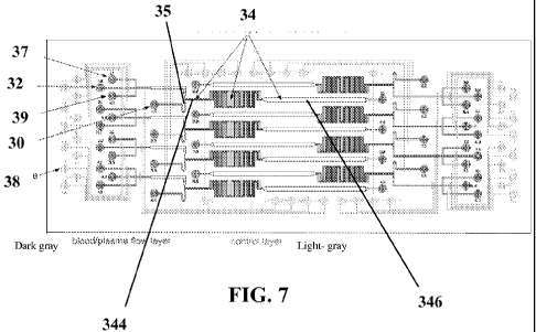
[0030] Figure 7 shows a schematic illustration of a microfluidic device
according
to an embodiment herein disclosed. The control layer is shown in light gray.
The
sample layer is shown in dark gray.
[0031] Figure 8 shows an exemplary schematic illustration of various phases of
a
process to manufacture a device herein disclosed.
6
CA 02694541 2010-01-11
WO 2009/012340 PCT/US2008/070232
[0032 ] Figure 9 shows a schematic exemplary schematic illustration of a
process to
manufacture and use a device integrated with DEAL technology according to an
embodiment herein disclosed.
[0033] Figure 10 shows a schematic exemplary schematic illustration of a
process
to manufacture and use a device according to an embodiment herein disclosed.
[0034] Figure 11 shows a photograph of a device according to the disclosure,
during separation of an exemplary fluid formed by diluted sheep blood.
[0035] Figure 12 shows a bright-field image of two assay lanes performed with
the
methods and devices herein disclosed
[0036] Figure 13 shows a dark field image of the assay illustrated in Figure
9.
[0037] Figure 14 shows an image of an assay channel region of a device
according
to an embodiment herein disclosed (top) and an assay performed on the device
according to an embodiment herein disclosed.
[0038] Figure 15 shows an exemplary measurement of a panel of blood biomarkers
from a finger-prick of whole blood. Panel (a) shows optical micrographs of a
device
herein disclosed while performing separation of plasma from fresh whole blood.
Panel
(b) shows a fluorescence image of blood barcodes in two adjacent microchannels
of a
device herein disclosed, on which both the unspiked and spiked fresh whole
blood
collected from a healthy volunteer were separately assayed.. The bars are
a1120 m in
width. Panel (c) shows fluorescence line profiles of the barcodes for both
unspiked
and spiked whole blood samples assayed as illustrated in Panel (b). The
distance
corresponds to the full length shown in Panel (b).
DETAILED DESCRIPTION
[0039] Devices, methods and systems for detecting target molecules in a sample
are herein disclosed.
[0040] The term "detect" or "detection" as used herein indicates the
determination of the existence, presence or fact of a target or signal in a
limited
portion of space, including but not limited to a sample, a reaction mixture, a
7
CA 02694541 2010-01-11
WO 2009/012340 PCT/US2008/070232
molecular complex and a substrate. A detection is "quantitative" when it
refers, relates
to, or involves the measurement of quantity or amount of the target or signal
(also
referred as quantitation), which includes but is not limited to any analysis
designed to
determine the amounts or proportions of the target or signal. A detection is
"qualitative" when it refers, relates to, or involves identification of a
quality or kind of
the target or signal in terms of relative abundance to another target or
signal, which is
not quantified.
[0041] The term "target" or "target molecule" as used herein indicates an
analyte of interest. The term "analyte" refers to a substance, compound or
component
whose presence or absence in a sample has to be detected. Analytes include but
are
not limited to biomolecules and in particular biomarkers. The term
"biomolecule" as
used herein indicates a substance compound or component associated to a
biological
environment including but not limited to sugars, amino acids, peptides
proteins,
oligonucleotides, polynucleotides, polypeptides, organic molecules, haptens,
epitopes,
biological cells, parts of biological cells, vitamins, hormones and the like.
The term
"biomarker" indicates a biomolecule that is associated with a specific state
of a
biological environment including but not limited to a phase of cellular cycle,
health
and disease state. The presence, absence, reduction, upregulation of the
biomarker is
associated with and is indicative of a particular state.
[0042] The term "sample" as used herein indicates a limited quantity of
something that is indicative of a larger quantity of that something, including
but not
limited to fluids from a biological environment, specimen, cultures, tissues,
commercial recombinant proteins, synthetic compounds or portions thereof.
Additionally exemplary samples include bodily fluids such as sputum, CSF,
sweat,
urine, semen, biopsy specimens, pap smear samples or any other sample obtained
from a human or a an animal being that contains a liquid component and a cell
component.
[0043] A further description of the devices methods and systems of the present
disclosure is provided with reference to applications wherein the sample is
blood, the
fluidic component is plasma and the targets are biomarkers. A person skilled
in the art
will appreciate the applicability of the features described in detail for
blood samples
and biomarkers for other biologic, organic and inorganic samples and targets.
The
8
CA 02694541 2010-01-11
WO 2009/012340 PCT/US2008/070232
devices methods and systems herein disclosed although designed to separate
plasma
from blood could be used to obtain particle-free fluid from any medium
containing
micron sized particles and detect molecules using capture agents of any kind.
[0044] In some embodiments, the present disclosure relates to a device that
combines plasma separation from whole blood with detection of multiple
biomarkers
from the plasma.
[0045] An exemplary device of the disclosure is shown in Figures 1-6.
Microfluidic flow channels are patterned on a chip, designed so that on one
region of
the chip (10) plasma is separated from whole blood, and in a second region of
the chip
(11), proteins or other analytes are measured from the separated plasma. In
the chip
of Figure 1, whole blood is introduced at a microfluidic channel inlet (12),
whole
blood minus a fraction of the plasma is removed from the chip via a
microfluidic
channel (13), or stored in a waste reservoir on chip (not shown). In the
depiction of
Figure 1, channel (13) is located in region (10). The reservoir can be located
at the
end of channel (13).
[ 0 0 4 6] Region (10) also includes a relatively wide or low-flow-resistance
channel
(15) fluidically connected with one or more relatively narrow, high-flow-
resistance
channels (14), which in turn are fluidically connected with channels (16).
Channels
(14) and (16) are included in region (11) of the chip depicted in Figure 1.
The relative
flow-resistances of channels (15, 14, 16) may be varied by design.
[0047] In the device of Figures 1-6, plasma separation from whole blood is
performed by controlling the plasma flow from the wider microfluidic channels
(15)
into the thin microfluidics channels (14) using the Zweifach-Fung effect which
is well
known in the art (Fung, Y.C. Stochastic flow in capillary blood vessels.
Microvasc.
Res. 5, 34-38 (1973); Svanes, K. & Zweifach, B.W. Variations in small blood
vessel
hematocrits produced in hypothermic rates by micro-occlusion. Microvascular
Research 1, 210-220 (1968); Yang, S., Undar, A. & Zahn, J.D. A microfluidic
device
for continuous, real time blood plasma separation. Lab on a Chip 6, 871-880
(2006)).
[0048] In the device of Figures 1-6, to achieve the Zweifach-Fung effect,
whole
blood is flowed through channel (15) in fluidic communication with channels
(14). If
the flow-resistances are the same, then whole blood will flow through both
sets of
9
CA 02694541 2010-01-11
WO 2009/012340 PCT/US2008/070232
channels. If the flow-resistance of the narrow channels (14) is greater than
the
resistance of channel (15), plasma will be conveyed from channel (15) to
channel
(14).
[0049] In some embodiments, the flow rate ratio between flowing channel (15)
and
assaying channel (14) can be as low as about 4:1. In other embodiments,
wherein the
flow rates ratio between the flow rate of channel (14) and the flow rate of
channel
(15) is at least, or also greater then about 20:1, the flow of plasma from
channel (15)
into channel (14) is maximized regardless of the inlet hematocrit. Also at a
flow rate
ratio of about 20:1 a good separation efficiency is observed (less than 20
cells
detectable in channels (14) and (16) upon visual inspection). In some
embodiments,
as the ratio between the flow rate of channel (14) and the flow rate of
channel (15) is
increased over the about 20:1 ratio, the separation efficiency is also
improved but the
yield of plasma (and accordingly input fluid to be assayed) is correspondingly
decreased.
[0050] In some embodiments, the ratio between the flow rate of channel (14)
and
the flow rate of channel (15) is increased over the about 20:1 ratio to values
that allow
maintaining a separation yield above 5%. In particular, in some embodiments,
the
flow ratio between the flowing channel (15) and the assaying channel (14) can
be over
about 100:1, from about 60:1 to about 100:1, from about 35:1 to about 60:1, or
from
20:1 to 35:1.
[0051] More particularly, in certain embodiments wherein a maximized
separation
efficiency and long assay time are desired or acceptable, a flow rate ratio
between
flowing channel (15) and assaying channel (14) over about 100:1 could be
selected. In
other embodiments wherein optimized separation yield and assaying time are
desired,
a flow rate ratio between flowing channel (15) and assaying channel (14) of
from
about 35:1 to about 60:1 can be selected.
[0052] A desired flow rate ratio for a sample and an experimental design of
choice
can be identified based on the radius of particles in the samples that,
according to the
experimental design of choice have to be separated from the fluidic component
that is
conveyed in the assaying channels. In particular, the flow rate ratio is
inversely
proportional to the radius of the particles to be separated as indicated in
Appendix A
CA 02694541 2010-01-11
WO 2009/012340 PCT/US2008/070232
of the present application, herein incorporated by reference in its entirety.
The term
"particle" as used herein identifies a small portion of matter that behaves as
a whole
unit in terms of its fluidic transport and properties, which includes cells,
such as red
blood cells, or aggregates thereof but can also include other particulate
matter of
organic or inorganic nature and aggregates thereof.
[0053] In the device of Figures 1-6, channel (14) is further fluidically
connected to
an assaying channel (16). In the embodiments, exemplified in Figures 1-6,
channel
(16) is an assaying channel that hosts one assaying region wherein an assay
for target
detection is performed.
[0054] The resistances of all the channels can be adjusted to achieve the
desired
flow rate ratio between channels (14) and (15), by adjusting the channel
dimensions
of various channels in, taking into account that plasma channels (14) will
have a
greater flow resistance if they are narrower and/or longer. Alternatively,
channel (15)
can be adjusted to be wider and/or shorter to decrease its flow resistance
(which
would also help to increase the resistance ratio between the two channels).
Furthermore, in some embodiments, channel (16) can have a width at least an
order of
magnitude (l Ox) greater than the width of plasma channel (14) to minimize the
impact
of channel (16) on the resistance and separation efficiency. However, width
and length
of channel (16), as well as channels (15) and (14) and the related flow
resistance can
be varied according to the desired chip configuration using the methods and
programs
herein disclosed.
[0055] In particular, appropriate flow rate ratio for a specific chip can be
identified
by a skilled person taking into account that in a pressure driven flow, the
flow rate Q
(m^3/s) within a microchannel is defined by Q=AP/R where OP is the pressure
difference for driving the flow between an inlet and an outlet (Pa), and R is
the
channel resistance (Pa s m^-3).
[0056] Appropriate dimensions of the channels of a microfluidic device to
obtain
the desired flow rate ratio can be identified by a skilled person taking into
account the
relationship between channel resistance and channel dimensions. For example
for a
rectangular microchannel with a low aspect ratio (w-h) the following equation
can be
considered:
11
CA 02694541 2010-01-11
WO 2009/012340 PCT/US2008/070232
:~~:
;:
t~~n~.~ i ~
;~~. ===
wherein R is the channel resistance, w is the width of the channel, h is the
height of
the channel, L is the length of the channel, and u is the viscosity of the
fluid. This
equation can be used to determine the resistances of all channels and to
control said
resistance to optimize the plasma separation according to the experimental
design.
[0057] A software program was created by the Applicants to allow a user to
input
the length, width, and height for each channel and the viscosity of the fluid,
to obtain
a corresponding resistance of the channel.
[0058] According to the program, dimensions for all channels within certain
ranges
and the desired input pressure (generally 5-l0psi) are initially input by a
user. For
example, for plasma separation the input channel depth can be about 10-20um,
the
width and length of channel (15) can be in the range of 15-35um, and in the
range of
100-500um respectively, the width of channel (13) can be between 60 and 200
um,
and the length of the plasma channels (14) and (16) can be between 5mm and
25mm
(see additional indications in Appendix A).
[0059] The program then computes resistances of all channels based on the
input
dimensions. The program inserts these values into a resistance matrix,
obtained as
indicated in Appendix A to the present application, which shows the system of
equations and associated matrices. The inverse of the resistance matrix is
then
multiplied by a pressure matrix (see Appendix A) to obtain the flow rates
matrix
which contains the flow rate for each channel. The program can also sum all
the
above plasma channel flow rates and divide over the channel (15) output flow
rate to
determine the percentage of the blood that is separated as plasma (this gives
the
yield). The program then outputs the resistances and flow rates for all the
channels,
the flow ratios, and a percentage plasma yield.
12
CA 02694541 2010-01-11
WO 2009/012340 PCT/US2008/070232
[0060] Suitability of the output parameters for an experimental design of
choice
can be identified by a skilled person upon reading of the present disclosure.
If the
parameters do not suit the experimental purpose, they can be adjusted
accordingly.
[0061] For example, for blood/plasma separation if the flow rate ratio is
below
20:1, and a good separation of plasma from whole blood is desired as well as a
high
yield of plasma and short assay time, new channel dimensions can be input into
the
program that are expected to increase the flow ratio (for example, lengthening
the
plasma channel, increasing the width of channel (13), etc). The process can
then be
repeated iteratively until the greatest flow rate ratio possible while still
maintaining
greater than 5% yield and flow rates greater than 100um/sec in channel (16),
are
achieved. The input dimensions that provide the flow rate ratio compatible
with the
desired design (also in view of the desired setting for target detection in
the assaying
channels - see below) can be used for the related device. Reference is also
made to the
Appendix A to the present application incorporated herein by reference in its
entirety.
[0062] In some embodiments, the width of the plasma channels can be held
constant (e.g. at 10 m in accordance with the Yang, Sung et al. Blood Plasma
Separation in Microfluidic Channels using Flow Rate Control. ASAIO Journal
2005;
51: 585-590) and while the length of the plasma channels (14) and/or the width
of
region (13) are varied.
[0063] In some embodiments, the length of the plasma channels (14) and the
width
of channels (13) were increased to obtain a desired flow ratio. In those
embodiments,
the orientation of channels (14) with respect to channels (15) can be varied
according
to the desired chip configuration, even if in some embodiments herein
illustrated the
channels are perpendicular.
[0064] In some embodiments the program can be operated with a software such as
Matlab, that already contains a built in matrix multiplication function.
[0065] In certain embodiments, the software program can also use the above
relation Q=OP/R and the conservation of mass transport law to determine the
volume
flow rate in each channel based on the resistance calculated for each channel.
Because
of the conservation of mass transport, the flow rates in the various channels
are
interdependent. In other words, if the flow entering channel 15 is defined as
Q15in,
13
CA 02694541 2010-01-11
WO 2009/012340 PCT/US2008/070232
the flow entering the first plasma channel of region 14 is Q14.1, and the flow
in
region 15 immediately below the first plasma channel as Q15.1, then
Q15in=Q14.1+Q15.1. Next, Q15.1=Q14.2+Q15.2, and so forth for all subsequent
plasma channels.
[0066] Also the software program herein described also allows devising a
system
of equations describing the pressure drop in each channel as a function of
known
resistances and unknown flow rates can be written in accordance with the
present
disclosure. In the end, the system of n equations and n unknowns can be solved
using
matrix algebra in which there is a flow matrix, a pressure matrix, and a
resistance
matrix, such that again Q=OP/R. These matrices of equations once introduced in
a
program such as Matlab can easily be solved, allowing identification of the
flow rates
in each channel given an input pressure. The program can then output the
ratios of the
flows, for example, Q14.1:Q15.1, Q14.2:Q15.2, etc.
[0067] A person skilled in the art, upon reading of the present disclosure,
will be
able to calculate the appropriate resistances of all channels for the various
embodiments based on the channel dimensions and to determine the volume flow
rates of fluid through each channel at a given inlet pressure, using for
example
software programs such as Matlab program.
[0068] In the device of Figures 1-6, separation of plasma from whole blood is
performed through controlled flow of the plasma from flowing channel (15)
flowing
to assaying channels (14) and (16).
[0069] The wording "flowing channel" as used herein indicates the portion of
the
device wherein the separation of one or more fluidic components of a sample
from the
sample is performed. For example, in embodiments herein disclosed it
identifies
channel (15) wherein the blood is flowed and from which the plasma component
is
conveyed into assaying channels (14) and (16)
[0070] The wording "assaying channel" as used herein indicates the portion of
the
device wherein the detection of the target is performed. For example, in
embodiments
herein disclosed assaying channels (14) and (16) are configured to receive
cell-free
plasma that can be assayed by underlying capture agents.
14
CA 02694541 2010-01-11
WO 2009/012340 PCT/US2008/070232
[0071] In particular, in the device of Figures 1-6, channels (14) and (16) can
carry
capture agents attached on the surface of the channel for detection of the
target in the
plasma fraction to be tested as also illustrated in the schematic illustration
of Figure 8
(see below) wherein capture agent spots (21) are indicated.
[0072] The wording "capture agents" as used herein indicate a molecule capable
of
specific binding with a predetermined target to form a detectable capture
agent target
complex. Exemplary capture agents include but are not limited to
polynucleotides and
proteins, and in particular antibodies.
[0073] The term "polynucleotide" as used herein indicates an organic polymer
composed of two or more monomers including nucleotides, nucleosides or analogs
thereof. The term "nucleotide" refers to any of several compounds that consist
of a
ribose or deoxyribose sugar, joined to a purine or pyrimidine base and to a
phosphate
group and that are the basic structural units of nucleic acids. The term
"nucleoside"
refers to a compound (as guanosine or adenosine) that consists of a purine or
pyrimidine base combined with deoxyribose or ribose and is found especially in
nucleic acids. The term "nucleotide analog" or "nucleoside analog" refers
respectively
to a nucleotide or nucleoside in which one or more individual atoms have been
replaced with a different atom or a with a different functional group.
Accordingly, the
term polynucleotide includes nucleic acids of any length DNA RNA analogs and
fragments thereof. A polynucleotide of three or more nucleotides is also
called
nucleotidic oligomers or oligonucleotide.
[0074] The term "polypeptide" as used herein indicates an organic polymer
composed of two or more amino acid monomers and/or analogs thereof. The term
"polypeptide" includes amino acid polymers of any length including full length
proteins and peptides, as well as analogs and fragments thereof. A polypeptide
of
three or more amino acids is also called a protein oligomer or oligopeptide.
As used
herein the term "amino acid", "amino acidic monomer", or "amino acid residue"
refers to any of the twenty naturally occurring amino acids including
synthetic amino
acids with unnatural side chains and including both D an L optical isomers.
The term
"amino acid analog" refers to an amino acid in which one or more individual
atoms
have been replaced, either with a different atom, isotope, or with a different
functional
group but is otherwise identical to its natural amino acid analog.
CA 02694541 2010-01-11
WO 2009/012340 PCT/US2008/070232
[0075] The term "protein" as used herein indicates a polypeptide with a
particular
secondary and tertiary structure that can participate in, but not limited to,
interactions
with other biomolecules including other proteins, DNA, RNA, lipids,
metabolites,
hormones, chemokines, and small molecules.
[0076] The term "antibody" as used herein refers to a protein that is produced
by
activated B cells after stimulation by an antigen and binds specifically to
the antigen
promoting an immune response in biological systems and that typically consists
of
four subunits including two heavy chains and two light chains. The term
antibody
includes natural and synthetic antibodies, including but not limited to
monoclonal
antibodies, polyclonal antibodies or fragments thereof. Exemplary antibodies
include
IgA, IgD, IgGl, IgG2, IgG3, IgM and the like. Exemplary fragments include Fab
Fv,
Fab' F(ab')2 and the like. A monoclonal antibody is an antibody that
specifically
binds to and is thereby defined as complementary to a single particular
spatial and
polar organization of another biomolecule which is termed an "epitope". A
polyclonal
antibody refers to a mixture of monoclonal antibodies with each monoclonal
antibody
binding to a different antigenic epitope. Antibodies can be prepared by
techniques
that are well known in the art, such as immunization of a host and collection
of sera
(polyclonal) or by preparing continuous hybridoma cell lines and collecting
the
secreted protein (monoclonal).
[0077] The wording "specific" "specifically" or specificity" as used herein
with
reference to the binding of a molecule to another refers to the recognition,
contact and
formation of a stable complex between the molecule and the another, together
with
substantially less to no recognition, contact and formation of a stable
complex
between each of the molecule and the another with other molecules.. Exemplary
specific bindings are antibody-antigen interaction, cellular receptor-ligand
interactions, polynucleotide hybridization, enzyme substrate interactions etc.
The term
"specific" as used herein with reference to a molecular component of a
complex,
refers to the unique association of that component to the specific complex
which the
component is part of. The term "specific" as used herein with reference to a
sequence
of a polynucleotide refers to the unique association of the sequence with a
single
polynucleotide which is the complementary sequence.
16
CA 02694541 2010-01-11
WO 2009/012340 PCT/US2008/070232
[0078] The term "attach" or "attached" as used herein, refers to connecting or
uniting by a bond, link, force or tie in order to keep two or more components
together,
which encompasses either direct or indirect attachment such that for example
where a
first molecule is directly bound to a second molecule or material, and the
embodiments wherein one or more intermediate molecules are disposed between
the
first molecule and the second molecule or material.
[0079] In some embodiments, the same area or different areas of channels (14)
and
(16) can be coated with different capture agents each bindingly
distinguishable from
another. In some embodiments, the same area or different areas of channels
(14) and
(16) can be coated with different capture agents each positionally
distinguishable from
another
[0080] The wording "bindingly distinguishable" as used herein with reference
to
molecules, indicates molecules that are distinguishable based on their ability
to
specifically bind to, and are thereby defined as complementary to a specific
molecule.
Accordingly, a first molecule is bindingly distinguishable from a second
molecule if
the first molecule specifically binds and is thereby defined as complementary
to a
third molecule and the second molecule specifically binds and is thereby
defined as
complementary to a fourth molecule, with the fourth molecule distinct from the
third
molecule.
[0081] The wording "positionally distinguishable" as used herein refers to
with
reference to molecules, indicates molecules that are distinguishable based on
the point
or area occupied by the molecules. Accordingly, positionally distinguishable
capture
agents are substrate polynucleotide that occupy different points or areas on
the
assaying channel and are thereby positionally distinguishable.
[0082] In embodiments, wherein bindingly and possibly also positionally
distinguishable capture agents are used detection of a plurality of biomarkers
can be
performed in a single channel or in a portion thereof.
[0083] In other embodiments, channel (14) and (16) of the device of Figures 1
to
6, does not include capture agents in all or part of the assaying channels. In
those
embodiments, blood can be flowed through flowing channel (15) and after
separation
17
CA 02694541 2010-01-11
WO 2009/012340 PCT/US2008/070232
of the plasma in channel (14), the analytes and in particular the biomolecules
are
attached and in particular adsorbed to channel (14) and/or (16) surface.
[0084] In some embodiments, detection of the attached analyte and/or capture
agent target complex is performed by providing a labeled molecule, which
includes
any molecule that can specifically bind a capture agent target complex to be
detected
(e.g. an antibody, aptamers, peptides etc) and a label that provides a
labeling signal,
the label compound attached to the molecule. The labeled molecule is contacted
with
the attached analyte and/or capture agent target complex and the labeling
signal from
the label compound bound to attached analyte and/or the capture agent-target
complex
on the substrate can then be detected, according to procedure identifiable by
a skilled
upon reading of the present disclosure and, in particular, of the Examples
section.
[0085] The terms "label" and "labeled molecule" as used herein as a component
of
a complex or molecule refer to a molecule capable of detection, including but
not
limited to radioactive isotopes, fluorophores, chemioluminescent dyes,
chromophores,
enzymes, enzymes substrates, enzyme cofactors, enzyme inhibitors, dyes, metal
ions,
nanoparticles, metal sols, ligands (such as biotin, avidin, streptavidin or
haptens) and
the like. The term "fluorophore" refers to a substance or a portion thereof
which is
capable of exhibiting fluorescence in a detectable image. As a consequence the
wording and "labeling signal" as used herein indicates the signal emitted from
the
label that allows detection of the label, including but not limited to
radioactivity,
fluorescence, chemolumiescence, production of a compound in outcome of an
enzymatic reaction and the likes.
[0086] In some embodiments, the detection method can be carried via
fluorescent
based readouts, in which the labeled antibody is labeled with fluorophore
which
includes but is not limited to small molecular dyes, protein chromophores and
quantum dots. In other embodiments, on-chip detection can be performed with
methods other than fluorescence based techniques. Exemplary suitable
techniques
include, colorimetric detection, enzyme-catalyzed production of different
colored or
fluorescent dyes (with different colors being associated with distinct
analytes),
microparticle/nanoparticle based detection using electron microscopy, AFM, or
dark-
field microscopy, magnetic detection using magnetic micro/nanoparticles,
electrical
detection methods.
18
CA 02694541 2010-01-11
WO 2009/012340 PCT/US2008/070232
[0087] In some embodiments, detection can be performed by methods that use
signal amplification such as gold nanoparticle based detection followed by
gold or
silver amplification. In particular, in some embodiments, in any of the
methods and
systems herein disclosed, detection can be carried out on gold nanoparticle-
labeled
secondary detection systems in which a common photographic development
solution
can amplify the gold nanoparticles as further described below. Also, if the
readout
comes from dark field scattering of gold particles, single molecule digital
proteomics
is enabled.
[0088] Additional techniques are identifiable by a skilled person upon reading
of
the present disclosure and will not be further discussed in details.
[00891 In embodiments wherein one or more targets and/or a plurality of
targets is detected described below in more details, the labeled molecule can
be
formed of a plurality of labeled molecules. Each labeled molecules comprises a
molecule that specifically binds one target of the one or more
targets/plurality of
targets and a label compound attached to the molecule, the label compound
providing
a labeling signal, each labeled molecule detectably distinguishable from
another.
[0090] The wording "detectably distinguishable" as used herein with reference
to labeled molecule indicates molecules that are distinguishable on the basis
of the
labeling signal provided by the label compound attached to the molecule.
Exemplary
label compounds that can be use to provide detectably distinguishable labeled
molecules, include but are not limited to radioactive isotopes, fluorophores,
chemoluminescent dyes, chromophores, enzymes, enzymes substrates, enzyme
cofactors, enzyme inhibitors, dyes, metal ions, nanoparticles, metal sols,
ligands (such
as biotin, avidin, streptavidin or haptens) and additional compounds
identifiable by a
skilled person upon reading of the present disclosure.
[0091] In embodiments, wherein bindingly distinguishable capture agents are
used
different analytes can be detected by use of detectably distinguishable
labeled
molecules each specific to a separate analyte of interest.
[0092] In some embodiments, binding of a target with a capture agent is
controlled
by adjusting the flow resistance of the various channels to control the
saturation
coverage of the capture agents by target in the plasma component conveyed in
the
19
CA 02694541 2010-01-11
WO 2009/012340 PCT/US2008/070232
assaying channels. In particular, in a device such as the one exemplarily
illustrated in
Figures 1 to 3, for a given concentration of detectable target in a solution,
and for a
given coverage of capture agents on the surface of channels (14) and (16), the
time
required to have saturation coverage of capture agent attached to the channels
by the
detectable targets is largely determined by two factors.
[0093] The first factor is given by the binding affinity which is the strength
of
interaction between a target and a capture agent (stronger interactions can
lead to
faster binding times). The second factor is the time required for a certain
target to
diffuse through a vessel volume to bind to a capture agent (target diffusion).
In the
devices methods and systems herein disclosed, flow conditions are controlled
so that
in a certain channel or portion thereof binding affinity or target diffusion
control
binding of the targets to the capture agents to various degrees.
[0094] For example, in devices wherein flow conditions are set up so that a
target
containing solution is not flowing through a channel, the time to saturation
coverage
of the target to capture agents on the channel surface is limited by the
diffusion of the
target in the solution. Depending upon the volume of the channel, targets
diffusion to
surface-bound capture agents to achieve saturation coverage can take from many
minutes to hours. Assays wherein the time to saturation coverage of the target
to
capture agent is limited by the target diffusion are herein also indicated as
diffusion
limited processes.
[0095] In other embodiments, wherein a target containing solution is flowed
through a channel at a sufficiently high flow velocity so that diffusion time
scales are
no longer relevant, only the strength of interaction (the affinity) between
targets and
capture agents that limits the time to saturation coverage. At this limit, the
time to
saturation coverage can be only 1-5 minutes. Assays wherein the time to
saturation
coverage of the target to capture agent is limited by the binding affinity of
the target
and the capture agents, are herein also indicated as affinity limited
processes.
[0096] In some embodiments of the present disclosure an affinity limited
process is
accomplished by having the fluid move above the detection region fast enough
that at
every moment, the target concentration above the capture agents remains
essentially
constant. More particularly, in some embodiment herein disclosed an affinity
limited
CA 02694541 2010-01-11
WO 2009/012340 PCT/US2008/070232
process can be performed by regulating the flow resistance to obtain a flow
velocity
of about 0.5mm/s. In other embodiments >0.5mm/s flow velocities in channels
(14)
and (16) can be obtained (regardless of their dimensions) by tuning the inlet
pressure
until this velocity is achieved. In the cases exemplified by Figures 1-6,
since channels
in (14) are very narrow compared to channel (16), flow velocity is much faster
in
channels (14) than in channels (16). Therefore an affinity limited process can
be run
in channels (14) even at low pressures, while diffusion limited processes can
be run in
channel (16).
[0097] Diffusion limited processes and affinity limited processes can differ
in ways
other than the timescale for completion.
[0098] A diffusion limited process, while slower, can be more sensitive than
an
affinity-limited process. In particular, in a diffusion limited process,
capture agents
such antibodies come in contact with more analytes per volume of solution than
in an
affinity limited process. Therefore, more analytes can be captured per unit
solution
volume when the process is diffusion limited, increasing the sensitivity of
detection,
especially when the sample concentration and volume is low. .
[0099] On the contrary, an affinity-limited process allows rapid detection of
a
certain target in samples including the target at high concentrations. In
particular, an
affinity limited process can be desired when detection of a target of interest
included
in a sample in high concentrations and volumes because, with an affinity
limited
process detection of said target of interest can be efficiently perform
without the need
of sampling many analytes per unit of volume.
[00100] The Damkohler number (Da), which is the ratio of hybridization rate
(Lr) to diffusion rate (Lm), can be used to determine if a process is affinity
limited or
diffusion-limited.
Da = Lr/Lm
[00101 ] The hybridization rate Lr is given by:
Lr kagfr
21
CA 02694541 2010-01-11
WO 2009/012340 PCT/US2008/070232
where ka is the reaction rate constant and gfr is the is the average density
of surface-
bound capture agents..
[001021 The diffusion mass transport rate L. is given by:
Lm= cube root[(UD2 )/(lwh2)]
where U is the flow rate, D is the diffusion constant, and l,w, and h are the
length,
width, and height respectively. When Da 1, (- 10) the reaction is occurring
much
more quickly than diffusion rate, so the process is diffusion-limited. When
Da 1(<0.1), the diffusion rate is fast compared to the reaction rate, and the
process
is affinity limited. In some embodiments, the wide channels (16) can be
designed to
give a Da>10 and the narrow channels (14) can be designed to give a Da<0.1 by
inputting the appropriate parameters of length, width, and height into the
above
equation. The pressure can be adjusted to obtain the appropriate flow rate.
[00103] A skilled person will appreciate that if the wide channel Da (Dal) is
>10
and the narrow channel Da (Da2) <0.1, the ratio of these two channel's Da
numbers
should be Da1:Da2>100. Also Lr, h, and D (diffusion constant) are equal in
both the
narrow and wide channels and
Da1:Da2=Lm2:Lm1 = cube root(U21iwi/Ui12W2)
wherein Lml = mass transport rate in the wide channel and Lm2 = mass transport
rate
in the narrow channel
[00104 ] In some embodiments, each narrow channel (14) can have a flow rate
that
is 1/5 of the flow rate of the wide channel (16) so U2=1/5U1 and the lengths
of
narrow and wide channels are often about equal. In those embodiments, the
equation
can be simplified to:
Da1:Da2=cube root(wi/5w2)
[00105] A skilled person can then solve for wl/w2 to obtain the ratio of wide
channel:narrow channel needed to obtain diffusion limitation in one channel
and
affinity limitation in the other channel.
22
CA 02694541 2010-01-11
WO 2009/012340 PCT/US2008/070232
[00106] However, the Damkohler number does not take into account the flow-
velocity dependence of the binding process. Therefore, in some embodiments
wherein
calculation of flow-velocity dependence is desired, a more suitable model is
the
Zimmermann model.
[00107] Reference is made to a passage from Zimmermann, M. et al. "Modeling
and Optimization of High Sensitivity, Low Volume Microfluidic-Based Surface
Immunoassays." Biomedical Microdevices 7:2, 99-110, 2005, pp.100-101, reported
in
the following paragraph [00108].
[ 0 010 8]"The flow of a liquid in a region over time t is characterized by a
velocity
vector field "? a pressure p and a density p. For laminar, incompressible and
viscous
fluids the density is constant. The flow is described by the Navier-Stokes
partial
differential equation system
- ;r i +, U< +;w slr
in dimensionless form with the Reynolds number Re and external forces External
forces such as gravity can be neglected in such miniaturized systems. For Re
2100,
flow is considered to be laminar and has a characteristic parabolic flow
profile with
zero flow velocity at the channel walls and peak flow velocity in the channel
center.
Here, Re is -0.07 for the maximum flow rates considered.
The bulk concentration C of a solute in a given solution is described by the
Convection-Diffusion equation of the
form
,;~~:! p \y
+
t^J
with a diffusion coefficient D, a source term 0 and the identical velocity
vector field
k.T
U given in equation (1). We have applied the Stokes-Einstein-relation D = 6-s!
~., with
the hydrodynamic radius Rh, the analyte viscosity rl and the Boltzmann
constant k to
23
CA 02694541 2010-01-11
WO 2009/012340 PCT/US2008/070232
estimate the diffusion co-efficient D of the analyte molecule to D = 10-6 cm2s-
' which
we used for all further calculations and which corresponds to the literature
(Metsamuronen et al., 2002) where comparable values for small molecules such
as
TNFa- are reported. The analyte viscosity was set to a high plasma viscosity
(Koenig
et al., 1998) of 2 mPa s.
The association and the dissociation from the capture site are described by
the rate
coefficients k, the analyte concentration C and the density of free binding
sites (OaX
Ot) on the surface, using an ordinary differential equation of the form,
;~{r
l! ~ i
-1 - k:x~ i~laxYt; ^ '
bd;
for monovalent receptors and ligands. koõis the rate constant for association,
k offis the
rate constant for dissociation, C is the concentration of free molecules in
the fluid, Ot
is the surface density at time t O,Y,aX is the maximum surface density of
molecules
calculated from the feature area of the individual capture molecules and is
assumed to
be constant over time. In this simulation we generally used 106 M-i s-i for k
oõ and 10-3
s-' for koff (Santora et al., 2001), but in some case these constants were
modified."
(Zimmermann, M. et al. "Modeling and Optimization of High Sensitivity, Low
Volume Microfluidic-Based Surface Immunoassays." Biomedical Microdevices 7:2,
99-110, 2005, pp.100-101)
[00109] In some embodiments, the equations from the Zimmermann passage
can be used to determine the flow rate associated with affinity limited vs.
diffusion
limited processes. In particular, using a simulation incorporating the
Zimmermann
equations, an analyte concentration of 1pM, and an antibody binding affinity
of
K 10^9, and feature sizes of 1500nm^2, flows above 0.5mm/sec can be considered
affinity limited whereas flows around 0.005mm/sec can be considered diffusion-
limited. More particularly flows of 0.05mm/sec can be considered at the
beginning of
the diffusion limited regime. As a consequence, a 10-100 fold difference in
flow
velocity can be identified between the diffusion limited and affinity limited
regimes.
[00110 ] Accordingly, in certain embodiments, wherein diffusion limited
processes and affinity limited process in assaying channels of a same device
are
desire, the affinity limited region can be about 10-100 times narrower than
the
24
CA 02694541 2010-01-11
WO 2009/012340 PCT/US2008/070232
diffusion-limited region (regardless of length, and all heights are constant),
wherein
the specific dimensions of the affinity limited region (plasma channels) are
also
dictated, by separation efficiency (possibly optimized with yield and flow
rate) (see
above and Appendix A). In some embodiments, the flow rate through the plasma
channels can be adjusted simply by increasing the pressure until fluid through
those
channels is moving at a speed of about 0.5mm/sec (affinity limited regime). In
some
embodiments, as long as the wide channel (16) is about 10-100 times wider than
the
combined width of the narrow channels (14), the flow velocity will be about
0.05-
0.005mm/sec (diffusion limited regime). In one embodiment, the narrow channels
(14) are l0um and there are 5 of them, for a combined width of 50um. In some
embodiments, the wide channel (16) can be 500um wide to obtain a 10-fold
reduction
in flow speed. In other embodiments, wherein channel (16)'s width is increased
to 2-
5mm, region (16) of the device would still be within the diffusion limited
regime
(0.005mm/s).
[00111 ] In accordance with the present disclosure, channels or portions
thereof
and in particular assaying channels (14) and (16) can be designed to host
either
diffusion-limited processes or affinity limited processes by varying the width
of the
channel.
[001121 In particular, in some embodiments, the channels are designed so that
binding of the capture agents to targets in the fluid component is an affinity
limited
process. For example, in the embodiments exemplarily illustrated. Figures 1-6,
channels (14) and (16) are in series so they will have the same volume flow
rate by
conservation of mass. Therefore, in those embodiments if channel (14) is much
narrower, e.g. 50-1000x, than channel (16), the fluid velocity must be much
faster
through (14) than through (16) in order for their volume flow rates to be
equal. This
velocity difference leads to a difference in analyte exploitation and binding
efficiency
between the two channels. It is clear from the above equation that varying the
1, w,
and h of the channel can be used to increase or decrease the Lm term, thereby
increasing or decreasing the Damkohler number. The 1, w, and h can be
increased to
decrease the Damkohler number far below 1 to obtain an affinity-limited
process and
far above 1 to obtain a diffusion-limited process.
CA 02694541 2010-01-11
WO 2009/012340 PCT/US2008/070232
[001131 In particular, in a device such as the one exemplified in Figures 1 to
6,
the two plasma assay regions (14, 16) on the chip can be harnessed for
different
purposes. When the chip is being operated for the separation of plasma from
whole
blood, the plasma flows rapidly through the thin channels (14), but only flows
slowly
through the reservoir (16). Thus, if (14) is utilized for measuring the levels
of plasma
proteins, then such an assay will be an affinity limited process, and
saturation
coverage of targets to surface-bound capture agents can be completed within a
few
minutes. By contrast, if (16) is the region utilized for measuring the levels
of plasma
proteins, then plasma flow through that region is slow, and the processes is
diffusion
limited but relatively more sensitive.
[00114] In particular, in the embodiments exemplified in Figures 1 to 6, the
greatest fluid velocity within the detection region (11) is in the plasma
channels (14)
and therefore the best binding efficiency occurs in this region, so the
signal/unit area
should be highest here. Velocity can be increased in this region by increasing
the inlet
pressure.
[001151 In the illustration of Figures 1 to 6, channel (14) is much narrower
than channel (16) so that the velocity in channel (14) will be above the
threshold
velocity (e.g. 0.5mm/s) for a given pressure while channel (16) should be wide
enough that the flow velocity is below the threshold velocity. Threshold
velocity is
that flow velocity at transition between diffusion limitation and affinity
limitation.
[00116] An embodiment of channels (14) and (16) is illustrated in more detail
in Figure 2. In the embodiment, illustrate in Figure 2, plasma channels (14)
are long
and narrow to achieve a high resistance relative to channel (15), thereby
accomplishing the plasma skimming. The dimensions of the 5 plasma channels
(14) of
Figure 2 can be about 10 m wide, about 25 mm long. Channel (16) can be about
500
m wide and about 10 mm long. The length of channel (15) can be 100um.
[00117 ] In some embodiments, the channel (15)'s width is in the range of
about
20-40 m, in those embodiments clogging of a sample such as blood decreased
considerably.
[00118] In some embodiments, wherein blood is treated with anticoagulants
immediately upon finger-prick blood draw, and the sample is immediately run
into the
26
CA 02694541 2010-01-11
WO 2009/012340 PCT/US2008/070232
blood inlet, the device can be run for extended time period, such as hours
without
clogging. In some embodiments, once the channel dimensions are determined
according to the experimental design of choice, and in particular within the
ranges
indicated above for optimized separation, yield and assay timing, the width of
channel
(15) can be increased to diminish a possible clogging effect. For example, for
sample
blood the width of channel (15) can be increased from 15 um, to 25 um, 35 um.
The
desired width can be selected in view of the dimension of the particle that
can clog
channels in the device, considering the desired separation efficiency, and
detection
settings. The above exemplary widths of channel (15) are indicated for
particles and
in particular cellular aggregates with sizes in the 0-25um range.
[00119 ] In some embodiments channels in region (14) are about 5 mm long. In
other embodiments the plasma channels are lengthened to about 15-25 mm, to
minimize the number of cells that flow over the detector, decreasing the
chance of
biofouling or signal interference in the detector region.
[00120] In embodiments, all plasma channels designed to be about l0um wide
and about l0um high regardless of length. Higher heights (e.g. as high as 15-
20 um
can be used without affecting device performance). In those embodiments, the
plasma
separation improved as the length of the channels was increased from 5um to
15um to
25 um.
[00121] In devices methods and systems herein disclosed, channels can be
designed so to control plasma separation from channel (15) to channel (14) as
herein
described and have predetermined channels or portions thereof configured to
host
affinity limited processes or diffusion limited processes. In those
embodiments, the
dimensions are optimized to control plasma separation and reactions performed
in
assaying channels of the device.
[001221 In some embodiments, the heights of the channels in the entire device
can be held constant (at about l0um), the width of plasma channels (14)
(affinity
limited region) can be held at about l0um, and the width of channel 16 10-100
times
larger than channels (14).
[00123] Other embodiments can be identified by a skilled person upon reading
of the present disclosure, also applicable to samples other than blood
considering that
27
CA 02694541 2010-01-11
WO 2009/012340 PCT/US2008/070232
for an n-fold difference between calculated affinity-limited and diffusion-
limited
velocities (based on the equations of the Zimmermann model), the wide plasma
channel (16) should be designed n times wider than the narrow plasma channels
(14).
[00124 ] Figure 3 shows an additional embodiment wherein channels (15), (14)
and (16) are illustrated in connection with other portions of the device, such
as blood
inlet (12), a lysis inlet (17), a primary reagent inlet (18), a labeled
molecule inlet (19)
and whole blood outlet (10).
[00125] In some embodiments, the blood sample is introduced into blood inlet
(12) and conveyed into the flowing channel (15). The plasma is then separated
into
assaying channels (14) and (16) wherein the biomarker is contacted and bound
with
the capture agent of choice in a detectable complex.
[001261 The whole blood is disposed through the whole blood outlet (10). A
lysis buffer can be also included in the lysis inlet (17) to prevent clogging
of the
whole blood by lysing the blood cells. A labeled molecule (e.g. a secondary
antibody)
can be added through the labeled molecule inlet (19) to detect the detectable
complex
of the assaying channels (14) and (16).
[00127] Each of the inlets shown in Figure 3 can be primed by closing the
main inlet control valve and allowing the sub-inlet solution to flow out of
inlet (6) to
remove any air bubbles prior to flowing into any of the eight devices. Each of
the
devices can be actuated independently in this design, such that up to 8 time
points of
serum protein expression can be obtained from a patient or mouse model.
[00128] Various embodiments can be envisioned including a different number
of inlets. In one embodiment, all the devices are connected by common inlets
as
mentioned before, so buffers and reagents can be delivered to all devices from
one
common inlet. In another embodiment, a number of completely independent
devices,
each having its own inlet, can be included in the same frame. Another
embodiment, a
plurality of devices is included in one frame, with a common buffer/reagent
inlet for
all the devices, with each having an independent blood/sample inlet for each
device
(to avoid cross-contamination of samples).
28
CA 02694541 2010-01-11
WO 2009/012340 PCT/US2008/070232
[001291 In other possible embodiments, devices can have a plurality of plasma
separation channels (14), e.g. from 2 to >100, and/or additional channels for
introducing reagents into the device before and after the blood separation is
performed. For example, in some embodiments, various reagents (capture,
detection,
blocking, wash agents, etc) can be introduced through channels in direct
fluidical
connection with channel (16) rather than having to flow reagents through the
thin,
long, high-resistance plasma channels (14). In those embodiments,
experiment/assay
times for region (16) as well as clogging of region (14) plasma channels can
be
decreased.
[00130] In other embodiments, reagents comprising capture agents, detection
agents, and other reagents identifiable by a skilled person can be mixed with
the
sample prior to introduction into the device rather than sequentially flowing
each fluid
separately.
[00131] In some embodiments, additional channels that can be envisaged
include a lysis buffer channel such as lysis channels (17) of Figure 3, that
enters
region (13) and lyses the cells as they enter that region to prevent cell-
clogging of the
outlet (and/or post-13 reservoir), helping to increase operation times. Lysing
cells
proximate to the outlet minimizes increasing resistance at outlet over time,
which
eventually leads to device failure thus prolonging the life of the device.
[00132] Other embodiments might include on-chip channels and reactors
before the separation region channels, in particular before channel (15), to
pre-treat
the blood before it enters the separation region. These channels may house
diluent
buffer to dilute the whole blood as it enters; other buffers; anticoagulant;
biological
solutions and fluids; solutions of proteins, DNA, RNA, or other biomolecules;
chemical reagents, oils, etc. These reagents might be housed in channels and
reservoirs on chip for addition to blood -with or without mixing- or for
addition to one
of the other immunoassay steps -with or without mixing-. For on-chip mixing of
whole blood, separated plasma or other immunoassay reagents either with each
other
or with any additives whether mentioned above or otherwise, on-chip rotary
mixers
driven by micromechanically controlled peristaltic pumps may be used (see for
example equipment described in Hong JW, et al. A nanoliter-scale nucleic acid
processor with parallel architecture. Nature Biotechnology.2004. 22,4: 435-
439. that
29
CA 02694541 2010-01-11
WO 2009/012340 PCT/US2008/070232
are contiguous with region (15), (13), (14), or (16). Or any other state-of-
the-art
microfluidic mixing components might be used: herring bone; cross over/zig-zag
channels; thin channels for diffusion based mixing, etc.
[00133] Other embodiments can include specific elements that enable or improve
introduction and circulation of a sample or reagents in the device including:
a pump to
apply an external pressure to the sample and/or reagents, a piston to apply
mechanical
pushing to one or more fluids in the device with a plug at the device inlet, a
peristaltic
pump on chip, a device that applies vacuum at a chip exit, a device that
provides
electrokinetic transport and/or any other equipment by which a force can be
applied
to drive the fluid through the channels.
[00134] Other embodiments can include on-chip (post 14 or post 16) additional
assaying channels/modules for amplification of a polynucleotide (e.g. DNA or
RNA)
or a polypeptide (e.g. proteins) for improved signal detection from blood,
plasma, or
serum. These could include on-chip modules that perform PCR, RT-PCR, immuno-
PCR, RCA, nanoparticle-based biobarcode detection, etc. In those embodiments
detection and/or quantitation of a certain target can be performed through the
capture
in those modules of PCR and RT-PCR amplicons, barcode DNAs in nanoparticle
based detection and/or of proteins, RNA, DNA, and/or any other surrogate/relay
biomolecule that is capturable by existing capture agents and is indicative of
the
presence of a biomolecule other than itself. An exemplary device including an
assaying channel is depicted in Figure 4, wherein channel (171) indicates a
region of
the chip's flow layer where PCR, RT-PCR, or immunoPCR can be performed. In the
exemplary embodiment of Figure 3A, the DNA products could then be detected by
the DNA capture strands in region (16).
[00135] In some embodiments, a plurality of devices can be included in the
same frame and in particular can be adapted to perform multiple assays, so
that an
assay step in one device is to be completed earlier than in other devices. In
those
embodiments, control valves can be included in the devices at the ends of
regions (13)
and/or (16) to minimize backflow and contamination of the assaying channels.
More
particularly in some embodiments, a mechanism can be included to control
fluids
movement between assays steps, so that at the end of an assay step, the valves
can be
turned on, stopping the movement of fluid while the device is readying for the
next
CA 02694541 2010-01-11
WO 2009/012340 PCT/US2008/070232
step. In some of those embodiments, the valve at the end of region (13) can
also have
the function of allowing reagents to be delivered only through the assaying
channels
(14) and (16) to minimize flowing of the reagent through channel (13), and
maximize
conveyance of the reagents into assaying channels (14) and (16). Exemplary
control
valves are illustrated in Figure 5, wherein control valve (113) and control
valve (116)
are schematically illustrated.
[00136] A control valve upstream of region (15) can also be included in each
of
a plurality of devices included in a same frame. In those embodiments, the
control
valve can be opened to allow sample and reagents to only enter the device in a
controlled fashion and can be closed to minimize backflow of sample into the
common inlet region or into other assay devices in the frame. An exemplary
schematic illustration of this valve is depicted in Figure 6, wherein valves
(112) and
(115) are shown.
[00137 ] In embodiments wherein a plurality of devices are included in a same
frame, an additional control valve actuating the common channel fed by
multiple
inlets (e.g. six) of the devices can also be included. This valve can be
closed to allow
priming of buffers and samples prior to flowing through the device and to
minimize
air bubbles. (see Figure 6)
[00138] In some embodiments the capture agents include one ore more
components. In particular, in some embodiments the capture agents can be
formed by
a substrate polynucleotide and a polynucleotide encoded-protein in application
of the
technology (herein also identified as DEAL) described in U.S. patent
application
Serial No. 11/888,502 herein incorporated by reference in its entirety.
[00139] Accordingly, the wording "substrate polynucleotide" as used herein
refers to a polynucleotide that is attached to a substrate so to maintain the
ability to
bind to its complementary polynucleotide. A substrate polynucleotide can be in
particular comprised of a sequence that specifically binds and is thereby
defined as
complementary with an encoding-polynucleotide of a polynucleotide encoded
protein.
[ 0 014 0] The wording "polynucleotide-encoded protein" refers to a
polynucleotide-protein complex comprising a protein component that
specifically
binds to, and is thereby defined as complementary to, a target and an encoding
31
CA 02694541 2010-01-11
WO 2009/012340 PCT/US2008/070232
polynucleotide attached to the protein component. In some embodiments, the
encoding polynucleotide attached to the protein is protein-specific. Those
embodiments can be used to perform assays that exploit the protein-specific
interaction to detect other proteins, cytokines, chemokines, small molecules,
DNA,
RNA, lipids, etc., whenever a target is known, and sensitive detection of that
target is
required. The term "polynucleotide-encoded antibody" as used herein refers to
a
polynucleotide-encoded protein wherein the protein component is an antibody.
[00141 ] In the polynucleotide-encoded proteins herein disclosed each protein
specifically binds to, and is thereby defined as complementary to, a pre-
determined
target, and each encoding polynucleotide-specifically binds to, and is thereby
defined
as complementary to, a pre-determined substrate polynucleotide.
[00142] In embodiments wherein the protein is an antibody, the protein-target
interaction is an antibody-antigen interaction. In embodiments wherein the
protein is
other than an antibody, the interaction can be receptor-ligand, enzyme-
substrate and
additional protein-protein interactions identifiable by a skilled person upon
reading of
the present disclosure. For example, in embodiments where the protein is
streptavidin,
the protein-target interaction is a receptor-ligand interaction, where the
receptor is
streptavidin and the ligand is biotin, free or attached to any biomolecules.
[ 00143 ] The advantages associated with the application of DEAL to the chip
herein
disclosed are multifold. First, the fact that polynucleotide hybridization is
utilized as
an assembly strategy allows for multiple proteins to be detected within the
same
microenvironment, since the capture agents for the distinct proteins can each
be
labeled with a different single stranded polynucleotide oligomer. Second,
antibodies
are stable within a relatively narrow range of salt concentration, pH, and
temperature,
which means that the surfaces onto which antibodies are attached are not
robust in the
face of drying and heating. Thus, antibodies generally must be attached to the
surface
immediately prior to use. The instability of antibodies also makes protein
assays
difficult to execute within microfluidics environments, since the antibodies
do not
survive the microfluidic fabrication process. Using polynucleotide
hybridization as an
assembly strategy circumvents these problems because polynucleotides are
stable
under fabrication conditions, and polynucleotide-patterned surfaces can be
prepared
ahead of time, dried out, heated, shipped around if need be, and so forth. In
the
32
CA 02694541 2010-01-11
WO 2009/012340 PCT/US2008/070232
present work, the thirteen-protein panel used in the serum test was designed
as
follows.
[00144] In some embodiments, wherein antibodies are conjugated to
polynucleotides, the stoichiometric proportions of SANH and antibody can be
modified/optimized for each antibody-polynucleotide conjugation reaction. If
the
same SANH:antibody ratio as was used in panels with just a few conjugates
(Kwong,
G. DEAL Encoded Antibody Libraries. JACS. 129(7):1959-1967, 2007.) is used in
all
conjugation reactions, some antibodies end up with few to no polynucleotides
and
others are overloaded. In those embodiments, the SANH:antibody ratio therefore
can
be optimized for each antibody by trying a range of SANH concentrations and
then
determining the number of polynucleotides attached to each antibody by gel
electrophoresis..
[00145] In some embodiments, wherein a plurality of substrate polynucleotide
is attached to an assaying channel, to minimize cross-talk between different
spots/stripes on the assaying channel, dye-conjugated polynucleotide, such as
DNAs
with sequence complementary to just one of the substrate polynucleotides can
be run
through the device to exclude sequences providing a signal greater than a
threshold
predetermined in view of the experimental design. For example, in some
embodiments the non-complementary sequences giving a signal greater than 4% of
the complementary sequences, were excluded. In those embodiments, selection of
substrate polynucleotides can be optimized to minimize cross-reactivity in
accordance
with the experimental design..
[001461 In some embodiments, to further minimize cross-talk between different
spots/stripes on an assaying channel, it was important to make sure that
polynucleotide-conjugated antibodies maintained their specificity after
conjugation.
Therefore, control assays were run to maximize the polynucleotide-conjugated
antibodies that maintain their specificity. In particular, in a control assay
a single
recombinant standard diluted in buffer can be run over the on-chip DEAL
detection
array. If spots/stripes other than the one specific for the recombinant show a
detectable signal, the related antibodies/DEAL conjugates were excluded from
future
use.
33
CA 02694541 2010-01-11
WO 2009/012340 PCT/US2008/070232
[001471 In some embodiments, to increase the dynamic range detectable within
the same detection region, the DNA loading during the slide-patterning can be
varied.
In particular, Polynucleotides associated with analytes at low concentrations
such as
cytokines that are found at concentrations in the femtomolar-picomolar range
can be
patterned with very high loading concentrations, whereas those associated with
high
concentration analytes such as albumin that occurs in the millimolar range can
be
patterned at low loading concentrations.
[00148] In some embodiments, DEAL capture agents to analytes with similar
blood concentrations are attached in the same lane, and a plurality of devices
can be
included in a same frame, each device measuring analytes within similar
concentration ranges. For example, one device can measure cytokines only,
while
another measures just high abundance proteins (such as albumin and
fibrinogen). In
those embodiments, all the devices can be connected by a common inlet so the
same
blood sample is run through multiple devices separately but simultaneously.
Each of
these devices can still maintain the same surface polynucleotide pattern in
their
detector region, but different antibodies are conjugated to the complementary
polynucleotides.
[ 0014 9] In some embodiments, the capture agents, which include but are not
limited to the capture agents according to the DEAL approach, array can be
printed
either as separate spots using a microarray patterning machine, or it can be
patterned
as a capture agent bar code using a separate microfluidic mold. Additional
details
concerning barcoding patterning are described in the related US Application
entitled
"Arrays, Substrates, Devices, Methods and Systems for Detecting Target
Molecules"
Serial No. to be assigned filed on July 16, 2008, herein incorporated by
reference in
its entirety.
[001501 In comparison to conventional spotting, the bar code method allows for
smaller feature sizes and higher feature densities, characteristics which
translate into
higher information densities as well as higher assay sensitivity.
[001511 An exemplary illustration of a barcoded pattern is provided by the
configuration of channel (16) in Figure 2. In particular, in the embodiment of
Figure
34
CA 02694541 2010-01-11
WO 2009/012340 PCT/US2008/070232
2, channel (16) is long enough to accommodate 4 iterations of a barcode
patterned on
the glass beneath it (see Figure 11 and related description below).
[001521 A further exemplary chip including a barcoded assaying channel is
shown in the schematic illustration of Figure 7 In the diagram, whole blood
flows
from an inlet (32) downward into the whole blood outlet (30), while separated
plasma
flows in the flowing channels (35) to the right through the narrow assay
channels (34)
which include affinity assay channels (344) and diffusion assay channels (346)
corresponding respectively to channel (14) and (16) of the device of Figure 1-
3. The
plasma eventually exits into a wider channel which covers the encoded DNA
array. A
reagent input (37) a reagent drain (39) and a control valve (38).
[001531 In the illustration of Figure 7, the microfluidic network is aligned
such
that the cell-free plasma channel regions overlap the array of capture agents.
In the
above design, 6 sub-inlets feed into the main inlet, such that one of up to 6
distinct
fluids can be fed into the device, one at a time, by actuating the appropriate
sub-inlet
control valve. This is not necessary for the disclosure, but it adds to both
the ease of
operation and the flexibility of the chip operation.
[00154 ] The device herein disclosed can be manufactured according to various
procedures herein disclosed. Figure 8 shows an exemplary process for
manufacturing
the device of Figures 1 to 6 which includes spotting (panel A), patterning
(Panel B)
and sealing (Panel C) the microfluidic chip. In the illustration of Figure 8,
the base of
the chip is a support or base layer (20). In some embodiments base layer (20)
is made
of a clear and smooth material, such as glass or smooth plastic that is molded
into the
substrate using techniques know to the skilled person. Arrays of protein
capture
agents (21) are then spotted onto the surface of substrate (20) using any one
of a
number of methods known in the art (see Panel A). In embodiments wherein
detection of multiple targets is desired, the capture agents are spotted so
that they are
positionally distinguishable one from the other so that the identities of the
target can
also be verified by their spatial location relative to one another.
[001551 In some embodiments, once the capture agents are deposited, a channel
layer (22) that contains a microfluidic design for the separation of plasma
from whole
blood is deposited on top of base layer (20) (Panel B). The channel layer is
designed
CA 02694541 2010-01-11
WO 2009/012340 PCT/US2008/070232
to form the walls of channels having as base portions of the substrate (20).
Channel
layer (22) is bonded to the support (20) so that leakage of fluid between
support (20)
and channel layer (22) is minimized and fluid only flows through the channels
(15,
14, 16).
[00156] In some embodiments, alignment markers can be co-deposited along
with the capture agent spots (21). Alignment markers are features that are
placed/deposited on the support (20) (e.g. a glass slide) and at complementary
positions on the device, to align the device to an exact location of choice on
the
support (20). In those embodiments the individual sets of protein capture
agent spots
(21) may be patterned arbitrarily over the surface of the substrate (20). As
long as
that patterning process results in a density of capture agents spots, such
that the
desired detection region covers at least one complete set of distinct spots
independently on the location of the device on the base layer. (20), then
alignment of
the channels on channel layer (22) with the capture agent spots (21) is
optional. This
is because, in the example of Figure 8, each assaying channel will be
guaranteed of
having at least one full set of capture agent spots in both the narrow channel
regions
(14) (for rapid measurements) and the large channel region (16) (for more
sensitive
measurements). The density for a specific device depends on the length of the
device's detection region, the size of the spots, and the spot spacing. For
example, for
twenty spots that are 20um diameter and spaced 30 um apart, the detection
region
(channel 16) would have to be at least (20um+30um)x20spots = 1mm. So for our
detection region, which is 10 mm, this would be considered a sufficiently high
density
to not require alignment marks.
[00157 ] Channel layer (22) and base layer (20) may be also fabricated from
the
same material, such as an etched glass wafer or a molded polymer material. The
array
of protein capture agents can then be spotted onto the channels. However, it
is
expected that such an approach would be difficult to carry out, since the thin
microfluidics channels (14) are typically smaller (10 to 20 micrometers wide)
than the
resolution of most capture agent spotting methods. A top layer (23) can then
be
sealed onto (22) so that fluid cannot flow in the spaces in between the
layers, except
for in the channel regions (Panel C).
36
CA 02694541 2010-01-11
WO 2009/012340 PCT/US2008/070232
[00158] In other embodiments, channel layer (22) and top layer (23) may be
made from the same film, which is then bonded onto base layer (20). However,
top
layer (23), whether or not joined with channel layer (22) and molded from the
same
material, or made from a different layer, top layer (23) can also be equipped
with
entrance and exit holes for the whole blood and the blood waste, as well as
for a hole
over (16) to permit the flow of plasma without pressure buildup.
[001591 In some embodiments, parts of the surfaces (14, 15, 16, 22, 23) that
are
not coated with capture agents and are part of the channels that are exposed
to either
blood or serum are coated with antifouling material, such as bovine serum
albumin,
polyethylene glycol material, or some other material that resists non-specific
protein
adsorption.
[00160] In some embodiments, the assaying channels are spotted with capture
agents. In other embodiments they are patterned and in particular barcoded. In
particular, in some embodiments the chip includes patterned DNA arrays to
assemble
(complementary) DNA'-labeled antibodies or other capture agents. Once the chip
is
fully assembled, the DNA'-labeled capture agents are assembled to the DNA
array via
flowing them the capture agents through the microfluidics channels and
allowing
them to assemble onto specific locations via DNA-hybridization.
[00161] The process, also shown in the schematic illustration of Figure 9 and
exemplified in Examples 2,4 and 5, allows an easier preparation of the protein
arrays
to be used in the device herein disclosed. Antibodies are not stable at
elevated
temperatures, or up dehydration. Both of those conditions can apply when
microfluidics chips are fabricated and assembled together. DNA, by contrast,
is stable
to moderate elevated temperatures (- 100-150 C) and dehydration.
[001621 In some embodiments, the chip may be batch fabricated using common
and inexpensive materials, such as glass or plastic.
[001631 In some embodiments, the chip has no moving parts and no electrical
inputs and outputs and the chip relies entirely on hydrodynamic flow design to
separate plasma from blood and to expedite the measurement of protein levels
from
that plasma.
37
CA 02694541 2010-01-11
WO 2009/012340 PCT/US2008/070232
[00164] In some embodiments a non-DEAL capture agent (e.g. in a capture
agent solution) can be patterned to the support before bonding the blood
separation
microfluidic device to the support. In a prophetic example, if at least two
distinct
antibodies or other protein capture agents (antigen, for example) are
patterned on a
slide such as base layer (20) of Figure 5, in a spatially separate or non-
spatially
separate fashion, the PDMS device is expected to be possibly placed on the
patterned
slide without heat treatment. The device is expected to still be functional
but will only
operate at a very low pressure (e.g. 1-2 psi) which is expected to affect fast
assay
times. In another prophetic example, peptides or aptamers can be pre-patterned
on a
slide and are expected to withstand the higher heat treatment associated with
strong
bonding of the PDMS device with the slide.
[001651 In some embodiments, after bonding the PDMS to a non-prepatterned
glass slide, introduce at least two distinct capture agents into the device
inlet and flow
the solution through regions (15), (14), and (16) and out the exit to coat the
walls with
capture agents. The sample can then be run and fluorophore-conjugated
detection
agents (detection antibody, protein, DNA, RNA, aptamer, etc) can be run
through.
Each analyte can then be detected by detectably distinguishable labels.
[00166] Other embodiments could include the capture agents being adsorbed to
the channel walls, rather than restricted to the slide surface.
[00167] In some embodiments, the chip design permits for multiple blood
based protein biomarkers to be measured from a very small aliquot (-2-3
microliters)
of whole blood. In those embodiments, the numbers of biomarkers measured are
limited by two factors. The first is the cross reactivity of the protein
capture agents -
if the agents are not sufficiently selective for their cognate proteins, then
cross-
reactivity will limit the assay sensitivity and the numbers of proteins that
can be
measured simultaneously. The other factor is the spot size and separation
distance of
the spotted capture agents.
[00168] In some embodiments, the chip design permits for multiple
measurements of each of the blood protein biomarkers, since each thin plasma
channel (14) can contain multiple sets of capture agent spots, and so multiple
channels
38
CA 02694541 2010-01-11
WO 2009/012340 PCT/US2008/070232
translate into multiple measurements. This increases the accuracy of the
measurements, without increasing the amount of blood needed.
[00169] In some embodiments, the chip design permits for the measurement of
blood protein biomarkers, within a few minutes. This time scale is faster than
most
chemical processes that can degrade molecules in the blood.
[00170] In some embodiments, the entire process, from blood sample
introduction, to protein levels measurement, may be automated. This minimizes
human intervention, associated error, etc., and reduces cost.
[00171 ] In some embodiments, multiple designs such as that shown in Figure 8
may be replicated on a single chip, further decreasing the cost of the
measurements by
allowing for multiple blood samples to be analyzed on the same chip, without
increased effort in chip fabrication.
[001721 The systems herein disclosed can be provided in the form of arrays or
kits of parts. An array sometimes referred to as a "microarray" includes any
one, two
or three dimensional arrangement of addressable regions bearing a particular
molecule
associated to that region. Usually the characteristic feature size is
micrometers.
Figures 13 and 14 provide exemplary microarrays.
[00173] In a kit of parts, capture agents and devices are comprised in the kit
independently. The capture agents, e.g. polynucleotide-encoded protein for
DEAL
technology, are included in one or more compositions, and each capture agent
is in a
composition together with a suitable vehicle carrier or auxiliary agent.
[00174] The device provided in the system can have substrate polynucleotide
attached thereto. In some embodiments, the substrate polynucleotides can be
further
provided as an additional component of the kit. Additional components can
include
labeled polynucleotides, labeled antibodies, labels, microfluidic chip,
reference
standards, and additional components identifiable by a skilled person upon
reading of
the present disclosure. In particular, the components of the kit can be
provided, with
suitable instructions and other necessary reagents, in order to perform the
methods
here disclosed. The kit will normally contain the compositions in separate
containers.
Instructions, for example written or audio instructions, on paper or
electronic support
39
CA 02694541 2010-01-11
WO 2009/012340 PCT/US2008/070232
such as tapes or CD-ROMs, for carrying out the assay, will usually be included
in the
kit. The kit can also contain, depending on the particular method used, other
packaged
reagents and materials (i.e. wash buffers and the like).
[00175] Further details concerning the identification of the suitable carrier
agent or auxiliary agent of the compositions, and generally manufacturing and
packaging of the kit, can be identified by the person skilled in the art upon
reading of
the present disclosure.
EXAMPLES
[00176] The methods and system herein disclosed are further illustrated in the
following examples, which are provided by way of illustration and are not
intended to
be limiting the scope of the present disclosure.
Example 1: Fabrication of a Chip for Plasma Separation and Analysis from a
Whole Blood sample
[00177] A device for plasma separation according with the present disclosure
was
manufactured as follows. Control valves and fluidic channels were fabricated
in
PDMS. The device was then bonded to a DNA microarray glass slide.
The design of the integrated separation-assay chip is shown in Figure 3. The
fluidic
channels were fabricated from PDMS and then bonded to a glass slide on which a
DEAL array has been printed. The microfluidic network was aligned such that
the
cell-free plasma channel regions overlap the array.
Example 2: Fabrication and use of an blood separation and analysis DEAL
device using
[00178] An integrated separation DEAL device was manufactured and used as
shown in the schematic illustration of Figure 9.
[00179] Strips of ssDNA oligomers were deposited onto a poly-lysine coated
glass
substrate using a layer of PDMS with microfluidic channels molded into it
(Panel a).
The PDMS was removed, leaving behind strips of ssDNA (panel b). An antibody
array was formed using DNA-hybridization to assemble ssDNA'-labeled antibodies
CA 02694541 2010-01-11
WO 2009/012340 PCT/US2008/070232
into the appropriate spatial locations (Panel c). Detection of a target
protein was then
performed following standard ELISA, ro sandwich-like protein assays (Panels d
to f)..
The exception is that all antibodies designed for protein detection are
labeled with red
fluorescent dyes (Cy5) for protein readout. One ssDNA strip has no associated
antibody - this is the alignment marker, and hybridization with ssDNA' labeled
with a
green fluorophore (Cy-3) provides a reference location for the antibody array.
Example 3: Manufacture and use of a chip including the DEAL Technology
[00180] An integrated microfluidic chip which performs blood handling and
rapid
plasma separation on low volumes of blood; and protein detection and
quantitation
using DEAL technology was manufactured and used.
[00181] According to a first series of experiments a microfluidic network was
placed in contact with a DNA array. Distinct antibodies tagged with signature
cDNA
sequences were then flowed over the DNA array, where they localized to the DNA
spots by hybridization of their ssDNA tag with the complementary ssDNA spot on
the
slide, forming a DNA Encoded Antibody Library (or DEAL) array. The blood
sample
(<10 uL) is delivered to the plasma skimming region of the device. The
separated
blood plasma was flowed over a region of the chip containing the DEAL array,
at
which point the various analytes to be targeted are bound by capture
antibodies at pre-
defined locations on the array. A solution of biotin- conjugated detection
antibodies
was then delivered to the DEAL array region to form an ELISA-like sandwich
with
the analyte and capture antibody. Gold nanoparticle-conjugated streptavidin
was then
directed to the array region, and the array was then developed by gold
amplification,
so that the resulting protein expression profile of the sample can be read
directly off
the slide. Alternatively, CyDye-conjugated streptavidin can be used and the
intensities
of DEAL array spots or bars can be measured using fluorescence.
[00182] According to a second series of experiments for protein detection
using
DEAL, the method was as follows: Capture antibodies (CAs) against the protein
of
interest were chemically labeled with single-stranded DNA (ssDNA) oligomers,
yielding ssDNA-CA conjugates. The coupling reaction is accomplished using
SFB/SANH-based conjugation chemistry to link amine termini on DNA oligomers to
the amine side-groups of proteins. A size-exchange column is used to purify
the
41
CA 02694541 2010-01-11
WO 2009/012340 PCT/US2008/070232
product by removing excess unreacted DNA molecules. Separately, the
complementary ssDNA oligomers are deposited in a barcode pattern on a poly-L-
lysine-coated glass slide using microchannel-guided patterning. At the
beginning of a
DEAL protein assay, incubation of ssDNA-CA conjugates with the complementary
ssDNA array assembles the capture agents onto those specific sites through DNA
hybridization.
[00183] This step transforms the DNA microarray into an antibody microarray
that
is ready for a protein sandwich assay. At this point, samples (i.e. plasma
separated
from human whole blood) can be applied onto the CA microarray and antigens can
be
captured. Finally, detection antibodies and/or fluorescent read-out probes are
introduced sequentially to complete the immuno-sandwich assay. DNA oligo
sequences are chosen with appropriate melting temperatures to optimize room-
temperature hybridization to complementary strands while minimizing cross-
hybridization (<5% in fluorescence signal).
[00184] In particular, a panel of blood protein biomarkers was detected from a
fingerprick of human blood. The protein panels used, along with the
corresponding
DNA codes, and their sequences are summarized in Table 1 and 2. These DNA
oligomers were synthesized by Integrated DNA Technologies (IDT), and purified
by
high pressure liquid chromatography (HPLC). The quality was confirmed by mass
spectrometry.
Table 1. List of protein panels and corresponding DNA codes.
Biomarker Panel
AA/AA' Interleukin-lbeta IIr10
BB/BB' Interleukin-6 IL-6
CC/CC' Interleukin-10 IIr10
DD/DD' Tumor necrosis factor-alpha TNF-a
EE/EE' Complement Component 3 C3
FF/FF C-reactive protein CRP
GG/GG' Plasminogen Plasminogen
HH/HH' Prostate specific antigen (total) PSA
42
CA 02694541 2010-01-11
WO 2009/012340 PCT/US2008/070232
Table 2. List of DNA sequences used for spatial encoding of antibodies
Sequence Tm (50mM
Sequence SEQ ID NO:
Name NaC1) C
AA' 5' NH3-AAAAAAAAAAGTCACAGACTAGCCACGAAG-3' 1 58
BB 5'-AAAAAAAAAAGCGTGTGTGGACTCTCTCTA-3' 2 58.7
BB' 5' NH3-AAAAAAAAAATAGAGAGAGTCCACACACGC-3' 3 57.9
CC 5'-AAAAAAAAAATCTTCTAGTTGTCGAGCAGG-3' 4 56.5
CC' 5' NH3-AAAAAAAAAACCTGCTCGACAACTAGAAGA-3' 5 57.5
DD 5'-AAAAAAAAAAGATCGTATGGTCCGCTCTCA-3' 6 58.8
DD' 5' NH3-AAAAAAAAAATGAGAGCGGACCATACGATC-3' 7 58
EE 5'-AAAAAAAAAAGCACTAACTGGTCTGGGTCA-3' 8 59.2
EE' 5' NH3-AAAAAAAAAATGACCCAGACCAGTTAGTGC-3' 9 58.4
FF 5'-AAAAAAAAAATGCCCTATTGTTGCGTCGGA-3' 10 60.1
FF' 5' NH3-AAAAAAAAAATCCGACGCAACAATAGGGCA-3' 11 60.1
GG 5'-AAAAAAAAAACTCTGTGAACTGTCATCGGT-3' 12 57.8
GG' 5' NH3-AAAAAAAAAAACCGATGACAGTTCACAGAG-3' 13 57
HH 5'-AAAAAAAAAAGAGTAGCCTTCCCGAGCATT-3' 14 59.3
HH' 5' NH3-AAAAAAAAAAAATGCTCGGGAAGGCTACTC-3' 15 58.6
* All amine-terminated strands were linked to antibodies to form DNA-antibody
conjugates using SFB/SANH coupling
chemistry as described by R. Bailey et al.' Codes AA-HH were used in the
experiment which examined fresh whole blood from a
healthy volunteer. Codes A-M were used for the molecular analyses of cancer
patient serum samples.
Example 4: Target detection from a single whole blood sample
[00185] An exemplary separation-analysis procedure was performed as follows. A
blood chip, composed of a blood separation microfluidic attached to a DNA
array-
spotted glass slide, was first encoded with three different human cytokine
capture
antibodies, forming an on-chip DEAL array. Sheep blood samples spiked with
human
cytokines were then delivered into the device. Separated plasma flowed over
the
array, and each cytokine target was captured on a different array spot.
[00186] The array was then developed for read-out by first selectively binding
biotin-functionalized antibodies to surface-bound targets. Spots containing
analyte
could then be visualized by treatment with streptavidin-gold nanoparticles and
gold
amplification are illustrated in Figures 12 and 13.
[00187] Figures 12 and 13 show a representative microarray slide showing the
simultaneous detection of three cytokine targets. The assay region is shown
for two of
the assay lanes on the chip.
[00188] In particular Figure 13 shows the bright-field and dark-field image of
two
assay lanes of the array of Figure 12 after gold amplification. Six DNA
sequences
43
CA 02694541 2010-01-11
WO 2009/012340 PCT/US2008/070232
(AF) were printed on the slide, but only three were encoded with capture
antibodies
(A-C). The concentrations of IFN-y, TNF-a, IL-2 in blood were 500 pM. I nM,
and 6
nM, respectively. Typically, the underlying array was exposed to analytes for
0.5 - 1 h
[00189] A similar separation and assay was performed using a microfluidic-
patterned DEAL barcode array. In contrast to conventional spots, a high-
density
barcode pattern was used in figure 11. Six DNA sequences printed, as 20 m-wide
stripes, were present on the slide. Arrow indicates flow direction during the
run. The
concentrations of IFN-y and TNF-a in blood were 500 pM and 1 nM, respectively.
The plasma was allowed to flow over the encoded stripes for 0.5 h. . Figure 11
shows
a typical result for a slide developed by gold amplification. Both cytokine
targets are
clearly present in both the bright and dark field images.
Example 5: Blood separation and multiparameter protein assay usin2 Blood
Separation/Protein Assay Chips includin2 DEAL Technolo2y
[00190] The compatibility of the DEAL technique with integrated microfluidics
yielded rapid blood separations and reliable measurements of a panel of
proteins. The
experimental procedure is detailed below.
[00191] a. Blocking: Prior to use of the BS/PA Chips, all microfluidic
channels
were blocked with the assay buffer solution (1% w/v BSA/PBS solution prepared
by
adding 98% pure Bovine Serum Albumin, Fraction V (Sigma) to 150 mM lx PBS
without calcium/magnesium salts (Irvine Scientific)) for 30-60minutes.
[00192] b. DEAL formation (introducing conjugates): A solution containing all
DNA-antibody conjugates was flowed through the assay channels of the BS/PA
chips
for -30-45min, and thus transformed the DNA barcode microarray into an
antibody
microarray, enabling the subsequent surface-bound immuno-assay. The unbound
conjugates were removed by flowing the assay buffer solution for 10 minutes.
The
DEAL-conjugate solution was prepared by mixing all synthesized conjugates inl%
BSA/PBS with a final concentration of 5 g/mL. The DNA coding oligomers were
pre-tested for orthogonality to ensure that cross-hybridization between non-
complementary oligomer strands yielded a fluorescence intensity that did not
exceed
5% of the complementary-pair signal intensity.
44
CA 02694541 2010-01-11
WO 2009/012340 PCT/US2008/070232
[00193] c. Collecting a finger-prick of blood: Finger pricks were carried out
using
BD Microtainer Contact-Activated Lancets (purple lancet - for low volume,
single
blood drop). Blood was collected with SAFE-T-FILL capillary blood collection
tubes
(RAM Scientific), which we pre-filled with a 25 mM EDTA solution as discussed
below. Two samples were prepared from the drop of whole blood.
[ 00194 ](i) Unspiked Blood Samples: The blood collection tube was pre-filled
with
80 L of 25 mM EDTA solution, and then 10 L of fresh human blood was
collected
in the EDTA-coated capillary, dispensed into the tube and rapidly mixed by
inverting
a few times.
[ 00195 ](ii) Spiked Blood Samples: The blood collection tube was pre-filled
with
40 L of 25 mM EDTA solution. Forty microliters of recombinant protein
solution,
containing all the protein standards, was added. Then, 2 uL of 0.5 M EDTA was
added to bring the total EDTA concentration up to 25mM. Finally, 10 L of
fresh
human blood was collected in an EDTA-coated capillary, added to the tube and
quickly mixed by inverting a few times. The final concentrations for all
protein
standards were on the order of lOnM. However, the quality of these "standards"
and
the affinity of capture antibodies vary substantially. The purpose of spiking
in protein
standards was only to contrast the signal at high protein concentrations with
that of as-
collected fresh whole blood.
[00196] d. Blood sample assay: These two blood samples were flowed into the
BS/PA chips within 1 minute of collection. The plasma was quickly separated
from
blood cells within the chip, and the proteins of interest were captured in the
downstream assay zone containing the DEAL barcode arrays. The entire process
from
finger prick to the completion of plasma protein capture was very rapid (<10
mins),
even though all steps were done by hand. Automated processes could expedite
the
entire process to < 5 minutes. The short time scale for the assay is largely
attributable
to the reduced diffusion barrier in a flowing microfluidic environment.
Conventional
immunoassays take 1-2 hours or more - and they first require that the blood
cells are
separated by centrifugation.
[001971 e. Applying detection antibodies: A mixture of biotin-labeled
detection
antibodies was flowed into the microfluidic devices for -30min to complete the
CA 02694541 2010-01-11
WO 2009/012340 PCT/US2008/070232
DEAL assay. The detection-antibody solution contained biotinylated detection
antibodies at -5 M prepared in 1% BSA/PBS. Afterwards, unbound detection
antibodies in the BS/PA chips were removed by flowing the assay buffer for 10
minutes.
[00198] f Fluorescence probes: Cy5 fluorescent dye-labeled streptavidin and
the
reference, Cy3-labeled complementary ssDNA (DNA code M/M'), were mixed
together and were then flowed into the BS/PA chips for 30min. Finally, the
assay
buffer was flowed for 10 minutes to remove unbound Steptavidin-Cy5.
[00199] g. Rinse: The PDMS blood chip device was removed from the DNA-
patterned glass slide. The slide was immediately dipped 6 times each in the
following
solutions in order: 1% BSA/PBS solution, lx PBS solution, 1/2 x PBS solution,
deionized Millipore H20. The slide was rinsed for a few seconds under a
Millipore
H20 stream, and then dried with a nitrogen gun.
[00200] h. Optical readout: The slide was scanned by an Axon Instruments
Genepix Scanner. The finest resolution (5 m) was selected. Two color channels
(the
green Cy3 channel and the red Cy5 channel) were turned on to collect
fluorescence
signals.
Example 6: Fabrication of a barcoded Chip for Plasma Separation and from
Whole Blood
[00201] The fabrication of the IBBCs was accomplished through a two-layer soft
lithography approach. A representative chip design is shown in Figure 7. The
silicon
master for the control layer (red) was fabricated by exposing a spin-coated
SU8 2010
negative photoresist film (-20 micrometers in thickness). Prior to molding,
the master
was silanized in a trimethylchlorosilane(TMCS) vapor box for 20min. A mixture
of
GE RTV 615 PDMS prepolymer part A and part B(5:1) was prepared, homogenized,
and then applied onto the control layer master. After degassing for 15min, the
PDMS
was cured at 80 C for 50 min. Then the solidified PDMS chips were cut off the
master
and the access holes were drilled with a 23 gauge stainless-steel hole.
46
CA 02694541 2010-01-11
WO 2009/012340 PCT/US2008/070232
[00202 ] Figure 7 shows in particular an AutoCAD design of an IBBC: flow layer
in red; control layer in green. Underneath the PDMS microfluidic chip is a
large-scale
DNA bar-code array.
[00203] The flow-layer master (blue) was fabricated using SPR 220 positive
photoresistive. After exposure and development, the photoresist pattern was
baked at
120 C in a convection oven to round the flow channels. The resultant flow
layer was
typically 15-20 m in thickness. Silanization treatment using TMCS was
performed
right before applying the fluid PDMS prepolymer. Next, a mixture of GE RTV 615
PDMS part A and part B(20:1) was prepared, homogenized, degassed, and then
spun
onto the flow layer master at 2000-3000 rpm for 1 min. It was cured at 80 C
for
30min, at which point the PDMS control layer was carefully aligned and placed
onto
the flow layer. Finally, an additiona160-min thermal treatment at 80 C was
performed
to bond the two PDMS layers together. The bilayer chip was then carefully
peeled off
of the flow-layer master and access holes were drilled.
[00204] Finally, the PDMS chip was bonded to the DEAL barcode slide via
thermal treatment at 80C for 4 hours, yielding a completed integrated blood
separation/protein assay (BS/PA) chip. In this chip, the DEAL barcode stripes
are
orientated perpendicular to the microfluidic assay channels. The BS/PA
features a
microfluidic biological fluid-handling module, specifically a whole blood
separation
unit, and a DEAL barcode array for highly multiplexed protein measurements. In
a
typical design, 8-12 identical blood separation and detection units were
integrated on
a single 2.5 cm x 7 cm chip.
Example 7: Patterning of Barcode Arrays
[00205] Using a microchannel-guided flow-patterning approach, DEAL barcode
arrays were fabricated. Although traditional inkjet spotting methods could
also have
been employed for spotting the DNA oligomers, the microchannel-guided flow-
patterning approach permits for the formation of arrays that are at least an
order of
magnitude denser than conventional microarrays.
[00206] This was accomplished by creating a polydimethylsiloxane (PDMS) mold
containing multiple parallel microfluidic channels, with each channel
conveying a
different biomolecule capture agent (Figure 10). The number of channels could
47
CA 02694541 2010-01-11
WO 2009/012340 PCT/US2008/070232
readily be expanded to include 100 or more different capture agents. Poly-
amine
coated glass surfaces were utilized, as poly-amine permits significantly
higher DNA
loading (with associated higher final assay sensitivity) than do more
traditional
aminated surfaces. DNA "bars" of 20-micrometer ( m) channel width were chosen
so
that they were compatible with a fluorescence microarray scanner for assay
readout.
That scanner had a resolution of 5 m. The fabrication details are as follows.
[00207] a. Mold fabrication. The microfluidic-patterning chips were made by
molding a PDMS elastomer from a master template, which was prepared using
photolithography to create a photoresist pattern on a silicon wafer. Such
methods are
standard practice.
[00208] b. PDMS patterning chip fabrication. The polydimethylsiloxane (PDMS)
elastomer molded in step 3a was then bonded onto a glass surface, which served
as
the channel floor. Prior to bonding, the glass surface was pre-coated with the
polyamine polymer, poly-L-lysine (Sigma-Aldrich), to increase DNA loading. The
coating process is described elsewhere. The number of microfluidic channels
determines the size of the barcode array. In the present work, the PDMS chip,
as
shown in Fig S2a, contains 13 to 20 parallel microchannels that wind back and
forth
to cover a large area (3cmx2cm) of the glass slide with the DNA barcode
microarray.
[00209] c. DEAL Barcode patterning. Solutions, each containing a different
strand
of primary DNA oligomers prepared in lx PBS buffer, were flowed into each of
the
microfluidic channels. The solution-filled chip was then placed in a
dessicator to
allow solvent (water) to evaporate completely through the gas-permeable PDMS,
leaving the DNA molecules behind. This process, which can be done days or
months
ahead of when the chips are actually used, can take several hours to complete.
Lastly,
the PDMS elastomer was removed from the glass slide, and the barcode-patterned
DNA was fixed to the glass surface by thermal treatment at 80C for 4 hours, or
by UV
crosslinking. It is noted that potassium phosphate crystals precipitated out
during
solution evaporation, but did not affect the quality of the DNA barcode
arrays. These
salts were readily removed by rapidly dipping the slide in deionized water
prior to
bonding the blood-assay chip to the slide.
48
CA 02694541 2010-01-11
WO 2009/012340 PCT/US2008/070232
[ 0 0210 ] The process is further shown in the schematic illustration of
Figure 10. (a)
Schematic depiction of microchannel-guided patterning of the ssDNA barcode
arrays.
Each individual ssDNA bar was patterned to be 20 m in width and to span the
full
dimensions of the glass substrate. (b) Integration of a DEAL barcode-patterned
glass
slide with the blood separation chip, to enable the detection multiple blood
proteins. .
The plasma channels from the blood separation chip are aligned perpendicular
to the
long DEAL barcode patterns, so that multiple barcodes are incorporated into
every
narrow plasma channel. No further alignment is necessary. (c) Mask design of a
13-
channel patterning chip. A-M denotes the channels for flowing the different
ssDNA
oligomers. (d) Validation of successful patterning of DNA molecules by
specific
hybridization of oligomer A to its fluorescent complementary strand A'. The
primary
strands B and C were pre-tagged with red and green dyes as references.
Example 8: Protein Analysis from separated plasma in barcoded chip
[00211] A barcoded chip including DEAL technology was used for the rapid
measurement of a panel of serum biomarkers from a finger-prick of whole blood.
In
particular, rapid measurement of a panel of blood biomarkers from a finger-
prick of
whole blood for two cases - fresh whole blood spiked with proteins, and fresh
whole
blood. All critical steps for the protein assay, starting from the actual
pricking of the
finger, were accomplished in less than 10 minutes.
[00212] The results are shown in Figure 15. In panel a. optical micrographs
showing the effective separation of plasma from fresh whole blood are shown. A
few
red blood cells were occasionally detected exiting the plasma channels, but
the plasma
was >99.99% free from cells, and the few cells present did not affect the
protein
assay. In panel (b) Fluorescence image of the blood barcodes in two adjacent
microchannels of an IBBC, on which both the unspiked and spiked fresh whole
blood
collected from a healthy volunteer were separately assayed. Eight plasma
proteins are
indicated. The bars are all 20 m in width. Panel (c) shows fluorescence line
profiles
of the barcodes for both unspiked and spiked whole blood samples. The distance
corresponds to the full length shown in b.
Example 9: Protein Analysis from Separated Plasma in antibody-coated channel
(prophetic)
49
CA 02694541 2010-01-11
WO 2009/012340 PCT/US2008/070232
[00213] The blood separation PDMS device is bonded directly to a plain glass
slide
or poly-L-lysine treated glass slide (that has not been pre-patterned). A
0.02mg/mL
solution of IL2 capture antibody (unconjugated to polynucleotide) is flowed
into a
single device and completely fills the volume of the device.
[00214 ] The solution incubates in the device at room temperature for lhr. lx
PBS
wash solution is then flowed in to remove unbound antibody. 1%BSA/PBS is then
flowed throughout the device for blocking lhr at RT. Blood spiked with 30nM
recombinant IL2 is then flowed into the device for lhr at RT, and plasma
containing
the spiked IL2 is separated from the whole blood and passes over the wide
channel
region. 1xPBS wash solution is flowed in for 10 minutes to remove unadhered
biomolecules.
[00215] Biotinylated IL-2 detection antibody (0.02mg/mL) in 1%BSA/PBS is then
flowed in for 1 hr at RT followed by 1xPBS wash solution. Cy5-conjugated
Streptavidin (0.02mg/mL) is then flowed in for lhr at RT followed by 1xPBS
wash
solution for 10 minutes. The PDMS device is removed and the slide is rinsed
with
PBS, then DI-H20. Slide is scanned in a fluorescence scanner (genepix) and
intensity
is quantified.
Example 10: Protein Analysis from Separated Plasma in a channel with no
capture a2ents (prophetic)
[00216] The blood separation PDMS device is bonded directly to a plain glass
slide
or poly-L-lysine treated glass slide (that has not been pre-patterned). The
device is
primed by completely filling its entire volume with lx PBS. Blood spiked with
30nM
recombinant IL2 is then flowed into the device for lhr at RT, and plasma
containing
the spiked IL2 is separated from the whole blood and passes over the wide
channel
region.
[00217] 1xPBS wash solution is flowed in for 10 minutes to remove unadhered
biomolecules. 1%BSA/PBS is then flowed throughout the device for blocking lhr
at
RT. Biotinylated IL-2 detection antibody (0.02mg/mL) in 1%BSA/PBS is then
flowed
in for 1 hr at RT followed by 1xPBS wash solution. Cy5-conjugated Streptavidin
(0.02mg/mL) is then flowed in for lhr at RT followed by 1xPBS wash solution
for 10
CA 02694541 2010-01-11
WO 2009/012340 PCT/US2008/070232
minutes. The PDMS device is removed and the slide is rinsed with PBS, then DI-
H20. Slide is scanned in a fluorescence scanner (genepix) and intensity is
quantified.
[ 00218 ] The examples set forth above are provided to give those of ordinary
skill
in the art a complete disclosure and description of how to make and use the
embodiments of the devices, systems and methods of the disclosure, and are not
intended to limit the scope of what the inventors regard as their disclosure.
[00219] In summary, according to some embodiments of the present application
microfluidic devices methods and systems for detecting a target in a fluidic
component of a sample are shown. In such devices, methods and systems, the
flow
resistance of various channels where the sample is introduced is adjusted to
control
separation of the fluidic component from the sample and/or performance of
assays for
the detection of the target in the fluidic component in a controlled fashion.
Such
performance is controlled by binding affinity of the target with capture
agents or
diffusion of the target in the fluidic component.
[00220] Modifications of the above-described modes for carrying out the
disclosure that are obvious to persons of skill in the art are intended to be
within the
scope of the following claims. All patents and publications mentioned in the
specification are indicative of the levels of skill of those skilled in the
art to which the
disclosure pertains. All references cited in this disclosure are incorporated
by
reference to the same extent as if each reference had been incorporated by
reference in
its entirety individually.
[00221] The entire disclosure of each document cited (including patents,
patent
applications, journal articles, abstracts, laboratory manuals, books, or other
disclosures) in the Background, Detailed Description, and Examples is hereby
incorporated herein by reference. Further, the hard copy of the sequence
listing
submitted herewith and the corresponding computer readable form are both
incorporated herein by reference in their entireties.
[00222] It is to be understood that the disclosures are not limited to
particular
compositions or biological systems, which can, of course, vary. It is also to
be
understood that the terminology used herein is for the purpose of describing
particular
embodiments only, and is not intended to be limiting. As used in this
specification and
51
CA 02694541 2010-01-11
WO 2009/012340 PCT/US2008/070232
the appended claims, the singular forms "a," "an," and "the" include plural
referents
unless the content clearly dictates otherwise. The term "plurality" includes
two or
more referents unless the content clearly dictates otherwise. Unless defined
otherwise,
all technical and scientific terms used herein have the same meaning as
commonly
understood by one of ordinary skill in the art to which the disclosure
pertains.
Although any methods and materials similar or equivalent to those described
herein
can be used in the practice for testing of the specific examples of
appropriate
materials and methods are described herein.
[00223] A number of embodiments of the disclosure have been described.
Nevertheless, it will be understood that various modifications may be made
without
departing from the spirit and scope of the present disclosure. Accordingly,
other
embodiments are within the scope of the following claims.
REFERENCES
- Yang, S., Undar, A. & Zahn, J.D. A microfluidic device for continuous, real
time
blood plasma separation. Lab on a Chip 6, 871-880 (2006).
- Svanes, K. & Zweifach, B.W. Variations in small blood vessel hematocrits
produced
in hypothermic rates by micro-occlusion. Microvascular Research 1, 210-220
(1968).
- Fung, Y.C. Stochastic flow in capillary blood vessels. Microvasc. Res. 5, 34-
38
(1973).
- Zimmermann, M., Delamarche, E., Wolf, M. & Hunziker, P. Modeling and
optimization of high-sensitivity, low-volume microfluidic-based surface
immunoassays. Biomedical Microdevices 7, 99-110 (2005).
- Hsieh, S.Y., Chen, R.K., Pan, Y.H. & Lee, H.L. Systematical evaluation of
the
effects of sample collection procedures on low-molecular-weight serum/plasma
proteome profiling. Proteomics 6, 3189-3198 (2006).
- Anderson, N.L. & Anderson, N.G. The human plasma proteome - History,
character, and diagnostic prospects. Molecular & Cellular Proteomics 1, 845-
867
(2002).
52
CA 02694541 2010-01-11
WO 2009/012340 PCT/US2008/070232
- Lathrop, J.T., Anderson, N.L., Anderson, N.G. & Hammond, D.J. Therapeutic
potential of the plasma proteome. Current Opinion in Molecular Therapeutics 5,
250-
257 (2003).
- Bailey, R.C., Kwong, G.A., Radu, C.G., Witte, O.N. & Heath, J.R. DNA-encoded
antibody libraries: A unified platform for multiplexed cell sorting and
detection of
genes and proteins. Journal of the American Chemical Society 129, 1959-1967
(2007).
- Thuillier, G. & Malek, C.K. Development of a low cost hybrid Si/PDMS multi-
layered pneumatic microvalve. Microsystem Technologies-Micro-and Nanosystems-
Information Storage and Processing Systems 12, 180-185 (2005).
- Thorsen, T., Maerkl, S.J. & Quake, S.R. Microfluidic large-scale
integration.
Science 298, 580-584 (2002).
- Hong, J.W. & Quake, S.R. Integrated nanoliter systems. Nature Biotechnology
21,
1179-1183 (2003).
- Heath, J.R. & Davis, M.E. Nanotechnology and cancer. Annual Review of
Medicine
59, 405 (2007).
- Gorelik, E. et al. Multiplexed immunobead-based cytokine profiling for early
detection of ovarian cancer. Cancer Epidemiology Biomarkers & Prevention 14,
981-
987 (2005).
- Pirrung, M.C. How to make a DNA chip. Angewandte Chemie-International
Edition
41, 1277-+ (2002).
- Dandy, D.S., Wu, P. & Grainger, D.W. Array feature size influences nucleic
acid
surface capture in DNA microarrays. Proceedings of the National Academy of
Sciences of the United States ofAmerica 104, 8223-8228 (2007).
53
CA 02694541 2010-01-11
WO 2009/012340 PCT/US2008/070232
APPENDIX A
The plasma skimming portion of the blood chip can be modeled as a resistor
network (see Figure Al which will be described making also reference to the
elements illustrated in application Figures 1 to 15 and in particular Figures
1-6).
.... .... ~ 4
u\..... _i ii '.. . .
'tCi3 ~~1:
...............
14
........... .... ; . ".
M
' ~. ............. : '' ~. ,, ' ir
, ; .
F>p ....
\L1F: `
, ...................... ..~.:
FIG. Al
The inlet pressure is given by Pi,,. Channel (15) of is divided into 5
sections
that each have a given resistance. The boundaries of the 5 sections of channel
(15) are
as follows:
Section "in": the section between the inlet and the first narrow channel has a
resistance Rin and is associated with a flow rate Qin. The pressure at the end
of this
segment is given by Pi.
54
CA 02694541 2010-01-11
WO 2009/012340 PCT/US2008/070232
Segment s1. the section between the first narrow channel and the second
narrow channel has a resistance Rsi and a flow rate Qsi. The pressure at the
end of this
segment is given by Pz.
Segment s2. the section between the second narrow channel and the third
narrow channel has a resistance Rsz and a flow rate Qs2. The pressure at the
end of this
segment is given by P3.
Segment s3. the section between the third narrow channel and the fourth
narrow channel has a resistance Rs3 and a flow rate Qs3. The pressure at the
end of this
segment is given by P4.
Segment A. the section between the fourth narrow channel and the fifth
narrow channel has a resistance Rs4 and a flow rate Qs4 The pressure at the
end of this
segment is given by P5.
Channel (13) has a resistance Roõti and a flow rate Qoõti.
The narrow plasma channel (region 14) resistances and flow rates are given by
the following:
Narrow Channel 1: Rpi , Qpi
Narrow Channel 2: Rpz , Qp2
Narrow Channel 3: Rp3, Qp3
Narrow Channel 4: Rp4, Qp4
Narrow Channel 5: Rp5, Qp5
The wide plasma channel (region 16) resistance and flow rate is Ro,,,2 and
Qoõtz. The pressure at the junction between the narrow and wide plasma
channels
(junction between regions 14 and 16) is given by Poõt.
Due to conservation of mass, the sum of the flow rates after a bifurcation in
the circuit must equal the sum of the flow rates coming out of the
bifurcation. For
example, the inlet flow rate Q;,, must equal the sum of the flow rates in the
first
narrow plasma channel (Qpi) and in section 1(Qs1) of channel (15). Similarly,
the
CA 02694541 2010-01-11
WO 2009/012340 PCT/US2008/070232
flow rate in section 1(Qsl) must equal the sum of Qp2 and Rs2, and so forth.
Continuing in this fashion, Applicants obtain the following 6 equations.
1. Qin=Qpl+Qs1
2. Qs1 = Qp2 + Qs2
3. Qs2 = Qp3 + Qs3
4. Qs3 = Qp4 + Qs4
5. Qs4 = Qp5 + Qoutl
6. Qout2 = Qpl + Qp2 +Qp3 + Qp4 + Qp5
Next, Applicants note that segments sl,s2,s3,and s4 all have the same
dimensions and, therefore, the same resistances.
7a. R1=RS2=RS3=RS4=RS
In addition, the 5 narrow plasma channels also have the same dimensions and,
therefore, the same resistances.
7b. Rp1=Rp2=Rp3=Rp4=Rp
Since the flows are being modeled by a resistor network, Applicants can
substitute the electrical version of Ohm's law - AV=IR (V=voltage, I=current,
R=resistance) - with the fluid flow version: AP=QR (P=pressure, Q=flow rate,
R=flow resistance). Applicants apply this equation for every channel and
segment in
the diagram. So, for example, the pressure drop across the resistor Rin can be
expressed as Pin - P1 = Q;,,Rin, and so forth. Applicants can write the
following 12
equations.
8. Pi. - P1 = QinRin
9= P1 - P2 = Qs1Rs
10. P2 - P3 = Qs2Rs
I I= Pin - I'1= Qs3Rs
12. Pin - I'1= Qs4Rs
13. P1- Pout2 = Qp1Rp
56
CA 02694541 2010-01-11
WO 2009/012340 PCT/US2008/070232
14. P2 - Pout2 = Qp2Rp
15. P3 - Pout2 = Qp3Rp
16. P4 - Pout2 = Qp4Rp
17. P5 - Pout2 = Qp5Rp
18. P5 = IoutlRoutl
19. PoUt2 = Iout2Rout2
Applicants can then re-write equations 8-19 as follows:
20. Pin = P1 + QinRin
21. P1 = P2 + Qs1R,
22. P2 = P3 + Qs2Rs
23. P3=P4+Qs3Rs
24. P4 = P5 + Qs4Rs
25. P1= Pout2 + Qp1Rp
26. P2 = Pout2 + Qp2Rp
27. P3 = Pout2 + Qp3Rp
28. P4 = Pout2 + Qp4Rp
29. P5 = Pout2 + Qp5Rp
Substituting equation 29 into equation 24 gives equation 31. Equation 31 can
then be substituted into equation 23 to obtain equation 32. Continuing in this
fashion
with serial substitutions into equations 22,21,and 20, the following list of
equations
can be obtained:
30. P5 = Pout2 + Qp5IZp
31. P4 = Pout2 + Qp5Rp+ Qs4Rs
32. P3 = Pout2 + Qp5Rp+ Qs4Rs+ Qs3Rs
33. P2 = Pout2 + Qp5Rp+ Qs4Rs+Qs3Rs+Qs2Rs
34. P1 = Pout2 + Qp5IZp+ Qs4Rs+Qs3Rs+Qs2Rs+Qs1Rs
35= Pin= I'out2+ Qp5Rp+ Qs4Rs+Qs3Rs+Qs2Rs+Qs1Rs+ QinRin
57
CA 02694541 2010-01-11
WO 2009/012340 PCT/US2008/070232
In similar fashion, one can substitute equation 5 into equation 4 to obtain
equation 42. Equation 42 can then be substituted into equation 3 to obtain
equation
43, and so forth to obtain equations 44 and 45.
36. Qs4 = Qp5 + Qoutl
37. Qs3 = Qp4 + Qp5 + Qoutl
38. Qs2 = Qp3 + Qp4 + Qp5 + Qoutl
39. Qsl = Qp2 +Qp3 + Qp4 + Qp5 + Qoutl
40. Qin = Qpl + Qp2 +Qp3 + Qp4 + Qp5 + Qoutl
Substituting equation 6 into equation 45, the following relation can be
obtained:
41 = Qin = Qout2 + Qoutl
The following substitutions can then be made. Equation 36 can be substituted
in for Qs4 in equations 31-35. Equation 37 can be substituted for Qs3 in
equations 32-
35. Substitute: 38 into 33-35; 39 into 34-35; 40 into 35. In so doing the
following set
of equations is obtained.
42. P5 = Pout2 + Qp5Rp
43. P4 = Pout2 + Qp5Rp+ (Qp5 + Qoutl)Rs
44. P3 = Pout2 + Qp5Rp+ (Qp5 + Qoutl)Rs+ (Qp4 + Qp5 + Qoutl) Rs
45. P2 = Pout2 + Qp5Rp+ (Qp5 + Qoutl)Rs+(Qp4 + Qp5 + Qoutl) Rs+ (Qp3 + Qp4 +
Qp5 + Qoutl)Rs
46. P1 = Pout2 + Qp5Rp+ (Qp5 + Qoutl)Rs+(Qp4 + Qp5 + Qoutl) Rs+(Qp3 + Qp4 +
Qp5
+ Qoutl)Rs+(Qp2 +Qp3 + Qp4 + Qp5 + Qoutl)Rs
47. Pin = Pout2 + Qp5Rp+ (Qp5 + Qoutl)Rs+(Qp4 + Qp5 + Qoutl) Rs+(Qp3 + Qp4 +
Qp5
+ Qoutl)Rs+(Qp2 +Qp3 + Qp4 + Qp5 + Qoutl)Rs + QinRin
Substituting equation 18 into 42 for the value of P5, Equation 28 into 43 for
value of P4, and so forth for P3, P2, P1, the following equations are
obtained:
48. QoutlRoutl = Pout2 + Qp5Rp
49. Pout2 + Qp4Rp = Pout2 + Qp5Rp+ (Qp5 + Qoutl)Rs
58
CA 02694541 2010-01-11
WO 2009/012340 PCT/US2008/070232
50. Pout2 + Qp3Rp = Pout2 + Qp5Rp+ (Qp5 + Qoutl)Rs+ (Qp4 + Qp5 + Qoutl) Rs
51 = Pout2 + Qp2Rp = Pout2 + Qp5Rp+ (Qp5 + Qoutl)Rs+lQp4 + Qp5 + Qoutl) Rs+
(Qp3
+ Qp4 + Qp5 + Qoutl)Rs
52. Pout2 + Qp1Rp = Pout2 + Qp5Rp+ (Qp5 + Qoutl)Rs+(Qp4 + Qp5 + Qoutl) Rs+(Qp3
+
Qp4 + Qp5 + Qoutl)Rs+(Qp2 +Qp3 + Qp4 + Qp5 + Qoutl)Rs
53. Pin = Pout2 + Qp5Rp+ (Qp5 + Qout1)Rs+(Qp4 + Qp5 + Qoutl) Rs+(Qp3 + Qp4 +
Qp5
+ Qoutl)Rs+(Qp2 +Qp3 + Qp4 + Qp5 + Qoutl)Rs + (Qpl + Qp2 +Qp3 + Qp4 + Qp5 +
Qoutl)Rin
The variable Pout2 cancels from both sides in equations 49-52, giving us Eqs.
55-5 8 below. In addition, one can substitute Eq. 19 into Eqs. 48 and 53 to
obtain the
following equations after simplification:
54. QoutlRoutl = Qout2Rout2 + Qp5Rp
55. Qp4Rp = Qp5Rp+ (Qp5 + Qoutl)Rs
56. Qp3Rp = Qp5Rp+ (Qp5 + Qoutl)Rs+ (Qp4 + Qp5 +Qoutl) Rs
57. Qp2Rp = Qp5Rp+ (Qp5 + Qoutl)R,+(Qp4 + Qp5 + Qoutl) R,+ (Qp3 + Qp4 + Qp5 +
Qoutl)Rs
58. Qp1Rp = Qp5Rp+ (Qp5 + Qoutl)Rs+(Qp4 + Qp5 + Qoutl) Rs+(Qp3 + Qp4 + Qp5
+Qoutl)Rs +(Qp2 +Qp3 + Qp4 + Qp5 + Qoutl)Rs
59. Pin = Qout2Rout2 + Qp5Rp+ (Qp5 + Qoutl)Rs+(Qp4 + Qp5 + Qouti) Rs+(Qp3 +
Qp4
+ Qp5 + Qoutl)Rs+(Qp2 +Qp3 + Qp4 + Qp5 + Qoutl)Rs + (Qpl + Qp2 +Qp3 + Qp4 +
Qp5 + Qoutl)Rin
Substituting Eq. 6 into Eqs. 48 and 53 for Qoõt2, one can obtain, after
simplification and re-arrangement, equations 60 and 65 below. In addition one
can re-
arrange equations 55-58 to obtain equations 61-64 below.
60. 0 = QoutlRoutl - Qp5(Rp+Rout2) - Qp4Rout2 - Qp3Rout2 - Qp2Rout2 - QplRout2
61. 0 = Qoutl (Rs) + Qp5(Rp + Rs) + - Qp4(Rp)
62. 0 = Qoutl(2Rs) + Qp5(Rp + 2Rs) + Qp4 (Rs) - Qp3(Rp)
63. 0 = Qoutl(3Rs) + Qp5(Rp + 3Rs) + Qp4 (2Rs) + Qp3(Rs) - Qp2(Rp)
64. 0 = Qoutl(4Rs) + Qp5(Rp + 4Rs) + Qp4 (3Rs) + Qp3(2Rs) + Qp2 (Rs) -
Qp1(Rp)
59
CA 02694541 2010-01-11
WO 2009/012340 PCT/US2008/070232
65. Pin = Qp5(Rp+4Rs+Rin +Rout2) + Qoutl(4Rs+Rin) + Qp4(3Rs+Rin+Rout2) +
Qp3(2Rs+Rin+Rout2) + Qp2(Rs+Rin+Rout2) + Qpl(Rin+Rout2)
The set of Eqs. 60-65 can be re-written in matrix notation in the form:
71. P=RQ, where P, R, and Q are the pressure, flow resistance, and flow rate
matrices respectively, as follows:
R=
[Rout1 -Rp - Rout2 -Routz -Rout2 -Rout2 -Rout2]
[RS Rp+Rs -Rp 0 0 0]
[2RS Rp+2Rs RS -Rp 0 0]
[3RS Rp+3Rs 2RS RS -Rp 0]
[4RS Rp+4Rs 3RS 2RS RS -Rp]
[4Rs+R;. Rp+4Rs+Rin+Rout2 3Rs+Rin+Rout2 2Rs+Rin+Rout2 Rs+Rin+Rout2 Rin+Rout2]
[Qoutl] [0]
[Qp5] [0]
Q - [Qp4] P [0]
[Qp3] [o]
[Qp2] [0]
[ Qp 1 ] [Pin]
The value of each of the resistances in the above matrix R is determined by
the
dimensions of the associated channel, according to the equation:
4 ~. .~
1
S
where R is the channel resistance; L,w, and h are the channel length, width,
and height respectively; is the fluid viscosity; and n is the number of
terms needed
before the value converges at about 4 decimal places. The user inputs the
dimensions
CA 02694541 2010-01-11
WO 2009/012340 PCT/US2008/070232
of each channel (L,w,h) and the viscosity of the fluid (in this case blood)
and the
program outputs the resistances for all the channels into the above resistance
matrix.
The flow rate matrix Q can be solved by matrix multiplication of the inverse
resistance matrix R-1 with the pressure matrix P (using Matlab). The user
inputs the
desired pressure P;,,.
72. Q= R-1P
This gives the flow rates Qot1,Qp5,Qp4,Qp3,Qp2,Qp1. These values can be
plugged into Eqs. 1-6 to obtain the values of QS1,QS2,QS3,Qs4,and Qoõt2. The
program
then takes the flow ratio of each narrow plasma channel over the channel 15
segment
downstream of it, as follows:
73. Qp1:Qs1
74. Qp2:Qs2
75. Qp3:Qs3
76. Qp4:Qs4
77. Qp5:Qout1
The program can calculate the yield of plasma from whole blood by dividing
the wide plasma channel flow rate Qout2 by the flow rate of whole blood going
out
channel 13 and multiplying that value by 100, as follows:
78. %yield=(Qoõt2/Qotl)x100
The program can also find the velocity (v) of fluid moving through any
channel by dividing the flow rate of the channel (Q) by the cross-sectional
area (A) of
the channel, as follows:
78. v=Q/A
It has been observed empirically that plasma skimmed by the narrow plasma
channels (region 14) has very few cells when the flow ratios in 73-77 above
are
>20:1.
61
CA 02694541 2010-01-11
WO 2009/012340 PCT/US2008/070232
In addition, yields above 5% were selected and flow velocities in the plasma
channel that are at least 0.5mm/sec (affinity limited regime) for fast assays.
The user
can modify the pressure P;,, until the desired flow velocity (>0.5mm/sec) in
Eq. 78 is
obtained. The upper limit on P;,, is usually about 25-30 psi for PDMS bonded
to glass.
Applicants input dimensions into the Matlab program for each of the channels
and run the program to identify flow ratios, yield, and velocities. According
to the
experimental design of choice the selected dimensions are as follows.
The height of all the channels is usually kept constant h=10um (but can be
adjusted up to 20um).
The width of the narrow plasma channels is usually kept constant at w=10 um,
but can be varied from 5-20 um.
The length of the narrow plasma channels can vary from 1 mm-100 mm, but
usually is within the range of about 10-25mm.
The length of channel (15) can be varied from 50 um to 1000 um.
The width of channel (15) can be varied from 10 um to 45 um. 25-35 um is
ideal for reducing clogging while maintaining separation efficiency.
Channel (13)'s length can be from 0.5mm-20mm. Generally, Applicants
stayed in the range of about 5-10mm.
Channel (13)'s width can be varied from 40 um to 500 um. Generally,
Applicants picked values in the range of about 80-150 um.
Channel (16) is intended to have a length of about 1-30mm, but usually in the
range of 5-10mm.
Channel (16) is intended to be about 10-100 times wider than the sum of
channel widths in region (14). As a result, the channel resistance is very
small and
does not affect the separation efficiency very much. Another consequence is
that the
flow velocity through this channel is 10-100 times slower. Therefore, if the
input
pressure or the channel (14) dimensions are adjusted to obtain a velocity of
about
0.5mm/sec (affinity limited process), the flow velocity in channel (16) will
be about
0.05-0.005mm/sec (diffusion-limited process).
The flow ratios (Eq. 73-77) needed to obtain a good separation scale inversely
with the radius of the particle. For example, the radius of a typical red
blood cell is
62
CA 02694541 2010-01-11
WO 2009/012340 PCT/US2008/070232
about 5-l0um and the related flow ratio threshold for good separation is
>20:1. If the
particles in the fluid being separated have twice the diameter (10-20 um), the
threshold flow ratio for good separation becomes >10:1. Conversely, if the
particles
have half the diameter (2.5-5 um) the threshold for good separation becomes
>40:1,
and so forth.
All channels have the same height, narrow plasma channels have the same
width, and channel (15)'S width is usually about 25-35um. Ordinarily, just the
narrow
plasma channel lengths and the width of channel (13) are varied to simplify
the
process of choosing channel dimensions. The program is run and values of flow
ratios, yield, and narrow channel velocity are obtained. For example, let's
say the
program outputs the following:
Flow ratios- 20:1 (threshold flow ratio)
Yield - 20% (>5%)
Flow velocity-0.2mm/sec (>0.5mm/sec needed for affinity limited process).
The flow ratio meets the predetermined threshold of 20:1 but the fact that the
yield is 20% means that the flow rate ration can be increased to obtain a
better
separation, without going below the predetermined limit of 5% yield. To
increase the
flow ratio, a user can increase the length of the narrow plasma channels or
increase
the width of channel (13). Increasing the length of region (14) channels will
increase
their resistance and slow down flow rate and flow velocity (which is already
below
our threshold). But then a user can just increase the inlet pressure (P;,,)
value in the
program to obtain a faster flow velocity through those channels.
One continues to iterate this process of changing channel (14) lengths and
channel (13) widths (and occasionally other channels' dimensions) and inlet
pressure
P;,, until flow ratios >20:1, yields>5%, and narrow channel flow velocities
>0.5mm/sec are obtained.
Applicants then set the width of channel (16) so that it will be <0.05mm/sec
needed for diffusion-limited flow in our system. For example, a possible
scenario is
that through iterations of running the program with different channel
dimensions,
Applicants found an optimization in which the flow velocity is exactly
0.5mm/sec
63
CA 02694541 2010-01-11
WO 2009/012340 PCT/US2008/070232
(affinity limited) in the narrow plasma channels. In this scenario, if
Applicants want
channel (16) to have a flow velocity of 0.05mm/sec (beginning of diffusion-
limited
flow regime), Applicants need to design channel (16) with 10 times the
combined
widths of the narrow plasma channels (in region 14). This is because given the
same
flow rate (since the regions are connected in series), the velocity will be
inversely
proportional to the width of the channels. A 10-fold increase in channel width
will
amount to a 10 fold decrease in flow velocity. More likely, a 100 times
increase in
channel width (for channel 16) will place the device settings more firmly in
the
diffusion-limited regime (<0.005mm/sec). In an exemplary embodiment
illustrated
herein, the narrow plasma channels are each 10um wide and there are 5 of them
for a
combined width of 50um. To obtain a 10-times slower flow velocity in the wide
plasma channel, Applicants have set the width 10 times greater (at 500um).
In a different system with different antibody binding constants and different
concentrations, different velocities will be obtained for affinity limited and
diffusion
limited flow (according to the 4 equations shown before from the Zimmermann
paper). These velocities should be calculated in advance so that the relative
ratios of
channel (14) and channel (16) widths can be determined. For an n-fold
difference
between calculated affinity-limited and diffusion-limited velocities (based on
the
equations of the Zimmermann model), the wide plasma channel should be designed
n
times wider than the narrow plasma channels.
* * *
64