Note: Descriptions are shown in the official language in which they were submitted.
CA 02695505 2010-02-03
WO 2009/020632 PCT/US2008/009475
ZNF206: A NOVEL REGULATOR OF EMBRYONIC STEM CELL
SELF-RENEWAL AND PLURIPOTENCY
Technical Field
The present invention relates to stem cell research, particularly to genes
involved
in regulation of self-renewal and pluripotency of stem cells, such as, for
example, human
embryonic stem cells.
Background Information
Several transcriptional factors have been implicated in human embryonic stem
cell
(hESC) self-renewal supporting a view that this process is regulated at the
level of
transcriptional control (Chambers, Cloning Stem Cells 6:386-391, 2004).
The transcription factor POU5F] (OCT4) is essential for embryonic stem cell
(ESC) pluripotency and appears to regulate a number of ESC properties. OCT4 is
specifically expressed in ESCs, pre-implantation embryos, epiblast, and germ
cells
(Okamoto et al., Cell 60:461-472, 1990; Scholer et al., EMBO J. 9:2185-2195,
1990).
Inactivation of OCT4 in mouse embryos and ESCs results in loss of pluripotency
and
spontaneous differentiation into the trophoblast lineage (Niwa et al., Nat.
Genet. 24:372-
376, 2000). Mouse ESCs (mESCs), even when constitutively expressing OCT4 from
an
exogenous promoter, still require LIF for self-renewal suggesting that LIF and
OCT4
function in different pathways. However, overexpression of OCT4 induces mESCs
to
differentiate into PE (Niwa et al., Nat. Genet. 24:372-376, 2000).
The homeodomain-containing transcription factor NANOG is another critical ESC
factor recently identified (Chambers et al., Cell 113:643-655, 2003; Mitsui et
al., Cell
113:631-642, 2003). The NANOG-deficient ICM fails to generate an epiblast and
only
produces extraembryonic primitive endoderm (PE). Similarly in culture, NANOG-
deficient ESCs lose pluripotency and differentiate to a PE lineage. Unlike
POU5F1/OCT-
4, NANOG overexpression can maintain ESC self-renewal without LIF (Chambers et
al.,
Cell 113:643-655, 2003). It has been proposed that NANOG maintains ESC self-
renewal
through repression of genes that promote differentiation, e.g., GATA4 and
GATA6,
which are upregulated in NANOG-deficient cells. That NANOG also binds
sequences in
the GATA6 gene supports this hypothesis (Mitsui et al., Cell 113:631-642,
2003).
Together, these observations suggest that NANOG is a critical factor
underlying
pluripotency in both ICM and ESCs by repressing their differentiation into PE,
and that
NANOG and OCT4 work together in the maintenance of the undifferentiated state
by
1
CA 02695505 2010-02-03
WO 2009/020632 PCT/US2008/009475
virtue of overlapping functions. Two cell fate decisions have to be made
during pre-
implantation development. The first is that cells of the morula remain
pluripotent or
differentiate into trophectoderm. The second is that cells of the ICM remain
pluripotent as
epiblast or differentiate into PE. OCT4 is the key determinant of the first
decision (since
OCT4-null ESCs differentiate into trophectoderm), while NANOG is the crucial
determinant of the second decision (since ESCs lacking NANOG differentiate
into PE)
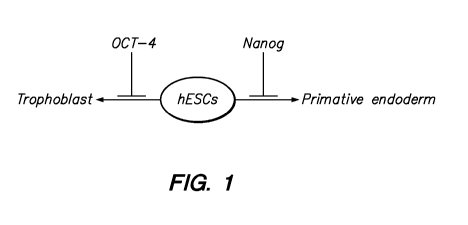
(Mitsui et al., Cell 113:631-642, 2003). Figure 1 shows transcription factors
involved in
controlling self-renewal of human embryonic stem cells by repressing early
lineage
commitment.
Two other transcription factors have been identified that interact with OCT4:
the
forkhead transcription factor FOXD3 and the Sry-related factor SOX2. FOXD3 is
expressed in the blastocyst and later in the post-implantation egg cylinder
epiblast.
FOXD3 physically interacts with OCT4 to activate the ostopontin enhancer,
which is
expressed in ESCs (Guo et al., Proc. Natl. Acad. Sci. U.S.A. 99:3663-3667,
2002). Sox2
is expressed in ESCs as well as in multipotent embryonic and extra-embryonic
lineages.
Disrupting Sox2 results in pre-implantation embryonic lethality (Avilion et
al., Genes
Dev. 17:126-140, 2003). Sox2 was identified as a co-factor of OCT4 for
activating FGF4,
which is restrictively expressed in undifferentiated ESCs, and is essential
for post-
implantation mouse development and limb patterning and growth (Yuan et al.,
Genes
Dev. 9:2635-2645, 1995). Transcriptional regulation of NANOG itself is also
regulated
by OCT4 and SOX2 (Rodda et al., J. Biol. Chem. 280: 24731-24737, 2005).
Another
OCT4 and SOX2 co-regulated gene is the ESC-specific transcription factor UTFI
(Nishimoto et al., Mol. Cell. Biol. 19:5453-5465, 1999). Taken together, these
studies
suggest that the SOX2-OCT4 complex is at the apex of a regulatory hierarchy of
the
"pluripotency genetic regulatory network".
Figure 1 shows transcription factors involved in controlling self-renewal by
repressing early lineage commitment.
In summary, ESC identity is determined by cell-intrinsic transcription factors
that
need to be expressed at particular levels in order to function appropriately.
However, the
molecular basis of the regulation of pluripotency and early lineage commitment
of hESCs
is still poorly understood. Additional intrinsic pathway-specific
transcription factors
presumably exist that maintain expression of the thousands of genes that are
expressed in
ESCs and control different types of renewal and differentiation pathways.
Understanding
how hESCs maintain their pluripotency and self-renewal and execute precise
2
CA 02695505 2010-02-03
WO 2009/020632 PCT/US2008/009475
differentiation programs will require extending our understanding of the
transcriptional
regulatory hierarchy of hESC function, including identifying new intrinsic
transcription
factors.
Summary of the Invention
We have identified zinc finger protein 206 (ZNF206), a novel repressor of
human
embryonic stem cell (hESC) differentiation. Repressing extra-embryonic
endoderm
development preserves the pluripotent state of human embryonic stem cells,
and,
conversely downregulating expression of ZNF206 in hESCs causes them to
upregulate
the expression of genes associated with the extra-embryonic endodermal
lineage, down-
regulate genes associated with the pluripotent state, and may lead to the
further
emergence of genes associated with even more differentiated lineages and
phenotypes.
According to one aspect of the invention, isolated polynucleotides are
provided
that comprise a sequence that has at least 90%, or 95%, or 100% nucleic acid
sequence
identity to a native ZNF206 polynucleotide and that hybridize selectively to
the native
ZNF206 polynucleotide. The isolated polynucleotide of claim 1 wherein the
sequence
that has at least 95% identity to a native ZNF206 polynucleotide. According to
another
embodiment, such isolated polynucleotides comprise a sequence at least 100
nucleotides
in length that has at least 90%, 95%, or 99% nucleic acid sequence identity to
a native
ZNF206 polynucleotide.
According to another embodiment of the invention, isolated polynucleotides are
provided that comprise at least 15, 20, or 30 contiguous nucleotides of a
native ZNF206
polynucleotide, wherein the isolated polynucleotide hybridizes selectively to
a native
ZNF206 polynucleotide. According to one embodiment, the isolated
polynucleotide
comprises a full-length protein-coding sequence of a native ZNF206 mRNA or
cDNA.
According to another embodiment, the isolated polynucleotide encodes a
polypeptide that
has ZNF206 activity.
According to another embodiment of the invention, cells are provided that
comprise any of the isolated polynucleotides described above. According to
another
embodiment, cells, vectors (including, but not limited to expression vectors),
probes and
primers are provided that comprise any of the isolated polynucleotides
described above.
Also provided are cells that comprise such vectors.
According to another embodiment of the invention, kits are provided that
comprise: (a) a first primer comprising at least 15 contiguous nucleotides of
a native
ZNF206 polynucleotide, wherein the first primer hybridizes selectively to a
native
3
CA 02695505 2010-02-03
WO 2009/020632 PCT/US2008/009475
ZNF206 polynucleotide; (b) a second primer comprising at least 15 contiguous
nucleotides of the native ZNF206 polynucleotide; and (c) suitable packaging
enclosing
the first primer and the second primer, wherein an amplification reaction
performed using
the first primer, the second primer, and a sample comprising a ZNF206 mRNA
produces
an amplification product that indicates the presence of the ZNF206 mRNA in the
sample.
According to another embodiment of the invention, isolated polypeptides of at
least 11 amino acids are provided that comprise at least 4, 5, 6, 7, 8, 9, or
10 contiguous
amino acids of a native ZNF206 polypeptide, and, that when introduced into a
mammal,
elicits production of an antibody that binds selectively to a native ZNF206
polypeptide.
According to another embodiment of the invention, isolated polypeptides are
provided
that comprise at least 11, 12, 15, 20, or 30 contiguous amino acids of a
native ZNF206
polypeptide, and, that when introduced into a mammal, elicits production of an
antibody
that binds selectively to a native ZNF206 polypeptide, including but not
limited to a full-
length native ZNG206 polypeptide.
According to another embodiment of the invention, isolated polypeptides are
provided that comprise a sequence that has at least 90%, 91, 92, 93, 94, 95%,
96%, 97%,
98%, 99% or 100% amino acid sequence identity to a native ZNF206 polypeptide,
wherein introduction of the isolated polypeptide into a mammal elicits
production of an
antibody that selectively binds to ZNF206. According to another embodiment of
the
invention, such isolated polypeptides comprise a sequence at least 15, 16, 17,
18, 19, 20,
30, 40 or more amino acids in length that has such a degree of amino acid
sequence
identity. According to another embodiment of the invention, such isolated
polypeptides
have ZNF206 activity.
According to another embodiment of the invention, isolated polynucleotides are
provided that encode any of the aforementioned polypeptides.
According to another embodiment of the invention, antibodies are provided that
bind selectively to a native ZNF206 polypeptide, including, but not limited
to,
monoclonal, polyclonal, chimeric, humanized, single-chain, and fragment
antibodies, for
example.
According to another embodiment of the invention, methods are provided for
making an antibody that binds selectively to a native ZNF206 polypeptide
comprising
introducing into a mammal (a) an expression vector comprising one of the
aforementioned polynucleotides, or (b) one of the aforementioned isolated
polypeptides,
thereby eliciting production of the antibody.
4
CA 02695505 2010-02-03
WO 2009/020632 PCT/US2008/009475
According to another embodiment of the invention, pharmaceutical compositions
are provided that comprise (a) a vector comprising a promoter suitable for
expression in
the cell operably linked to an isolated polynucleotide comprising a sequence
that has at
least 90% nucleic acid sequence identity to a native ZNF206 polynucleotide and
that
hybridizes selectively to the native ZNF206 polypeptide, wherein expression of
the
polynucleotide in the cell causes a reduction in ZNF206 polypeptide levels in
the cell, and
(b) a pharmaceutically acceptable carrier.
According to another embodiment of the invention, methods are provided for
making a medicament for treating a patient with a cancer or at risk for
developing the
cancer, the method comprising formulating the medicament with a
pharmaceutically
effective amount of a vector comprising a promoter suitable for expression in
the cell
operably linked to an isolated polynucleotide comprising a sequence that has
at least 90%
nucleic acid sequence identity to a native ZNF206 polynucleotide and that
hybridizes
selectively to the native ZNF206 polypeptide, wherein expression of the
polynucleotide in
the cell causes a reduction in ZNF206 polypeptide levels in the cell.
According to another embodiment of the invention, methods are provided for
detecting the presence of a ZNF206 polynucleotide in a sample comprising the
ZNF206
polynucleotide, the method comprising contacting the sample with a probe or
primer
comprising a polynucleotide sequence that binds selectively to the ZNF206
polynucleotide and detecting binding of the probe or primer to the ZNF206
mRNA. One
such embodiment, comprises (a) contacting the sample with a first primer that
comprises
the polynucleotide sequence that hybridizes selectively to the ZNF206 mRNA and
a
second primer comprising a polynucleotide sequence that hybridizes to the
ZNF206
mRNA, (b) performing an amplification reaction to produce an amplification
product that
indicates the presence of the ZNF206 mRNA in the sample, and (c) detecting the
amplification product, including, but not limited to, a PCR reaction.
According to another embodiment of the invention, methods are provided for
detecting the presence of a ZNF206 polypeptide in a sample comprising the
ZNF206
polypeptide, the method comprising (a) contacting the sample with an antibody
(including, but not limited to, a monoclonal antibody) that binds selectively
to the
ZNF206 polypeptide and (b) detecting binding of the antibody to the ZNF206
polypeptide. Such methods may, for example, comprise performing ELISA or bio-
barcode assays.
5
CA 02695505 2010-02-03
WO 2009/020632 PCT/US2008/009475
According to another embodiment of the invention, methods are provided for
assessing the pluripotency of a cell by various means. According to one such
embodiment, such methods comprise (a) measuring ZNF206 polypeptide or
polynucleotide levels in a sample comprising the cell, and (b) comparing the
ZNF206
polypeptide or polynucleotide levels in the sample to a reference. According
to another
such embodiment, such methods comprise measuring the ZNF206 polypeptide level
in
the cell by contacting a sample comprising the cell with an antibody that
binds selectively
to ZNF206 polypeptide (including but not limited to a monoclonal antibody) and
measuring binding of the antibody to ZNF206 polypeptide in the sample, such
as, for
example, by an ELISA or bio-barcode assay. According to another embodiment,
such
methods comprise measuring the ZNF206 mRNA level in the cell by contacting a
sample
comprising the cell with a probe or primer that hybridizes selectively to
ZNF206 mRNA
and measuring hybridization of the probe or primer to the ZNF206 mRNA in the
sample.
According to another embodiment, such methods comprise measuring the ZNF206
mRNA level in the cell by (a) contacting the sample comprising the cell with
one or more
primers that comprise a polynucleotide sequence that hybridizes selectively to
the
ZNF206 mRNA, (b) performing an amplification reaction (including but not
limited to a
PCR reaction or bio-barcode assay) to produce an amplification product that
indicates the
presence of ZNF206 mRNA in the sample, and (c) measuring the amplification
product.
In any of the foregoing methods for assessing the pluripotency of a cell, the
sample may
be, for example, a tissue sample.
According to another embodiment of the invention, methods are provided for
maintaining or increasing the pluripotency of a cell comprising expressing in
the cell a
vector comprising (a) a promoter suitable for expression in the cell operably
linked to (b)
an isolated polynucleotide comprising a sequence at least 100 nucleotides in
length that
has at least 90% nucleic acid sequence identity to a native ZNF206
polynucleotide,
wherein expression of the polynucleotide in the cell produces a polypeptide
that reduces
or prevents differentiation of the cell.
According to another embodiment of the invention, methods are provided for
promoting differentiation of a cell comprising reducing ZNF206 expression of
the cell.
According to one embodiment, such a method comprises expressing in the cell a
vector
comprising (a) a promoter suitable for expression in the cell operably linked
to (b) an
isolated polynucleotide comprising a sequence that has at least 90% nucleic
acid sequence
identity to a native ZNF206 polynucleotide and that hybridizes selectively to
the native
6
CA 02695505 2010-02-03
WO 2009/020632 PCT/US2008/009475
ZNF206 polypeptide, wherein expression of the polynucleotide in the cell
causes a
reduction in ZNF206 polypeptide levels in the cell.
According to another embodiment of the invention, methods are provided for
diagnosing a cancer characterized by elevated levels of ZNF206 comprising (a)
obtaining
a sample comprising a cell, (b) determining ZNF206 polypeptide or
polynucleotide levels
in the sample, and (c) comparing the ZNF206 polypeptide or polynucleotide
levels in the
sample with a reference.
According to another embodiment of the invention, methods are provided for
treating a cancer characterized by elevated levels of ZNF206 comprising
administering to
a patient in need of such treatment a composition comprising a vector
comprising (a) a
promoter suitable for expression in a cell of the patient operably linked to
(b) an isolated
polynucleotide comprising a sequence at least 100 nucleotides in length that
has at least
90% nucleic acid sequence identity to a native ZNF206 polynucleotide, wherein
expression of the polynucleotide in the cell reduces ZNF206 polypeptide levels
in the
cell.
According to another embodiment of the invention, methods are provided for
diagnosing a disease state resulting from a mutation in a ZNF206
polynucleotide
comprising (a) providing a sample from a patient comprising a cell and (b)
determining
whether the sample comprises a mutated ZNF206 polynucleotide. The presence of
a
mutated ZNF 206 polynucleotide in the sample may be determined, for example
by:
contacting the sample with a polynucleotide probe or primer that hybridizes
specifically
to a mutated ZNF206 polynucleotide sequence; by contacting the sample with one
or
more primers that comprise a polynucleotide sequence that hybridizes
selectively to the
mutated ZNF206 polynucleotide, and performing an amplification reaction (e.g.,
a PCR
or bio-barcode assay) to produce an amplification product that indicates the
presence of
the mutated ZNF206 polynucleotide in the sample; by detecting a restriction
fragment
length polymorphism; or by contacting the sample with an antibody probe that
hybridizes
specifically to a mutated ZNF polypeptide sequence encoded by the mutated ZNF
polynucleotide.
Any of the aforementioned methods may be automated.
The foregoing and other aspects of the invention will become more apparent
from
the following detailed description, accompanying drawings, and the claims.
Unless otherwise defined, all technical and scientific terms used herein have
the
same meaning as commonly understood by one of ordinary skill in the art to
which this
7
CA 02695505 2010-02-03
WO 2009/020632 PCT/US2008/009475
invention pertains. Although methods and materials similar or equivalent to
those
described herein can be used in the practice or testing of the present
invention, suitable
methods and materials are described below. All publications, patent
applications, patents,
and other references mentioned herein are incorporated by reference in their
entirety. In
case of conflict, the present specification, including definitions, will
control. In addition,
the materials, methods, and examples are illustrative only and not intended to
be limiting.
Brief Description of the Figures
Figure 1 shows transcription factors involved in controlling self-renewal of
human
embryonic stem cells by repressing early lineage commitment.
Figure 2 shows high and unique expression of ZNF206 in undifferentiated hESCs.
[A] ZNF206 and NANOG were highly expressed in hESC line WA09 (H9) but not in
PE-
like (PEL) cells derived from them. [B] Quantitative RT-PCR analysis of ZNF206
expression in H9 hESCs, in PEL cells derived from H9 hESCs, and in adult human
tissues.
Figure 3 shows that ZNF206 expression is downregulated upon hESC
differentiation into extraembryonic endoderm cells. HESCs (from lines WA09
[H9] and
WA01 [H 1]) were treated for various times -- 0, 48, 96 hrs -- with BMP2
(50ng/ml)
followed by Quantitative RT-PCR to analyze the expression of NANOG [A], ZNF206
[B], GATA6 [C], and GATA4 [D].
Figure 4 shows the predicted protein sequence of three isoforms of ZNF206. The
ZNF206 gene contains five introns and five exons. [A] Primers were
specifically
designed to amplify and to clone the different spliced ZNF206 mRNA isoforms
expressed
in undifferentiated hESCs. [B] Four different ZNF206 mRNA isoforms were cloned
from
undifferentiated hESCs. Isoform 1 is 2568bp, isoform 2 is 2343bp, and isoform
3 is
2075bp. [C] Isoform 1 and 4 are predicted to encode truncated ZNF206 proteins
containing a "Novel" and "SCAN" domain. The Novel domain contains a
sumoylation
site and the SCAN domain has been previously reported to mediate protein-
protein
interactions. On the other hand, ZNF206 isoform 2 is predicted to contain 780
amino
acids containing the Novel, SCAN and 14 C2H2 Zinc finger domains. The C2H2
zinc
finger domains often mediate DNA binding.
Figure 5 shows a diagram of three C-terminally tagged ZNF206 lentivirus
expression vectors that we have successfully made.
Figure 6 shows the knock-down efficiency of lentiviral ZNF206 shRNA
expression constructs. Human kidney 293FT-ZNF206-V5 expressing cell lines were
8
CA 02695505 2010-02-03
WO 2009/020632 PCT/US2008/009475
infected with lentiviral particles containing ZNF206 shRNA expression
constructs. After
puromycin selection and expansion of infected 293FT cells, we performed
quantitative
RT-PCR [A] and Western blot analysis [B].
Figure 7 shows the generation of a polyclonal rabbit antibody against the
human
ZNF206 proteins. Underlined is the peptide (amino acids 71_1-726) used to
immunize
rabbits against the human ZNF206 protein. The polyclonal antibody detects a
protein that
is approximately 80 kD in size in undifferentiated hES cell line H9 and not in
the hES-
derived PEL differentiated cells.
Figure 8 shows the effects of ZNF206 knockdown on OCT-4 and NANOG
expression in hESCs. hESCs were infected with three different shRNA lentiviral
expression particles (ZNF206 shRNA-A, ZNF206 shRNA-B, ZNF206 shRNA-C) and the
control lentiviral empty vector. Four days after infection of undifferentiated
hESC lines
H9 (WA09) and H 1(WA01), the mRNA and protein expression of ZNF206, Oct-4 and
NANOG were determined by quantitative RT PCR.
Figure 9 shows the hypothesized Role of ZN206 in hESCs. [A] In our model,
OCT4 is the key inhibitor of trophoblast differentiation in hESCs (since
specific down-
regulation of OCT-4 in hESCs leads to trophoblast differentiation), while
NANOG and
ZNF206 are key inhibitiors of extra-embryonic endoderm lineage differentiation
(since
specific down-regulation of NANOG or ZNF206 leads to extra-embryonic endoderm
lineage differentiation). For example, down-regulation of ZNF206 expression in
hESCs
causes them to upregulate genes associated with the extra-embryonic endoderm
lineage
(e.g., GATA4, GATA6, SOX17, AFP and HNF4A). [B] We further hypothesize that
extra-embryonic endoderm differentiation may be the earliest default pathways
for
hESCs, particularly when dissociated into single cells and grown in defined,
serum-free,
feeder-free conditions. This default lineage may then help instruct the
emergence of other
lineages, e.g., neuroectoderm (perhaps giving the appearance of being
default).
Figure 10 shows the DNA sequence for four isoforms of ZNF206.
Detailed Description of the Invention
Definitions and Methods
The following definitions and methods are provided to better define the
present
invention and to guide those of ordinary skill in the art in the practice of
the present
invention. Unless otherwise noted, terms are to be understood according to
conventional
usage by those of ordinary skill in the relevant art. Definitions of common
terms in
9
CA 02695505 2010-02-03
WO 2009/020632 PCT/US2008/009475
molecular biology may also be found in Rieger et al., Glossary of Genetics:
Classical and
Molecular, 5th edition, Springer-Verlag: New York, 1991; and Lewin, Genes V,
Oxford
University Press: New York, 1994. The nomenclature for DNA bases as set forth
at 37
CFR 1.822 is used. The standard one- and three-letter nomenclature for amino
acid
residues is used.
Polynucleotides
As used herein, the term "ZNF206 polynucleotide" refers to the ZNF206 genomic
DNA, mRNA, and cDNA corresponding to the mRNA as present in humans (including
any of the several human isoforms of ZNF206) or non-human species, such as,
for
example, in the chimpanzee, mouse or chicken (Bernot et al., Genomics 50:147-
160,
1998). Also encompassed by the term "ZNF206 polynucleotides" are, for example:
fragments or portions of the ZNF206 mRNA or cDNA, including but not limited
to, a
ZNF206 polynucleotide; fragments that encode antigenic determinants of ZNF206
(e.g.,
those that elicit antibodies that bind selectively to ZNF206 polypeptide);
probes and
primers that hybridize selectively to ZNF206 polynucleotides; etc. Also
included are
mutated or variant polynucleotides that include one or more nucleotide
insertions,
deletions, or substitutions from the wild-type ZNF206 sequence, but that, for
example:
retain the ability to bind selectively to ZNF206; encode a polypeptide that
includes a
ZNF206 antigenic determinant; encode a polypeptide having ZNF206 activity;
etc.
As used herein, the term "hybridizes selectively" refers to binding of a
probe,
primer or other polynucleotide, under stringent hybridization conditions, to a
target
polynucleotide, such as a native, or wild-type, ZNF206 mRNA or cDNA, to a
substantially higher degree than to other polynucleotides. Probes and primers
that
hybridize selectively to ZNF206 include sequences that are unique to ZNF206.
In
particular, a probe that "hybridizes selectively" to ZNF206 does not hybridize
substantially to ZNF206 under stringent hybridization conditions and therefore
can be
used to distinguish a ZNF206 polynucleotide (e.g., a ZNF206 mRNA) from a
ZNF206
polynucleotide. Similarly, a primer that "hybridizes selectively" to ZNF206,
when used
in an amplification reaction such as PCR, results in amplification of ZNF206
without
resulting in substantial amplification of ZNF206 under suitable amplification
conditions.
Thus, all or substantially all of a ZNF206-selective probe or primer
hybridizes to the
target ZNF206 polynucleotide under suitable conditions, as can be determined
given the
sensitivity of a particular procedure. Similarly, as used herein, the term
"selective for" in
CA 02695505 2010-02-03
WO 2009/020632 PCT/US2008/009475
reference to a polynucleotide, indicates that the polynucleotide hybridizes
selectively to a
target polynucleotide.
Similarly, a probe or primer that includes a sequence that is unique to ZNF206
hybridizes selectively to ZNF206. In particular, a probe that hybridizes
selectively to
ZNF206 does not hybridize substantially to ZNF206 under stringent
hybridization
conditions and therefore can be used to distinguish a ZNF206 polynucleotide
(e.g., a
ZNF206 mRNA) from a ZNF206 polynucleotide. Similarly, a primer that hybridizes
selectively to a ZNF206 polynucleotide, when used in an amplification reaction
such as
PCR, results in amplification of the ZNF206 polynucleotide without resulting
in
substantial amplification of ZNF206 polynucleotide. Thus, all or substantially
all of a
ZNF206-selective probe or primer hybridizes to the target ZNF206
polynucleotide, as can
be determined given the sensitivity of a particular procedure.
As used herein, the terms "wild-type" or "native" in reference to a
polynucleotide
are used interchangeably to refer to a polynucleotide that has 100% sequence
identity
with a reference polynucleotide that can be found in a cell or organism, or a
fragment
thereof.
Polynucleotide (e.g., DNA or RNA) sequences may be determined by sequencing
a polynucleotide molecule using an automated DNA sequencer. A polynucleotide
sequence determined by this automated approach can contain some errors. The
actual
sequence can be confirmed by resequencing the polynucleotide by automated
means or by
manual sequencing methods well known in the art.
Unless otherwise indicated, each "nucleotide sequence" set forth herein is
presented as a sequence of deoxyribonucleotides (abbreviated A, G, C and T).
However,
the term "nucleotide sequence" of a DNA molecule as used herein refers to a
sequence of
deoxyribonucleotides, and for an RNA molecule, the corresponding sequence of
ribonucleotides (A, G, C and U) where each thymidine deoxynucleotide (T) in
the
specified deoxynucleotide sequence in is replaced by the ribonucleotide
uridine (U).
By "isolated" polynucleotide is intended a polynucleotide that has been
removed
from its native environment. For example, recombinant polynucleotides
contained in a
vector are considered isolated for the purposes of the present invention.
Further examples
of isolated polynucleotides include recombinant polynucleotides maintained in
heterologous host cells or purified (partially or substantially)
polynucleotides in solution.
Isolated RNA molecules include in vivo or in vitro RNA transcripts of the DNA
11
CA 02695505 2010-02-03
WO 2009/020632 PCT/US2008/009475
molecules of the present invention. Isolated polynucleotides according to the
present
invention further include such molecules produced synthetically.
Polynucleotides can be in the form of RNA, such as mRNA, or in the form of
DNA, including, for instance, cDNA and genomic DNA. The DNA can be double-
stranded or single-stranded. A single-stranded DNA or RNA can be a coding
strand, also
known as the sense strand, or it can be a non-coding strand, also referred to
as the anti-
sense strand. Polynucleotides can include non-naturally occurring nucleotide
or
ribonucleotide analogs.
The term "fragment" (of a polynucleotide) as used herein refers to
polynucleotides
that are part of a longer polynucleotide having a length of at least about 15,
20, 25, 30, 35,
or 40 nucleotides (nt) in length, which are useful, for example, as probes and
primers.
Thus, for example, a fragment of ZNF206 at least 20 nucleotides in length
includes 20 or
more contiguous nucleotides from the nucleotide sequence of the ZNF206 full-
length
cDNA. Such DNA fragments may be generated by the use of automated DNA
synthesizers or by restriction endonuclease cleavage or shearing (e.g., by by
sonication) a
full-length ZNF206 cDNA, for example.
Also encompassed by the present invention are isolated polynucleotides that
hybridize under stringent hybridization conditions to a ZNF206 polynucleotide
such as,
for example, a ZNF206 transcript (i.e., mRNA). By "stringent hybridization
conditions" is
intended overnight incubation at 42 C. in a solution comprising: 50%
formamide, 5x
SSC (750 mM NaCI, 75 mM trisodium citrate), 50 mM sodium phosphate (pH7.6), 5
x
Denhardt's solution, 10% dextran sulfate, and 20 g/ml denatured, sheared
salmon sperm
DNA, followed by washing the filters in 0.1x SSC at about 65 C.
Alternatively, stringent
hybridizations are conditions used for performance of a polymerase chain
reaction (PCR).
Such hybridizing polynucleotides are useful diagnostically as a probe
according to
conventional DNA hybridization techniques or as primers for amplification of a
target
sequence by the polymerase chain reaction (PCR).
As used herein, the term "hybridizes (or binds) specifically" is used
interchangeably with the term "hybridizes (or binds) selectively" means that
most or
substantially all hybridization of a probe or primer is to a particular
polynucleotide in a
sample under stringent hybridization conditions.
The present invention also provides polynucleotides that encode all or a
portion of
a polypeptide, e.g., a full-length ZNF206 polypeptide or a portion thereof.
Such protein-
coding polynucleotides may include, but are not limited to, those sequences
that encode
12
CA 02695505 2010-02-03
WO 2009/020632 PCT/US2008/009475
the amino acid sequence of the particular polypeptide or fragment thereof and
may also
include together with additional, non-coding sequences, including for example,
but not
limited to introns and non-coding 5' and 3' sequences, such as the
transcribed, non-
translated sequences that play a role in transcription, mRNA processing--
including
splicing and polyadenylation signals, e.g., ribosome binding and stability of
mRNA; an
additional coding sequence which codes for additional amino acids, such as
those which
provide additional functionalities. In addition, the sequence encoding the
polypeptide can
be fused to a heterogeneous polypeptide or peptide sequence, such as, for
example a
marker sequence that facilitates purification of the fused polypeptide. One
example of
such a marker sequence is a hexa-histidine peptide, such as the tag provided
in a pQE
vector (Qiagen, Inc.). As described in Gentz et al., Proc. Natl. Acad. Sci.
USA 86:821-
824 (1989), for instance, hexa-histidine provides for convenient purification
of the fusion
protein. The "HA" tag is another peptide useful for purification which
corresponds to an
epitope derived from the influenza hemagglutinin (HA) protein (Wilson et al.,
Cell
37:767, 1984).
The present invention further relates to variants of the native, or wild-type,
polynucleotides of the present invention, which encode portions, analogs or
derivatives of
a ZNF206 polypeptide. Variants can occur naturally, such as a natural allelic
variant, i.e.,
one of several alternate forms of a gene occupying a given locus on a
chromosome of an
organism. Non-naturally occurring variants can be produced, e.g., using known
mutagenesis techniques or by DNA synthesis. Such variants include those
produced by
nucleotide substitutions, deletions or additions. The substitutions, deletions
or additions
can involve one or more nucleotides. The variants can be altered in coding or
non-coding
regions or both. Alterations in the coding regions can produce conservative or
non-
conservative amino acid substitutions, deletions or additions. Also included
are silent
substitutions, additions and deletions, which do not alter the properties and
activities of
the ZNF206 polypeptide or portions thereof.
Further embodiments of the invention include isolated polynucleotide molecules
have, or comprise a sequence having, a high degree of sequence identity with a
native, or
wild type, ZNF206 polynucleotide, for example, at least 90%, 95%, 96%, 97%,
98% or
99% identical thereto.
A polynucleotide is considered to have a nucleotide sequence at least, for
example, 95% "identical" to a reference nucleotide sequence if it is identical
to the
reference sequence except that it includes up to five mutations (additions,
deletions, or
13
CA 02695505 2010-02-03
WO 2009/020632 PCT/US2008/009475
substitutions) per each 100 nucleotides of the reference nucleotide sequence.
These
mutations of the reference sequence can occur at the 5' or 3' terminal
positions of the
reference nucleotide sequence or anywhere between those terminal positions,
interspersed
either individually among nucleotides in the reference sequence or in one or
more
contiguous groups within the reference sequence. Nucleotide sequence identity
may be
determined conventionally using known computer programs such as the BESTFIT
program (Wisconsin Sequence Analysis Package, Version 8 for Unix, Genetics
Computer
Group, University Research Park, 575 Science Drive, Madison, Wis. 53711.
BESTFIT
uses the local homology algorithm of Smith and Waterman, Adv. Appl. Math.
2:482-489
(1981), to find the best segment of homology between two sequences. When using
BESTFIT or any other sequence alignment program to determine whether a
particular
sequence is, for instance, 95% identical to a reference sequence according to
the present
invention, the parameters are set, of course, such that the percentage of
identity is
calculated over the full length of the reference nucleotide sequence and that
gaps in
homology of up to 5% of the total number of nucleotides in the reference
sequence are
allowed.
Recombinant Constructs; Vectors and Host Cells
The present invention also provides recombinant polynucleotide constructs that
comprise a ZNF206 polynucleotide, including but not limited to vectors. The
present
invention also provides host cells comprising such vectors and the production
of ZNF206
polypeptides or fragments thereof by recombinant or synthetic techniques.
"Operably Linked". A first nucleic-acid sequence is "operably linked" with a
second nucleic-acid sequence when the first nucleic-acid sequence is placed in
a
functional relationship with the second nucleic-acid sequence. For instance, a
promoter is
operably linked to a coding sequence if the promoter affects the transcription
or
expression of the coding sequence. Generally, operably linked DNA sequences
are
contiguous and, where necessary to join two protein coding regions, in reading
frame.
"Recombinant". A "recombinant" polynucleotide is made by an artificial
combination of two otherwise separated segments of sequence, e.g., by chemical
synthesis or by the manipulation of isolated segments of polynucleotides by
genetic
engineering techniques. Techniques for nucleic-acid manipulation are well-
known (see,
e.g., Sambrook et al., 1989, and Ausubel et al., 1992). Methods for chemical
synthesis of
polynucleotides are discussed, for example, in Beaucage and Carruthers, Tetra.
Letts.
22:1859-1862, 1981, and Matteucci et al., J. Am. Chem. Soc. 103:3185, 1981.
Chemical
14
CA 02695505 2010-02-03
WO 2009/020632 PCT/US2008/009475
synthesis of polynucleotides can be performed, for example, on commercial
automated
oligonucleotide synthesizers.
Recombinant vectors are produced by standard recombinant techniques and may
be introduced into host cells using well known techniques such as infection,
transduction,
transfection, transvection, electroporation and transformation. The vector may
be, for
example, a phage, plasmid, viral or retroviral vector. Retroviral vectors may
be
replication competent or replication defective. In the latter case, viral
propagation
generally will occur only in complementing host cells.
Expression vectors include sequences that permit expression of a polypeptide
encoded by a polynucleotide of interest in a suitable host cell. Such
expression may be
constitutive or non-constitutive, e.g., inducible by an environmental factor
or a chemical
inducer that is specific to a particular cell or tissue type, for example.
Expression vectors
include chromosomal-, episomal- and virus-derived vectors, e.g., vectors
derived from
bacterial plasmids, bacteriophage, yeast episomes, yeast chromosomal elements,
viruses
such as baculoviruses, papova viruses, vaccinia viruses, adenoviruses, fowl
pox viruses,
pseudorabies viruses and retroviruses, and vectors derived from combinations
thereof,
such as cosmids and phagemids.
In expression vectors, a polynucleotide insert is operably linked to an
appropriate
promoter. The promoter may be a homologous promoter, i.e., a promoter or
functional
portion thereof, that is associated with the polynucleotide insert in nature,
for example, a
ZNF206 promoter with a ZNF206 or ZNF206 protein coding region. Alternatively,
the
promoter may be a heterologous promoter, i.e., a promoter or functional
portion thereof,
that is not associated with the polynucleotide insert in nature, for example,
a bacterial
promoter used for high-level protein expression in bacterial cells (or, for
that matter, any
promoter other than a ZNF206 promoter) operably linked to a ZNF206 protein
coding
region. The expression constructs will further contain sites for transcription
initiation,
termination and, in the transcribed region, a ribosome binding site for
translation. The
coding portion of the mature transcripts expressed by the constructs will
include a
translation initiating AUG at the beginning and a termination codon
appropriately
positioned at the end of the polypeptide to be translated.
Vectors may include one or more selectable marker suitable for selection of a
host
cell into which such a vector has been introduced. Such markers include
dihydrofolate
reductase or neomycin resistance for eukaryotic cell culture and tetracycline
or ampicillin
resistance genes for culturing in E. coli and other bacteria. Representative
examples of
CA 02695505 2010-02-03
WO 2009/020632 PCT/US2008/009475
appropriate hosts include bacterial cells, such as E. coli, Streptoniyces and
Salmonella
ryphimurium cells; fungal cells, such as yeast cells; insect cells such as
Drosophila S2 and
Spodoptera Sf9 cells; animal cells such as CHO, COS and Bowes melanoma cells;
and
plant cells. Appropriate culture media and conditions for the above-described
host cells
are known in the art.
Bacterial promoters suitable include the E. coli lacI and lacZ promoters, the
T3
and T7 promoters, the gpt promoter, the lambda PR and PL promoters and the trp
promoter. Eukaryotic promoters include the CMV immediate early promoter, the
HSV
thymidine kinase promoter, the early and late SV40 promoters, the promoters of
retroviral
LTRs, such as those of the Rous sarcoma virus (RSV), and metallothionein
promoters,
such as the mouse metallothionein-I promoter.
For secretion of the translated protein into the lumen of the endoplasmic
reticulum, into the periplasmic space or into the extracellular environment,
appropriate
secretion signals may be incorporated into the expressed polypeptide. The
signals may be
endogenous to the polypeptide or they may be heterologous signals.
A polypeptide of interest may be expressed in a modified form, such as a
fusion
protein, and may include not only secretion signals but also additional
heterologous
functional regions. For instance, a region of additional amino acids,
particularly charged
amino acids, may be added to the N-terminus of the polypeptide to improve
stability and
persistence in the host cell, during purification or during subsequent
handling and storage.
Also, peptide moieties may be added to the polypeptide to facilitate
purification. Such
regions may be removed prior to final preparation of the polypeptide. The
addition of
peptide moieties to polypeptides to engender secretion or excretion, to
improve stability
and to facilitate purification, among others, are familiar and routine
techniques in the art.
An expressed polypeptide of interest can be recovered and purified from
recombinant cell cultures by well-known methods including ammonium sulfate or
ethanol
precipitation, acid extraction, anion or cation exchange chromatography,
phosphocellulose chromatography, hydrophobic interaction chromatography,
affinity
chromatography, hydroxylapatite chromatography and lectin chromatography.
Polypeptides of the present invention include naturally purified products,
products
of chemical synthetic procedures, and products produced by recombinant
techniques from
a prokaryotic or eukaryotic host, including, for example, bacterial, yeast,
higher plant,
insect and mammalian cells. Depending upon the host employed in a recombinant
production procedure, the polypeptides of the present invention may be
glycosylated or
16
CA 02695505 2010-02-03
WO 2009/020632 PCT/US2008/009475
may be non-glycosylated. In addition, polypeptides of the invention may also
include an
initial modified methionine residue, in some cases as a result of host-
mediated processes.
Polynucleotide constructs can also be used to reduce expression of ZNF206 in a
cell. For example, antisense constructs, ribozymes, short interfering RNA
(siRNA) or
small hairpin RNA (shRNA), and other such constructs can be used for this
purpose.
A "small interfering RNA" or "short interfering RNA" (siRNA) or "short hairpin
RNA" (shRNA) is a double-stranded RNA molecule that is complementary to a
target
nucleic acid sequence, for example, VEGF-C. A double-stranded RNA molecule is
formed by the complementary pairing between a first RNA portion and a second
RNA
portion. The length of each portion generally is less than 30 nucleotides in
length (e.g.,
29, 28, 27, 26, 25, 24, 23, 22, 21, 20, 19, 18, 17, 16, 15, 14, 13, 12, 11, or
10 nucleotides).
In some embodiments, the length of each portion is 19 to 25 nucleotides in
length. In
some siRNA molecules, the complementary first and second portions of the RNA
molecule are the "stem" of a hairpin structure. The two portions can be joined
by a linking
sequence, which can form the "loop" in the hairpin structure. The linking
sequence can
vary in length. In some embodiments, the linking sequence can be 5, 6, 7, 8,
9, 10, 11, 12
or 13 nucleotides in length. The first and second portions are complementary
but may not
be completely symmetrical, as the hairpin structure may contain 3' or 5'
overhang
nucleotides (e.g., a 1, 2, 3, 4, or 5 nucleotide overhang).
RNA molecules have been shown by many researchers to be effective in
suppressing mRNA accumulation. siRNA-mediated suppression of nucleic acid
expression is specific as even a single base pair mismatch between siRNA and
the
targeted nucleic acid can abolish the action of RNA interference. siRNAs
generally do not
elicit anti-viral responses.
There are well-established criteria for designing siRNAs (see, e.g., Elbashire
et
al., Nature, 411:494 8, 2001; Amarzguioui et al., Biochem. Biophys. Res.
Commun.,
316:1050 8, 2004; Reynolds et al., Nat. Biotech., 22:326-30, 2004). Details
can be found
in the websites of several commercial vendors such as Ambion, Dharmacon,
GenScript,
and OligoEngine. The sequence of any potential siRNA candidate generally is
checked
for any possible matches to other nucleic acid sequences or polymorphisms of
nucleic
acid sequence using the BLAST alignment program (see ncbi.nlm.nih.gov on the
World
Wide Web). Typically, a number of siRNAs have to be generated and screened in
order to
compare their effectiveness.
17
CA 02695505 2010-02-03
WO 2009/020632 PCT/US2008/009475
Once designed, the siRNAs of the present invention can be generated by any
method known in the art, for example, by in vitro transcription,
recombinantly, or by
synthetic means (e.g., having either a TT or a UU overhang at the 3' end).
siRNAs can be
generated in vitro by using a recombinant enzyme, such as T7 RNA polymerase,
and
DNA oligonucleotide templates, or can be prepared in vivo, for example, in
cultured cells
(see, for example, Elbashir et al., supra; Brummelkamp et al., supra; and Lee
et al., Nat.
Biotech., 20:500-505, 2002).
In addition, strategies have been described for producing a hairpin siRNA from
vectors containing a RNA polymerase III promoter. Various vectors have been
constructed for generating hairpin siRNAs in host cells using either an H 1-
RNA or an
snU6 RNA promoter. A RNA molecule as described above (e.g., a first portion, a
linking
sequence, and a second portion) can be operably linked to such a promoter.
When
transcribed by RNA polymerase III, the first and second portions form a
duplexed stem of
a hairpin and the linking sequence forms a loop. The pSuper vector
(OligoEngines Ltd.,
Seattle, Wash.) also can be used to generate siRNA.
A TTTTT penta-nucleotide usually is attached to the end of the second portion
(i.e., the antisense strand) in a vector to serve as a terminator for RNA
polymerase III
transcription. For that reason, siRNA candidates that contain more than three
consecutive
Ts should be avoided since four or more consecutive Ts in the template nucleic
acid
triggers termination of RNA polymerase III transcription.
Several techniques can be used to test the effect of different siRNA
constructs on
cellular mRNA and/or protein levels. For example, dual-GFP transfection, CHO-
cell
double transfection based on an antibody/epitope specificity, quantitative RT-
PCR,
Northern blots, Western blots, immunofluorescence, and Hygro/Neo selection.
These
methods are well known in the art.
Polypeptides
As used herein, the phrase "a ZNF206 polypeptide" refers to a polypeptide at
least
10, 11, 12, 12, 14, 15, 20, 30, 40, 49, 50, 100 or more amino acid residues in
length and
have a high degree of sequence identity with the full-length native, or wild-
type, ZNF206
polypeptide or a fragment thereof. Included are variant forms of ZNF206
polypeptides
that include deletions, insertions or substitutions of one or more amino acid
residues in a
native ZNF206 polypeptide sequence, including without limitation polypeptides
that
exhibit activity similar, but not necessarily identical, to an activity of the
full-length
18
CA 02695505 2010-02-03
WO 2009/020632 PCT/US2008/009475
native, or wild-type, ZNF206 polypeptide or fragment thereof as measured in a
relevant
biological assay.
As used herein, the terms "wild-type" or "native" in reference to a peptide or
polypeptide are used interchangeably to refer to a polypeptide that has 100%
sequence
identity with a reference polypeptide that can be found in a cell or organism,
or a
fragment thereof.
As used herein, the term "ZNF206 activity" refers to a biological activity of
a
native ZNF206 polypeptide including, but not limited to, repressing PE or PE-
like
differentiation, regulation of pluripotency gene expression, DNA binding,
etc..
As used herein, the terms "peptide" and "oligopeptide" are considered
synonymous and, as used herein, each term refers to a chain of at least two
amino acids
coupled by peptidyl linkages. As used herein, the terms "polypeptide" and
"protein" are
considered synonymous and each term refers to a chain of more than about ten
amino
acid residues. All oligopeptide and polypeptide formulas or sequences herein
are written
- from left to right and in the direction from amino terminus to carboxy
terminus.
As used herein, the term "isolated" polypeptide or protein refers to a
polypeptide
or protein removed from its native environment. For example, recombinantly
produced
polypeptides and proteins expressed in host cells are considered isolated for
purposes of
the invention as are native or recombinant polypeptides and proteins which
have been
substantially purified by any suitable technique.
As used herein, the term "binds selectively" is interchangeable with the term
"binds specifically, and, when used in reference to a ZNF206 polypeptide,
refers to
binding of an antibody, ligand, receptor, substrate, or other binding agent to
the target
ZNF206 polypeptide to a substantially higher degree than to other
polypeptides.
According to some embodiments, all or substantially all binding of an antibody
or other
binding agent is to the target ZNF206 polynucleotide, as can be determined
given the
sensitivity of a particular procedure. An antibody, ligand, receptor,
substrate or other
binding agent is said to be "selective for" or specific for" a polypeptide or
other target
molecule, such as ZNF206, if it binds selectively to the target molecule.
The amino acid sequence of a ZNF206 polypeptide or peptide can be varied
without significant effect on the structure or function of the protein. In
general, it is
possible to replace residues which contribute to the tertiary structure of the
polypeptide or
peptide, provided that residues performing a similar function are used. In
other instances,
19
CA 02695505 2010-02-03
WO 2009/020632 PCT/US2008/009475
the type of residue may be completely unimportant if the alteration occurs at
a non-
critical region of the protein.
Thus, the invention further includes variations of ZNF206 polypeptide or
peptide
that show substantial ZNF206 activity. Such mutants include deletions,
insertions,
inversions, repeats, and type substitutions (for example, substituting one
hydrophilic
residue for another, but not strongly hydrophilic for strongly hydrophobic as
a rule).
Small changes or such "neutral" amino acid substitutions will generally have
little effect
on activity.
Typically seen as conservative substitutions are the replacements, one for
another,
among the aliphatic amino acids Ala, Val, Leu and Ile; interchange of the
hydroxyl
residues Ser and Thr, exchange of the acidic residues Asp and Glu,
substitution between
the amide residues Asn and Gln, exchange of the basic residues Lys and Arg and
replacements among the aromatic residues Phe, Tyr.
Guidance concerning which amino acid changes are likely to be phenotypically
silent (i.e., are not likely to have a significant deleterious effect on a
function) can be
found, for example, in Bowie et al., Science 247:1306-1310, 1990.
Thus, a fragment, derivative or analog of a native, or wild-type ZNF206
polypeptide, may be (i) one in which one or more of the amino acid residues
are
substituted with a conserved or non-conserved amino acid residue and such
substituted
amino acid residue may or may not be one encoded by the genetic code, or (ii)
one in
which one or more of the amino acid residues includes a substituent group, or
(iii) one in
which the mature polypeptide is fused with another compound, such as a
compound to
increase the half-life of the polypeptide (for example, polyethylene glycol),
or (iv) one in
which the additional amino acids are fused to the mature polypeptide, such as
an IgG Fc
fusion region peptide or leader or secretory sequence or a sequence that is
employed for
purification of the mature polypeptide or a proprotein sequence.
Charged amino acids may be substituted with another charged amino acid.
Charged amino acids may also be substituted with neutral or negatively charged
amino
acids, resulting in proteins with reduced positive charge. The prevention of
aggregation is
highly desirable to avoid a loss of activity and increased immunogenicity
(Pinckard et al.,
Clin Exp. Immunol. 2:331-340, 1967; Robbins et al., Diabetes 36:838-845, 1987;
Cleland
et al., Crit. Rev. Therapeutic Drug Carrier Systems 10:307-377, 1993).
The replacement of amino acids can also change the selectivity of protein
binding
to cell surface receptors. Ostade et al., Nature 361:266-268 (1993) describes
certain
CA 02695505 2010-02-03
WO 2009/020632 PCT/US2008/009475
mutations resulting in selective binding of TNF-a to only one of the two known
types of
TNF receptors.
It is well known in the art that one or more amino acids in a native sequence
can
be substituted with other amino acid(s), the charge and polarity of which are
similar to
that of the native amino acid, i.e., a conservative amino acid substitution,
resulting in a
silent change. Conservative substitutes for an amino acid within the native
polypeptide
sequence can be selected from other members of the class to which the amino
acid
belongs. Amino acids can be divided into the following four groups: (1) acidic
amino
acids, (2) basic amino acids, (3) neutral polar amino acids, and (4) neutral,
nonpolar
amino acids. Representative amino acids within these various groups include,
but are not
limited to, (1) acidic (negatively charged) amino acids such as aspartic acid
and glutamic
acid; (2) basic (positively charged) amino acids such as arginine, histidine,
and lysine; (3)
neutral polar amino acids such as glycine, serine, threonine, cysteine,
cystine, tyrosine,
asparagine, and glutamine; and (4) neutral nonpolar (hydrophobic) amino acids
such as
alanine, leucine, isoleucine, valine, proline, phenylalanine, tryptophan, and
methionine.
Conservative amino acid substitution within the native polypeptide sequence
can be made
by replacing one amino acid from within one of these groups with another amino
acid
from within the same group. In one aspect, biologically functional equivalents
of the
proteins or fragments thereof of the present invention can have ten or fewer,
seven or
fewer, five or fewer, four or fewer, three or fewer, two, or one conservative
amino acid
changes. The encoding nucleotide sequence will thus have corresponding base
substitutions, permitting it to encode biologically functional equivalent
forms of the
proteins or fragments of the present invention.
It is understood that certain amino acids may be substituted for other amino
acids
in a protein structure without appreciable loss of interactive binding
capacity with
structures such as, for example, antigen-binding regions of antibodies or
binding sites on
substrate molecules. Because it is the interactive capacity and nature of a
protein that
defines that protein's biological functional activity, certain amino acid
sequence
substitutions can be made in a protein sequence and, of course, its underlying
DNA
coding sequence and, nevertheless, a protein with like properties can still be
obtained. It
is thus contemplated by the inventors that various changes may be made in the
peptide
sequences of the proteins or fragments of the present invention, or
corresponding DNA
sequences that encode said peptides, without appreciable loss of their
biological utility or
21
CA 02695505 2010-02-03
WO 2009/020632 PCT/US2008/009475
activity. It is understood that codons capable of coding for such amino acid
changes are
known in the art.
In making such changes, the hydropathic index of amino acids may be
considered.
The importance of the hydropathic amino acid index in conferring interactive
biological
function on a protein is generally understood in the art (Kyte and Doolittle,
J. Mol. Biol.
157:105-132, 1982). It is accepted that the relative hydropathic character of
the amino
acid contributes to the secondary structure of the resultant protein, which in
turn defines
the interaction of the protein with other molecules, for example, enzymes,
substrates,
receptors, DNA, antibodies, antigens, and the like. Each amino acid has been
assigned a
hydropathic index on the basis of its hydrophobicity and charge
characteristics (Kyte and
Doolittle, J. Mol. Biol. 157:105-132, 1982); these are: isoleucine (+4.5),
valine (+4.2),
leucine (+3.8), phenylalanine (+2.8), cysteine/cystine (+2.5), methionine
(+1.9), alanine
(+1.8), glycine (-0.4), threonine (-0.7), serine (-0.8), tryptophan (-0.9),
tyrosine (-1.3),
proline (-1.6), histidine (-3.2), glutamate (-3.5), glutamine (-3.5),
aspartate (-3.5),
asparagine (-3.5), lysine (-3.9), and arginine (4.5). In making such changes,
the
substitution of amino acids whose hydropathic indices may be within 2, or 1,
or within
0.5.
It is also understood in the art that the substitution of like amino acids can
be
made effectively on the basis of hydrophilicity. U.S. Pat. No. 4,554,101
states that the
greatest local average hydrophilicity of a protein, as govern by the
hydrophilicity of its
adjacent amino acids, correlates with a biological property of the protein.
As detailed in U.S. Pat. No. 4,554,101, the following hydrophilicity values
have
been assigned to amino acid residues: arginine (+3.0), lysine (+3.0),
aspartate (+3.0±1),
glutamate (+3.0±1), serine (+0.3), asparagine (+0.2), glutamine (+0.2),
glycine (0),
threonine (-0.4), proline (-0.5±1), alanine (-0.5), histidine (-0.5),
cysteine (-1.0),
methionine (-1.3), valine (-1.5), leucine (-1.8), isoleucine (-1.8), tyrosine
(-2.3),
phenylalanine (-2.5), and tryptophan (-3.4). In making changes to a native
polypeptide or
peptide sequence, the substitution of amino acids whose hydrophilicity values
may be
within 2, or within 1, or within 0.5.
Of course, the number of amino acid substitutions a skilled artisan would make
depends on many factors, including those described above. Generally speaking,
the
number of substitutions for any given ZNF206 polypeptide will not be more than
50, 40,
30, 20, 10, 5, 3, or 2.
22
CA 02695505 2010-02-03
WO 2009/020632 PCT/US2008/009475
Amino acids in the ZNF206 protein of the present invention that are essential
for
function can be identified by methods known in the art, such as site-directed
mutagenesis
or alanine-scanning mutagenesis (Cunningham and Wells, Science 244:1081-1085,
1989). The latter procedure introduces single alanine mutations at every
residue in the
molecule. The resulting mutant molecules are then tested for biological
activity such as in
vitro or in vivo ligand or receptor binding or other characteristic biological
activities.
Sites that are critical for ligand-receptor binding can also be determined by
structural
analysis such as crystallization, nuclear magnetic resonance or photoaffinity
labeling
(Smith et al., J. Mol. Biol. 224:899-904, 1992; de Vos et al. Science 255:306-
312, 1992).
The polypeptides and peptides of the present invention include native, or wild-
type polypeptides and peptides, and polypeptides or peptide variants that are
at least 75%,
80%, 85%, 90%, 95%, 96%, 97%, 98% or 99% identical to (or have such a degree
of
identity with) the native ZNF206 polypeptide and fragments thereof.
By a polypeptide having an amino acid sequence at least, for example, 95%
"identical" to a reference amino acid sequence is intended that the amino acid
sequence of
the polypeptide is identical to the reference sequence except that the
polypeptide
sequence may include up to five amino acid alterations per each 100 amino
acids of the
reference amino acid sequence of the reference polypeptide. In other words, to
obtain a
polypeptide having an amino acid sequence at least 95% identical to a
reference amino
acid sequence, up to 5% of the amino acid residues in the reference sequence
may be
deleted or substituted with another amino acid, or a number of amino acids up
to 5% of
the total amino acid residues in the reference sequence may be inserted into
the reference
sequence. These alterations of the reference sequence may occur at the amino-
or
carboxy-terminal positions of the reference amino acid sequence or anywhere
between
those terminal positions, interspersed either individually among residues in
the reference
sequence or in one or more contiguous groups within the reference sequence.
As a practical matter, whether any particular polypeptide has a particular
degree
of amino acid sequence identity when compared to a reference polypeptide can
be
determined conventionally using known computer programs such the Bestfit
program
(Wisconsin Sequence Analysis Package, Version 8 for Unix, Genetics Computer
Group,
University Research Park, 575 Science Drive, Madison, Wis. 53711. When using
Bestfit
or any other sequence alignment program to determine whether a particular
sequence is,
for instance, 95% identical to a reference sequence according to the present
invention, the
parameters are set, of course, such that the percentage of identity is
calculated over the
23
CA 02695505 2010-02-03
WO 2009/020632 PCT/US2008/009475
full length of the reference amino acid sequence and that gaps in homology of
up to 5%
of the total number of amino acid residues in the reference sequence are
allowed.
Fragments of the polypeptides described herein may, for example, comprise: the
full-length amino acid sequence of ZNF206; a less than full-length amino acid
sequence
that retains ZNF206 activity; a sequence that comprises one or more antigenic
determinants of ZNF206, for example, those that elicit antibodies that bind
selectively to
ZNF206; etc. Also included are fragments that include both sequences that are
unique to
ZNF206 and sequences from another protein. The polypeptide fragments of the
present
invention can be used for numerous purposes, for example, to elicit antibody
production
in a mammal, as molecular weight markers on SDS-PAGE gels or on molecular
sieve gel
filtration columns using methods well known to those of skill in the art, etc.
Polypeptides of the present invention can be used to raise, or elicit,
polyclonal and
monoclonal antibodies that bind selectively to a native ZNF206 polypeptide,
which are
useful in diagnostic assays for detecting ZNF206 expression or for other
purposes.
Further, such polypeptides can be used in the yeast two-hybrid system to
"capture"
binding proteins (Fields and Song, Nature 340:245-246, 1989). For eliciting
ZNF206-
specific antibody production, the fragment may comprise, for example, a
polypeptide of
at least 11 amino acids, including at least 4, 5, 6, 7, 8, 9, 10, 11, or more
contiguous
amino acids of a native ZNF206 polypeptide. Of course, longer fragments with
complete
sequence homology with the ZNF206 polypeptide, including fragments
constituting the
full-length ZNF206 polypeptide, may be used for eliciting antibody production.
Alternatively, for eliciting ZNF206-specific antibody production, a longer
polypeptide
may be employed that has at least 70%, or 80%, or 85%, or 90%, or 95%, or 100%
amino
acid sequence identity to a native ZNF206 polypeptide. Such a longer
polypeptide may
be at least 15, or 20, or 30, or 40 or more amino acids in length.
In another aspect, the invention provides a peptide or polypeptide comprising
an
epitope-bearing portion of a polypeptide of the invention. The epitope of this
polypeptide
portion is an immunogenic or antigenic epitope of a polypeptide of the
invention. An
"immunogenic epitope" is defined as a part of a protein that elicits an
antibody response
when the whole protein is the inimunogen. These immunogenic epitopes are
believed to
be confined to a few loci on the molecule. On the other hand, a region of a
protein
molecule to which an antibody can bind is defined as an "antigenic epitope."
The number
of immunogenic epitopes of a protein generally is less than the number of
antigenic
24
CA 02695505 2010-02-03
WO 2009/020632 PCT/US2008/009475
epitopes. See, for instance, Geysen et al., Proc. Natl. Acad. Sci. USA 81:3998-
4002,
1984).
As to the selection of peptides or polypeptides bearing an antigenic epitope
(i.e.,
that contain a region of a protein molecule to which an antibody can bind), it
is well
known in that art that relatively short synthetic peptides that mimic part of
a protein
sequence are routinely capable of eliciting an antiserum that reacts with the
partially
mimicked protein. See, for instance, Sutcliffe et al., Science 219:660-666,
1983). Peptides
capable of eliciting protein-reactive sera are frequently represented in the
primary
sequence of a protein, can be characterized by a set of simple chemical rules,
and are
confined neither to immunodominant regions of intact proteins (i.e.,
immunogenic
epitopes) nor to the amino or carboxyl terminals. Peptides that are extremely
hydrophobic
and those of six or fewer residues generally are ineffective at inducing
antibodies that
bind to the mimicked protein; longer, soluble peptides, especially those
containing proline
residues, usually are effective (Sutcliffe et al., supra, at 661).
Antigenic epitope-bearing peptides and polypeptides of the invention are
useful
for eliciting the production of antibodies, including monoclonal antibodies,
which bind
selectively to a polypeptide of the invention. A high proportion of hybridomas
obtained
by fusion of spleen cells from donors immunized with an antigen epitope-
bearing peptide
generally secrete antibody reactive with the native protein (Sutcliffe et al.,
supra, at 663).
The antibodies raised by antigenic epitope-bearing peptides or polypeptides
are useful to
detect the mimicked protein, and antibodies to different peptides may be used
for tracking
the fate of various regions of a protein precursor which undergoes post-
translational
processing. The peptides and anti-peptide antibodies may be used in a variety
of
qualitative or quantitative assays for the mimicked protein, for instance in
competition
assays since it has been shown that even short peptides (e.g., about 9 amino
acids) can
bind and displace the larger peptides in immunoprecipitation assays. See, for
example,
Wilson et al., Cell 37:767-778, 1984). The anti-peptide antibodies of the
invention also
are useful for protein purification, e.g., by adsorption chromatography using
known
methods.
Antigenic epitope-bearing peptides and polypeptides of the invention designed
according to the above guidelines may contain a sequence of at least 7, 8, 9,
10, 11, 12,
13, 14, 15, 20 or 30 or more amino acids contained within the amino acid
sequence of a
polypeptide of the invention. However, peptides or polypeptides comprising a
larger
portion of an amino acid sequence of a polypeptide of the invention,
containing about 30
CA 02695505 2010-02-03
WO 2009/020632 PCT/US2008/009475
to about 50 amino acids, or any length up to and including the entire amino
acid sequence
of a polypeptide of the invention, also are considered epitope-bearing
peptides or
polypeptides of the invention and also are useful for inducing antibodies that
react with
the mimicked protein.
The amino acid sequence of the epitope-bearing peptide may be selected to
provide substantial solubility in aqueous solvents (i.e., sequences including
relatively
hydrophilic residues and highly hydrophobic sequences may be avoided).
The epitope-bearing peptides and polypeptides of the invention may be produced
by any conventional means for making peptides or polypeptides including
recombinant
means using nucleic acid molecules of the invention. For instance, a short
epitope-bearing
amino acid sequence may be fused to a larger polypeptide which acts as a
carrier during
recombinant production and purification, as well as during immunization to
produce anti-
peptide antibodies. Epitope-bearing peptides also may be synthesized using
known
methods of chemical synthesis. For instance, Houghten has described a simple
method for
synthesis of large numbers of peptides, such as 10-20 mg of 248 different 13
residue
peptides representing single amino acid variants of a segment of the HA 1
polypeptide
which were prepared and characterized (by binding studies employing an enzyme-
linked
immunosorbent assay [ELISA]) in less than four weeks (Houghten, Proc. Natl.
Acad. Sci.
USA 82:5131-5135, 1985; and U.S. Pat. No. 4,631,211). In this procedure the
individual
resins for the solid-phase synthesis of various peptides are contained in
separate solvent-
permeable packets, enabling the optimal use of the many identical repetitive
steps
involved in solid-phase methods. A completely manual procedure allows 500-1000
or
more syntheses to be conducted simultaneously.
Epitope-bearing peptides and polypeptides of the invention are used to induce
antibodies according to methods well known in the art. See, for instance,
Sutcliffe et al.,
supra; Wilson et al., supra; Chow et al., Proc. Natl. Acad. Sci. USA 82:910-
914; and
Bittle et al., J. Gen. Virol. 66:2347-2354, 1985). Generally, animals may be
immunized
with free peptide; however, anti-peptide antibody titer may be boosted by
coupling of the
peptide to a macromolecular carrier, such as keyhole limpet hemacyanin (KLH)
or
tetanus toxoid. For instance, peptides containing cysteine may be coupled to
carrier using
a linker such as m-maleimidobenzoyl-N-hydroxysuccinimide ester (MBS), while
other
peptides may be coupled to carrier using a more general linking agent such as
glutaraldehyde. Animals such as rabbits, rats and mice are immunized with
either free or
carrier-coupled peptides, for instance, by intraperitoneal and/or intradermal
injection of
26
CA 02695505 2010-02-03
WO 2009/020632 PCT/US2008/009475
emulsions containing about 100 g peptide or carrier protein and Freund's
adjuvant.
Several booster injections may be needed, for instance, at intervals of about
two weeks, to
provide a useful titer of anti-peptide antibody which can be detected, for
example, by
ELISA assay using free peptide adsorbed to a solid surface. The titer of anti-
peptide
antibodies in serum from an immunized animal may be increased by selection of
anti-
peptide antibodies, for instance, by adsorption to the peptide on a solid
support and
elution of the selected antibodies according to methods well known in the art.
Immunogenic epitope-bearing peptides of the invention, i.e., those parts of a
protein that elicit an antibody response when the whole protein is the
immunogen, are
identified according to methods known in the art. For instance, Geysen et al.
(1984),
supra, discloses a procedure for rapid concurrent synthesis on solid supports
of hundreds
of peptides of sufficient purity to react in an enzyme-linked immunosorbent
assay.
Interaction of synthesized peptides with antibodies is then easily detected
without
removing them from the support. In this manner a peptide bearing an
immunogenic
epitope of a desired protein may be identified routinely by one of ordinary
skill in the art.
For instance, the immunologically important epitope in the coat protein of
foot-and-
mouth disease virus was located by Geysen et al. with a resolution of seven
amino acids
by synthesis of an overlapping set of all 208 possible hexapeptides covering
the entire
213 amino acid sequence of the protein. Then, a complete replacement set of
peptides in
which al120 amino acids were substituted in turn at every position within the
epitope
were synthesized, and the particular amino acids conferring specificity for
the reaction
with antibody were determined. Thus, peptide analogs of the epitope-bearing
peptides of
the invention can be made routinely by this method. U.S. Pat. No. 4,708,781 to
Geysen
(1987) further describes this method of identifying a peptide bearing an
immunogenic
epitope of a desired protein.
U.S. Pat. No. 5,194,392 to Geysen (1990) describes a general method of
detecting
or determining the sequence of monomers (amino acids or other compounds) which
is a
topological equivalent of the epitope (i.e., a "mimotope") which is
complementary to a
particular paratope (antigen binding site) of an antibody of interest. More
generally, U.S.
Pat. No. 4,433,092 to Geysen (1989) describes a method of detecting or
determining a
sequence of monomers which is a topographical equivalent of a ligand which is
complementary to the ligand binding site of a particular receptor of interest.
Similarly,
U.S. Pat. No. 5,480,971 discloses linear CI_7-alkyl peralkylated oligopeptides
and sets and
libraries of such peptides, as well as methods for using such oligopeptide
sets and
27
CA 02695505 2010-02-03
WO 2009/020632 PCT/US2008/009475
libraries for determining the sequence of a peralkylated oligopeptide that
preferentially
binds to an acceptor molecule of interest. Thus, non-peptide analogs of the
epitope-
bearing peptides of the invention also can be made routinely by these methods.
Polypeptides of the present invention and the epitope-bearing fragments
thereof
described above can be combined with parts of the constant domain of
immunoglobulins
(IgG), resulting in chimeric polypeptides. These fusion proteins facilitate
purification and
show an increased half-life in vivo. This has been shown, e.g., for chimeric
proteins
consisting of the first two domains of the human CD4-polypeptide and various
domains
of the constant regions of the heavy or light chains of mammalian
immunoglobulins (EPA
394,827; Traunecker et al., Nature 331:84-86, 1988). Fusion proteins that have
a
disulfide-linked dimeric structure due to the IgG part can also be more
efficient in binding
and neutralizing other molecules than the monomeric ZNF206 protein or protein
fragment
alone (Fountoulakis et al., J. Biochem. 270:3958-3964, 1995).
Antibodies
ZNF206-selective antibodies for use in the present invention can be raised
against
the intact ZNF206 or an antigenic polypeptide fragment thereof, which may
presented
together with a carrier protein, such as an albumin, to an animal system (such
as rabbit or
mouse) or, if it is long enough (at least about 25 amino acids), without a
carrier.
As used herein, the term "antibody" (Ab) or "monoclonal antibody" (Mab) is
meant to include intact molecules as well as antibody fragments (or "fragment
antibodies") (such as, for example, Fab and F(ab')2 fragments) which are
capable of
selectively binding to ZNF206. Fab and F(ab')2 fragments lack the Fc
portion of
intact antibody, clear more rapidly from the circulation, and may have less
non-specific
tissue binding of an intact antibody (Wahl et al., J. Nucl. Med. 24:316-325,
1983). Also
included are single-chain antibodies.
The antibodies of the present invention may be prepared by any of a variety of
methods. For example, cells expressing the ZNF206 or an antigenic fragment
thereof can
be administered to an animal in order to induce the production of sera
containing
polyclonal antibodies. In one method, a preparation of ZNF206 protein is
prepared and
purified as described above to render it substantially free of natural
contaminants. Such a
preparation is then introduced into an animal in order to produce polyclonal
antisera of
greater specific activity.
The antibodies of the present invention include monoclonal antibodies (or
ZNF206 binding fragments thereof). Such monoclonal antibodies can be prepared
using
28
CA 02695505 2010-02-03
WO 2009/020632 PCT/US2008/009475
hybridoma technology (Colligan, Current Protocols in Immunology, Wiley
Interscience,
New York (1990-1996); Harlow and Lane, Antibodies: A Laboratory Manual, Cold
Spring Harbor Press, Cold Spring Harbor, N.Y. (1988), Chapters 6-9, Current
Protocols
in Molecular Biology, Ausubel, infra, Chapter 11). In general, such procedures
involve
immunizing an animal (for example, a mouse or rabbit) with a ZNF206 antigen or
with a
ZNF206-expressing cell. Suitable cells can be recognized by their capacity to
bind anti-
ZNF206 antibody. Such cells may be cultured in any suitable tissue culture
medium, such
as Earle's modified Eagle's medium supplemented with 10% fetal bovine serum
(inactivated at about 56 C), and supplemented with about 10 g/l of
nonessential amino
acids, about 1,000 U/ml of penicillin, and about 100 g/ml of streptomycin.
The
splenocytes of such mice are extracted and fused with a suitable myeloma cell
line. Any
suitable myeloma cell line may be employed in accordance with the present
invention.
After fusion, the resulting hybridoma cells are selectively maintained in HAT
medium,
and then cloned by limiting dilution as described by Wands et al.,
Gastroenterology
80:225-232, 1981); Harlow and Lane, infra, Chapter 7. The hybridoma cells
obtained
through such a selection are then assayed to identify clones which secrete
antibodies
capable of binding the ZNF206 antigen.
Alternatively, additional antibodies capable of binding to the ZNF206 antigen
may be produced in a two-step procedure through the use of anti-idiotypic
antibodies.
Such a method makes use of the fact that antibodies are themselves antigens,
and
therefore it is possible to obtain an antibody which binds to a second
antibody. In
accordance with this method, ZNF206-selective antibodies are used to immunize
an
animal, such as a mouse. The splenocytes of such an animal are then used to
produce
hybridoma cells, and the hybridoma cells are screened to identify clones which
produce
an antibody whose ability to bind to the ZNF206-selective antibody can be
blocked by the
ZNF206 antigen. Such antibodies comprise anti-idiotypic antibodies to the
ZNF206-
selective antibody and can be used to immunize an animal to induce formation
of further
ZNF206-selective antibodies.
It will be appreciated that Fab and F(ab')2 and other fragments of the
antibodies of
the present invention may be used according to the methods disclosed herein.
Such
fragments are typically produced by proteolytic cleavage, using enzymes such
as papain
(to produce Fab fragments) or pepsin (to produce F(ab')2 fragments).
Alternatively,
29
CA 02695505 2010-02-03
WO 2009/020632 PCT/US2008/009475
ZNF206-binding fragments can be produced through recombinant DNA technology or
protein synthesis.
Diagnostic Methods
The present invention provides methods for detecting the presence of ZNF206
polynucleotides (for example, ZNF206 mRNA) or polypeptides in a sample, such
as a
biological sample from an individual; for quantitating ZNF206 polynucleotides
or
polypeptides in a sample; for determining a ZNF206/ZNF206 polynucleotide or
polypeptide ratio in a sample, etc.
In the methods of the present invention, a measurement of ZNF206 polypeptide
or
polynucleotide or a ZNF206/ZNF206 ratio is compared to a "reference."
Depending on
the embodiment of the invention, such a reference can include a measurement in
a control
sample; a standard value obtained by measurements of a population of
individuals; a
baseline value determined for the same individual at an earlier timepoint,
e.g., before
commencing a course of treatment; or any other suitable reference used for
similar
methods.
As used herein, the term "individual" or "patient" refers to a mammal,
including,
but not limited to, a mouse, rat, rabbit, cat, dog, monkey, ape, human, or
other mammal.
By "biological sample" is intended any biological sample obtained from an
individual, including but not limited to, a body fluid, cell, tissue, tissue
culture, or other
source that contains ZNF206 protein or mRNA. Methods for obtaining such
biological
samples from mammals are well known in the art.
Detection of mRNA. Total cellular RNA can be isolated from a biological sample
using any suitable technique such as the single-step guanidinium-thiocyanate-
phenol-
chloroform method described in Chomczynski and Sacchi, Anal. Biochem. 162:156-
159
(1987). Levels of mRNA encoding ZNF206 are then assayed using any appropriate
method. These include Northern blot analysis, S 1 nuclease mapping, the
polymerase
chain reaction (PCR), reverse transcription in combination with the polymerase
chain
reaction (RT-PCR), and reverse transcription in combination with the ligase
chain
reaction (RT-LCR).
Northern blot analysis can be performed as described in Harada et al., Cell
63:303-312, 1990). Briefly, total RNA is prepared from a biological sample as
described
above. For the Northern blot, the RNA is denatured in an appropriate buffer
(such as
glyoxal/dimethyl sulfoxide/sodium phosphate buffer), subjected to agarose gel
electrophoresis, and transferred onto a nitrocellulose filter. After the RNAs
have been
CA 02695505 2010-02-03
WO 2009/020632 PCT/US2008/009475
linked to the filter by a UV linker, the filter is prehybridized in a solution
containing
formamide, SSC, Denhardt's solution, denatured salmon sperm, SDS, and sodium
phosphate buffer. ZNF206 cDNA labeled according to any appropriate method
(such as a
32P-multiprimed DNA labeling system is used as probe. After hybridization
overnight, the
filter is washed and exposed to x-ray film. cDNA for use as probe according to
the
present invention is described in the sections above.
S1 mapping can be performed as described in Fujita et al., Cel149:357-367,
1987). To prepare probe DNA for use in S 1 mapping, the sense strand of above-
described
cDNA is used as a template to synthesize labeled antisense DNA. The antisense
DNA can
then be digested using an appropriate restriction endonuclease to generate
further DNA
probes of a desired length. Such antisense probes are useful for visualizing
protected
bands corresponding to the target mRNA (i.e., mRNA encoding ZNF206). Northern
blot
analysis can be performed as described above.
According to one embodiment, levels of mRNA encoding ZNF206 are assayed
using a polynucleotide amplification method, including but not limited to a
polymerase
chain reaction (PCR). One PCR method that is useful in the practice of the
present
invention is the RT-PCR method described in Makino et al., Technique 2:295-
301, 1990),
for example. By this method, the radioactivity of the DNA products of the
amplification,
i.e., the "amplification products" or "amplicons," in the polyacrylamide gel
bands is
linearly related to the initial concentration of the target mRNA. Briefly,
this method
involves adding total RNA isolated from a biological sample in a reaction
mixture
containing a RT primer and appropriate buffer. After incubating for primer
annealing, the
mixture can be supplemented with a RT buffer, dNTPs, DTT, RNase inhibitor and
reverse
transcriptase. After incubation to achieve reverse transcription of the RNA,
the RT
products are then subject to PCR using labeled primers. Alternatively, rather
than labeling
the primers, a labeled dNTP can be included in the PCR reaction mixture. PCR
amplification can be performed in a DNA thermal cycler according to
conventional
techniques. After a suitable number of rounds to achieve amplification, the
PCR reaction
mixture is electrophoresed on a polyacrylamide gel. After drying the gel, the
radioactivity
of the appropriate bands (corresponding to the mRNA encoding ZNF206 is
quantified
using an imaging analyzer. RT and PCR reaction ingredients and conditions,
reagent and
gel concentrations, and labeling methods are well known in the art.
According to one embodiment of an amplification method of the invention,
primers are employed that selectively amplify a ZNF206 polynucleotide in a
sample, for
31
CA 02695505 2010-02-03
WO 2009/020632 PCT/US2008/009475
example, a primer pair including at least one primer that selectively
hybridizes to ZNF206
mRNA (e.g., that includes sequences from the region of the ZNF206 mRNA that
encodes
the ZNF206 polypeptide. The second primer can include any sequence from the
target
ZNF206 polynucleotide, whether such a sequence is unique to ZNF206 or is
shared by
ZNF206 and another polynucleotide. This embodiment is useful for amplifying
only a
ZNF206 transcript (mRNA) in a sample, for example.
According to another embodiment of the invention, primers are employed that
selectively amplify a ZNF206 polynucleotide, for example, a primer pair that
includes at
least one primer that selectively hybridizes to ZNF206 mRNA. The second primer
can
include any sequence from the target ZNF206 polynucleotide, whether such a
sequence is
unique to ZNF206 or is shared by ZNF206 and another polynucleotide. This
embodiment
is useful for amplifying only a ZNF206 transcript (mRNA) in a sample, for
example.
According to another embodiment of the invention, primers are employed that
amplify both a ZNF206 polynucleotide and a second reference polynucleotide.
For
example, two primer pairs (e.g., four primers) can be used, one pair that
selectively
amplifies ZNF206 and a second pair that selectively amplifies the reference
polynucleotide, so as to produce amplification products that can be
distinguished from
one another, for example by length. This embodiment is useful, for example,
for
determining the ratio of ZNF206 mRNA to a reference mRNA in a sample.
The skilled artisan will be able to produce additional primers, primer pairs,
and
sets of primers for PCR and other amplification methods based on the sequences
taught
herein.
One embodiment of the present invention is a kit that includes primers useful
for
amplification methods according to the present invention. Such kits also
include suitable
packaging, instructions for use, or both.
Another PCR method useful for detecting the presence of and/or quantitating
ZNF206 mRNA and protein in a biological sample is through the use of "bio-
barcode"
nanoparticles. For detection and/or quantitation of proteins, for example, two
types of
capture particles are employed: one is a micro-size magnetic particle bearing
an antibody
selective for a target protein, and the other is a nanoparticle with attached
antibodies
selective for the same protein. The nanoparticle also carries a large number
(e.g., -100) of
unique, covalently attached oligonucleotides that are bound by hybridization
to
complementary oligonucleotides. The latter are the "bio-barcodes" that serve
as markers
for a selected protein. Because the nanoparticle probe carries many
oligonucleotides per
32
CA 02695505 2010-02-03
WO 2009/020632 PCT/US2008/009475
bound protein, there is substantial amplification, relative to protein. There
is a second
amplification of signal in a silver enhancement step. The result is 5-6 orders
of magnitude
greater sensitivity for proteins than ELISA-based assays, by detecting tens to
hundreds of
molecules. See, e.g., U.S. Patent No. 6,974,669. See also, e.g., Stoeva et
al., J. Am.
Chem. Soc. 128:8378-8379, 2006, for an example of detection of protein cancer
markers
with bio-barcoded nanoparticle probes. The bio-barcode method can also be used
for
detecting and/or quantitating mRNA and other polynucleotides in a sample
(Huber et al.,
Nucl. Acids Res. 32:e137, 2004; Cheng et al., Curr. Opin. Chem. Biol. 10:11-
19, 2006;
Thaxton et al., Clin. Chim. Acta 363:120-126, 2006; U.S. Patent 6,974,669).
Detection of polypeptide. Assaying the presence of, or quantitating, ZNF206
polypeptide in a biological sample can occur using any method known in the
art.
Antibody-based techniques are useful for detecting the presence of and/or
quantitating ZNF206 levels in a biological sample. For example, ZNF206
expression in
tissues can be studied with classical immunohistological methods. In these,
the specific
recognition is provided by the primary antibody (polyclonal or monoclonal) but
the
secondary detection system can utilize fluorescent, enzyme, or other
conjugated
secondary antibodies. As a result, an immunohistological staining of tissue
section for
pathological examination is obtained. Tissues can also be extracted, e.g.,
with urea and
neutral detergent, for the liberation of ZNF206 for Western-blot or dot/slot
assay
(Jalkanen et al., J. Cell. Biol. 101:976-985, 1985; Jalkanen et al., J. Cell.
Biol. 105:3087-
3096, 1987). In this technique, which is based on the use of cationic solid
phases,
quantitation of ZNF206 can be accomplished using isolated ZNF206 as a
standard. This
technique can also be applied to body fluids. With these samples, a molar
concentration
of ZNF206 will aid to set standard values of ZNF206 content for different
tissues, fecal
matter, body fluids (serum, plasma, urine, synovial fluid, spinal fluid), etc.
The normal
appearance of ZNF206 amounts can then be set using values from healthy
individuals,
which can be compared to those obtained from a test subject.
Other antibody-based methods useful for detecting ZNF2061evels include
immunoassays, such as the enzyme linked immunosorbent assay (ELISA), the
radioimmunoassay (RIA), and the "bio-barcode" assays described above. For
example,
ZNF206-selective monoclonal antibodies can be used both as an immunoadsorbent
and as
an enzyme-labeled probe to detect and quantify the ZNF206. The amount of
ZNF206
present in the sample can be calculated by reference to the amount present in
a standard
preparation using a linear regression computer algorithm. Such an ELISA for
detecting a
33
CA 02695505 2010-02-03
WO 2009/020632 PCT/US2008/009475
tumor antigen is described in lacobelli et al., Breast Cancer Research and
Treatment
11:19-30, 1988. In another ELISA assay, two distinct selective monoclonal
antibodies can
be used to detect ZNF206 in a sample. In this assay, one of the antibodies is
used as the
immunoadsorbent and the other as the enzyme-labeled probe.
The above techniques may be conducted essentially as a "one-step" or "two-
step"
assay. The "one-step" assay involves contacting ZNF206 with immobilized
antibody and,
without washing, contacting the mixture with the labeled antibody. The "two-
step" assay
involves washing before contacting the mixture with the labeled antibody.
Other
conventional methods may also be employed as suitable. It is usually desirable
to
immobilize one component of the assay system on a support, thereby allowing
other
components of the system to be brought into contact with the component and
readily
removed from the sample.
Suitable enzyme labels include, for example, those from the oxidase group,
which
catalyze the production of hydrogen peroxide by reacting with substrate.
Glucose oxidase,
for example, has good stability and its substrate (glucose) is readily
available. Activity of
an oxidase label may be assayed by measuring the concentration of hydrogen
peroxide
formed by the enzyme-labeled antibody/substrate reaction. Besides enzymes,
other
suitable labels include radioisotopes, such as iodine (1251, 1211), carbon
(14C), sulfur (35S),
tritium (3H), indium (112In), and technetium (99Tc), and fluorescent labels,
such as
fluorescein and rhodamine, and biotin.
In addition to assaying ZNF2061evels in a biological sample obtained from an
individual, ZNF206 can also be detected in vivo by imaging. Antibody labels or
markers
for in vivo imaging of ZNF206 include those detectable by X-radiography, NMR
or ESR.
For X-radiography, suitable labels include radioisotopes such as barium or
cesium, which
emit detectable radiation but are not overtly harmful to the subject. Suitable
markers for
NMR and ESR include those with a detectable characteristic spin, such as
deuterium,
which may be incorporated into the antibody by labeling of nutrients for the
relevant
hybridoma.
A ZNF206-selective antibody or antibody fragment which has been labeled with
an appropriate detectable imaging moiety, such as a radioisotope (for example,
131I1112In999mTc), a radio-opaque substance, or a material detectable by
nuclear magnetic
resonance, is introduced (for example, parenterally, subcutaneously or
intraperitoneally)
into the mammal to be examined for a disorder. It will be understood in the
art that the
size of the subject and the imaging system used will determine the quantity of
imaging
34
CA 02695505 2010-02-03
WO 2009/020632 PCT/US2008/009475
moieties needed to produce diagnostic images. In the case of a radioisotope
moiety, for a
human subject, the quantity of radioactivity injected will normally range from
about 5 to
20 millicuries of 99mTc. The labeled antibody or antibody fragment will then
preferentially accumulate at the location of cells which contain ZNF206. In
vivo tumor
imaging is described in Burchiel et al., "Immunopharmacokinetics of
Radiolabeled
Antibodies and Their Fragments" (Chapter 13 in Tumor Imaging: The
Radiochemical
Detection of Cancer, Burchiel and Rhodes, eds., Masson Publishing Inc., 1982).
Where in vivo imaging is used to detect enhanced levels of ZNF206 for
diagnosis
in humans, one may use "humanized" chimeric monoclonal antibodies. Such
antibodies
can be produced using genetic constructs derived from hybridoma cells
producing the
monoclonal antibodies described above. Methods for producing chimeric
antibodies,
including humanized chimeric antibodies, are known in the art. See, for
review, Morrison,
Science 229:1202, 1985; Oi et al., BioTechniques 4:214, 1986; Cabilly et al.,
U.S. Pat.
No. 4,816,567; Taniguchi et al., EP 171496; Morrison et al., EP 173494;
Neuberger et al.,
WO 8601533; Robinson et al., WO 8702671; Boulianne et al., Nature 312:643,
1984;
Neuberger et al., Nature 314:268, 1985.
Further suitable labels for the ZNF206-selective antibodies of the present
invention are provided below. Examples of suitable enzyme labels include
malate
dehydrogenase, staphylococcal nuclease, delta-5-steroid isomerase, yeast-
alcohol
dehydrogenase, alpha-glycerol phosphate dehydrogenase, triose phosphate
isomerase,
peroxidase, alkaline phosphatase, asparaginase, glucose oxidase, beta-
galactosidase,
ribonuclease, urease, catalase, glucose-6-phosphate dehydrogenase,
glucoamylase, and
acetylcholine esterase.
Examples of suitable radioisotopic labels include 3H, "'In1125111311, 32P,
355, 14C,
51Cr, 57-ro , 58CO, 59Fe, 75Se, 152Eu, 90Y, 67Cu, 217Ci, 211At, 212Pb, 47SC,
09Pd, etc. ' uIn has
advantages where in vivo imaging is used since its avoids the problem of
dehalogenation
of the ' 251- or ' 31 I-labeled monoclonal antibody by the liver. In addition,
this
radionucleotide has a more favorable gamma emission energy for imaging
(Perkins et al.,
Eur. J. Nucl. Med. 10:296-301, 1985); Carasquillo et al., J. Nucl. Med. 28:281-
287,
1987). For example, "' In coupled to monoclonal antibodies with 1-(P-
isothiocyanatobenzyl)-DPTA has shown little uptake in non-tumorous tissues,
particularly the liver, and therefore enhances specificity of tumor
localization (Esteban et
al., J. Nucl. Med. 28:861-870, 1987).
CA 02695505 2010-02-03
WO 2009/020632 PCT/US2008/009475
Examples of suitable non-radioactive isotopic labels include 157Gd, 55Mn,
162Dy,
SZTr, and 56Fe.
Examples of suitable fluorescent labels include 152Eu label, fluorescein,
isothiocyanate, rhodamine, phycoerythrin, phycocyanin, allophycocyanin, o-
phthaldehyde, and fluorescamine.
Examples of suitable toxin labels include diphtheria toxin, ricin, and cholera
toxin.
Examples of chemiluminescent labels include luminal, isoluminal, aromatic
acridinium
ester, imidazole, acridinium salt, oxalate ester, luciferin, luciferase, and
aequorin.
Examples of nuclear magnetic resonance contrasting agents include heavy metal
nuclei such as Gd, Mn, and Fe.
Typical techniques for binding the above-described labels to antibodies are
provided by Kennedy et al. (Clin. Chim. Acta 70:1-31, 1976), and Schurs et al.
(Clin.
Chim. Acta 81:1-40, 1977). Coupling techniques mentioned in the latter are the
glutaraldehyde method, the periodate method, the dimaleimide method, the m-
maleimidobenzyl-N-hydroxy-succinimide ester method.
Dia ng osing disease states resulting from mutations in ZNF206
Given the effect of ZNF206 on the pluripotency of a cell, mutations in ZNF206
may result in an aberrant pluripotency state in a cell, leading to cancerous
or other disease
states. According to one embodiment of the invention, methods are provided for
diagnosing a disease state resulting from a mutation in a ZNF206
polynucleotide
comprising (a) providing a sample from a patient comprising a cell and (b)
determining
whether the sample comprises a mutated ZNF206 polynucleotide. The presence of
a
mutated ZNF 206 polynucleotide in the sample may be determined, for example
by:
contacting the sample with a polynucleotide probe or primer that hybridizes
specifically
to a mutated ZNF206 polynucleotide sequence; by contacting the sample with one
or
more primers that comprise a polynucleotide sequence that hybridizes
selectively to the
mutated ZNF206 polynucleotide, and performing an amplification reaction (e.g.,
a PCR
or bio-barcode assay) to produce an amplification product that indicates the
presence of
the mutated ZNF206 polynucleotide in the sample; by detecting a restriction
fragment
length polymorphism; or by contacting the sample with an antibody probe that
hybridizes
specifically to a mutated ZNF polypeptide sequence encoded by the mutated ZNF
polynucleotide.
36
CA 02695505 2010-02-03
WO 2009/020632 PCT/US2008/009475
Pharmaceutical compositions and methods
The Oct3/4 gene, a POU (Pit-Oct-Unc) family of transcription factors was once
thought to be expressed only in embryonic stem cells and in tumor cells. With
the
availability of normal adult human stem cells, tests for the expression of
Oct3/4 gene and
the stem cell theory in human carcinogenesis became possible. Human breast,
liver,
pancreas, kidney, mesenchyme, and gastric stem cells, HeLa and MCF-7 cells,
and canine
tumors were tested with antibodies and polymerase chain reaction (PCR) primers
for
Oct3/4. Adult human breast stem cells, immortalized nontumorigenic and tumor
cell
lines, but not normal differentiated cells, expressed Oct3/4. Adult human
differentiated
cells lose their Oct-4 expression. Oct3/4 is expressed in a few cells found in
the basal
layer of human skin epidermis. The data demonstrate that normal adult stem
cells and
cancer stem cells maintain expression of Oct3/4, consistent with the stem cell
hypothesis
of carcinogenesis. These Oct-4-positive cells may represent "cancer stem
cells."
(Carcinogenesis, 26:495-502, 2005). One therapeutic strategy is to suppress
the Oct-4
gene in order to cause such "cancer stem cells" to differentiate.
Expression of a ZNF206-encoding construct in an ESC is a way of maintaining
the cell in a pluripotent state and preventing differentiation of the ESC,
particularly
default differentiation towards the extra-embryonic lineage. In fact, ZNF206
expression
in differentiated cells may be used to "reprogram" such cells to become
pluripotent. The
ability to reduce ZNF206 expression, and thereby promote the differentiation
of
pluripotent cells has pharmaceutical applications. Reducing ZNF206 expression
may be
used to treat certain cancers, or to reduce the risk of developing a cancer,
characterized by
cells that that have elevated levels of ZNF206 expression. In support of this
approach,
pluripotent stem cells were induced from mouse embryonic or adult fibroblasts
by
introducing stem cell transcription factors Oct 3/4, SOX2, c-Myc and K1f4
(Takahashi
and Yamanka, Cell 126:663-676, 2006; Wernig et al., In vitro reprogramming of
fibroblasts into a pluripotent ES-cell-like state, Nature advance online
publication 6 June
2007 [doi:10.1038/nature05944]; Okita et al., Generation of germline-competent
induced
pluripotent stem cells, Nature advance online publication 6 June 2007
[doi:10.1038/nature05934]). ZNF could be used to induce pluripotent stem cells
from
human embryonic or adult cells, such as, for example, fibroblast cells, by
itself or in
combination with one or more stem cell transcription factors such as Oct 3/4,
SOX2, c-
Myc or K1f4, for example, under ES cell culture conditions.
37
CA 02695505 2010-02-03
WO 2009/020632 PCT/US2008/009475
The invention will be better understood by reference to the following
Examples,
which are intended to merely illustrate the best mode now known for practicing
the
invention. The scope of the invention is not to be considered limited thereto.
EXAMPLE 1
Materials and methods
Human embryonic stem cell (hESC) culture. hESC lines WA01 (Hl) and WA09
(H9) (WiCell, Madison WI) were initially maintained on irradiated mouse
embryonic
fibroblast (MEF) feeder cells in medium that consisted of DMEM/F-12 (80%),
Knockout
Serum Replacement (20%), L-alanyl-L-glutamine (GlutaMax; 2 mM), MEM
nonessential
amino acids (1X), b-Mercaptoethanol (100 mM) (all from Invitrogen, Carlsbad,
CA.), and
bFGF (4 ng/ml) (PeproTech Inc., Rocky Hill, NJ) as described
previously(Thomson et al.,
1998), then transferred to human feeder layers (HS27 line, ATCC). For feeder-
free
growth, cells were transferred to Matrigel (growth factor-reduced, Becton
Dickinson,
Bedford, MA.) or human purified laminin-coated dishes, and cultured in the
same
medium with a higher concentration of bFGF (20 ng/ml). HESCs were mechanically
passaged every 5 to 7 days by cutting undifferentiated hESC colonies into
small pieces
using a 27 G PrecisionGlide Needle attached to a 1 ml syringe (Becton
Dickinson,
Bedford, MA).
Isolation of hESC-derived PEL cells. WA09 hESC-derived PEL cells were
isolated from the differentiated cells surrounding the periphery of
undifferentiated hESC
colonies grown in feeder-free defined culture. A two-step mechanical/enzymatic
treatment method was employed: first, all of the morphologically distinct hESC
colonies
were mechanically dissected away from the cultures, then the remaining cells
were lifted
by brief treatment with 0.05% trypsin and then transferred to new Matrigel- or
laminin-
coated plates containing hESC medium. The PEL cells were further purified by
repeating
the isolation procedure multiple times until no morphologically hESC-like
cells were
observed. POU5F1/OCT4 staining confirmed that no positive cells remained and
GATA6 staining showed that the PEL cells homogeneously expressed this marker.
The
PEL cells maintained a normal diploid karyotype identical to the parental hESC
cells for
at least 20 passages. For production of feeder layers, PEL cells were
irradiated in the
same manner as human fibroblast cell lines.
38
CA 02695505 2010-02-03
WO 2009/020632 PCT/US2008/009475
Production of lentivirus particles and infection of hESCs. Briefly, lentiviral
vectors were produced by co-transfecting the transfer vector pFUGW, the HIV-1
packaging vector 8.9, and the VSVG envelope glycoprotein into 293 fibroblasts
and
concentrated as described previously. Undifferentiated hESCs (line WA01
[passage 49]
and line WA09 [passage 45]) that had been growing in feeder-free culture for 4
days were
incubated with lentiviral vector particles and polybrene (6 g/ml; Sigma)
overnight and
the medium was changed the next day. After 7 days of continuous culturing in
the defined
conditions, hESC colonies that displayed homogenous expression of eGFP were
each
mechanically picked and individually transferred to wells of 6 well plates.
The eGFP-
positive undifferentiated hESC subcultures were maintained under the defined
culture
conditions. For testing growth of colonies from single cells, eGFP-positive
colonies were
dissociated and sorted by FACS into 96 well plates (see below). Colonies that
were
observed to be derived from single cells were expanded and characterized.
Fluorescence Activated Cell Sorting (FACS) and single-cell culture.
Undifferentiated eGFP-hESCs were dissociated with 0.05% trypsin/0.53 mM EDTA
(Invitrogen) into a suspension of single cells and small clusters. Dissociated
cells were
filtered through 85- m Nitex mesh to remove aggregates and then sorted on a
FACSVantage SE equipped with DiVa electronics and software (Becton Dickinson
Biosciences). The GFP signal was excited with an argon laser tuned to 488 nm
at 200 mW
of power and the emission signal was collected through a 530/30 bandpass
filter. The
eGFP-positive cells were sorted into wells of a 96 well plate (1 eGFP
cell/well) at 15 psi
using a 100- m nozzle tip. Propidium iodide was used to exclude dead cells and
only live
cells were used for sorting. PEL cell conditioned medium was generated by 48
hours
incubation at 37 C in serum-free medium containing ITS supplement (Invitrogen)
and
100 ng/ml bFGF but no serum or serum replacement. Colony-forming efficiency
was
measured by plating a known number of cells (1000) into 6-well dishes
containing the
appropriate feeder layer or conditioned medium.
Microarray analysis. RNA was isolated from cultured cells using the Qiagen
RNEasy kit (Qiagen, Inc, Valencia, CA). Two PEL cultures, 2 undifferentiated
hESC
(WA09) cultures, and 2 HS27 human foreskin fibroblast (HFF) cultures were
harvested
separately and served as biological replicates. To assure that only
undifferentiated hESCs
were isolated, colonies were isolated by hand using a micropipette. Sample
preparation
and analysis was performed as previously described (Cai et al., Stem Cells
24:516-530,
2006; Schwartz et al., Stem Cells Dev. 14:517-534, 2005). Briefly,
amplification was
39
CA 02695505 2010-02-03
WO 2009/020632 PCT/US2008/009475
performed using 100 ng of total RNA using the Illumina RNA Amplification kit
(Ambion, Inc., Austin, TX) following the manufacturer's instructions; labeling
was done
by incorporating of biotin- 1 6-UTP (Perkin Elmer Life and Analytical
Sciences, Boston,
MA) present at a ratio of 1:1 with unlabeled UTP. _ Labeled, amplified
material (700 ng
per array) was hybridized to the Illumina Sentrix Human 6 BeadChip according
to the
manufacturer's instructions (Illumina, Inc., San Diego, CA). Arrays were
washed, and
then stained with Amersham fluorolink streptavidin-Cy3 (GE Healthcare Bio-
Sciences,
Little Chalfont, UK) according to methods provided by the manufacturer. Arrays
were
scanned with an Illumina BeadArray Reader confocal scanner and array data
processing
and analysis were performed using Illumina BeadStudio software. The Illumina
BeadArrays have an average of 30 beads of each type (50-mer complementary
oligonucleotides) in each array, so for each set of biological replicates we
obtained
approximately 60 independent measurements of hybridization for each
transcript.
Differential expression of individual genes between groups was calculated by
the t-test.
RT-PCR. Expression of several gene transcripts was probed by semiquantitative
RT-PCR. Initial denaturation was carried out at 94 C for 2 minutes, followed
by 35 cycles
of PCR (94 C for 30 seconds, 55 C for 30 seconds, 72 C for 1 minute). Primers
used and
their expected products are:
Product Size Primers
(bp)
Activin A (Inhibin 262 5'-CTTGAAGAAGAGACCCGAT-3'
beta A) 5'-CTTCTGCACGCTCCACTAC-3'
Activin Receptor 556 5'-ACACGGGAGTGCATCTACTACAACG-3'
IIB (ACTRIIB-2B) 5'-TTCATGAGCTGGGCCTTCCAGACAC-3';
AFP 676 5'-AGAACCTGTCACAAGCTGTG-3'
5'-CACAGCAAGCTGAGGATGTC-3'
beta-Actin 400 5'-TGGCACCACACC TTTCTACAATGAGC-3'
5'-GCACAGCTTCTCCTTAA TGTCACGC-3'
CDX2 563 5'-GAACCTGTGCGAGTGGATGCG-3'
5'-GGTCTATGGCTGTGGGTGGGAG-3'
DNMT3B 433 5'-CTCTTACCTTACCATCGACC-3'
5'-CTCCAGAGCATGGTACATGG-3'
GATA4 218 5'-CATCAAGACGGAGCCTGGCC-3'
5' -TGACTGTCGGCCAAGACCAG-3'
HNF4 762 5'-GCTTGGTTCTCGTTGAGTGG-3'
5'-CAGGAGCTTATAGGGGCTCAGAC-3'
LIN-28 420 5'-AGTAAGCTGCACATGGAAGG-3'
5'-ATTGTGGCTCAATTCTGTGC-3'
CA 02695505 2010-02-03
WO 2009/020632 PCT/US2008/009475
SOX2 370 5'-CCGCATG TACAACATGATGG-3'
5'-CTTCTTCATGAGCGTCT TGG-3'
GATA-6 213 5'-CCATGACTCCAACTTCCACC-3'
5' -ACGGAGGACGTGACTTCGGC-3'
NANOG 493 5'-GGCAAACAACCCACTTCTGC-3'
5'-TGTT CCAGGCCTGATTGTTC-3'
POU5F1 247 5'-CGTGAAGCTGGAGAAGGAGAAGCTG-3'
5'-CAAGGGCCGCAGCTTACACATGTTC-3'
SOX 17 181 5'-CGCACGGAATTTGAACAGTA -3'
5'-GGATCAGGGACCTGTCACAC-3'
Immunocytochemistry. Cultures were fixed with 4% paraformaldehyde and
blocked in 1X PBS containing 0.2% Triton X-100 and 2% BSA. The cells were
incubated with the primary antibody in 0.1% Triton X-100 in PBS at 4 C
overnight.
Then, secondary antibody (Invitrogen) was added and incubated at RT for 45
min. After
staining with DAPI, cells were visualized with a fluorescence microscope.
Primary
antibody to AFP, GATA6, POU51/OCT4, SSEA-4, and Tra-1-81 were obtained from
Santa Cruz Biotechnology.
Teratoma formation. Approximately 104 hESCs were injected beneath the kidney
capsule of adult male Severe Combined Immunodeficient (SCID) mice. After 21 to
90
days, mice were sacrificed and teratomas were dissected, fixed in Bouin's
fixative
overnight, processed for paraffin sections and stained with hematoxylin and
eosin.
Sections were examined for evidence of tissue differentiation using bright
field light
microscopy and photographed as appropriate. All procedures involving mice were
carried
out in accordance with Institutional and NIH guidelines.
Results
Identification of ZNF206 as a potential transcriptional repressor of PE-like
differentiation. The molecular mechanisms regulating early lineage commitment
from the
ICM (or its in vitro counterpart, the human embryonic stem cell [hESC]) to
primitive
endoderm (PE) are poorly understood. NANOG is the only known transcription
factor
that regulates hESC self-renewal by inhibiting PE differentiation.
To identify other transcription factors that could act as specific repressors
of the
PE (with similar proprieties as NANOG) we performed microarray analysis on an
isolated
population of hESC derived PE-like (PEL) cells and on their parental
undifferentiated
clonally-related hESC line. From these analyses, we found many genes that were
uniquely expressed in hESCs and not expressed in PEL cells. Among the many
genes that
41
CA 02695505 2010-02-03
WO 2009/020632 PCT/US2008/009475
exhibited unique expression was a zinc finger protein (ZNF206) and NANOG
(Figure
2A).
The identification of NANOG among the genes uniquely expressed in
undifferentiated hESCs offered us confidence that our microarray analysis had,
indeed,
revealed genes that might be involved in regulating self-renewal and early
lineage
commitment to the PE. Since our goal was to find novel transcription factors
that might
act as transcriptional repressors, we decided to focus on ZNF206 since zinc
finger
proteins often act as transcriptional regulators. Therefore, we hypothesized
that it may be
a novel repressor of PE (or PE-like) differentiation. To determine whether
ZNF206 is
uniquely expressed in hESCs, we performed quantitative RT-PCR on many human
tissues
and found it to be expressed only in hESCs and not in differentiated PEL cells
or any of
the differentiated human tissues tested (Figure 2B).
To test further whether ZNF206 expression is regulated during early
differentiation into the PE, we treated hESCs with BMP2, a factor previously
reported to
induce hESCs to differentiate into PE (Pera et al., J. Cell Sci. 117:1269-
1280, 2004).
Indeed, NANOG and ZNF206 expression were both down-regulated in BMP2-treated
hESCs (Figure 3A and 3B) while expression of PE markers GATA6 and GATA4 were
induced (Figure 3C and 3D). The similarity in the expression patterns of NANOG
and
ZNF206 suggested to us that ZNF206 may have a similar function as NANOG in
promoting self-renewal by inhibiting PE differentiation.
Human ZNF206 Cloning and Expression Analysis. Figure 4 shows the predicted
protein sequence of three isoforms of ZNF206. The ZNF206 gene contains five
introns
and five exons. To begin to understand the function of human ZNF206, primers
were
specifically designed to amplify and to clone the different spliced ZNF206
mRNA
isoforms expressed in undifferentiated hESCs by RT-PCR (Figure 4A). Four
different
ZNF206 mRNA isoforms were cloned from undifferentiated hESCs; isoform 1 is
2568bp,
isoform 2 is 2343bp, and isoform 3 is 2075bp (Figure 4B). These isoforms
likely result
from alternative splicing that takes place in undifferentiated hESCs. The
ZNF206
isoform 2 is predicted encode the 780 amino-acid full-length functional ZNF206
protein
that contains the Novel and SCAN domains and 14 C2H2 zinc fingers (Figure 4C).
The
Novel domain contains a sumoylation site, and the SCAN domain has been
previously
reported to mediate protein-protein interactions. Zinc fingers often mediate
DNA binding.
ZNF206 isoform 3 is predicted to encode a protein that contains a SCAN domain
and 13
C2H2 zinc fingers (Figure 4C). ZNF206 isoforms 1 and 4 are predicted to encode
short
42
CA 02695505 2010-02-03
WO 2009/020632 PCT/US2008/009475
truncated proteins containing the Novel and SCAN domains but lacking the 14
C2H2 zinc
finger domains (Figure 4C).
The ZNF206 mRNA transcripts for the four isoforms are similar in size; however
isoform 2 is the predominant form expressed by undifferentiated hESCs. As a
result, we
focused on ZNF206 isoform 2 and generated various ZNF2061entivirus expression
constructs containing different C-terminal tags, one a V5 tag, another a eGFP
fluorescent
protein, and third containing a TAP tag (Figure 5). To begin analyzing the
localization of
ZNF206 protein, we transfected human 293T kidneys cells and human cervical
HeLa
cells with lentiviral vectors expressing ZNF206-eGFP and ZNF206-V5 protein.
Our
expression experiments show that both the ZNF206-eGFP and the ZNF206-V5 tagged
protein localizes to the nucleus.
Knockdown of ZNF206 protein causes the down-regulation of pluripotency genes
in hESCs. To determine the functional role of ZNF206 in hESCs, we decided to
knockdown its expression in undifferentiated hESCs by expressing short hairpin
RNAs
(shRNAs) specifically directed against the human ZNF206 mRNA. Sense and
antisense
oligos for ZNF206 shRNA were annealed to form a linker for ligation into pEN_H
1 Entry
vector. We successfully generated three gateway entry clones. Each candidate
ZNF206
shRNA clone was fully sequenced to ensure that they retained 100% homology to
the
ZNF206 target gene. The H 1 Pol III-ZNF206 Cassettes were then subcloned into
the
lentiviral expression construct pDSL_hpUGIP (a shRNA lentiviral expression
destination
vector obtained from ATTC) via the Gateway LR recombination reaction
(Invitrogen)
(Figure 6A). We then tested their ability to specifically knockdown the
expression of
ZNF206 in 293FT-ZNF206-V5 expressing cells and performed quantitative RT-PCR
and
Western blot analysis using an anti-V5 antibody (Invitrogen). The V5 antibody
recognizes the C-terminal V5 epitope of the ZNF206-V5 fusion protein and
allowed us to
see the protein knock-down efficiency. Our results indicated that two
lentiviral shRNA
constructs specifically down-regulated ZNF206 mRNA and protein expression but
only
the lentiviral shRNA ZNF206 C expression construct was effective at down-
regulating
ZNF206 protein at >90% (Figure 6A, B).
To evaluate endogenous ZNF206 expression in undifferentiated hESC's we
generated a custom rabbit polyclonal anti-peptide polyclonal antibody raised
against
amino acids 711-726 of human ZNF206 protein sequence (Figure 7A) we found that
this
antibody specifically detected a protein that was approximately 80 kD in
undifferentiated
43
CA 02695505 2010-02-03
WO 2009/020632 PCT/US2008/009475
hESC's and not in the hESC-derived PEL differentiated cells, corresponding to
the
predicted full size of the human ZNF206 protein.
To evaluate the effects of ZNF206 down-regulation, we infected
undifferentiated
H9 (NIH WA09) and H1 (NIH WA01) hESC lines with three different ZNF206 shRNA
lentivirus expression particles (ZNF 206 shRNA-A, ZNF 206 shRNA-B, or ZNF 206
shRNA-C) or lentivirus carrying "empty" control vectors. Four days after
infection of
undifferentiated hESCs, we evaluated their effects on ZNF206, OCT-4, and NANOG
mRNA levels (Figure 8). The protein expression was evaluated by using the
commercial
antibodies for OCT4, and NANOG. The results of the knockdown experiments
indicated
that infection of undifferentiated hESCs with ZNF206 shRNA-C lentivirus
particles was
the most potent down-regulator of ZNF206 mRNA and protein expression levels.
In
addition, we preliminarily observed that OCT-4 and NANOG expression were also
indirectly down-regulated as a result of knocking down ZNF206 protein
expression.
SSEA-4, a surface marker on undifferentiated hESCs was also down-regulated.
Since
OCT-4 and NANOG expression are required to maintain hESCs undifferentiated and
pluripotent, our results strongly suggested that ZNF206 expression is
associated with
(and perhaps essential) for hESC self-renewal and pluripotency.
Down-regulation of ZNF206 protein expression induces hESCs to differentiate
along the extra-embryonic endodermal lineaRe. Since ZNF206 is differentially
expressed
between undifferentiated hESCs and primitive endoderm-like (PEL) cells (Figure
2A), we
decided to also determine if knocking down ZNF206 expression in
undifferentiated
hESCs causes them to differentiate along the extra-embryonic endoderm lineage.
To
determine this, we infected H9 hESCs with ZNF206 shRNA-C lentiviral expression
particles. Consistent with our previous experiments, after four days, the hESC
colonies
that were infected with the ZNF206 shRNA-C lentiviral expression particles had
a
differentiated morphology. Analysis of the ZNF206 shRNA-C infected hESC
colonies by
immunofluorescence indicated that the knockdown of ZNF206 caused the majority
of the
hES cells to expressed SSEA-1, a specific surface marker of differentiated
hESCs and,
within the positive population of SSEA-1-expressing cells, were cells that co-
expressed
GATA6 (an early marker of the primitive endoderm lineage). Further analysis
using RT-
PCR indicated that down-regulating the expression of ZNF206 in hESCs causes
them to
up-regulate the expression of genes associated with the extra-embryonic
endodermal
lineage, e.g., GATA4, GATA6, SOX7, Couptfl and Couptfll.
44
CA 02695505 2010-02-03
WO 2009/020632 PCT/US2008/009475
The role of ZN206 in hESCs and, by extension, human embryonic development.
As indicated in the model depicted in Figure 9, our studies show that extra-
embryonic
endoderm lineage appears to be the earliest default pathway for hESC
differentiation
(even prior to neuroectoderm - indeed, perhaps helping to instruct the
formation of
neuroectoderm), particularly when hESCs are dissociated into single cells and
grown in
defined, serum-free, feeder-free conditions. This default lineage may then
help instruct
the emergence of other lineages, e.g., neuroectoderm (perhaps giving the
appearance of
being default). In our model, OCT4 is the key inhibitor of trophoblast
differentiation in
hESCs (since specific down-regulation of OCT-4 in hESCs leads to trophoblast
differentiation), while NANOG and ZNF206 are key inhibitiors of extra-
embryonic
endoderm lineage differentiation (since specific down-regulation of NANOG or
ZNF206
leads to extra-embryonic endoderm lineage differentiation). For example, down-
regulation of ZNF206 expression in hESCs causes the upregulation of genes in
the hESCs
that are associated with the extra-embryonic endoderm lineage (e.g., GATA4,
GATA6,
SOX17, AFP and HNF4A). Repressing extra-embryonic endoderm development
preserves the pluripotent state of hESCs (and perhaps, by extension, the ICM),
and,
conversely downregulating expression of ZNF206 in hESCs causes them to
upregulate
the expression of genes associated with the extra-embryonic endodermal
lineage, down-
regulate genes associated with the pluripotent state, and perhaps lead to the
further
emergence of genes associated with even more differentiated lineages and
phenotypes.
Figure 10 provides the nucleotide sequence of four isoforms of ZNF206.
EXAMPLE 2
As discussed in Example 1 above, the discovery of ZNF206 was one of the
byproducts of having devised an entirely defined medium for growing human
embryonic
stem cells (hESCs). Briefly, we determined the minimal essential components of
a
defined culture system that could stably maintain hESCs in a self-renewing
pluripotent
state and serve as a platform for directing such hESCs towards particular
differentiated
cell types efficiently and exclusively using small molecules inducers, without
an
intervening multi-lineage embryoid body (EB) stage. In this culture system,
hESCs
spontaneously form an autogenic supportive niche composed of what proved to be
primitive endoderm (PE) cells that could, in turn, support efficient clonal
expansion and
long-term self-renewal of hESCs, presumably providing paracrine support in
vitro, much
as the PE does for epiblast in vivo. High-throughput genomic and proteomic
analysis of
CA 02695505 2010-02-03
WO 2009/020632 PCT/US2008/009475
this clonally-related hESC-derived PE - when compared with the
undifferentiated starting
hESCs -- allowed us to identify a novel Zinc finger protein (ZNF206) that was
found to
maintain hESC renewal and pluripotency by repressing PE lineage commitment.
Activin A is the predominant paracrine factor enabling hESC growth
Our further analysis suggests that Activin A, which is secreted by hESC-
derived
primitive endoderm-like (PEL) cells (and the signal transduction pathway it
activates) is
the predominant paracrine factor enabling hESC clonal growth in a feeder-free
minimal
essential chemically-defined culture system.
Table 1 below provides a selective list of potential hESC growth-supporting
proteins identified specifically in PEL- (but not human fibroblast [Hs27]-)
conditioned
medium (CM) by MudPit (Multidimensional Protein Identification Technology)
proteomic analysis followed by Western blotting analysis. To meet the
criteria, a peptide
had to be detected three or more times (sequence count) and 10 percent or more
of the
protein sequence had to be detected (sequence coverage).
Table 1: Potential hESC growth supporting proteins
Hs27-CM PEL-CM
Accession Protein name Seqcount SpecCount SeCov Seqcount SpecCount SeCov
number (%) (%)
1P100009720 Leukemia x x x 3 9 10
inhibitory
factor
IPI00008780 Stanniocalcin- x x x 4 16 22
2
IP100028670 Inhibin (3 A x x x 12 24 30
(Activin A)
1P100007960 Periostin x x x 57 244 46
1P100215630 Versican x x x 6 15 10
IP100220156 Transforming x x x 3 5 11
growth factor
R2
SpecCount = number of times a peptide for the corresponding protein was
identified.
X=Not detected
Activin A added to our minimum essential defined culture medium (before
spontaneous PE formation) can substitute in large measure for PE paracrine
factors to
maintain hESC pluripotency as assessed by the ability to promote hESC colony
formation
from a single cell. We found that the PE-mediated activation of the Activin-A
receptorIIA/B-Smad2/3 signaling pathway is required to maintain
undifferentiated hESC
growth. When specific inhibitors of Activin A (anti-Activin A, soluble
ACVR2A/B-FC
46
CA 02695505 2010-02-03
WO 2009/020632 PCT/US2008/009475
receptors, or SB-431542) were added to PE culture medium, the PE culture
medium lost
its ability to support clonal hESC expansion for both WA09 (H9) and WA01 (H1)
cells.
Hence, ZNF206 appears to regulate not only the emergence of extra-embryonic
endoderm, but also the spontaneous secretion of members of the critical
Activin pathway.
shRNA-mediated knock-down of ZNF206 causes hESCs to lose pluripotency
Using Western blots, we determined that short-hairpin (sh)RNA-mediated knock-
down of ZNF206 causes hESCs to lose pluripotency and differentiate into extra-
embryonic endoderm. ZNF206 knock-down alone was sufficient to abrogate Oct-4
and
Nanog expression, suggesting it may work either upstream or in a critical
complex with
these known canonical "pluripotency genes", and likely establishing ZNF206 as
an
equally pivotal mediator of pluripotence -- perhaps even essential for the
proper
expression and functioning of Oct-4 and Nanog.
RT-PCR was used to demonstrate the new expression of extra-embryonic lineage
markers (GATA4, GATA6, SOX7, AFP and 14NF4A) coincident with the loss of
pluripotency marker expression (Oct-4, Nanog, Sox2); however, expression of
trophoblastic markers (i.e., Cdx2, HCGa, HGG(3) was not turned on.
Immunofluorescence staining was used to illustrate the effect of ZNF206 on the
actual expression of markers within H9 (WA09) hESC colonies infected with
ZNF206
shRNA-C lentiviral expression particles. Immunofluorescence demonstrated the
expression of the differentiation hESC surface marker SSEA-1 and the emergence
of
expression of the primitive endoderm (PE) early marker GATA-6 ectopically
within the
formerly undifferentiated colong (i.e., PE-like cells). These studies
confirmed that
knockdown of ZNF206 induces hESCs to differentiate alone the extra-embryonic
endodermal lineage.
Indeed, ZNF206-shRNA treated hESCs and PE cells have overlapping global
gene expression profiles. Microarray gene expression was used to compare hESCs
(line
WA09 [H9]) treated with ZNF206 shRNA expression particles and human heart,
brain,
and liver tissues and hESC-derived primitive endoderm cells. The gene profiles
of
primitive endoderm and hESCs in which ZNF206 was suppressed were virtually
identical. However there was very little overlap when such ZNF206-suppressed
HSCs
were compared with other cell types.
Overexpression of ZNF206 in PE cells induces dedifferentiation into
pluripotent cells
Most intriguing, however, is the role that ZNF206 may play in a reprogramming
process. As indicated above, we determined that ZNF206 could maintain hESC
renewal
47
CA 02695505 2010-02-03
WO 2009/020632 PCT/US2008/009475
and pluripotence by repressing constitutive PE lineage commitment. We also
found that
overexpression of ZNF206 alone in PE cells induced them to dedifferentiate-
become
"reprogrammed-back into pluripotent cells, as demonstrated in dedifferentiated
PE cells
that were immunostained for intracellular (Oct4, alkaline phosphatase) and
surface
markers of pluripotence (SSEA-4, Tra-1-80, Tra-1-60). Cells reprogrammed with
the
single factor ZNF206 not only looked like hESCs but also appeared to be
identical to
induced pluripotent somatic cells (IPSCs) generated from skin fibroblasts
using the
classical "four-factor cocktail" of Oct4, c-myc, Sox-2 andflf-4.
This result becomes intriguing in light of recent reports that their most
efficient
reprogramming occurs in "fibroblasts" generated from hESCs. We suspect these
are not
actually fibroblasts but rather PE, suggesting that ZNF206 may be a simpler
biologically
faithful method for dedifferentiation. In other words, under some
circumstances, this
single factor ZNF206 may be sufficient for generating induced pluripotent
somatic cells
(iPSCs), rather than the four factors usually required. The reprogrammed cells
obtained
by this method appear to be identical to those obtained using Oct4, c-myc, sox-
2, &flf4
retrovirally transduced into skin cells.
All publications, patents and patent applications are incorporated herein by
reference. While in the foregoing specification, this invention has been
described in
relation to certain embodiments thereof, and many details have been set forth
for purposes
of illustration, it will be apparent to those skilled in the art that the
invention is
susceptible to additional embodiments and that certain of the details herein
may be varied
considerably without departing from the basic principles of the invention.
48