Note: Descriptions are shown in the official language in which they were submitted.
CA 02695526 2010-02-02
WO 2008/021212 PCT/US2007/017753
HIGH SPEED SWELLING, PRESSURE EXERTING HEMOSTATIC .DEVICE
STATEMENT REGARDING FEDERALLY SPONSORED RESEARCH
This invention was made with U.S. Government support under coniract number~=
W81XWH-05-C-0044, monitored by U.S. Army'Institute of Surgical Research. The
Government has certain rights in the invention.
FIELD OF INVENTION
The present invention relates to devices and methods for the; treatment of
injuries
which produce bleeding, including high volume, high pressure bleeding in
:proxirnal:
eYtremities.
BACKGROUND.OF THE INVENTION
It is known that up to 10% of battlefield fatalities occur because soldiers
bleed to
death due to wounds inflicted on their proximal extremities, where it is often
not possible
to apply standard first aid methods, such as a tourniquet. For example, often,
the only =
way to treat injuries to the femoral artery is to locate the artery and clarnp
it. In
battlefield conditions, performing such work is not alwayspossible; nor is it
simple to;do:.
Soldiers often operate in environments where it is cold, wet, and dark, making
the
medic's job that much more difficult. An:injury to a major artery must be
treated quickly
to prevent life-threatening hemorrhage.
The average sized adult male's blood volume is approximately 6 liters: The
loss
of about 20% of this blood volume, without fluid replenishment; to ensure
blood pressure
is maintained, is potentially fatal. With fluid replenishment it is possible
for a person in
good health to lose up to 50% of the blood volume without a transfusion arid
still
survive, as long as the total circulation fluid volume remains around 6
liters. However,
this type of intervention is often not possible in the field.
Many of these deaths could be prevented through the development of devices and
techniques suitable for application in the field as temporary measures for
immediate
treatment. This is a problem that has, and continues to, receive much
attention. :
Castaneda et al. (Castaneda, F., Swischuk, J. L., Smouse, H. B.,Brady,
T.,:"Gelatin : ;
1 . , . . .
CA 02695526 2010-02-02
WO 2008/021212 PCT/US2007/017753
Sponge Closure Device Versus Manual Compression After Peripheral Arterial
Catheterization Procedures," J. Vasc. Interv.. Radiol., Vol. 14, No. 12,
Deceniber:2003)
evaluated the safety and efficacy of a porcine gelatin sponge intended to be
used'as an
alternative to manual compression after a single interventional iadiology:
practice. Theii
"QuickSeal" system delivers the extravascular sponge over a wire. Although
this
system appeared to provide benefit, it is unlikely that such an approach
would, be of use.
on the battlefield because it requires an operating theater environment arid
'a'small, clean
wound.
Another study into the effectiveness of Arterial Puncture Closing Devices
(APCD's) conducted by Koreny et al. (Koreny, M., R.iedmuller,; E., Nikfardjam,
M.,
Siostrzonek, P., Mullner, M, "Arterial Puricfure Closing Devices Compared With
Standard Manual Compression After Cardiac Catheterization," JAlI2A, Vol: 291,
No: 3,
January 2004) showed that many of the devices intended to accelerate the
healing process
after procedures such as coronary angiography and percutaneous vascular
interventions:.
are not very effective, and in some cases liave negative effects. ` The study
concluded that
the APCD's analyzed showed only marginal, evidence that they are effective and
the're is
reason for concern that they may actually increase the risk of hematoma and
pseudoaneurysm.
U.S. Patent Publication No. 2004/0013715 discloses an example of a:hemostatic
:
device containing a swellable polymer. However, the device described by the
this paterit
publication does not appear to be ideally suited to preventing the clotting
and gelling of:
blood from inhibiting absorption of blood by the polymer and preventing
maximal
swelling of the device.
While these and other conventional hemostatic materials and methods for
controlling bleeding are potentially useful: in certain situations and under
certain
conditions, a need exists for improved hemostatic devices and methods fortheir
use.:
SUMMARY OF THE INVENTION
The present invention relates to hemostatic devices for treatinga:wound, '
comprising at least one porous membrane forming at least one enclosure having
ari
interior and an exterior; a plurality of absorbent polymer particles contained
in the:
interior of the enclosure, the polymer particles collectively forming a
polymeric mass and
2
CA 02695526 2010-02-02
WO 2008/021212 PCT/US2007/017753
being configured to swell in the presence of a fluid; and a plurality.of
wicking elements'
contained in enclosure, the wicking elements capable of transporting fluid
into an interior
region of the polymeric mass.
The present invention also relates to hemostatic devices for treating. a
wourid;
comprising, at least one porous membrane defining at least one enclosure
having an
interior and an exterior, and a plurality of hemostatic units contained in
the; interioT of;tlie
at least one enclosure, wherein each hemostatic unit contains a plurality
of:polymer
particles collectively forming a polymerictniass configured to swell:in the
presence oftt:
fluid.
The present invention also provides methods for treating a wound,
comprisirig:.;
forming a hemostatic device containing a plurality of polymer particles within
at least,-
one enclosure formed by one or more porous membranes, the polymer particles
collectively forming a polymeric mass and configured to swell iin the
pr'esence :of a= fluid,
the hemostatic device further coritaining a plurality of wicking elements
contained:in the;
enclosure, the wicking elements capable of transporting fluid into an
interiorregion of,
the polymeric mass; and inserting the herriostatic device into a wound cavity.
The present invention also provides methods for treating a wound, comprisin.g
inserting any invventive device described herein into a wound cavity:
BRIEF DESCRIPTION OF THE DRAWINGS The accompanying drawings are schematic and
are not intended to be drawn -to
scale. In the figures, each identical, or substantially similar component that
is illustrated
in various figures is typically represented by a single numeral:or notation.
:For purposes
of clarity, not every component is labeled:in- every figure, nor is every
component of each
embodiment of the invention shown where illustration is not necessary to allow
those of
ordinary skill in the art to understand the invention. In the drawings: FIG. 1
shows a hemostatic device 'and a method of its assembly, according to one;
embodiment of the invention;
FIG. 1 A shows a cross-sectional view of the hemostatic device of F:IG': 1;
FIG. 2 shows a hemostatic device and a method of its assembly, according to
another embodiment of the invention;
FIG. 2A shows a cross-sectional view of the hemostatic device of F.IG. 2;
3
CA 02695526 2010-02-02
WO 2008/021212 PCT/US2007/017753
FIG. 3 shows one embodiment of a hemostatic device which swells; upon
absorption of a fluid; : . :
FIGS. 4A-B show hemostatic devices containing a plurality of swellable
hemostatic units, according to certain embodiments of the invention;
FIG. 5 is a graph showing the free.swell absorption kinetics of a
superabsoibent;
polymer in 0.9% salt water solution, according to one embodiment of the
inv;ention;
FIG. 6 is a graph showing the absorption kinetics of a superabsorbent polymer
in
0:9% salt water solution under a pressure equivalent to 15 mm Hg, accordiing
to orie: ;'.
embodiment of the invention;
FIG. 7 is a graph showing the absdrption kinetics of a superabsorbent polyiner
in;
0.9% salt water solution under a pressure equivalent to 60 mm Iig, accor.ding
ao one
embodiment of the invention; . .
FIG. 8 is a graph showing the Mean Arterial Pressure (MAP) measured duririgl
the
test treatment method described in Example .2.
FIG. 9 is a graph showing the survival rates and time of death of anirrials
havirig
wounds treated with hemostatic devices of the invention for (a) the hemostat
group an
d,
(b) the control group in Example 3.
FIG. 10 is a graph showing the average of the mean arterial pressure for (a)
the ;
hemostat group and (b) the control group, measured over the duration of the,
experirnents:
conducted in Example 3.
FIG. 11 is a graph describing the (i) pre-treatment and (ii) post-treatrrient
mass:.
and time normalized blood loss for (a) the' hemostat group and (b) the
contr'ol group in
Example 3. ' = _ .
FIG. 12 is a graph describing the post-treatment blood loss for (a) the
hemostat
group and (b) the control group in Example 3.
FIG. 13 is a graph showing the- average of the mean arterial pressure for (a)
the
hemostat group and (b) the control group, measured over the duration of the
experinieinfs.
conducted in Example 4.
FIG. 14 is a graph describing the (i) pre-treatment and (ii) post-treatment
mass;
and time normalized blood loss for (a) the hemostat group and (b) the control
group iri: -.
Example 5.
FIG. 15 is a graph describing the post-treatment blood.loss for (a) the
hemostat
4
CA 02695526 2010-02-02
WO 2008/021212 PCT/US2007/017753
. . ; . , , =
group and (b) the control group in Example 4.
. ; = . = ; . = ...
DETAILED DESCRIPTION OF THE INVENTION :;;:.=,
Generally disclosed herein are hemostatic devices and.methods for absorption
of
fluids (e.g., blood) using the devices. In some cases, the hemostatic device's
of the.
invention utilize superabsorbent polyniers; to absorb fluids, causing
the'devices to swell:
The devices, when swollen, can be used to exert pressure on the' walls of a
cavity; to
substantially reduce or stop the flow of fluid into and from the c`avity. -In
certain
embodiments, the present invention provides hemostatic devices that, wheri
placed in or
on a wound, are capable of exerting sufficient pressure on the interior
surface of the
wound cavity in order to stop, or substantially reduce, the loss of blood.
Int. some cases,`
the devices may also facilitate clotting of blood by, for example; absorbing:
fluid.;
The hemostatic devices and rnethods of certain einbodiments niay
be`particularly;
advantageous for treatment of battle-inflicted and traumatic wounds in.which
there is :`.
substantial damage and the wound cavity is substantially irregular in shape.
Devices and'
methods of certain embodiments of the invention can provide Ahe opportunity to
treat
wounds in the proximal extremities and torso, where a tourniguet'cannot be
used: =;:;
Certain embodiments of the present invention may also be used!to serve as;a
critical
emergency first aid device to extend the time available for treatment of a
wound to
enable enough time for transport of the victim to a suitable facility for
treatnient. Certairi
embodiments of the inventive devices are :able to conform to any wound, sliape
and may
be adjustable in size.
In some embodiments, devices of the invention advantageously have;the ability
to
exert controlled pressure to and/or in a wourid independently, i.e., without
manual
compression, making embodiments of the' invention useful in the treatrnent of
certain
non-compressible wounds, such as non-compressible abdominal wounds. Another
advantage of certain devices of the invention is the ability to enhance
coagulation or:
clotting of blood. In some embodiments, devices of the invention may, also
fie' useful in;
the treatment of traumatic pelvic injuries. In an illustrative embodiment; a
device of the
invention may be introduced through a retroperitoneal approach in the pelv:is:
via a-
suprapubic incision. It is possible that the hemostat may represent a
better:alternative
than laparotomy pads for effective packing in such embodiments.
~ .. . . .
CA 02695526 2010-02-02
WO 2008/021212 PCT/US2007/017753
One basic function of devices of certain embodiments of the` present
irivention is
to serve as a blood absorption and/or pressure exertion device. (e. g., a
plug) in .wounds ':.
that exhibit high-pressure bleeding, such as wounds that may be inflicted on
the
battlefield by flying shrapnel or gunshots; and in traumatic accidents of any
kind. Tliese
wounds may be highly irregular and present.treatment problems; if they:
are:.suffered in' the
proximal extremities. For example, a particular area of interest.for certain
emliodimerits
of this invention relates to groin iniuries, Which often cannot be-treated
with a tourniquet:
and, as a result, cause a high percentage of deaths by exsanguination. Devices
and
methods of certain embodiments of the invention can enable the quick
treatnient of ::.
severe bleeding by the application of pressure directly on the walls of the
wound cavity
and on damaged blood vessels by certain devices of the invention: The pressure
applied :=
ma), be sufficient to balance that which drives the blood flow
inconditions'.of severe.
bleeding. Whilst under pressure, devices of,certain embodiments of the
invention can
form a seal within the wound cavity, impeding the flow of blood through the
cavity and
effectively plugging the wound. In other embodiments, agents ihat promote -
bloQd:
clotting, as well as any other medicinal agents, may be includedt Such;devices
can have
the added advantage of exerting pressure only where needed without cutting
blood'flow:
to the surrounding areas.
In some embodiments, devices: of the invention comprise a plurality of
superabsorbent polymer particles contained within an enclosure; such as:a
membrane,:for
example, which may be elastic or inelastic. Fluid, in such embodiments; can
pass
`.
through the membrane and contact the polyriner particles, whicli.swell upori
absorption of
the fluid, causing the device to undergo geoinetric changes and. to increase
;in size.: FIG;
3 shows an illustrative embodiment, whereiri the device, in the absence of
fluid, may be
substantially flat, andjn the presence of fluid, may adopt a swollen,
inflated,shape upon
absorption of the fluid. As a result of this:swelling, the hemostatic device
becomes
swollen and thereby can be used to apply pressure to a wound cavity.
One advantageous feature of certain embodiments of the present invention
relates.
to the ability to achieve enhanced swelling of superabsorbent polyrner
particles within.the
hemostatic device, enabling the device to.rapidly produce an enhariced amount
of
pressure on or within a wound. In some cases, devices whieh-employ swelling
action -of
superabsorbent polymers to absorb, for example, blood, may experience
'premature
6
CA 02695526 2010-02-02
WO 2008/021212 PCT/US2007/017753
. ` :
gelling of the blood at the surface of the device due to the coagulation
properties:of blood
around a foreign object. The swelling behavior of superabsorbent polymers may,
be :, ~=
dramatically different in, for example,:water, than it is in blood due to the
particulate
content and coagulation properties of blood, which can give rise to differerit
swelling
kinetics. For example, in water, regardless of the salinity which affects
ultimate swollen
. . : . ; . .
volume, a polymeric mass comprising a plur'ality. of
superabsorbent:polymer.particles
contained in a bag may swell freelysince-the flow paths do not become
obstzucted-~until`
sufficient pressure is built up to compress the polymer particles :onto each
other. Jin
blood, however, the polymer particles have a tendency to swell: quickly on
;the outer
surface of the polymeric mass, but the,coagulation and particulate content
of:the blood
may agglomerate in the flow channels between particles and can cause,the outer
layer to
gel and block the fizrther ingress of blood,. leaving the interior of the
polymeric mass: dry
and unswollen. That is, clotted or coagulated blood may form a layer on
tlie.outer
surface of the device and prevent further absorption of blood, siich that' a
portion of the
superabsorbent polymer material located in interior portions of the device is
prevented
from contacting the blood and, thus, does not swell. In some cases, a
sufficiently large
portion of the superabsorbent polymer material is prevented froin swelling
such that:tlie;
ability of the device to eaert adequate pressure on the wound is hampered.
Without being bound by any particul'ar theory or mecliariism of action;:it is:
believed that certain embodiments of the hemostatic devices of the
present;inentiori
cause rapid absorption of aqueous component(s) of the blood during swelling,
resulting'
,
in dehydration of the blood. This, in some embodiments, can 'facilifate,
accelerate, or,
otherwise enhance clotting. The rate of this process may be modulated; for.
example;. to:
reduce or prevent premature clotting that may prevent optimal swelling.
Accordingly, some embodimerits of the invention make use of wiclking
eleinerits,
to prornote the transport of fluid (e.g., blood: fluids) into -interior
portions of a hemostatic `
device to achieve enhanced swelling of the device. In sorrie certain
embodiments,
wicking elements are used to facilitate the exertion of pressure iin/on a
wound as well as
for enhancing the clotting of blood. A "wicking element," as used herein,
is=given:its
ordinary meaning in the art and refers to a hydrophilic material having :the
ability to.
transport fluid via capillary action. The wicking elements may be in. the form
of fibers:
(or yarn/thread comprising multiple fibers), beads, tubes, sheets; or the
like;. In one
7
CA 02695526 2010-02-02
WO 2008/021212 PCT/US2007/017753
. = ' = ' , , .
. . = , ; .
embodiment, the wicking elements transport fluid from; an= outer surface
of'a'pblyme=ric
mass formed of absorbent polymer particles;within the hemostat, which outer
surface is'
in direct contact with the fluid, to an inner; portion not in direct contact,
or inot initially in,
direct contact, with the fluid. Examples of suitable hydrophilic materials for
forrizing the
wicking elements include, but are not lim'ited'to; polyester, ny.lon, acrylie,
eelTulosic;`or;
other not naturally hydrophilic materials that have been, rendered
hydrophilic,for
. . , .
example, via a surface coating. Inclusion,of: Nvicking elements in
hernostaticdevices
described herein may increase the rate:and ainount of fluid absorbed by
devices of the'
invention and the degree of swelling by increasing the exposure of
superabsorbent
polymer particles to fluid.
As described more fully below, hemostatic devices of the inverition: may be
constructed and arranged in various configurations. In some embodiments; one
or; rriore
hemostatic devices may be inserted into the wound cavity individually and/or
grouped
together as a multi hemostatic unit devvice: For example, in certain
embodiments, a
plurality of hemostatic units, each comprising a small hemostatic device, may
be
enclosed in a containment structure such as a membrane, bag,. or otlier
enclosure to form
a larger hemostatic device. In other embodiments, a plurality of hemostatie
units
comprising isolated polymeric masses of absorbent polymeric particles, may.:be
ericlosed'
by one or more membranes such that they:comprise separate compartments
within.a. ;:.
multi-compartment hemostatic device. Siinilar to the use of wieking elements;
partitioning the overall mass of absorbent polymer material in the hemostati'c
device:into
; . . .
a plurality of discrete, smaller hemostatic units, can serve to reduce
premature'
coagulation and blood solids from preventing blood fluids from:being able;to
gain.access
to all of the absorbent polymer material of the device. In certa'iri
embodirnents, the use of
wicking elements may be combined with a multi-hemostatic unit construction
of.the
hemostatic device to even further enhance: the degree of fluid uptake arid'
swelling
achieved by the device in use.
In certain embodiments, the present invention provides ~a hemostatio device
for.
treating a wound, wherein the device comprises an enclosure comprising
a;porous
membrane having an interior and an exterior, and at least one,; and more
typically a
plurality, of polymer particles contained in the interior of the enclosure.
:Iri~ certain;
embodiments, the polymer particles and any, optional wicking elements
associate together
. , . . .
8
CA 02695526 2010-02-02
WO 2008/021212 PCT/US2007/017753
. . : ~ = ' = , = =
to collectively form a polymeric massand are configured to sw.ell in the
presence of;a: ;=
fluid, such as blood. The hemostatic device 'may further comprise a plurality
of wickirig
elements (e.g,. wicking fibers, wicking bead=s, etc.) contained in;theinterior
of.the
membrane enclosure, wherein the wicking elements are capable' of trarisporting
fluid mto
an interior region of the polymeric mass. =In! one embodiment, the device is
constructed':
by blending and optionally bonding (e.g. with a polymer such as propylene,
glycol as
described in more detail below) a plurality of polymer particles
(e.gõ'superabsorbent='.:
polymer particles) with a plurality of wicking fibers and insertirig the
material: into:a
porous membrane enclosure, such as a porous bag constructed from, for example,
a::.
honeycomb Lycra knit. The device may be designed to absorb blood from;high
volurrie,
high pressure bleeding wounds and to swell and exert pressure directly on the
bleeding
site. The device may also form a mechanical seal at the site, for example, if
the swollen
polymer particles fill in any empty space between the particles, to
effect'ively seal the :;;.
flow of blood.
In some cases, the hemostatic device' is specifically designed for
large,.trautnatic
wounds. In some cases, the device is designed to address other types of
bleeding
wounds. In some embodiments, the device may be inserted directly into a wound
cavity.:
In other embodiments, the device may be arranged within in a bandage placed
onto or
over a wound. :
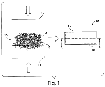
FIGS. 1 and 1 A show anillustrative embodiment of a hemostatic device.
Hemostatic device 10 comprises a first membrane 12, a second membrane!14, and.
absorbent material 16 contained between the membranes 12 and 14. Material .16
may,
:
comprise a blend of superabsorbent polymer particles 11 and wicking element=s,
such'as`
wicking fibers 13, and may further include other fibrous fillers. As
describ'edherein; the:
wicking material may reduce coagulation of blood on the outer' surface=15
of:the device;
which can reduce premature gelling and promote.transport of fluid (e.g., blood
fluids):
past the outer surface 15 of hemostatic device 10 and into absorbent material
16.
Membranes 12 and 14 may be joined along their outer edges to forni a
seal:l8'=by~ gluing,
sewing, heat sealing, or any other suitable;sealing method know~n to those
skilled in the
art.
FIGS. 2 and 2A illustrate another embodiment of the invention, wherein
hemostatic device 20 is shaped like a disc and comprises a first membrane 22
and a:.:
= . . =
9
CA 02695526 2010-02-02
WO 2008/021212 PCT/US2007/017753
. ' = . ! ' ; . = . . ~ .
second membrane 24 joined along their outer edges, while encapsulating a
material 26
between membranes 22 and 24. Material 26, may comprise a blend of
superabsorb.erit ;
polymer particles and wicking fibers, as described above. 'Membranes 22 and
24. cari be
joined as described above to form sea1. 28:; It should be understood that
devices of the;
invention can be made in a variety of shapes, sizes, and configurations
:suitable for: a~' `. ..
particular application. In some cases, the device may have dimensions aength,
width) in the range of about 5 mm to about 200 mm, with a typical range of
about 20 mm
., .
to 100 mm. The device thickness may, range frorim about 1 mm to about 50;rrim,
with a:
typical range being 5 mm to about 20 mm. : :. : .
A single hemostatic device or multiple hemostatic devices, rriay:be utilized
for .,
, ~ . , .
absorption of fluid in the treatment of a wound. In some embodiments; the use
of
multiple hemostatic devices may be advantageous in that the devices may be
readily
adjusted to any size, shape, or configuration;of a wound by simply adding or-
removing
individual hemostatic devices. Additionally, a higher degree of shape
conformability'
may be obtained using, for example, multiple, small devices than with a
single, larger~. .:
device. A plurality of hemostatic devices, may be carried in a small dispenser
and could'
be extracted, as needed, and inserted into the wound. Alternatively; the
devices may be
packaged in a tightly rolled configuration; thus pi'oviding initially very
thiri swelling
devices that could be inserted in the rolled-up configuration even into
tight'wourid:
entries, such as bullet wounds, for example.:
In some embodiments, devices of the invention rnay achieve enhance:d'swelling;
of the superabsorbent polymer particles by the inclusion of a plurality of
hemostatic u.nits
in a single hemostatic device to increase the;total surface area of
superabso',rbent polymer.'
particles exposed to fluid relative to a hemostatic device having;the same
quantity :of
absorbent material but' in a single hemostatic unit. This
"compartmentalization" of.
multiple hemostatic units within a single device can allow
for.more:'efficierit~absorption.
of fluid. For example, such a device may comprise at least one porous membrane
formed
into an enclosure having an interior and an exteribr, and a plurality of
hemostatic units
contained in the interior of the enclosure, wherein each hemostatic unit
coritains a 30 plurality of polymer particles collectively forming a polymeric
'mass that are configured :
to swell in the presence of a fluid. Some or all of the hemostatic units may:
optionally .;!
comprise wicking elements capable of tra.nsporting fluid into an` interior
regiori of the .:;
CA 02695526 2010-02-02
WO 2008/021212 PCT/US2007/017753
polymeric mass of the unit. :=
, . =
In one embodiment, a hemostatic device can comprise, a;first porou$ membrane
and a second porous membrane sealed.together at selected locations` to!form
aplurality of
compartments, wherein each compartment comprises a hemostatic unit, as shown
inFIG;
4A. Hemostatic device 40 contains a plurali,ty of individual hemostatic units
42, each :' .
formed by sealing a portion of membrane 44 to a,portion of inembrane!46 to
form sealing
borders 48 defining the compartments. Eacli hemostatic unit 42 may,comprise a
pluiali'ty
of superabsorbent polymer particles, as described above: The; hemostatie
device :40 can:
be folded as required to have a similar,effect as, for example, the insertion
of multiple
smaller hemostatic devices, or can simply be laid over a wound opening an'd
:then pushed
in to ensure contact all around the wound surface with excess device material
simply;
protruding from the wound. In some embodiments, the sealing, borders between,;
individual hemostatic units may comprise'perforations to act as'a tear-off
device,;
allowing the device size to be tailored as needed.
In another embodiment, illustrated, in FIG. 4B, a hemostatic device 60 may,
'
comprise a plurality of hemostatic units 52, wherein each hemostatic unit
comprises at'
least one porous membrane forming an enclosure that has an interior containing
absorbent polymer particles. The hemostatic units may be, in turn, contained
within:the
interior of an enclosure formed by another: membrane or other porous
material;or. net-like
structure. Hemostatic device 50 comprises a plurality of hemostatic units 52
contairied'iri
a membrane enclosure 54. Upon contact with a fluid (e.g., blood),
hemostat'ic'device 5Q
adopts a swollen configuration 60.
In some cases, the hemostatic units may achieve acceptable swelling pi-
operties
without addition of wicking elements,:since 'increasing the total'surface area
of polymer.'
particles exposed to blood (e.g., via "compartmentalizing") can . reduce the
depth-of fluid*;
penetration required to wet the polymeric.mass tliroughout. Also, faster
ab=sorption of
fluid may be achieved by compartmentalization. In some cases, the: swelling
and
consequent exertion of pressure may be obtained through a largely inelastic
geometry
change of a number of smaller hemostatic:units, which, when acting
togeth:er;.:produce a
single device capable of swelling to between 30 and 50'times its original
volume.
It should be understood that, in some cases,.it may be preferred that- the
individual
hemostatic units further comprise wicking fi:bers; as described herein, to
combine the
11.
CA 02695526 2010-02-02
WO 2008/021212 PCT/US2007/017753
. ' = ; = ~ = , = :.
effects of both the capillary action of the wicking fibers with the increase
in exposed
surface area provided by the compartmentaliza.tion techniques described
atiove.
Without being bound by any particular theory or mechanism of:act"i,on, it is
believed that, the physical swelling function'with this "compartmentalized"
design may:
occurs at three levels. At the most basic level, the swelling is believed;to
driven by the
absorption of fluid through outer membrane.54 of the device and into the
individual
. ; . .
hemostat elements 52 contained within the de=vice. : In such embodiments,
wi'cking: f bers:
; . . =
may not be required since the polymer, volume in each hemostatic. unit may be
small:
enough to be able to absorb and swell 'fully before gelling/coagulation of
fluid on the
surface of membrane 54 and/or the hemostatic unit occurs. At the next level;
each
hemostatic unit may swell to a maximum -bloat'driven by the polymer vvithin'
and reach a`
maximum swollen size, thereby acting as an'independent swelling element!'of
the overall
hemostatic device. At the uppermost level, a number of these
independenthemostatic
units interact with each other within a confiried space, defined b y the
enclosure forrried
by the outer membrane of the overall liemostatic device. Initially, each
hemostatic=unit;
may be free to expand, but as they gain volume, they begin to ekert'pressure
:on each ,=.
other. At this level the device begins to act as a single unit, capable of
exeirting ancl
transferring pressure throughout its external'geometry. This approach presents
a
potentially significant advantage for enhancing swelling speed. By dividirig
the polymer
mass into discrete hemostatic units, the polymer is distributed more
evenly'through the
wound than it would be if it was contained in a single mass
without;wicking=filler.; This
can result in a much larger surface area of exposed polymer, which can
significantly!
increase swelling speed. With this approach the swelling speed;to maximum
bloat of the
individual hemostatic units has been measured to be less than 30 seconds for
certain:
geometries_ Since this swelling happens throughout the overall
~hemostatic;device; the
overall hemostatic device itself can bloat, foi= example to its maximum=
bloat,: in less than'
seconds, potentially giving it the capability to grow at a rate of
between:600% and ;. ,
1000% per minute. Furthermore, the addition of wicking elements, such as
wickirig
fibers, in the hemostatic units may also result in a further increase in
swelling speed.,
30 As described herein, the plurality of hemostatic units may be contaihed in
a
porous container or enclosure. In some cases, the enclosure may bei formed of
a:
membrane that may be a highly stretchable meinbrane or net-like structuie,:
which woukl:
12 ; . :
CA 02695526 2010-02-02
WO 2008/021212 PCT/US2007/017753
not substantially limit the expansion of the device as a whole, but wbich
would contain ;
the hemostatic units in a single device. Such a membrane or riet may .be:made
of any
suitable material; in certain embodiments, it,may be formed of a biocompatible
and
highly elastic polymer. Such suitable poly'mers are well known to those
ski:'lled in the art
and are commercially available. In other cases, the membrane may .be fornied
of :'.
substantially non-stretchable membrane, butt may be sized -to provid'e an
enclosure,with :
enough excess volume, so that it does not substantially limit the
expansion:ofthe
hemostatic device as a whole.
The hemostatic units can be made.in:any suitable size such that;they may'be
contained with an enclosure or container of a desired size. For ;example,' for
larger: :..:
wounds, the size of the individual hemostatic units may be at least 20 mm in
diameter`
before swelling, and the membrane enclosure coritaining the plurality of these
elements
may, for example, be an essentially square enclosure at least 20 mm x 20 mm in
size. 'Of
course, the size of the membrane enclosure may be selected aind varied as
would be
apparent to those skilled in the art, to provide sufficient volume to
accomrriodate'a-
desired number of hemostatic units of a particular size, accountiiig for the
degree of
swelling of the hemostatic units and giveri tlie maximum degree'of stretch of
the:
membrane material. Similarly, membrane enclosures of various shapes may be
prov.ided;
depending on the type of Nvound to be treated, etc., as would be apparent'to
tliqse s:killed
in the art. For example, for smaller wounds; the individual herriostatic units
may be 5.
mm in diameter and the membrane containing the plurality of these elernents
may be 'at
least 5 mm x 5 mm, depending on the factors noted above. It should be
understood that
the device may have any size or shape required to suit a particular:
application:
Another advantageous feature of certain devices and methods of the invention
is 25 the ability to provide for the three-dimensional absorption of fluid.
tWhile inariy kno:wrc
absorbing devices are limited to a two dimensional geometry,-resulting in
planar
transport, certain devices and methods disclosed herein can provide three-
dimensional .'
flow of fluid through the device, resulting, in more efficient absorption of
fluid. and,
therefore, more efficient exertion of pressure on'a wound and/or sealing
ofalie'wourid:
Absorption rates of certain hemostatic devices described` herein,may be at
least 20
to 21 grams, or more, of blood per gram of polymer within 5 minutes (or, in
some cases,
more than 5 minutes) in free-swelling conditions. This represents an average
swellirig
, . . . .
13
CA 02695526 2010-02-02
WO 2008/021212 PCT/US2007/017753
rate of at least 400% per minute in blood in free swelling conditions.
Ty:pical: absorption;
rates of certain devices, when swollen in deionized water in free-swelling
conditions can;
achieve swelling ratios of as high as 140 grams of water per grain of polymer
in 3.
minutes, representing an average swell ratio.of 4500% per minute. This
comparison'
demonstrates the preference to conduct testing of hemostatic devices iri blood
rather;tlian
-, . ~ . ~ . .
water. Experimental data indicate that the swell behavior appears to be
essentialiy;=
. . , .
exponential, and the majority of swelling takes place soon a$er immersion
;of1he device
in the fluid. It has also been observed that e~cternal pressure terids- to
have a signifcant
effect on the swelling ratio, as swelling can depend strongly on the, balance;
between ;:.:
internal pressure within the device and e:cternal pressure. Typical polymer
swelling
curves in 0.9% salt water, which mimics the salinity of human blood, are shown
iri FIGS:
5-7 for blends of sodium polyacrylate polymers comprising high surface, area
.particles.
and poly-anionic beads in a 1:1 ratio. FIG. 5 is a- graph of the swelling
curve for the
sodium polyacrylate polymer blend in 0.9% salt water at 0 mm Hg pressure.
FIG.~6'isa
graph of the swelling curve for the sodiurri polyacrylate polymer blend: in
6.9%0 'salt water
at 15 mm Hg pressure. FIG. 7 is a graph of the swelling curve for the sodium
polyacrylate polymer blend in 0.9% salt water at 60 mm Hg pressure. Different
results
can be achieved by varying the ratio of the p:olyrner blend comp:onents.
Another aspect of the present invention provides methods for treating wounds
using one or more hemostatic devices, as described herein. Such methods may
involve;.
inserting one or more hemostatic devices into, near, or onto the surface of a
wound:
Superabsorbent polymers and polymer particles that may be used iri the present
invention to form hemostatic devices may` be selected and/or designed to exert
substaritial
pressure to/within a wound upon swelling: and/or form a tight seal; pressure
effective
enough, for example, to stop high pressure bleeding from transe.cted
principal.art eries:
The polymer may be used as an actuating and sealing element, and, ;in some:
cases, may:
preferably be a superabsorbent hydrogel. These polymers, for example poTymers
based'
on sodium polyacrylate, are known to be able to absorb hundreds of tirnes
their weight, in
fluid. : : . .
A short description of the properties and behavior of certain hydrogelsis
provided
below, which hydrogels may be suitable for use in certain embodiments of tlie'
hemostatic
device discussed herein. It should be noted that the list is not eahaustive,
and those of
,
14
CA 02695526 2010-02-02
WO 2008/021212 PCT/US2007/017753
ordinary skill in the art may readily select:or= form other suitable.
absorbent'materials:
using available information regarding the. absorbency and swelling,properties
of various
materials and no more than routine experimentation and:screening tests.
Polymer gels are typically characterized by long chain polymermoiecules that:
.are
crosslinked to form a network. This network can trap and hold: fluid, which
can give.gels
properties somewhere between those of solids and liquids.. Dependingon
the'level of
crosslinking, various properties of a particular gel can be tailorecl. For
:example, a highl.y
crosslinked gel generally is structurally str orig and =tends to resist
releasing : fl'uid under *
pressure, but may exhibit slow transition tirries. A lightly crosslinked gel
rimay=be weaker:
structurally, but may react more quickly during its phase transition. In the
design of:gels
for a particular application, the degree of crosslinking may be adjusted to
achieve the
desired compromise between speed of absorptiori and level of structural
integrity. :Those:
of ordinary skill in the art would be able to identify methods for;
modulatingthe degree of
crosslinking in such gels.
A property of gels in powder form that may be particularly useful in the
preserit
invention is their ability to block flow:of fluid and/or gas. Gels in powder
form, when'
dry, can allow the passage of air and water in spaces that exist between the
packed '.
particles, provided the particles are of sufficient size to produce
sufficieritly large spaces.;
However, when this powder mass is brouglit into contact with a fluid
rimediurri such as=
water, the particles at the surface of the mass begin to absorb fluid, swell,
and softem. :If
the motion of the gels is somewhat restrained by restricting ari overall
change in volume,,.
the swelling particles can fill in the empty spaces between the particles;
effectively
sealing off the flow path.
As long as fluid medium is present, the gel tends to swell to regain a'
conditiorr of;
equilibrium, which can ensure that the seal is maintained. =In ce'rtain
embodiments,of 'tlie
present invention, fast swelling superabsorbent polymers are used in the
creation of;a
hemostatic device that is activated by the presence of water, either fresh
water or water
with a sodium ion concentration rangiing from about 0 to 10%. Such properties
enalile
the polymer to swell in the presence of blood by absorbing the wwater
coritent. o'f the'b1ood
while tolerating the presence of a significant concentration of other species
in.blood; such
as sodium ions, for example. .
The nature of the fluid, more specif cally the concentration of sodiuni ions,
in
CA 02695526 2010-02-02
WO 2008/021212 PCT/US2007/017753
. . . = =
part, determines the degree of absorption and swelling ratio. For example,
the=polymer;.
may absorb 400 to 500 times its weight in distilled or deionized;water, but
this may dr,op:
to 100 times if the water is ordinary tap water, or. 50 times or less if the
water has a
significant content of sodium ions. This is because the water absorption is
~driven by a;
property called osmotic pressure, which the polymer strives to maintain
balanced at zero
differential with respect to the surrounding environrrient: Osmotic pressure
at'.
equilibrium has been shown to be related to a combinatiori of the rubber
=elasticity of the
polymer network, the polymer-polymer arid polymer-solvent affinity,= and the
ionizat'iori.
of the polymer network. The rubber elasticity of the network can provide a
mechanical`
restoring force to changes in volume. The affinity of the polymer for itself,
and the
solvent can determine whether this component of osmotic pressure drives it to:
absorb : `
fluid or not. Finally, the ionization of the 'network can determine the
drivirig force; tliat ;
attempts to balance the ionization level oflthe polymer with that of the
soh%ent in thc .
surroundings. The ionization of the polymer network can provide the
opportunity .ta
tailor the polymer's behavior. By modifying the ionization of the
polymer~it;is possible,
to affect the types of fluids that can be absorbed and the degree to which
the.y sre
absorbed. For a given ionization, if the fluid contains a higher ionic
concentration than'
the polymer, this component would not tend: to drive absorption: On -the`
other hand,' if
the fluid is deionized water, the driving force for absorption would be
greater and the
swelling ratio correspondingly large.
In some embodiments, the polymer particles comprise polyacrylates:: For
example, sodium polyacrylate-based superabsorbent polymers can be modified to
provide
polymer particles having a greater affinity for sodium ions than the sodiurri.
ions have for
water. In an illustrative embodiment, a polyacrylate-based superabsorbent
polymer witli
convoluted surface topology is used. This polymer is commerciall.y available
in various
suitable forms. In some cases, the polyrrier may be a blend of a commercially
available
sodium polyacrylate polymer with poly-ariionic beads (PAB), as supplied
by"Champion'
Enterprises, Ft. Wayne, IN, that have an affinity for sodium ions, and a
surfactant,
humectant, or other agent that assists in the absorption of water.; Since the
blend can be:
tailored to suit the application, and each component is useful depending on;
the
application, the range of blend ratios can be between about 0% and 100% of
sodium:
polyacrylate and PAB, for example, 0:100, 1:99, 2:98, 3:97, and so `on:'
Accordirig: to tlie!
16
CA 02695526 2010-02-02
WO 2008/021212 PCT/US2007/017753
. : s = _ , , = ,
present invention, one suitable ratio is=50:50õ i.e. approximately.equal parts
of sodium
polyacrylate polymer and PAB. Such'a polymer blend can exhibit suitable; speed
and :
swelling ratio requirements for functioning in blood. In.some cases, ce
rtairi;blends of the
, , . . . . .
sodium polyacrylate polymer and PABs can.tolerate very high sodium ion
concentrations,
e.g. up to about 10% sodium ions, including, levels found typically in blood.
Such.
polymers may be provided in powder form with average particle sizes ranging,
for
example, between 1 micron and 1000 microns, or, in some cases, between 200
microris
and 400 microns. The average particle size may be determined by fractionatirig
the.
polymer using sieves that encompass the minimum and maximum sizes of';the
desired;
size range in order to exclude particles outside the desired size range. In
some cases, th'e;
polymer particle size is selected such that; when contained in hemostatic
devices of the -
invention, the polymer particles may expand freely and completely,-but may be
prevented
from escaping into the blood stream through the walls of an enclosure
containing them,::
as described more fully below. Typical swelling kinetics curves for the
polyiners used in
this invention, in 0.9% salt water, are shown in FIGS. 5-6.
As used herein, "particle size" refers:to the largest characteristic dimension
(i.e. >
of a line passing through the geometric ceinter of the particle e.g:,
diameter) that can be :
measured along any orientation of a particle (e.g., a polymer particle):
Particle size as'
used herein may be measured or estimated, for example, using a sieve analysis,
wherein
particles are passed through openings of a standard size in a screen.' The
particle-size .
distribution may be reported as the weight percentage of particles retained on
each' of a
series of standard sieves of decreasing'size, and the percentage of particles
:passed of the
finest size. That is, the average particle size:may correspond to the
50%:point in.the.
weight distribution of particles.
It will be apparent to those skilled=inthe art that a wide r'.ange of
swellable
polymers can be used in devices -and methods of the present invention,
'd'epending orithe
desired performance and intended use. For example, polysaccharides,
isopropylacrylamides, and/or butylacrylamides may also be used within the
context of the
invention. Sodium polyacrylate polymers have been used in diapers and: other'
absorberit
devices for many years because they have'a high swelling capability,.can swell
in a
matter of seconds, retain the fluid effectively under pressure, and generally;
show rio
adverse reaction on the human body.
17
CA 02695526 2010-02-02
WO 2008/021212 PCT/US2007/017753
Wicking elements that may be suitable for use in the present
inventiori;include; `'.
but are not limited to, fibers such as Nylon fibers, highly convoluted wicking
PET;fibe
polypropylene fibrous filler material, other hydrophilic materials capable of
transporting
fluids via capillary action, and the like. Wicking elements may be
hydrophilic, or coated;
or otherwise treated to render them or at least their surfaces hydrophilic..
Those of
, . : ~., . .
ordinary skill in the art would be able to select appropriate materials
toprovide wicking
elements for use in the present invention based on the teaching and
direction:provided -'
herein. A screening test for suitable wicking materials may involve
imiriersing an :.
enclosed structure (e.g, a membrane(s) forining an enclosure(s))+ containing a
hydrophilic
material wicking element candidate and swellable polymer material in aluid and
.
examining the swollen material to determine whether or not thehydrophilic
matzrial:.
successfully transported fluid to the interior portions of the structure.
This;may be:
accomplished by, for example, cutting the swollen structure in half to examine
the
difference between the portions close to the surface of the structure and the
portions in
the interior of the structure.
As described herein, wicking fibers and polymer particles may be combined ..
:;
together to produce the hemostat devices.': In some cases, a device comprises
a plurality`
of wicking fibers and a plurality of polymer particles which interact to forni
a'polymerie
mass, which is in the form of a fibrous structure. The interaction may involve
covalent
bonding, ionic bonding, hydrogen bonding, dative bonding, electrostatic
iriteractions, vari
der Waals interactions, other types of bondirng or interactions, and/or the
like,. Irr some
cases, at least a portion of the plurality of absorbent polymer particles are
bonded to afi
least a portion of the plurality of wicking elements. Such bonding may produce
a fibrous
structure comprising intertwined fibers with:polyrrier particles bonded to
tlie-fibers. The
bonding between wicking fibers and polymer particles can also provide
sufficient spacing
between polymer particles to facilitate' and/or maximize fluid
absorption.throughout:the:
device.
The wicking fibers and polymer particles may be combined alone, or:in the
presence of additional materials,.such as boriding agents (e.g., propylene
gTycol,
adhesives, etc.), solvents, and the like.' Those df ordinary skill in the art
wquld be alile to,
identify materials and methods suitable for use in the fonnation:of stntctures
comprising
wicking fibers and polymer particles, as described herein. For exaniple, the
wicking
18
CA 02695526 2010-02-02
WO 2008/021212 PCT/US2007/017753
fibers, polymer particles, and at least one bonding agent may be combined,11
such that' a<,
cohesive structure is formed and/or sufficient bonding between wicking f Uers
and
polymer particles occurs, without reducing or-diminishing the absorption
pioperties of,
the structure (e.g., by encapsulating the polymer).
In some embodiments, wicking fiber.s are used, whereiri the diameter of the .'
wicking fibers may preferably be in the range of approximately: 10-150
microns. The
blending of wicking fibers and polymer particles may be performed: by mixing
them:
together in the presence of a bonding agerit, such as propylene glycol, for
exa.rnple: iri a,~
mass ratio of approximately 1:10 (fiber:polymer) with the polymer partieles.:
=This:may
be achieved by dissolving the propylene glycol in a sufficient quantity of
alcohol, mixing
with the polymer particles, and allowing the:alcohol to evaporate. The polymer
particl:es:,
become coated with a small amount of propylene glycol, and are then added to
the:
wicking fibers and shaken in a closed container, resulting in an-ev.enly
distributed cloud
of polymer particles within a fibrous structure. As described here, the
fibrous. structure s
may maintain spacing between the polymer particles, which, wlien comtiined
:with.the:
capillary action of the wicking fibers, increases the area for fluid flow to
erisure complete
absorption and swelling. Those of ordinary :skill in the art would be able to
select
additional methods and materials for bonding of polymer particles to fibers.
The mass ratio of polymer particles to'wicking elements may be such that the`
wicking paths are maintained for a sufficiently long enough time to 'enable
the,entice:
polymeric mass in the hemostatic device to swell. In some embodiments, the
preferred'
blend ratio of wicking element mass to polymer particle mass'ranges from
approximately
1:10 to 1:2, with a ratio of 3 grams of wicking element to 10 grams of polymer
particles
being one example. In one embodimeizt, the mass ratio of wicking elements .1o
polymer
particles is 1:3.3. Devices of the invention may advantageously maintain a
high ratio of
absorbing material mass (e.g., superabsorbent polymer particle`mass) to
wi`cking elemerit;
mass.
Membranes used for containing swelling materials and, optionally, wicking f
bers
as described herein, or for containing multiple hemostatic units in' a~multi-
unit'.device,
may be selected to suit a particular application. The membranes may comprise
an :elastic
material or a sufficiently sized non-elastic material, such that the membrane
may enable
swelling of the polymer particles, and the'device may undergo a change in size
and/or =,
19
CA 02695526 2010-02-02
WO 2008/021212 PCT/US2007/017753
shape upon absorption of a fluid (e.g., blood), but may also be resistant to
tearing and%ar
. . . ; . , . .
puncturing. For example, in some cases, the membrane may be;an elastic
materiat - tfiat
may stretch in accordance with the swellirig :polymer particles enclosed
within the.
membrane. Examples of such elastic materials include, for example,
polyisoprene;
polybutadiene, polydimethyl siloxane,,latex'rubber, and copolymer materials;
sucli as:
copolymers of polyurethane and polyethlyene glycol*(Lycra), and the like. ~
In: some cas;s;
the membranes are made from a non-stretch or substantially nori-elastic
materal. In, such
cases, the membranes may then be configured and sized such that the device;may
still
.:: .
undergo a change in size and/or shape: For example, the device.may be provided
such,
that there is excess non-elastic material which can be folded or rolled,
and,;upon
absorption of fluid, the device may swell and increase in size. Examples* of
suitable no
elastic membrane materials include nylon, polypropylene, .polyethylene,
polyethylene :;.;
terephthalate (PET), polycarbonate, acrylic polymers, polystyrene, cellulos'e
or cellulose `
esters, polysulfone, or the like. In certain embodiments, the meinbrane may be
in the
form of a continuous sheet that comprisesa plurality of pores therethrough; In
other:
embodiments, the membrane may comprise a plurality of fibers; configured, ;for
example:
in the form of a non-woven felt or matt or, alternatively, in the form of a=
knitted "or-
woven fabric or in the form of a screen. . ...
Membranes suitable for use in the*present invention rriay also contain pores
or .
other openings through the membrane:to facilitate more rapid passage of fluid;
through
the membrane. A wide variety of suitable membrane materials having a:wide -
variety of
average pore size and pore size distribution are readily commereially
available from a:
number of suppliers. In some cases, the pores of-the membrane may be
suffi'ciently small
to contain the polymer, or polymer and wicking fiber mixture; without leakage,
while
sufficiently large to enable free fluid flow: In some cases, the membrane niay
possess
pores large enough to enable the uninhibited flow of blood. In some
embodiments,, the;
pore size may be larger than the size of the polymer particles, .for
exarnple,:if thepolyiner
particles are blended with materials (e:g., wicking fibers) such that the
blerid'is bonded
together and thereby able to be contained.;': Also, while the use of larger
pore sizes may
result in the loss of a small number of particles through the
membrane,~the;blood-
pressure gradient within the wound cavity may ensure that any particles that
are lost;froin
the device will remain in or near the wound cavity. Moreover, even in tlie
absence of
CA 02695526 2010-02-02
WO 2008/021212 PCT/US2007/017753
such pressure, blood vessels will normally collapse, thereby effectively
preventing the ':.
entrance of loose particles into the circulatory system even if they did leak
from the
membrane. In some cases, the membrane;is;a microporous membrane. In,'soine
cases,.
the membrane may contain pores having an effective average diameter:of about
0.15 mm
. . . -.,..
to 1.0 mm, more preferably 0.2 mm - 0.5 mm. Pore sizes of the,membranes may be
measured by, for example, a microscope, via a porometer device, via.particle
retention:
tests, or otherwise, as would be apparent to those* skilled in the art.
Nominal .pore size -for
a particular membrane material is typically specified by the manufacturer and
:supplier'.of
the commercially available membrane rnaterials.
-0 The membrane may be formed from a single continuous'piece or a plurality'
of .;;
joined pieces. For example, at least two membranes can be joiined along their
outer
edges, with the swelling materials contained inside. Alternatively, a single:
membrarie:
can be folded or otherwise formed into an.enclosure for containing'polymer
particles in:!
its interior.
Other characteristics that may be desirable for materials 'used as membraries
in the
present invention include hydrophilicity, biocompatibility, sufficient
elasticity withoui.
excessive stiffness, and the ability to seal the membrane to itself or other
membraries
using conventional industrial methods:
In an illustrative embodiment, a commercially available; horieycomb knif.
Lyera;:
fabric may be selected as the membrane. For example, SL-485. Micro Mesh Lycra;
available from various suppliers, which is:a blend of 80% Polyester and 20%
Lycra, with
a stretch of 25% in width and 50% in length; may be selected as' the:
membrane. It should
be understood that other cornmercially available or custom made membranes
ma.y. al'so be
used.
A number of alternative designs for achieving constructions,that enable
wicking',
and/or absorption of blood into a hemostatic device are possible. In some
embodiments,
a single layer or multiple layers of superabsorbent polymer may, be bonded to
a strip of
material (e.g., a hydrophilic fabric strip) located in the interior of the
device with.a:-
surfactant or water-soluble adhesive. Similarly, in another embodiment,;the
polymers.:
may be bonded to an interior surface of a surrounding membrane of the device,
with a~
surfactant or water soluble adhesive. Such approaches may provide as
extensive. a,
polymer surface as possible inside the device such =that the maxi;mum amount
of polymer
21
CA 02695526 2010-02-02
WO 2008/021212 PCT/US2007/017753
may contact the fluid, while reducing the length of the path the fluid has
ao.`.travel i:n ord'e'r
to make contact with the polymer. Also, bonding the polymer to a surface
within the device may prevent it from agglomerating. This may enable a more
effective infiltration?
of fluid into the polymeric mass.
These above descriptions of applications for the inventive devices are not
intended to be exhaustive, and merely illustrate some of the possible
embodiments and
uses of this invention. It will be apparent to:those skilled in the;art that
certain
embodiments of this invention may be well suited for the temporary emergency
pluggirig
of any leak within the pressure range of the device in which water, seawater,
or other .;`
solvent able to swell the absorbent materi al, forms at least part of the
leaking fluid; It -
will also be apparent to those skilled in the art that by utilizing polyrners
tliat srvell in tlie:
presence of organic compounds, the device may be employed.in instances
where:tlie'
leaking fluid is gasoline or oil, for example.
The function and advantage of these and other embodiments of the present '
invention may be more fully understood froin the examples below. 'The
following:
examples, while illustrative of certain embodiments of the inverition, do not
ehempl'ify
the full scope of the invention.
EXAMPLES
Example 1: Manufacture of hemostatic device :
A hemostatic device was tested on an animal using a Fatal Groin Injury
1VIOde1; as
described below. During this test four hemostats were used, although any
suitable:'
number may be used. The hemostats used comprised a 4" x 4" SL-485:Micro,Mesh
Lycra bag containing 10 grams of a 50-50,blend of sodium polyacrylate
superabsorbent:
polymers in two fon-ns. One form is a high surface area particle of arbitrary
shape, which
provides speed and initial volume swell, and the other form is a poly-anioriic
bead form,:
which provides structural integrity and pressure (supplied by Cliampion
Eriterprises,:Ft.~ :
Wayne, IN). The blend was then treated with 1. 'gram of propylene glycol -;and
313': grams
of polypropylene fluff filler. The hemostatic devices weighed 1=5 grams each.
The hemostatic devices were manufactured by enclosing a blend of polymer and
fiber, as described herein, in a bag formed by bonding two 10 cm by, 10 cro
sheets of
Lycra knit fabric. It should be clear that this size and weight were
selected;. for the
22
CA 02695526 2010-02-02
WO 2008/021212 PCT/US2007/017753
. = = ~ , : . =
purposes of testing and that the sizes, contents and ratios can be'scaledup or
down to suit
any alternative application, test or wound type. The typical construction arid
coristruction
process used in prototyping the preferred embodiment of this device involves
formirig' a
blend of the propylene fibers with sodium: polyacrylate superabsorbent
.polymer particles
attached to fibers using the propylene glycof. The polymer and 'fiber
composite is,th`en inserted into the Lycra bag, which was then either sealed
with a4atex-based adhesive; to
form the final hemostatic device. Alternatively, the bag may be; heat-sealed,
whereiri
fabric stretch and other performance characteristics may be
substantially:retained since
the heat seal takes place fiber on fiber:
Example 2: Live testing of the hemostatic:device .:..
A pig (46.3 kg) was anesthetized and instrumented at the neck to read all
;relevanf
vital signs. Ports were inserted for the introduction of fluids. A complex
groin injury:
was inflicted to produce uncontrolled hemorrhage. This injury included
~transaction of
the proximal thigh soft tissues (skin, quadriceps and adductor muscles), and
complete' ;
division of the femoral artery and vein just below the inguinal ligament. :
This.was-
achieved by incising these structures ~uitha sharp scalpel (as described in,
Alam,' H. :B:,.
et al., "Application of a Zeolite Hemostatic Agent Achieves 100% Survival in:
a Lethal
Model of Complex Groin Injury in Swine;" J Trauma, Vol. 56, No. 5, May,: 2004;
incorporated herein by reference).
The wound was produced and bleeding was allowed to occur freel': After.3'.
minutes of uncontrolled bleeding, the four test hemostatic devices
construc;ted as
described in Example 1 were inserted into. the wound. Once the hemostats:were
in place,
a standard gauze dressing (67 grams) was.used to pack the wound.
Subsequeritly,
manual compression was applied for 54 minutes; after which pressure was
removed and
the wound was observed. Fluid resuscitation began 15 minutes after the'initial
creation
of the injury, and the pig received 2 liters of staridard 0.9% saline
solution,;admini"stered
intravenously, over a period of 30 minutes. The pig was monitored for a total
of= 120 :'. .
minutes after the time of injury. Midway through the experiment, the sweliing
of the :
hemostatic devices was apparent. At the end of the experiment the pig:was
euthanized.
At the completion of the test, the swelling.had forced the gauze bandage.out
of the= ~
wound site. The top of the bandages remained dry even as blood pressure
increased
23
CA 02695526 2010-02-02
WO 2008/021212 PCT/US2007/017753
, =
: , i ' . i = . ' ' =
during resuscitation. After removal of the gauze bandages at tlie end of the
test, the
hemostats were removed and studied. Bleeding.vessels had to be clarriped :off
as the' '.;
.
hemostatic devices were removed to prevent further'bleeding.. :; .
The devices, which had been remo'ved from the wound s;ite, were observed'to'
absorb blood throughout the device, as observed by examination of'the, swollen
polymeric material.
Blood loss during the initial 3 minutes was measured continiuously by
suctioning
the blood into a collection container. Bloodloss during the reniainder of
tlie4est'(3-120
minutes) was measured by weighing all the dressings before and after the
experiment arid
adding the difference to the total blood loss.; The blood loss is shown in
Table 1.
Blood pressure was monitored throughout the duration of the test. The rneari
arterial pressure (MAP) was calculated using the formula (2*D+S)/3, where,D'
is:tlie;
diastolic pressure and S is the systolic pressure. FIG. 8 shows a graph of
tlie IV1AP
measured during the test treatment method described in this exainple. '' .
The hemostats showed an excellent ability to control and stop the bleeding-
even',
after increasing the blood pressure with the resuscitation fluids. :
Throughout:the te'st;
there was clear visual evidence of the devices swelling inside the cavity and
applying
pressure. Upon removal of the devices at the erid of the test, several vessels
inside the
wound began bleeding heavily again, indicating that the hemostats were
successfully
sealing the bleeding sites the test. The pig survived the entire 120 minute
duration of tlie:
experiment.
Table 1: Summary of blood loss and blood absorption by hemostats
Initial Final 'Net'blood Blood loss (ml) Normalized blood
weight (g) weight (g) absorbed (g) (I g= 0.95 mi) loss (nil/kg bod), wt),
Initial blood loss - - -' 812 17.5
Standard gauze 67 110 43 41
Hemostat # 1 15 83 68 : 65
Hemostat #2 15 76 61 58
Hemostat #3 15 134 1~19' 113
Hemostat #4 15 69 54 51
Total absorption - - - ; 328 7.1;
Example 3: Clinical testing of the hemostatic device - complete' vessel
transection
A hemostatic device, as described in Example 1, was tested in 20 pigs using a
24
CA 02695526 2010-02-02
WO 2008/021212 PCT/US2007/017753
model of fatal groin injury developed by Alam (as described in; Alam, H. B.;
et al.; .':
"Application of a Zeolite Hemostatic Agent . Achieves 100% Survival in a
Lethal Model'
of Complex Groin Injury in Swine," J'Traunza, Vol. 56, No. 5, May 2004,
mcorporated:,
herein by reference), inflicted under anaesthetic, to' produce uncontrolled
hemorrhage.::
: . . . :~
The injury included transection of the proximal thigh soft tissues (skin;
quadriceps and
adductor muscles) and complete -division of the femoral artery and veiri just
below: tfie`: '.
inguinal ligament. After injury, the animal was randomized into one of tvvo
group,s, '(a):a.
hemostat group and (b) a control group. The control group was treated using
a'standard
gauze dressing and the hemostat group was treated. using the hemostat with the
standard
gauze dressing. Three minutes of uncontrolled bleeding were allowed and then
the
wound was packed with either standard gauze dressing for the control group,
or,
hemostats and the standard gauze dressing for the hemostat group.
Subsequently, manual
compression was applied to the wound for 5 minutes, after which pressure was
remo.ved
and the wound was observed. Fluid resuscitation was applied 15 miriutes afteir
the; iriitiO
creation of the injury, and each pig receivedapproximately 2liters, adjusted
for body :..
weight, of standard 0.9 fo saline solution administered intravenously over
a;period bf 30:
minutes. Each pig was monitored for a totaT of 120 minutes after the-time of
injury.
During the resuscitation and observation period, any reasonable;procedure that
could be
applied in the field to ensure survival was;applied as required.
Thiswas:done;to simulate
field conditions as closely as possible. During the experiment a: number
of'physiological`
indicators such as blood pressure, blood gas; andpulse were monitored and
recorded for
later interpretation. At the end of each experiment each pig was euthanized.
The data measured during the experiments was studied to highlight;trends in
device efficacy. The key indicator of device efficacy was the survival rate
measure& for
both the control group and the hemostat group. FIG. 9 shows'a graph ofth'e
survival
rates and time of death of animals of (a) the hemostat group and (b);the
coritrol group. The control group, which counted n=9 animals, achieved a
survival rate of`44% usirig the
standard gauze dressing and standard resuscitation procedures. The
hemostat'group;,
which counted n=9 animals, achieved a survival rate of 100% by using
the;hemostat:
device, a standard dressing and standard resuscitation procedures. FIG.
10shows a grapli
of the average of the mean arterial pressure for (a) the hemostat group and
(b) the control
group, measured over the duration of each experiment. The data for the eontrol
group::
CA 02695526 2010-02-02
WO 2008/021212 PCT/US2007/017753
showed the average only for the surviving animals, meaning that the
calcul'ation of.tlie :.
average at any point in time was done only for animals that survived at;that
time.' Forthe
surviving animals, the hemostat group showed a significantly higher average
MAP~, ;=; ::
particularly in the early stages of resuscitation. This vvas a resul.t
of.reduced post-
treatment bleeding in the hemostat group and illustrated the efficacy of
the";hemostatic
devices in stopping bleeding and holding pressure, which contributed to the
overall liigli
survival figures achieved in the hemostat group.
The normalized blood loss (mass and time), measured before and after
application of the wound dressings is suminarized by the graphs in FIGS.
F1=12. The
pre-treatment blood loss (0-3 min) was obtained by dividing the=total rnassof
blood ,
collected before treatment by the mass of the animal and by the `pre-treatment
time; The
post-treatment blood loss (3-120 min) was obtained by dividing; thetotal mass
of blood :*
collected after start of treatment by the animal mass and the time to 'death,
after removing
the initial 3-minute period. This, calculation accounted for the animals
that'=died before
the end of the experiment and, consequently, stopped bleeding (Ahtija et al,
"Testing of
Modified Zeolite Hemostatic Dressings in a Large Animal Model of Lethal Groiin
'
Injury," Journal of Trauma, Injury, Infection, and Critical Care; December
2006).. Tlie;
difference in post-treatment blood loss showed that the hemostat made.a
sigriificarit
difference in its reduction.
FIG. 11 shows a graph describing the (i) pre-treatment and (ii) post=.
treatment '`.
mass and time normalized blood loss for (a) the hemostat group;and (b) the
control :=:
group. FIG. 12 shows a graph describing the post-treatment blood loss, for;(a)
the '=': :
hemostat group and (b) the control group. The post-treatment blood loss was
scaled,in ;
FIG. 12 so that the relative difference between the hemostat and control
grqups could be
more easily observed. The hemostat group, on average, bled one fifth of the
amou.nt ":*;
measured in the control group after the application of treatment and
resuscitation, clearly:
demonstrating the efficacy of the device.
The above results demonstrated that the swelling hemostat devices described
herein work well in this swine model of lethal groin injury with-complete
transect'ion of
the femoral vessels. This was demonstrated by the difference in survival
nurnbers (100%
for the hemostat versus 44% for the control group), by the difference in post-
treatment
blood loss (0.06 mUkg/min for the hernostat=versus 0.34 ml/kg/min for the
control :
26
CA 02695526 2010-02-02
WO 2008/021212 PCT/US2007/017753
group), and by the significantly higher MAP achieved during
resuscitationby;the
hemostat group. No secondary side effect:s, such as exothermic heating, were
observed'
. . ~ .
Additionally, there was no need for drying, wetting, cleaning, or other
treafinent of the
wound site.
Example 4: Clinical testingof the hemostatic device duringpartial vessel
transectiori.
In this example, the hemostat devices, as described in Example 3, were tested
using a modified model of fatal groin injury:in pigs. The model~ requires,
partial
transection of the femoral arteries in order to prevent vasoconstriction and
collapse;
ensuring continued severe bleeding. In addition, the time period of free
bleeding and the
period of application of external pressure were reduced, resulting in
higher;pressure :
bleeding at the time of treatment. Thus, tlie device needed to provide
sufficient pressure
to remain in contact with the bleeding site. The test protocol/procedure was
identical to.
that described in the Example 3, except fo'r the wound, which eritailed a
part'ial
transection of the femoral vessels.
FIG. 13 shows a graph of the a=verage of the Mean Arterial Pressure for (a)
th'e :
hemostat group and (b) the control group, :measured over the duration of each
experiment. The hemostat group showed a significantly higher average MAP,
particularly in the early recovery stage before resuscitation and the later
stages= of
resuscitation. This illustrates the efficacy;of the swelling hemostatic device
in`stoppirig
bleeding and holding pressure, which contributed to the overall-higfi survival
achieved in
testing.
FIG. 14 shows a graph describing the (i) pre-treatment (0-1 min) and (ii) post-
treatment
(1-120 min) mass and time norrimalized blood loss for (a) the hemostat
groiap.:
and (b) the control group. FIG. 15 shows a graph describing the post-
treatriient tilood
loss for (a) the hemostat group and (b) the' control group scaled to better
show 'the
differences. The pre-treatment rate of blood'loss correlated well with the
p're-treat'rnerit'
rate of blood loss in Example 3. In addition; the relative reduction in post-
:treatment
blood loss also correlated well with the post-treatment blood loss in Example
3, even
though the absolute rate of post treatment blood loss was lower.: These
resultsindicate
that the swelling hemostatic device was able to effectively control hemorrhage
in the
partial transection model with reduced application of external pressure: ' :=
27
CA 02695526 2010-02-02
WO 2008/021212 PCT/US2007/017753
While several embodiments of the'invention have been described and
illustrated;
herein, those of ordinary skill in the art will readily envision a variety,
ofothermeans arid
structures for performing the functions and/or obtaining the results or
advantages
described herein, and each of such variations, modifications and iinprovements
is
deemed to be within the scope of the present invention. More generally,
tliose, skilled'in
the art would readily appreciate that all parameters, dimensions, materials,
:arid
configurations described herein are meant to be exemplary and that actual
parameters,~:
dimensions, materials, and configurations -vvill depend upon specific
applications for;
which the teachings of the present invention:are used. Those skilled in
the;art-will:
recognize, or be able to ascertain using no rriore than routine
experimeritation, mariy'
equivalents to the specific embodiments of the invention described herein. ;
It is;
therefore, to be understood that the foregoing embodiments are presented
by:way of `
example only and that, within the scope of the appended claims'and`
equivalents thereto;
the invention may be practiced otherwise than as-specifically described. The
present
invention is directed to each individual feature, system, material and/or
me'thod descrilied
herein. In addition, any combination of two or more such features, systems,
materials
and/or methods, provided that such features, systems, materials
and/or.niethods are not ;
mutually inconsistent, is included within the scope of the preserit
inveritioti,
In the claims (as well as in the specification above), all transitional
phrases; or
phrases of inclusion, such as "comprising;" "including," "carrying,"
"havirig,"
"containing," "composed of," "made of," "formed of," "involving" and the; like
shall be
interpreted to be open-ended, i.e., to mean `iricluding but not limited to":
arid; therefore,:.
encompassing the items listed thereafter and equivalents thereof as well as
additional
items. Only the transitional phrases or phrases of inclusion "consisting of
and , :.
"consisting essentially of' are to be interpreted as closed or semi-closed
phrases,
respectively. The indefinite articles "a" and "an," as used hereift in the
specificationand;
in the claims, unless clearly indicated to the contrary, should be;u.nderstood
to`:mea.n ;"at.
least one."
All references cited herein, including patents and published applications, are
:
;..'
incorporated herein by reference. In cases where the present specification
arid a
document incorporated by reference and/or referred to herein include
conflicting:
disclosure, and/or inconsistent use of terrriinology, and/or the
incorporated%referenced.;
28
CA 02695526 2010-02-02
WO 2008/021212 PCT/US2007/017753
. : = ~'
documents use or define terms differently than they are used or defined in the
preserit
specification, the present specification, shall control.
What is claimed is:
= = = = . . . : . .
. = . ~ ; :
29