Note: Descriptions are shown in the official language in which they were submitted.
CA 02695679 2014-12-17
- 1 -
BRANCHED STENT GRAFT SYSTEM
Description
Technical Field
This invention relates to a prosthetic system for implantation within a human
or animal body for the repair of damaged vessels, ducts, or other
physiological
passageways.
Background of the Invention
Throughout this specification, when discussing the application of this
invention to the aorta or other blood vessels, the term "distal", with respect
to a
prosthesis, is intended to refer to a location that is, or a portion of the
prosthesis that
when implanted is, further downstream with respect to blood flow; the term
"distally"
means in the direction of blood flow or further downstream. The term
"proximal" is
intended to refer to a location that is, or a portion of the prosthesis that
when
implanted is, further upstream with respect to blood flow; the term
"proximally"
means in the direction opposite to the direction of blood flow or further
upstream.
The functional vessels of human and animal bodies, such as blood vessels
and ducts, occasionally weaken or even rupture. For example, the aortic wall
can
weaken, resulting in an aneurysm. Upon further exposure to hemodynamic forces,
such an aneurysm can rupture. In Western European and Australian men who are
between 60 and 75 years of age, aortic aneurysms greater than 29 mm in
diameter
are found in 6.9% of the population, and those greater than 40 mm are present
in
1.8% of the population.
One surgical intervention for weakened, aneurismal, or ruptured vessels
involves the use of a prosthetic device to provide some or all of the
functionality of
the original, healthy vessel, and/or preserve any remaining vascular integrity
by
replacing a length of the existing vessel wall that spans the site of vessel
failure.
It is preferable that these prostheses seal off the failed portion of the
vessel.
For weakened or aneurysmal vessels, even a small leak in the prosthesis may
lead
to
CA 02695679 2010-02-05
WO 2009/020653
PCT/US2008/009548
- 2 -
the pressurization of, or flow in, the treated vessel which can aggravate the
condition
the prosthesis was intended to treat. A prosthesis of this type can, for
example, treat
aneurysms of the abdominal aortic, iliac, or branch vessels such as the renal
arteries.
A prosthetic device can be of a unitary construction or be comprised of
multiple
prosthetic modules. A modular prosthesis allows a surgeon to accommodate a
wide
variation in vessel morphology while reducing the necessary inventory of
differently
sized prostheses. For example, aortas vary in length, diameter, and angulation
between the renal artery region and the region of the aortic bifurcation.
Prosthetic
modules that fit each of these variables can be assembled to form a
prosthesis,
obviating the need for a custom prosthesis or large inventories of prostheses
that
accommodate all possible combinations of these variables. A modular system may
also accommodate deployment options by allowing the proper placement of one
module before the implantation of an adjoining module.
Modular systems are typically assembled in situ by overlapping the tubular
ends of the prosthetic modules so that the end of one module sits partially
inside the
other module, preferably forming circumferential apposition through the
overlap
region. This attachment process is called "tromboning." The connections
between
prosthetic modules are typically maintained by the friction forces at the
overlap region
and enhanced by the radial force exerted by the internal prosthetic module on
the
external prosthetic modules where the two overlap. The fit may be further
enhanced
by stents fixed to the modules at the overlap region.
A length of a vessel which may be treated by these prostheses may have one
or more branch vessels, i.e. vessels anastomosed to the main vessel. The
celiac,
superior mesenteric, left common carotid, and renal arteries, for example, are
branch
vessels of the aorta; the hypogastric artery is a branch vessel of the common
iliac
artery. If these branch vessels are blocked by the prosthesis, the original
blood
circulation is impeded and the patient can suffer. If, for example, the celiac
artery is
blocked by the prosthesis, the patient can experience abdominal pain, weight
loss,
nausea, bloating, and loose stools associated with mesenteric ischemia. The
blockage of any branch vessel is usually associated with unpleasant or even
life
threatening symptoms.
CA 02695679 2010-02-05
WO 2009/020653
PCT/US2008/009548
- 3 -
When treating a vessel with a prosthetic device, it is therefore preferable to
preserve the original circulation by providing a prosthetic branch that
extends from the
prosthesis to a branch vessel so that the blood flow into the branch vessel is
not
impeded. For example, the aortic section of the ZENITH abdominal aortic
prosthesis
(Cook, Inc., Bloomington, IN.), described below, can be designed to extend
above the
renal arteries and to have prosthetic branches that extend into the renal
arteries.
Alternatively, the iliac branches of the ZENITH device can be designed to
extend into
the corresponding hypogastric arteries. Branch extension prosthetic modules
("branch
extensions") can form a tromboning connection to the prosthetic branch to
complete
o the prosthesis. Furthermore, some aneurysms extend into the branch
vessels.
Deploying prosthetic branches and branch extensions into these vessels may
help
prevent expansion and/or rupture of these aneurysms. High morbidity and
mortality
rates are associated with these aneurysms.
Aortic arch stent grafts are used in treating dissection and aneurismal
dilation of
the aortic arch. Many of these grafts have branches that maintain the patency
of the
branch arteries originating in the arch (the innominate, left common carotid,
and left
subclavian arteries) and help direct the flow of blood into the branch
arteries. Many of
these branched grafts have branches that project outward from the prosthesis.
Implanting the stent grafts in the branch arteries provides a challenge to
surgeons
because of the anatomic features of the aortic arch. Blood flow from the
branch
arteries must not be interrupted for an extended length of time because they
supply
blood to the brain. Implanting branch stents that mate with the branches
presents
challenges because the natural orientation of the aortic arch must be matched
or
simulated by the stent grafts.
A surgeon may access the aortic arch through the branch arteries to implant
small vessel stents. Guide wires are used to link the small vessel stents in
the branch
arteries with the branches of the aortic arch stent. However, much time may be
lost in
threading the guide wires through the openings of the aortic arch stent
branches and
through the branch arteries. A surgeon will often manipulate the guide wire
around
the difficult angles in the aortic arch stent channels before being able to
connect with
the delivery catheter of the branched stent.
CA 02695679 2014-12-17
- 4-
Summary of the Invention
The present invention provides an endovascular prosthetic device for
implantation in an aortic arch.
The preferred endovascular prosthetic device comprises a primary prosthesis
comprising a major lumen and at least one socket for receiving a secondary
prosthesis. The secondary prosthesis is to be deployed in a branch artery. The
at
least one socket has at least a portion that extends into the major lumen and
is
configured or angled in a proximal direction to direct blood flowing from the
heart to
a branch artery. The at least one socket has a fenestration in its wall to
accommodate a guide wire. The guide wire passes through the major lumen from a
distal location into the at least one socket through the fenestration to
facilitate
placement of the secondary prosthesis in the branch artery. On some
embodiments
there is at least one fenestration in the wall of the socket.
In another aspect of the invention, there is provided an endovascular system
for implantation in an aortic arch. The system comprises an endovascular
prosthetic device, as described above, and includes the secondary prosthesis
for
implantation in a branch artery. The secondary prosthesis can be small vessel
prosthetic grafts, such as stent grafts.
In yet another aspect of the invention, there is provided an endovascular
prosthetic device comprising a prosthetic device with first and second
sockets, and
one guide wire extending through the fenestration in the first socket and then
through the fenestration in the second socket.
Another aspect of the invention provides an endoluminal prosthetic device
having three sockets. Each socket has at least one fenestration through which
a
guide wire extends. In some embodiments, there are three guide wires, one for
each socket. In other embodiments, there is one guide wire extending through
the
fenestrations in each socket. The sockets in the prosthetic device correspond
to the
innominate, left common carotid, and left subclavian arteries that branch from
the
aortic arch.
Brief Description of the Drawing
Embodiments of the present invention are described below, by way of
example only, with reference to the accompanying drawings, in which:
CA 02695679 2010-02-05
WO 2009/020653
PCT/US2008/009548
- 5 -
Figure 1A illustrates a prosthetic device with two sockets and two guide wires
extending through the sockets and holes into the major lumen;
Figure 1B is an illustration of a prosthetic device with the first and second
sockets sharing a common anastomosis;
Figure 1C is an illustration of a prosthetic device with branches having
diameters that taper to a smaller size from the opening to the proximal
portion of the
branch;
Figure 2A is a schematic representation of a prosthetic device with two
sockets
and one guide wire that is threaded through the fenestrations in the wall of
each
socket;
Figure 2B illustrates the guide wire threaded though the second socket and
corresponding hole;
Figure 3 is an illustration of a compacted prosthetic device in a large
aneurysm
of the aortic arch;
Figure 4 is a schematic drawing of the prosthetic device in a semi-deployed
configuration;
Figure 5 is an illustration of a guide wire being snared in the innominate
artery;
Figure 6 is a schematic representation of sheaths placed into the sockets over
their respective guide wires;
Figure 7 illustrates second guide wires being placed into the prosthetic
device
through the sheaths;
Figure 8 is an illustration of secondary prostheses being implanted into the
sockets through their respective sheaths;
Figure 9 is a view of the side branch stents in the prosthetic device without
the
second guide wires;
Figure 10 is an illustration of the secondary prostheses in their fully
expanded
configuration; and
Figures 11A and 11B are side views of a prosthesis with a major socket
containing two minor sockets.
Detailed Description
The term "endoluminal" describes objects that are found or can be placed
inside a
lumen in the human or animal body. The term "endovascular" describes objects
that
CA 02695679 2010-02-05
WO 2009/020653
PCT/US2008/009548
- 6 -
are or can be placed within a blood vessel. A lumen can be an existing lumen
or a
lumen created by surgical intervention. This includes lumens such as blood
vessels,
parts of the gastrointestinal tract, ducts such as bile ducts, parts of the
respiratory
system, etc. A "prosthetic device" is thus a prosthesis that can be placed
inside one of
these lumens.
The term "stent" means any device or structure that adds rigidity, expansion
force, or support to a prosthesis. A Z-stent is a stent that has alternating
struts and
peaks (i.e., bends) and defines a generally cylindrical lumen. The "amplitude"
of a Z-
stent is the distance between two bends connected by a single strut. The
"period" of a
Z-stent is the total number of bends in the Z-stent divided by two, or the
total number
of struts divided by two.
The term "endoleak" refers to a leak around or through a prosthetic device.
Endoleaks can occur through the fabric of a prosthesis, through the
interconnections
of a modular prosthesis, or around the ends of the prosthesis, inter alia.
Endoleakage
may result in the repressurizing of an aneurysm.
The term "branch vessel" refers to a vessel that branches off from a main
vessel. Examples are the celiac and renal arteries which are branch vessels to
the
aorta (i.e., the main vessel in this context). As another example, the
hypogastric
artery is a branch vessel to the common iliac, which is a main vessel in this
context.
Thus, it should be seen that "branch vessel" and "main vessel" are relative
terms.
The term "prosthetic branch" refers to a portion of a prosthesis that is
anastomosed to the prosthetic trunk and shunts blood into and/or through a
branch
vessel.
= Some embodiments of the endovascular prosthetic system of the present
invention comprise a prosthetic device comprising structural support. In some
embodiments this structural support is a stent. In one embodiment, the stent
may be
formed by a plurality of discontinuous stent elements. In another embodiment,
the
stent may be formed from a single stent element. The stent may be located on
the
exterior of the device, the interior of the device, or both. The stent may be
balloon-
expandable or a self-expanding stent. Typically, the stent has a circular
cross-section
when fully expanded so as to conform to the generally circular cross-section
of a body
lumen. In one example, the stent may comprise struts and acute bends or apices
that
CA 02695679 2010-02-05
WO 2009/020653
PCT/US2008/009548
- 7 -
are arranged in a zig-zag configuration in which the struts are set at angles
to each
other and are connected by the acute bends. The present invention can be used
with
a wide variety of stent configurations, including, but not limited to, shape
memory alloy
stents, expandable stents, and stents formed in situ.
Preferably, the stent is formed from Nitinol, stainless steel, tantalum,
titanium,
gold, platinum, inconel, iridium, silver, tungsten, cobalt, chromium, or
another
biocompatible metal, or alloys of any of these. Examples of other materials
that may
be used to form stents include carbon or carbon fiber; cellulose acetate,
cellulose
nitrate, silicone, polyethylene teraphthalate, polyurethane, polyamide,
polyester,
polyorthoester, polyanhydride, polyether sulfone, polycarbonate,
polypropylene, high
molecular weight polyethylene, polytetrafluoroethylene, or another
biocompatible
polymeric material, or mixtures or copolymers of these; polylactic acid,
polyglycolic
acid, or copolymers thereof; a polyanhyd ride, polycaprolactone,
polyhydroxybutyrate
valerate, or another biodegradable polymer, or mixtures or copolymers of
these; a
protein, an extracellular matrix component, collagen, fibrin, or another
biologic agent;
or a suitable mixture of any of these. Preferably, the stent is a Nitinol or
stainless steel
stent.
The term "stent graft" refers to a type of endoluminal device riicle of a
tubular
graft material and supported by at least one stent.
The stent graft material is preferably made of woven polyester having a twill
weave and a porosity of about 350 ml/min/cm2 (available from Vascutek Ltd.,
Renfrewshire, Scotland, UK). The stent graft material is preferably made of
seamless
woven polyester. The prosthetic trunk and stent graft material can also be
made of
any other at least substantially biocompatible material, including such
fabrics as other
polyester fabrics, polytetrafluoroethylene (PTFE), expanded PTFE, and other
synthetic
materials known to those of skill in the art. Naturally occurring
biomaterials, such as
collagen, are also highly desirable, particularly a derived collagen material
known as
extracellular matrix (ECM), such as small intestinal submucosa (SIS). Other
examples
of ECMs are pericardium, stomach submucosa, liver basement membrane, urinary
bladder submucosa, tissue mucosa, and dura mater. SIS is particularly useful,
and
can be made in the fashion described in U.S. Patent No. 4,902,508 to Badylak
et al.;
U.S. Patent No. 5,733,337 to Carr; U.S. Patent No. 6,206,931 to Cook et al.;
U.S.
CA 02695679 2014-12-17
- 8 -
Patent No. 6,358,284 to Fearnot et al.; 17 Nature Biotechnology 1083 (Nov.
1999);
and WIPO Publication WO 98/22158 of May 28, 1998, to Cook et at. It is also
preferable that the material is non-porous so that it does not leak or sweat
under
physiologic forces.
Graft materials may also include porous polymer sheet of a biocompatible
material. Examples of biocompatible polymers from which porous sheets can be
formed include polyesters, such as poly(ethylene terephthalate), polylactide,
polyglycolide, and copolymers thereof; fluorinated polymers, such as
polytetrafluoroethylene (PTFE), expanded PTFE (ePTFE) and poly(vinylidene
fluoride); polysiloxanes, including polydimethyl siloxane; and polyurethanes,
including polyetherurethanes, polyurethane ureas, polyetherurethane ureas,
polyurethanes containing carbonate linkages, and polyurethanes containing
siloxane segments. In addition, materials that are not inherently
biocompatible may
be subjected to surface modifications in order to render the materials
biocompatible.
Examples of surface modifications include graft polymerization of
biocompatible
polymers from the material surface, coating of the surface with a crosslinked
biocompatible polymer, chemical modification with biocompatible functional
groups,
and immobilization of a compatibilizing agent such as heparin or other
substances.
Thus, any polymer that may be formed into a porous sheet can be used to make a
graft material, provided the final porous material is biocompatible. Polymers
that
can be formed into a porous sheet include polyolefins, polyacrylonitrile,
nylons,
polyaramids, and polysulfones, in addition to polyesters, fluorinated
polymers,
polysiloxanes, and polyurethanes as listed above. Preferably the porous sheet
is
made of one or more polymers that do not require treatment or modification to
be
biocompatible. More preferably, the porous sheet includes a biocompatible
polyurethane. Examples of biocompatible polyurethanes include Thoralon
(Thoratec, Pleasanton, California), Biospane, Bionatee, Elasthane , Pursil
And
Carbosil (Polymer Technology Group, Berkeley, CA).
Preferably the porous polymeric sheet contains the polyurethane Thoralon .
As described in U.S. Patent Application Publication No. 2002/0065552, Thoralon
is a polyetherurethane urea blended with a siloxane-containing surface
modifying
CA 02695679 2014-12-17
- 9 -
additive. Specifically, the polymer is a mixture of base polymer BPS-215 and
an
additive SMA-300. The concentration of additive may be in the range of 0.5% to
5%
by weight of the base polymer. The BPS-215 component (Thoratec) is a
segmented polyether urethane urea containing a soft segment and a hard
segment.
The soft segment is made of polytetramethylene oxide (PTMO) and the hard
segment is made from the reaction of 4,4'-diphenylmethane diisocyanate (MDI)
and
ethylene diamine (ED). The SMA-300 component (Thoratec) is a polyurethane
comprising polydimethylsiloxane as a soft segment and the reaction product of
MDI
and 1 ,4-butanediol as a hard segment. A process for synthesizing SMA-300 is
described, for example, in U.S. Pat. Nos. 4,861 ,830 and 4,675,361. A porous
polymeric sheet can be formed from these two components by dissolving the base
polymer and additive in a solvent such as dimethylacetamide (DMAC) and
solidifying the mixture by solvent casting, or by coagulation, in a liquid
that is a non-
solvent for the base polymer and additive.
Thoralon has been used in certain vascular applications and is
characterized by thromboresistance, high tensile strength, low water
absorption, low
critical surface tension, and good flex life. Thoralon is believed to be
biostable and
to be useful in vivo in long term blood contacting applications requiring
biostability
and leak resistance. Because of its flexibility, Thoralon is useful in larger
vessels,
such as the abdominal aorta, where elasticity and compliance is beneficial.
In addition to Thoralon , other polyurethane ureas may be used as a porous
sheet. For example, the BPS-215 component with a MDI/PTMO mole ratio ranging
from about 1.0 to about 2.5 may be used. Such polyurethane ureas preferably
include a soft segment and a hard segment formed from a diisocyanate and
diamine. For example, polyurethane ureas with soft segments such as
polyethylene
oxide, polypropylene oxide, polycarbonate, polyolefin, polysiloxane (i.e.
polydimethylsiloxane), and other polyether soft segments made from higher
homologous series of diols may be used. Mixtures of any of the soft segments
may
also be used. The soft segments also may have either alcohol end groups or
amine
end groups. The molecular weight of the soft segments may vary from about 500
to
about 5,000 g/mole.
CA 02695679 2010-02-05
WO 2009/020653
PCT/US2008/009548
- 10 -
The diisocyanate used as a component of the hard segment may be
represented by the formula OCN-R-NCO, where ¨R¨ may be aliphatic, aromatic,
cycloaliphatic, or a mixture of aliphatic and aromatic moieties. Examples of
diisocyanates include tetramethylene diisocyanate, hexamethylene diisocyanate,
-- trimethyhexamethylene diisocyanate, tetramethylxylylene diisocyanate, 4,4'-
decyclohexylmethane diisocyanate, dimer acid diisocyanate, isophorone
diisocyanate,
metaxylene diisocyanate, diethylbenzene diisocyanate, decamethylene 1,10
diisocyanate, cyclohexylene 1,2-diisocyanate, 2,4-toluene diisocyanate, 2,6-
toluene
diisocyanate, xylene diisocyanate, m-phenylene diisocyanate, hexahydrotolylene
-- diisocyanate (and isomers), naphthylene-1,5-diisocyanat- e, 1-methoxyphenyl
2,4-
diisocyanate, 4,4'-biphenylene diisocyanate, 3,3-dimethoxy-4,4'-biphenyl
diisocyanate,
and mixtures thereof.
The diamine used as a component of the hard segment includes aliphatic
amines, aromatic amines, and amines containing both aliphatic and aromatic
moieties.
For example, diamines include ethylene diamine, propane diamines,
butanediamines,
hexanediamines, pentane diamines, heptane diamines, octane diamines, m-
xylylene
diamine, 1,4-cyclohexane diamine, 2-methypentamethylene diamine, 4,4'-
methylene
dianiline, and mixtures thereof. The amines may also contain oxygen and/or
halogen
atoms in their structures.
In addition to polyurethane ureas, other polyurethanes, preferably those
having
a chain extended with diols, may be used as a porous sheet. Polyurethanes
modified
with cationic, anionic, and aliphatic side chains may also be used. See, for
example,
U.S. Pat. No. 5,017,664. Polyurethanes may need to be dissolved in solvents
such as
dimethyl formamide, tetrahydrofuran, dimethyacetamide, dimethyl sulfoxide, or
-- mixtures thereof.
The soft segments of these polyurethanes may contain any of the soft
segments mentioned above, such as polytetramethylene oxide, polyethylene
oxide,
polypropylene oxide, polycarbonate, polyolefin, polysiloxane (i.e.,
polydimethylsiloxane), other polyether soft segments made from higher
homologous
-- series of diols, and mixtures of these soft segments. The soft segments may
have
amine end groups or alcohol end groups.
CA 02695679 2014-12-17
-11 -
The hard segment may be formed from any of the diisocyantes listed above, such
as 4,4'-diphenylmethane diisocyanate, tetramethylene diisocyanate,
hexamethylene
diisocyanate, trimethyhexamethylene diisocyanate, tetramethylxylylene
diisocyanate, 4,4'-decyclohexylmethane diisocyanate, dimer acid diisocyanate,
isophorone diisocyanate, metaxylene diisocyanate, diethylbenzene diisocyanate,
decamethylene 1 ,10 diisocyanate, cyclohexylene 1 ,2-diisocyanate, 2,4-toluene
diisocyanate, 2,6-toluene diisocyanate, xylene diisocyanate, m-phenylene
diisocyanate, hexahydrotolylene diisocyanate (and isomers), naphthylene-1 ,5-
diisocyanate, 1-methoxyphenyl 2,4-diisocyanate, 4,4'-biphenylene diisocyanate,
3,3-dimethoxy-4,4'-biphenyl diisocyanate, and mixtures thereof.
The hard segment may be formed from one or more polyols. Polyols may be
aliphatic, aromatic, cycloaliphatic, or may contain a mixture of aliphatic and
aromatic
moieties. For example, the polyol may be ethylene glycol, diethylene glycol,
thethylene glycol, 1 ,4-butanediol, neopentyl alcohol, 1 ,6-hexanediol, 1 ,8-
octanediol, propylene glycols, 2,3-butylene glycol, dipropylene glycol,
dibutylene
glycol, glycerol, or mixtures thereof.
In addition, the polyurethanes may also be end-capped with surface active
end groups, such as, for example, polydimethylsiloxane, fluoropolymers,
polyolefin,
polyethylene oxide, or other suitable groups. See, for example, the surface
active
end groups disclosed in U.S. Pat. No. 5,589,563.
The porous polymeric sheet may contain a polyurethane having siloxane
segments, also referred to as a siloxane-polyurethane. Examples of
polyurethanes
containing siloxane segments include polyether siloxane-polyurethanes,
polycarbonate siloxane-polyurethanes, and siloxane-polyurethane ureas.
Specifically, examples of siloxane-polyurethane include polymers such as Elast-
Eon
2 and Elast-Eon 30 (Aortech Biomaterials, Victoria, Australia);
polytetramethyleneoxide (PTMO) and polydimethylsiloxane (PDMS) polyether-
based aromatic siloxane-polyurethanes such as Pursi10-10, -20, and -40 TSPU;
PTMO and PDMS polyether-based aliphatic siloxane-polyurethanes such as Pursil
AL-50 and AL-10 TSPU ; aliphatic, hydroxy-terminated polycarbonate and PDMS
polycarbonate-based siloxane-polyurethanes such as Carbosi10-10, -20, and -40
TSPU (all available from Polymer Technology Group). The PursiI0, Pursil-AL ,
and
= CA 02695679 2014-12-17
- 12 -
Carbosile polymers are thermoplastic elastomer urethane copolymers containing
siloxane in the soft segment, and the percent siloxane in the copolymer is
referred
to in the grade name. For example, Pursil-100 contains 10% siloxane. These
polymers are synthesized through a multi-step bulk synthesis in which PDMS is
incorporated into the polymer soft segment with PTMO (Pursile) or an aliphatic
hydroxy-terminated polycarbonate (Carbosile). The hard segment consists of the
reaction product of an aromatic diisocyanate, MDI, with a low molecular weight
glycol chain extender. In the case of Pursil-AL the hard segment is
synthesized
from an aliphatic diisocyanate. The polymer chains are then terminated with a
siloxane or other surface modifying end group. Siloxane-polyurethanes
typically
have a relatively low glass transition temperature which provides for
polymeric
materials having increased flexibility relative to many conventional
materials. In
addition, the siloxane-polyurethane can exhibit high hydrolytic and oxidative
stability, including improved resistance to environmental stress cracking.
Examples
of siloxane-polyurethanes are disclosed in U.S. Patent Application Publication
No.
2002/0187288.
The porous polymer sheet may contain polytetrafluoroethylene or expanded
polytetratfluoroethylene (ePTFE). Films or sheets of ePTFE are typically
porous
without the need for further processing. The structure of ePTFE can be
characterized as containing nodes connected by fibrils. Porous ePTFE can be
formed, for example, by blending PTFE with an organic lubricant and
compressing it
under relatively low pressure. Using a ram type extruder, the compressed
polymer
is then extruded through a die, and the lubricant is removed from the extruded
polymer by drying or other extraction method. The dried material is then
rapidly
stretched and/or expanded at elevated temperatures. This process can provide
for
ePTFE having a microstructure characterized by elongated nodes interconnected
by fibrils. Typically, the nodes are oriented with their elongated axis
perpendicular to
the direction of stretch. After stretching, the porous polymer is sintered by
heating it
to a temperature above its crystalline melting point while maintaining the
material in
its stretched condition. This can be considered as an amorphous locking
process for
permanently setting the microstructure in its expanded or stretched
configuration.
The structure and porosity of ePTFE is disclosed, for example, in U.S. Patent
= CA 02695679 2014-12-17
- 13 -
Nos. 6,547,815 B2; 5,980,799; and 3,953,566. Structures of porous hollow
fibers
can be formed from PTFE, and these porous hollow fibers can be assembled to
provide a cohesive porous sheet. Porous hollow fibers containing PTFE are
disclosed, for example, in U.S. Patent No. 5,024,671.
Polymers can be processed to be porous sheets using standard processing
methods, including solvent-based processes such as casting, spraying and
dipping,
and melt extrusion processes. Extractable pore forming agents can be used
during
processing to produce porous sheets. Examples of extractable pore forming
agents
include inorganic salts such as potassium chloride (KCI) and sodium chloride
(NaCI), organic salts, and polymers such as poly(ethylene glycol) (PEG) and
polyvinylpyrrolidone (PVP). Pore forming agents may have a particle size from
about 10 pm to about 500 pm, from about 20 pm to about 100 pm, and from about
10 pm to about 40 pm. The amount of pore forming agent relative to the polymer
may be from about 20 percent by weight (wt%) to about 90 wt%, and from about
40
wt% to about 70 wt%. These sizes and amounts of pore forming agents can
provide
for a high degree of porosity following extraction of the pore forming agent.
The
porosity can be from about 20 wt% to about 90 wt% and from about 40 wt% to
about 70 wt% of the final product.
Porous sheets may be in the form of a microporous, open-celled structure in
which the pores are substantially interconnected. Microporous structures can
be
formed by extrusion of a mixture of polymer and one or more blowing agents.
Microcellular polymeric foams can be produced by exposing the polymer to super-
critical CO2 under high temperature and pressure to saturate the polymer with
the
super-critical CO2, and then cooling the polymer. Microcellular foams can be
produced as described, for example, in U.S. Patent Nos. 4,473,665 and
5,160,674.
The foaming process can be carried out on extruded polymer tube by first
dissolving
an inert gas such as nitrogen or CO2 under pressure into the polymer, and then
forming microvoids by quickly decreasing the solubility of the gas in the
polymer by
changing the pressure or temperature, thus inducing thermodynamic instability.
Examples of microporous polymeric structures are disclosed, for example, in
U.S.
Patent No. 6,702,849.
CA 02695679 2014-12-17
- 14 -
Porous Thoralon can be formed by mixing the polyetherurethane urea, the
surface modifying additive and a particulate substance in a solvent.
Preferably the
particulate is insoluble in the solvent, and the particulate may be any of a
variety of
different particulates or pore forming agents. For example, the solvent may be
DMAC, and the particulate may be an inorganic salt. The composition can
contain
from about 5 wt% to about 40 wt% polymer, and different levels of polymer
within
the range can be used to fine tune the viscosity needed for a given process.
The
composition can contain less than 5 wt% polymer for some spray application
embodiments. The particulates can be mixed into the composition. For example,
the
io mixing can be performed with a spinning blade mixer for about an hour
under
ambient pressure and in a temperature range of about 18 C to about 27 C. The
entire composition can be cast as a sheet or coated onto an article such as a
mandrel or a mold. In one example, the composition can be dried to remove the
solvent, and then the dried material can be soaked in distilled water to
dissolve the
particulates and leave pores in the material. In another example, the
composition
can be coagulated in a bath of distilled water. Since the polymer is insoluble
in the
water, it will rapidly solidify, trapping some or all of the particulates. The
particulates
can then dissolve from the polymer, leaving pores in the material. It may be
desirable to use warm water for the extraction, for example water at a
temperature
of about 60 C. The resulting void-to-volume ratio can be substantially equal
to the
ratio of salt volume to the volume of the polymer plus the salt. The resulting
pore
diameter can also be substantially equal to the diameter of the salt grains.
The present invention provides an endovascular prosthetic device that can
be used as a part of a system for treating a vasculature. The preferred
embodiment
of device comprises a primary prosthesis comprising major lumen. There is also
a
socket that has at least a portion that extends into the major lumen and that
portion
is angled in a proximal direction to direct blood flowing from the heart to
the branch
artery. The socket receives a secondary prosthesis that will be deployed in a
branch
artery. There is also a hole through a distal side of the socket to
accommodate a
guide wire that passes through the major lumen from a distal location into the
socket
CA 02695679 2010-02-05
WO 2009/020653
PCT/US2008/009548
- 15 -
and into the branch artery. The guide wire is configured to facilitate
placement of the
secondary prosthesis in the branch artery. In some embodiments, the prosthetic
device can be implanted without a guide wire and the guide wire is then
inserted
manually once the prosthetic device is in place.
There are also embodiments for use in the aorta with fenestrations for
secondary prostheses to be implanted in the renal or iliac arteries. Such
embodiments also comprise pre-loaded guide wires.
Some embodiments provide a system for treating an aneurysm in the aortic
arch. The system includes the endovascular prosthetic device as described
above
and also the secondary prosthesis for implantation in a branch artery. There
are also
embodiments wherein the primary prosthesis has at least one socket
corresponding to
a branch artery and at least one secondary prosthesis for implantation in a
branch
artery. Some embodiments comprise two sockets or three sockets with two or
three
secondary prostheses for implantation in branch arteries.
The endovascular prosthetic device is preferably designed and manufactured
for implantation in a constrained configuration and deployment to an expanded
configuration. The guidewire is pre-loaded with the device to reduce the time
usually
taken to maneuver a guide wire through an aortic branch artery and through a
socket
angled in a proximal direction. The hole in the socket in the device is
through the
distal side of that portion of the socket extending into the major lumen. The
guide wire
extends through the major lumen, hole and socket while in the constrained and
expanded configuration.
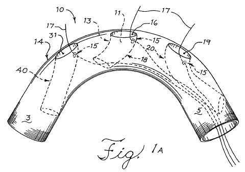
Turning to Figure 1A, a prosthetic device is illustrated with a primary
prosthesis
10 comprising a major lumen 12 extending therethrough from the proximal end 3
to
the distal end 5 of the primary prosthesis 10. The major wall 14 contains the
major
lumen 12 and occludes an aneurysm once deployed. First 31, second 16, and
third
19 openings are shown in the major wall 14 that correspond to the first 40,
second 18,
and third 20 sockets, which are all of a tubular form, and to three branch
arteries that
branch away from the vessel in which the primary prosthesis 10 is deployed.
Although
the embodiment illustrated has three tubular sockets, other embodiments
provide
primary prostheses with one or two openings corresponding to one or two
sockets as
well as other embodiments having more than three sockets. In other
embodiments,
CA 02695679 2014-12-17
- 16 -
there is at least one socket in the major wall 14. There are also embodiments
wherein the primary prosthesis 10 further comprises a structural support
around at
least a portion of the major wall 14. The structural support can be a stent in
some
embodiments.
At least a portion of the first 40, second 18, and third 20 sockets extend
into
the major lumen 12 from the openings 31, 16, and 19. While the first 40 and
second
18 sockets are angled in a proximal direction, the third 19 socket is angled
in a
distal direction in the figure shown. The sockets, therefore, are arranged in
fluid
communication with the major lumen 12. There may be other embodiments in which
the sockets are angled in directions suitable for other specified treatments.
The first
40, second 18, and third 20 sockets mate with the proximal ends of secondary
prostheses to form a secure seal with the primary prosthesis 10 at the
openings.
The sockets 40, 18, and 20 are angled to receive the flow of blood and direct
it
through their minor lumens 11 into the branch arteries. The sockets 40, 18,
and 20
are further designed to be hemodynamically effective and to minimize blood
turbulence in the primary prosthesis 10. As such the sockets can have an
internal
helical design as described in U.S. Patent Publication No. 2006/0247761. Also,
the
sockets can have baffles straddling either side of the socket to direct blood
flow
around sections of the internal helical socket that may cause turbulent blood
flow.
The sockets have fenestrations 15 that are in fluid communication with the
minor
lumens 11 and the major lumen 12. The fenestrations 15 are located in the
distal
sides 5 of the minor walls 13 of the sockets, the portion that extends into
the major
lumen. Although Figure 1 illustrates an embodiment with three sockets 40, 18,
and
20, there are other embodiments comprising at least one socket or two sockets.
In
the embodiment illustrated, there is a first socket 40 and opening 31
configured to
direct blood flow into the innominate artery. The second socket 18 and opening
16
are configured to direct blood flow into the left common carotid artery. The
third
socket 20 and opening 19 are configured to direct blood flow into the left
subclavian
artery.
The sockets may be bifurcated. Figure 1 B is an illustration of an
embodiment where the first 40 and second 18 sockets share a common
anastomosis. The sockets can also taper from a large diameter to a smaller
diameter so as to have in effect a
CA 02695679 2010-02-05
WO 2009/020653
PCT/US2008/009548
- 17 -
frusto-conical form. In Figure 1C, the diameter of the opening 31 of the first
socket 40
is larger than the remainder of the branch. In some embodiments, the branches
taper
from a range of about 13mm to 15mm to a range of about 9mm to 11mm. In some
embodiments, the branch tapers from about 14mm to about 10mm. It will be
appreciated that in many embodiments the reduction in diameter from the wide
end to
the narrow end of each socket is relatively low. Similarly, in other
embodiments the
taper may be in the other direction.
Guide wires 17 extend from the distal end 5 of the primary prosthesis 10
through the fenestrations 15 to extend into the minor lumens 11 of the sockets
and out
of the primary prosthesis 10 through the openings in the major wall 14.
Because of
their arrangement in the described embodiments, upon placement and deployment,
the guide wires 17 will be positioned in the target vessels for snaring with a
double
lumen catheter or some other guide wire. The guide wires 17 can have angled
tips,
flexible tips, compliant tips, or blunt tips.
Figure 2A shows an embodiment having one guide wire 17 threaded through
the fenestrations 15 of the first 40, second 18, and third 20 branches.
Although the
embodiment shown has two fenestrations 15 on the second 18 and third 20
branches,
there are also embodiments having only one fenestration 15 per socket. The
guide
wire 17 is used to guide and deploy a secondary prosthesis, such as a side
branch
graft, into the first 31 opening of the first 40 socket. After deployment of
the first
secondary prosthesis, the guide wire 17 is pulled out of the first 40 socket
and into the
second 18 branch. In such an embodiment, the guide wire 17 tip preferably
comprises
Nitinol or other shape memory alloy or other shape memory material. This
allows the
guide wire 17 tip to assume an orientation pointing out of the second 16
opening.
As seen in Figure 2B, the guide wire 17 has been pulled from the first socket
40
and the second socket 18 and into the third 20 socket. This may be done after
the
guide wire has been used to deploy a secondary prosthesis 60 in the second
socket
18. As the guide wire 17 tip retains its shape, it points out of the third
opening 19 just
as it pointed out of the first opening 16. The guide wire 17 can now be used
to place a
secondary prosthesis into the third socket 20.
The fenestrations 15 in the branches do not hinder blood flow once the
prosthesis 10 is properly deployed. Once a secondary prosthesis, usually a
tubular
CA 02695679 2010-02-05
WO 2009/020653
PCT/US2008/009548
- 18 -
prosthesis such as a side branch graft, is positioned and deployed in a
socket, the
guide wire 17 is retracted from the fenestration 15. The proximal end of the
secondary
prosthesis occludes the fenestration 15 such that blood flow is not
detrimentally
affected.
The devices disclosed herein can be deployed into the aortic arch by methods
known in the art. Figure 3 illustrates the system of the present invention
with a
primary prosthesis 10 being introduced into an aortic arch having a descending
aneurysm 37. A main guide wire 32 is inserted into the femoral artery (right
or left)
through an incision and is guided through the descending aorta, the aortic
arch, and
the ascending aorta. The main guide wire 32 is guided to the aortic valve of
the heart
in some methods. The guide wires, now individually labeled 33, 35, and 39, are
seen
extending out of the three openings 31, 16, and 19.
In Figure 4, the primary prosthesis 10 is partially expanded. Although not
shown, this can be accomplished with ties partially constraining the
prosthesis. Holes
31, 16, and 19 are aligned with the innominate 30, left common carotid 34, and
left
subclavian 42 arteries, respectively. Ties 38 are used to constrain the
primary
prosthesis 10. The guide wires 33, 35, and 39 are appropriately positioned in
the
arteries 30, 34, and 42 for snaring. Diagnostic imaging can be used to confirm
the
proper placement of all the elements. Radiopaque markers can be placed to mark
the
positions of the first 31, second 16, and third 19 openings. Radiopaque
markers can
also be placed at other locations on the primary prosthesis 10 to assist in
marking the
position of the implant. For instance, in some embodiments the radiopaque
markers
can be placed on the proximal 3 and distal 5 ends of the primary prosthesis
10.
Guide wire 33 projects into the innominate 30 artery where the guide wire 33
is
captured by a snare 50 as shown in Figure 5. Once snared, the surgeon uses the
snare 50 to pull guide wire 33 through the innominate 30 artery towards the
snare's 50
entry point. As illustrated in Figure 6, a sheath 63 is then placed over the
guide wire
33 and advanced through the innominate 30 artery to the opening 31. This
sheath 63
is used to advance another guide wire 17 into the first socket 40 as shown in
Figure 7.
Snares are used to capture the remaining guide wires 35 and 39 so that one or
more
sheaths 63 are advanced into the remaining openings 16 and 19. In the
embodiment
illustrated, the sheaths 63 are advanced to openings 16 and 19 in the second
18 and
CA 02695679 2010-02-05
WO 2009/020653
PCT/US2008/009548
- 19 -
third 20 sockets while the primary prosthesis 10 is partially constrained.
Once the
sheaths 63 have been advanced to their respective openings, the primary
prosthesis
is fully expanded, as shown in Figure 8. The primary prosthesis 10 can be
expanded using means known in the art including, but not limited to, balloon
5 expansion or by the loosening of constraining wire.
Over guide wire 17, a secondary prosthesis 60 is advanced to the opening 31
of the first socket 40. In such embodiments, the prosthetic systems comprise
at least
one secondary prosthesis 60 having a proximal end 72 and a distal end with a
lumen
therethrough, the proximal end 72 being sealingly engaged with at least one
socket in
10 the major wall 14 and the distal end extending into the lumen of a
branch artery. The
secondary prosthesis 60 is a side branch graft in some embodiments and
comprises a
stent in many embodiments. Figure 9 illustrates the system with the sheaths 63
removed. The proximal end 72 of the secondary prosthesis 60 is placed within
the
socket 40, as shown in Figure 10, such that the fenestration 15 will be
occluded by the
secondary prosthesis 60 once it is expanded. The secondary prosthesis 60 is
then
expanded to substantially occupy the minor lumen 11 of the first socket 40 and
the
innominate artery 30.
The secondary prosthesis 60 may also be implanted using other methods
known in the art. The secondary prosthesis 60 can be self-expanding or
expanded by
balloon catheter and deployed such that the proximal end 72 is sealingly
engaged with
the opening 16. Figure 9 illustrates other secondary prostheses 60 placed
within the
remaining sockets 18 and 20. Figure 10 is an illustration of three secondary
prostheses fully expanded in their respective sockets.
There are also embodiments of the prosthetic systems disclosed that comprise
a prosthetic device comprising two sockets extending into the major lumen from
corresponding holes and arranged in fluid communication with the major lumen.
In
such embodiments, the two sockets may correspond to any two of the innominate,
left
common carotid, or left subclavian arteries. Two secondary prostheses would
also
accompany this embodiment for placement in the two sockets. The system further
comprises two guide wires, one for each socket. In some embodiments, there is
one
guide wire that is threaded through the holes of each socket.
CA 02695679 2013-08-06
-20-
There is an endovascular prosthetic device comprising a primary prosthesis
with a primary
lumen; a major socket in the primary prosthesis having a major lumen at least
a portion of
which extends into the primary lumen and comprising at least one minor socket
with a minor
lumen at least partially within the major lumen; and a fenestration in the
wall of the major
socket to accommodate a guide wire passing through the major, minor, and
primary lumens and
being configured to facilitate placement of a secondary prosthesis in a branch
artery. The minor
socket and the major socket can share a common distal wall. In such instances,
the fenestration
can be located in the distal wall in direction communication of the minor
lumen and primary
lumen.
The prosthetic device can also have a socket that extends into the major lumen
with a
major opening 113 and at least one minor opening. In Figures 11A and 11B, the
prosthetic
device 100 has a socket 105 with one major opening 113 and two minor openings
116, 119 in
the distal portion of the socket 105. Each minor opening 116, 119 corresponds
to minor sockets
130, 132, respectively, that include minor lumens 123, 125, respectively,
resting within the
lumen 127 of the socket 105. The minor sockets can extend in the same
direction as the lumen
of the socket they rest within. In Figures 11A and 11B, the minor sockets 130,
132 extend in
the same proximal direction as major lumen 127.
Figure 11A shows one fenestration 115 in the distal side of the socket 105
just below
the major opening 113 through which a guide wire 107 has been placed. In
Figure 11B, the
guide wire 107 has been placed through three fenestrations: one fenestration
115 in the distal
wall of minor socket 130 and two fenestrations 118, 117 in the proximal and
distal walls of
minor socket 132. The distal wall of minor socket 132 forms a portion of the
distal wall of the
major socket 105. Fenestration 117 is just below the minor opening 119 and is
in fluid
communication with the primary lumen 122 of the prosthetic device 100 and
minor lumen 125.
Throughout this specification various indications have been given as to
preferred and
alternative embodiments of the invention. However, it should be understood
that the invention
is not limited to any one of these. It is therefore intended that the
foregoing detailed description
be regarded as illustrative rather than
CA 02695679 2010-02-05
WO 2009/020653
PCT/US2008/009548
- 21 -
limiting, and that it be understood that it is the appended claims, including
all
equivalents, that are intended to define the scope of this invention.