Note: Descriptions are shown in the official language in which they were submitted.
CA 02696081 2010-02-10
~
1
Method for depositing nanoparticles on a support
Object of the invention
[0001] The present invention relates to a method for
depositing and attaching nanoparticles on any support.
State of the art
[0002] It is generally recognized that the term of
<< nanoparticle >> describes an aggregate of small molecules, or
an assembly of a few tens to a few thousand of atoms, forming
a particle, the dimensions of which are of the order of one
nanometer, i.e. smaller than 1,000nm (lu), preferably less
than 100 nm. Because of their size, these particles have
particular physical, electrical, chemical and magnetic
properties and impart to the supports on which they are
applied, novel physical, electrical, chemical, magnetic and
mechanical properties.
[0003] Nanoparticles are of an increasing interest
because of their involvement in the development of many
devices used in very different fields, such as for example the
detection of biological or chemical compounds, the detection
of gases or chemical vapors, the elaboration of fuel cells or
of devices for storing hydrogen, the making of electronic or
optical nanostructures, of novel chemical catalysts, of bio-
sensors or so-called smart coatings, such as self-cleaning
coatings or which have a particular biological activity, for
example an anti-bacterial activity.
[0004] There exist many techniques with which
nanoparticles of different nature may be deposited on various
supports. There exist solution chemistry methods such as those
described for example in the article Deposition of PbS
particles from a nonaqueous chemical bath at room
temperature >> of T. Chaudhuri et al. Materials Letters (2005),
CA 02696081 2010-02-10
2
59 (17) pp 2191-2193, and in the article << Deposition of gold
nanoparticles on silica spheres by electroless metal plating
technique >> of Y. Kobayashi et al., Journal of Colloid and
Interface Science (2005), 283 (2) pp 601-604.
[0005] There also exist electrochemistry methods as for
example those described in the article << Deposition of
clusters and nanoparticles onto boron-doped diamond electrodes
for electrocatalysis >> of G. Sine et al., Journal of Applied
Electrochemistry, (2006) 36 (8) pp 847-862, and in the article
<< Deposition of platinum nanoparticles on organic
functionalized carbon nanotubes grown in situ on carbon paper
for fuel cell >> of M. Waje et al., Nanotechnology (2005), 16
(7) pp 395-400.
[0006] These may also be vacuum deposition techniques
involving a plasma as in particular described in the article <<
Platinum nanoparticles interaction with chemically modified
highly oriented pyrolytic graphite surfaces >> of D. Yang et
al., Chemistry of materials (2006) 18 (7) pp 1811-1816, and in
the article Au nanoparticles supported on HOPG: An XPS
characterization >>, of D. Barreca et al. Surface Science
Spectra (2005) 10 pp 164-169.
[0007] These techniques have many drawbacks, which may
for example be problems related to the reproducibility of the
method used, problems of distribution, homogeneity and
regularity of the deposition of nanoparticles. These
techniques are also complex to apply. Generally, they are
expensive, because, inter alia, of the necessity of generating
a vacuum, even a partial vacuum, and they are difficult to
apply on an industrial scale. Further the deposition of
nanoparticles usually comprises a step for activating the
support, which, in the techniques described earlier, requires
preliminary treatment which is very often complex and which
may take several hours or even days.
CA 02696081 2010-02-10
3
[0008] Furthermore, all these techniques pose
environmental problems, for solution chemistry as well as
electrochemistry, notably because of the use of solvents and
chemical reagents which pollute, and problems of large energy
consumption, as regards vacuum techniques using a plasma.
[0009] In particular, document W02007/122256 describes
the deposition of nanoporous layers by projecting a colloidal
solution in a thermal plasma jet, a plasma for which the
neutral species, the ionized species and the electrons have a
same temperature. In this document, it is specified that the
particles of the colloidal solution are at least partly melted
in order to be able to adhere to the substrate. In particular,
the plasma jet described has a gas temperature comprised
between 5,O00 K to 15,000 K. A non-negligible thermal effect
will therefore be noted both on the substrate and on the
particles of the sol.
Objects of the invention
[0010] The present invention proposes a method for
depositing nanoparticles on a support which does not have the
drawbacks of the state of the art.
[0011] The present invention proposes a rapid,
inexpensive method and easy to apply.
[0012] The present invention also proposes a
minimization of the heat stresses both on the substrate and on
the nanoparticles.
[0013] The present invention also proposes a deposition
method which improves homogeneity of the deposit, and more
particularly the dispersion of the nanoparticles on the
substrate.
Summary of the invention
[0014] The present invention discloses a method using a
colloidal solution (or suspension) of nanoparticles for
CA 02696081 2010-02-10
4
depositing nanoparticles on a support, and using atmospheric
plasma for depositing nanoparticles on a support.
[0015] The present invention relates to a method for
depositing nanoparticles on a support comprising the following
steps:
- taking a colloidal solution (or suspension) of nanoparticles
and,
- nebulizing said colloidal solution (or suspension) of
nanoparticles on a surface of said support in an atmospheric
plasma.
[0016] By << nanoparticle >> is meant an aggregate of
small molecules, or an assembly of a few hundred to a few
thousand atoms, forming a particle, for which the dimensions
are of the order of one nanometer, generally smaller than
100nm.
[0017] By << colloidal solution >> is meant a homogeneous
suspension of particles in which the solvent is a liquid and
the solute a solid homogeneously disseminated as very fine
particles. Colloidal solutions may take various forms, a
liquid, gel, or slurry. Colloidal solutions are intermediate
between suspensions, which are heterogeneous media comprising
microscopic particles dispersed in a liquid, and true
solutions, in which the solute(s) is (are) in the state of
molecular division in the solvent. Also, in the liquid form,
the colloidal solutions are sometimes called << sols >>.
[0018] In a preferred embodiment of the present
invention, the atmospheric plasma is an atmospheric
non-thermal plasma.
[0019] By << non-thermal plasma >> or << cold plasma >> is
meant a partly or totally ionized gas which comprises
electrons, (molecular or atomic) ions, atoms or molecules, and
radicals, out of thermodynamic equilibrium, the electron
temperature of which (a temperature of several thousand or
several tens of thousands of Kelvins) is significantly higher
CA 02696081 2010-02-10
than that of the ions and of the neutral particles (a
temperature close to room temperature up to a few hundred
Kelvins.
[0020] By atmospheric plasma >> or, atmospheric non-
5 thermal plasma >> or further << atmospheric cold plasma >> is
meant a partly or totally ionized gas which comprises
electrons, (molecular or atomic) ions, atoms or molecules, and
radicals, out of the thermodynamic equilibrium, the electron
temperature of which is significantly higher than that of the
ions and of the neutral particles (the temperatures are
similar to those described for a << cold plasma ), and for
which the pressure is comprised between about 1 mbar and about
1,200 mbars, preferably between about 800 and about 1,200
mbars.
[0021] According to a particular embodiment of the
invention, the method includes one or more of the following
characteristics:
- the plasma comprises a plasmagenic gas and the macroscopic
temperature of said plasmagenic gas in said plasma may vary
between about -20 C and about 600 C, preferably between -10 C
and about 400 C and preferably between room temperature and
about 400 C;
- the method further comprises a step for activating the
surface of the support by submitting said surface of said
support to atmospheric plasma;
- the activation of the surface of the support and the
nebulization of the colloidal solution are concomitant;
- the activation of the surface of the support is preceded
with a step for cleaning said surface of said support;
- the nebulization of the colloidal solution of nanoparticles
is accomplished in the discharge area or the post-discharge
area of the atmospheric plasma;
- the plasma is generated by an atmospheric plasma torch;
CA 02696081 2010-02-10
6
- the nebulization of the colloidal solution of nanoparticles
is accomplished in a direction substantially parallel to the
surface of the support;
- the nanoparticles are nanoparticles of a metal, of a metal
oxide, of a metal alloy or of a mixture thereof;
- the nanoparticles are nanoparticles of at least one
transition metal, of its corresponding oxide, of an alloy of
transition metals or of a mixture thereof;
- the nanoparticles are selected from the group formed by
magnesium (Mg), strontium (Sr), titanium (Ti), zirconium (Zr),
lanthanum (La), vanadium (V), niobium (Nb), tantalum (Ta),
chromium (Cr), molybdenum (Mo), tungsten (W), manganese (Mn),
rhenium (Re), iron (Fe), ruthenium (Ru), osmium (Os), cobalt
(Co), rhodium (Rh), iridium (Ir), nickel (Ni), palladium (Pd),
platinum (Pt), copper (Cu), silver (Ag), gold (Au), zinc (Zn),
cadmium (Cd), aluminium (Al), indium (In), tin (Sn), lead
(Pb), the corresponding oxides thereof, or an alloy of these
metals;
- the nanoparticles are selected from the group formed by
titanium dioxide (titania (Ti02)), copper oxide (CuO), ferrous
oxide (FeO), ferric oxide (Fe203), iron oxide (Fe304), iridium
dioxide (Ir02), zirconium dioxide (Zr02), aluminium oxide
(A1203) ;
- the nanoparticles are selected from the group formed by a
gold/platinum (AuPt), platinum/ruthenium (PtRu),
cadmium/sulfur (CdS), or lead/sulfur (PbS) alloy;
- the support is a solid support, a gel or nanostructured
material;
- the support is selected from the group formed by a
carbonaceous support, carbon nanotubes, metal, metal alloy,
metal oxide, zeolite, semiconductor, polymer, glass and/or
ceramic;
- the support is silica, carbon, titanium, alumina, or multi-
walled carbon nanotubes;
CA 02696081 2010-02-10
7
- the atmospheric plasma is generated from a plasmagenic gas
selected from the group formed by argon, helium, nitrogen,
hydrogen, oxygen, carbon dioxide, air or a mixture thereof;
[0022] In a preferred embodiment of the present
invention, the colloidal solution comprises a surfactant.
[0023] By surfactant >>, << tenside >> or surface agent
>> is meant a compound modifying the surface tension between
two surfaces. Surfactant compounds are amphiphilic molecules,
i.e. they have portions of different polarity, one is
lipophilic and apolar, and the other one hydrophilic and
polar. This type of molecules allows stabilization of
colloids. There exist cationic, anionic, amphoteric or
non-ionic surfactants. An example of such a surfactant is
sodium citrate.
[0024] The present invention moreover discloses the use
of a colloidal solution of nanoparticles for depositing
nanoparticles on a support by means of an atmospheric plasma.
[0025] According to particular embodiments, the use of
the colloidal solution of nanoparticles includes one or more
of the following characteristics:
- the colloidal solution is nebulized in the discharge or
post-discharge area of atmospheric plasma;
- the atmospheric plasma is generated by an atmospheric plasma
torch.
[0026] The present invention also describes the use of
atmospheric plasma for depositing nanoparticles on a support,
said nanoparticles being in the form of a colloidal solution
of nanoparticles, and said colloidal solution being nebulized
at the surface of said support in said atmospheric plasma.
Short description of the figures
[0027] Fig. 1 illustrates the size distribution of gold
particles of a colloidal solution.
CA 02696081 2010-02-10
8
[0028] Fig. 2 illustrates an image obtained by
transmission electron microscopy (TEM) of a colloidal solution
of gold particles.
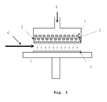
[0029] Fig. 3 schematically illustrates an atmospheric
plasma torch.
[0030] Fig. 4 illustrates X photoelectron spectroscopy
(XPS) spectra of the surface of HOPG graphite after deposition
of gold nanoparticles via plasma according to the method of
the present invention. (a) global spectrum, (b) deconvoluted
spectrum of the Au 4f level, (c) deconvoluted spectrum of the
0 ls level, (d) deconvoluted spectrum of the C ls level.
[0031] Fig. 5 illustrates atomic force microscopy (AFM)
images of a sample of HOPG graphite, a) before and b) after
depositing gold nanoparticles according to the method of the
present invention.
[0032] Fig. 6 illustrates images of high resolution
electron microscopy of secondary electrons (Field Emission Gun
Scanning Electron Microscope (FEG-SEM)) of HPOG graphite a)
before, b) and c) after depositing gold nanoparticles
according to the method of the present invention. (a)
magnification x 2,000, (b) magnification x 25,000, (c)
magnification x 80,000. Energy dispersion spectroscopic
analysis (EDS) is collected on nanoparticles.
[0033] Fig. 7 illustrates the comparison of the
experimental XPS spectrum of the Au 4f level shown in
Fig. 4(b) and of the modeled spectrum by using a growth model
of the Volmer-Weber type.
[0034] Fig. 8 illustrates an X photoelectron
spectroscopy (XPS) spectrum of the surface of the HOPG
graphite after depositing gold nanoparticles without using a
plasma (comparative).
[0035] Fig. 9 illustrates an image obtained by high
resolution electron microscopy of secondary electrons
CA 02696081 2010-02-10
9
(FEG-SEM) of a HOPG graphite sample after depositing gold
nanoparticles without using plasma (comparative).
[0036] Fig. 10 illustrates an image (magnification
x 100,000) obtained by high resolution electron microscopy of
secondary electrons (FEG-SEM) of a steel sample after
depositing gold nanoparticles according to the method of the
present invention.
[0037] Fig. 11 illustrates an image (magnification
x 3,000) obtained by high resolution electron microscopy of
secondary electrons of a glass sample after depositing gold
nanoparticles (FEG-SEM) according to the method of the present
invention.
[0038] Fig. 12 illustrates an image (magnification
x 50,000) obtained by high resolution electron microscopy of
secondary electrons (FEG-SEM) of a PVC polymer sample after
depositing gold nanoparticles according to the method of the
present invention.
[0039] Fig. 13 illustrates an image (magnification
x 10,000) obtained by high resolution electron microscopy of
secondary electrons (FEG-SEM) of an HDPE polymer sample after
depositing gold nanoparticles according to the method of the
present invention.
[0040] Fig. 14 illustrates an image (magnification x
10,000) obtained by high resolution electron microscopy of
secondary electrons (FEG-SEM) of a steel sample after
depositing gold nanoparticles, in the absence of plasma
(comparative).
[0041] Fig. 15 illustrates an image obtained by
transmission electron microscopy (TEM) of a sample of carbon
nanotubes before (a) and after depositing gold nanoparticles
according to the method of the present invention (b).
[0042] Fig. 16 illustrates an X photoelectron
spectroscopy (XPS) spectrum of the surface of carbon nanotubes
CA 02696081 2010-02-10
after depositing gold nanoparticles according to the method of
the present invention.
[0043] Fig. 17 illustrates an image obtained by
transmission electron microscopy (TEM) of a sample of carbon
5 nanotubes after depositing platinum nanoparticles according to
the method of the present invention.
[0044] Fig. 18 illustrates an X photoelectron
spectroscopy (XPS) spectrum of the surface of carbon nanotubes
after depositing platinum nanoparticles according to the
10 method of the present invention.
[0045] Fig. 19 illustrates an image (magnification
x 120,000) from high resolution electron microscopy of
secondary electrons (FEG-SEM) of a HOPG graphite sample after
depositing rhodium particles according to the method of the
present invention.
[0046] Fig. 20 illustrates an X photoelectron
spectroscopy (XPS) spectrum of the HOPG graphite surface after
depositing rhodium nanoparticles according to the method of
the present invention.
[0047] Fig. 21 illustrates an electron microscopy image
(magnification x 100,000) of secondary electrons (FEG-SEM) of
a steel sample after depositing platinum nanoparticles
according to the method of the present invention.
[0048] Fig. 22 illustrates an electron microscopy image
(magnification x 100,000) of secondary electrons (FEG-SEM) of
a PVC sample after depositing rhodium nanoparticles according
to the method of the present invention.
[0049] Fig. 23 illustrates an electron microscopy image
(magnification x 100,000) of secondary electrons (FEG-SEM) of
an HDPE sample after depositing rhodium nanoparticles
according to the method of the present invention.
CA 02696081 2010-02-10
11
Detailed description of several embodiments of the invention
[0050] The method for depositing nanoparticles according
to the invention involves a colloidal solution or suspension
of nanoparticles which is deposited on any support by means of
an atmospheric plasma, said atmospheric plasma may be
generated by any adequate device making use of atmospheric
plasma.
[0051] This method has many advantages. For example, it
allows a so-called <<clean>> deposit to be made, i.e. without
using any so-called polluting solvents. Advantageously, the
deposition of nanoparticles according to the invention only
requires low energy consumption. Surprisingly, the deposition
of nanoparticles is rapid because the activation of the
support and the nebulization of the nanoparticles, also
possibly the preliminary cleaning of the support, are
accomplished in the atmospheric plasma, or in the flow of
atmospheric plasma, in a single step or in a single continuous
process.
[0052] Surprisingly, the method according to the
invention allows the nanoparticles to be strongly adhered to
the support. With this technique, it is possible to control
the properties of the interface and to adjust the deposition
of nanoparticles on the support. Further, this method does not
require expensive installations and it is easily applied
industrially.
[0053] The colloidal solution of nanoparticles may be
prepared by any technique and/or any adequate means.
[0054] In the method according to the invention, the
support, on which the colloidal solution of nanoparticles is
deposited, is any adequate material which may be covered with
nanoparticles, any material regardless of its nature and/or
its form. Preferably, this is a solid support, gel or
nanostructured material.
CA 02696081 2010-02-10
12
[0055] In the method according to the invention, the
plasma is any adequate atmospheric plasma. This is a plasma
generated at a pressure comprised between about 1 mbar and
about 1,200 mbars, preferably between 800 and 1,200 mbars.
Preferably, this is an atmospheric plasma, the macroscopic
temperature of the gas of which may vary for example between
room temperature and about 400 C. Preferably, the plasma is
generated by an atmospheric plasma torch.
[0056] An atmospheric plasma does not require a vacuum,
which makes it inexpensive and easy to maintain. With
atmospheric plasma, it is possible to clean and activate the
surface of the support, either by functionalizing it, for
example by generating oxygen-containing, nitrogen-containing,
sulfur-containing and/or hydrogen-containing groups, or by
generating surface defects, for example vacancies, steps,
and/or pits. These surface groups may for example comprise
very reactive radicals having a short lifetime.
[0057] These reactive groups at the surface of the
substrate may then react with the surface of the
nanoparticles, or, with the surfactants present at their
surfaces. The nanoparticles themselves may be activated by the
plasma, either directly by forming radicals from the hydration
water, or by reactions with a surfactant attached to the
surface of the nanoparticle.
[0058] Preferably, in the method according to the
invention, the activation of the support and the nebulization
of the colloidal solution are accomplished concomitantly, i.e.
in the plasma, or in the plasma flow, generated by a device
making use of atmospheric plasma. Thus, nebulization of the
colloidal solution occurs at the same time, or else
immediately after the activation of the support by the
atmospheric plasma.
[0059] Nebulization of the colloidal solution may be
accomplished either in the discharge area or in the
CA 02696081 2010-02-10
13
post-discharge area of the atmospheric plasma. Preferably,
nebulization of the colloidal solution is accomplished in the
post-discharge area of the plasma, since in certain cases,
this may have additional advantages. With this, it is possible
to not contaminate the device generating the plasma. With
this, it is possible to facilitate the treatment of polymeric
supports, to avoid degradation to the support to be covered
and also for example to not cause melting, oxidation,
degradation and/or aggregation of nanoparticles.
[0060] Nebulization of the colloidal solution is any
adequate nebulization and may be accomplished in any direction
(orientation) relatively to the surface of the support.
Preferably, nebulization is accomplished in a direction
substantially parallel to the support, but it may also be
accomplished for example under an angle of about 45 , or for
example under an angle of about 75 , relatively to the surface
of the support to be treated.
[0061] Example 1:
Gold nanoparticles were deposited on highly oriented pyrolytic
graphite (HOPG), a support which has chemical properties
similar to those of multi-walled carbon nanotubes (MWCNTs).
[0062] Highly oriented pyrolytic graphite (HOPG) is
commercially available (MikroMasch - Axesstech, France) . With
ZYB quality, this graphite, with a size of 10 mm x 10 mm x 1
mm, has an angle called a mosaic spread angle >> of 0.8 0.2
and a lateral grain >> size greater than 1 mm. A few surface
layers of the graphite are detached beforehand with an
adhesive tape before the graphite sample is immersed in an
ethanol solution for 5 minutes under ultrasonication.
[0063] The colloidal suspension is for example prepared
according to the method for thermal reduction of the citrate
as described in the article of Turkevich et al. J. Faraday
Discuss. Chem. Soc. (1951), 11 page 55, according to the
following reaction:
CA 02696081 2010-02-10
14
6 HAuC14 + K3C6H507 + 5 H20 -> 6 Au + 6 CO2 + 21 HC1 + 3 KCl,
wherein the citrate acts as a reducing agent and as a
stabilizer. Conventionally, a gold solution is prepared by
adding 95 mL of an aqueous 134 mM tetrachloroauric acid
solution (HAuC14,3H20, Merck) and 5 mL of an aqueous 34 mM
trisodium citrate solution (C6H807Na3=2H20, Merck) with 900 mL
of distilled water. The thereby obtained solution is then
brought to its boiling point for 15 minutes. With a pale
yellow color, the gold solution then becomes of a red color
within one to three minutes.
[0064] With this method for thermal reduction of the
citrate, it is possible to obtain a stable dispersion of gold
particles, the gold concentration of which is 134mM, and the
particles of which have an average diameter of about 10 nm and
about 10o polydispersity (Fig. 1).
[0065] Deposition of the colloidal gold suspension on
highly oriented pyrolytic graphite is carried out with a
plasma source AtomfloTM-250 (Surfx Technologies LLC). As
described in Fig. 3, the diffuser of the plasma torch
comprises two perforated aluminium electrodes, with a diameter
of 33 mm, and separated by a gap with a width of 1.6 mm. In
this specific example, the diffuser is placed inside a sealed
chamber under an argon atmosphere at room temperature. The
upper electrode 1 of the plasma source is connected to a
generator of= radiofrequencies, for example 13,56MHz, while the
lower electrode 2 is earthed.
[0066] The plasma torch operates at 80 W and the plasma
3 is formed by supplying the torch upstream from the electrode
with argon 4 at a flow rate of 30 L/min. The space between the
HOPG graphite sample 5 lying on a sample-holder 7 and the
lower electrode 2 is 6 1 mm. This space is under atmospheric
pressure.
[0067] Before depositing the nanoparticles, the graphite
support is subject to a flow of plasma from the plasma torch,
CA 02696081 2010-02-10
for about 2 minutes for example, which allows the support to
be cleaned and activated. 3 to 5 mL of colloidal suspension is
nebulized in the post-discharge area of the plasma torch and
in a direction 6 substantially parallel to the sample
5 (Fig. 3). The colloidal suspension is injected for about 5
minutes, with periodic pulses of about one second, spaced out
by about 15 seconds. The samples 5 are then washed in an
ethanol solution under ultrasonication for about 5 minutes.
[0068] An X photoelectron spectroscopy (XPS) analysis
10 of the HOPG graphite surface covered with nanoparticles was
carried out on a ThermoVG Microlab 350 apparatus, with an
analytical chamber at a pressure of 10-9 mbars and an Al Ka X-
ray source (hy =1,486.6 eV) operating at 300 W. The spectra
were measured with a recording angle of 90 and were recorded
15 with a pass energy in the analyzer of 100 eV and an X-ray beam
size of 2 mm x 5 mm. The determination of the chemical state,
as for it, was made with a pass energy analyzer of 20 eV. The
charge effects on the measured positions of the binding energy
were corrected by setting the binding energy of the spectral
envelope of carbon, C(ls), to 284.6 eV, a value generally
recognized for accidental contamination of the carbon surface.
Carbon, oxygen and gold spectra were deconvoluted by using a
Shirley base line model and a Gaussian-Lorentzian model.
[0069] The XPS spectra of the surface of the HOPG
graphite covered with nanoparticles are illustrated in Fig. 4.
Fig. 4a) shows the presence of carbon at a percentage of
77.8%, of oxygen at a percentage of 14.9%, of potassium at a
percentage of 3.2% and of gold at a percentage of 1.0%. Silica
traces have also been detected; these are impurities
incorporated into the HOPG graphite samples. This analysis
indicates strong adhesion of gold on the HOPG graphite
although the samples were washed in an ethanol solution under
ultrasonication. It should be noted that with or without the
CA 02696081 2010-02-10
16
ultrasonic cleaning step with ethanol, the amount of gold
deposited on the HOPG graphite is similar.
[0070] The gold spectrum, Au(4f) (Fig. 4 b), was
deconvoluted relatively to the spin-orbit doublets Au4f5/2-
Au4f7/2 with a set intensity ratio of 0.75:1 and with a
separation energy of 3.7 eV. The single component Au4f7/2 is
localized at 83.7 eV, which allows this to be ascribed without
any ambiguity to gold metal. This means that the gold clusters
have been significantly oxidized during the treatment with the
plasma.
[0071] The carbon spectrum, C(ls), illustrated in Fig. 4
d) comprises a main peak at 283.7 eV which is ascribed to a
carbon-carbon (sp2) bond. The peaks localized at 284.6 eV,
285.8 eV and 288.6 eV may respectively be ascribed to C-C
(sp3), C-0, and 0-C=0 bonds. The presence of observed C-0 and
O-C=0 bonds probably originates either from the short exposure
of the samples to ambient oxygen during their handling, or
from the presence of a small amount of oxygen during the
plasma treatment as suggested by the post-discharge
characterization by optical emission spectrometry (data not
shown). This explanation is consistent with the oxygen
spectrum, 0(ls), which shows the presence of O-C bonds (533.5
eV) and 0=C bonds (531.9 eV).
[0072] The morphology of the surface of HOPG graphite
covered with nanoparticles was studied by producing atomic
force microscopy images recorded by a PicoSPMO LE apparatus
with a Nanoscope IIIa controller (Digital Instruments, Veeco)
operating under the conditions of the ambient medium. The
microscope is equipped with a 25 pm analyzer and operates in
contact mode. The cantilever used is a low frequency silica
probe NC-AFM Pointprobe0 from Nanosensors (Wetzlar-
Blankenfeld, Germany) having an integrated pyramidal tip with
a radius of curvature of 110 nm. The spring constant of the
cantilever ranges between 30 and 70 N m-1 and its measured free
CA 02696081 2010-02-10
17
resonance frequency is 163.1 kHz. The images were recorded at
scanning frequencies from 0.5 to 1 line per second.
[0073] The atomic force microscopic images (lum x lpm)
before and after depositing the nanoparticles by plasma
treatment are illustrated in Fig. 5. As shown by Fig. 5 b),
the graphite is covered with clusters, or islets, of gold
which are either isolated and which have a diameter larger
than 0.Olum (10 nm), or branched. These islets are
homogeneously dispersed with a covering rate of about 12%.
[0074] In order to confirm the nature of the islets and
to obtain highly magnified images, images from scanning
electron microscopy coupled with an energy dispersion X-ray
spectrometer (EDS) were produced by means of a JEOL JSM-7000F
apparatus equipped with a spectrometer (EDS, JED-2300F) . This
instrument, operating with an acceleration voltage of 15kV and
a magnification of 80,000 times, not only allows analysis of
the morphology of surface structures, which may thereby be
observed with optimum contrast, but also determination of the
distribution of the size of the islets. Energy dispersion X-
ray spectrometry analysis (EDS), as for it, allows their
chemical composition to be apprehended.
[0075] Before their analysis, the graphite samples are
deposited beforehand on a copper strip of a sample-holder
before being introduced into the analysis chamber under a
pressure of about 10-8 mbar.
[0076] As shown by Fig. 6a, in the initial state,
several steps are observable with a magnification of 20,000
times. Further, as shown by Fig. 6b, many clusters,
illustrated by bright spots, and having a homogeneous
distribution, are present at the surface of the graphite after
depositing nanoparticles according to the method of the
invention. With greater magnification (80,000 times,
Fig. 6c)), it is easy to perceive aggregates and isolated
nanoparticles with a diameter of about 10 nm. Energy
CA 02696081 2010-02-10
18
dispersion X-ray spectrometry analysis (Fig. 6d)) confirms
that the bright spots are gold nanoparticles. It is also
important to note that the aggregates are organized in packets
of clusters of gold nanoparticles which have the same particle
diameter as those of the initial colloidal suspension
(Fig. 1).
[0077] The morphology of the deposit, at a depth
resolution of the order of one nanometer, was also quantified
by analyzing the signal of the Au 4f peak (Fig. 7), a method
proposed by Tougaard et al., in an article in J. Vac. Sci.
Technol (1996) 14 page 1415.
[0078] Table 1 summarizes the characteristics of the
structure of the gold islets on the HOPG graphite resulting
from the analysis of three Au4f spectra with the QUASES-
Tougaard software, which are expressed as a covering rate (t =
thickness of the contamination C layer) and as a height of the
gold islets (h) . The growth mode is of the Volmer-Weber type
(3D islets structure)
Table 1:
Samples Height of the Covering Carbon thickness
gold islets percentage (%) (contamination layer)
h (nm)
(nm)
A 10.6 9.9 1.0
B 11.1 15.0 0.6
C 9.2 6.0 0.2
[0079] Surprisingly, the height of the gold islets (h)
varies between 9.2 and 10.6 nm, values substantially identical
with the average nanoparticle diameter of the colloidal
suspension (Fig. 1). Further, it seems that about 12% of the
surface of the support is covered with gold islets of about 10
nm. It should be noted that a gold covering percentage of
about 10% is consistent with the covering rate as determined
by atomic force microscopy and by scanning electron
CA 02696081 2010-02-10
19
microscopy. Thus, the analysis of the spectral Au 4f curve
with the QUASES software shows good correlation between
experimental and theoretical data.
[0080] Example 2 (comparative):
A deposition of gold nanoparticles on HOPG according to the
method of Example 1 is carried out, except for the
nanoparticle deposition step which is carried out without
using any atmospheric plasma (Figs. 8 and 9). After deposition
of nanoparticles and before analysis, the obtained samples are
washed with ethanol for about 5 minutes with ultrasonic waves.
[0081] As shown by Fig. 8, as compared with Fig. 4a, the
XPS spectrum of the sample obtained after nebulization of the
colloidal gold solution without using any atmospheric plasma,
demonstrates the presence of carbon and oxygen and the absence
of gold; this is confirmed by the atomic force microscopy
image (AFM) of the relevant sample (Fig. 9 as compared with
Figs. 5b or 6b).
[0082] Example 3 (comparative):
A deposition of gold nanoparticles on steel according to the
method of Example 1 is carried out, except for the
nanoparticle deposition step which is carried out without the
use of any atmospheric plasma. After depositing the
nanoparticles and before analysis, the obtained samples are
washed with ethanol for about 5 minutes with ultrasonic waves.
In Fig. 14, the absence of nanoparticles at the surface of the
steel is noted.
[0083] In the following examples, the method used is the
one described in Example 1, only the supports (substrates)
used and the nature of the colloidal solutions are different.
[0084] Example 4:
Gold nanoparticles were deposited on a steel support according
to the method described in Example 1, with ultrasonic
cleaning. In Fig. 10 the presence of nanoparticles is noted.
[0085] Example 5:
CA 02696081 2010-02-10
Gold particles were deposited on a glass support according to
the method described in Example 1. In Fig. 11 the presence of
nanoparticles after ultrasonic cleaning is noted.
[0086] Example 6:
5 Gold particles were deposited on a PVC support according to
the method described in Example 1, with ultrasonic cleaning.
The microscopy image of Fig. 12 was obtained after having
covered the sample with a metal layer. In Fig. 12 the presence
of nanoparticles is noted.
10 [0087] Example 7:
Gold particles were deposited on an HDPE support (Fig. 13)
according to the method described in Example 1, with
ultrasonic cleaning. The microscopy image of Fig. 13 was
obtained after having covered the sample with a metal layer.
15 In Fig. 13 the presence of nanoparticles is noted.
[0088] Example 8:
Gold nanoparticles were deposited on a carbon nanotube support
accordirlg to the method described in Example 1, after
ultrasonic cleaning. In Fig. 15 the presence of spherical
20 nanoparticles of about 10 nm is noted after ultrasonic
cleaning. This presence of gold is confirmed by the XPS
spectrum in Fig. 16.
[0089] In the following examples, colloidal platinum and
rhodium solutions provided by G.A. Somorjai (Department of
Chemistry, University of California, Berkeley (USA)) were used
(R. M. Rioux, H. Song, J. D. Hoefelmeyer, P. Yang and G. A.
Somorjai, J. Phys. Chem. B 2005, 109, 2192-2202; Yuan Wang,
Jiawen Ren, Kai Deng, Linlin Gui, and Youqi Tang, Chem. Mater.
2000, 12, 1622-1627.).
[0090] Example 9:
Platinum nanoparticles were deposited on a carbon nanotube
support according to the method described in Example 1. In
Fig. 17 the presence of spherical nanoparticles of about 10 nm
CA 02696081 2010-02-10
21
is noted. This presence of platinum is confirmed by the XPS
spectrum in Fig. 18.
[0091] Example 10:
Rhodium nanoparticles were deposited on an HOPG carbon support
according to the method described in Example 1. In Fig. 19,
the presence of spherical nanoparticles of about 10 nm is
noted after ultrasonic cleaning. This presence of rhodium is
confirmed by the XPS spectrum in Fig. 20.
[0092] Example 11:
Rhodium nanoparticles were deposited on a PVC support
according to the method described in Example 1, with
ultrasonic cleaning. The microscopy image of Fig. 22 was
obtained after having covered the sample with a metal layer.
In Fig. 22, the presence of nanoparticles is noted.
[0093] Example 12:
Gold nanoparticles were deposited on an HDPE support according
to the method described in Example 1, with ultrasonic
cleaning. The microscopy image of Fig. 23 was obtained after
having covered the sample with a metal layer. In Fig. 23, the
presence of nanoparticles is noted.
CA 02696081 2010-02-10
2~U
Poids relatif (u.a.) Relative weight (a.u.)
Diametre des particules Particle diameter
Intensite (CPS) Intensity (CPS)
Energie de liaison (eV) Binding energy (eV)
Au metal Metal Au
Analyse EDX (5keV) EDX analysis (5keV)
Spectre experimental Experimental spectrum
Modele de croissance V-W V-W growth model
Hauteur de l'ilot d'or = h Height of the gold islet = h
Epaisseur de la couche de C de Thickness of the
contamination = t contamination C layer = t
Nanoparticules d'or (10 nm) Gold nanoparticles (10 nm)
Caracteristique du support Support characteristic
Presence d'or (faible quantite Presence of gold (small
en accord avec TEM) amount consistent with TEM)
Presence de rhodium Presence of rhodium