Note: Descriptions are shown in the official language in which they were submitted.
CA 02696139 2014-12-03
MATERIALS AND METHODS FOR DELIVERING COMPOSITIONS TO
SELECTED TISSUES
TECHNICAL FIELD
[0003J The invention relates to delivery systems and, more
particularly, to a- device that can deliver preprogrammed quantities
of a composition over time without the need for external power or
RACKGROUND
[00041 Drug delivery to a location of infection, disease or a
disorder to ameliorate symptoms or cure the disease and disorder are
important.
SUMMARY
[0005] Provided herein are minimally invasive controlled drug
delivery systems and methods for use in delivery of a particular
drug or drugs to the eye that include porous film or porous film
particles having pores configured and dimensioned to at least
partially receive at least one drug therein. Embodiments include
devices and methods for treating intraoculer diseases where porous
film particles impregnated with a particular drug are sized and
configured to permit intraocular injection of the loaded porous film
particles. Other embodiments include devices and methods for
treating extraocuiar diseases, where one of a porous film,
biodegradable polymer replica , porous Si02-polymer composite, or
porous Si-polymer composite impregnated with a particular drug is
configured to contact a portion of the eye, such as the ocular
1
CA 02696139 2010-02-10
WO 2009/009563
PCT/US2008/069474
surface or retrobulbar surface, and controllably release the drug
for surface delivery of the drug. Advantageously, release of the
drug is also monitorable such that the amount of drug remaining in
the porous substrate can be accurately quantified.
[0006] The disclosure provides a composition comprising: a
silicon material comprising a plurality of pores selectively
dimensioned to obtain a desired reflective wavelength and/or rate of
drug delivery; and a drug or biologically active material within the
pores. In one embodiment, the silicon material comprises a silicon
dioxide material. In another embodiment, the material comprises a
particulate size of between about 1 pm and 100 pm. The composition
can further comprises a polymeric material capping the pores.
[0007] The disclosure also provides a multilayer silicon
composition comprising a silicon material; a first surface and a
second surface on the silicon material; a plurality of pores of a
first tunable size on the first surface; a plurality of pores of a
second tunable size on the second surface; and a drug or biological
agent disposed within the pores on the first and/or second surface.
In one embodiment, the silicon composition comprises a particle size
between 1 pm and 100 pm. In one embodiment, the silicon material is
a silicon dioxide. In yet another embodiment, the composition
further comprises a polymer capping the pores on the first side
and/or second side.
[0008] The disclosure further provides a method of preparing a
device for controlled drug delivery to a location of the eye
comprising: providing a porous nanostructured silicon-containing
template having pores configured to receive a particular drug, said
template being sized and configured to be delivered into or upon a
surface of the eye; and loading the template with the drug. The
method can further comprise providing one of a silicon template, a
Si02 template, and a Si02/polymer composite template. In yet another
embodiment, the method can further comprise disposing one of an
organic polymer, an inorganic polymer, and a bio polymer in the
template. In yet a further embodiment, the method can comprise
removing the silicon-containing template from the polymer by one of
chemical corrosion and dissolution. The method can further comprise
sizing and configuring the template to be a carrier configured to be
2
CA 02696139 2010-02-10
WO 2009/009563
PCT/US2008/069474
included in a contact lens. The method can comprise placing the
contact lens in abutment with a front extraocular surface. In one
embodiment, the method comprises
[0009] sizing and configuring the template to be a scleral
plaque for the retrobulbar surface of the eye. The method can
comprise suturing the scleral plaque to the retrobulbar surface.
The method can comprise injecting the particles intraocularly. The
method can comprise configuring the particles to have a monitorable
optical response depending on a quantity of drug disposed in the
pores. In yet another embodiment, the method can further comprise
trapping the drug or drugs in the pores by oxidizing the porous
template around the drug or drugs. The oxidizing can be performed
at repeated intervals by performing layered oxidation. For example,
a biological agent or drug can be trapped in the pores by controlled
addition of oxidants. Oxidation of the freshly prepared (hydride-
terminated) porous Si material results in an effective shrinking of
the pores. This occurs because the silicon oxide formed has a
larger volume than the Si starting material. If a drug is also
present in the solution that contains the oxidant, the drug becomes
trapped in the pores.
[0010] The disclosure also provides a minimally invasive
controlled drug delivery device for delivering a particular drug or
drugs to a particular location of the eye, said device comprising: a
porous film template having pores configured and dimensioned to at
least partially receive at least one drug therein; and wherein said
template is dimensioned to be delivered into or onto the eye.
[0011] The disclosure provides a device for the controlled
release of an active ingredient comprising: a) a polymer layer
comprising a plurality of nano-apertures; b) a base comprising a
non-porous substrate layer; and c) at least one reservoir juxtaposed
between the polymer layer and the base, wherein the reservoir is in
fluid communication with the nano-apertures of the polymer layer and
is configured to contain an active ingredient.
[0012] The disclosure further provides a method of producing a
hydrophilic, porous silicon oxide substrate comprising heating a
porous silicon substrate to a temperature above 80 C in the presence
3
CA 02696139 2010-02-10
WO 2009/009563
PCT/US2008/069474
of an agent suitable for oxidizing the silicon substrate thereby
producing a hydrophilic, porous silicon oxide substrate.
[0013] The disclosure also provides a method of producing a
hydrophobic, porous silicon substrate comprising heating a porous
silicon substrate to a temperature above 80 C in the presence of an
agent suitable for hydrosilylating the silicon substrate thereby
producing a hydrophobic, porous silicon substrate.
[0014] The disclosure provides a pulse therapy method for
treating a subject, the method comprising: a) identifying a subject
having a condition and selecting one or more active ingredients
suitable for treating the condition; b) correlating the quantity and
type of active ingredients with a pulse therapy dosing profile
suitable for treating the condition; c) configuring a device of the
disclosure to obtain a device suitable for delivering the dosing
profile of b) to the subject; and e) implanting or explanting the
device in or on a target tissue associated with the subject.
BRIEF DESCRIPTION OF DRAWINGS
[0015] Figure 1A-B show methods and reactions for generating
porous Si. (A) Shows a schematic of the etch cell used to prepare
porous Si. The electrochemical half reactions are shown, and the
equivalent circuit for etching of a p-type Si wafer is shown at
right. (B) represents a chemical reaction for the oxidation of the
porous Si around a candidate molecule according to one embodiment of
the disclosure.
[0016] Figure 2 illustrates a chemical modification reaction
whereby a candidate molecule is attached to an inner pole wall
according to another embodiment of the invention
[0017] Figure 3A-B shows representations of photo-measurements
and polymer composites. (a) shows a schematic demonstrating the
change in a reflectance spectrum from a single layer of porous Si
upon introduction of a molecular species into the porous matrix. The
change in refractive index of the composite film results in a red
shift of the Fabry-Perot interference fringes. The reverse process
can also be monitored, yielding a blue shift in the spectrum. (B) is
a schematic diagram illustrating a templated synthesis of polymer
photonic crystals using porous Si masters according to an embodiment
of the disclosure.
4
CA 02696139 2010-02-10
WO 2009/009563
PCT/US2008/069474
[0018] Figure 4 is a graph illustrating a correlation between
the optical thickness of an alkylated porous silicon film to the
concentration of drug appearing in phosphate buffered saline
solution over 2 hours.
[0019] Figure 5 shows a cross-sectional scanning electron
micrograph image of an intact porous Si film prior to removal from
the bulk silicon substrate and fracture into microparticles. The
pores are aligned along the <100> direction of the original silicon
crystal.
[0020] Figure 6A-B depicts unoxidized and oxidized porous Si
particles. (A) shows fresh porous Si particles in a droplet of 5%
dextrose solution. Particle clumping is observed due to the
hydrophobic nature of the unmodified porous Si particles. (B) shows
oxidized porous Si particles in a droplet of 5% dextrose solution.
These particles were observed to be more dispersed in solution,
presumably because of the hydrophilic nature of the 5i02 surface.
[0021] Figure 7A-D depicts intravitreal injections of porous Si
particles. (A) is a photograph taken under a surgical microscope
immediately after intravitreal injection of fresh porous Si
particles. Particles can be observed suspended in the center of the
vitreous. A few small air bubbles mixed with the porous Si particles
are present at the top of the vitreous cavity. (B) is a fundus
photograph taken one week after the injection, showing porous Si
particles dispersed in the vitreous. (C) is a fundus photograph
taken 2 weeks after injection, indicating that most of the particles
have disappeared and those remaining were barely observable. (D)
depicts a light microscopic graph showed normal retina detached from
the retinal pigment epithelium during the histology processing.
(25X, H&E staining).
[0022] Figure 8A-B provides images of intravitreal
hydrosilylated Si particles. (A) is a photograph taken under a
surgical microscope immediately after intravitreal injection of
hydrosilylated porous Si particles. Particles can be observed
suspended in the center of the vitreous. (B) is a fundus photograph
obtained 3 months after injection. The particles are dispersed in
the vitreous and many demonstrated a distinctive blue color
indicative of partial degradation and dissolution.
CA 02696139 2014-12-03
[0023] Figure 9A-C provide imagco of ocular tissue following Si
particle injection. (A) shows a surgical microscope image of a
dissected rabbit eye cup, winh hydrosilylated porous Si particles
distributed on a normal iooking retina. Photograph was obtained 4
months after injection. Two retina folds are present, caused during
dissection. (B) shows a scanning Plectron microseope image of nhe
hydrosilyiated porous Si particles sampled from a rabbit eye 4
months after intravitroal irjection. The sharp edges and pitted
surface of the particles indicate a very slow erosion process. (C)
shows a light microscopic: photograph of the retina and choroid from
a rabbit eye harvested 9 months after intravitreal injection of
hydrosilylated porous Si particles. Normal choricretinal morphology
and structure; are observed. (62.5x, H&F. staining).
[0024] Figure 10A-O provides images of oxidized porous Si
particles following injection. (A) is a photograph taken under a
surgical microscope immediately after intrayitreal injection of
oxidized porous Si particles. Particles can be observed suspended in
the center of the vitreous above the optic nerve. (El) is a fundus
photograph of a rabbit eye at 2 weeks after intravitreal injection
of oxidized porous Si particles. any violet particles
and a normal
fundus can be seon. Tho particles were initially green upon
irjection. The violet color indicates that r,ignilicaut oxidation
and dissolution oe the par.ticles has occurred. Some of the
particles hove lost their vivid reflectance completely and appear
brown in color. (C) is a fundus photograph of the same rabbit eye, 9
weeks after intravitraal injection of oxidized porous Si particles.
Many of the particles have degraded and are no longer observed. The
tundus appears normal. (0) is a light microscopic photograph of the
retina and choroid from a rabbit eye harvested 4 months after
intrvitrc51 injection of oxidized porous Sl parlic2c:, Normol
choriorotinal morphology and structures are observed with a slight
artificial reniral detachoenr. (25X, li4X staining).
[0025] Figure provides data related to the release of
bevacizumab (AvastinT54) from Si02 particles.
[0026] Figure 22 provides data related to the unique features of
the porous Si microparticies provided herein. Such features inicnde
spectral encoding for self-reporz3ng capability and tunable
6
CA 02696139 2014-12-03
nonostuctures for c.ontrolling release ratc,tor accommodating -various
payloads and versatile characteristic for attaching drugs, targeting moieties
and other molecules.
[0027] Figure 13 provides data for the release profile of
.A.vastinTm from a Si micropartic Le provided herein. The assumptions were
Volume of injection (particles excipient) 0.1cc
Mass of particles injected 0.0258g
Drug half-life in vitreous (AVASTIN) 5days
Drug loading (mg drug/g particle) 17mg/g
Volume of vitreous 4.5mL
Mass of drug injected 438600nanograms
Initial concentration (total drug in particles) 97466.66667nanograms/mL
The particle parameters were
Particles approximated as squares.
Thickness 20microns
x-dimension 20microns
Porosity 50percent
Number of particles 30000particles
Density of Si 2.33g/ml_
Area per particle 0.000004cm2
Free volume per particle 0.000000004cm3
Total free volume 0.00012cm3
The results were
..õ
Release rate half-life [73765- days
Theoretical duration of drug release 303 !days
Steady-State concentration 35702.0757nanogramsimL
DETAILED DESCRIPTIoN
(0028] As used to rein and in Lne appended claimL, the singular
forms "a," "and," and "the" include piurai referents unless the
(.;o::LexL clearly dictates otherwise. Thus, for :Aample, reference to
"a pore" includes a plurality of such pores and refsrence to "the
drug" includes reference to one or more drugn known to tnose skilled
in the art, and so forth.
[002.9 Also, !the use of "or" mean:: "an6/o' unle5s slated
otherwise. Similzirly, "ccmprise," "compriser-" "comprising"
"include," "includes," and 'Including" ere ,aterchanacable and not
intended to be limiting.
CA 02696139 2014-12-03
[0030] It is to be further understood that where descriptions of
various embodiments use the term "comprising," those skilled in the
art would understand that in some specific instances, an embodiment
can be alternatively described using language "consisting
essentially of" or "consisting of."
[0031] Unless defined otherwise, all technical and scientific
terms used herein have the same meaning as commonly understood to
one of ordinary skill in the art to which this disclosure belongs.
Although methods and materials similar or equivalent to those
described herein can be used in the practice of the disclosed
methods and compositions, the exemplary methods, devices and
materials are described herein.
[0032] The publications discussed above and throughout the text
are provided solely for their disclosure prior to the filing date of
the present application. Nothing herein is to be construed as an
admission that the inventors are not entitled to antedate such
disclosure by virtue of prior disclosure.
[0033] The ability to deliver drugs locally to the site of need
and over a prolonged period of time is important as a therapeutic
method for many ailments and diseases. Many drugs are more effective
if delivered at a specific site since they can be delivered in
concentrated dosages at the point pf interest, while maintaining an
7a
CA 02696139 2014-12-03
overall low dosage within the total body. Additionally, many drugs
cannot te delivered by oral moans because the molcculcs are too
fragile to survive the digestive process, or because the mclecules
do not pass efficiently through the walls of the digestive organs.
Some drug therapies require lung term dosing over the course of many
months or years requiring frequent visits to a clinician for
treatment. Furthermore, some drugs require delivery in places that
are inconvenient for injection, such as in the eye or in internal
organs. In all these cases, sustained drug rilivery through an
implant or attached device would be of great benefit to patients
undergoing treatment.
[0034] hn important application of drug delivery implant is age
related macular degeneration (AMD). Age related macular degeneration
is the leading cause of blindness in people over age 65. The
National Eye Institute estimates that there are 1.6 million
individuals with AMD in the United States alone. Macular
degeneration is the physical disturbance of the center of the retina
called the macula, the part of the retina which is capable of the
most acute and detailed vision. Currently, there is no known cure
for AMD. However, new therapies are being developed which show
promise in controlling the progression of the disease. Some of
these treatments include frequent administration of protein-based .
drug formulations such as Lucentismt(ranibizumab) and AvastinTm
(bevacizumab) directly into the eye. Since these drugs consist of
laroe protein molecules which cannot be administered through oral
formulations, patients sofEcring from AMD have to receive injections
directly into their eyes once every month. Tne highly invasive
nature of the nreatment and limitations in conLcolling an effective
z:rug concentration in the eye over a prolonged period of time still
leave the delivery methods tar from ideal.
[0035] In addition tu AND, diabeLln reLinopatty, retinovascler
disease, and other types of retinal Cegenera%ions are amenable to
treatment by dreg delivery implant. This is because many of those
diseases also need local therapy with the same types c.:.: oompounds
that are used for AMU. Treating diseases associated with
intraccular scarring such as ret.inal detacoment (?VR) ard glaJcome
8
CA 02696139 2010-02-10
WO 2009/009563
PCT/US2008/069474
can also be accomplished through sustained release of drug within
the eye to prevent unwanted proliferation.
[0036] Delivery of drugs into vitreous via liposomes or slow
release crystalline lipid prodrugs extend the drug vitreous half-
life, but traditional liposomes or self-assembling liposomes often
decrease vitreous clarity when used, cannot be easily customized to
release drugs with different physicochemical properties, and do not
"report" drug release information. Accordingly, the current state
of art does not provide a satisfactory way to construct a small
device and implement methods for the delivery of a drug in a
predetermined time dependent manner.
[0037] Each of the features and teachings disclosed below can be
utilized separately or in conjunction with other features and
teachings to provide a drug delivery device for delivering time
dependent dosing. Representative examples of the disclosure, which
examples utilize many of these additional features and teachings
both separately and in combination, will now be described in further
detail with reference to the attached drawings. This detailed
description is merely intended to teach a person of skill in the art
further details for practicing aspects of the present teachings and
is not intended to limit the scope of the invention. Therefore,
combinations of features and steps disclosed in the following detail
description may not be necessary to practice the invention in the
broadest sense, and are instead taught merely to particularly
describe representative examples of the present teachings.
[0038] Moreover, the various features of the representative
examples and the dependent claims may be combined in ways that are
not specifically and explicitly enumerated in order to provide
additional useful embodiments of the present teachings. In addition,
it is expressly noted that all features disclosed in the description
and/or the claims are intended to be disclosed separately and
independently from each other for the purpose of original
disclosure, as well as for the purpose of restricting the claimed
subject matter independent of the compositions of the features in
the embodiments and/or the claims. It is also expressly noted that
all value ranges or indications of groups of entities disclose every
possible intermediate value or intermediate entity for the purpose
9
CA 02696139 2010-02-10
WO 2009/009563
PCT/US2008/069474
of original disclosure, as well as for the purpose of restricting
the claimed subject matter.
[0039] As an initial starting point it is important to
understand the difference between silicon, silicon oxide and silica
(i.e., silicon dioxide). This is mentioned because there is a
fundamental difference in the compositions, uses and biological
activity of these materials.
[0040] Silicon is the chemical element that has the symbol Si
and atomic number 14. Silicon occasionally occurs as the pure free
element in nature, but is more widely distributed as various forms
of silicon dioxide (silica) or silicates. Silicon is used in the
electronics industry where substantially pure and highly pure
silicon are used for the formation of wafers. Pure silicon is used
to produce ultra-pure silicon wafers used in the semiconductor
industry, in electronics and in photovoltaic applications. Ultra-
pure silicon can be doped with other elements to adjust its
electrical response by controlling the number and charge (positive
or negative) of current carriers. Such control is desirable for
transistors, solar cells, integrated circuits, microprocessors,
semiconductor detectors and other semiconductor devices which are
used in electronics and other high-tech applications. In photonics,
silicon can be used as a continuous wave Raman laser medium to
produce coherent light. Hydrogenated amorphous silicon is used in
the production of low-cost, large-area electronics in applications
such as LCDs, and of large-area, low-cost thin-film solar cells.
Accordingly, most commonly purchased silicon is in the form of
silicon wafers. Silicon when metabolized by the body is converted
to silane, a compound that when accumulated has toxic effects.
[0041] Silicon oxide typically refers to a silicon element
linked to a single reactive oxygen species (e.g., a radical). Such
silicon oxide compounds are useful for the additional of carbon or
other desirable elements wherein a bond is formed between the
reactive oxygen and the desired element or chemical side chain.
Silicon oxides are useful for the formation of hydrogenated silicon
oxycarbide (H:Si0C) films having low dielectric constant and a light
transmittance. Such Si-O-X (wherein X is any suitable element other
than oxygen) compounds are formed using complex reactions including
CA 02696139 2010-02-10
WO 2009/009563
PCT/US2008/069474
reacting a methyl-containing silane in a controlled oxygen
environment using plasma enhanced or ozone assisted chemical vapor
deposition to produce the films. In contrast, a dioxide (further
described below) comprises two (2) oxygens linked to a silicon atom.
[0042] Silicon dioxide refers to the compound 5i02 (sometime
referred to as silica). Silicon dioxide is formed when silicon is
exposed to oxygen (or air). A thin layer (approximately 1 nm or 10
A) of so-called 'native oxide' is formed on the surface when silicon
is exposed to air under ambient conditions. Higher temperatures and
alternate environments are used to grow layers of silicon dioxide on
silicon. Silicon dioxide is inert and harmless. When silica is
ingested orally, it passes unchanged through the gastrointestinal
tract, exiting in the feces, leaving no trace behind. Small pieces
of silicon dioxide are equally harmless, so long as they are not
large enough to mechanically obstruct the GI tract or fluid flow, or
jagged enough to lacerate the GI lining, vessel or other tissue.
Silicon dioxide produces no fumes and is insoluble in vivo. It is
indigestible, with zero nutritional value and zero toxicity.
Silicon dioxide has covalent bonding and forms a network structure.
Hydrofluoric acid (HF) is used to remove or pattern silicon dioxide
in the semiconductor industry.
[0043] Silicon is an essential trace element that is linked to
the health of bone and connective tissues. The chemical species of
relevance to the toxicity of porous Si are silane (SiH4) and
dissolved oxides of silicon; three important chemical reactions of
these species are given in Eq. (1)-(3). The surface of porous Si
contains Si-H, SiH2, and SiH2 species that can readily convert to
silane. Silane is chemically reactive (Eq. (1)) and toxic,
especially upon inhalation. Like silane, the native SiHx species on
the porous Si surface readily oxidize in aqueous media. Silicon
itself is thermodynamically unstable towards oxidation, and even
water has sufficient oxidizing potential to make this reaction
spontaneous Eq. (2). The passivating action of 5i02 and Si-H (for
samples immersed in HF solutions) make the spontaneous aqueous
dissolution of Si kinetically slow. Because of its highly porous
nanostructure, oxidized porous Si can release relatively large
amounts of silicon-containing species into solution in a short time.
11
CA 02696139 2010-02-10
WO 2009/009563
PCT/US2008/069474
The soluble forms of Si02 exist as various silicic acid compounds
with the orthosilicate (Si044-) ion as the basic building block (Eq.
(3)), and these oxides can be toxic in high doses. Because the body
can handle and eliminate silicic acid, the important issue with
porous Si-based drug delivery systems is the rate at which they
degrade and resorb.
SiH4 + 2H20 Si02 + 4H2 (1)
Si + 02 5i02 (2)
5i02 + 2H20 Si(OH)4 (3)
[0044] Surface chemistry plays a large role in controlling the
degradation properties of porous Si in vivo. After Si is
electrochemically etched, the surface is covered with reactive
hydride species. These chemical functionalities provide a versatile
starting point for various reactions that determine the dissolution
rates in aqueous media, allow the attachment of homing species, and
control the release rates of drugs. The two most important
modification reactions are chemical oxidation (Eq. (2)) and grafting
of Si-C species.
[0045] The various embodiments provided herein are generally
directed to systems and methods for producing a drug delivery device
that can deliver time dependent dosing without the need for
electronics or power. Accordingly, the disclosure recognizes and
addresses an important and unmet medical need for a minimally
invasive, controllable and monitorable drug delivery system and
methods of using the system that would enable long acting local
treatment of both extraocular and intraocular diseases.
[0046] Traditional methods of intraocular drug delivery include
the use of liposomes or self-assembling liposomes, which often
decrease vitreous clarity when used, cannot be easily customized to
release drugs with different physicochemical properties, and do not
"report" drug release information.
[0047] Advantageously, the disclosure provides devices and
methods for treating both intraocular and extraocular diseases that
promote sustained release of a pharmacological candidate, or drug,
that is impregnated on nanostructured silicon, such as Si, 5i02,
polymer-templated, Si/polymer, or 5i02/polymer composites.
12
CA 02696139 2010-02-10
WO 2009/009563
PCT/US2008/069474
[0 0 4 8] In one aspect, the devices and methods are also self-
reporting such that drug release and quantity remaining can be
monitored. Embodiments include minimally invasive, self-reporting,
controlled delivery systems for delivering a drug or drugs to
surfaces of the eyes, both the ocular surface (cornea and
conjunctiva) and the scleral surface, as well as intraocular
portions of the eye, including the retina, choroids, lens, ciliary
body, anterior chamber, and vitreous. Such devices include not only
Si photonic crystals that include an active ingredient, but also
biodegradable polymer imprints made from porous silicon templates.
[0049] For intraocular diseases, such as glaucoma, age-related
macular degeneration (ARMD), choroidal neovascularization (CNV),
uveitis, diabetic retinopathy, retinovasclar disease, and other
types of retinal degenerations, drug delivery to the vitreous,
retina, and choroid is a challenging task due to the formidable
obstacles posed by the blood-retinal barrier and the tight junctions
of the retinal pigment epithelium. Only small fractions of drug
administered systemically reach the target, requiring large and
potentially toxic doses when delivered systemically. Another
challenge to retinal drug delivery is the fact that drug levels
should be sustained for prolonged periods at the target site. This
is difficult using intravitreal injections because the short half-
life of most intravitreal injectable drugs. Intraocular implants
have provided sustained vitreoretinal drug levels for treating
certain retinal diseases. However, this route demands intraocular
surgery that is known to cause intraocular complications when
placing and replacing the implant.
[0050] For ocular surface diseases, such as viral keratitis,
chronic allergic conjunctivitis, glaucoma, and scleritis, some of
the same problems persist. Systemic administration of drug requires
potentially toxic doses, and topical treatments have a short half-
life, requiring numerous and frequent doses. For treating ocular
surface diseases biodegradable polymer imprints may be made from
porous silicon templates. The silicon-free polymer may be used in
drug delivery contact lenses or implants at an appropriate location
on or associated with the eye, including the ocular surface and
retrobulbar surface.
13
CA 02696139 2010-02-10
WO 2009/009563
PCT/US2008/069474
[ 0 0 5 1 ] Photonic crystals have widespread application in
optoelectronics, chemical and biological sensors, high-throughput
screening, and drug delivery applications. These photonic crystals
are especially advantageous because of the relative ease with which
the optical properties, pore size, and surface chemistry can be
manipulated. Moreover, position, width, and intensity of spectral
reflectivity peaks may be controlled by the current density waveform
and solution composition used in the electrochemical etch, thus
rendering possible the preparation of films of porous Si photonic
crystals that display any color within the visible light band with
high color saturation, which is a desirable feature for information
displays.
[0052] The term photonic crystal refers to a material in which a
spatially repeating pattern produces a distinctive spectral pattern.
A photonic crystal comprises small porous silicon particles that
have been machined and sized to small crystals for intraocular
injection.
[0053] The disclosure provides compositions and methods for
injection of porous microscopic nanostructured silicon particles
impregnated with a particular drug or drugs. While the disclosure
contemplates use of numerous porous microscopic particles, typical
particles include porous silicon or silicon dioxide particles
(referred to as "smart dust"), which are nanostructure that allows
maintenance of sustained intraocular therapeutic drug levels with
minimal invasiveness and elimination of systemic side effects. In
addition to configuring the nanostructure to suit individual
applications, the disclosure also contemplates chemically modifying
the particles and the particular drug or drugs to tune and control
release profiles of the particles. Intraocular injection allows
monitoring of drug levels non-invasively.
[0054] Porous silicon is advantageous in that porous silicon
films have a large free volume (typically 50-80%). Thus having a
high capacity for a drug can be custom designed at the nanoscale to
deliver one or more drugs at a variety of customizable release rates
and the photonic properties of a nanostructured material as a means
to non-invasively determine the rate and amount of drug delivered.
The porous silicon photonic crystal particles of the disclosure can
14
CA 02696139 2014-12-03
he impregnated with a drug or plurality of dregs, and sebsequently
introduced into the retina, choroids, lens ciliary body, anterior
chamber, and vitreous of the eye via injection. For details of coded
phetonie particles and methods of preparing the same, see published
U.S. applicaticu serial numbere: 20050042764 enciteed, "Optically
encoded particlee," 20050009374 entitled, "Direct pazterning of
silicon by photoelectrochemical etching," 20070143695 entitled
"Optically encoded particles, system and high-throughput screening,"
and 20070051815 entitled "Optically encoded particles with grey
scale spectra".
[0055] The "smart dust" photonic crystal particles can be
optimized for intravitreal delivery of one or more of a vase array
of drugs including anti-cancer drags and other small molecule drugs,
inhibitory nucleic acids, peptides and polypeptides. For example,
the discloeure demonstrates pigment epithelium derived factor
(PEDF), an 8-moss peptide fragment of urokireese :uPA), dexamethasone,
and a host cf other drugs, small molecules, proteins (e.g.,
antibodies such as bevacizumab and Fab fragments of antibodies such
as ranibizumab), peptides, and nucleic acids can be used. These
smart dust photonic crystals may be impregnated with drugs by eithee
trapping one or more of the drugs in porous Si smart dust, or the
pores themselves may be chemically modified to bind the candidate
drug.
[005C] Photonic eryntain arc produced from porous silicon and
porous silicon/polymer composites, or porous Si film ex polymer
replica or Si-polymer composite may be generated as o sheet for an
exopeant. Pulsed electrochemical ezching of a silicon chip produces
a multilayered porous nanostructure. A convenient feature of porous
Si is that the average pore size can be controlled over a wide range
by appropriate choice of current, HF concentraeion, wafer
resistivity, and electrode configuration used in tee electrochemical
etch. This tunabilety of the pore dimensions, porosity, and surface
area is especially advantageous.
[0057] The thickness, pore size, and porosity of a given film is
controlled by the current density, duration of the elee cycle, and
etchant solution composition. In addition, a porous silicon film
can be used as a template to generate an imprinr of biologically
CA 02696139 2010-02-10
WO 2009/009563
PCT/US2008/069474
compatible or bioresorbable materials. The porous silicon film or
its imprint possess a sinusoidally varying porosity gradient,
providing sharp features in the optical reflectivity spectrum that
can be used to monitor the presence or absence of chemicals trapped
in the pores. It has been shown that the particles (smart dust) made
from the porous silicon films by mechanical grinding or by
ultrasonic fracture still carry the optical reflectivity spectrum.
[0058] Porous Si is a product of an electrochemical anodization
of single crystalline Si wafers in a hydrofluoric acid electrolyte
solution. Pore morphology and pore size can be varied by controlling
the current density, the type and concentration of dopant, the
crystalline orientation of the wafer, and the electrolyte
concentration in order to form macro-, meso-, and micropores. Pore
sizes ranging from 1 nm to a few microns can be prepared. The type
of dopant in the original silicon wafer is important because it
determines the availability of valence band holes that are the key
oxidizing equivalents in the reaction shown in Fig. 1. In general
the relationships of dopant to morphology can be segregated into
four groups based on the type and concentration of the dopant: n-
type, p-type, highly doped n-type, and highly doped p-type. By
"highly doped," is meant dopant levels at which the conductivity
behavior of the material is more metallic than semiconducting. For
n-type silicon wafers with a relatively moderate doping level,
exclusion of valence band holes from the space charge region
determines the pore diameter. Quantum confinement effects are
thought to limit pore size in moderately p-doped material. For both
dopant types the reaction is crystal face selective, with the pores
propagating primarily in the <100> direction of the single crystal.
For example, electrochemically driven reactions use an electrolyte
containing hydrofluoric acid. Application of anodic current oxidizes
a surface silicon atom, which is then attacked by fluoride. The net
process is a 4 electron oxidation, but only two equivalents are
supplied by the current source. The other two equivalents come from
reduction of protons in the solution by surface SiF2 species. Pore
formation occurs as Si atoms are removed in the form of SiF4, which
reacts with two equivalents of F- in solution to form SiF62 .
16
CA 02696139 2010-02-10
WO 2009/009563
PCT/US2008/069474
[0 0 5 9] The porosity of a growing porous Si layer is proportional
to the current density being applied, and it typically ranges
between 40 and 80%. Pores form at the Si/porous Si interface, and
once formed, the morphology of the pores does not change
significantly for the remainder of the etching process. However, the
porosity of a growing layer can be altered by changing the applied
current. The film will continue to grow with this new porosity until
the current changes.
[0060] This feature allows the construction of layered
nanostructures simply by modulating the applied current during an
etch. For example, one dimensional photonic crystals consisting of a
stack of layers with alternating refractive index can be prepared by
periodically modulating the current during an etch.
[0061] Stain etching is an alternative to the electrochemical
method for fabrication of porous Si powders. The term stain etching
refers to the brownish or reddish color of the film of porous Si
that is generated on a crystalline silicon material subjected to the
process. In the stain etching procedure, a chemical oxidant
(typically nitric acid) replaces the power supply used in the
electrochemically driven reaction. HF is typically used as an
ingredient, and various other additives are used to control the
reaction. Stain etching generally is less reproducible than the
electrochemical process, although recent advances have improved the
reliability of the process substantially. Porous Si powders
prepared by stain etch are commercially available
(http:--vestaceramics.net).
[0062] For in vivo applications, it is often desirable to
prepare porous Si in the form of particles. The porous layer can be
removed from the Si substrate with a procedure commonly referred to
as "electropolishing" or "lift-off." The etching electrolyte is
replaced with one containing a lower concentration of HF and a
current pulse is applied for several seconds. The lower
concentration of HF results in a diffusion limited situation that
removes silicon from the crystalline Si/porous Si interface faster
than pores can propagate. The result is an undercutting of the
porous layer, releasing it from the Si substrate. The freestanding
porous Si film can then be removed with tweezers or a vigorous
17
CA 02696139 2010-02-10
WO 2009/009563
PCT/US2008/069474
rinse. The film can then be converted into microparticles by
ultrasonic fracture. Conventional lithography or microdroplet
patterning methods can also be used if particles with more uniform
shapes are desired.
[0063] The ability to easily tune the pore sizes and volumes
during the electrochemical etch is a unique property of porous Si
that is very useful for drug delivery applications. Other porous
materials generally require a more complicated design protocol to
control pore size, and even then, the available pore sizes tend to
span a limited range. With electrochemically prepared porous Si,
control over porosity and pore size is obtained by adjusting the
current settings during the etch. Typically, larger current density
produces larger pores. Large pores are desirable when incorporating
sizable molecules or drugs within the pores. Pore size and porosity
is important not only for drug loading; it also determines
degradation rates of the porous Si host matrix.
[0064] Smaller pores provide more surface area and expose more
sites for attack of aqueous media. The smaller porous filaments
within the film yield greater dissolution rates, providing a
convenient means to control degradation rates of the porous Si host.
[0065] Surface chemistry plays a large role in controlling the
degradation properties of porous Si in vivo. Immediately after Si is
electrochemically etched, the surface is covered with reactive
hydride species. These chemical functionalities provide a versatile
starting point for various reactions that determine the dissolution
rates in aqueous media, allow the attachment of homing species, and
control the release rates of drugs. The two most important
modification reactions are chemical oxidation (Eq. (2)) and grafting
of Si-C species.
[0066] With its high surface area, porous Si is particularly
susceptible to air or water oxidation. Once oxidized, nanophase 5i02
readily dissolves in aqueous media, and surfactants or nucleophiles
accelerate the process. Si-0 bonds are easy to prepare on porous Si
by oxidation, and a variety of chemical or electrochemical oxidants
can be used. Thermal oxidation in air tends to produce a relatively
stable oxide, in particular if the reaction is performed at >600 C.
18
CA 02696139 2010-02-10
WO 2009/009563 PCT/US2008/069474
Ozone oxidation, usually performed at room temperature, forms a more
hydrated oxide that dissolves quickly in aqueous media.
[0067] Milder chemical oxidants, such as dimethyl sulfoxide
(DMSO, Eq. (4)), benzoquenone, or pyridine, can also be used for
this reaction. Mild oxidants are sometimes preferred because they
can improve the mechanical stability of highly porous Si films,
which are typically quite fragile.
H
DMSO
si
si -(CH)2S ssi
si Si si Si (4)
[0068] The mechanical instability of porous Si is directly
related to the strain that is induced in the film as it is produced
in the electrochemical etching process, and the volume expansion
that accompanies thermal oxidation can also introduce strain. Mild
chemical oxidants presumably attack porous Si preferentially at Si-
Si bonds that are the most strained, and hence most reactive. As an
alternative, nitrate is a stronger oxidant, and nitric acid
solutions are used extensively in the preparation of porous Si
particles from silicon powders by chemical stain etching.
[0069] Slow oxidation of the porous Si surface by dimethyl
sulfoxide (DMSO), when coupled with dissolution of the newly formed
oxide by HF, is a mild means to enlarge the pores in porous Si
films. Aqueous solutions of bases such as KOH can also be used to
enlarge the pores after etching. Electrochemical oxidation, in which
a porous Si sample is anodized in the presence of a mineral acid
such as H2504, yields a fairly stable oxide. Oxidation imparts
hydrophilicity to the porous structure, enabling the incorporation
and adsorption of hydrophilic drugs or biomolecules within the
pores. Aqueous oxidation in the presence of various ions including
Ca2-' generates a calicified form of porous Si that has been shown to
be bioactive and is of particular interest for in vivo applications.
Calcification can be enhanced by application of a DC electric
current.
[0070] The porous smart silicon dust can be oxidized to increase
stability and injected into animal eyes. The smart silicon dust can
be variously modified to be a long-lasting intraocular drug delivery
19
CA 02696139 2010-02-10
WO 2009/009563
PCT/US2008/069474
vehicle to carry various therapeutic compounds. In addition,
biodegradable porous polymer imprints made from porous silicon
templates can be used as a drug delivery implant to be placed at an
appropriate location in the eye. The drug can be added into the
imprint solution before casting or engineered into the pores after
casting.
[0071] Carbon grafting stabilizes porous Si against dissolution
in aqueous media, but the surface must still avoid the non-specific
binding of proteins and other species that can lead to opsonization
or encapsulation. Reactions that place a polyethylene glycol (PEG)
linker on a porous Si surface have been employed to this end. A
short-chain PEG linker yields a hydrophilic surface that is capable
of passing biomolecules into or out of the pores without binding
them strongly. The distal end of the PEG linker can be modified to
allow coupling of other species, such as drugs, cleavable linkers,
or targeting moieties, to the material.
[0072] The oxides of porous Si are easy to functionalize using
conventional silanol chemistries. When small pores are present (as
with p-type samples), monoalkoxydimethylsilanes (RO-Si(Me)2-R') can
be more effective than trialkoxysilanes ((R0)25i-R') as surface
linkers. This is because trialkoxysilanes oligomerize and clog
smaller pore openings, especially when the reagent is used at higher
concentrations.
[0073] Whereas Si-C chemistries are robust and versatile,
chemistries involving Si-0 bonds represent an attractive alternative
two reasons. First, the timescale in which highly porous 5i02 is
stable in aqueous media is consistent with many short-term drug
delivery applications¨typically 20 min to a few hours. Second, a
porous 5i02 sample that contains no additional stabilizing
chemistries is less likely to produce toxic or antigenic side
effects. If it is desired that the porous Si material be stable in
vivo for long periods (for example, an extended release formulation
or an in vivo biosensor), Si-C chemistries such as hydrosilylation
with endcapping or thermal carbonization with acetylene is useful.
If a longer-lived oxide matrix is desired, silicon oxides formed at
higher temperatures (>700 C) are significantly more stable in
CA 02696139 2010-02-10
WO 2009/009563
PCT/US2008/069474
aqueous media than those formed at lower temperatures or by ozone
oxidation.
[0074] Either silicon smart dust or the episcleral one-way
releasing plaque of biodegradable polymer imprint of silicon smart
dust provide a device and method for intravitreal drug delivery that
promotes sustained intraocular therapeutic drug levels with minimal
invasiveness and elimination of systemic side effects. Impregnation
of the porous material may proceed in several ways.
[0075] The candidate drug may be "physically" trapped within the
pores, or, the pores themselves may be chemically modified to bind
the candidate drug.
[0076] More specifically, "physical trapping" is similar to
building a ship in a bottle, where the "ship" is the candidate drug
and the "bottle" is the nanometer-scale pores in the porous Si
matrix. Small molecules can be trapped in the porous matrix by
oxidizing the porous Si around the molecule. The relevant reaction
is illustrated in FIG. 1, where "0" in the equation is a molecular
oxidant such as 02, dimethyl sulfoxide, hydrogen peroxide, or water.
Since oxidation of silicon adds two atoms of oxygen per atom of Si
to the material, there is a significant increase in volume of the
matrix upon oxidation. This has the effect of swelling the pore
walls and shrinking the free volume inside the pores, and under the
appropriate conditions, molecules present in the pores during
oxidation become trapped in the oxide matrix. One aspect of the
trapping process is the increased concentration of the active
ingredient which occurs during the trapping process. The crystals
may present a negatively charged environment and an active
ingredient, such as proteins and other drugs, may be concentrated in
the crystals to levels much higher than the free concentration of
the active ingredient in solution. This can result in 10 to 100
fold or more increase in active ingredient concentration when
associated with a crystal. For example, Avastin which has a
commercial concentration of 2.5 mg per 0.1 cc can be concentrated by
association with the crystal structures described herein. The
oxidizing can be performed at repeated intervals by performing
layered oxidation. For example, a biological agent or drug can be
trapped in the pores by controlled addition of oxidants. Oxidation
21
CA 02696139 2010-02-10
WO 2009/009563
PCT/US2008/069474
of the freshly prepared (hydride-terminated) porous Si material
results in an effective shrinking of the pores. This occurs because
the silicon oxide formed has a larger volume than the Si starting
material. If a drug is also present in the solution that contains
the oxidant, the drug becomes trapped in the pores.
[0077] Furthermore the porous silicon oxide can comprise a
higher concentration of a biological agent or drug (e.g., Avastin)
than a non-oxidized Si hydride material. For example, the oxide
treatment causes the oxidized porous Si material to absorb larger
quantities of the drug Avastin than are absorbed by the freshly
prepared (hydride-terminated) porous Si material.
[0078] The free volume in a porous Si film is typically between
50 and 80%. Oxidation should reduce this value somewhat, but the
free volume is expected to remain quite high. Most of the current
drug delivery materials are dense solids and can deliver a small
percentage of drug by weight. The amount of drug that can be loaded
into the porous Si material is expected to be much larger than, for
example, surface-modified nanoparticles or polylactide (PLA)
polymers. Experiments can quantify the amount of each of the drugs
that can be loaded into the smart dust delivery vehicle.
[0079] During chemical modification, a molecule is attached to
the inner pore walls via covalent bonds. In the porous Si system,
proteins, DNA, and various small molecules can be attached following
several different procedures. One embodiment uses electrochemical
modification. For example, reduction of 1-iodo-6-
(trifluoroacetylamino) hexane at a p-type porous silicon cathode
leads to attachment of the trifluoroacetamidohexyl group. Subsequent
acid-catalyzed hydrolysis should lead directly to the surface-bound
amine species. The reactions are represented by the equation
illustrated in FIG. 2.
[0080] The surface amine can then be functionalized with a drug,
polypeptide or peptide. As demonstrated in the specific non-
limiting examples, below, the surface amine is functionalized with
an 8-mer peptide fragment of uPA using standard peptide coupling
methods.
[0081] Various approaches to load a molecular payload into a
porous Si host have been explored, and they can be grouped into the
22
CA 02696139 2010-02-10
WO 2009/009563
PCT/US2008/069474
following general categories: covalent attachment, physical
trapping, and adsorption.
[0082] Covalent attachment provides a convenient means to link a
biomolecular capture probe to the inner pore walls of porous Si for
biosensor applications, and this approach can also be used to attach
drug molecules. As described elsewhere herein, linking a biomolecule
via Si-C bonds tends to be a more stable route than using Si-0 bonds
due to the susceptibility of the Si-0 species to nucleophilic
attack.
[0083] The versatility of the hydrosilylation reaction for
preparing functional porous Si surfaces was recognized early in the
history of porous Si surface chemistry. One of the more common
approaches is to graft an organic molecule that contains a carboxyl
species on the distal end of a terminal alkene. The alkene end
participates in the hydrosilylation reaction, bonding to the Si
surface and leaving the carboxy-terminus free for further chemical
modification. A favorite linker molecule is undecylenic acid, which
provides a hydrophobic 10 carbon aliphatic chain to insulate the
linker from the porous Si surface. The drug payload can be attached
directly to the carboxy group of the alkene, or it can be further
separated from the surface with a PEG linker. Due to the stability
of the Si-C bond, hydrosilylation is good way of attaching a payload
to porous Si. The payload is only released when the covalent bonds
are broken or the supporting porous Si matrix is degraded. For drug
delivery this introduces a complication in that the drug may not
release from the linker, resulting in a modified version of the drug
being introduced into the body. In addition, a drug may be
susceptible to attack by silane generated during the degradation of
the porous Si scaffolding or by residual reactive species on the
porous Si material itself.
[0084] If the drug to be trapped is relatively robust, it can be
locked into place by oxidation of the porous Si host matrix. The
locking procedure takes advantage of the fact that when porous Si is
oxidized to 5i02 there is a volume expansion to accommodate the
extra oxygen atoms. This volume expansion serves to shrink the
pores, trapping anything that happens to be in them at the time.
High pH and nucleophilic nature of ammonia enhance oxidation of
23
CA 02696139 2010-02-10
WO 2009/009563
PCT/US2008/069474
freshly etched porous Si in aqueous solutions. Similar oxidation can
be induced by vapor phase pyridine. Nucleophilic groups present on
drug payloads may also participate in this reaction, as can
oxidizing species such as quinones. The silicic acid generated
during dissolution Eq. (3) can participate in sol-gel type
reactions-essentially reprecipitation of the silicic acid, but in
the form of various inorganic silicates. Common ions such as Ca2-' and
Mg2-' in solution can participate in silicate precipitation reactions
Eq. (5), and these types of precipitates are known to be bioactive.
Si(OH)4+ 2Ca2'- Ca25iO4 + 41-K (5)
[0085] Once formed, mild thermal treatments can be used to
dehydrate the oxide or silicate matrix. Heating tends to densify and
rigidify the structure by forming strong Si-O-Si linkages (Eq. (6)).
/OH HO 0
utibõ_s/ 11:10s1,/ .0, `'
SI-ftosui
/ --""1111V
(6)
[0086] As-formed porous Si has a hydride-terminated surface that
is very hydrophobic. Oxidized porous Si is hydrophilic, and
chemically modified porous Si surfaces can be hydrophobic,
hydrophilic, or both (amphiphilic), depending on the specific
functional group(s) attached. The nature of the surface plays a
critical role in determining the amount of drug that can be loaded
and the rate at which it is released. Silicon oxide surfaces tend to
present a negative surface charge to an aqueous solution due to the
low pKa of 5i02. Often referred to as "electrostatic adsorption,"
attractive coulombic forces from this negative surface provide a
means to extract positively charged ions from solution and
concentrate them at the interface.
[0087] Whereas covalent attachment and oxidative trapping
approaches described above tend to trap their payloads fairly
irreversibly, electrostatic adsorption represents essentially an ion
exchange mechanism that holds molecules more weakly. Electrostatics
is a useful means to affect more rapid drug delivery, as opposed to
covalent or physical trapping approaches that release drug over a
period of days, weeks, or months.
24
CA 02696139 2010-02-10
WO 2009/009563
PCT/US2008/069474
[0088] The affinity of a porous Si particle for a particular
molecule can be controlled with surface chemistry. The surface of
oxidized porous Si has a point of zero charge at a pH of around 2,
and so it presents a negatively charged surface to most aqueous
solutions of interest. At the appropriate pH, porous 5i02
spontaneously adsorbs positively charged proteins such as serum
albumin, fibrinogen, protein A, immunoglobulin G (IgG), or
horseradish peroxidase, concentrating them in the process. For
example, a 0.125 mg/mL solution of the monoclonal antibody
bevacizumab (trade name Avastin, an anti-cancer drug) spontaneously
concentrates in suitably prepared porous Si02 by a factor of >100.
[0089] Porous Si can also be made hydrophobic, and hydrophobic
molecules such as the steroid dexamethasone or serum albumin can be
loaded into these nanostructures. Hydrophilic molecules can also be
loaded into such materials with the aid of the appropriate
surfactant. The native hydride surface of porous Si is hydrophobic.
Such techniques have been used for short-term loading and release.
Because water is excluded from these hydrophobic surfaces, aqueous
degradation and leaching reactions tend to be slow. The grafting of
alkanes to the surface by hydrosilylation is commonly used to
prepare materials that are stable in biological media; this
stability derives in large part from the ability of the hydrophobic
moieties to locally exclude water or dissolved nucleophiles.
[0090] By way of example only, binding and release of: 1)
Avastin (bevacizumab); 2) a DNA 16-mer; 3) IgG (using a Protein A
receptor); and 4) biotinylated bovine serum albumin (using a
streptavidin receptor) have been demonstrated using this
methodology. The high surface area and optical interferometric means
of detection lead to very high sensitivity for many of these
systems, and the fact that the materials are constructed from single
crystal Si substrates means they can be readily prepared using Si
microfabrication technologies.
[0091] In addition to having pore characteristics (thickness,
pore size, and porosity) that may be controlled by the current
density, duration of the etch cycle, and etchant solution
composition, the porous silicon film may also be used as a template
to generate an imprint of biologically compatible or bioresorbable
CA 02696139 2014-12-03
materials (see e.g., Li 1.2:: al., Nanoscructured ras::ing of organic
and bin-polymers in porous silicon templates; U.S. Patent
Application Publication No. 20060236436; and Li, et al., Polymer
Replicas of Photonic Porous Silicon For Sensing and Drug Delivery
Applications. Science 2003, (299), 2045-2047). Dot): the porous
silicon film and/or its imprint possess a sinusoidally varying
porosity gradient, previding sharp features in the optical
reflectivity spectrum that can be used to monitor the presence or
absence of chemicals trapped in the pores. It !:as been shown that
the particles (smart dust) made from the porous silicon ftlms by
mechanical grinding or by ultrasonic fracture still carry the
optical reflectivity spectrum. These porous silicon particles can be
oxidized to increase stability and injected into animal eyes without
toxicity to the intraocular tissues since siaca is a mineral needed
by the body for building bones and connective tissue.
[0092] A porous film can be lifted off the silicon subs.orate,
and can then ho broken into micron-sized particles having a size
conducive to intraocular injection. For example, in one embodiment,
the micron-sized particles are sized and configured such that they
may be injected into the eye with a 25 or 27-gauge needle. The
particles act as one-dimensional photonic crystals, displaying an
optical reflectivity spectrum that is determined by the waveform
used in che electrochemical etch. This spectrum acts as an optical
barcode that can be cbserved through human Li6SUC using, to:
example, an inexpensive CCD spectrometer and a white light source.
For the drug delivery methods and systems of the disclosure, a drug
is impregnated and trapped in the pores, and the optical code may be
used r_o report on the release rate of the drug in the vitreous. For
details 02 sensing molecular transport in or out. of the particles,
cm: for sensing degradation of the particles, see publinhed U.S.
patents seria; numbers: 6,24e,539, 6,897,965, ir1:1 6,720,177,
"Porous semicouductor-based optical intcrferometric sensor".
In this manner, the amount of
drug may be quantified to determine how much remains within the
particles, and whetner zaiministration of additionai doses is
necessary.
26
=
CA 02696139 2010-02-10
WO 2009/009563
PCT/US2008/069474
[0 0 9 3] Advantageously, the optical interference spectrum used in
particle identification can be measured with inexpensive and
portable instrumentation (a CCD spectrometer or a diode laser
interferometer). Removal of the drug from the pores results in a
change in the refractive index of the porous film and will be
observed as a wavelength shift in the spectral code of the dust
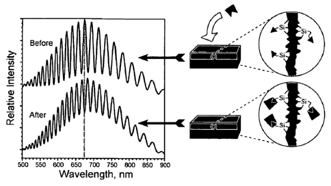
particle (see, e.g., Fig. 3A). Characteristic color changes are thus
indicative of drug quantity remaining in the pores. Thus, the term
photonic crystal is used for the- film that has been machined and
sized to small crystals for intraocular injection.
[0094] A spectrometric method of detection of the oxidized
"smart dust" injected into the rabbit eyes was also investigated.
One eyepiece of the surgical microscope was connected to the input
of a fiber-optic based spectrophotometer and this allows us to
accurately focus the detecting light on the intraocular "smart dust"
particles. The disclosure also provides a camera for monitoring the
color change of the crystal outfitted with a spectrometer to
quantitate the drug release. In yet another embodiment, a scanning
laser ophthalmoscope which scans the retina and inner eye with a
monochromatic light is outfitted with the appropriate wavelength to
scan and detect reflectance spectrum changes allowing quantification
of drug release.
[0095] In addition, to the use of porous silicon as a drug
delivery composition, porous Si is an attractive candidate for use
as a template because of the tunability of the porosity and average
pore size. Additionally, elaborate 1, 2, and 2.5-dimensional
photonic crystals are readily prepared in porous Si. Porous Si
composites (e.g., porous Si and a polymer) show great promise for
improving the mechanical stability and control over release rates of
a delivery system. Either the composite itself or a nanostructure
derived from the composite by removal of the porous Si template can
be used. Porous Si combined with a biocompatible polymer can yield
improved control over drug release kinetics and improved stability
in aqueous media, and the use of biopolymers that are selectively
cleaved by specific proteases provides the possibility of tissue-
specific action.
27
CA 02696139 2010-02-10
WO 2009/009563
PCT/US2008/069474
[0 0 9 6] Removal of the porous Si or porous 5i02 template from a
polymer or biopolymer imprint can be achieved (depending upon the
polymer used) by chemical dissolution using aqueous KOH or HF,
respectively, providing a free-standing porous polymer film with the
optical characteristics of the master. Whether or not the process
replicates the nanostructure of the master is highly dependent on
the processing conditions and the type of polymer used. Also, the
ability of the polymer to release from the master is dependent on
the interfacial chemistry and tortuosity of the pore network.
[0097] Two synthetic approaches can be used to generate a
template polymeric delivery composition or a porous Si polymer
composite. In one aspect, the polymer is synthesized within the
porous matrix. In another aspect, a pre-formed polymer is infused
into the matrix by melt- or solution-casting. For drug delivery
applications, it is important to use a biocompatible polymer. Any
number of biocompatible polymers can be used in the methods and
compositions of the disclosure as described herein. For example,
hydrogels can be used. Hydrogels are commonly used in ophthalmologic
devices, biosensors, biomembranes, and controlled drug delivery.
Water-swollen, crosslinked polymeric networks can undergo volume
phase transitions in response to environmental changes such as pH,
ionic strength, temperature, or electric fields.
[0098] Polymer replicas can be implanted on the sclera for
trans-scleral drug release. It has been shown in rabbit eyes that
polymer replicas are biocompatible and may safely and effectively
remain in the eye for multiple months, if not years. Measurement of
the decay in intensity of the peaks in the photonic crystal spectrum
should provide a monitor of the rate of drug release from an
implanted biocompatible polymer. In order to test the above
hypothesis, drug-impregnated poly(L-lactide) (PL) films, cast from
thermally oxidized porous silicon templates, can be prepared
following a scheme, designated generally at 10, illustrated in FIG.
3. Specifically, a template (such as electropolished porous
silicon), generally at 12, is provided, having pores 14 dimensioned
to suit a particular application. A polymer, generally at 16, is
loaded into the pores 14 to form a polymer-template composite. The
template 12 is subsequently removed, leaving a polymer-based
28
CA 02696139 2014-12-03
photonic film 16. Replication of the optical spectrum in the
biocogpatible polymer upon removal of the porous silicon template
can be used to confirm the replication process. The release
characteristics of the polymers can be studied.
[0099] Any number of polymeric materials can be used in the
generation of a polymeric porous structure of the disclosure.
Including, for example, nylon (polyamides), dacranTm (polyesters),
polystyrene, polypropylene, polycaprolactone, polyacrylates,
polyvinyl compounds (e.g., polyvinylchloride), polycarbonate (PVC),
polytecrafluorethylene (PTFE, teflonTm), thermanox (TPX), poly(N-
isopropylscrylamide), nitrocellulose, cotton, polyglycolic acid
(PGA), zeta, collagen (in the form of sponges, braids, or woven
threads, etc.), cellulose, gelatin, poly lactic acid, poly glycolic
acid, copolymers of poly lactic or poly glycolic. acid, or other
naturally occurring biodegradable materials or synthetic materials,
including, for example, a variety of polyhydroxyalkanoates. Again,
any number of polymers can work provided the polymer is transparent
at the wavelengths of interest for the photcnic application. If the
template is to be removed, the polymer should be a solid and not a
liquid. Typical polymers can include, for example, Poly dimethyl
siloxane (PDMS), poly lactic acid (PLA), PLGA, polypropylene,
polyethylene, polystyrene, and clear epoxy.
[00100] The degradation of the photonic structure in these films
can be characterized in pH 7.4 aqueous buffer solutions, in vitro
and in vivo. In accelerated degradation studies, polymer imprints
impregnated with caffeine were studied. The intensity of the rugate
peak displays an approximately exponential decay when the polymer is
dissolved in pH 10 buffer. Simultaneous measurement of the decay of
the spectral peak 4nd the appearance of caffeine in the solution
(caffeine absorption feature at 274 am) confirmed that the drug was
reLeased on a time scale comparable to polymer degiadatior:.
[00101] Embodiments of the disclosure also contemplate vectorial
drug delivery. The polymer-based photonic film shown in FIG. 3
contains A polymer "cap" 16 on one side of the film. Films prepared
in this manner will leech drug out one side of the film, allowing
greater control of the drag delivery parameters. Manufacturing
variables are channel sizes and packing.
29
CA 02696139 2010-02-10
WO 2009/009563
PCT/US2008/069474
[ 0 0 1 02 ] For intraocular delivery of drugs, a doctor or clinician
may look through the iris of the eye and into the clear part of the
eye to observe the colors of the injected particles. In this manner,
the amount of drug remaining or the degree to which the particles
have dissolved may be monitored, which in turns permits the doctor
or clinician to forecast the length of time before the particles
completely dissolve, and to predict when the patient may need
subsequent injections.
[00103] Other embodiments include use of a porous silicon or
silicon/polymer composite at a particular location of the eye, or
using the porous silicon or silicon/polymer composite as a template
to generate other biologically compatible or biologically resorbable
materials for similar use. Biodegradable polymer imprints may be
made from porous silicon templates, which may be used as drug
delivery contact lenses or implants at an appropriate location of
the eye, including the ocular surface and retrobulbar surface.
[00104] Another embodiment of the disclosure include drug(s)
impregnated in porous films configured to be worn or attached on the
front of the eye. A contact lens formed of impregnated porous thin
film material, for example, comprises and embodiment of the
disclosure. While another embodiment encompasses a contact lens, it
also contemplates other similarly curved solid template
correspondingly shaped with a front surface of the eye, as well as
being configured to join the eye at the sclera as an episcleral
plaque. The particular drug or drugs to be used with the polymer
imprint may be added to the imprint solution prior to casting or
engineered into the pores of the imprint after casting. Accordingly,
the embodiment of the disclosure provides a system and method of
drug delivery wherein porous silicon films can be variously modified
to be a long-lasting intraocular drug delivery vehicle to carry
various therapeutic compounds. In addition, biodegradable porous
polymer imprints made from porous silicon templates can be used as a
drug delivery implant to be placed at an appropriate location in the
eye. The drug can be added into the imprint solution before casting
or engineered into the pores after casting.
[00105] For the extraocular drug delivery, the emphasis on
optical reporting declines. With the episcleral plaque, for example,
CA 02696139 2010-02-10
WO 2009/009563
PCT/US2008/069474
delivery is retrobulbar, and it is not as easy to use an optical
instrument to "read" these films. In this retrobulbar embodiment,
the ability of the nanostructure to set the rate of dissolution or
drug release is a property. Because the electrochemical process used
to construct porous Si can control the nanostructure to such a
precise degree, precise control of the dissolution and/or drug
release profile of the particles or of the composites is conferred.
Thus, for example, the disclosure provides a contact lens configured
and arranged to cover a front extraocular surface, where a rim, or
"carrier," of the contact lens would be either a silicon or
silicon/polymer composite film impregnated with drug(s). The wearer
would receive a sustained and monitorable release of drug through
the contact lens. Another embodiment includes the use of episcleral
plaques.
[00106] An episcleral plaque is an extraocular way to deliver
drugs and the intraocular dust injection promotes monitoring of drug
levels non-invasively. The disclosure provides use of a silicon or
silicon/polymer composite film impregnated with drugs to be affixed
or adhered to a retrobulbar surface of the eye. The patient would
thereby receive a sustained and monitorable release of drug through
the episcleral plaque.
[00107] While the disclosure provides for use with a virtually
unlimited number of pharmaceutical candidates, several exemplary
drugs will be discussed herein. For example, drug delivery for drugs
used in treating ARMD and uveitis will be shown for purposes of
illustration. These diseases require prolonged intraocular
therapeutic drug levels to halt the progress of the disease and the
deterioration of eyesight. However, the promising drugs for treating
these diseases all share a common problem, which is the transient
intraocular therapeutic level requires frequent intravitreal
injections. These promising drugs include angiostatic steroids,
metalloproteinase inhibitors, VEGF binding drugs, PEDF, an 8-mer
peptide fragment of urokinase (uPA) and dexamethasone. These drugs
may also be used to treat, for example, diabetic retinopathy. In
particular, PEDF, the 8-mer peptide fragment of uPA and
dexamethasone all have short intravitreal half lives.
31
CA 02696139 2010-02-10
WO 2009/009563
PCT/US2008/069474
[ 0 0 1 0 8 ] Other drugs or "active ingredient" that can be used with
the smart dust of the disclosure include any one or any combination
of the following, but are not limited to, anti-angiogenic compounds
such as bevacizumab, ranibizumab, pegaptanib, and other compounds in
the angiogenic cascade. Also included are glucocorticosteroids such
as dexamethasone, triamcinolone acetonide, fluocinolone acetonide
and other comparable compounds in the corticosteroid and cortisene
families. Also included are compounds such as antacids, anti-
inflammatory substances, coronary dilators, cerebral dilators,
peripheral vasodilators, anti-infectives, psychotropics, anti-
manics, stimulants, anti-histamines, laxatives, decongestants,
vitamins, gastrointestinal sedatives, anti-diarrheal preparations,
anti-anginal drugs, vasodilators, anti-arrhythmics, anti-
hypertensive drugs, vasoconstrictors and migraine treatments, anti-
coagulants and anti-thrombotic drugs, analgesics, anti-pyretics,
hypnotics, sedatives, anti-emetics, anti-nauseants, anti-
convulsants, neuromuscular drugs, hyper- and hypoglycemic agents,
thyroid and anti-thyroid preparations, diuretics, anti-spasmodics,
uterine relaxants, mineral and nutritional additives, anti-obesity
drugs, anabolic drugs, erythropoietic drugs, anti-asthmatics,
bronchodilators, expectorants, cough suppressants, mucolytics, drugs
affecting calcification and bone turnover and anti-uricemic drugs.
Specific drugs include gastro-intestinal sedatives such as
metoclopramide and propantheline bromide; antacids such as aluminum
trisilicate, aluminum hydroxide, ranitidine and cimetidine; anti-
inflammatory drugs such as phenylbutazone, indomethacin, naproxen,
ibuprofen, flurbiprofen, diclofenac, dexamethasone, prednisone and
prednisolone; coronary vasodilator drugs such as glyceryl
trinitrate, isosorbide dinitrate and pentaerythritol tetranitrate;
peripheral and cerebral vasodilators such as soloctidilum,
vincamine, naftidrofuryl oxalate, co-dergocrine mesylate,
cyclandelate, papaverine and nicotinic acid; anti-infective
substances such as erythromycin stearate, cephalexin, nalidixic
acid, tetracycline hydrochloride, ampicillin, flucloxacillin sodium,
hexamine mandelate and hexamine hippurate; neuroleptic drugs such as
flurazepam, diazepam, temazepam, amitryptyline, doxepin, lithium
carbonate, lithium sulfate, chlorpromazine, thioridazine,
32
CA 02696139 2010-02-10
WO 2009/009563
PCT/US2008/069474
trifluperazine, fluphenazine, piperothiazine, haloperidol,
maprotiline hydrochloride, imipramine and desmethylimipramine;
central nervous stimulants such as methylphenidate, ephedrine,
epinephrine, isoproterenol, amphetamine sulfate and amphetamine
hydrochloride; antihistamic drugs such as diphenhydramine,
diphenylpyraline, chlorpheniramine and brompheniramine; anti-
diarrheal drugs such as bisacodyl and magnesium hydroxide; the
laxative drug, dioctyl sodium sulfosuccinate; nutritional
supplements such as ascorbic acid, alpha tocopherol, thiamine and
pyridoxine; anti-spasmodic drugs such as dicyclomine and
diphenoxylate; drugs affecting the rhythm of the heart such as
verapamil, nifedipine, diltiazem, procainamide, disopyramide,
bretylium tosylate, quinidine sulfate and quinidine gluconate; drugs
used in the treatment of hypertension such as propranolol
hydrochloride, guanethidine monosulphate, methyldopa, oxprenolol
hydrochloride, captopril and hydralazine; drugs used in the
treatment of migraine such as ergotamine; drugs affecting
coagulability of blood such as epsilon aminocaproic acid and
protamine sulfate; analgesic drugs such as acetylsalicylic acid,
acetaminophen, codeine phosphate, codeine sulfate, oxycodone,
dihydrocodeine tartrate, oxycodeinone, morphine, heroin, nalbuphine,
butorphanol tartrate, pentazocine hydrochloride, cyclazacine,
pethidine, buprenorphine, scopolamine and mefenamic acid; anti-
epileptic drugs such as phenytoin sodium and sodium valproate;
neuromuscular drugs such as dantrolene sodium; substances used in
the treatment of diabetes such as tolbutamide, disbenase glucagon
and insulin; drugs used in the treatment of thyroid gland
dysfunction such as triiodothyronine, thyroxine and
propylthiouracil, diuretic drugs such as furosemide, chlorthalidone,
hydrochlorthiazide, spironolactone and triamterene; the uterine
relaxant drug ritodrine; appetite suppressants such as fenfluramine
hydrochloride, phentermine and diethylproprion hydrochloride; anti-
asthmatic and bronchodilator drugs such as aminophylline,
theophylline, salbutamol, orciprenaline sulphate and terbutaline
sulphate; expectorant drugs such as guaiphenesin; cough suppressants
such as dextromethorphan and noscapine; mucolytic drugs such as
carbocisteine; anti-septics such as cetylpyridinium chloride,
33
CA 02696139 2010-02-10
WO 2009/009563
PCT/US2008/069474
tyrothricin and chlorhexidine; decongestant drugs such as
phenylpropanolamine and pseudoephedrine; hypnotic drugs such as
dichloralphenazone and nitrazepam; anti-nauseant drugs such as
promethazine theoclate; haemopoietic drugs such as ferrous sulphate,
folic acid and calcium gluconate; uricosuric drugs such as
sulphinpyrazone, allopurinol and probenecid; and calcification
affecting agents such as biphosphonates, e.g., etidronate,
pamidronate, alendronate, residronate, teludronate, clodronate and
alondronate.
[00109] Insofar as the disclosure contemplates including a
virtually unlimited number of drugs, in vitro pharmacokinetic
studies can be used to determine the appropriate configuration of
the porous silicon film and its dust for each drug. The drug
conjugated porous silicon film and its dust can be aliquoted into
vitreous samples in cell culture dishes. Intensity of reflected
light from the porous silicon film or its dust can be measured using
a low power spectrophotometer, at the same time free drug in the
vitreous sample can be measured, as a function of time for the
porous film or dust immersed in the vitreous sample. Correlation
between spectrophotometer change and drug concentration in vitreous
can be determined and used for in vivo PK studies.
[00110] For biocompatible polymer imprints of the porous silicon
film, drug can be impregnated in the polymer casting solution. Then
the free standing polymer porous film can further conjugate with
drug molecules to fill the pores. In vitro PK studies can be
performed in a similar way as with the porous silicon film or its
dust.
[00111] Optimized porous silicon smart dust adapted to the drug
candidate will not be toxic after intravitreal injection and the
vitreous drug half-life will be in the range of weeks to months and
the drug level will sustain above the EC for months. A method
includes preparing porous Si photonic crystal particles, loading the
pores of those crystal particles with one or more drugs, and
injecting the particles into the vitreous via syringe. The amount of
drug loaded in the particles may then be monitored via one or more
of a plurality of methods, such as by visual inspection, digital
imaging, laser eye scan, or spectroscopic observation. Any of these
34
CA 02696139 2010-02-10
WO 2009/009563
PCT/US2008/069474
four methods are non-invasive, allowing the practitioner or
clinician to observe the particles through the pupil of the eye.
[00112] More particularly, one method of the disclosure proceeds
as follows. Porous Si photonic crystals are formed from a porous
silicon film that is electrochemically etched in a single crystal Si
substrate by application of a sinusoidal current density-time
waveform. The waveform varies between 15 and 45mA/cm2, with 70
repeats and a periodicity of 12.5 s. The one-dimensional photonic
crystal that results has a color that depends on the waveform
parameters. The conditions described above produce a film that has a
strong reflectivity maximum in the green region of the spectrum.
This is a convenient color for visual observation in the eye, though
any color or pattern of colors (multiple spectral peaks) can be
incorporated into the films. The spectral features can range in
wavelength from 300 nm to 10,000 nm. The film is removed from the Si
substrate using a pulse of current. Particles with dimensions in the
range 1 pm to 270 pm are generated by ultrasonication.
[00113] The photonic crystals are then loaded with a drug or
drugs. The pores of the photonic crystals are large enough to allow
infiltration of drugs such as, for example, dexamethasone. Drug can
be loaded into the film or particles by infiltration from solution.
In a typical preparation, the drug loading solution comprised 6x10⁻²
M dexamethasone in methanol. 25 pL of the solution was pipetted onto
the porous Si film and the solvent was allowed to evaporate in air.
The film was briefly rinsed with deionized water to remove any
excess drug remaining on the surface that had not infiltrated the
pores.
[00114] Once the drug is loaded into the pores of the photonic
crystals, the photonic crystals are then injected into the patient.
In another aspect, the loaded photonic crystals are oxidized to
entrap the drug. The drug-loaded crystals are placed in an
appropriate excipient and injected into the vitreous. After
intravitreal injection, the porous silicon particles floated in the
vitreous affording an ophthalmoscopically clear view of the fundus
without any observed toxicity. The particles may last in the
vitreous for up to four months without any noticeable abnormalities.
CA 02696139 2010-02-10
WO 2009/009563
PCT/US2008/069474
[0 0 1 1 5] The optical interference spectrum used in particle
identification can readily be measured with inexpensive and portable
instrumentation such as a CCD spectrometer or a diode laser
interferometer. Removal of the drug from the porous nanostructure
results in a change in- the refractive index of the porous film and
is observed as a wavelength shift in the spectrum, or a shift in the
code, of the dust particle. The high surface area and optical
interferometric means of detection lead to very high sensitivity for
this system. Furthermore, particles can be encoded to reflect
infrared light that can penetrate living tissues and enable
noninvasive sensing through opaque tissue.
[00116] The described devices, systems and methods also encompass
the pulsatile delivery of active ingredients, such as pharmaceutical
compounds. By "pulsatile" is meant that a plurality of drug doses
are released at spaced apart intervals of time. Accordingly, the
devices and systems are designed, configured and manufactured to
possess release profiles (e.g., release kinetics) suitable for
treating specific conditions or multiple conditions. It is
understood that such devices and systems can include a plurality of
active ingredients each possessing a specific release profile
suitable for treating multiple conditions. A pulsatile delivery
system is capable of providing, for example, one or more immediate
release pulses at predetermined time points after a controlled lag
time or at specific sites. The system or device allows for pulsatile
drug delivery, and the administration of differing sized dosages of
active ingredients at different times automatically, pursuant to a
pre-programmed dosage profile utilized to design, configure and
manufacture a device or system provided herein. Exemplary release
profiles include those that correspond to desired peaks and troughs
related to disease symptoms.
[00117] Accordingly, provided herein are devices, systems and
methods designed to facilitate the controlled release of an active
ingredient in a biological system. In some aspects, the active
ingredient is a pharmaceutical compound. The compound can be
included in a suitable matrix or carrier. The matrix or carrier can
further include hydrophilic binders, water-soluble diluents,
36
CA 02696139 2010-02-10
WO 2009/009563
PCT/US2008/069474
surfactants, lubricants, disintegrants, antioxidants, or non water-
soluble diluents, or any combination thereof.
[00118] The term "active ingredient" is intended to mean any
compound having a therapeutic effect, and which is suitable for
administration in a device provided herein. Active ingredients
include non-peptide organic molecules, small peptides and peptide
mimetics, and the like, as well as their pharmaceutically acceptable
salts. The active ingredient itself may be stable upon storage or
under stress conditions, but when formulated with one or more
carriers it shows stability problems, e.g., it starts to degrade.
[00119] The term "carrier" is intended to mean such carriers
which are commonly used in the pharmaceutical chemistry for
preparing pharmaceutical formulations, see, e.g., Remington: The
Science and Practice of Pharmacy, 19th Edition (1995); "Drugs and
the pharmaceutical sciences", vol. 81, 1997. In particular such one
or more carriers are selected from, but not limited to, hydrophilic
binders, water-soluble diluents, surfactants, lubricants,
disintegrants, antioxidants, non water-soluble diluents and/or other
fillers known to the skilled person.
[00120] The term "pharmaceutically acceptable salt" represents
salt forms of an active ingredient that are physiologically suitable
for pharmaceutical use. The pharmaceutically acceptable salts can
exist in conjunction with an active ingredient as acid addition
primary, secondary, tertiary, or quaternary ammonium, alkali metal,
or alkaline earth metal salts. Within the disclosure, the active
ingredient may be prepared in the form of a salt such as
pharmaceutically acceptable salts, especially acid-addition salts,
including salts of organic acids and mineral acids. Examples of such
salts include salts of organic acids such as formic acid, fumaric
acid, acetic acid, propionic acid, glycolic acid, lactic acid,
pyruvic acid, oxalic acid, succinic acid, malic acid, maleic acid,
tartaric acid, citric acid, benzoic acid, salicylic acid and the
like. Suitable inorganic acid-addition salts include salts of
hydrochloric, hydrobromic, sulphuric and phosphoric acids and the
like. The acid addition salts may be obtained as the direct products
of compound synthesis. In the alternative, the free base may be
dissolved in a suitable solvent containing the appropriate acid, and
37
CA 02696139 2010-02-10
WO 2009/009563
PCT/US2008/069474
the salt isolated by evaporating the solvent or otherwise separating
the salt and solvent.
[00121] The term "hydrophilic binder" represents binders commonly
used in the formulation of pharmaceuticals, such as
polyvinylpyrrolidone, copolyvidone (cross-linked
polyvinylpyrrolidone), polyethylene glycol, sucrose, dextrose, corn
syrup, polysaccharides (including acacia, tragacanth, guar, and
alginates), gelatin, and cellulose derivatives (including
hydroxypropyl methylcellulose, hydroxypropyl cellulose, and sodium
carboxymethylcellulose).
[00122] The term "water-soluble diluent" represents compounds
typically used in the formulation of pharmaceuticals, such as sugars
(including lactose, sucrose, and dextrose), polysaccharides
(including dextrates and maltodextrin), polyols (including mannitol,
xylitol, and sorbitol), and cyclodextrins.
[00123] The term "non water-soluble diluent" represents compounds
typically used in the formulation of pharmaceuticals, such as
calcium phosphate, calcium sulfate, starches, modified starches and
microcrystalline cellulose.
[00124] The term "non water-soluble diluent with non-swelling
properties" represents the non water-soluble diluents as indicated
above, but excluding starches and modified starches and the like.
[00125] The term "surfactant", as used herein, represents ionic
and nonionic surfactants or wetting agents commonly used in the
formulation of pharmaceuticals, such as ethoxylated castor oil,
polyglycolyzed glycerides, acetylated monoglycerides, sorbitan fatty
acid esters, poloxamers, polyoxyethylene sorbitan fatty acid esters,
polyoxyethylene derivatives, monoglycerides or ethoxylated
derivatives thereof, diglycerides or polyoxyethylene derivatives
thereof, sodium docusate, sodium laurylsulfate, cholic acid or
derivatives thereof, lecithins, alcohols and phospholipids.
[00126] The term "antioxidant" represents the three groups of
antioxidants, true antioxidants, reducing agents and antoxidant
synergists, such as tocopherols, tocopherolesters, alkyl gallates,
butylated hydroxyanisole, butylated hydroxytoluene, ascorbic acid,
citric acid, edetic acid and its salts, lecithin and tartaric acid.
38
CA 02696139 2010-02-10
WO 2009/009563
PCT/US2008/069474
[ 0 0 1 2 7 ] The term "disintegrant" represents compounds such as
starches, clays, celluloses, alginates, gums, cross-linked polymers
(such as cross-linked polyvinylpyrrolidone and cross-linked sodium
carboxymethylcellulose), sodium starch glycolate, low-substituted
hydroxypropyl cellulose, and soy polysaccharides. Preferably, the
disintegrant is a modified cellulose gum such as e.g. cross-linked
sodium carboxymethylcellulose.
[00128] The drug or photonic nanocrystal of the disclosure can be
formulated for in vivo delivery using the compositions and methods
described above.
[00129] Although certain embodiments of the invention have been
described, additional embodiments and examples are provided below.
Such specific examples are not intended to limit the invention.
EXAMPLES
[00130] Porous silicon dust was injected into rabbit vitreous and
no toxicity was found compared with the fellow eyes that received
the same volume of phosphate-buffered saline (PBS) injection. The
porous silicon film was etched using a sinusoidal current varying
between 15 and 45mA/cm2, with 70 repeats and a periodicity of 12.5
s. The film was sonicated into a dust that ranged from 1 pm to 270
pm. After intravitreal injection, the porous silicon particles
floated in the vitreous affording an ophthalmoscopically clear view
of the fundus without any observed toxicity. The particles lasted in
the vitreous for one week without any noticeable abnormalities.
[00131] Thermally oxidized silicon dust was also injected into
the vitreous of four rabbits. This chemical modification of the
porous silicon film was proposed as one of the alternative methods
to increase the residence time of the porous silicon dust in
vitreous. This approach demonstrated a great increase of the
residence time of the particles in the rabbit eye compared to the
previous incompletely hydrosilylated smart dust (from less than 7
days to longer than 3 weeks). In addition, by increasing the
sonication time during preparation, smaller and more uniform smart
dust particles were produced, which can be delivered into vitreous
by the 25 or 27-gauge needle that is commonly used for intravitreal
injection in the clinic.
39
CA 02696139 2010-02-10
WO 2009/009563
PCT/US2008/069474
[00132] Additional data supports use of completely hydrosilated
porous Si photonic crystals that have no toxicity by clincial
examination or electroretinograms or histology at 3 1/2 months post
injection, inclusive of shorter times. For example, 100 microliters
of the material were injected, and the characteristic color of the
crystals is seen making it clear that one can use this
characteristic for monitoring drug release in the eye.
[00133] Intravitreal injection of 100 pl of oxidized porous Si
photonic crystal particles in 5% dextrose was performed. The
measured size of the smart dust ranged from 10 to 45pm with an
average of 30pm; approximately 30,000 particles were injected into
each rabbit eye. The injected particles appeared purplish green
floating in the vitreous. From the second day some of the particles
aggregated and sank onto the inferior retina. No toxicity was seen
and the smart dust particles were still visible at the last
examination 34 weeks later with at least half of the originally
injected material remaining, as assessed by ophthalmoscopy. It is
therefore anticipated that the particles would be safe and effective
for at least a year if not two years. Thus, this preliminary thermal
oxidation modification has greatly extended the time of intravitreal
residence compared to the previous incompletely hydrosilylated smart
dust. The data demonstrated that the porous silicon particle was
safe as an intravitreal drug delivery vehicle. Modifications such as
oxidation and silicon-carbon chain conjugation can be used to
further increase the stability of the silicon dust and can make it a
long-lasting slow release intravitreal drug delivery system.
[00134] A preliminary study was performed on a rat CNV model
using systemic administration of an 8-mer peptide derived from
urokinase plasminogen activator (uPA) to block the uPA-urokinase
plasminogen activator receptor (uPAR) interaction. This 8-mer
peptide was administrated subcutaneously twice daily at 200 mg/kg/d
beginning at the time of induction of CNV (with laser) to introduce
CNV in Brown Norway rats. Two weeks after laser treatment,
simultaneous FA and ICG using scanning laser angiography was
performed to identify the leaking laser bums. The results showed
that this 8-mer peptide reduced the laser induced CNV by 70%
compared to the control group (44.7% of laser burns leak in control
CA 02696139 2010-02-10
WO 2009/009563
PCT/US2008/069474
group versus 13.4% in treated group, p<0.001). Administration of
the drug intravitreally using a proposed porous silicon smart dust
should maintain the desired intraocular drug level.
[00135] Thermal Oxidation of Porous Si Particles: Preliminary
studies of porous Si particles oxidized and annealed at 300 C for 2
hours in air show that the material is stable in aqueous pH 11
buffer for several days, and recent results indicate that this
approach can dramatically increase the residence time of the
particles in the rabbit eye. In addition, by increasing the
sonication time during preparation, smaller and more uniform smart
dust particles were produced which can be delivered into vitreous by
the 28.5 gauge needle that is commonly used for intravitreal
injection in the clinic. Intravitreal injection of 100 pl of
oxidized porous Si photonic crystal particles in 5% dextrose was
performed. The measured size of the smart dust ranged from 10 to
45pm with a average of 30pm; approximately 30,000 particles were
injected into each rabbit eye. The color of the injected particles
floating in the vitreous was clearly visible, which is indicative of
drug release and degradation by hydrolysis. Degradation by
hydrolysis is especially advantageous in that no enzymes are
necessary to degrade the particles. From the second day some of the
particles aggregated and sank onto the inferior retina. No toxicity
was noticed and the smart dust particles were still visible until
the last examination, which indicates that this preliminary thermal
oxidation has more than tripled the time of intravitreal residence
compared to the previous incompletely hydrosilylated smart dust.
Experiments can be performed to quantify the residence time and
correlate it with the chemical modification conditions such as
thermal oxidation time, temperature, and ambient atmosphere.
[00136] Electrochemical Grafting of Organic Reagents: The
hydride-terminated surface of p-type or p++-type porous silicon can
be stabilized by electrochemical reduction of acetonitrile solutions
of various organo halides. Reduction of 6-iodo-ethylhexanoate, 1-
iodo-6- (trifluoroacetylamino) hexane, iodomethane, 1-bromohexane,
or ethyl 4-bromobutyrate at a porous Si cathode results in removal
of the halogen and attachment of the organic fragment to the porous
Si surface via a Si-C bond. A two- step procedure was devised
41
CA 02696139 2010-02-10
WO 2009/009563
PCT/US2008/069474
involving attachment of the functional group of interest followed by
attachment of methyl groups (by reduction of iodomethane) to
residual, more sterically inaccessible sites on the porous Si
surface and found that electrochemical alkylation greatly improves
the stability of porous Si against oxidation and corrosion in
various corrosive aqueous media, and that the methyl capping
procedure provides the most stable porous Si material yet reported.
This chemistry also allows covalent attachment of the candidate
drugs for the release studies.
[00137] Thermal Hydrosilylation of Organoalkenes: This approach
provides a porous Si material that is stable even in boiling aqueous
pH 10 solutions. This chemistry was extended to the dust particles
and find similar levels of stability. Parameters of the reaction may
be adjusted in order to identify the key parameters leading to this
instability. In particular, the surface coverage (essentially the
efficiency of the chemical reaction), the type of organic species
grafted to the surface (alkyl carboxylates, alkyl esters, and alkyl
halides), and the chain length of the alkyl species can be
investigated. Reaction conditions such as the presence of added
radical initiators, transition metal catalysts, and photoassisted
hydrosilylation can be explored.
[00138] For each modified porous silicon film, its sonicated dust
can be intravitreally injected into 3 rabbit eyes with the fellow
eyes used for control. After injection, the toxicity can be
monitored by slit lamp, indirect ophthalmoscope, ERG, and pathology.
In addition, a remote spectrometer probe can be used to determine
the clearance rate of the silica dust in vitreous on living animals
through the dilated pupil. The spectrometer probe is believed to
render more accurate information since the small particles may not
be seen using indirect ophthalmoscope.
[00139] A spectrometric method of detection of the oxidized
"smart dust" injected into the rabbit eyes was also investigated.
One eyepiece of the surgical microscope was connected to the input
of a fiber-optic based spectrophotometer and this allows us to
accurately focus the detecting light on the intraocular "smart dust"
particles. The disclosure also provides a camera for monitoring the
color change of the crystal outfitted with a spectrometer to
42
CA 02696139 2010-02-10
WO 2009/009563
PCT/US2008/069474
quantitate the drug release. In yet another embodiment, a scanning
laser ophthalmoscope which scans the retina and inner eye with a
monochromatic light is outfitted with the appropriate wavelength to
scan and detect reflectance spectrum changes allowing quantification
of drug release. The preliminary data showed a feasibility of this
approach and the specific wavelength of a porous Si photonic film
was detected with a 1 nm spectral resolution. This resolution is
sufficient to determine concentration of a species such as a large
protein in the porous Si film to micromolar concentration levels. As
an alternative, the probe can be adapted to a fundus camera which is
used for clinical retinal imaging. For the rabbit or rodent eyes,
the fundus can be photographed using a fundus camera without
anesthesia.
[00140] In in vitro experiments, the optical codes of the porous
Si photonic crystal particles can be read using digital imaging
cameras. Since the color of the particles provides an indirect
measure of the amount of drug loaded, the most accurate measure is
obtained using a spectrometer. However, the color resolution in a
digital camera is sufficient to measure the loading to an accuracy
of 10%, which is sufficient for the present application. In order to
measure the degree of loading in porous Si "smart dust," the color
of the particles can be recorded using a color digital camera
connected to the fundus camera. Software to process the digital
images and extract concentration information can be obtained with
minor modifications to commercially available software. The
advantage of this approach is that it requires only minor
modification to existing readily available medical equipment, and it
allows acquisition of data from a large number of particles
simultaneously. If higher resolution concentration information is
needed, the illumination light can be filtered using a monochromator
or bandpass filters, providing spectral resolution equivalent to
that which can be obtained with a spectrometer.
[00141] The long-lasting porous silicon film and its imprint can
be further optimized for delivery of three candidate drugs (PEDF, an
8-mer peptide fragment of uPA, and dexamethsasone) by controlling
the pore size and morphology. These parameters are easily controlled
using the appropriate anodic electrochemical etching current
43
CA 02696139 2010-02-10
WO 2009/009563
PCT/US2008/069474
density, duration of the etch cycle, and etchant solution
composition. Since the imprint and its porous silicon template share
the similar nanostructures, it is assumed that imprints from
optimized porous silicon can also be appropriate for delivering
those drug candidates.
[00142] Additional in vivo data regarding the "smart dust"
material after intraocular injection and new in vitro data
concerning the release of dexamethasone from "smart dust"
formulations is as follows. In vivo studies The new formulation of
"smart dust" particles containing a silicon dioxide shell have been
observed in the vitreous of living rabbits for 16 weeks and they are
showing evidence of dissolution without any evidence of toxicity by
slit lamp, indirect ophthalmoscopic examinations or by light or
electron microscopy. More than half of the particles appear to be
present at this time point indicating excellent potential as a long
acting drug delivery system. Injection of "smart dust" particles
containing a hydrosilylated alkyl shell into the living rabbit eye
has shown no evidence of toxicity for up to five weeks of ongoing
examination.
[00143] Additional in vivo studies demonstrated the increased
stability of "smart dust" particles containing a hydrosilylated
alkyl shell. These chemically modified particles also exhibit slower
release rates for a drug. Release of dexamethasone from the modified
porous silicon matrix is slowed by a factor of 20 compared to
unmodified porous silicon.
[00144] Chemistries have also been developed to expand the pores
in order to accommodate larger molecules within the pores, such as a
modified Fab fragment of human IgG. The pore expansion procedure
involves the enlargement of pores by treatment with
dimethylsulfoxide (DMSO) containing hydrofluoric acid (HF). The
porosity increases approximately 10% after the expansion treatment,
and it was found that this chemistry allows admission of large
molecules such as human IgG (150 kDa) and bovine serum albumin (67
kDa). As will be clear to artisans, the invention makes use the
optical properties of porous silicon photonic crystals to monitor
drug delivery rates. The shift in the reflectivity spectrum of the
film coincides with release of a drug. Optical measurements were
44
CA 02696139 2010-02-10
WO 2009/009563
PCT/US2008/069474
carried out while concurrent absorbance measurements were obtained
as the drug-infused porous silicon films were introduced in buffered
aqueous solutions. There is a linear correlation between the
increase of drug concentration in solution (/.e. drug diffusing from
the pores) and a change in the optical thickness of the porous
silicon film.
[00145] The optical properties of porous Si have been
investigated for numerous applications including chemical and
biological sensors. Porous Si is a biocompatible and bioresorbable
material that has also been investigated for in-vivo drug delivery
and biomedical device applications. Recently, a technique to
produce micro particulate photonic crystals from porous Si was
developed. The distinctive particle spectrum can be observed through
human tissue, (Li, Cunin et al., Science 299(5615):2045-7 (2003))
and it can be used to monitor the loading and release of various
organic or biomolecules including dexamethasone, IgG and bovine
serum albumin. This optical method of monitoring molecular loading
and release is well suited for ophthalmic applications. The drug can
be housed in the porous matrix while the optical spectrum allows
non-invasive measurement of the release rate. This is the first
study to characterize the intraocular properties of porous silicon
particles that are capable of acting as a self-reporting drug
delivery system in living animal eyes.
[00146] Fabrication of Porous Silicon Particles: Porous silicon
particles were fabricated by an electrochemical etch of single-
crystalline, degenerately B-doped p-type silicon (Siltronix Inc.,
<100> orientation, -1mQ.cm resistivity) in a 48% aqueous HF :
ethanol (3:1 by volume) electrolyte solution. An optical rugate
structure was electrochemically etched into the Si wafer using a
sinusoidal current modulation of 15-45 mA/cm2, with 70 repeats and a
periodicity of 12.5 seconds. The films were removed from the bulk
silicon substrate by electropolishing in a 3.3% HF in ethanol
solution using a current density of 200 mA/cm2 for 2 min. The
manufactured porous Si film was generally 20 microns thick (see
Figure 5) with a porosity of 67% as determined by gravimetric
analysis. The freestanding films were then ultrasonically fractured
using an ultrasonic cleaner (5 min.) to produce particles ranging in
CA 02696139 2010-02-10
WO 2009/009563
PCT/US2008/069474
size from 1-270 microns with over 70% particles falling in the range
of 15-30 microns (estimated by optical microscopy). For a 20 micron
particle, there is an estimated free volume of 4x10⁻⁹cm3 available
for drug loading per particle, with a total free volume of 1.2x10-
4cm3 per particle injection in the rabbit vitreous.
[00147] Chemical Modification of Porous Si Particles: Unmodified
porous silicon is known to be unstable in aqueous media because of
rapid oxidation of the reactive hydride species present on the
surface. In this work, two different chemical modification
reactions were performed in order to stabilize the particles. The
first method involves surface alkylation by means of thermal
hydrosilylation with 1-dodecene, and the second method is thermal
oxidation.
[00148] Surface Alkylation of Porous Si Particles: Thermal
hydrosilylation was carried out on porous Si particles immediately
after their preparation, following the method of Buriak (Buriak,
Adv. Mater. 11(3):265-267 (2002)). The particles were placed in a
Schlenk flask containing 1-dodecene and freeze-pump-thaw cycles were
performed to remove oxygen. The reaction flask was filled with
nitrogen and the mixture was heated at 120 C for 2 hours. The
particles were rinsed thoroughly with dichloromethane and ethanol
and then dried in air. The product was characterized by FTIR,
confirming the presence of alkyl species on the surface of the
particles.
[00149] Thermal Oxidation of PSi Particles: Oxidation was carried
out on porous Si particles immediately after their preparation.
Oxidation was accomplished by heating at 80 C in an oven in ambient
air for 24 hours.
[00150] Animal studies: Eleven New Zealand Red rabbits were used
to study the safety and stability of the porous silicon particles in
the rabbit vitreous. All of the animal handlings were carried out in
adherence to the ARVO Statement for the Use of Animals in Ophthalmic
and Vision Research. Using injection methods previously published,
one eye of each animal was injected with the porous Si particles,
and the fellow eye was injected with the same volume of 5% dextrose
to serve as the control. Three rabbits were injected with fresh
(not chemically modified) porous Si particles, five rabbits were
46
CA 02696139 2010-02-10
WO 2009/009563
PCT/US2008/069474
injected with hydrosilylated porous Si particles, and three were
used to evaluate the oxidized porous Si particles. All of the
particles were suspended in ethanol for sterilization. Prior to
injection, the ethanol was evaporated and lmL of 5% dextrose was
added to -120 mg of the particles. One drop (-6 pL) of sample was
taken for particle sizing and counting by light microscopy (Figure
6, Panel A and Panel B). A 25 gauge needle was used to deliver 100
L of the suspension (roughly 12 mg particles) into the rabbit
vitreous through the pars plana under direct view of a surgical
microscope. After intravitreal injection, the eyes were monitored
with indirect ophthalmoscope, tonometer, and biomicroscopic slitlamp
on day 3 and once each subsequent week thereafter. Fundus
photography was carried out in the selected rabbit eyes at different
intervals after injection to assess degradation of the porous Si
particles. The electroretinogram (ERG) was recorded from all eyes of
the animals prior to animal sacrifice. After animal sacrifice, the
eye globes were enucleated for histology evaluation. The vitreous
containing the hydrosilylated porous Si particles was excised from
selected eyes, and the particles were examined by scanning electron
microscopy.
[00151] Observation of unmodified porous Si particles in the
rabbit eye: A 100 pL aliquot of porous Si particles in 5% dextrose
solution was injected into the vitreous of three eyes of three
rabbits using a 25 gauge needle. The particles ranged in size from 1
to 270 m and the estimated number of particles per injection was
approximately 12,000. The particles were suspended in the vitreous
at the injection site (Figure 7, Panel A) and observed to disperse
into the surrounding vitreous during the following 2 to 3 days
(Figure 7, Panel B). No toxic effects were observed, and the
particles degraded completely in 3 to 4 weeks (Figure 7, Panel C).
Pathologic examination by light microscopy revealed no indications
of toxicity (Figure 7, Panel D).
[00152] Observation of hydrosilylated porous Si particles in the
rabbit eye: A 100 pL aliquot of hydrosilylated porous Si particles
in 5% dextrose solution was injected into the vitreous of five eyes
of five rabbits using a 25 gauge needle. The particles ranged in
size from 1 to 300 m (longest dimension) with an estimated 1900
47
CA 02696139 2010-02-10
WO 2009/009563 PCT/US2008/069474
particles per injection. The hydrosilylated particles became
distributed throughout the vitreous within 2 to 3 days while
displaying a vivid green color. Degradation was observed to be much
slower than for the unmodified porous Si particles (Figure 8). Four
months after injection the animals were sacrificed, and the
particles were analyzed by optical and by scanning electron
microscopy. Approximately 50% of the viewable particles appeared
blue-green in color (Figure 9, Panel A). The scanning electron
microscope images revealed sharp edges on the particles but a pitted
surface, indicating some degree of erosion (Figure 9, Panel B). The
other three rabbits were sacrificed for histopathology. ERG
examination, tonometry, and histology did not show any indications
of toxicity (Figure 9, Panel C) (see Table 1 below).
[00153] Observation of oxidized porous silicon particles in the
rabbit eye: A 100 pL aliquot of oxidized porous Si particles in 5%
dextrose solution was injected into the vitreous of three eyes of
three rabbits using a 25 gauge needle. The particles ranged from 10
to 40 pm and an estimated 30,000 particles were injected. The
oxidized particles showed similar dispersion in the vitreous as the
unmodified and the hydrosilylated particles. Faster degradation
rates were observed for the oxidized particles than for the
hydrosilylated particles (see Table 1 below). Two weeks after
injection, 20% of the particles showed evidence of degradation and
roughly 80% of them were reflecting purple light (Figure 10, Panel
B). Nine weeks after injection, over 80% of the observable
particles lost their vivid reflective property and appeared degraded
and brown. The particles had settled into the inferior vitreous or
retina (Figure 10, Panel C). The ERG, tonometry, and histology did
not reveal any indications of toxicity (Figure 10, Panel D) (see
Table 1 below):
Table 1. Characterization of the different porous Si particle types
used in intravitreal injection
Maximum Bio-
Number Estimated
vitreous microscopy
Particle of vitreous half- Intraocular
residence & Indirect ERG Pathology
type eyes life by pressure
time ophthalmol
tested ophthalmoscopy
scopy
Unmodifi
18.7 4 (at
ed 3 4 weeks 1 week Normal
Normal Normal
week 4)
(Fresh)
48
CA 02696139 2014-12-03
NA
:.17 weeks 16 weeks
.1q 2 ia.L. 17
NA NA N,vr7=1 Ncrmai Norm,'
weeks)
12 Lc 16 16.7-15 (AL
weeks Somal Nermn1 Nonni
wuek& wftek 4)
20 4 tat wook
= NA Normal notm.)1
rmal
..
[00154] The present-. studies
demonstrate that porous Si particles
can be safely injected into rabbit vitreous, ar.d the unmodified
particles degrade in three to four weeks without evidence of
toxicity. Chemical modification of the particle and pore surface,
either by grafting of dodecyl species (hydrosilylation) or by
converston to 5i02 (thermal oxidation) dramatically increases the
stability E.nd vitrecus residence time of the particles. This
indicates that hydrosilylated or oxidized porous 5)* particles may be
used as a long-lasting intravitreal drug delivery vehicle.
Fuv:.he.:_more, by controlling the extent of oxidation or
hydrosilylation, the vitreous residence time of the particles may be
manipulated to fit the specific treatment modality.
[00155] Porous Si has been studied previously in physiological
aquernis solutions and was found to dissolve into the form of
orthosilicic acid, which is vital for normal bone and connective
tissue homeos toe is. ("Dissolution of different forms of partially porous
silicon wafers under simulated
physiological conditions", Anderson ot al. (2003) phys. stat sot. (a), 197:331-
51 However, porous
Si ti isso 1 ution has never been studied in vitreous, which is a
complex biological solution with constant fluid turn over. This type
of condition is not easily duplicated in an in vitro setting .
Thererore, the di sso ti on and the associated potenti al toxicity
must be studied directly in the living eyes.
[(MSS) The =,:nd que photoni c properties of porous Si make 1.A:is
i deal for drug del.very by imparting a potential self --
reporting feature within the delivery systen., The wlyelongth of the
spectral peak reflected iron porous Si photonic crystals is
dependent on she refractive index (n) of the porous Si matrix ("Smart
Dust: Self-assembling, self-orienting photonic crystals of porous Si. Link and
Sailor (2003) Proc. Hall Acad.
Sd. USA 100:10007-10). Chanqes ih ref racti vs index c,i the poros Si .1 r
ocfhirs as ar.p.:encs so 1 uti on (n = 1 . 34) replaces orga:iic molecules or
CA 02696139 2014-12-03
proteins (n - 1.4) in the pores results in a blue shift. of the
reflectivity peak, producing an observable color change. A spectral
blue shift is also expected as the Si matrix (n - 3.5) is oxidized
to 3102 (n - 1.7) or as the 5i02 matrix dissolves. In the present
case, the initla3 green color of the photonic crystals is observed
to turn blue or violet after neverel days to weeks in vitreous
(depending on the surface chemistry) indicating dissolution of the
porous matrix. After extended periods in vitreous, some of the
particles lose their vivid reflectance and appear brown in color.
The brown color is attributed to light absorption by residual Si in
a particle whose photonic signature has shifted into the ultraviolet
range. It Is also possible that the signature spectrum of the
photos-sic crystal no longer exists due to extensive degradation of
the periodic nanostructure. This unique signature spectrum of the
photonic crystal could be utilized to monitor drug release through
the transparent optical medium of the eye using a simple CCD
spectrometer device that would provide a non-invasive method to
monitor drug release. This would be an advantage over other drug
delivery materials such as biodegradable and bioerodible polymeric
microparticles.
[00157] The fact that certain preparations of the porous Si
particles have long vitreous lifetimes and display no apparent
toxie.Lty indicates that porous Si may be used as an intravitreal
delivery material. With the advent of many intravitreal Injectable
the ouch as dexamethasone, pegaptnib (Macuge&14),
bevacirumab (AvastinTm), and the recently FDA-approved ranibizumab
tLucentislm), repeated intravizreal injections can potentially
generate serious problems. These procedures impose life quality
issues with patients and raise the risk of intraoeular infections.
Trapping such compositions in porous Si micropartieles by an
encapenl_ant or by covalent or electrostatic interactions between the
drug and the porous Si particles, allows for the composition to be
slowly released as the particles degrade. This would eliminate the
necesity of frequent injections.
[0015S] A ICO pl intravitreal Injection as used in the present
rabbit stuhies typically contains -30,000 poreas Si particles, each
apprwcimalely 50 microns square and 20 microns chick. It was
CA 02696139 2010-02-10
WO 2009/009563
PCT/US2008/069474
calculated that at least -50 mg of deximethasone per gram of porous
Si material can be loaded. Assuming that the porous Si particles
display first order dissolution kinetics and that drug release
occurs concomitant with particle dissolution, then the steady-state
concentration of drug in the eye can be approximated using the
dissolution mechanisms of Dove and Crerar (Geochimica Et
Cosmochimica Acta 69(21):4963-4970 (2005)). With this model, the
dissolution of the porous Si particles can be approximated by this
overall reaction:
5i02 + 2H20 - H45iO4
Where the species H45iO4 represents the water-soluble form of silicic
acid. The rate expression for this reaction is dependent on the
total surface area of the particles exposed to solution and the mass
flow rate of silicic acid out of the system. For the particulate
system, the appearance of silicic acid in solution was assumed to
correlate with the appearance of drug in solution, and that the
total surface area of particles exposed to solution is proportional
to the number of particles, N. As the particles dissolve, the drug
would be released, and the steady-state concentration of drug in the
eye can be calculated based on the following relationship that has
been adapted from Dove's model:
Md = [ tin (drug) / t1/2 (particle) ] xNxL
Where Md is the mass of free drug in the vitreous, N is the number
of particles injected per eye, L is the mass of drug loaded per
particle, and t4/2(drug) is the half-life of free drug in vitreous,
and t4/2(particle) is the half-life of the particles in vitreous.
[00159] In general the longer the particle half-life, as
demonstrated for the hydrosilylated particles and oxidized
particles, the lower the steady-state concentration of drug. For
particles with a 60-day half-life and an initial loaded drug mass of
600 pg in 12 mg of PSi particles (-30,000 particles), the steady-
state concentration of dexamethasone with a vitreous half-life of
3.48 h would be 1 pg/mL in rabbit vitreous (1.4 ml), which is above
the therapeutically relevant dose of >5 ng/mL. The particles may
deliver a drug at therapeutically relevant quantities for at least 3
half-lives of the particles (180 days).
51
CA 02696139 2010-02-10
WO 2009/009563
PCT/US2008/069474
[ 0 0 1 60 ] Drugs with a longer vitreous half-life such as Avastin (5
days) should be able to further extend the period between
injections. For Avastin, a loading capacity of about 1-10, 10-20,
20-50, 50-100, or 100-500 mg of drug per gram of particles may be
suitable for treating conditions responsive to the drug. In one
example, the initial amount of drug in a 0.1 cc injection of porous
Si loaded with Avastin may be approximately 100 g of drug. If the
half-life of the particles is 60 days, then the steady-state
concentration of drug in vitreous would be - 8 g/mL. The
therapeutically relevant dose of Avastin as an intraocular treatment
is generally considered > 22ng/mL. It is understood that the
skilled artisan can readily determine the loading capacity of the
particles provided herein based upon various factors, including the
type of active ingredient to be associated with the particles. It
is also understood that the dosage of an active ingredient
associated with treating a particular disorder can be modified
according to various methods known to the skilled artisan.
[00161] The disclosure demonstrates the intravitreal
biocompatibility of porous Si microparticles and the feasibility of
porous Si as a platform for an intraocular drug delivery system. As
noted herein, fresh porous Si particles (3 eyes), oxidized porous Si
(porous SiM particles (3 eyes), and hydrosilylated porous Si
particles (5 eyes) were tested in rabbit eyes. No toxicity was found
by using slitlamp to monitor the anterior segment, or by using
indirect ophthalmoscope to monitor posterior segment. The lack of
toxicity was also confirmed by eletroretinography and histology by
light microscopy. The hydrosilylated and oxidized particles were
observable in the vitreous until the end of the 4 month study.
[00162] The current study also demonstrates that the Si materials
are typically converted into particulate form by ultrasonication.
By extending the sonication time, a more evenly distributed and
smaller particles (mean size of 20 pm) can be produced and they are
more compatible with the intravitreal injection method. Further,
the two chemical modifications made to the porous Si materials
(oxidation and hydrosilylation) led to dramatically increased
intravitreal stability and slower degradation. The estimated
vitreous half-life increased from one week (fresh particles) to five
52
CA 02696139 2014-12-03
weeks (oxidized particles) and to 16 weeks (hydrosilylated
particles).
[00163] Also provided herein are novel methods for producing
porous SiO2 particles by oxidation of porous Si at 800 C. Particles
manufactured in this manner are more hydrophilic than the previous
oxidized ones which were processed at 220 C. This new type of porous
Si02 was injected into 6 rabbit eyes and no toxicity (including ERG)
was observed during the 5 month ongoing study. This new type of
porous Si02 allowed more efficient loading of the IgG-based drug
Avastin, a candidate drug for treatment of macular degeneration.
[00164] The porous 3102 particles oxidized at 800 C. were loaded
with Avastin and 100p1 of particles (containing 225 pg avastin) were
injected into 3 rabbit eyes. 20 weeks after injection, the vitreous
Avastin level was still 50 ng/ml which is higher than the IC50 of
Avastin (22ng/m1).
[00165] A porous Si-polymer composite plague was prepared and
surgically implanted on the rabbit eye globe under conjunctiva and
Tenon. These plaques have the same optical feature and nano pore
structure as their porous Si film templates and the nano pores open
to only one side of the plaque, allowing unidirectional drug
release. These plaques are well tolerated by the rabbit eyes.
Accordingly, the compositions and methods provided herein achieve
slow release and long lasting drug delivery to treat macular
degeneration, diabetic macular edema, choroidal neovascularization
and retinal vein occlusion and uveitis etc vitreoretinal diseases.
(00166) These embodiments are meant to be illustrative examples
and not exhaustive of the types of useful drug delivery structures
that can be manufactured using the materials and methods described
herein. The structures and methods discussed above will have great
utility for a variety of applications including, but not limited to,
controlled, sustained and programmable drug delivery.
(00167] While the invention is susceptible to various
modifications, and alternative forms, specific examples thereof have
been shown in the drawings and are herein described in detail.
53