Note: Descriptions are shown in the official language in which they were submitted.
CA 02696160 2015-04-27
52392-75
PEGylation by the Dock and Lock (DNL) Technique
Related Applications
WWI This application claims priority to U.S. Patent Application Serial No.
11/925,408,
filed October 26, 2007.
BACKGROUND
[0002J The efficacy of a therapeutic agent may be enhanced by improving its
bioavailability
via several means, one of which is PEGylation, a process of chemically linking
polyethylene
glycol (PEG) to the therapeutic agent of interest, with the resulting
conjugate exhibiting an
increased serum half-life. Additional advantages of the PEGylated products may
also include
lower immunogenicity, decreased dosing frequency, increased solubility,
enhanced stability,
and reduced renal clearance. Because the most common reactive sites on
proteins (including
peptides) for attaching PEG are the E amino groups of lysine and the a amino
group of the N-
terminal residue, early methods of PEGylation resulted in modification of
multiple sites,
yielding not only monoPEGylated conjugates consisting of mixtures of
positional isomers,
such as PEGINTRONTm (Grace et al., J. Biol. Chem. 2005; 280:6327) and PEGASYS
(Dhalluin et al., Bioconjugate Chem. 2005; 16:504), but also adducts
comprising more than
one PEG chain. Site-specific attachment of a single PEG to the a amino group
of the N-
terminal residue was reported to be the predominant product upon reacting PEG-
aldehyde
(PEG-ALD) at low pH with IFN-131 b (Basu etal., Bioconjugate Chem.
2006;17:618) or IFN-
131a (Pepinsky etal., J. Pharmacol. Exp. Ther. 2001;297:1059). Similar
strategies were
applied to prepare N-terminally linked PEG to G-CSF (Kinstler et al., Pharm.
Res.
1996;13:996) or type I soluble tumor necrosis factor receptor (Kerwin et al.,
Protein Sci.
2002;11:1825). More recently, a solid-phase process for PEGylation of the N-
terminus of
recombinant interferon alpha-2a was reported (Lee et al., Bioconjug. Chem.
Oct. 18, 2007,
epub).
[0003] Site-directed PEGylation of a free cysteine residue introduced into a
target protein has
also been achieved with PEG-maleimide (PEG-MAL) for several recombinant
constructs
including IL-2 (Goodson and Katre, Biotechnology. 1990:8:343); IFN-a2
(Rosendahl etal.,
1
CA 02696160 2010-02-10
WO 2009/055653 PCT/US2008/081085
Bioconjugate Chem. 2005;16:200); GM-CSF (Doherty et al., Bioconjugate Chem.
2005;16:1291); scFv (Yang etal., Protein Eng. 2003;16:761), and miniantibodies
(Kubetzko
et al., J. Biol. Chem; 2006;201:35186). A popular approach for improving the
therapeutic
efficacy of an enzyme has been to prepare conjugates containing multiple PEG
of small size,
as known for methioninase (Yang et al., Cancer Res. 2004;64:6673); L-methione
y-lyase
(Takakura eta!,, Cancer Res. 2006:66:2807): arginine deaminase (Wang et al.,
Bioconjugate
Chem. 2006;17:1447); adenosine deaminase (Davis etal., Clin. Exp. Immunol.
1981;46:649);
L-asparaginase (Bendich etal., Clin. Exp. Immunol. 1982;48:273); and liver
catalase
(Abuchowski etal., J. Biol. Chem. 1977;252:3582).
[0004] PEGylations of bovine serum albumin (Abuchowski etal., J. Biol. Chem.
1977;252:3578); hemoglobin (Manjula etal., Bioconjugate Chem. 2003;14:464);
visomant
(Mosharraf et al., Int. J. Pharm. 2007;336:215); small molecules such as
inhibitors of integrin
a4131 (Pepinsky etal., J. Pharmacol. Exp. Ther. 2005;312:742); lymphoma-
targeting peptides
(DeNardo etal., Clin. Cancer. Res. 2003;9(Suppl.):3854s); anti-VEGF aptamer
(Bunka and
Stockley, Nat. Rev. Microbiol. 2006;4:588) and oligodeoxynucleotides (Fisher
et al., Drug
Metab. Dispos. 2004;32:983) have also been described. However, there exists a
need for a
general method of PEGylation that would produce exclusively a monoPEGylated
conjugate
composed of a single PEG linked site-specifically to a predeteimined location
of the
candidate agent and retains the bioactivity of the unmodified counterpart.
SUMMARY OF THE INVENTION
[0005] The present invention discloses methods and compositions for producing
PEGylated
compounds with selected numbers of attached PEG residues that are attached at
selected
locations of a candidate agent. In preferred embodiments, the agents are
monoPEGylated. In
more preferred embodiments, the target to be PEGylated may be attached to a
DDD
(dimerization and docking domain) sequence and a PEG moiety may be attached to
an AD
(anchor domain) sequence as described in more detail below. Dimers of the DDD
sequence
bind with high affinity to monomers of the AD sequence, resulting in formation
of a
monoPEGylated effector moiety dimer. The stoichiometry of binding and location
of the
PEG residue are determined by the specificity of the DDD/AD interaction.
[0006] In more preferred embodiments, the monoPEGylated complex may be
covalently
stabilized by introduction of cysteine residues at appropriate locations in
the DDD and AD
2
SUBSTITUTE SHEET (RULE 26)
CA 02696160 2010-02-10
WO 2009/055653 PCT/US2008/081085
sequences, to form disulfide bonds that stabilize the complex. In other
embodiments, the
PEG reagents may be capped at one end with a linear or branched methoxy group
(m-PEG).
[0007] In other preferred embodiments, the PEGylated complex made by the DNL
method
shows a rate of clearance from serum that is at least an order of magnitude
slower than the
unPEGylated effector moiety. In certain alternative embodiments, the PEGylated
complex
may be alternatively constructed with the PEG moiety attached to the DDD
sequence and the
effector moiety attached to the AD sequence, resulting in a stoichiometry of 2
PEG to 1
effector moiety per complex.
[0008] The skilled artisan will realize that virtually any physiologically or
therapeutically
active agent to be administered in vivo may be stabilized by PEGylation,
including but not
limited to enzymes, cytokines, chemokines, growth factors, peptides,
apatamers,
hemoglobins, antibodies and fragments thereof. Exemplary agents include MIF,
HMGB-1
(high mobility group box protein 1), TNF-a, IL-1, IL-2, IL-3, IL-4, IL-5, IL-
6, IL-7, IL-8, IL-
9, IL-10, IL-11, IL-12, IL-13, IL-15, IL-16, IL-17, IL-18, IL-19, IL-23, IL-
24, CCL19,
CCL21, IL-8, MCP-1, RANTES, MIP-1A, MIP-1B, ENA-78, MCP-1, IP-10, Gro-13,
Eotaxin,
interferon-a, -13, G-CSF, GM-CSF, SCF, PDGF, MSF, Flt-3 ligand,
erythropoietin,
thrombopoietin, hGH, CNTF, leptin, oncostatin M, VEGF, EGF, FGF, P1GF,
insulin, hGII,
calcitonin, Factor VIII, IGF, somatostatin, tissue plasminogen activator, and
LIF.
[0009] The monoPEGylated complexes, are suitable for use in a wide variety of
therapeutic
and diagnostic applications. Methods of use of monoPEGylated complexes may
include
detection, diagnosis and/or treatment of a disease or other medical condition.
Such
conditions may include, but are not limited to, cancer, hyperplasia, diabetes,
diabetic
retinopathy, macular degeneration, inflammatory bowel disease, Crohn's
disease, ulcerative
colitis, rheumatoid arthritis, sarcoidosis, asthma, edema, pulmonary
hypertension, psoriasis,
corneal graft rejection, neovascular glaucoma, Osler-Webber Syndrome,
myocardial
angiogenesis, plaque neovascularization, restenosis, neointima formation after
vascular
trauma, telangiectasia, hemophiliac joints, angiofibroma, fibrosis associated
with chronic
inflammation, lung fibrosis, deep venous thrombosis or wound granulation.
[0010] In particular embodiments, the disclosed methods and compositions may
be of use to
treat autoimmune disease, such as acute idiopathic thrombocytopenic purpura,
chronic
idiopathic thrombocytopenic purpura, dennatomyositis, Sydenham's chorea,
myasthenia
3
SUBSTITUTE SHEET (RULE 26)
CA 02696160 2013-10-01
52392-75
gravis, systemic lupus erythematosus, lupus nephritis, rheumatic fever,
polyglandular
syndromes, bullous pemphigoid, juvenile diabetes mellitus, Henoch-Schonlein
purpura, post-
streptococcal nephritis, erythema nodosum, Takayasu's arteritis, Addison's
disease,
rheumatoid arthritis, multiple sclerosis, sarcoidosis, ulcerative colitis,
erythema multiforme,
IgA nephropathy, polyarteritis nodosa, ankylosing spondylitis, Goodpasture's
syndrome,
thromboangitis obliterans, Sjogren's syndrome, primary biliary cirrhosis,
Hashimoto's
thyroiditis, thyrotoxicosis, scleroderma, chronic active hepatitis,
polymyositis/dermatomyositis, polychondritis, pemphigus vulgaris, Wegener's
granulomatosis, membranous nephropathy, amyotrophic lateral sclerosis, tabes
dorsalis, giant
cell arteritis/polymyalgia, pernicious anemia, rapidly progressive
glomerulonephritis,
psoriasis or fibrosing alveolitis.
[00111 It is anticipated that any type of tumor and any type of tumor antigen
may be targeted.
Exemplary types of tumors that may be targeted include acute lymphoblastic
leukemia, acute
myelogenous leukemia, biliary cancer, breast cancer, cervical cancer, chronic
lymphocytic
leukemia, chronic myelogenous leukemia, colorectal cancer, endometrial cancer,
esophageal,
gastric, head and neck cancer, Hodgkin's lymphoma, lung cancer, medullary
thyroid cancer,
non-Hodgkin's lymphoma, multiple myeloma, renal cancer, ovarian cancer,
pancreatic
cancer, glioma, melanoma, liver cancer, prostate cancer, and urinary bladder
cancer.
4
CA 02696160 2017-01-20
52392-75
[0011a] The invention as claimed relates to:
- a PEGylated complex comprising: a) an effector moiety attached to a
dimerization and docking domain (DDD) moiety, wherein the effector moiety is
interferon-a
and the DDD moiety is from human protein kinase A (PKA) regulatory subunit
RIa,
RIIa or RIIP; and b) a PEG moiety attached to an anchor domain (AD) moiety,
wherein the
AD moiety is from an A-kinase anchoring protein (AKAP); wherein two effector
moiety-DDD moieties of a) bind to one PEG-AD moiety of b) to form the
PEGylated
complex, wherein the clearance rate of the PEGylated complex from serum is at
least an order
of magnitude slower than the clearance rate of the unPEGylated effector
moiety;
- a PEGylated complex comprising: a) an effector moiety attached to an
anchor domain (AD) moiety, wherein the effector moiety is interferon-a and the
AD moiety is
from an A-kinase anchoring protein (AKAP); and b) a PEG moiety attached to a
dimerization
and docking domain (DDD) moiety, wherein the DDD moiety is from human protein
kinase A
(PKA) regulatory subunit Ma, RIP, Mkt or RIIf3; wherein two PEG-DDD moieties
of b) bind
to one effector moiety-AD moiety of a) to form the PEGylated complex, wherein
the
clearance rate of the PEGylated complex from serum is at least an order of
magnitude slower
than the clearance rate of the unPEGylated effector moiety;
- a method of PEGylating an effector moiety comprising: a) attaching an
effector moiety to a dimerization and docking domain (DDD) moiety, wherein the
effector
moiety is interferon-a and the DDD moiety is from human protein kinase A (PKA)
regulatory
subunit RIa, RIP, Rlla or RII13; b) attaching a PEG moiety to an anchor domain
(AD) moiety,
wherein the AD moiety is from an A-kinase anchoring protein (AKAP); and c)
allowing two
effector moiety-DDD moieties of a) to bind to one PEG-AD moiety of b) to form
a PEGylated
complex comprising two effector moiety-DDD moieties and one PEG-AD moiety,
wherein
the clearance rate of the PEGylated complex from serum is at least an order of
magnitude
slower than the clearance rate of the unPEGylated effector moiety;
- a method of PEGylating an effector moiety comprising: a) attaching an
effector moiety to an anchor domain (AD) moiety, wherein the effector moiety
is interferon-a
4a
CA 02696160 2017-01-20
52392-75
and the AD moiety is from an A-kinase anchoring protein (AKAP); b) attaching a
PEG
moiety to a dimerization and docking domain (DDD) moiety, wherein the DDD
moiety is
from human protein kinase A (PKA) regulatory subunit RIa, RIP, RIIcc or RIII3;
and c)
allowing two PEG-DDD moieties of b) to bind to one effector moiety-AD moiety
of a) to
form a PEGylated complex comprising one effector moiety-AD moiety and two PEG-
DDD
moieties, wherein the clearance rate of the PEGylated complex from serum is at
least an order
of magnitude slower than the clearance rate of the unPEGylated effector
moiety; and
- use of the PEGylated complex as described herein for treating cancer.
BRIEF DESCRIPTION OF THE DRAWINGS
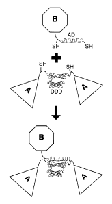
[0012] FIG. 1. Cartoon illustration of the DNL method. Triangles depict
component A, which forms a homodimer (a2) mediated by the dimerization and
docking
domain (DDD). The location of free thiol groups (SH) of the engineered
cysteine residues is
indicated. Octagons depict component B containing an anchor domain (AD)
peptide. The
DNL reaction results in the generation of a covalent trimeric structure via
binding of DDD
and AD peptides and subsequent formation of disulfide bridges.
[0013] FIG. 2. Cartoon drawing of IMP362. A 20 kDa PEG (starburst),
AD2 peptide
(helix), EDANS fluorescent tag (oval) and the positions of free sulfhydryl
groups (SH) are
indicated.
[0014] FIG. 3. Cartoon drawing of IMP413. A 30 kDa PEG (starburst),
AD2 peptide
(helix), EDANS fluorescent tag (oval) and the positions of free sulfhydryl
groups (SH) are
indicated.
4b
CA 02696160 2010-02-10
WO 2009/055653 PCT/US2008/081085
[0015] FIG. 4. Analysis of roller bottle production and purification by anti-
IFNa immunoblot
and ELISA. Samples were diluted as indicated and 5 u.1 were subjected to
reducing SDS-
PAGE and immunoblot analysis with polyclonal anti-IFNa. The dilution, total
volume and
fraction analyzed of the total volume (f) for each sample is given. The amount
of protein in
each band was estimated from standards and divided by the total volume to give
the total
protein estimate. Total protein measurements determined by ELISA are also
given.
[0016] FIG. 5. Cartoon drawing of a2b-362. IFNa2b groups (pentagons), 20 kDa
PEG
(starburst), AD2 and DDD2 peptides (helices) and EDANS fluorescent tag (oval)
are
indicted.
[0017] FIG. 6. Cartoon drawing of cc2b-413. IFNa2b groups (pentagons), 30 kDa
PEG
(starburst), AD2 and DDD2 peptides (helices) and EDANS fluorescent tag (oval)
are
indicted.
[0018] FIG. 7. Dose-response curves showing in vitro growth inhibition of
Burkitt's
lymphoma (Daudi) cells after 4-days in culture in the presence of either rhIFN-
a2b standard,
IFN-a2b-DDD2 or a2b-362. MTS dye was added to the plates, which were incubated
for 3 h
before measuring the 0D490. The % of the signal obtained from untreated cells
was plotted
vs. the log of the molar concentration. The 50% effective concentration (EC50)
values were
obtained by sigmoidal fit non-linear regression using Graph Pad Prism
software.
[0019] FIG. 8. Evaluation of the pharmacokinetic properties of IFNa
constructs. Each
reagent (test and control) was administered to Swiss-Webster mice at equimolar
protein doses
as a single bolus i.v. injection of 3 jig for rhuIFN-a2a, 5 fig for
PEG1NTRONTm , 11 jig for
a2b-362, and 13 lag tor a2b-413. Serum samples were isolated at the times
indicated and the
serum concentrations of IFN-a were determined by ELISA. The pM concentration
was
plotted vs. hours post injection. Data represents the mean value from two
mice.
[0020] FIG. 9. Evaluation of the therapeutic efficacy of IFNa constructs in
mice bearing
Burkitt's lymphoma (Daudi). Eight-week-old female SCID mice were injected iv.
with 1.5 x
107 Daudi cells. Groups of 5 mice were administered PEGINTRONTm , a2b-362 and
a2b-
413 at doses of 3,500, 7,000 or 14,000 Units once per week for 4 weeks.
Therapy
commenced 1 day after the Daudi cells were transplanted. Injection times are
indicated with
arrows. Survival curves and median survival are shown for each group.
SUBSTITUTE SHEET (RULE 26)
CA 02696160 2010-02-10
WO 2009/055653 PCT/US2008/081085
[0021] FIG. 10. Evaluation of the dosing schedule for therapy of tumor-bearing
mice. Eight-
week-old female SCID mice were injected iv. with 1.5 x 10⁷ Daudi-cells. Groups
of 6-7 mice
were administered 14,000 IU of either PEGINTRONTm or a2b-413 via a s.c.
injection.
Therapy was commenced 1 day after the Daudi cells were administered to the
mice. Groups
were dosed once a week (q7dx4), once every other week (q2wkx4) or once every 3
weeks
(q3wkx4). Injection times are indicated with arrows. All the mice received 4
injections in
total. Survival curves and median survival are shown for each group.
[0022] FIG. 11. Cartoon drawing of G-CSF-413. G-CSF groups (pentagons), 30 kDa
PEG
(starburst), AD2 and DDD2 peptides (helices) and EDANS fluorescent tag (oval)
are
indicted.
[0023] FIG. 12. Cartoon drawings of h679-Fab-DDD2 (A), dimeric EPO-DDD2 (B),
which
combine to create EPO-679 (C) by the DNL method. The variable and constant
domains of
h679 Fab (ovals), AD2 and DDD2 helices, EPO groups (pentagons) and free
sulfhydryl
groups (SH) are indicated.
[0024] FIG. 13. Stimulation of cell growth by EPO-constructs. EPO-responsive
TF1 cells (1
x 104) were cultured for 72 hours in the presence of rhEPO, EPO-DDD2 or EPO-
679. The
relative viable cell density was detelmined by MTS assay. The log of the
concentration in
U/mL is plotted vs. 0D490.
[0025] FIG. 14. Cartoon drawing of EPO-413. EPO groups (pentagons), 30 IcDa
PEG
(starburst), AD2 and DDD2 peptides (helices) and EDANS fluorescent tag (oval)
are
indicted.
[0026] FIG. 15. Structure of IMP-421.
[0027] FIG. 16. Structure of mPEG2-MAL-40K.
Dock and Lock (DNL) method
[0028] The DNL method exploits specific protein/protein interactions that
occur between the
regulatory (R) subunits of cAMP-dependent protein kinase (PKA) and the
anchoring domain
(AD) of A-kinase anchoring proteins (AKAPs) (Baillie etal., FEBS Letters.
2005; 579: 3264.
Wong and Scott, Nat. Rev. Mol. Cell Biol. 2004; 5: 959). PKA, which plays a
central role in
one of the best studied signal transduction pathways triggered by the binding
of the second
6
SUBSTITUTE SHEET (RULE 26)
CA 02696160 2010-02-10
WO 2009/055653 PCT/US2008/081085
messenger cAMP to the R subunits, was first isolated from rabbit skeletal
muscle in 1968
(Walsh et al.,J. Biol. Chem. 1968;243:3763). The structure of the holoenzyme
consists of
two catalytic subunits held in an inactive form by the R subunits (Taylor, J.
Biol. Chem.
1989;264:8443). Isozymes of PKA are found with two types of R subunits (RI and
RI), and
each type has a and p isoforms (Scott, Phannacol. Ther. 1991;50:123). The R
subunits have
been isolated only as stable dimers and the dimerization domain has been shown
to consist of
the first 44 amino-terminal residues (Newlon etal., Nat. Struct. Biol.
1999;6:222). Binding
of cAMP to the R subunits leads to the release of active catalytic subunits
for a broad
spectrum of serine/threonine kinase activities, which are oriented toward
selected substrates
through the compartmentalization of PKA via its docking with AKAPs (Scott
etal., J. Biol.
Chem. 1990;265;21561)
[0029] Since the first AKAP, microtubule-associated protein-2, was
characterized in 1984
(Lohmann etal., Proc. Natl. Acad. Sci USA. 1984;81:6723), more than 50 AKAPs
that
localize to various sub-cellular sites, including plasma membrane, actin
cytoskeleton,
nucleus, mitochondria, and endoplasmic reticulum, have been identified with
diverse
structures in species ranging from yeast to humans (Wong and Scott, Nat. Rev.
Mol. Cell
Biol. 2004;5:959), The AD of AKAPs for PKA is an amphipathic helix of 14-18
residues
(Carr etal., J. Biol. Chem. 1991;266:14188). The amino acid sequences of the
AD are quite
varied among individual AKAPs, with the binding affinities reported for RII
dimers ranging
from 2 to 90 nM (Alto etal., Proc. Natl. Acad. Sci. USA. 2003;100:4445).
Interestingly,
AKAPs will only bind to dimeric R subunits. For human RIIa, the AD binds to a
hydrophobic surface formed by the 23 amino-terminal residues (Colledge and
Scott, Trends
Cell Biol. 1999; 6:216). Thus, the dimerization domain and AKAP binding domain
of human
RIIa are both located within the same N-terminal 44 amino acid sequence
(Newlon et al.,
Nat. Struct. Biol. 1999;6:222; Newlon etal., EMBO J. 2001;20:1651), which is
tenned the
DDD herein.
DDD of Human RI[Ia and AD of AKAPs as Linker Modules
[0030] We have developed a platform technology to utilize the DDD of human
RlIa and the
AD of a certain amino acid sequence as an excellent pair of linker modules for
docking any
two entities, referred to hereafter as A and B, into a noncovalent complex,
which could be
further locked into a stably tethered structure through the introduction of
cysteine residues
7
SUBSTITUTE SHEET (RULE 26)
CA 02696160 2010-02-10
WO 2009/055653 PCT/US2008/081085
into both the DDD and AD at strategic positions to facilitate the formation of
disulfide bonds,
as illustrated in Fig.1 . The general methodology of the "dock-and-lock"
approach is as
follows. Entity A is constructed by linking a DDD sequence to a precursor of
A, resulting in a
first component hereafter referred to as a. Because the DDD sequence would
effect the
spontaneous formation of a dimer, A would thus be composed of a2. Entity B is
constructed
by linking an AD sequence to a precursor of B, resulting in a second component
hereafter
referred to as b. The dimeric motif of DDD contained in a2 will create a
docking site for
binding to the AD sequence contained in b, thus facilitating a ready
association of a2 and b to
form a binary, trimeric complex composed of a2b. This binding event is made
irreversible
with a subsequent reaction to covalently secure the two entities via disulfide
bridges, which
occurs very efficiently based on the principle of effective local
concentration because the
initial binding interactions should bring the reactive thiol groups placed
onto both the DDD
and AD into proximity (Chimura et al., Proc. Natl. Acad. Sci. USA.
2001;98:8480) to ligate
site-specifically.
[0031] By attaching the DDD and AD away from the functional groups of the two
precursors, such site-specific ligations are also expected to preserve the
original activities of
the two precursors. This approach is modular in nature and potentially can be
applied to link.
site-specifically and covalently, a wide range of substances, including
peptides, proteins,
nucleic acids, and PEG. The DNL method was disclosed in U.S. provisional
patent
applications 60/728,292, filed October 20, 2005; 60/751,196, filed December
16, 2005; and
60/782,332, filed March 14, 2006; and U.S. patent applications 11/389,358,
filed March 24,
2006; 11/391,584, filed March 28, 2006; 11/478,021, filed June 29, 2006;
11/633,729, filed
December 5, 2006 and 11/925,408, filed October 26, 2007.
[00321 In preferred embodiments. as illustrated in the Examples below, the
effector moiety to
be PEGylated is a protein or peptide, which can be linked to a DDD or AD unit
to form a
fusion protein or peptide. A variety of methods are known for making fusion
proteins,
including nucleic acid synthesis, hybridization and/or amplification to
produce a synthetic
double-stranded nucleic acid encoding a fusion protein of interest. Such
double-stranded
nucleic acids may be inserted into expression vectors for fusion protein
production by
standard molecular biology techniques (see, e.g. Sambrook et al., Molecular
Cloning, A
laboratory manual, 2nd Ed, 1989). In such preferred embodiments, the AD and/or
DDD moiety
may be attached to either the N-terminal or C-terminal end of an effector
protein or peptide.
8
SUBSTITUTE SHEET (RULE 26)
CA 02696160 2010-02-10
WO 2009/055653 PCT/US2008/081085
However, the skilled artisan will realize that the site of attachment of an AD
or DDD moiety to
an effector moiety may vary, depending on the chemical nature of the effector
moiety and the
part(s) of the effector moiety involved in its physiological activity. Site-
specific attachment of a
variety of effector moieties may be performed using techniques known in the
art, such as the use
of bivalent cross-linking reagents and/or other chemical conjugation
techniques.
PEGylation by DNL
[0033] In a preferred method, the target to be PEGylated is linked to a DDD
sequence to
generate the DDD module. A PEG reagent of a desirable molecular size is
derivatized with a
related AD sequence and the resulting PEG-AD module is combined with the DDD
module
to produce the PEGylated conjugate that consists of a single PEG tethered site-
specifically to
two copies of the effector moiety via the disulfide bonds formed between DDD
and AD. The
PEG reagents may be capped at one end with a methoxy group (m-PEG), can be
linear or
branched, and may contain one of the following functional groups: propionic
aldehyde,
butyric aldehyde, ortho-pyridylthioester (OPTE). N-hydroxysuccinimide (NHS),
thiazolidine-
2-thione, succinimidyl carbonate (SC), maleimide, or ortho-pyridyldisulfide
(OPPS). Among
the effector moieties that may be of interest for PEGylation are enzymes,
cytokines,
chemokines, growth factors, peptides, aptamers, hemoglobins, antibodies and
antibody
fragments. The method is not limiting and a wide variety of agents may be
PEGylated using
the disclosed methods and compositions. PEG of various sizes and derivatized
with a variety
of reactive moieties may be obtained from commercial sources as discussed in
more detail in
the Examples below.
Cytokines and Other Immunomodulators
[0034] In certain preferred embodiments, the effector moiety to be PEGylated
is an
immunomodulator. An immunomodulator is an agent that when present, alters,
suppresses or
stimulates the body's immune system. Immunomodulators of use may include a
cytokine, a
stem cell growth factor, a lymphotoxin, a hematopoietic factor, a colony
stimulating factor
(CSF), an interferon (IFN), erythropoietin, thrombopoietin and a combination
thereof.
Specifically useful are lymphotoxins such as tumor necrosis factor (TNF),
hematopoietic
factors, such as interleukin (IL), colony stimulating factor, such as
granulocyte-colony
stimulating factor (G-CSF) or granulocyte macrophage-colony stimulating factor
(GM-CSF),
9
SUBSTITUTE SHEET (RULE 26)
CA 02696160 2010-02-10
WO 2009/055653 PCT/US2008/081085
interferon, such as interferons-a, -13 or -7, and stem cell growth factor,
such as that designated
"S1 factor".
[0035] In more preferred embodiments, the effector moieties to be PEGylated
are cytokines,
such as lymphokines, monokines, growth factors and traditional polypeptide
hormones.
Included among the cytokines are growth hormones such as human growth hormone,
N-
methionyl human growth hointone, and bovine growth hormone; parathyroid
hormone;
thyroxine; insulin; proinsulin; relaxin; prorelaxin; glycoprotein hormones
such as follicle
stimulating hormone (FSH), thyroid stimulating hormone (TSH), and luteinizing
hormone
(LH); hepatic growth factor; prostaglandin, fibroblast growth factor;
prolactin; placental
lactogen, OB protein; tumor necrosis factor-a and -13; mullerian-inhibiting
substance; mouse
gonadotropin-associated peptide; inhibin; activin; vascular endothelial growth
factor;
integrin; thrombopoietin (TP0); nerve growth factors such as NGF-13; platelet-
growth factor;
transforming growth factors (TGFs) such as TGF- a and TGF- 13; insulin-like
growth factor-I
and -II; erythropoietin (EPO); osteoinductive factors; interferons such as
interferon-a., -13, and
-7; colony stimulating factors (CSFs) such as macrophage-CSF (M-CSF);
interleukins (ILs)
such as IL-1, IL-la, IL-2, IL-3, IL-4, IL-5, IL-6, IL-7, IL-8, IL-9, IL-10, IL-
11, IL-12; IL-13,
IL-14, IL-15, IL-16, IL-17, IL-18, IL-21, IL-25, LIF, kit-ligand or FLT-3,
angiostatin,
thrombospondin, endostatin, tumor necrosis factor (TNF, such as INF-a) and LT.
The
PEGylation of exemplary cytokines is described below in Examples 2 through 12.
[0036] The amino acid sequences of protein or peptide immunomodulators, such
as
cytokines, are well known in the art and any such known sequences may be used
in the
practice of the instant invention. The skilled artisan is aware of numerous
sources of public
information on cytokine sequence. For example, the NCBI database contains both
protein
and encoding nucleic acid sequences for a large number of cytokines and
immunomodulators,
such as erythropoietin (GenBank NM 000799), IL-1 beta (GenPept AAH08678), GM-
CSF
(GenPept AAA52578), TNF-a (GenPept CAA26669) and virtually any of the peptide
or
protein inununomodulators listed above. It is a matter of routine for the
skilled artisan to
identify an appropriate amino acid and/or nucleic acid sequence for
essentially any protein or
peptide effector moiety of interest.
Antibodies and Antibody Fragments
[0037] In other embodiments, antibodies or antigen-binding fragments of
antibodies may be
PEGylated. Antigen-binding antibody fragments are well known in the art, such
as F(abr)2,
SUBSTITUTE SHEET (RULE 26)
CA 02696160 2015-04-27
52392-75
F(ab)2, Fab', Fab, Fv, scFv and the like, and any such known fragment may be
used. As used
herein, an antigen-binding antibody fragment refers to any fragment of an
antibody that binds
with the same antigen that is recognized by the intact or parent antibody.
Techniques for
preparing AD and/or DDD conjugates of virtually any antibody or fragment of
interest are
known (e.g., U.S. Patent Application Serial No, 11/633,729).
100381 An antibody or fragment thereof may be used which is not conjugated to
a therapeutic
agent ¨ referred to as a "naked" antibody or fragment thereof. In alternative
embodiments,
antibodies or fragments may be conjugated to one or more therapeutic and/or
diagnostic
agents. A wide variety of such therapeutic and diagnostic agents are known in
the art, as
discussed in more detail below, and any such known therapeutic or diagnostic
agent may be
used.
100391 Techniques for preparing monoclonal antibodies against virtually any
target antigen
are well known in the art. See, for example, Kohler and Milstein, Nature 256:
495 (1975),
and Coligan etal. (eds.), CURRENT PROTOCOLS IN IMMUNOLOGY, VOL. 1, pages
2.5.1-2.6.7 (John Wiley & Sons 1991). Briefly, monoclonal antibodies can be
obtained by
injecting mice with a composition comprising an antigen, removing the spleen
to obtain B-
lymphocytes, fusing the B-lymphocytes with myeloma cells to produce
hybridomas, cloning
the hybridomas, selecting positive clones which produce antibodies to the
antigen, culturing
the clones that produce antibodies to the antigen, and isolating the
antibodies from the
hybridoma cultures.
[0040] MAbs can be isolated and purified from hybridoma cultures by a variety
of well-
established techniques. Such isolation techniques include affinity
chromatography with
Protein-A Sepharosem, size-exclusion chromatography, and ion-exchange
chromatography.
See, for example, Coligan at pages 2.7.1-2.7.12 and pages 2.9.1-2.9.3. Also,
see Baines et
al., "Purification of Immunoglobulin G (IgG)," in METHODS IN MOLECULAR
BIOLOGY, VOL. 10, pages 79-104 (The Humana Press, Inc. 1992).
[0041] After the initial raising of antibodies to the immunogen, the
antibodies can be
sequenced and subsequently prepared by recombinant techniques. Humanization
and
chimerization of murine antibodies and antibody fragments are well known to
those skilled in
the art. The use of antibody components derived from humanized, chimeric or
human
antibodies obviates potential problems associated with the immunogenicity of
murine constant
regions.
Chimeric Antibodies
11
CA 02696160 2010-02-10
WO 2009/055653 PCT/US2008/081085
[0042] A chimeric antibody is a recombinant protein in which the variable
regions of a
human antibody have been replaced by the variable regions of, for example, a
mouse
antibody, including the complementarity-determining regions (CDRs) of the
mouse antibody.
Chimeric antibodies exhibit decreased immunogenicity and increased stability
when
administered to a subject. General techniques for cloning murine
immunoglobulin variable
domains are disclosed, for example, in Orlandi et al., Proc. Nat'l Acad. Sci,
USA 86: 3833
(1989). Techniques for constructing chimeric antibodies are well known to
those of skill in
the art. As an example, Leung et al., Hybridoma /3:469 (1994), produced an LL2
chimera
by combining DNA sequences encoding the Võ and VH domains of murine LL2, an
anti-
CD22 monoclonal antibody, with respective human lc and IgGi constant region
domains.
Humanized Antibodies
[0043] Techniques for producing humanized MAbs are well known in the art (see,
e.g., Jones
et al., Nature 321: 522 (1986), Riechrnann et al., Nature 332: 323 (1988),
Verhoeyen et al.,
Science 239: 1534 (1988), Carter etal., Proc. Nat'l Acad Sci. USA 89: 4285
(1992), Sandhu,
Crit. Rev. Biotech. 12: 437 (1992), and Singer et al., J. Immun. 150: 2844
(1993)). A
chimeric or murine monoclonal antibody may be humanized by transferring the
mouse CDRs
from the heavy and light variable chains of the mouse immunoglobulin into the
corresponding variable domains of a human antibody. The mouse framework
regions (FR) in
the chimeric monoclonal antibody are also replaced with human FR sequences. As
simply
transferring mouse CDRs into human FRs often results in a reduction or even
loss of antibody
affinity, additional modification might be required in order to restore the
original affinity of the
murine antibody. This can be accomplished by the replacement of one or more
some human
residues in the FR regions with their murine counterparts to obtain an
antibody that possesses
good binding affinity to its epitope. See, for example, Tempest et al.,
Biotechnology 9:266
(1991) and Verhoeyen etal., Science 239: 1534 (1988), Generally, those human
FR amino
acid residues that differ from their murine counterparts and are located close
to or touching
one or more CDR amino acid residues would be candidates for substitution.
Human Antibodies
[0044] Methods for producing fully human antibodies using either combinatorial
approaches
or transgenic animals transformed with human immunoglobulin loci are known in
the art
(e.g., Mancini et al., 2004, New illicrobiol. 27:315-28; Conrad and Scheller,
2005, Comb.
Chem. High Throughput Screen. 8:117-26; Brekke and Loset, 2003, Curr. Opin.
Phamacol.
3:544-50). A fully human antibody also can be constructed by genetic or
chromosomal
12
SUBSTITUTE SHEET (RULE 26)
CA 02696160 2015-04-27
52392-75
transfection methods, as well as phage display technology, all of which are
known in the art.
See for example, McCafferty et al., Nature 348:552-553 (1990). Such fully
human
antibodies are expected to exhibit even fewer side effects than chimeric or
humanized
antibodies and to function in vivo as essentially endogenous human antibodies.
In certain
embodiments, the claimed methods and procedures may utilize human antibodies
produced
by such techniques.
[0045] In one alternative, the phage display technique may be used to generate
human
antibodies (e.g., Dantas-Barbosa et al., 2005, Genet. Mol. Res. 4:126-40).
Human antibodies
may be generated from normal humans or from humans that exhibit a particular
disease state,
such as cancer (Dantas-Barbosa et al., 2005). The advantage to constructing
human
antibodies from a diseased individual is that the circulating antibody
repertoire may be biased
towards antibodies against disease-associated antigens.
[0046] In one non-limiting example of this methodology, Dantas-Barbosa et al.
(2005)
constructed a phage display library of human Fab antibody fragments from
osteosarcoma
patients. Generally, total RNA was obtained from circulating blood lymphocytes
(Id.).
Recombinant Fab were cloned frnm the , 7 and lc chain antibody repertoires
and inserted
into a phage display library (Id.). RNAs were converted to cDNAs and used to
make Fab
cDNA libraries using specific primers against the heavy and light chain
immunoglobulin
sequences (Marks et al., 1991, Mol. Biol. 222:581-97). Library construction
was
performed according to Andris-Widhopf et al. (2000, In: Phage Display
Laboratory Manual,
Barbas et al. (eds), 1' edition, Cold Spring Harbor Laboratory Press, Cold
Spring Harbor, NY
pp. 9.1 to 9.22). The final Fab fragments were digested with restriction
endonucleases and
inserted into the bacteriophage genome to make the phage display library. Such
libraries may
be screened by standard phage display methods, as known in the art.
[0047] Phage display can be performed in a variety of formats, for their
review, see e.g.
Johnson and Chiswell, Current Opinion in Structural Biology 3:5564-571 (1993).
Human
antibodies may also be generated by in vitro activated B-cells. See U.S.
Patent Nos.
5,567,610 and 5,229,275. The skilled
artisan will realize that these techniques are exemplary and any known method
for making
and screening human antibodies or antibody fragments may be utilized.
[0048] In another alternative, transgenic animals that have been genetically
engineered to
produce human antibodies may be used to generate antibodies against
essentially any
immunogenic target, using standard immunization protocols. Methods for
obtaining human
13
CA 02696160 2010-02-10
WO 2009/055653
PCT/US2008/081085
antibodies from transgenic mice are disclosed by Green et al, Nature Genet.
7:13 (1994),
Lonberg et al., Nature 368:856 (1994), and Taylor et al., Int. Itnmun. 6:579
(1994). A non-
limiting example of such a system is the XenoMouse (e.g., Green et al., 1999,
J. Inzmunol.
Methods 231:11-23) from Abgenix (Fremont, CA). In the XenoMouse and similar
animals,
the mouse antibody genes have been inactivated and replaced by functional
human antibody
genes, while the remainder of the mouse immune system remains intact.
[0049] The XenoMouse was transformed with germline-configured YACs (yeast
artificial
chromosomes) that contained portions of the human IgH and lgkappa loci,
including the
majority of the variable region sequences, along accessory genes and
regulatory sequences.
The human variable region repertoire may be used to generate antibody
producing B-cells,
which may be processed into hybridomas by known techniques. A XenoMouse
immunized
with a target antigen will produce human antibodies by the normal immune
response, which
may be harvested and/or produced by standard techniques discussed above. A
variety of
strains of XenoMouse are available, each of which is capable of producing a
different class
of antibody. Transgenically produced human antibodies have been shown to have
therapeutic
potential, while retaining the pharmacokinetic properties of normal human
antibodies (Green
et at., 1999). The skilled artisan will realize that the claimed compositions
and methods are
not limited to use of the XenoMouse system but may utilize any transgenic
animal that has
been genetically engineered to produce human antibodies.
Antibody Fragments
[0050] Antibody fragments which recognize specific epitopes can be generated
by known
techniques. Antibody fragments are antigen binding portions of an antibody,
such as F(alS)2,
Fab', F(ab)-,, Fab, Fv, slTv and the like. F(ab')2fragments can be produced by
pepsin digestion
of the antibody molecule and Fab' fragments can be generated by reducing
disulfide bridges
of the F(ab')2fragments. Alternatively, Fab' expression libraries can be
constructed (Huse et
al., 1989, Science, 246:1274-1281) to allow rapid and easy identification of
monoclonal Fab'
fragments with the desired specificity. F(ab)2
fragments may be generated by papain
digestion of an antibody and Fab fragments obtained by disulfide reduction.
[0051] A single chain Fv molecule (scFv) comprises a VL domain and a VH
domain. The
VL and VH domains associate to form a target binding site. These two domains
are further
covalently linked by a peptide linker (L). Methods for making scFv molecules
and designing
suitable peptide linkers are described in US Patent No. 4,704,692, US Patent
No. 4,946,778,
R. Raag and M. Whitlow, "Single Chain Fvs." FASEB Vol 9:73-80 (1995) and R.E.
Bird and
B.W. Walker, "Single Chain Antibody Variable Regions," TIBTECH, Vol 9: 132-137
(1991).
14
SUBSTITUTE SHEET (RULE 26)
CA 02696160 2010-02-10
WO 2009/055653 PCT/US2008/081085
[0052] An antibody fragment can be prepared by proteolytic hydrolysis of the
full length
antibody or by expression in E. coli or another host of the DNA coding for the
fragment. An
antibody fragment can be obtained by pepsin or papain digestion of full length
antibodies by
conventional methods. These methods are described, for example, by Goldenberg,
U.S.
Patent Nos. 4,036,945 and 4,331,647 and references contained therein. Also,
see Nisonoff et
al., Arch Biochem. Biophy.s. 89: 230 (1960); Porter, Biochem. J. 73: 119
(1959), Edelman et
al., in METHODS IN ENZYMOLOGY VOL. 1, page 422 (Academic Press 1967), and
Coligan at pages 2.8.1-2.8.10 and 2.10.-2.10.4.
Known Antibodies
[0053] Antibodies of use may be commercially obtained from a wide variety of
known
sources. For example, a variety of antibody secreting hybridoma lines are
available from the
American Type Culture Collection (ATCC, Manassas, VA). A large number of
antibodies
against various disease targets, including but not limited to tumor-associated
antigens, have
been deposited at the ATCC and/or have published variable region sequences and
are
available for use in the claimed methods and compositions. See, e.g., U.S.
Patent Nos.
7,312,318; 7,282,567; 7,151,164; 7,074,403; 7,060,802; 7,056,509; 7,049,060;
7,045,132;
7,041,803; 7,041,802; 7,041,293; 7,038,018; 7,037,498; 7,012,133; 7,001,598;
6,998,468:
6,994,976; 6,994,852; 6,989,241; 6,974,863; 6,965,018; 6,964,854; 6,962,981;
6,962,813;
6,956,107; 6,951,924; 6,949,244; 6,946,129; 6,943,020; 6,939,547; 6,921,645;
6,921,645;
6,921,533; 6,919,433; 6,919,078; 6,916,475; 6,905,681; 6,899,879; 6,893,625;
6,887,468;
6,887,466; 6,884,594; 6,881,405; 6,878,812; 6,875,580; 6,872,568; 6,867,006;
6,864,062;
6,861,511; 6,861,227; 6,861,226; 6,838,282; 6,835,549; 6,835,370; 6,824,780;
6,824,778;
6,812,206; 6,793,924; 6,783,758; 6,770,450; 6,767,711; 6,764,688; 6,764,681;
6,764,679;
6,743,898; 6,733,981; 6,730,307; 6,720,15; 6,716,966; 6,709,653; 6,693,176;
6,692,908;
6,689,607; 6,689,362; 6,689,355; 6,682,737; 6,682,736; 6,682,734; 6,673,344;
6,653,104;
6,652,852; 6,635,482; 6,630,144; 6,610,833; 6,610,294; 6,605,441; 6,605,279;
6,596,852;
6,592,868; 6,576,745; 6,572;856; 6,566,076; 6,562,618; 6,545,130; 6,544,749;
6,534,058;
6,528,625; 6,528,269; 6,521,227; 6,518,404; 6,511,665; 6,491,915; 6,488,930;
6,482,598;
6,482,408; 6,479,247; 6,468,531; 6,468,529; 6,465,173; 6,461,823; 6,458,356;
6,455,044;
6,455,040, 6,451,310; 6,444,206' 6,441,143; 6,432,404; 6,432,402; 6,419,928;
6,413,726;
6,406,694; 6,403,770; 6,403,091; 6,395,276; 6,395,274; 6,387,350; 6,383,759;
6,383,484;
6,376,654; 6,372,215; 6,359,126; 6,355,481; 6,355,444; 6,355,245; 6,355,244;
6,346,246;
6,344,198; 6,340,571; 6,340,459; 6,331,175; 6,306,393; 6,254,868; 6,187,287;
6,183,744;
SUBSTITUTE SHEET (RULE 26)
CA 02696160 2010-02-10
WO 2009/055653 PCT/US2008/081085
6,129,914; 6,120,767; 6,096,289; 6,077,499; 5,922,302; 5,874,540; 5,814,440;
5,798,229;
5,789,554; 5,776,456; 5,736,119; 5,716,595; 5,677,136; 5,587,459; 5,443,953,
5,525,338.
These are exemplary only and a wide variety of other antibodies and their
hybridomas are
known in the art. The skilled artisan will realize that antibody sequences or
antibody-
secreting hybridomas against almost any disease-associated antigen may be
obtained by a
simple search of the ATCC, NCBI and/or USPTO databases for antibodies against
a selected
disease-associated target of interest. The antigen binding domains of the
cloned antibodies
may be amplified, excised, ligated into an expression vector, transfected into
an adapted host
cell and used for protein production, using standard techniques well known in
the art.
Therapeutic Agents
[0054] In alternative embodiments, therapeutic agents such as cytotoxic
agents, anti-
angiogenic agents, pro-apoptotic agents, antibiotics, hormones, hormone
antagonists,
chemokines, drugs, prodrugs, toxins, enzymes or other agents may be PEGylated
as described
herein. PEGylated forms of therapeutic agents have been disclosed, for
example, for SN38
(Zhao et al., Bioconjug Chem 2008, 10:849-59), unease (Biggers & Scheinfeld,
Cuff Opin
Investig Drugs 2008, 9:422-29), docetaxel (Liu et al., 2008, J Pharm Sci
97:3274-90) and
camptothecin (Haverstick et al., 2007, Bioconjug Chem 18:2115-21). Drugs of
use may
possess a pharmaceutical property selected from the group consisting of
antimitotic, antikinase,
alkylating, antimetabolite, antibiotic, alkaloid, anti-angiogenic, pro-
apoptotic agents and
combinations thereof
[0055] Exemplary drugs of use may include 5-fluorouracil, aplidin, azaribine,
anastrozole,
anthracyclines, bendamustine, bleomycin, bortezomib, bryostatin-1, busulfan,
calicheamycin,
camptothecin, carboplatin, 10-hydroxycamptothecin, carmustine, celebrex,
chlorambucil,
cisplatin (CDDP), Cox-2 inhibitors, irinotecan (CPT-11), SN-38, carboplatin,
cladribine,
camptothecans, cyclophosphamide, cytarabine, dacarbazine, docetaxel,
dactinomycin,
daunorubicin, doxorubicin, 2-pyrrolinodoxorubicine (2P-DOX), cyano-morpholino
doxorubicin, doxorubicin glucuronide, epirubicin glucuronide, estramustine,
epidophyllotoxin, estrogen receptor binding agents, etoposide (VP16),
etoposide glucuronide,
etoposide phosphate, floxuridine (FUdR), 3',51-0-dioleoyl-FudR (ITUdR-d0),
fludarabine,
flutamide, famesyl-protein transferase inhibitors, gemcitabine, hydroxyurea,
idarubicin,
ifosfamide, L-asparaginase, lenolidamide, leucovorin, lomustine,
mechlorethamine,
melphalan, mercaptopurine, 6-mercaptopurine, methotrexate, mitoxantrone,
mithramycin,
mitomycin, mitotane, navelbine, nitrosurea, plicomycin, procarbazine,
paclitaxel, pentostatin,
16
SUBSTITUTE SHEET (RULE 26)
CA 02696160 2010-02-10
WO 2009/055653 PCT/US2008/081085
PSI-341, raloxifene, semustine, streptozocin, tamoxifen, taxol, temazolomide
(an aqueous
form of DTIC), transplatinum, thalidomide, thioguanine, thiotepa, teniposide,
topotecan,
uracil mustard, vinorelbine, vinblastine, vincristine and vinca alkaloids.
[00561 Toxins of use may include ricin, abrin, alpha toxin, saporin,
ribonuclease (RNase),
e.g., onconase, DNase I, Staphylococcal enterotoxin-A, pokeweed antiviral
protein, gelonin,
diphtheria toxin, Pseudomonas exotoxin, and Pseudomonas endotoxin.
[0057] Chemokines of use may include RANTES, MCAF, MIP1-alpha, MIP1-Beta and
IP-10.
[0058] In certain embodiments, anti-angiogenic agents, such as angiostatin,
baculostatin,
canstatin, maspin, anti-VEGF antibodies, anti-P1GF peptides and antibodies,
anti-vascular
growth factor antibodies, anti-Flk-1 antibodies, anti-Fit-1 antibodies and
peptides, anti-Kras
antibodies, anti-cMET antibodies, anti-MU' (macrophage migration-inhibitory
factor)
antibodies, laminin peptides, fibronectin peptides, plasminogen activator
inhibitors, tissue
metalloproteinase inhibitors, interferons, interleukin-12, IP-10, Gro-13,
thrombospondin, 2-
methoxyoestradiol, proliferin-rclated protein, carboxiamidotriazole, CM101,
Marimastat,
pentosan polysulphate, angiopoietin-2, interferon-alpha, herbimycin A,
PNU145156E, 16K
prolactin fragment, Linomide, thalidomide, pentoxifylline, genistein, TNP-470,
endostatin,
paclitaxel, accutin, angiostatin, cidofovir, vincristine, bleomycin, AGM-1470,
platelet factor
4 or minocyclinc may be of use.
[0059] Other useful therapeutic agents may comprise oligonucleotides,
especially antisense
oligonucleotides that preferably are directed against oncogenes and oncogene
products, such
as bc1-2 or p53.
Conjugation
[0060] A variety of techniques and compositions for conjugation of small
molecules to
proteins or peptides, such as AD or DDD peptides, are known and may be used.
Therapeutic
agents can be attached, for example to reduced SH groups and/or to
carbohydrate side chains.
A therapeutic agent can be attached to a reduced protein or peptide comprising
a cysteine
residue via disulfide bond formation. Alternatively, such agents can be
attached using a
heterobifunctional cross-linker, such as N-succinyl 3-(2-
pyridyldithio)propionate (SPDP).
Yu et al., Int. ,I. Cancer 56: 244 (1994). General techniques for such
conjugation are well-
known in the art. See, for example, Wong, CHEMISTRY OF PROTEIN CONJUGATION
AND CROSS-LINKING (CRC Press 1991); Upeslacis et al., "Modification of
Antibodies by
Chemical Methods," in MONOCLONAL ANTIBODIES: PRINCIPLES AND
17
SUBSTITUTE SHEET (RULE 26)
CA 02696160 2010-02-10
WO 2009/055653 PCT/US2008/081085
APPLICATIONS, Birch et at. (eds.), pages 187-230 (Wiley-Liss, Inc. 1995);
Price,
"Production and Characterization of Synthetic Peptide-Derived Antibodies," in
MONOCLONAL ANTIBODIES: PRODUCTION, ENGINEERING AND CLINICAL
APPLICATION, Ritter et al. (eds.), pages 60-84 (Cambridge University Press
1995).
Therapeutic Use
[0061] The compositions described herein are particularly useful for treatment
of various
disease states. In preferred embodiments, the diseases may be autoimmune
diseases or
cancer, such as hematopoietic cancers or solid tumors. Exemplary non-limiting
diseases that
may be treated using the disclosed compositions and methods include indolent
forms of B-
cell lymphomas, aggressive forms of B-cell lymphomas, non-Hodgkin's lymphoma,
multiple
myeloma, chronic lymphatic leukemias, acute lymphatic leukemias, acute
myelogenous
leukemia, chronic lymphocytic leukemia, chronic myelogenous leukemia,
Hodgkin's
lymphoma, Waldenstrom's macroglobulinemia, as well as GVHD, cryoglobulinemia,
hemolytic anemia, allosensitization, and organ transplant rejection. Also
included are class
III autoimmune diseases such as immune-mediated thrombocytopenias, such as
acute
idiopathic thrombocytopenic purpura and chronic idiopathic thrombocytopenic
purpura,
deimatomyositis, Sjogren's syndrome, multiple sclerosis, Sydenham's chorea,
myasthenia
gravis, systemic lupus erythematosus, lupus nephritis, rheumatic fever,
rheumatoid arthritis,
polyglandular syndromes, bullous pemphigoid, diabetes mellitus, Henoch-
Schonlein purpura,
post-streptococcal nephritis, erythema nodosum, Takayasu's arteritis,
Addison's disease,
sarcoidosis, ulcerative colitis, erythema multiforme, IgA nephropathy,
polyarteritis nodosa,
ankylosing spondylitis, Goodpasture's syndrome, thromboangitis obliterans,
primary biliary
cirrhosis, Hashimoto's thyroiditis, thyrotoxicosis, scleroderma, chronic
active hepatitis,
polymyositis/dermatomyositis, polychondritis, pemphigus vulgaris, Wegener's
granulomatosis, membranous nephropathy, amyotrophic lateral sclerosis, tabes
dorsalis, giant
cell arteritis/polymyalgia, pernicious anemia, rapidly progressive
glomerulonephritis and
fibrosing alveolitis.
[0062] Solid tumors that may be treated include neuroblastoma, malignant
melanoma, breast,
ovarian, cervical, uterine, endometrial, prostatic, lung, kidney, colorectal,
gastric, bladder,
glioma, sarcoma, brain, esophageal, epithelial, osteosarcoma, testicular,
liver and pancreatic
cancers.
18
SUBSTITUTE SHEET (RULE 26)
CA 02696160 2010-02-10
WO 2009/055653 PCT/US2008/081085
Kits
[0063] Various embodiments may concern kits containing components suitable for
treating
or diagnosing diseased tissue in a patient. Exemplary kits may contain at
least PEGylated
therapeutic agent as described herein. If the composition containing
components for
administration is not formulated for delivery via the alimentary canal, such
as by oral
delivery, a device capable of delivering the kit components through some other
route may be
included. One type of device, for applications such as parenteral delivery, is
a syringe that is
used to inject the composition into the body of a subject. Inhalation devices
may also be used.
In certain embodiments, a PEGylated therapeutic agent may be provided in the
form of a
prefilled syringe or autoinjection pen containing a sterile, liquid
formulation or lyophilized
preparation.
[0064] The kit components may be packaged together or separated into two or
more
containers. In some embodiments, the containers may be vials that contain
sterile,
lyophilized formulations of a composition that are suitable for
reconstitution. A kit may also
contain one or more buffers suitable for reconstitution and/or dilution of
other reagents. Other
containers that may be used include, but are not limited to, a pouch, tray,
box, tube, or the
like. Kit components may be packaged and maintained sterilely within the
containers.
Another component that can be included is instructions to a person using a kit
for its use.
EXAMPLES
[0065] The following examples are provided to illustrate, but not to limit,
the claims of the
present invention.
Example 1. Generation of PEG-AD2 modules
Synthesis of IMP35 0
CGQIEYLAKQIVDNAIQQAGC(SS-tbu)-NH2 (SEQ ID NO:1) MH+2354
[0066] IMP350, incorporating the sequence of AD2, was made on a 0.1 rnmol
scale with
Sieber Amide resin using Fmoe methodology on a Protein Technologies PS3
peptide
synthesizer. Starting from the C-terminus the protected amino acids used were
Fmoc-Cys(t-
Buthio)-0H, Fmoc-Gly-OH, Fmoc-Ala-OH, Fmoc-Gln(Trt)-0H, Fmoc-Gln(Trt)-0H, Fmoc-
Ile-OH, Fmoc-Ala-01-1, Fmoc-Asn(Trt)-0H, Fmoc-Asp(OBut)-0H, Fmoc-Val-OH, Fmoc-
19
SUBSTITUTE SHEET (RULE 26)
CA 02696160 2010-02-10
WO 2009/055653 PCT/US2008/081085
Ile-OH, Fmoc-Gln(Trt)-0II, Fmoc-Lys(Boc)-0H, Fmoc-Ala-OH, Fmoc-Leu-OH, Fmoc-
Tyr(But)-0H, Fmoc-Glu(0But)-0H, Fmoc-Ile-OH, Fmoc-Gln(Trt)-0H, Fmoc-Gly-OH and
Fmoe-Cys(Trt)-0H. The peptide was cleaved from the resin and purified by
reverse phase
(RP)-HPLC.
Synthesis of PEG20-IMP350
[0067] IMP350 (0.0104 g) was mixed with 0.1022 g of mPEG-OPTE (20kDa, Nektar
Therapeutics) in 7 mL of 1 M Tris buffer at pH 7.81. Acetonitrile, 1 mL, was
then added to
dissolve some suspended material. The reaction was stirred at room temperature
for 3 h and
then 0.0527 g of TCEP was added along with 0.0549 g of cysteine. The reaction
mixture was
stirred for 1.5 h and then purified on a PD-10 desalting column, which was
equilibrated with
20% methanol in water. The sample was eluted, frozen and lyophilized to obtain
0.0924 g of
crude PEG20-IMP350 (MH+ 23508 by MALDI).
Synthesis of IMP360
CGQIEYLAKQIVDNAIQQAGC(SS-tbu)G-EDANS (SEQ ID NO:1) WI+ 2660
[0068] IMP 360, incorporating the AD2 sequence, was synthesized on a 0.1 mmol
scale with
Fmoc-Gly-EDANS resin using Fmoc methodology on a Protein Technologies PS3
peptide
synthesizer. The Fmoc-Gly-OH was added to the resin manually using 0.23 g of
Fmoc-Gly-
OI I, 0.29 g of HATU, 26 jaL of DIEA, 7.5 mL of DMF and 0.57 g of EDANS resin
(Nova
Biochem). The reagents were mixed and added to the resin. The reaction was
mixed at room
temperature for 2.5 hr and the resin was washed with DMF and IPA to remove the
excess
reagents. Starting from the C-terminus the protected amino acids used were
Fmoc-Cys(t-
Buthio)-0H, Fmoc-Gly-OH, Fmoc-Ala-OH, Fmoc-Gln(Trt)-0II, Fmoc-Gln(Trt)-0H,
Fmoc-
Ile-OH, Fmoc-Ala-OH, Fmoc-Asn(Trt)-0H, Fmoc-Asp(OBut)-0H, Fmoc-Val-OH, Fmoc-
Fmoc-Gln(Trt)-0H, Fmoc-Lys(Boc)-0H, Fmoc-Ala-OH, Fmoc-Leu-OH, Fmoc-
Tyr(But)-0H, Fmoc-Glu(0But)-0H, Fmoc-Ile-OH, Fmoc-Gln(Trt)-0H, Fmoc-Gly-OH and
Fmoc-Cys(Trt)-0H. The peptide was cleaved from the resin and purified by RP-
HPLC.
Synthesis of IMP362 (PEG20-IMP360)
[0069] A cartoon diagram of IMP362 is provided in FIG. 2. For synthesis of
IMP362,
IMP360 (0.0115 g) was mixed with 0.1272 g of mPEG-OPTE (20kDa, Nektar
Therapeutics)
SUBSTITUTE SHEET (RULE 26)
CA 02696160 2010-02-10
WO 2009/055653 PCT/US2008/081085
in 7 mL of 1 M tris buffer, pH 7.81. Acetonitrile (1 mL) was then added to
dissolve some
suspended material. The reaction was stirred at room temperature for 4 h and
then 0.0410 g of
TCEP was added along with 0.0431 g of cysteine. The reaction mixture was
stirred for 1 h
and purified on a PD-10 desalting column, which was equilibrated with 20 cYc.
methanol in
water. The sample was eluted, frozen and lyophilized to obtain 0.1471 g of
crude IMP362
(MH+ 23713).
Synthesis of DIM 3 (PEG30-11IP360)
[0070] A cartoon diagram of IMP 413 is provided in FIG. 3. For synthesis of
IMP 413, IMP
360 (0.0103 g) was mixed with 0.1601 g of mPEG-OPTE (30kDa, Nektar
Therapeutics) in 7
mL of 1 M tris buffer at pH 7.81. Acetonitrile (1 mL) was then added to
dissolve some
suspended material. The reaction was stirred at room temperature for 4.5 h and
then 0.0423 g
of TCEP was added along with 0.0473 g of cysteine. The reaction mixture was
stirred for 2 h
followed by dialysis for two days. The dialyzed material was frozen and
lyophilized to obtain
0.1552 g of crude IMP413 (MH+ 34499).
Synthesis of IMP421
IMP 421 Ac-C-PEG3-C(S-tBu)GQIEYLAKQIVDNAIQQAGC(S-tBu)G-NH2 (SEQ ID
NO:9)
[0071] The peptide IMP421, MW 2891 was made on NovaSyne TGR resin (487.6 mg,
0.112 mmol) by adding the following amino acids to the resin in the order
shown: Fmoc-Gly-
OH, Fmoc-Cys(t-Buthio)-0H, Fmoc-Gly-OH, Fmoc-Ala-OH, Fmoc-Gln(Trt)-0H, Fmoc-
Gln(Trt)-0H, Fmoc-Ile-OH, Fmoc-Ala-OH, Fmoc-Asn(Trt)-0H, Fmoc-Asp(OBut)-0H,
Fmoc-Val-OH, Fmoe-Ile-OH, Fmoc-Gln(Trt)-0H, Fmoc-Lys(Boe)-OH, Fmoc-Ala-OH,
Fmoc-Leu-OH, Fmoc-Tyr(But)-0H, Fmoc-Glu(0But)-0H, Fmoc-Ile-OH, Fmoc-Gln(Trt)-
OH, Fmoe-Gly-OH, Fmoc-Cys(t-Buthio)-0H, Fmoc-NH-PEG3-COOH, Fmoc-Cys(Trt)-0H.
The N-terminal amino acid was protected as an acetyl derivative. The peptide
was then
cleaved from the resin and purified by RP-HPLC to yield 32.7 mg of a white
solid.
Synthesis of IMP457
[0072] IMP 421 (SEQ ID NO:9, FIG. 15), incorporating the sequence of AD2, was
synthesized by standard chemical means. To a solution of 15.2 mg (5.26 mop
IMP 421
21
SUBSTITUTE SHEET (RULE 26)
CA 02696160 2010-02-10
WO 2009/055653 PCT/US2008/081085
(F.W. 2890.50) and 274.5 mg (6.86 mop mPEG2-MAL-40K in 1 mL of acetonitrile
was
added 7 mL 1 M Tris pH 7.8 and allowed to react at room temperature for 3 h.
The excess
mPEG2-MAL-40K (FIG. 16) was quenched with 49.4 mg L-cysteine, followed by S-S-
tBu
dcprotection over one hour with 59.1 mg TCEP. The reaction mixture was
dialyzed overnight
at 2-8 'V using two 3-12 mL capacity 10K Slide-A-Lyzer dialysis cassettes (4
ml into each
cassette) into 5 L of 5 mM ammonium acetate, pH 5Ø Three more 5 L buffer
changes of 5
mM ammonium acetate, pH 5.0 were made the next day with each dialysis lasting
at least 21/2
h. The purified product (19.4 mL) was transferred into two 20 mL scintillation
vials, frozen
and lyophilized to yield 246.7 mg of a white solid. MALDI-TOF gave results of
mPEG2-
MAL-40K 42,982 and IMP-457 45,500.
Example 2. Generation of DDD module based on Interferon (IFN)-a2b
Construction of IFN-a2b-DDD2-pdHL2 for expression in mammalian cells
[0073] The cDNA sequence for IFN-a2b was amplified by PCR, resulting in a
sequence
comprising the following features, in which XbaI and BamHI are restriction
sites, the signal
peptide is native to IFN-a2b, and 6 His is a hexahistidine tag: XbaI---Signal
peptide--
IFNa2b ---6 His---BamHI. The resulting secreted protein consists of IFN-a2b
fused at its C-
terminus to a polypeptide consisting of SEQ ID NO:2.
KSHHHHHHGSGGGGSGGGCGIIIQIPPGLTELLOGYTVEVLRQQPPDLVEFAVEYFTR
LREARA (SEQ ID NO:2)
[0074] PCR amplification was accomplished using a full length human IFNa2b
cDNA clone
(Invitrogen Ultimate ORF human clone cat# HORFO1Clone ID 10H3 5221) as a
template and
the following oligonucleotides as primers:
IFNA2 Xba I Left
5'-TCTAGACACAGGACCTCATCATGGCCTTGACCTTTGCTTTACTGG-3' (SEQ ID
NO:3)
IFNA2 BamHI right
22
SUBSTITUTE SHEET (RULE 26)
CA 02696160 2015-04-27
52392-75
5'-
GGATCCATGATGGTGATGATGGTGTGACTTTTCCTTACTTCTTAAACTTTCTTGC-
3' (SEQ ID NO:4)
[0075] The PCR amplimer was cloned into the pGemT vector (Promega). A DDD2-
pdHL2
mammalian expression vector was prepared for ligation with IFN-a2b by
digestion with XbaI
and Barn HI restriction endonucleases. The IFN-a2b amplimer was excised from
pGemT
with XbaI and Barn HI and ligated into the DDD2-pdHL2 vector to generate the
expression
vector IFN-a2b-DDD2-pdHL2..
Mammalian expression of IFN-a2b-DDD2
[00761 1FN-a2b-DDD2-pdHL2 was linearized by digestion with SalI enzyme and
stably
transfected into Sp/BEE myeloma cells by electroporation (see. e.g., U.S.
Patent Application
Serial No. 11/487,215, filed 7/14/06). Two clones were
found to have detectable levels of IFN-a2b by ELISA. One of the two clones,
designated 95,
was adapted to growth in serum-free media without substantial decrease in
productivity. The
clone was subsequently amplified with increasing methotrexate (MTX)
concentrations from
0.1 to 0.8 iM over five weeks. At this stage, it was sub-cloned by limiting
dilution and the
highest producing sub-clone (95-5) was expanded. The productivity of 95-5
grown in shake-
flasks was estimated to be 2.5 mg/L using commercial rIFN-a2b (Chemicon IF007,
Lot
06008039084) as a standard.
Purification of IFN-a2b-DDD2 from batch cultures grown in roller bottles
[0077] Clone 95-5 was expanded to 34 roller bottles containing a total of 20 L
of serum-free
Hybridoma SFM with 0.8 M MTX and allowed to reach terminal culture. The
supernatant
fluid was clarified by centrifugation, filtered (0.2 p.M). The filtrate was
diafiltered into 1X
Binding buffer (10 mM imidazole, 0.5 M NaC1, 50 mM NaH2PO4, pH 7.5) and
concentrated
to 310 mL in preparation for purification by immobilized metal affinity
chromatography
(IMAC). The concentrate was loaded onto a 30-mL Ni-NTA column, which was
washed with
500 mL of 0.02% TweenTm 20 in IX binding buffer and then 290 mL of 30 mM
imidazole,
0.02% Tween 20, 0.5 M NaC1, 50 mM NaH2PO4, pH 7.5. The product was eluted with
110
mL of 250 mM imidazole, 0.02% Tween 20, 150 mM NaCI, 50 mM NaH2PO4, pH 7.5.
Approximately 6 mg of IFNa2b-DDD2 was purified. FIG. 4 shows the results of an
anti-
23
CA 02696160 2015-04-27
52392-75
IFNa immunoblot and EL1SA used to quantify IFNa2b-DDD2.
Characterization of IFN-a2b-DDD2
[0078] The purity of IFN-a2b-DDD2 was assessed by SDS-PAGE under reducing
conditions
(not shown). The Coomassie blue-stained gel showed that the batch produced
from roller
bottles was purer than an earlier batch (not shown). IFN-a2b-DDD2 was the most
heavily
stained band and accounted for approximately 50% of the total protein (not
shown). The
product resolved as a doublet with an Mr of ¨26 kDa, which is consistent with
the calculated
MW of IFN-a2b-DDD2-SP (26IcDa). There was one major contaminant with a Mr of
34 kDa
and many faint contaminating bands (not shown).
Example 3. Generation of PEGylated IFN-a2b by DNL
Preparation and purification of a2b-362 (IFN-a2b-DDD2-IMP362)
[0079] A cartoon drawing depicting the structure of a2b-362 having two copies
of IFNa2b
coupled to a 20 kDa PEG is provided in FIG. 5. A DNL reaction was performed by
the
addition of 11 mg of reduced and lyophilized IMP362 in 10-fold molar excess to
2.25 mg
(3.5 ml) of IFN-a2b-DDD2 in 250 mM imidazole, 0.02% Tween 20, 150 mM NaC1, 1
mM
EDTA, 50 mM NaH2PO4, pH 7.5. After 6 h at room temperature in the dark, the
reaction
mixture was dialyzed against CM Loading Buffer (150 mM NaC1, 20 mM NaAc, pH
4.5) at
4 C in the dark. The solution was loaded onto a 1-mL lli-TrapTm CM-FF column
(Amersham),
which was pre-equilibrated with CM Loading buffer. After sample loading, the
column was
washed with CM loading buffer to baseline, followed by washing with 15 mL of
0.25 M
NaC1, 20 mM NaAc, pH 4.5, The PEGylated product was eluted with 12.5 mL of 0.5
M
NaC1, 20 mM NaAc, pH 4.5.
[0080] The conjugation process was analyzed by SDS-PAGE with Coomassie blue
staining,
fluorescence imaging and anti-IFNa immunoblotting (not shown). To normalize
the samples
for direct protein mass comparison, each fraction eluted from the CM-FF column
was
concentrated to 3.5 mL to match the reaction volume. Under non-reducing
conditions, the
Coomassie blue-stained gel revealed the presence of a major band at a Mr of
110 kDa in the
reaction mixture, which was absent in the unbound or 0.25 M NaC1 wash
fraction, but
evident in the 0.5 M NaC1 fraction (not shown). Fluorescence imaging, which
was used to
24
CA 02696160 2010-02-10
WO 2009/055653 PCT/US2008/081085
detect the EDANS tag on IMP362, demonstrated that the 110 kDa band contained
IMP362
and the presence of excess IMP362 in the reaction mixture and the unbound
fraction, which
did not stain with Coomassie blue (not shown). Anti-IFNa inamunoblotting
confirmed the
association of IFN-a2b with the 110 kDa band (not shown). These data together
indicate that
the DNL reaction resulted in the site-specific and covalent conjugation of
IMP362 with a
dimer of IFN-a2b. Under reducing conditions, which breaks the disulfide
linkage, the
components of the DNL structures were resolved (not shown). The calculated MW
of a2b-
362 was ¨75 kDa, which matches well the mass of 76,728 Da determined by MALDI
TOF.
The observed discrepancy between the calculated mass and the estimated Mr by
SDS-PAGE
is due to PEG, which is known to inflate the molecular size when PEGylated
products are
analyzed by SDS-PAGE or SE-HPLC. Overall, the DNL reaction resulted in a near
quantitative yield of a homogeneous product that was > 90% pure after
purification by cation-
exchange chromatography (not shown).
Preparation and purification of a2b-457 (IFN-a2b-DDD2-11t4P457)
[00811 A DNL reaction was performed by the addition of 2.5 mg of reduced and
lyophilized
IMP457 in 10-fold molar excess to 1 mg (1.7 ml) of IFN-a2b-DDD2 in 250 mM
imidazole,
0.02% Tween 20, 150 mM NaC1, 1 mM EDTA, 50 mM NaH2PO4, pH 7.5. After 60 hat
room
temperature, 1mM oxidized glutathione was added to the reaction mixture, which
was then
held for an additional 2 h. The mixture was diluted 1:20 with CM Loading
Buffer (150 mM
NaC1, 20 mM NaAc, pH 4.5) and titrated to pH 4.5 with acetic acid. The
solution was loaded
onto a 1-ntI, Hi-Trap CM-FF column (Amersham), which was pre-equilibrated with
CM
Loading Buffer. After sample loading, the column was washed with CM Loading
Buffer to
baseline, followed by washing with 15 mL of 0.25 M NaCl, 20 mM NaAc, pH 4.5.
The
PEGylated product was eluted with 20 mL of 0.5 M NaC1, 20 mIVI NaAc, pH 4.5.
The a2b-
457 was concentrated to 2 mL and diafiltered into 0.4 M PBS, pH 7.4. The final
yield was
approximately 1 mg of a2b-457 of >90% purity as determined by SDS-PAGE and
IFNa
ELISA.
Preparation and purification of a2b-413 (IFN-a2b-DDD2-IMP413)
[0082] A cartoon drawing depicting the structure of a2b-413 having two copies
of IFNa2b
coupled to a 30 kDa PEG is provided in FIG. 6. a2b-413 was prepared as
described
immediately above using IMP413 instead of IMP362.
SUBSTITUTE SHEET (RULE 26)
CA 02696160 2010-02-10
WO 2009/055653 PCT/US2008/081085
Example 4. Evaluation of the in vitro potency of IFN-a2b-DDD2, a2b-362, and
a2b-413
In vitro anti-proliferative assay
[0083] IFN-a2b-DDD2 and a2b-362 were assayed for inhibition of growth of
Burkitt's
lymphoma (Daudi) cells. Briefly, IFN-a2b standard (Chemicon 1E007, Lot
06008039084),
IFN-a2b-DDD2 (batch 010207) and a2b-362 (batch 010807) were each diluted to
500 pM in
RPM! 1640 media supplemented with 10% FBS, from which three-fold serial
dilutions in
triplicate were made in 96-well tissue culture plates (50 pl sample/well).
Daudi cells were
diluted to 4 x 10⁵ cells/mL and 50 1AL were added to each well (20K/well). The
concentration
range for each test reagent was 500 pM to 0.008 pM. After 4 days at 37 C, MTS
dye was
added to the plates (20 pi., per well) and after 3 h the plates were read with
an Envision plate
reader (Perkin Elmer, Boston MA) at 490 nm. Dose-response curves were
generated (FIG. 7)
and 50% effective concentration (EC50) values were obtained by sigmoidal fit
non-linear
regression using Graph Pad Prism software (Advanced Graphics Software,
Encinitas, CA).
The calculated EC50 for IFNa2b-DDD2 and a2b-362 were similar (¨ 16 pM) and
about 5-
fold less potent than the IFN-a2b standard (EC50-4 pM). In a similar
experiment the a2b-
413 had similar potency as a2b-362.
Anti-viral assay
[0084] Duplicate samples were analyzed in a viral challenge assay using
encephalomyocarditis (EMC) virus on A549 cells by an independent analytical
laboratory
(PBL Interferon Source, Piscataway, NJ). Plates were stained with crystal
violet and the OD
was measured by spectrophotometry on a 96-well plate reader following
solubilization of the
dye. The data were analyzed with Graph Pad Prizm software using a sigmoidal
fit (variable
slope) non-linear regression. The anti-viral titer was determined by
comparison of EC50
values with that of an IFNa standard. The specific anti-viral activities were
calculated at 1.2
x 108 U/mg and 8.8 x 10⁶ U/mg for a2b-362 and a2b-413, respectively.
Example 5. In vivo evaluation of a2b-413 and a2b-362
Pharinaco kinetics
[0085] The study was performed in adult female Swiss-Webster mice (-35 g).
There were 4
different treatment groups of 2 mice each. Each reagent (test and control) was
administered
26
SUBSTITUTE SHEET (RULE 26)
CA 02696160 2010-02-10
WO 2009/055653 PCT/US2008/081085
at equimolar_protein doses (3 ig of rhuIFN-a2a, 5 lag of PEGINTRONTm , 11 g of
a2b-362,
and 13 p,g of a2b-413) as a single bolus i.v. injection. Mice were bled via
the retro-orbital
method at various time-points (pre-dose, 5-min, 2-, 8-, 24-, 48-, 72-, 96-,
and 168-h post-
injection). The blood was allowed to clot, centrifuged, and the serum was
isolated and stored
at ¨70 C until assayed for IFN-a concentration and subsequent PK-analysis.
[0086] Concentrations of IFN-a in the serum samples were determined using a
human
interferon alpha ELISA kit following the manufacturer's instructions (PBL
Interferon
Source). Briefly, the serum samples were diluted appropriately according to
the human 1FN-
a standard provided in the kit. An antibody coupled to the microtiter plate
wells captured
interferon. A second antibody was then used to reveal the bound interferon,
which was
quantified by anti-secondary antibody conjugated to horseradish peroxidase
(HRP) following
the addition of Tetramethyl benzidine (TMB) substrate. The plates were read at
450nm, and
the results are shown in FIG. 8.
[0087] The PK properties of each agent are summarized in Table 1. As expected,
rhIFN-a2a
had the most rapid clearance from the blood of injected mice. Its clearance
was
approximately 3-fold faster than the PEGINTRONTm and more than 13-fold faster
than the
DNL-IFN reagents. The PEGINTRONTm was in turn cleared greater than 4-fold
faster than
a2b-362 or a2b-413. There was little difference in the elimination rates
between a2b-362
and a2b-413.
100881 In terms of mean residence time (MRT), there is a clear correlation
with size among
the various reagents. The 19-kDa rhIFN-a2a had a MRT that was 7-fold less than
the 31 kDa
PEG1NTRONTm (0.7 h versus 5.1 h, respectively), which had a 2-fold lower MRT
when
compared to the 70 kDa a2b-362 (10.3 h). The MRT for the 80 kDa a2b-413 (21.7
h) was 2-
fold longer than a2b-362. Finally, a test for bioequivalence showed that none
of the reagents
tested were the same in terms of PK, indicating that the differences are
genuine (i.e.,
circulating half-life for a2b-413 > a2b-362 > PEGINTRONTm > rhIFN-a2a).
27
SUBSTITUTE SHEET (RULE 26)
CA 02696160 2010-02-10
WO 2009/055653 PCT/US2008/081085
Table 1. Blood Pharmacokinetic Analysis of Interferon-a2b Containing DNL
Molecules
Administered as Intravenous Injections to Naïve Swiss-Webster Mice.
Dose Elimination
Animal (pmol) Cmax Ty,,, Tv,i3 AUC008-. Rate MRT
Number (04) (hours) (hours)
(h*pM) (1/h)
(h)
Recombinant Human Interferon-a2a
- Animal No. 1 160 16,411 0.29 10.53 7,011 2.34 0.63
Animal No. 2 160 21,835 0.31 7.14 10,147 2.15 0.78
Mean 160 19,123 0.30 8.84 8,579 2.25 0.71
PEG-INTRON
Animal No. 1 160 87,090 0.53 6.29 137,790 0.63
5.42
Animal No. 2 160 105,774 0.43 5.11 150,905 0.70
4.79
Mean 160 96,432 0.48 5.70 144,348 0.67
5.11
IFN-a2b-IMP362
Animal No. 1 320 60,833 1.72 7.54 379,462 0.16
9.03
Animal No. 2 320 97,089 1.43 10.14 570,336 0.17
11.56
Mean 320 78,961 1.58 8.84 474,899 0.17
10.30
IFN-a2b-IMP413
Animal No. 1 320 152,923 0.69 12.85 1,012,470 0.15
16.75
Animal No. 2 320 100,495 4.03 28.53 1,179,056 0.09
26.56
Mean 320 126,709 2.36 20.69 1,095,763 0.12
21.66
Anti-tumor therapeutic efficacy
[0089] An initial in vivo tumor therapy study demonstrated that the DNL-
PEGylated
interferons were more potent and longer-lasting compared to PEGINTRONTm .
Eight-week-
old female C.B.-17 SCID mice were injected i.v. with a human Burkitt's
lymphoma cell-line
(Daudi) at 1.5 x 10⁷ cells per animal. There were 10 different treatment
groups of 5 mice
each. Equivalent units of activity of PEGINTRONTm , a2b-362 and a2b-413 were
administered once every 7 days via s.c. injection in either the left or right
flank at three
different doses (3500, 7000, and 14000 Units). Therapy commenced 1 day after
the Daudi
cells were transplanted.
100901 Mice were observed daily for signs of distress and paralysis. They were
weighed
weekly. In the event a mouse or mice lost greater than 15% of its body weight
(but <20%) it
was weighed every 2 days until it either gained back its weight to <15% loss
or was
28
SUBSTITUTE SHEET (RULE 26)
CA 02696160 2010-02-10
WO 2009/055653 PCT/US2008/081085
sacrificed due to >20% loss. Mice were also terminated when hind-limb
paralysis developed
or if they became otherwise moribund.
[0091] Survival curves generated from this study are shown in FIG. 9.
PEGINTRONTm ,
cc2b-362, and a2b-413 all demonstrated significant improvement in survival
when compared
to saline control mice (P<0.0016). Except for the 3,500 IU dose of a2b-362,
both cc2b-413
and a2b-362 were superior to PEGINTRONTm when administered at equal activity
doses
(P-(0.0027). a2b-362 showed more than twice the potency of PEGINTRONTm . Doses
of
7,000 IU and 3,500 IU of a2b-362 were superior to 14,000 IU (P=0.0016) and
7,000 IU
(P=0.0027) doses of PEGINTRONTm , respectively. a2b-413 is more than four
times as
potent as PEGINTRONTm since a 3,500 IU dose of the former was superior to
14,000 IU of
the latter (P=0.0027). a2b-413 was significantly better than a2b-362
(P<0.0025) when
administered at equivalent doses. However, there were no statistically
significant differences
among the three doses of a2b-413, even though the 14,000 IU dose resulted in a
median
survival of 60 days in comparison to the 3,500 IU dose and its 46-day median
survival
(P-0.1255). The in vivo efficacy observed for a2b-362, a2b-413, and
PEGINTRONTm thus
correlate well with the PK data.
[0092] The increased bioavailability of a2b-362 and a2b-413 demonstrated by PK
analysis
contributes to the enhanced in vivo anti-tumor potency of DNL-PEGylated IFNa.
In turn,
these two factors allow for a less frequent dosing schedule used in tumor
therapy. This was
demonstrated with a similar in vivo tumor therapy study as above, in which
equal units of
activity of PEGINTRONTm or a2b-413 were administered with varied dosing
schedules.
This study was performed in 8-week-old female SCID mice injected i.v. with
Daudi 1.5 x 10⁷
cells. There were 7 different treatment groups of 6-7 mice each. Each reagent
(test and
control) was administered 14,000 IU via a s.c. injection in either the left or
right flank.
Therapy was commenced 1 day after the Daudi-cells were administered to the
mice. One set
of mice was dosed once a week for 4 weeks (q7dx4), another dosed on a bi-
weekly schedule
over 8 weeks (q2wkx4), while the third set of mice was dosed once every 3
weeks over 12
weeks (q3wkx4). All the mice received a total of 4 injections.
[0093] Survival curves generated from this study are shown in FIG. 10. All
animals that
received either folin of interferon at any of the various schedules had
significantly improved
survival in comparison to saline control mice (P<0.0009). Importantly, all the
IFN-IMP413-
29
SUBSTITUTE SHEET (RULE 26)
CA 02696160 2010-02-10
WO 2009/055653 PCT/US2008/081085
treated mice had significantly improved survival when compared to those
animals treated at
the same schedule with PEGINTRONTm (P<0.0097). Of note, those mice treated
every other
week with IFN-IMP413 (q2wkx4) not only had significantly improved survival in
comparison to those treated with PEGINTRONTm at the same schedule (MST= >54
days
versus 28 days, respectively; P=0.0002), but were also significantly better
than those animals
treated weekly (q7dx4) with PEGINTRONTm (MST=36.5 days; P=0.0049). Further,
survival of mice treated every three weeks with IFN-IMP413 (q3wkx4) was
significantly
better than those treated with PEGINTRONTm every two weeks (MST= 54 days
versus 28
days; P=0.002) and approaches significance when compared to those treated
weekly with
PEGINTRONTm (P=0.0598).
[0094] These studies demonstrated that DNL-PEGylation of IFNa2b resulted in
improved
and long-lasting efficacy, even when compared with other PEGylated forms of
IFNa2b,
allowing for less frequent dosing. Similar enhancements are realized when this
technology is
applied to other cytokines (such as G-CSF and EPO), growth factors, enzymes,
antibodies,
immunomodulators, hormones, peptides, drugs, interference RNA,
oligonueleotides, vaccines
and other biologically active agents.
Example 6. Generation of DDD module based on Granulocyte-Colony Stimulating
Factor (G-CSF)
Construction of G-CSF-DDD2-pdHL2 for expression in mammalian cells
[0095] The cDNA sequence for G-CSF was amplified by PCR resulting in sequences
comprising the following features, in which XbaI and BamHI are restriction
sites, the signal
peptide is native to human G-CSF, and 6 His is a hexahistidine tag: XbaI---
Signal peptide---
G-CSF ---6 His---BamHI. The resulting secreted protein consisted of G-CSF
fused at its C-
terminus to a poly-peptide consisting of SEQ ID NO:5.
KSHHHHHHGSGGGGSGGGCGIIIQIPPGLTELLQGYTVEVLRQQPPDLVEFAVEYFTR
LREARA (SEQ ID NO:2)
[0096] PCR amplification was accomplished using a full-length human G-CSF cDNA
clone
(Invitrogen IMAGE human cat# 97002R0 Clone ID 5759022) as a template and the
following oligonucleotides as primers:
SUBSTITUTE SHEET (RULE 26)
CA 02696160 2010-02-10
WO 2009/055653 PCT/US2008/081085
G-CSF XbaI Left
5'-TCTAGACACAGGACCTCATCATGGCTGGACCTGCCACCCAG-3' (SEQ ID NO:5)
G-CSF BamHI-Right
5'-GGATCCATGATGGTGATGATGGIGTGACTTGGGCTGGGCAAGGTGGCGTAG-3'
(SEQ ID NO:6)
[0097l The PCR amplimer was cloned into the pGemT vector. A DDD2-pdHL2
mammalian
expression vector was prepared for ligation with G-CSF by digestion with XbaI
and Barn HI
restriction endonucleases. The G-CSF amplifier was excised from pGemT with
XbaI and
Barn HI and ligated into the DDD2-pdHL2 vector to generate the expression
vector G-CSF-
DDD2-pdHL2.
Mammalian expression of G-CSF-DDD2
[00981 G-CSF-pdHL2 was linearized by digestion with Sall enzyme and stably
transfected
into Sp/EEE myeloma cells by electroporation. Clones were selected with media
containing
0.15 1.t1MMTX. Clone #4 was shown to produce 0.15 mg/L of G-CSF-DDD2 by
sandwich
EL1SA.
Purification of G-(SF-DDD2 from batch cultures grown in roller bottles
[00991 Approximately 3 mg of G-CSF-DDD2 is purified as descried in Example 2.
Clone 4
is expanded to 34 roller bottles containing a total of 20 L of Hybridoma SFM
with 0.4 uM
MTX and allowed to reach terminal culture. The supernatant fluid is clarified
by
centrifugation, filtered (0.2 iaM), diafiltered into 1X Binding buffer (10 mM
Imidazole, 0.5 M
NaCl, 50 mM NaH2PO4, pH 7.5 and concentrated. The concentrate is loaded onto a
Ni-NTA
column, which is washed with 0.02% Tween 20 in 1X binding buffer and then 30
mM
imidazole, 0.02% Tween 20, 0.5 M NaC1, 50 mM NaH2PO4, pH 7.5. The product is
eluted
with 250 mM imidazole, 0.02% Tween 20, 150 mM NaCl, 50 mM NaH2PO4, pH 7.5.
Example 7. Generation of PEGylated G-CSF by DNL
[001001 A cartoon drawing depicting the structure of G-CSF-413 having two
copies of
G-CSF coupled to a 30 kDa PEG is provided in FIG. 11. A DNL reaction is
performed by the
addition of reduced and lyophilized IMP413 in 10-fold molar excess to G-CSF-
DDD2 in
31
SUBSTITUTE SHEET (RULE 26)
CA 02696160 2010-02-10
WO 2009/055653
PCT/US2008/081085
PBS. After 6 h at room temperature in the dark, the reaction mixture is
purified by
immobilized metal affinity chromatography using Ni-NTA.
Example 8. Generation of DDD module based on Erythropoeitin (EPO)
Construction of G-CSF-DDD2-pdHL2 for expression in mammalian cells
[001011 The cDNA
sequence for EPO was amplified by PCR resulting in sequences
comprising the following features, in which XbaI and BamHI are restriction
sites, the signal
peptide is native to human EPO, and 6 His is a hexahistidine tag: XbaI---
Signal peptide---
EPO ---6 His---BamHI. The resulting secreted protein consists of EPO fused at
its C-terminus
to a polypeptide consisting of SEQ ID NO:2.
[01001 PCR amplification was accomplished using a full-length human EPO cDNA
clone as
a template and the following oligonucleotides as primers:
EPO Xba I left
5'-TCTAGACACAGGACCTCATCATGGGGGTGCACGAATGTCC-3' (SEQ ID NO:7)
EPO BamHI Right
5'-GGATCCATGATGGTGATGATGGIGTGACTTICTGTCCCCTGTCCTGCAG-3'
(SEQ ID NO:8)
[0101] The PCR amplimer was cloned into the pGemT vector. A DDD2-pdHL2
mammalian
expression vector was prepared for ligation with EPO by digestion with Xbar
and Barn HI
restriction endonucleases. The EPO amplimer was excised from pGemT with Xbar
and Barn
HI and ligated into the DDD2-pdHL2 vector to generate the expression vector
EPO-DDD2-
pdHL2.
Mammalian expression of EPO-DDD2
[0102] EPO-pdHL2 was linearized by digestion with Sall enzyme and stably
transfected into
Sp/EEE myeloma cells by electroporation. Clones were selected with media
containing 0.15
1.1M MTX. Clones # 41, 49 and 37 each were shown to produce ¨0.5 mg/L of EPO
by an
ELISA using Nunc Immobilizer Nickel-Chelate plates to capture the His-tagged
fusion
protein and detection with anti-EPO antibody.
32
SUBSTITUTE SHEET (RULE 26)
CA 02696160 2010-02-10
WO 2009/055653 PCT/US2008/081085
Purification of EPO from batch cultures grown in roller bottles
[0103] Approximately 2.5 mg of EPO-DDD2 was purified by IMAC from 9.6 liters
of
serum-free roller bottle culture as described in Example 2. SDS-PAGE and
immunoblot
analysis indicated that the purified product constituted approximately 10% of
the total protein
following IMAC (not shown). Under reducing conditions the EPO-DDD2 polypeptide
was
resolved as a broad band with a M, (40 ¨ 45 kDa) greater than its calculated
mass (28 kDa)
due to extensive and heterogeneous glycosylation. Under non-reducing
conditions the EPO-
DDD2 primarily resolved as a disulfide-linked covalent dimer (mediated by
DDD2) with a
M, of 80-90 kDa.
Example 9. DNL conjugation of EPO-DDD2 with a Fab-AD2 module
[0104] h679 is a humanized monoclonal antibody that is highly specific for the
hapten HSG
(histamine-succinyl-glycine). Production of an h679-Fab-AD2 module, which is
depicted in
the cartoon drawing in FIG. 12A, has been described previously (Rossi et. al,
Proc. Natl.
Acad. Sci. USA. 2006; 103:6841). A cartoon drawing depicting the dimeric
structure of EPO-
DDD2 is provided in FIG. 12B. A small-scale preparation of EPO-679 (EPO-DDD2 x
h679-
Fab-AD2) was made by DNL. EPO-DDD2 (1 mg) was reacted overnight with h679-Fab-
AD2 (1 mg) in PBS containing linM reduced glutathione and 2 mM oxidized
glutathione.
The DNL conjugate was purified by HSG-based affinity chromatography as
described
previously (Rossi et. al, Proc. Natl. Acad, Sci. USA. 2006; 103:6841). A
cartoon drawing
depicting the structure of EPO-679 with two EPO moieties and h679-Fab is
provided in FIG.
12C. Coomassie blue staining of SDS-PAGE gels demonstrated the creation of EPO-
679 (not
shown). The DNL product, which resolved as a broad band with a M, of 150 ¨ 170
kDa under
non-reducing conditions, was highly purified and consisted only of the three
constituent
polypeptides (EPO, h679-Fd-AD2 and h679 Kappa) as demonstrated by SDS-PAGE
under
reducing conditions (not shown).
Example 10. Biological activity of EPO-DDD2 and EPO-679
[0105] EPO-DDD2 and EPO-679 were assayed for their ability to stimulate the
growth of
EPO-responsive TF1 cells (ATCC) using recombinant human EPO (Calbiochem) as a
positive control. TF1 cells were grown in RPMI 1640 media supplemented with
20% FBS
without GM-CSF supplementation in 96-well plates containing 1 x 10⁴
cells/well. The
concentrations (units/m1) of the EPO constructs were determined using a
commercial kit
33
SUBSTITUTE SHEET (RULE 26)
CA 02696160 2010-02-10
WO 2009/055653 PCT/US2008/081085
(Human erythropoietin ELISA kit, Stern Cell Research, Cat# 01630). Cells were
cultured in
the presence of rhEPO, EPO-DDD2 or EPO-679 at concentrations ranging from 900
U/ml to
0.001 Eiml for 72 hours. The viable cell densities were compared by MTS assay
using 20 IA
of MTS reagent/well incubated for 6 hours before measuring the 0D490 in a 96-
well plate
reader. Dose response curves and EC50 values were generated using Graph Pad
Prism
software (FIG. 13). Both EPO-DDD2 and EPO-679 showed in vitro biological
activity at
approximately 10% of the potency of rhEPO.
Example 11. Generation of PEGylated EPO by DNL
10106] A cartoon drawing depicting the structure of EPO-413 having two copies
of EPO
coupled to a 30 kDa PEG is provided in FIG. 14. A DNL reaction is performed by
the
addition of reduced and lyophilized IMP413 in 10-fold molar excess to EPO-DDD2
in PBS.
After 6 h at room temperature in the dark, the reaction mixture is purified by
immobilized
metal affinity chromatography using Ni-NTA.
Example 12. Production of 2-PEG:1-Effector moiety Complexes
[01071 In alternative embodiments, it is desirable to make PEGylated complexes
with a
stoichiometry of 2 PEG moieties to 1 effector moiety. Such PEGylated complexes
are
readily made by the methods of Examples 1-3 above, by attaching the PEG moiety
to the
DDD sequence and the active agent to the AD sequence. A PEGylated complex with
a 2:1
stoichiometry of PEG to IFN-cc2b is prepared by a modification of the methods
of Examples
1-3. The complex exhibits stability in serum and shows interferon activity
that is lower than
the PEGylated complex with a 1:2 stoichiometry of PEG to IFN-a2b. However,
clearance
rate for the bi-PEGylated complex is slower than the clearance rate for the
mono-PEGylated
complex.
34
SUBSTITUTE SHEET (RULE 26)
CA 02696160 2010-02-10
WO 2009/055653 PCT/US2008/081085
Example 13. Production of Antibody Fragments For PEGylation
[0108] Fab antibody fragments may be produced as fusion proteins containing
either a DDD
or AD sequence. Independent transgenic cell lines are developed for each Fab
fusion protein.
Once produced, the modules can be purified if desired or maintained in the
cell culture
supernatant fluid. Following production, any (Fab-DDD)2 module can be combined
with any
PEG-AD module, or any Fab-AD module can be combined with any (PEG-DDD)2 module
to
generate a PEGylated Fab construct.
DDD1: SHIQIPPGLTELLQGYTVEVLRQQPPDLVEFAVEYFTRLREARA (SEQ ID
NO:10)
DDD2: CGHIQIPPGLTELLQGYTVEVLRQQPPDLVEFAVEYFTRLREARA (SEQ ID
NO:11)
AD1: QIEYLAKQIVDNAIQQA (SEQ ID NO:12)
AD2: CGQ1EYLAKQIVDNAIQQAGC (SEQ ID NO:1)
[0109] The plasmid vector pdHL2 has been used to produce a number of
antibodies and
antibody-based constructs. See Gillies et al., J Immunol Methods (1989),
125:191-202;
Losman et al., Cancer (Phila) (1997), 80:2660-6. The di-cistronic mammalian
expression
vector directs the synthesis of the heavy and light chains of IgG. The vector
sequences are
mostly identical for many different IgG-pdHL2 constructs, with the only
differences existing
in the variable domain (VH and VL) sequences. Using molecular biology tools
known to
those skilled in the art, these IgG expression vectors can be converted into
Fab-DDD or Fab-
AD expression vectors. To generate Fab-DDD expression vectors, the coding
sequences for
the hinge, CH2 and CH3 domains of the heavy chain are replaced with a sequence
encoding
the first 4 residues of the hinge, a 14 residue Gly-Ser linker and the first
44 residues of human
Rffix (referred to as DDD1). To generate Fab-AD expression vectors, the
sequences for the
hinge, CH2 and CH3 domains of IgG are replaced with a sequence encoding the
first 4
residues of the hinge, a 15 residue Gly-Ser linker and a 17 residue synthetic
AD called
AKAP-LS (referred to as AD1), which was generated using bioinformatics and
peptide array
technology and shown to bind RIfcc dimers with a very high affinity (0.4 nM).
See Alto, et al.
Proc. Natl. Acad. Sci., U.S.A (2003), 100:4445-50.
SUBSTITUTE SHEET (RULE 26)
CA 02696160 2010-02-10
WO 2009/055653 PCT/US2008/081085
[0110] Two shuttle vectors were designed to facilitate the conversion of IgG-
pdHL2 vectors
to either Fab-DDD1 or Fab-AD1 expression vectors, as described below.
Preparation of CHI
[0111] The CHI domain was amplified by PCR using the pdHL2 plasmid vector as a
template. The left PCR primer consists of the upstream (5') of the CH1 domain
and a SacII
restriction endonuclease site, which is 5' of the CHI coding sequence. The
right primer
consists of the sequence coding for the first 4 residues of the hinge (PKSC)
followed by
GGGGS with the final two codons (GS) comprising a Barn HI restriction site.
5' of CHI Left Primer
5'GAACCTCGCGGACAGTTAAG-3' (SEQ ID NO:13)
CHI +G4S-Bam Right
5'GGATCCTCCGCCGCCGCAGCTCTTAGGTTTCTTGTCCACCTTGGTGTTGCTGG-3'
(SEQ ID NO:14)
[0112] The 410 bp PCR amplimer was cloned into the pGemT PCR cloning vector
(Promega,
Inc.) and clones were screened for inserts in the T7 (5') orientation.
Construction of (G4S)2DDD1
[0113] A duplex oligonucleotide, designated (04S)2DDD1, was synthesized by
Sigma
Genosys (Haverhill, UK) to code for the amino acid sequence of DDD1 preceded
by 11
residues of the linker peptide, with the first two codons comprising a BamHI
restriction site.
A stop codon and an EagI restriction site are appended to the 3'end. The
encoded polypeptide
sequence is shown below.
GSGGGGSGGGGSHIQIPPGLTELLQGYTVEVIA2QQPPDLVEFAVEYFTRLREARA
(SEQ ID NO:15)
[0114] The two oligonucleotides, designated RIIA1-44 top and RIIA1-44 bottom,
that
overlap by 30 base pairs on their 3' ends, were synthesized (Sigma Genosys)
and combined
to comprise the central 154 base pairs of the 174 bp DDD I sequence. The
oligonucleotides
were annealed and subjected to a primer extension reaction with Taq
polymerase.
36
SUBSTITUTE SHEET (RULE 26)
CA 02696160 2010-02-10
WO 2009/055653 PCT/US2008/081085
RHAI -44 top
5"GTGGCGGGTCTGGCGGAGGTGGCAGCCACATCCAGATCCCGCCGGGGCTCACG
GAGCTGCTGCAGGGCTACACGGTGGAGGTGCTGCGACAG-3' (SEQ ID NO:16)
RIIA1-44 bottom
5'GCGCGAGCTTCTCTCAGGCGGGTGAAGTACTCCACTGCGAATTCGACGAGGTC
AGGCGGCTGCTGTCGCAGCACCTCCACCGTGTAGCCCTG-3' (SEQ ID NO:17)
[0115] Following primer extension, the duplex was amplified by PCR using the
following
primers:
G4S Barn-Left
5'-GGATCCGGAGGTGGCGGGTCTGGCGGAGGT-3' (SEQ ID NO: l)
1-44 stop Eag Right
5' -CGGCCGTCAAGCGCGAGCTTCTCTCAGGCG-3 ' (SEQ ID NO:19)
[0116] This amplimer was cloned into pGemT and screened for inserts in the 17
(5')
orientation.
Construction of (G4S)2-AD1
[0117] A duplex oligonucleotide, designated (G4S)2-AD1, was synthesized (Sigma
Genosys)
to code for the amino acid sequence of AD1 preceded by 11 residues of the
linker peptide
with the first two codons comprising a BamHI restriction site. A stop codon
and an EagI
restriction site are appended to the 3'end. The encoded polypeptide sequence
is shown below.
GSGGGGSGGGGSQIEYLAKQIVDNAIQQA (SEQ ID NO:20)
[0118] Two complimentary overlapping oligonucleotides, designated AKAP-IS Top
and
AKAP-IS Bottom, were synthesized.
AKAP-IS Top
5'GGATCCGGAGGTGGCGGGTCTGGCGGAGGTGGCAGCCAGATCGAGTACCTGGC
37
SUBSTITUTE SHEET (RULE 26)
CA 02696160 2010-02-10
WO 2009/055653 PCT/US2008/081085
CAAGCAGATCGTGGACAACGCCATCCAGCAGGCCTGACGGCCG-3' (SEQ ID
NO :21)
AK4P-IS Bottom
5'CGGCCGTCAGGCCTGCTGGATGGCGTTGTCCACGATCTGCTTGGCCAGGTACTC
GATCTGGCTGCCACCTCCGCCAGACCCGCCACCTCCGGATCC-3' (SEQ ID NO:22)
[0119] The duplex was amplified by PCR using the following primers:
G4S Barn-Left
'-GGATCCGGAGGTGGCGGGICTGGCGGAGGT-3' (SEQ ID NO:23)
AK71P-IS stop Fag Right
5'-CGGCCGTCAGGCCTGCTGGATG-3' (SEQ ID NO:24)
[0120] This amplimer was cloned into the pGemT vector and screened for inserts
in the T7
(5') orientation.
Ligating DDD1 with CH1
[0121] A 190 bp fragment encoding the DDD1 sequence was excised from pGemT
with
BamHI and NotI restriction enzymes and then ligated into the same sites in CH1-
pGemT to
generate the shuttle vector CH1-DDD1-pGemT.
Ligating AD! with CH1
[0122] A 110 bp fragment containing the ADI sequence was excised from pGemT
with
BamHI and NotI and then ligated into the same sites in CH1-pGemT to generate
the shuttle
vector CH1-AD1-pGemT.
Cloning CH1-DDD1 or CH1-AD1 into pdHL2-based vectors
[0123] With this modular design either CHI-DDD1 or CHI -AD! can be
incorporated into
any IgG construct in the pdHL2 vector. The entire heavy chain constant domain
is replaced
with one of the above constructs by removing the SacII/EagI restriction
fragment (CH1-CH3)
from pdHL2 and replacing it with the SacII/EagI fragment of CHI -DDD1 or CH1-
AD1,
which is excised from the respective pGemT shuttle vector.
38
SUBSTITUTE SHEET (RULE 26)
CA 02696160 2010-02-10
WO 2009/055653 PCT/US2008/081085
Construction of C-DDD1-Fd-hMN-14-pdHL2
[0124] The hMN-14 antibody is a humanized CEA-binding antibody comprising the
murine
MN-14 CDR sequences (see, e.g., U.S. Patent No. 6,676,924). C-DDD1-Fd-hMN-14-
pdHL2
is an expression vector for production of a stable dimer that comprises two
copies of a fusion
protein C-DDD1-Fab-hMN-14, in which DDD1 is linked to hMN-14 Fab at the
carboxyl
terminus of CH1 via a flexible peptide spacer. The plasmid vector hlvfN14(I)-
pdHL2, which
has been used to produce hIVIN-14 IgG, was converted to C-DDD1-Fd-hMN-14-pdHL2
by
digestion with SacII and EagI restriction endonucleases to remove the CH1-CH3
domains
and insertion of the CH1-DDD1 fragment, which was excised from the CH1-DDD1-
SV3
shuttle vector with SacII and EagI.
C-DDD2-Fd-hMN-14-pdHL2
[0125] C-DDD2-Fd-hMN-14-pdHL2 is an expression vector for production of C-DDD2-
Fab-
hMN-14, which possesses a dimerization and docking domain sequence of DDD2
appended
to the carboxyl terminus of the Fd via a 14 amino acid residue Gly/Ser peptide
linker. The
fusion protein secreted is composed of two identical copies of hMN-14 Fab held
together by
non-covalent interaction of the DDD2 domains.
[0126] The expression vector was engineered as follows. Two overlapping,
complimentary
oligonucleotides, which comprise the coding sequence for part of the linker
peptide
(GGGGSGGGCG, SEQ ID NO:25) and residues 1 ¨ 13 of DDD2, were made
synthetically.
The oligonucleotides were annealed and phosphory-lated with T4 PNK, resulting
in overhangs
on the 5' and 3' ends that are compatible for ligation with DNA digested with
the restriction
endonucleases BamHI and PstI, respectively.
G4S-DDD2 top
5'GATCCGGAGGTGGCGGGTCTGGCGGAGGTTGCGGCCACATCCAGATCCCGCCG
GGGCTCACGGAGCTGCTGCA-3' (SEQ ID NO:26)
G4S-DDD2 bottom
5'GCAGCTCCGTGAGCCCCGGCGGGATCTGGATGTGGCCGCAACCTCCGCCAGAC
CCGCCACCTCCG-3' (SEQ ID NO:27)
[0127] The duplex DNA was ligated with the shuttle vector CH1-DDD1-pGemT,
which was
prepared by digestion with BamHI and PstI, to generate the shuttle vector CHI -
DDD2-
39
SUBSTITUTE SHEET (RULE 26)
CA 02696160 2010-02-10
WO 2009/055653 PCT/US2008/081085
pGcmT. A 507 bp fragment was excised from CH1-DDD2-pGemT with SacII and EagI
and
ligated with the IgG expression vector hMN14(I)-pdHL2, which was prepared by
digestion
with SacII and EagI. The final expression construct is C-DDD2-Fd-hMN-14-pdHL2.
Example 14. PEGylation of Fab Antibody Fragments
[01281 The C-DDD2-Fd-hMN14-pdHL2 vector is transfected into SpIEEE cells (see
U.S.
Patent Application Serial No. 11/487,215) and used to produce C-DDD2-Fab-hMN-
14. The
di-cistronic expression vector directs the synthesis and secretion of both hMN-
14 kappa light
chain and C-DDD2-Fd-hMN-14, which combine to form C-DDD2-Fab-hMN14. The C-
DDD2-Fab-hMN-14 spontaneously foims dimers, which are mixed with an equimolar
amount of IMP362, IMP413 or IMP457, resulting in the production of a PEGylated
C-
DDD2-Fab-hMN-14 dimer. The hMN-14 Fab moiety retains its binding specificity
for the
CEA antigen. Injection into nude mice bearing a CEA-expressing tumor shows
that the
PEGylated C-DDD2-Fab-hMN-14 exhibits a significantly prolonged circulating
half-life
compared to non-PEGylated hMN-14 F(ab)2, resulting in improved efficacy with a
less
frequent dosing schedule.
Example 15. Creation of C-H-AD2-IgG-pdHL2 expression vectors.
[0129] The pdHL2 mammalian expression vector has been used to mediate the
expression of
many recombinant IgGs. A plasmid shuttle vector was produced to facilitate the
conversion
of any IgG-pdHL2 vector into a C-H-AD2-IgG-pdHL2 vector. The gene for the Fc
(CH2 and
CH3 domains) was amplified using the pdHL2 vector as a template and the
oligonucleotides
Fc Bg1H Left and Fc Bam-EcoRI Right as primers.
Fc BgIII Left
5'-AGATCTGGCGCACCTGAACTCCTG-3' (SEQ ID NO:28)
Fc Barn-EcoRI Right
5'-GAATTCGGATCCTTTACCCGGAGACAGGGAGAG-3' (SEQ ID NO:29)
[0130] The amplimer was cloned in the pGemT PCR cloning vector. The Fc insert
fragment
was excised from pGemT and ligated with AD2-pdHL2 vector to generate the
shuttle vector
Fc-AD2-pdHL2.
SUBSTITUTE SHEET (RULE 26)
CA 02696160 2010-02-10
WO 2009/055653 PCT/US2008/081085
[0131] To convert any IgG-pdHL2 expression vector to a C-H-AD2-IgG-pdHL2
expression
vector, an 861 bp BsrGI / NdeI restriction fragment is excised from the former
and replaced
with a 952 bp BsrGI / Ndel restriction fragment excised from the Fc-AD2-pdHL2
vector.
BsrGI cuts in the CH3 domain and NdeI cuts downstream (3') of the expression
cassette.
Example 16. Production of C-H-A02-hLL2 IgG
[0132] Epratuzumab, or hLL2 IgG, is a humanized anti-human CD22 MAb (see,
e.g., U.S.
Patent Nos. 5,443,953; 5,789,554; 6,187,287; 7,074,403). An expression vector
for C-H-
AD2-hLL2 IgG was generated from hLL2 IgG-pdHL2, as described in Example 15,
and used
to transfect Sp2/0 myeloma cells by electroporation. Following transfection,
the cells were
plated in 96-well plates and transgenic clones were selected in media
containing
methotrexate. Clones were screened for C-H-AD2-hLL2 IgG productivity by a
sandwich
ELISA using 96-well microtitre plates coated with an hLL2-specific anti-
idiotype MAb and
detection with peroxidase-conjugated anti-human IgG. Clones were expanded to
roller bottles
for protein production and C-H-AD2-hLL2 IgG was purified from the spent
culture media in
a single step using Protein-A affinity chromatography. SE-HPLC analysis
resolved two
protein peaks (not shown). The retention time of the slower eluted peak (8.63
mm) is similar
to hLL2 IgG. The retention time of the faster eluted peak (7.75 min) is
consistent with a ¨300
kDa protein. It was later determined that this peak represents disulfide
linked dimers of C-H-
AD2-hLL2-IgG. This dimer is reduced to the monomeric form during the DNL
reaction.
SDS-PAGE analysis demonstrated that the purified C-H-AD2-hLL2-IgG consisted of
both
monomeric and disulfide-linked dimeric forms of the module (not shown).
Protein bands
representing these two forms are evident by SDS-PAGE under non-reducing
conditions,
while under reducing conditions all of the forms are reduced to two bands
representing the
constituent polypeptides (Heavy chain-AD2 and kappa chain). No other
contaminating bands
were detected.
Example 17. Production of C-H-AD2-hA20 IgG
[0133] hA20 IgG is a humanized anti-human CD20 MAb (see, e.g., U.S. Patent No.
7,154,164). An expression vector for C-H-AD2-hA20 IgG was generated from hA20
IgG-
pDHL2, as described in Example 15, and used to transfect Sp2/0 myeloma cells
by
electroporation. Following transfection, the cells were plated in 96-well
plates and transgenic
clones were selected in media containing methotrexate. Clones were screened
for C-H-AD2-
41
SUBSTITUTE SHEET (RULE 26)
CA 02696160 2010-03-03
hA20 IgG productivity by a sandwich ELISA using 96-well microtitre plates
coated with an
hA20-specific anti-idiotype MAb and detection with peroxidase-conjugated anti-
human IgG.
Clones were expanded to roller bottles for protein production and C-11-AD2-
hA20 IgG was
purified from the spent culture media in a single step using Protein-A
affinity
chromatography. SE-HPLC and SDS-PAGE analyses gave very similar results to
those
obtained for C-H-AD2-hLL2 IgG in Example 16.
Example 18. Production of PEGylated IgG
[0134] The C-H-AD2-hLL2 IgG or C-H-AD2-hA20 IgG antibodies are mixed with PEG-
DDD2 to form PEGylated tiLL2 or hA20. The hLL2 and hA20 retain their binding
specificity for the CD22 and CD20 antigens, respectively. Injection into nude
mice bearing a
CD20- or CD22-expressing lymphomas shows that the PEGylated antibodies exhibit
a
significantly prolonged circulating half-life compared to non-PEGylated
antibody, resulting
in improved efficacy with a less frequent dosing schedule.
SEQUENCE LISTING IN ELECTRONIC FORM
In accordance with Section 111(1) of the Patent Rules, this description
contains a sequence listing in electronic form in ASCII text format
(file: 52392-75 Seq 24-FEB-10 vl.txt).
A copy of the sequence listing in electronic form is available from the
Canadian Intellectual Property Office.
The sequences in the sequence listing in electronic form are reproduced
in the following table.
SEQUENCE TABLE
<110> IBC PHARMACEUTICALS, INC.
<120> PEGYLATION BY THE DOCK AND LOCK (DNL) TECHNIQUE
<130> IBC118W03
<140>
<141>
<150> 11/925,408
<151> 2007-10-26
<160> 33
<170> PatentIn version 3.5
42
CA 02696160 2010-03-03
<210> 1
<211> 21
<212> PRT
<213> Artificial Sequence
<220>
<223> Description of Artificial Sequence: Synthetic
peptide
<400> 1
Cys Gly Gin Ile Glu Tyr Leu Ala Lys Gin Ile Val Asp Asn Ala Ile
1 5 10 15
Gin Gin Ala Gly Cys
<210> 2
<211> 63
<212> PRT
<213> Artificial Sequence
<220>
<223> Description of Artificial Sequence: Synthetic
peptide
<400> 2
Lys Ser His His His His His His Gly Ser Gly Gly Gly Gly Ser Gly
1 5 10 15
Gly Gly Cys Gly His Ile Gin Ile Pro Pro Gly Leu Thr Glu Leu Leu
20 25 30
Gin Gly Tyr Thr Val Glu Val Leu Arg Gin Gin Pro Pro Asp Leu Val
35 40 45
Glu Phe Ala Val Glu Tyr Phe Thr Arg Leu Arg Glu Ala Arg Ala
50 55 60
<210> 3
<211> 45
<212> DNA
<213> Artificial Sequence
<220>
<223> Description of Artificial Sequence: Synthetic
oligonucleotide
<400> 3
tctagacaca ggacctcatc atggccttga cctttgcttt actgg 45
<210> 4
<211> 55
<212> DNA
<213> Artificial Sequence
<220>
<223> Description of Artificial Sequence: Synthetic
oligonucleotide
<400> 4
ggatccatga tggtgatgat ggtgtgactt ttccttactt cttaaacttt cttgc 55
<210> 5
<211> 41
42a
CA 02696160 2010-03-03
<212> DNA
<213> Artificial Sequence
<220>
<223> Description of Artificial Sequence: Synthetic
oligonucleotide
<400> 5
tctagacaca ggacctcatc atggctggac ctgccaccca g 41
<210> 6
<211> 51
<212> DNA
<213> Artificial Sequence
<220>
<223> Description of Artificial Sequence: Synthetic
oligonucleotide
<400> 6
ggatccatga tggtgatgat ggtgtgactt gggctgggca aggtggcgta g 51
<210> 7
<211> 40
<212> DNA
<213> Artificial Sequence
<220>
<223> Description of Artificial Sequence: Synthetic
oligonucleotide
<400> 7
tctagacaca ggacctcatc atgggggtgc acgaatgtcc 40
<210> 8
<211> 49
<212> DNA
<213> Artificial Sequence
<220>
<223> Description of Artificial Sequence: Synthetic
oligonucleotide
<400> 8
ggatccatga tggtgatgat ggtgtgactt tctgtcccct gtcctgcag 49
<210> 9
<211> 22
<212> PRT
<213> Artificial Sequence
<220>
<223> Description of Artificial Sequence: Synthetic
peptide
<220>
<221> MOD_RES
<222> (1)..(1)
<223> Cys(S-tBu)
4 2b
CA 02696160 2010-03-03
<220>
<221> MOD_RES
<222> (21)..(21)
<223> Cys(S-tBu)
<400> 9
Cys Gly Gin Ile Glu Tyr Leu Ala Lys Gin Ile Val Asp Asn Ala Ile
1 5 10 15
Gin Gin Ala Gly Cys Gly
<210> 10
<211> 44
<212> PRT
<213> Artificial Sequence
<220>
<223> Description of Artificial Sequence: Synthetic
peptide
<400> 10
Ser His Ile Gin Ile Pro Pro Gly Leu Thr Glu Leu Leu Gin Gly Tyr
1 5 10 15
Thr Val Glu Val Leu Arg Gin Gin Pro Pro Asp Leu Val Glu Phe Ala
20 25 30
Val Glu Tyr Phe Thr Arg Leu Arg Glu Ala Arg Ala
35 40
<210> 11
<211> 45
<212> PRT
<213> Artificial Sequence
<220>
<223> Description of Artificial Sequence: Synthetic
peptide
<400> 11
Cys Gly His Ile Gin Ile Pro Pro Gly Leu Thr Glu Leu Leu Gin Gly
1 5 10 15
Tyr Thr Val Glu Val Leu Arg Gln Gin Pro Pro Asp Leu Val Glu Phe
20 25 30
Ala Val Glu Tyr Phe Thr Avg Leu Arg Glu Ala Arg Ala
35 40 45
<210> 12
<211> 17
<212> PRT
<213> Artificial Sequence
<220>
<223> Description of Artificial Sequence: Synthetic
peptide
<400> 12
Gin Ile Glu Tyr Leu Ala Lys Gin Ile Val Asp Asn Ala Ile Gin Gln
1 5 10 15
Ala
42c
CA 02696160 2010-03-03
<210> 13
<211> 20
<212> DNA
<213> Artificial Sequence
<220>
<223> Description of Artificial Sequence: Synthetic
oligonucleotide
<400> 13
gaacctcgcg gacagttaag 20
<210> 14
<211> 53
<212> DNA
<213> Artificial Sequence
<220>
<223> Description of Artificial Sequence: Synthetic
oligonucleotide
<400> 14
ggatcctccg ccgccgcagc tcttaggttt cttgtccacc ttggtgttgc tgg 53
<210> 15
<211> 55
<212> PRT
<213> Artificial Sequence
<220>
<223> Description of Artificial Sequence: Synthetic
peptide
<400> 15
Gly Ser Gly Gly Gly Gly Ser Gly Gly Gly Gly Ser His Ile Gin Ile
1 5 10 15
Pro Pro Gly Leu Thr Glu Leu Leu Gin Gly Tyr Thr Val Glu Val Leu
20 25 30
Arg Gin Gin Pro Pro Asp Leu Val Glu Phe Ala Val Glu Tyr Phe Thr
35 40 45
Arg Leu Arg Glu Ala Arg Ala
50 55
<210> 16
<211> 92
<212> DNA
<213> Artificial Sequence
<220>
<223> Description of Artificial Sequence: Synthetic
oligonucleotide
<400> 16
gtggcgggtc tggcggaggt ggcagccaca tccagatccc gccggggctc acggagctgc 60
tgcagggcta cacggtggag gtgctgcgac ag 92
<210> 17
<211> 92
<212> DNA
<213> Artificial Sequence
4 2d
CA 02696160 2010-03-03
<220>
<223> Description of Artificial Sequence: Synthetic
oligonucleotide
<400> 17
gcgcgagctt ctctcaggcg ggtgaagtac tccactgcga attcgacgag gtcaggcggc GO
tgctgtcgca gcacctccac cgtgtagccc tg 92
<210> 18
<211> 30
<212> DNA
<213> Artificial Sequence
<220>
<223> Description of Artificial Sequence: Synthetic
oligonucleotide
<400> 18
ggatccggag gtggcgggtc tggcggaggt 30
<210> 19
<211> 30
<212> DNA
<213> Artificial Sequence
<220>
<223> Description of Artificial Sequence: Synthetic
oligonucleotide
<400> 19
cggccgtcaa gcgcgagctt ctctcaggcg 30
<210> 20
<211> 29
<212> PRT
<213> Artificial Sequence
<220>
<223> Description of Artificial Sequence: Synthetic
peptide
<400> 20
Gly Ser Gly Gly Gly Gly Ser Gly Gly Gly Gly Ser Gin Ile Glu Tyr
1 5 10 15
Leu Ala Lys Gin Ile Val Asp Asn Ala Ile Gin Gln Ala
20 25
<210> 21
<211> 96
<212> DNA
<213> Artificial Sequence
<220>
<223> Description of Artificial Sequence: Synthetic
oligonucleotide
<400> 21
ggatccggag gtggcgggtc tggcggaggt ggcagccaga tcgagtacct ggccaagcag 60
atcgtggaca acgccatcca gcaggcctga cggccg 96
42e
CA 02696160 2010-03-03
<210> 22
<211> 96
<212> DNA
<213> Artificial Sequence
<220>
<223> Description of Artificial Sequence: Synthetic
oligonucleotide
<400> 22
cggccgtcag gcctgctgga tggcgttgtc cacgatctgc ttggccaggt actcgatctg 60
gctgccacct ccgccagacc cgccacctcc ggatcc 96
<210> 23
<211> 30
<212> DNA
<213> Artificial Sequence
<220>
<223> Description of Artificial Sequence: Synthetic
oligonucleotide
<400> 23
ggatccggag gtggcgggtc tggcggaggt 30
<210> 24
<211> 22
<212> DNA
<213> Artificial Sequence
<220>
<223> Description of Artificial Sequence: Synthetic
oligonucleotide
<400> 24
cggccgtcag gcctgctgga tg 22
<210> 25
<211> 10
<212> PRT
<213> Artificial Sequence
<220>
<223> Description of Artificial Sequence: Synthetic
peptide
<400> 25
Gly Gly Gly Gly Ser Gly Gly Gly Cys Gly
1 5 10
<210> 26
<211> 73
<212> DNA
<213> Artificial Sequence
<220>
<223> Description of Artificial Sequence: Synthetic
oligonucleotide
42f
CA 02696160 2010-03-03
. . .
<4'..0> 26
gatccggagg tggcgggtct ggcggaggtt gcggccacat ccagatcccg ccggggctca 60
cggagctgct gca 73
<210> 27
<211> 65
<212> DNA
<213> Artificial Sequence
<220>
<223> Description of Artificial Sequence: Synthetic
oligonucleotide
<400> 27
gcagctccgt gagccccggc gggatctgga tgtggccgca acctccgcca gacccgccac 60
ctccg 65
<210> 28
<211> 24
<212> DNA
<213> Artificial Sequence
<220>
<223> Description of Artificial Sequence: Synthetic
oligonucleotide
<400> 28
agatctggcg cacctgaact cctg 24
<210> 29
<211> 33
<212> DNA
<213> Artificial Sequence
<220>
<223> Description of Artificial Sequence: Synthetic
oligonucleotide
<400> 29
gaattcggat cctttacccg gagacaggga gag 33
<210> 30
<211> 21
<212> PRT
<213> Artificial Sequence
<220>
<223> Description of Artificial Sequence: Synthetic
peptide
<220>
<221> MOD_RES
<222> (21)..(21)
<223> Cys(SS-tBu)
<400> 30
Cys Gly Gin Ile Glu Tyr Leu Ala Lys Gln Ile Val Asp Asn Ala Ile
1 5 10 15
Gin Gin Ala Gly Cys
42g
CA 02696160 2010-03-03
<210> 31
<211> 22
<212> PRT
<213> Artificial Sequence
<220>
<223> Description of Artificial Sequence: Synthetic
peptide
<220>
<221> MOD_RES
<222> (21)..(21)
<223> Cys(SS-tBu)
<220>
<221> MOD_RES
<222> (22)..(22)
<223> Gly-EDANS
<400> 31
Cys Gly Gin Ile Glu Tyr Leu Ala Lys Gin Ile Val Asp Asn Ala Ile
1 5 10 15
Gin Gin Ala Gly Cys Gly
<210> 32
<211> 6
<212> PRT
<213> Artificial Sequence
<220>
<223> Description of Artificial Sequence: Synthetic
6xHis tag
<400> 32
His His His His His His
1 5
<210> 33
<211> 10
<212> PRT
<213> Artificial Sequence
<220>
<223> Description of Artificial Sequence: Synthetic
peptide
<400> 33
Gly Gly Gly Gly Ser Gly Gly Gly Gly Ser
1 5 10
42h