Note: Descriptions are shown in the official language in which they were submitted.
CA 02696164 2016-02-18
WO 2009/023185 PCT/US2008/009619
Cancer Treatment Using Humanized Antibodies that Bind to EphB4
BACKGROUND OF THE INVENTION
Angiogenesis, the development of new blood vessels from the endothelium of a
preexisting vasculature, is a critical process in the growth, progression, and
metastasis of
solid tumors within the host. During physiologically normal angiogenesis, the
autocrine,
paracrine, and amphicrine interactions of the vascular endothelium with its
surrounding
stromal components are tightly regulated both spatially and temporally.
Additionally, the
levels and activities of proangiogenic and angiostatic cytokines and growth
factors are
maintained in balance. In contrast, the pathological angiogenesis necessary
for active tumor
growth is sustained and persistent, representing a dysregulation of the normal
angiogenic
system. Solid and hematopoietic tumor types are particularly associated with a
high level of
abnormal angiogenesis.
It is generally thought that the development of tumor consists of sequential,
and
interrelated steps that lead to the generation of an autonomous clone with
aggressive growth
potential. These steps include sustained growth and unlimited self-renewal.
Cell populations
in a tumor are generally characterized by growth signal self-sufficiency,
decreased sensitivity
to growth suppressive signals, and resistance to apoptosis. Genetic or
cytogenetic events that
initiate aberrant growth sustain cells in a prolonged "ready" state by
preventing apoptosis.
SUMMARY OF THE INVENTION
This application provides, inter alia, antibodies, e.g., modified antibodies,
or antigen-
binding fragments thereof that bind to the extracellular domain of EphB4. The
modified anti-
EphB4 antibodies, or antigen-binding fragments thereof are less immunogenic
compared to
their unmodified parent antibodies in a given species, e.g., a human. The
antibodies and
antigen binding fragments are useful in therapeutic treatments for affecting
EphB4 function
in order to inhibit angiogenesis and tumor growth.
CA 02696164 2010-02-11
WO 2009/023185 PCT/US2008/009619
In one embodiment, the application provides a deimmunized antibody or antigen
binding fragment thereof that binds the extracellular domain of EphB4,
including a heavy
chain variable region and a light chain variable region, wherein each variable
region has
between 2 to 20 amino acid substitutions in the framework region in comparison
to a
nonhuman or parent antibody that binds the extracellular domain of EphB4.
In one embodiment, the deimmunized antibody or antigen binding fragment
thereof
has one or more complementarity determining regions (CDRs) from a nonhuman or
parent
antibody that binds the extracellular domain of EphB4. In one embodiment,
between 1-5
substitutions are present in the complementarity determining regions (CDRs).
In one embodiment, one or more substitutions reduces the number of T-cell
epitopes
in the deimmunized antibody or antigen binding fragment thereof as compared to
the
nonhuman or parent antibody. In one embodiment, one or more substitutions
reduces the
number of B-cell epitopes in the deimmunized antibody or antigen binding
fragment thereof
as compared to the nonhuman or parent antibody. In one embodiment, one or more
substitutions introduces one or more regulatory T-cell epitopes in the
deimmunized antibody
or antigen binding fragment thereof as compared to the nonhuman or parent
antibody.
In one embodiment, the heavy chain variable region of the deimmunized antibody
or
antigen binding fragment thereof has 20 or fewer amino acid substitutions in
comparison to a
nonhuman or parent antibody that binds the extracellular domain of EphB4. In
another
embodiment, the heavy chain variable region of the deimmunized antibody or
antigen
binding fragment thereof has 19 or fewer, 18 or fewer, 17 or fewer, 16 or
fewer, 15 or fewer,
14 or fewer, 13 or fewer, 12 or fewer, 11 or fewer, 10 or fewer, 9 or fewer, 8
or fewer, or 7 or
fewer amino acid substitutions in comparison to a nonhuman or parent antibody
that binds the
extracellular domain of EphB4. In one embodiment, the heavy chain variable
region has at
least 2, at least 3, at least 4, at least 5, at least 6, at least 7, at least
8, at least 9, at least 10, at
least 11, at least 12, or at least 13 amino acid substitutions in comparison
to a nonhuman or
parent antibody.
In one embodiment, the light chain variable region of the deimmunized antibody
or
antigen binding fragment thereof has 20 or fewer amino acid substitutions in
comparison to a
nonhuman or parent antibody that binds the extracellular domain of EphB4. In
another
embodiment, the light chain variable region of the deimmunized antibody or
antigen binding
fragment thereof has 19 or fewer, 18 or fewer, 17 or fewer, 16 or fewer, 15 or
fewer, 14 or
2
CA 02696164 2016-02-18
WO 2009/023185 PCT/US2008/009619
fewer, 13 or fewer, 12 or fewer, 11 or fewer, 10 or fewer, 9 or fewer, 8 or
fewer, or 7 or
fewer amino acid substitutions in comparison to a nonhuman or parent antibody
that binds the
extracellular domain of EphB4. In one embodiment, the light chain variable
region has at
least 2, at least 3, at least 4, at least 5, at least 6, at least 7, at least
8, at least 9, at least 10, at
least 11, at least 12, or at least 13 amino acid substitutions in comparison
to a nonhuman or
parent antibody..
In one embodiment, the deimmunized antibody or antigen binding fragment
thereof
binds to the extracellular domain of EphB4 with a similar or greater binding
affinity than
mouse monoclonal antibody #131, ATCC deposit number PTA-6214.
In one embodiment, the substitutions in the deimmunized antibody or antigen
binding
fragment thereof result in an increase in the sequence identity between the
framework region
of the antibody or antigen binding fragment and a human germline gene sequence
that is
homologous to said framework region.
In one embodiment, the deimmunized antibody or antigen binding fragment
thereof
inhibits the formation of tubes by cultured endothelial cells. In another
embodiment, the.
deimmunized antibody or antigen binding fragment thereof inhibits the
vascularization of a
tissue in vivo. In another embodiment, the deimmunized antibody or antigen
binding
fragment thereof decreases the growth of a human tumor xenograft in a mouse.
In a further
embodiment, the deimmunized antibody or antigen binding fragment thereof
inhibits the
vascularization of tissue implanted in the cornea of an animal or the
vascularization of a
MatrigelTM tissue plug implanted in an animal. In one embodiment, the
deimmunized
antibody or antigen binding fragment thereof promotes apoptosis.
In some embodiments, the effector function of the deimmunized antibody or
antigen
binding fragment thereof is altered. In another embodiment, the effector
function of the
deimmunized antibody or antigen binding fragment thereof is increased. In
another
embodiment, the effector function of the deimmunized antibody or antigen
binding fragment
thereof is decreased. In some embodiments, the deimmunized antibody or antigen
binding
fragment comprises a heavy chain constant region. In some embodiments, the N-
glycosylation in the Fc region is removed. In some embodiments, the Fe region
comprises a
mutation within the N-glycosylation recognition sequence, whereby the Fc
region of the
antibody or polypeptide is not N-glycosylated. In some embodiments, the Fc
region is
PEGylated. In some embodiments, the heavy chain constant region is a human
heavy chain
3
CA 02696164 2010-02-11
WO 2009/023185 PCT/US2008/009619
IgG2a constant region containing the following residues: serine at positions
330 and 331. In
some embodiments, the heavy chain constant region is a human heavy chain IgG4
comprising
the following mutations: proline at position 233, valine at position 234, and
alanince at
position 235. These amino acid positions are based on Kabat numbering
In one embodiment, the deimmunized antibody or antigen binding fragment
thereof
inhibits EphB4 dimerization or multimerization. In one embodiment, the
deimmunized
antibody or antigen binding fragment thereof inhibits the EphrinB2 stimulated
autophosphorylation of EphB4. In one embodiment, the deimmunized antibody or
antigen
binding fragment thereof stimulates EphB4 kinase activity.
In one embodiment, the deimmunized antibody or antigen binding fragment
thereof
binds to the first fibronectin-like domain of EphB4. In one embodiment, the
deimmunized
antibody or antigen binding fragment thereof binds to the second fibronectin-
like domain of
EphB4.
In one embodiment, the deimmunized antibody or antigen binding fragment
thereof is
conjugated to a cytotoxic agent. In one embodiment, the cytotoxic agent is
selected from the
group consisting of a radioactive agent, a molecule of plant, fungal or
bacterial origin, such as
for example saporin , a biological protein, vinblastine, 4-
desacetylvinblastine, vincristine,
leurosidine, and vindesine. In certain embodiments, the antibody or antigen
binding fragment
thereof is conjugated to a cytotoxic agent through a stable linker which
releases the cytotoxic
agent inside cancer cells.
In one embodiment, the variable region of the antibody or antigen binding
fragment
has between 2 to 20 amino acid substitutions in comparison to a nonhuman or
parent
antibody that binds the extracellular domain, wherein said nonhuman or parent
antibody also
provides one or more CDRs in the deimmunized antibody or antigen binding
fragment
thereof. In a further embodiment, the deimmunized antibody or antigen binding
fragment
thereof includes a heavy chain variable region and a light chain variable
region, wherein each
variable region has between 2 to 20 amino acid substitutions in comparison to
a nonhuman or
parent antibody that binds the extracellular domain of EphB4, and the
deimmunized antibody
or antigen binding fragment thereof has one or more complementarity
determining regions
(CDRs) from said nonhuman or parent antibody. In a further embodiment, the
deimmunized
antibody or antigen binding fragment thereof is less immunogenic in a human
subject than
said nonhuman or parent antibody.
4
CA 02696164 2016-02-18
WO 2009/023185 PCT/US2008/009619
In one embodiment, the nonhuman or parent antibody is mouse monoclonal #47 or
mouse monoclonal #131; and PTA-6214,
respectively. In a further embodiment, one or more of the substitutions in the
heavy chain
variable region occurs at an amino acid position selected from the group
consisting of
positions 5, 12, 40, 66, 75, and 83 according to the Kabat numbering system.
In a further
embodiment, one or more substitutions in the heavy chain variable region is
selected from the
group consisting of valine at position 5, lysine at position 12, alanine at
position 40, arginine
at position 66, threonine at position 75, and arginine at position 83, said
positions according
to the Kabat numbering system. In a further embodiment, one or more
substitutions in the
light chain variable region occurs at an amino acid position selected from the
group
consisting of positions 45, 74, and 100, according to the Kabat numbering
system. In another
embodiment, one or more substitutions in the light chain variable region is
selected from the
group consisting of lysine at position 45, threonine at position 74, and
glutamine at position
100, according to the Kabat numbering system.
In one embodiment, the heavy chain variable region includes a) an FR1 selected
from
the group consisting of amino acids 1-30 of SEQ ID NO:1, SEQ ID NO:3, SEQ ID
NO:4,
SEQ ID NO:10, SEQ ID NO:1 I, and SEQ ID NO:13, b) an FR2 selected from the
group
consisting of amino acids 36-49 of SEQ ID NO:1, SEQ ID NO:3, SEQ ID NO:5, SEQ
ID
NO:10, and SEQ ID NO:13; c) an FR3 selected from the group consisting of amino
acids 67-
98 of SEQ ID NO:1, SEQ ID NO:2, SEQ ID NO:3, SEQ ID NO:4, SEQ ID NO:5, SEQ ID
NO:10, SEQ ID NO:1 1, SEQ ID NO:12, and SEQ ID NO:14; and d) an FR4 selected
from
the group consisting of amino acids 113-123 of SEQ ID NO:1 and SEQ ID NO:10.
In one embodiment, the light chain variable region includes a) an FRI selected
from
the group consisting of amino acids 1-23 of SEQ ID NO:6, SEQ ID NO:15, and SEQ
ID
NO:16, b) an FR2 selected from the group consisting of amino acids 35-49 of
SEQ ID NO:6,
SEQ ID NO:7, SEQ ID NO:9, SEQ ID NO:15, and SEQ ID NO:18; c) an FR3 selected
from
the group consisting of amino acids 57-88 of SEQ ID NO:6, SEQ ID NO:7, SEQ ID
NO:8,
SEQ ID NO: and SEQ ID NO:17; and d) an FR4 selected from the group
consisting of
amino acids 98-107 of SEQ ID NO:6 and SEQ ID NO:15.
In one embodiment, the heavy chain variable region of the deimmunized antibody
or
antigen binding fragment thereof includes a CDR I including SEQ ID NO:19, a
CDR2
including SEQ ID NO:20, and a CDR3 including SEQ ID NO:21; and wherein the
light chain
5
CA 02696164 2010-02-11
WO 2009/023185 PCT/US2008/009619
includes a CDR1 including SEQ ID NO:22, a CDR2 including SEQ ID NO:23, and a
CDR3
including SEQ ID NO:24. In a further embodiment, the deimmunized antibody or
antigen
binding fragment thereof is less immunogenic in a human subject than mouse
monoclonal
antibody #47. In a further embodiment, the deimmunized antibody or antigen
binding
fragment thereof binds the extracellular domain of EphB4 with a binding
affinity which is at
least 80%, at least 90%, or at least 100% of the binding affinity of mouse
monoclonal
antibody #47 binding to the extracellular domain of EphB4.
In a further embodiment, the deimmunized antibody or antigen binding fragment
thereof inhibits binding of EphB4 to the extracellular portion of EphrinB2. In
a further
embodiment, the deimmunized antibody or antigen binding fragment thereof
inhibits EphB4
dimerization or multimerization. In a further embodiment, the deimmunized
antibody or
antigen binding fragment thereof inhibits the EphrinB2 stimulated
autophosphorylation of
EphB4. In a further embodiment, the deimmunized antibody or antigen binding
fragment
thereof stimulates EphB4 kinase activity. In a further embodiment, the
deimmunized
antibody or antigen binding fragment thereof binds to the first fibronectin-
like domain of
EphB4. In a further embodiment, the binds to the second fibronectin-like
domain of EphB4.
In a further embodiment, the deimmunized antibody or antigen binding fragment
thereof is conjugated to a cytotoxic agent. In a further embodiment, the
cytotoxic agent is
selected from the group consisting of a radioactive agent, a molecule of
plant, fungal or
bacterial origin such as saporin, a biological protein, vinblastine, 4-
desacetylvinblastine,
vincristine, leurosidine, and vindesine. In certain embodiments, the antibody
or antigen
binding fragment thereof is conjugated to a cytotoxic agent through a stable
linker which
releases the cytotoxic agent inside cancer cells.
In a further embodiment, the deimmunized antibody or antigen binding fragment
thereof inhibits the formation of tubes by cultured endothelial cells. In a
further embodiment,
the deimmunized antibody or antigen binding fragment thereof inhibits the
vascularization of
a tissue in vivo. In a further embodiment, the deimmunized antibody or antigen
binding
fragment thereof decreases the growth of a human tumor xenograft in a mouse.
In a further
embodiment, the deimmunized antibody or antigen binding fragment thereof
inhibits the
vascularization of tissue implanted in the cornea of an animal or the
vascularization of a
Matrigel tissue plug implanted in an animal. In a further embodiment, the
deimmunized
antibody or antigen binding fragment thereof promotes apoptosis.
6
CA 02696164 2010-02-11
WO 2009/023185 PCT/US2008/009619
In some embodiments, the effector function of the deimmunized antibody or
antigen
binding fragment thereof is altered. In another embodiment, the effector
function of the
deimmunized antibody or antigen binding fragment thereof is increased. In
another
embodiment, the effector function of the deimmunized antibody or antigen
binding fragment
thereof is decreased. In some embodiments, the deimmunized antibody or antigen
binding
fragment comprises a heavy chain constant region. In some embodiments, the N-
glycosylation in the Fc region is removed.
In a further embodiment, the heavy chain variable region of antibody or
antigen
binding fragment thereof includes a) an FR1 selected from the group consisting
of amino
acids 1-30 of SEQ ID NO:1, SEQ ID NO:3, and SEQ ID NO:4, b) an FR2 selected
from the
group consisting of amino acids 36-49 of SEQ ID NO:1, SEQ ID NO:3, and SEQ ID
NO:5;
c) an FR3 selected from the group consisting of amino acids 67-98 of SEQ ID
NO:1, SEQ ID
NO:2, SEQ ID NO:3, SEQ ID NO:4, and SEQ ID NO:5; and d) an FR4 consisting of
amino
acids 113-123 of SEQ ID NO:1; and the light chain variable region includes a)
an FR1
consisting of amino acids 1-23 of SEQ ID NO:6, b) an FR2 selected from the
group
consisting of amino acids 35-49 of SEQ ID NO:6, SEQ ID NO:7, and SEQ ID NO:9;
c) an
FR3 selected from the group consisting of amino acids 57-88 of SEQ ID NO:6,
SEQ ID
NO:7, and SEQ ID NO:8; and d) an FR4 consisting of amino acids 98-107 of SEQ
ID NO:6.
In a further embodiment, the heavy chain variable region of the antibody or
antigen
binding fragment includes an amino acid sequence selected from the group
consisting of: a)
SEQ ID NO: 1; b) SEQ ID NO:2; c) SEQ ID NO: 3, d) SEQ ID NO: 4, and e) SEQ ID
NO:5.
In a further embodiment, the light chain variable region of the antibody or
antigen
binding fragment includes an amino acid sequence selected from the group
consisting of: a)
SEQ ID NO: 6; b) SEQ ID NO:7; c) SEQ ID NO: 8, and d) SEQ ID NO: 9.
In a further embodiment, the heavy chain variable region of the antibody or
antigen
binding fragment includes an amino acid sequence selected from the group
consisting of: a)
SEQ ID NO: 1; b) SEQ ID NO:2; c) SEQ ID NO: 3, d) SEQ ID NO: 4, and e) SEQ ID
NO:5;
and the light chain variable region of the antibody or antigen binding
fragment includes an
amino acid sequence selected from the group consisting of: a) SEQ ID NO: 6; b)
SEQ ID
NO:7; c) SEQ ID NO: 8, and d) SEQ ID NO: 9..
In a further embodiment, the heavy chain variable region of the antibody or
antigen
binding fragment includes the amino acid sequence of SEQ ID NO: 3, and the
light chain
7
CA 02696164 2010-02-11
WO 2009/023185
PCT/US2008/009619
variable region includes an amino acid sequence selected from the group
consisting of: a)
SEQ ID NO: 7 and b) SEQ ID NO:8.
In a further embodiment, the heavy chain variable region includes the amino
acid
sequence of SEQ ID NO: 4, and the light chain variable region includes an
amino acid
sequence selected from the group consisting of: a) SEQ ID NO: 7 and b) SEQ ID
NO:8.
In a further embodiment, the heavy chain variable region includes the amino
acid
sequence of SEQ ID NO: 3, and the light chain variable region includes the
amino acid
sequence of SEQ ID NO:8.
In one embodiment, the heavy chain variable region of the antibody or antigen
binding fragment includes a CDR I including SEQ ID NO:25, a CDR2 including SEQ
ID
NO:26, and a CDR3 including SEQ ID NO:27; and the light chain variable region
includes a
CDR1 including SEQ ID NO:28, a CDR2 including SEQ ID NO:29, and a CDR3
including
SEQ ID NO:30. In a further embodiment, the deimmunized antibody or antigen
binding
fragment thereof is less immunogenic in a human subject than mouse monoclonal
antibody
#131.
In a further embodiment, the deimmunized antibody or antigen binding fragment
thereof binds the extracellular domain of EphB4 with a binding affinity which
is at least 80%,
at least 90%, or at least 100% of the binding affinity of mouse monoclonal
antibody #131
binding to the extracellular domain of EphB4.
In a further embodiment, the deimmunized antibody or antigen binding fragment
thereof inhibits binding of EphB4 to the extracellular portion of EphrinB2. In
a further
embodiment, the deimmunized antibody or antigen binding fragment thereof
inhibits EphB4
dimerization or multimerization. In a further embodiment, the deimmunized
antibody or
antigen binding fragment thereof inhibits the EphrinB2 stimulated
autophosphorylation of
EphB4. In a further embodiment, the deimmunized antibody or antigen binding
fragment
thereof stimulates EphB4 kinase activity. In a further embodiment, the
deimmunized
antibody or antigen binding fragment thereof binds to the first fibronectin-
like domain of
EphB4. In a further embodiment, the deimmunized antibody or antigen binding
fragment
thereof binds to the second fibronectin-like domain of EphB4.
In a further embodiment, the deimmunized antibody or antigen binding fragment
thereof is conjugated to a cytotoxic agent. In a further embodiment, the
cytotoxic agent is
selected from the group consisting of a compound that emits radiation, a
molecule of plant,
8
CA 02696164 2010-02-11
WO 2009/023185 PCT/US2008/009619
fungal or bacterial origin, such as saporin, a biological protein,
vinblastine, 4-
desacetylvinblastine, vincristine, leurosidine, and vindesine. In certain
embodiments, the
antibody or antigen binding fragment thereof is conjugated to a cytotoxic
agent through a
stable linker which releases the cytotoxic agent inside cancer cells.
In a further embodiment, the antibody or antigen binding fragment inhibits the
formation of tubes by cultured endothelial cells. In a further embodiment, the
deimmunized
antibody or antigen binding fragment thereof inhibits the vascularization of a
tissue in vivo.
In a further embodiment, the deimmunized antibody or antigen binding fragment
thereof
decreases the growth of a human tumor xenograft in a mouse. In a further
embodiment, the
deimmunized antibody or antigen binding fragment thereof inhibits the
vascularization of
tissue implanted in the cornea of an animal or the vascularization of a
Matrigel tissue plug
implanted in an animal. In a further embodiment, the deimmunized antibody or
antigen
binding fragment thereof promotes apoptosis.
In some embodiments, the effector function of the deimmunized antibody or
antigen
binding fragment thereof is altered. In another embodiment, the effector
function of the
deimmunized antibody or antigen binding fragment thereof is increased. In
another
embodiment, the effector function of the deimmunized antibody or antigen
binding fragment
thereof is decreased. In some embodiments, the deimmunized antibody or antigen
binding
fragment comprises a heavy chain constant region. In some embodiments, the N-
glycosylation in the Fc region is removed.
In a further embodiment, the heavy chain variable region of the deimmunized
antibody or antigen binding fragment thereof includes a) an FR1 selected from
the group
consisting of amino acids 1-30 of SEQ ID NO:10, SEQ ID NO:11, and SEQ ID
NO:13, b) an
FR2 selected from the group consisting of amino acids 36-49 of SEQ ID NO:10,
and SEQ ID
NO:13; c) an FR3 selected from the group consisting of amino acids 67-98 of
SEQ ID
NO:10, SEQ ID NO:11, SEQ ID NO:12, and SEQ ID NO:14; and d) an FR4 consisting
of
amino acids 113-123 of SEQ ID NO:10; and wherein the light chain variable
region includes
a) an FR1 selected from the group consisting of amino acids 1-23 of SEQ ID
NO:15, and
SEQ ID NO:16, b) an FR2 selected from the group consisting of amino acids 35-
49 of SEQ
ID NO:15, and SEQ ID NO:18; c) an FR3 selected from the group consisting of
amino acids
57-88 of SEQ ID NO:15, and SEQ ID NO:17; and d) an FR4 consisting of amino
acids 98-
107 of SEQ ID NO:15.
CA 02696164 2010-02-11
WO 2009/023185
PCT/US2008/009619
In a further embodiment, the heavy chain variable region includes an amino
acid
sequence selected from the group consisting of: a) SEQ ID NO: 10; b) SEQ ID
NO:11; c)
SEQ ID NO: 12, d) SEQ ID NO: 13, and e) SEQ ID NO:14.
In a further embodiment, the light chain variable region includes an amino
acid
sequence selected from the group consisting of: a) SEQ ID NO: 15; b) SEQ ID
NO:16, c)
SEQ ID NO: 17, and d) SEQ ID NO: 18.
In a further embodiment, the heavy chain variable region includes the amino
acid
sequence of SEQ ID NO: 13, and the light chain variable region includes an
amino acid
sequence selected from the group consisting of: a) SEQ ID NO:17 and b) SEQ ID
NO:18.
In a further embodiment, the heavy chain variable region includes the amino
acid
sequence of SEQ ID NO: 14, and the light chain variable region includes an
amino acid
sequence selected from the group consisting of: a) SEQ ID NO:17 and b) SEQ ID
NO:18. In
a further embodiment, the heavy chain variable region includes the amino acid
sequence of
SEQ ID NO: 14, and the light chain variable region includes the amino acid
sequence of SEQ
ID NO:18.
In one embodiment, the deimmunized antibody or antigen binding fragment that
binds
to the extracellular domain of EphB4 with the same or greater affinity than
the parent or
nonhuman antibody comprises a heavy chain variable region and a light chain
variable
region. The deimmunized antibody or antigen binding fragment has one or more
of the
following characteristics: a) each variable region is derived entirely from
one or more human
antibodies; b) each variable region has a reduced number of T-cell epitopes
compared to the
parent or nonhuman antibody; and c) each variable region has a reduced number
of B-cell
epitopes compared to the parent or nonhuman antibody. In one embodiment, each
variable
region is a composite of segments from one or more human antibodies. In one
embodiment,
the human antibody segments are from 2 to 35 amino acids in length. In one
embodiment, the
human antibody segments do not comprise an entire CDR or individual framework
region. In
a further embodiment, one or more of the following residues are present in the
heavy chain
variable region: valine at position 5, lysine at position 12, alanine at
position 40, arginine at
position 66, threonine at position 75, and arginine at position 83, said
positions according to
the Kabat numbering system. In a further embodiment, one or more of the
following residues
are present in the light chain variable region: lysine at position 45,
threonine at position 74,
and glutamine at position 100, said positions according to the Kabat numbering
system.
CA 02696164 2010-02-11
WO 2009/023185 PCT/US2008/009619
In a further embodiment, the heavy chain variable region comprises a CDR1
comprising SEQ ID NO:25, a CDR2 comprising SEQ ID NO:26, and a CDR3 comprising
SEQ ID NO:27; and the light chain variable region comprises a CDR1 comprising
SEQ ID
NO:28, a CDR2 comprising SEQ ID NO:29, and a CDR3 comprising SEQ ID NO:30. In
a
further embodiment, the heavy chain variable region comprises a) an FR1
selected from the
group consisting of amino acids 1-30 of SEQ ID NO:10, SEQ ID NO:11, and SEQ ID
NO:13, b) an FR2 selected from the group consisting of amino acids 36-49 of
SEQ ID
NO:10, and SEQ ID NO:13; c) an FR3 selected from the group consisting of amino
acids 67-
98 of SEQ ID NO:10, SEQ ID NO:1 I, SEQ ID NO:12, and SEQ ID NO:14; and d) an
FR4
consisting of amino acids 113-123 of SEQ ID NO:10; and the light chain
variable region
comprises a) an FRI selected from the group consisting of amino acids 1-23 of
SEQ ID
NO:15, and SEQ ID NO:16, b) an FR2 selected from the group consisting of amino
acids 35-
49 of SEQ ID NO:15, and SEQ ID NO:18; c) an FR3 selected from the group
consisting of
amino acids 57-88 of SEQ ID NO:15, and SEQ ID NO:17; and d) an FR4 consisting
of amino
acids 98-107 of SEQ ID NO:15.
In another embodiment, the heavy chain variable region comprises a CDR1
comprising SEQ ID NO:19, a CDR2 comprising SEQ ID NO:20, and a CDR3 comprising
SEQ ID NO:21; and wherein the light chain comprises a CDR1 comprising SEQ ID
NO:22, a
CDR2 comprising SEQ ID NO:23, and a CDR3 comprising SEQ ID NO:24. In a further
embodiment, the heavy chain variable region comprises a) an FR1 selected from
the group
consisting of amino acids 1-30 of SEQ ID NO:1, SEQ ID NO:3, and SEQ ID NO:4,
b) an
FR2 selected from the group consisting of amino acids 36-49 of SEQ ID NO:1,
SEQ ID
NO:3, and SEQ ID NO:5; c) an FR3 selected from the group consisting of amino
acids 67-98
of SEQ ID NO:1, SEQ ID NO:2, SEQ ID NO:3, SEQ ID NO:4, and SEQ ID NO:5; and d)
an
FR4 consisting of amino acids 113-123 of SEQ ID NO:1; and the light chain
variable region
comprises a) an FR1 consisting of amino acids 1-23 of SEQ ID NO:6, b) an FR2
selected
from the group consisting of amino acids 35-49 of SEQ ID NO:6, SEQ ID NO:7,
and SEQ ID
NO:9; c) an FR3 selected from the group consisting of amino acids 57-88 of SEQ
ID NO:6,
SEQ ID NO:7, and SEQ ID NO:8; and d) an FR4 consisting of amino acids 98-107
of SEQ
ID NO:6.
In one embodiment, the deimmunized antibody or antigen binding fragment threof
that binds to the extracellular domain of EphB4 is less immunogenic than the
#131 antibody
11
CA 02696164 2016-02-18
WO 2009/023185 PCT/US2008/009619
obtained from a hybridoma having an ATCC deposit number PTA-614 and binds with
the
same or greater affinity than the antibody obtained from a hybridoma. In a
further
embodiment, heavy chain variable region of the deimmunized antibody or antigen
binding
fragment comprises a CDR1 comprising SEQ ID NO:25, a CDR2 comprising SEQ ID
NO:26, and a CDR3 comprising SEQ ID NO:27; and the light chain variable region
comprises a CDR1 comprising SEQ ID NO:28, a CDR2 comprising SEQ ID NO:29, and
a
CDR3 comprising SEQ ID NO:30. In a further embodiment, the heavy chain
variable region
comprises a) an FR1 selected from the group consisting of amino acids 1-30 of
SEQ ID
NO:10, SEQ ID NO:11, and SEQ ID NO:13, b) an FR2 selected from the group
consisting of
amino acids 36-49 of SEQ ID NO:10, and SEQ ID NO:13; c) an FR3 selected from
the group
consisting of amino acids 67-98 of SEQ ID NO:10, SEQ ID NO:11, SEQ ID NO:12,
and
SEQ ID NO:14; and d) an FR4 consisting of amino acids 113-123 of SEQ ID NO:10;
and the
light chain variable region comprises a) an FRI selected from the group
consisting of amino
acids 1-23 of SEQ ID NO:15, and SEQ ID NO:16, b) an FR2 selected from the
group
consisting of amino acids 35-49 of SEQ ID NO:15, and SEQ ID NO:] 8; c) an FR3
selected
from the group consisting of amino acids 57-88 of SEQ ID NO:15, and SEQ ID
NO:17; and
d) an FR4 consisting of amino acids 98-107 of SEQ ID NO:15.
In one embodiment, the deimmunized antibody or antigen binding fragment
thereof
that binds to the extracellular domain of EphB4 is less immunogenic than the
#47 antibody
obtained from a hybridoma and binds with the
same or greater affinity than the antibody obtained from a hybridoma. In a
further
embodiment, the heavy chain variable region of the deimmunized antibody or
antigen
binding fragment comprises a CDR1 comprising SEQ ID NO:19, a CDR2 comprising
SEQ
ID NO:20, and a CDR3 comprising SEQ ID NO:21; and the light chain comprises a
CDR1
comprising SEQ ID NO:22, a CDR2 comprising SEQ ID NO:23, and a CDR3 comprising
SEQ ID NO:24. In a further embodiment, the heavy chain variable region
comprises a) an
FR1 selected from the group consisting of amino acids 1-30 of SEQ ID N0:1, SEQ
ID NO:3,
and SEQ ID NO:4, b) an FR2 selected from the group consisting of amino acids
36-49 of
SEQ ID NO:1, SEQ ID NO:3, and SEQ ID N0:5; c) an FR3 selected from the group
consisting of amino acids 67-98 of SEQ ID NO:], SEQ ID N0:2, SEQ ID NO:3, SEQ
ID
NO:4, and SEQ ID N0:5; and d) an FR4 consisting of amino acids 113-123 of SEQ
ID N0:1;
and the light chain variable region comprises a) an FR I consisting of amino
acids 1-23 of
12
CA 02696164 2010-02-11
WO 2009/023185 PCT/US2008/009619
SEQ ID NO:6, b) an FR2 selected from the group consisting of amino acids 35-49
of SEQ ID
NO:6, SEQ ID NO:7, and SEQ ID NO:9; c) an FR3 selected from the group
consisting of
amino acids 57-88 of SEQ ID NO:6, SEQ ID NO:7, and SEQ ID NO:8; and d) an FR4
consisting of amino acids 98-107 of SEQ ID NO:6.
In one embodiment, the deimmunized antibody or antigen binding fragment that
binds
to the extracellular domain of EphB4 has a heavy chain variable region that
comprises one or
more on the following: valine at position 5, lysine at position 12, alanine at
position 40,
arginine at position 66, threonine at position 75, and arginine at position
83, said positions
according to the Kabat numbering system. In a further embodiment, the
deimmunized
antibody or antigen binding fragment has a light chain variable region that
comprises one or
more on the following: lysine at position 45, threonine at position 74, and
glutamine at
position 100, said positions according to the Kabat numbering system.
In one embodiment, a method of reducing the growth rate of a tumor in a
subject is
provided. In a further embodiment the method includes administering to the
subject a
therapeutically effective amount of a deimmunized antibody or antigen binding
fragment
thereof disclosed herein. In one embodiment, the subject is a human subject.
In one
embodiment, the tumor includes cells expressing a higher level of EphB4 than
noncancerous
cells of a comparable tissue.
In one embodiment, the application provides a method of promoting apoptosis
and
thereby treating a subject suffering from cancer. In a further embodiment, the
method
includes administering to the subject a therapeutically effective amount of
the deimmunized
antibody or antigen binding fragment thereof disclosed herein. In one
embodiment, the
subject is a human subject. In one embodiment, the cancer includes cancer
cells expressing
EphB4 at a higher level than noncancerous cells of a comparable tissue. The
cancer may be a
metastatic cancer. In a further embodiment, the cancer is selected from the
group consisting
of colon carcinoma, breast tumor, mesothelioma, prostate tumor, squamous cell
carcinoma,
Kaposi sarcoma, ovarian cancer, and leukemia. In one embodiment, the cancer is
an
angiogenesis-dependent cancer or an angiogenesis independent cancer. In one
embodiment,
the antibody or antigen-binding fragment may be co-administered with one or
more
additional anti-cancer chemotherapeutic agents that inhibit cancer cells in an
additive or
synergistic manner with the antibody or antigen binding fragment.
13
CA 02696164 2010-02-11
WO 2009/023185 PCT/US2008/009619
In certain embodiments, the disclosure provides methods for treating a subject
suffering from a cancer, including: (a) identifying in the subject a tumor
having a plurality of
cancer cells that express EphB4 and/or EphrinB2; and (b) administering to the
subject an
antibody or antigen-binding fragment which binds to an extracellular domain of
an EphB4
protein.
In one embodiment, a method of inhibiting angiogenesis in a subject is
provided. In a
further embodiment, the method includes administering to a subject in need
thereof an
effective amount of the antibody disclosed herein. In one embodiment, the
subject is a human
subject. In a further embodiment, the subject is diagnosed with macular
degeneration.
In one embodiment, a method may comprise contacting a cell with an amount of a
deimmunized antibody or antigen-binding fragment sufficient to inhibit
angiogenesis.
In certain aspects, the disclosure provides methods for treating a subject
suffering
from an angiogenesis-associated disease, including administering to the
subject a
deimmunized antibody or antigen-binding fragment which binds to an
extracellular domain
of an EphB4 protein. The antibody or antigen-binding fragment may be
formulated with a
pharmaceutically acceptable carrier. An angiogenesis related disease or
unwanted
angiogenesis related process may be selected from the group consisting of
angiogenesis-
dependent cancer, benign tumors, inflammatory disorders, chronic articular
rheumatism and
psoriasis, ocular angiogenic diseases, Osler-Webber Syndrome, myocardial
angiogenesis,
plaque neovascularization, telangiectasia, hemophiliac joints, angiofibroma,
wound
granulation, wound healing, telangiectasia psoriasis scleroderma, pyogenic
granuloma,
cororany collaterals, ischemic limb angiogenesis, rubeosis, arthritis,
diabetic
neovascularization, fractures, vasculogenesis, and hematopoiesis. An antibody
or antigen-
binding fragment may be co-administered with at least one additional anti-
angiogenesis agent
that inhibits angiogenesis in an additive or synergistic manner with the
antibody or antigen-
binding fragment.
In a further embodiment of the methods of treatment, the deimmunized antibody
or
antigen binding fragment thereof is administered systemically. In a further
embodiment, the
deimmunized antibody or antigen binding fragment thereof is administered
locally.
In one embodiment, a pharmaceutical composition including a deimmunized
antibody
or antigen binding fragment thereof disclosed herein is provided. In a further
embodiment,
the composition may also include any pharmaceutically acceptable carriers or
excipients.
14
CA 02696164 2010-02-11
WO 2009/023185 PCT/US2008/009619
In one embodiment the use of the deimmunized antibodies or antigen binding
fragments thereof disclosed herein in the manufacture of a medicament for
treating cancer is
provided. In a further embodiment, the cancer is selected from the group
consisting of colon
carcinoma, breast tumor, mesothelioma, prostate tumor, squamous cell
carcinoma, Kaposi
sarcoma, ovarian cancer, and leukemia. In a further embodiment, a use of the
deimmunized
antibodies or antigen binding fragments thereof disclosed herein in the
manufacture of a
medicament for inhibiting angiogenesis is provided.
In one embodiment the deimmunized antibody or antibody binding fragment may
inhibit an activity of the EphB4. An antibody may be designed to inhibit the
interaction
between Ephrin B2 and EphB4. An antagonist antibody will generally affect Eph
and/or
Ephrin signaling. For example, an antibody may inhibit clustering or
phosphorylation of
EphB4. In one embodiment, the deimmunized antibody or antibody binding
fragment may
also increase activity of the EphB4. An agonist antibody, for example, may
upregulate
EphB4 signaling.
In certain aspects the disclosure provides methods of inhibiting signaling
through
Ephrin B2/EphB4 pathway in a cell. A method may comprise contacting the cell
with an
effective amount of antibody or antibody binding fragment which binds to an
extracellular
domain of an EphB4 protein and inhibits an activity of the EphB4.
In certain embodiments, the deimmunized antibody or antibody binding fragment
may
be a polyclonal antibody, a monoclonal antibody or antibody fragment, a
recombinant
antibody, a diabody, a chimerized or chimeric antibody or antibody fragment, a
humanized
antibody or antibody fragment, a fully human antibody or antibody fragment, a
CDR-grafted
antibody or antibody fragment, a single chain antibody, an Fv, an Fd, an Fab,
an Fab', or an
F(ab')2, and synthetic or semi-synthetic antibodies.
In certain embodiments, the deimmunized antibody or antibody fragment binds to
an
extracellular domain of an EphB4 protein with a dissociation constant (KD) of
at least about 1
x 10-3 M, at least about 1 x 10⁻⁴ M, at least about 1 x 10⁻⁵ M, at least about
1 x 10⁻⁶ M, at least
about 1 x 10⁻⁷ M, at least about 1 x 10⁻⁸ M, at least about 1 x 10⁻⁹ M, at
least about 1 x 10⁻¹
M, at least about 1 x 10-11 M, or at least about 1 x 10-12 M, to an
extracellular domain of an
EphB4 protein.
In certain aspects, the deimmunized antibody or antibody fragment disclosed
herein
may be covalently linked (or otherwise stably associated with) an additional
functional
CA 02696164 2010-02-11
WO 2009/023185 PCT/US2008/009619
moiety, such as a label or a moiety that confers desirable pharmacokinetic
properties.
Exemplary labels include those that are suitable for detection by a method
selected from the
group consisting of: fluorescence detection methods, positron emission
tomography detection
methods and nuclear magnetic resonance detection methods. Labels may, for
example, be
selected from the group consisting of: a fluorescent label, a radioactive
label, and a label
having a distinctive nuclear magnetic resonance signature. Moieties such as a
polyethylene
glycol (PEG) moiety may be affixed to an antibody or antigen binding portion
thereof to
increase serum half-life.
In certain embodiments, the deimmunized antibody or antibody fragment includes
an altered
constant region, wherein said antibody or antigen-binding fragment exhibits
decreased
effector function relative to an anti-Eph4B antibody with a native constant
region. In certain
embodiments, decreased effector function includes one or more properties of
the following
group: decreased antibody-dependenT-cell-mediated cytotoxicity (ADCC), and
decreased
complement dependent cytotoxicity (CDC) compared to an anti-Eph4B antibody
with a
native constant region.
In certain embodiments, the deimmunized antibody or antigen binding fragment
thereof includes an altered constant region, wherein said antibody or antigen-
binding
fragment exhibits increased effector function relative to an anti-Eph4B
antibody with a native
constant region. In certain embodiments, increased effector function includes
one or more
properties of the following group: increased antibody-dependenT-cell-mediated
cytotoxicity
(ADCC), and increased complement dependent cytotoxicity (CDC), compared to an
anti-
Eph4B antibody with a native constant region.
In certain embodiments, the deimmunized antibody or antigen-binding fragment
thereof has an anti-cancer activity. In certain embodiments, the anti-cancer
activity may be
inhibiting tumor growth, inhibiting cancer cell proliferation, inhibiting
cancer cell migration,
inhibiting metastasis of cancer cells, inhibiting angiogenesis, or causing
tumor cell death.
In one embodiment, the application provides a diagnostic composition including
an
antibody of the application for detecting prostate cancer.
In one embodiment, the disclosure provides a deimmunized antibody or antigen
binding fragment thereof that binds to an epitope situated in the
extracellular portion of
EphB4. The deimmunized antibody or antigen binding fragment thereof may bind
to an
epitope situated within amino acids 16-198 of the EphB4 sequence of Figure 1.
For example,
16
CA 02696164 2010-02-11
WO 2009/023185 PCT/US2008/009619
the epitope may be situated within the globular domain (amino acids 29-197 of
Figure 1) of
EphB4, which binds to EphrinB2. The deimmunized antibody or antigen binding
fragment
thereof may inhibit the binding of EphB4 to the extracellular portion of
EphrinB2. The
deimmunized antibody or antigen binding fragment thereof may bind to an
epitope situated
within amino acids 327-427 or 428-537 of the EphB4 sequence of Figure 1. For
example, the
deimmunized antibody or antigen binding fragment thereof may bind to the first
fibronectin-
like domain (amino acids 324-429 of Figure 1) or the second fibronectin-like
domain (amino
acids 434-526 of Figure 1) of EphB4.
In other embodiments the antibody or antigen binding fragment is clinically
acceptable for administration to a human.
In other embodiments, polynucleotides including a nucleotide sequence encoding
the
deimmunized antibody or antigen binding fragment thereof disclosed herein are
provided. In
other embodiments, polynucleotides that hybridize under stringent conditions
to
polynucleotides encoding the deimmunized antibody or antigen binding fragment
thereof
disclosed herein are provided.
In other embodiments, vectors including one or more nucleotide sequences
encoding
the deimmunized antibody or antigen binding fragment thereof disclosed herein
are provided.
In other embodiments, isolated cells including a vector that expresses the
deimmunized antibody or antigen binding fragment thereof disclosed herein are
provided.
The application contemplates combinations of any of the foregoing aspects and
embodiments.
BRIEF DESCRIPTION OF THE DRAWINGS
Figure 1 shows the amino acid sequence of the EphB4 precursor protein.
(Genbank accession
number NP 004435 and SEQ ID NO:53)
Figures 2A-2D show amino acid alignments comparing the variable regions from
the parental
mouse monoclonal antibodies and the deimmunized variants. Figure 2A depicts
the heavy
chain variable region of mouse monoclonal antibody #47 (SEQ ID NO:49) aligned
with 5
deimmunized variants; Figure 2B depicts the light chain variable region of #47
(SEQ ID
NO:50) aligned with 4 deimmunized variants; Figure 2C depicts the heavy chain
variable
region of mouse monoclonal antibody #131 (SEQ ID NO:51) aligned with 5
deimmunized
variants; Figure 2D depicts the light chain variable region of #131 (SEQ ID
NO:52) aligned 4
17
CA 02696164 2010-02-11
WO 2009/023185 PCT/US2008/009619
deimmunized variants. Shaded residues indicate amino acids that differ from
the parent
mouse monoclonal antibody.
Figure 3A depicts the results of extracellular EphB4 sandwich ELISA comparing
the binding
of a chimeric #47 antibody with 4 deimmunized #17 variant antibodies. The
numbers indicate
the sequence of the variable region. For example, "3/7" indicates an antibody
with a heavy
chain variable region of SEQ ID NO: 3 and a light chain variable region of SEQ
ID NO:7.
Figure 3B shows the concentration of each antibody where 50% binding in the
ELISA is
reached.
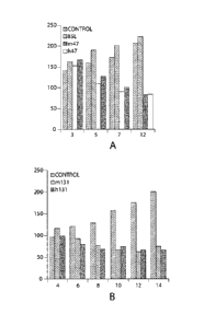
Figure 4A depicts the results of extracellular EphB4 sandwich ELISA comparing
the binding
of a chimeric #131 antibody with 4 deimmunized #131 variant antibodies. Figure
4B shows
the concentration of each antibody where 50% binding in the ELISA is reached.
Figure 5 shows a western blot of an SDS gel loaded with lysate from HT29 cells
that were
treated with 10 mg/ml of antibody (Lane 1: no antibody treatment, Lane 2:
mouse
monoclonal #131, Lane 3: chimeric #131, Lane 4: an exemplary deimmunized #131
antibody, Lane 5: mouse monoclonal #47, Lane 6: chimeric #47, Lane 7: and
exemplary
deimmunized #47 antibody, Lane 8 indicated the molecular markers with the
weight in I(Da).
The blot was probed with an anti-EphB4 primary antibody.
Figures 6A and 6B depict the results of an in vivo squamous cell carcinoma
xenograft assay.
Tumor volume is expressed on the Y-axis as mm3 and the X-axis corresponds to
the number
of days following the beginning of treatment. Treatment with the mouse
monoclonal
antibodies #47 and #131 are compared with an exemplary deimmunized antibodies
and
control treatment. .
DETAILED DESCRIPTION OF THE INVENTION
I. Definitions
A "subject" refers to a vertebrate, such as for example, a mammal, or a human.
Though the antibodies and antigen binding fragments of the present application
are primarily
concerned with the treatment of human subjects, they may also be employed for
the treatment
of other mammalian subjects such as dogs and cats for veterinary purposes.
As used herein, the terms "antibody" and "antibodies" (immunoglobulins)
encompass,
but are not limited to, monoclonal antibodies (including full-length
monoclonal antibodies),
polyclonal antibodies, multispecific antibodies (e.g., bispecific antibodies)
formed from at
18
CA 02696164 2016-02-18
WO 2009/023185
PCT/US2008/009619
least two intact antibodies, human antibodies, humanized antibodies, camelised
antibodies,
chimeric antibodies, single-chain Fvs (scFv), single-chain antibodies, single
domain
antibodies, domain antibodies, Fab fragments, F(ab ' )2 fragments, antibody
fragments that
exhibit the desired biological activity, disulfide-linked Fvs (sdFv),
intrabodies, and epitope-
binding fragments or antigen binding fragments of any of the above. Antibodies
include
immunoglobulin molecules and immunologically active fragments of
immunoglobulin
molecules, i.e., molecules that contain an antigen-binding site.
Immunoglobulin molecules
can be of any type (e.g., IgG, IgE, IgM, IgD, IgA and IgY), class (e.g., IgGl,
IgG2, Ig03,
IgG4, IgAl and IgA2) or subclass.
The term "an antigen-binding fragment" refers to any portion of an antibody
that
retains binding to the antigen. An exemplary antigen-binding fragment of an
antibody is the
heavy chain and/or light chain CDR, or the heavy and/or light chain variable
region.
The term "immunogenicity" refers to the ability of an antibody or antigen
binding
fragment to elicit an immune response (humoral or cellular) when administered
to a recipient
and includes, for example, the HAMA response. A HAMA response is initiated
when T-cells
from a subject make an immune response to the administered antibody. The T-
cells then
recruit B-cells to generate specific"anti-antibody"antibodies.
The term "T-cell epitopes" refers to specific peptide sequences which either
bind with
reasonable efficiency to MHC class II molecules or which are able to stimulate
T-cells via
presentation on MHC class II.
The term "B-cell epitopes" refers to peptide sequences recognized by B-cells.
In
general these sequences are solvent accessible.
The term deimmunization is a process that reduces the immunogenicity of a
compound to a given species. A deimmunized antibody is an antibody that has
lower
immunogenicity in a given species than the corresponsing parent or nonhuman
antibody.
As used herein, the terms Ephrin and Eph are used to refer, respectively, to
ligands
and receptors. They can be from any of a variety of animals (e.g., mammals/non-
mammals,
vertebrates/non-vertebrates, including humans). The nomenclature in this area
has changed
rapidly and the terminology used herein is that proposed as a result of work
by the Eph
Nomenclature Committee.
19
CA 02696164 2016-02-18
WO 2009/023185 PCT/US2008/009619
II. Overview
The Eph family receptors are a family of receptor protein-tyrosine kinases
which are
related to Eph, a receptor named for its expression in an erythropoietin-
producing human
hepatocellular carcinoma cell line. They are divided into two subgroups on the
basis of the
relatedness of their extracellular domain sequences and their ability to bind
preferentially to
Ephrin-A proteins or Ephrin-B proteins. Receptors which interact
preferentially with Ephrin-
A proteins are EphA receptors and those which interact preferentially with
Ephrin-B proteins
are EphB receptors.
Eph receptors have an extracellular domain composed of the ligand-binding
globular
domain, a cysteine rich region followed by a pair of fibronectin type III
repeats. The
cytoplasmic domain consists of a juxtamembrane region containing two conserved
tyrosine
residues; a protein tyrosine kinase domain; a sterile a-motif (SAM) and a PDZ-
domain
binding motif. EphB4 is specific for the membrane-bound ligand Ephrin B2
(Sakano, S. et al
1996; Brambilla R. et al 1995). Ephrin B2 belongs to the class of Eph ligands
that have a
transmembrane domain and cytoplasmic region with five conserved tyrosine
residues and
PDZ domain. Eph receptors are activated by binding of clustered, membrane
attached
ephrins (Davis S et al, 1994), indicating that contact between cells
expressing the receptors
and cells expressing the ligands is required for Eph activation.
Upon ligand binding, an Eph receptor dimerizes and autophosphorylate the
juxtamembrane tyrosine residues to acquire full activation (Kalo MS et al,
1999).
In addition to forward signaling through the Eph receptor, reverse signaling
can occur
through the ephrin Bs. Eph engagement of ephrins results in rapid
phosphorylation of the
conserved intracellular tyrosines (Bruckner K, 1997) and somewhat slower
recruitment of
PDZ binding proteins.
The EphB4 precursor protein is depicted in Figure 1. Amino acids 16-198 of the
EphB4 sequence of Figure 1 correspond to the Globular Domain (GD) of EphB4
that binds to
EphrinB2. Amino acids 239-321 correspond to the cysteine rich domain and amino
acids
324-429 and 434-526 correspond to the first fibronectin-like domain (FNDI) and
the second
fibronectin-like domain (FND2) of EphB4 respectively.
Several studies have shown that high expression of Eph/ephrins may be
associated
with increased potentials for tumor growth, tumorigenicity, and metastasis (
Kiyokawa E, 1994; Tang XX, 1999; Stephenson SA, 2001;
CA 02696164 2016-02-18
WO 2009/023185 PCVLIS2008/009619
Berclaz G, 1996). Application 10/949,720 demonstrates that EphB4
antibodies cause apoptosis, decrease angiogenesis, and inhibit tumor growth in
a xenografl
head and neck carcinoma tumor type.
The disclosure provides deimmunized antibodies and antigen binding fragments
that
may be used to treat cancer as well as angiogenesis related disorders and
unwanted
angiogenesis related processes.
Deimmunized antibodies and antigen binding fragments may be used to inhibit
EphB4 function in vitro and in vivo. The disclosure provides antibodies that
act as receptor
antagonists, such as by inhibiting EphB4 and EphB2 interaction. The disclosure
also
provides antibodies and antigen binding portions thereof that act as agonists
and activate
EphB4 kinase activity (typically assessed by evaluating EphB4 phosphorylation
state).
Surprisingly, such antibodies also inhibit EphB4 functions in cell based and
in vivo assays.
Accordingly, such antibodies and antigen binding fragments may be used to
inhibit EphB4
function in vitro and in vivo, and for treating cancer or disorders associated
with unwanted
angiogenesis. While not wishing to be limited to any particular mechanism, it
is expected
that antibodies which stimulate EphB4 kinase activity, also affect EphB4
removal from the
membrane, thus decreasing overall EphB4 levels.
III. Antibodies
Antibodies are proteins produced by lymphocytes known as B-cells in
vertebrates in
response to stimulation by antigens. The basic structural unit of an antibody
(or rather
immunoglobulin (Ig)) molecule consists of four polypeptide chains which come
together in
the shape of a capital letter "Y". Two of the four chains are identical light
(L) chains and two
are identical heavy (1-1) chains. There are five different kinds (isotypes) of
heavy chains
which divide antibodies into five classes, namely, IgA, IgD, IgE, IgG and IgM.
In addition,
there are two different isotypes of light chains designated .kappa. and
.lambda.. Each class of
heavy chains can combine with either of the light chains. The heavy and light
chains each
contain a variable region (VH and VL, respectively) that is involved in
antigen binding and a
constant (C) region. The antigen binding site is composed of six hypervariable
regions (or
rather complementarity determining regions (CDRs)). Three CDRs from the heavy
chain and
three CDRs from the light chain are respectively positioned between four
relatively
conserved anti-parallel .beta.-sheets which are called framework regions (FR
I, FR2, FR3 and
21
CA 02696164 2010-02-11
WO 2009/023185 PCT/US2008/009619
FR4), on each chain. By convention, numbering systems have been utilized to
designate the
location of the component parts of VH and VL chains. The Kabat definition is
based on
sequence variability and the Chothia definition is based on the location of
structural loop
regions. The Kabat definition for numbering is used herein.
In certain aspects, the present application provides deimmunized antibodies
and
antigen binding fragments against EphB4. Is some embodiments the deimmunized
antibody
or antigen binding fragment binds to an extracellular domain of EphB4. It is
understood that
antibodies may be Fab, Fv, scFv, Fab' and F(ab)2, monoclonal and polyclonal
antibodies,
engineered antibodies (including chimeric, single chain, CDR-grafted,
humanized, fully
human antibodies, and artificially selected antibodies), and synthetic or semi-
synthetic
antibodies produced using phage display or alternative techniques.
In one embodiment of the application, the antibody fragments are truncated
chains
(truncated at the carboxyl end). In certain embodiments, these truncated
chains possess one or
more immunoglobulin activities (e.g., complement fixation activity). Examples
of truncated
chains include, but are not limited to, Fab fragments (consisting of the VL,
VH, CL and CH1
domains); Fd fragments (consisting of the VH and CHI domains); Fv fragments
(consisting
of VL and VH domains of a single chain of an antibody); dab fragments
(consisting of a VH
domain); isolated CDR regions; (Fab)2 fragments, bivalent fragments
(comprising two Fab
fragments linked by a disulphide bridge at the hinge region). The truncated
chains can be
produced by conventional biochemical techniques, such as enzyme cleavage, or
recombinant
DNA techniques, each of which is known in the art. These polypeptide fragments
may be
produced by proteolytic cleavage of intact antibodies by methods well known in
the art, or by
inserting stop codons at the desired locations in the vectors using site-
directed mutagenesis,
such as after CH1 to produce Fab fragments or after the hinge region to
produce (Fab1)2
fragments. Single chain antibodies may be produced by joining VL- and VH-
coding regions
with a DNA that encodes a peptide linker connecting the VL and VH protein
fragments
This application also provides fragments of anti-EphB4 antibodies, which may
comprise a portion of an intact antibody, such as for example, the antigen-
binding or variable
region of the intact antibody. Examples of antibody fragments include Fab,
Fab', F(abl,
and Fv fragments; diabodies; linear antibodies (Zapata et al., Protein
Eng.1995; 8(10): 1057-
1062); single-chain antibody molecules; and multispecific antibodies formed
from antibody
fragments.
22
CA 02696164 2010-02-11
WO 2009/023185 PCT/US2008/009619
Papain digestion of antibodies produces two identical antigen-binding
fragments, called
"Fab" fragments, each with a single antigen-binding site, and a residual "Fe"
fragment,
whose name reflects its ability to crystallize readily. Pepsin treatment of an
antibody yields
an F(ab')2 fragment that has two antigen-combining sites and is still capable
of cross-linking
antigen.
"Fv" usually refers to the minimum antibody fragment that contains a complete
antigen-
recognition and -binding site. This region consists of a dimer of one heavy-
and one light-
chain variable region in tight, non-covalent association. It is in this
configuration that the
three CDRs of each variable region interact to define an antigen-binding site
on the surface of
the VH-VL dimer. Collectively, the CDRs confer antigen-binding specificity to
the antibody.
However, even a single variable region (or half of an Fv comprising three CDRs
specific for
an antigen) has the ability to recognize and bind antigen, although likely at
a lower affinity
than the entire binding site.
Thus, in certain embodiments, the antibodies disclosed in the application may
comprise 1, 2, 3, 4, 5, 6, or more CDRs that recognize the extracellular
domain of EphB4.
The Fab fragment also contains the constant domain of the light chain and the
first
constant domain (CH1) of the heavy chain. Fab' fragments differ from Fab
fragments by the
addition of a few residues at the carboxy terminus of the heavy chain CH1
domain including
one or more cysteines from the antibody hinge region. Fab'-SH is the
designation herein for
Fab' in which the cysteine residue(s) of the constant domains bear a free
thiol group. F(ab')2
antibody fragments originally were produced as pairs of Fab' fragments that
have hinge
cysteines between them. Other chemical couplings of antibody fragments are
also known.
"Single-chain Fv" or "scFv" antibody fragments comprise the VH and VL domains
of an
antibody, wherein these domains are present in a single polypeptide chain. In
certain
embodiments, the Fv polypeptide further comprises a polypeptide linker between
the VH and
VL domains that enables the scFv to form the desired structure for antigen
binding. For a
review of scFv see Pluckthun in The Pharmacology of Monoclonal Antibodies,
vol. 113,
Rosenburg and Moore, eds. (Springer-Verlag: New York, 1994), pp. 269-315.
SMIPs are a class of single-chain peptides engineered to include a target
binding
region and effector domain (CH2 and CH3 domains). See, e.g., U.S. Patent
Application
Publication No. 20050238646. The target binding region may be derived from the
variable
23
CA 02696164 2010-02-11
WO 2009/023185 PCT/US2008/009619
region or CDRs of an antibody, e.g., an anti-EphB4 antibody of the
application.
Alternatively, the target binding region is derived from a protein that binds
EphB4.
The term "diabodies" refers to small antibody fragments with two antigen-
binding
sites, which fragments comprise a heavy-chain variable region (VH) connected
to a light-
chain variable region (VL) in the same polypeptide chain (VH-VL). By using a
linker that is
too short to allow pairing between the two domains on the same chain, the
domains are
forced to pair with the complementary domains of another chain and create two
antigen-
binding sites. Diabodies are described more fully in, for example, EP 404,097;
WO
93/11161; and Hollinger et al., Proc. Natl. Acad. Sci. USA, 90: 6444-6448
(1993).
It is well known that the binding to a molecule (or a pathogen) of antibodies
with an
Fc region assists in the processing and clearance of the molecule (or
pathogen). The Fc
portions of antibodies are recognized by specialized receptors expressed by
immune effector
cells. The Fc portions of IgG1 and IgG3 antibodies are recognized by Fc
receptors present on
the surface of phagocytic cells such as macrophages and neutrophils, which can
thereby bind
and engulf the molecules or pathogens coated with antibodies of these isotypes
(Janeway et
al., Immunobiology 5th edition, page 147, Garland Publishing (New York,
2001)).
The anti-EphB4 antibodies of the present application include antibodies having
all
types of constant regions, including IgM, IgG, IgD, IgA and IgE, and any
isotype, including
IgGl, IgG2a, IgG2b, IgG3 and IgG4. The light chains of the antibodies can
either be kappa
light chains or lambda light chains.
In certain embodiments, single chain antibodies, and chimeric, humanized or
primatized (CDR-grafted) antibodies, as well as chimeric or CDR-grafted single
chain
antibodies, comprising portions derived from different species, are also
encompassed by the
present disclosure as antigen-binding fragments of an antibody. The various
portions of these
antibodies can be joined together chemically by conventional techniques, or
can be prepared
as a contiguous protein using genetic engineering techniques. For example,
nucleic acids
encoding a chimeric or humanized chain can be expressed to produce a
contiguous protein.
See, e.g., U.S. Pat. Nos. 4,816,567 and 6,331,415; U.S. Pat. No. 4,816,397;
European Patent
No. 0,120,694; WO 86/01533; European Patent No. 0,194,276 B1; U.S. Pat. No.
5,225,539;
and European Patent No. 0,239,400 BI. See also, Newman et al., BioTechnology,
10: 1455-
1460 (1992), regarding primatized antibody. See, e.g., Ladner et al., U.S.
Pat. No. 4,946,778;
and Bird et al., Science, 242: 423-426 (1988)), regarding single chain
antibodies.
24
CA 02696164 2010-02-11
WO 2009/023185 PCT/US2008/009619
In certain aspects, the present application provides antibodies and antigen
binding
fragments having binding specificity for an EphB4 or a portion of EphB4. In
some aspects
the antibodies and antigen binding fragments bind to one or more specific
domains of EphB4.
For example, an antibody or antigen binding fragment binds to one or more
extracellular
domains of EphB4 (such as the globular domain, the cystein-rich domain, and
the first
fibronectin type 3 domain, and the second fibronectin type 3 domain). In some
aspects, the
immunoglobulins can bind to EphB4 with a dissociation constant (KD) of at
least about 1x10-
6, 1x10⁻⁷, 1x10⁻⁸, 1x10⁻⁹ M or less. In certain embodiments antibodies and
antigen binding
fragments disclosed herein are specific for EphB4, with minimal binding to
other members of
the Eph or Ephrin families.
In certain embodiments, the present application provides EphB4 antagonist
antibodies. As
described herein, the term "antagonist antibody" refers to an antibody that
can inhibit one or
more functions of an EphB4, such as a binding activity (e.g., ligand binding)
and a signaling
activity (e.g., clustering or phosphorylation of EphB4, stimulation of a
cellular response, such
as stimulation of cell migration or cell proliferation). For example, an
antagonist antibody
can inhibit (reduce or prevent) the interaction of an EphB4 receptor with a
natural ligand
(e.g., Ephrin B2 or fragments thereof). In some embodiments, antagonist
antibodies directed
against EphB4 can inhibit functions mediated by EphB4, including endothelial
cell migration,
cell proliferation, angiogenesis, and/or tumor growth. In certain embodiments,
the antagonist
antibody binds to an extracellular domain of EphB4.
In other embodiments, antibodies or antigen binding fragments are EphB4
agonists.
In some embodiments antibodies or antigen binding fragments activate or
enhance EphB4
kinase activity, even independent of EphrinB2. In some instances, such
antibodies may be
used to stimulate EphB4. However, applicants note that in most cell-based and
in vivo
assays, such antibodies surprisingly behaved like antagonist antibodies. Such
antibodies
appear to bind to the fibronectin type III domains, particularly the region of
amino acids 327-
427 of Fig. I. In some embodiments, antibodies or antigen binding fragments
that bind to the
fibronectin type III domains of EphB4 can inhibit functions mediated by EphB4,
including
endothelial cell migration, cell proliferation, angiogenesis, and/or tumor
growth.
In certain embodiments, single chain antibodies, and chimeric, humanized or
primatized (CDR-grafted) antibodies, as well as chimeric or CDR-grafted single
chain
CA 02696164 2010-02-11
WO 2009/023185 PCT/US2008/009619
antibodies, comprising portions derived from different species, are also
encompassed by the
disclosure as antigen binding portions of an antibody.
In addition, antigen binding fragments of antibodies, including fragments of
chimeric,
humanized, primatized or single chain antibodies, can also be produced.
Antigen binding
fragments of the subject antibodies retain at least one binding function
and/or modulation
function of the full-length antibody from which they are derived. Certain
antigen binding
fragments retain the ability to inhibit one or more functions characteristic
of an EphB4, such
as a binding activity, a signaling activity, and/or stimulation of a cellular
response. For
example, in one embodiment, an antigen binding fragment of an EphB4 antibody
can inhibit
the interaction of EphB4 with one or more of its ligands (e.g., Ephrin B2)
and/or can inhibit
one or more receptor-mediated functions, such as cell migration, cell
proliferation,
angiogenesis, and/or tumor growth.
In one aspect, the deimmunized antibody or antigen binding fragment is a mouse
antibody. In one aspect, the heavy and light chain variable regions each
contain 2 to 20 amino
acid substitutions. In one aspect, the substitutions comprise replacing at
least one mouse
amino acid with at least one corresponding human amino acid. In one aspect,
the human
amino acid is chosen based on identifying a human germline gene that is
homologous to the
mouse variable region. In one aspect, a homologous human germline gene is
independently
identified for each of the four framework regions of the mouse variable
region.
The term "humanized antibody and antigen binding fragment" as used herein
refers to
an antibody or antigen binding fragment comprising portions of antibody of
different origin,
wherein at least one portion is of human origin. Accordingly, one embodiment
relates to a
deimmunized antibody having binding specificity for an EphB4 (e.g., human
EphB4), said
antibody comprising an antigen binding region of nonhuman origin (e.g.,
rodent) and at least
a portion of an antibody of human origin (e.g., a human framework region, a
human constant
region or portion thereof). For example, the deimmunized antibody can comprise
portions
derived from an antibody of nonhuman origin with the requisite specificity,
such as a mouse,
and from antibody sequences of human origin (e.g., a chimeric antibody),
joined together
chemically by conventional techniques (e.g., synthetic) or prepared as a
contiguous
polypeptide using genetic engineering techniques (e.g., DNA encoding the
protein portions of
the chimeric antibody can be expressed to produce a contiguous polypeptide
chain).
26
CA 02696164 2010-02-11
WO 2009/023185
PCT/US2008/009619
In certain embodiments, the framework regions are derived from the closest
human
germline framework regions. In certain embodiments, the antibody or antigen
binding
fragment comprises the FR1, FR2, FR3, and FR4 regions from the closest human
germline
gene. In certain embodiments each framework region is independently selected
from the
human germline gene closest to the particular framework region. In certain
embodiments,
residues that affect antigen binding affinity in the framework regions are
sustituted with the
corresponsing residues from the nonhuman or parent antibody.
In one aspect a deimmunized antibody or antigen binding fragment contains one
or
more antibody chains comprising a CDR of nonhuman origin (e.g., one or more
CDRs
derived from an antibody of nonhuman origin) and a framework region derived
from a light
and/or heavy chain of human origin, e.g., germline antibody genes (e.g., CDR-
grafted
antibodies with or without framework changes). In one embodiment, the
deimmunized
antibody can compete with murine monoclonal antibody for binding to an EphB4
polypeptide. Chimeric or CDR-grafted single chain antibodies are also
encompassed by the
term humanized antibody.
In one aspect a deimmunized antibody or antigen binding fragment contains one
or
more antibody chains comprising a CDR of nonhuman origin (e.g., one or more
CDRs
derived from an antibody of nonhuman origin) and a framework region of
nonhuman origin.
In one embodiment the nonhuman framework region is substituted with at least
one amino
acid from a corresponding human framework region. In one embodiment, the
substitution of
a human amino acid residue for a nonhuman residue reduces the immunogenicity
of the
antibody in a human subject.
In one embodiment, a deimmunized antibody or antigen binding fragment thereof
is
provided that binds the extracellular domain of EphB4, including a heavy chain
variable
region and a light chain variable region, wherein each variable region has
between 2 to 20
amino acid substitutions in comparison to a nonhuman or parent antibody that
binds the
extracellular domain of EphB4. The variable region encompasses three CDR
regions
interspersed with four framework regions. In one aspect the substitutions are
in the
framework region.
In one embodiment, a nonhuman or parent antibody is compared to a database of
human germline antibody genes, such as from V BASE, and highly homologous
individual
framework regions are identified. Structural models may be generated of the
nonhuman or
27
CA 02696164 2016-02-18
WO 2009/023185 PCT/US2008/009619
parent antibody variable region using such programs as SwissPdb, WAM (Web
Antibody
Modelling), and AbM. Residues, such as those, for example, that do not play a
role in
interacting with CDRs or antigen or contribute to antigrn binding affinity,
may be substituted
by the corresponding residue from the human germline gene.
In one embodiment, the individual framework regions, instead of the whole
framework, in the variable region amino acid sequence of the nonhuman or
parent antibody
are compared to corresponding sequences in a collection of human antibodies.
The human
framework with the highest degree of homology is selected to replace the
original framework
of the nonhuman or parent antibody. This technique, known as "framework
patching", is
described in detail in US Patent Application No. US 2005/0033028.
In one embodiment, referred to as "framework shuffling", a combinatorial
library with
CDR variable regions from the nonhuman or parent antibody are fused in frame
into a pool of
individual human germline frameworks (Dall'Acqua et al., Methods, 36:43
(2005)). The
libraries are then screened to identify clones that encode humanized
antibodies which bind
the extracellular domain of EphB4 with similar or greater binding affinity
compared to the
nonhuman or parent antibody.
In one embodiment, the nonhuman or parent antibody is analyzed in order to
identify
potential T-cell epitopes. T-cell epitopes can be idenified using peptide
threading software
that predicts MCH class II binding motifs. Computational binding prediction
algorithms
include iTopeTm, Tepitope, SYFPEITHI, and MHCpred. In one embodiment,
homologous
individual human framework regions are analyzed for potential T-cell epitopes
in parallel.
Epitopes that are identified in both the nonhuman or parent variable region
and the human
germline genes may be disregarded. Epitopes identified in only the nonhuman or
parent
variable region are then flagged for potential replacement.
In one embodiment, the nonhuman or parent antibody is analyzed in order to
identify
potential B-cell epitopes. Potential B-cell epitopes can be recognized by
identifying residues
in the non-human or parent antibody framework region that are at least
partially solvent
accessible and differ from corresponding homologous human antibody framework
residues.
In one embodiment, potential B-cell epitopes are eliminated by replacing the
solvent
accessible nonhuman or parent antibody framework residues with corresponding
human
residues.
28
CA 02696164 2010-02-11
WO 2009/023185 PCT/US2008/009619
In one embodiment substitutions introduced into the deimmunized antibody
comprise
amino acid substitutions, deletions or insertions. In one embodiment, each
substitution results
in the replacement of one amino acid with the corresponding amino acid from a
homologous
human germline gene or from a human variable region consensus sequence. In one
embodiment, a nonhuman or parent antibody or antigen binding fragment is
deimmunized by
substituting from 2 to 20 amino acids with corresponding amino acids from a
homologous
human germline gene or from a human variable region consensus sequence. In one
embodiment, the substitutions may remove one or more T-cell or B-cell
epitiopes. In another
embodiment, the substitutions may introduce a regulatory T-cell epitope. In
one embodiment,
the deimmunized nonhuman or parent antibody or antigen binding fragment
demonstrates a
reduced immunogenicity response over the nonhuman or parent antibody or
antigen binding
fragment when administered to a human subject.
In one embodiment, the heavy and light chain variable regions of the
deimmunized
antibody or antigen binding fragment are derived entirely from one or more
human
antibodies, as described in W02006/08246. In one embodiment, the variable
regions are
composed of segments of amino acid sequence from one or more human antibodies.
In one
embodiment the human segments are two or more amino acids in length. In one
embodiment,
the human segments are 100 or fewer amino acids in length. In further
embodiments, the
human segments are 50 or fewer, 40 or fewer, or 30 or fewer amino acids in
length.
In one embodiment, each variable region has a reduced number of T-cell
epitopes
compared to the parent or nonhuman antibody. In one embodiment, each variable
region has
a reduced number of B-cell epitopes compared to the parent or nonhuman
antibody.
In one aspect, the deimmunized antibody or antigen binding fragment comprises
the
CDR regions of mouse monoclonal antibody #47. Mouse monoclonal antibody #47
has been
described and characterized in US2005/0249736, which is hereby incorporated by
reference
in its entirety. The CDR regions for the heavy chain of mouse monoclonal
antibody #47 are
defined as SEQ ID NO:19 (CDR1), SEQ ID NO:20 (CDR2), and SEQ ID NO:21 (CDR3).
The CDR regions for the light chain of mouse monoclonal antibody #47 are
defined as SEQ
ID NO:22 (CDR I ), SEQ ID NO:23 (CDR2), and SEQ ID NO:24 (CDR3). In one
aspect, the
deimmunized antibody or antigen binding fragment comprises one or more
framework
regions (FR1-FR4) of mouse monoclonal antibody #47.
29
CA 02696164 2010-02-11
WO 2009/023185 PCT/US2008/009619
In one aspect the deimmunized antibody or antigen binding fragment is less
immunogenic (or rather, elicits a reduced HAMA response) than mouse monoclonal
antibody
#47 in a human subject. Assays to determine immunogenicity are well within the
knowledge
of the skilled person. Art-recognized methods of determining immune response
can be
performed to monitor a HAMA response in a particular subject or during
clinical trials.
Subjects administered deimmunized antibodies can be given an immunogenicity
assessment
at the beginning and throughout the administration of said therapy. The HAMA
response is
measured, for example, by detecting antibodies to the deimmunized therapeutic
reagent, in
serum samples from the subject using a method known to one in the art,
including surface
plasmon resonance technology (BIACORE) and/or solid-phase ELISA analysis.
Alternatively, in vitro assays designed to measure a T-cell activation event
are also indicative
of immunogenicity. One assay, by way of example, is the T-cell proliferation
assay. In this
assay PBMCs from donors representing >80% of HLA-DR alleles in the world are
screened
for proliferation in response to an antibody or antibody fragment.
In one aspect the deimmunized antibody or antigen binding fragment binds the
extracellular domain of EphB4 with a binding affinity which is at least 80% or
at least 90%
of the binding affinity of mouse monoclonal antibody #47. In another
embodiment, the
deimmunized antibody or antigen binding fragment binds the extracellular
domain of EphB4
with a binding affinity which is at least 100%, or rather with a greater
binding affinity than
mouse monoclonal antibody #47.
The determination of binding affinity is well within the knowledge of a
skilled
person. Art recognized methods include enzyme-linked immunosorbent assays
(ELISAs),
radioimmunoprecipitation (RIP) assays, and the BIAcore biosensor assay.
Example 2
describes in more detail the determination of binding affinity using the
sandwich ELISA.
In another aspect, the deimmunized antibody or antigen binding fragment
comprises a
heavy chain that comprises an amino acid sequence defined as SEQ ID NO:1, SEQ
ID NO:2,
SEQ ID NO:3, SEQ ID NO:4, or SEQ ID NO:5 and a light chain that comprises an
amino
acid sequence defined as SEQ ID NO:6, SEQ ID NO:7, SEQ ID NO:8, or SEQ ID
NO:9. In
one embodiment the heavy chain is SEQ ID NO:3 and the light chain is SEQ ID
NO:7. In
one embodiment the heavy chain is SEQ ID NO:3 and the light chain is SEQ ID
NO:8. In
one embodiment the heavy chain is SEQ ID NO:4 and the light chain is SEQ ID
NO:7. In
one embodiment the heavy chain is SEQ ID NO:4 and the light chain is SEQ ID
NO:8. In
CA 02696164 2016-02-18
WO 2009/023185 PCT/US2008/009619
some of the embodiments the deimmunized antibody or antigen binding fragment
binds to the
extracellular domain of EphB4. In some of the embodiments the deimmunized
antibody or
antigen binding fragment is less immunogenic to a human subject than the mouse
monoclonal
antibody #47.
In one aspect, the deimmunized antibody or antigen binding fragment comprises
the
CDR regions of mouse monoclonal antibody #131. Mouse monoclonal antibody #131
has
been described and characterized in US2005/0249736.
The CDR regions for the heavy chain of mouse monoclonal
antibody #131 are defined as SEQ ID NO:25 (CDR1), SEQ ID NO:26 (CDR2), and SEQ
ID
NO:27 (CDR3). The CDR regions for the light chain of mouse monoclonal antibody
#131
are defined as SEQ ID NO:28 (CDR I ), SEQ ID NO:29 (CDR2), and SEQ ID NO:30
(CDR3). In one aspect, the deimmunized antibody or antigen binding fragment
comprises
one or more framework regions (FRI -FR4) of mouse monoclonal antibody #131.
In one aspect the deimmunized antibody or antigen binding fragment binds the
extracellular domain of EphB4 with a binding affinity which is at least 80% or
at least 90%
of the binding affinity of mouse monoclonal antibody #131. In another
embodiment, the
deimmunized antibody or antigen binding fragment binds the extracellular
domain of EphB4
with a binding affinity which is at least 100%, or rather with a greater
binding affinity than
mouse monoclonal antibody #131.
In one aspect the deimmunized antibody or antigen binding fragment is less
immunogenic (or rather, elicits a reduced HAMA response) than mouse monoclonal
antibody
#131 in a human subject.
In another aspect, the deimmunized antibody or antigen binding fragment
comprises a
heavy chain that comprises an amino acid sequence defined as SEQ ID NO:10, SEQ
ID
NO:11, SEQ ID NO:12, SEQ ID NO:13, or SEQ ID NO:14 and a light chain that
comprises
an amino acid sequence defined as SEQ ID NO:15, SEQ ID NO:16, SEQ ID NO:17, or
SEQ
ID NO:18. In one embodiment the heavy chain is SEQ ID NO:13 and the light
chain is SEQ
ID NO:17. In one embodiment the heavy chain is SEQ ID NO:13 and the light
chain is SEQ
ID NO:18. In one embodiment the heavy chain is SEQ ID NO:14 and the light
chain is SEQ
ID NO:17. In one embodiment the heavy chain is SEQ ID NO:14 and the light
chain is SEQ
ID NO:18. In some of the embodiments the deimmunized antibody or antigen
binding
fragment binds to the extracellular domain of EphB4. In some of the
embodiments the
31
CA 02696164 2010-02-11
WO 2009/023185 PCT/US2008/009619
deimmunized antibody or antigen binding fragment is less immunogenic to a
human subject
than the mouse monoclonal antibody #131.
In some embodiments, the deimmunized antibodies inhibit the formation of tubes
by
cultured endothelial cells. Inhibition can be determined by any method known
to a person
skilled in the art, including the following. Matrigel (60 I of 10mg/m1;
Collaborative Lab,
Cat. No. 35423) is placed in each well of an ice-cold 96-well plate. The plate
is allowed to sit
at room temperature for 15 minutes then incubated at 37 C for 30 minutes to
permit Matrigel
to polymerize. In the mean time, human umbilical vein endothelial cells are
prepared in
EGM-2 (Clonetic, Cat. No. CC3162) at a concentration of 2x10⁵ cells/ml. The
deimmunized
antibody or antigen binding fragment is prepared at 2x the desired
concentration (5
concentration levels) in the same medium. Cells (500 1) and 2x antibody (500
1) were
mixed and 200 I of this suspension is placed in duplicate on the polymerized
Matrigel. After
24 h incubation, triplicate pictures are taken for each concentration using a
Bioquant Image
Analysis system. Protein addition effect (IC50) is assessed compared to
untreated controls by
measuring the length of cords formed and number of junctions.
In some embodiments, the deimmunized antibody or antigen binding fragment
inhibits the vascularization of a tissue in vivo. Inhibition can be determined
by any method
known to a person skilled in the art, including the following. In vivo
angiogenesis can be
assayed in mice as growth of blood vessels from subcutaneous tissue into a
Matrigel plug
containing the deimmunized antibody or antigen binding fragment. Matrigel
rapidly forms a
solid gel at body temperature, trapping the factors to allow slow release and
prolonged
exposure to surrounding tissues. Matrigel (8.13 mg/ml, 0.5 ml) in liquid form
at 4 C is
mixed with Endothelial Cell Growth Supplement (ECGS), deimmunized antibody
plus ECGS
or Matrigel plus vehicle alone (PBS containing 0.25% BSA). Matrigel (0.5m1) is
injected
into the abdominal subcutaneous tissue of female nu/nu mice (6 wks old) along
the peritoneal
mid line. At day 6, mice are sacrificed and plugs are recovered and processed
for histology.
Typically the overlying skin is removed, and gels are cut out by retaining the
peritoneal lining
for support, fixed in 10% buffered formalin in PBS and embedded in paraffin.
Sections of 3
m are cut and stained with H&E or Masson's trichrome stain and examined under
light
microscope
In some embodiments, the deimmunized antibody or antigen binding fragment
decreases the growth of a human tumor xenograft in a mouse. Inhibition of
tumor growth can
32
CA 02696164 2016-02-18
WO 2009/023185 PCT/US2008/009619
be determined by any method known to a person skilled in the art, including
the methods
described in the examples.
In some embodiments, the deimmunized antibody or antigen binding fragment
inhibits the vascularization of tissue implanted into the cornea of an animal.
Inhibition can be
determined by any method known to a person skilled in the art, including the
mouse corneal
micropocket assays performed according to that detailed by Kenyon et al.,
1996. (see US
publication 2005/0249736)
In some embodiments, the deimmunized antibody or antigen binding fragment
promotes apoptosis. Apoptosis can be examined in vitro using various methods
including
TUNEL staining and the Cell Death Detection ELISAplus Kit (Roche, Piscataway,
NJ).
In certain aspects, the present application provides the hybridoma cell lines,
as well as
to the monoclonal antibodies produced by these hybridoma cell lines. The cell
lines
disclosed have uses other than for the production of the monoclonal
antibodies. For example,
the cell lines can be fused with other cells (such as suitably drug-marked
human myeloma,
mouse myeloma, human-mouse heteromyeloma or human lymphoblastoid cells) to
produce
additional hybridomas, and thus provide for the transfer of the genes encoding
the
monoclonal antibodies. In addition, the cell lines can be used as a source of
nucleic acids
encoding the anti-EphB4 immunoglobulin chains, which can be isolated and
expressed (e.g.,
upon transfer to other cells using any suitable technique (see e.g., Cabilly
et al., U.S. Pat. No.
4,816,567; Winter, U.S. Pat. No. 5,225,539)). For instance, clones comprising
a rearranged
anti-EphB4 light or heavy chain can be isolated (e.g., by PCR) or cDNA
libraries can be
prepared from mRNA isolated from the cell lines, and cDNA clones encoding an
anti-EphB4
immunoglobulin chain can be isolated. Thus, nucleic acids encoding the heavy
and/or light
chains of the antibodies or portions thereof can be obtained and used in
accordance with
recombinant DNA techniques for the production of the specific immunoglobulin,
immunoglobulin chain, or variants thereof (e.g., humanized immunoglobulins) in
a variety of
hosT-cells or in an in vitro translation system. For example, the nucleic
acids, including
cDNAs, or derivatives thereof encoding variants such as a humanized
immunoglobulin or
immunoglobulin chain, can be placed into suitable prokaryotic or eukaryotic
vectors (e.g.,
expression vectors) and introduced into a suitable hosT-cell by an appropriate
method (e.g.,
transformation, transfection, electroporation, infection), such that the
nucleic acid is operably
linked to one or more expression control elements (e.g., in the vector or
integrated into the
33
CA 02696164 2010-02-11
WO 2009/023185 PCT/US2008/009619
hosT-cell genome). For production, hosT-cells can be maintained under
conditions suitable
for expression (e.g., in the presence of inducer, suitable media supplemented
with appropriate
salts, growth factors, antibiotic, nutritional supplements, etc.), whereby the
encoded
polypeptide is produced. If desired, the encoded protein can be recovered
and/or isolated
(e.g., from the hosT-cells or medium). It will be appreciated that the method
of production
encompasses expression in a hosT-cell of a transgenic animal (see e.g., WO
92/03918,
GenPharm International, published Mar. 19, 1992).
The present antibodies and antigen binding fragments can be utilized to
directly kill or
ablate cancerous cells in vivo. Direct killing involves administering the
antibodies (which are
optionally fused to a cytotoxic drug) to a subject requiring such treatment.
In some
embodiments, the cancer comprises cancer cells expressing EphB4 at a higher
level than
noncancerous cells of a comparable tissue. Since the antibodies recognize
EphB4 on cancer
cells, any such cells to which the antibodies bind are destroyed. Where the
antibodies are
used alone to kill or ablate cancer cells, such killing or ablation can be
effected by initiating
endogenous host immune functions, such as CDC and/or ADCC. Assays for
determining
whether an antibody kills cells in this manner are within the purview of those
skilled in the
art.
Accordingly in one embodiment, the antibodies of the present disclosure may be
used
to deliver a variety of cytotoxic compounds. Any cytotoxic compound can be
fused to the
present antibodies. The fusion can be achieved chemically or genetically
(e.g., via expression
as a single, fused molecule). The cytotoxic compound can be a biological, such
as a
polypeptide, or a small molecule. As those skilled in the art will appreciate,
for small
molecules, chemical fusion is used, while for biological compounds, either
chemical or
genetic fusion can be employed.
Non-limiting examples of cytotoxic compounds include therapeutic drugs, a
compound emitting radiation, molecules of plant, fungal, or bacterial origin,
biological
proteins, and mixtures thereof. The cytotoxic drugs can be intracellularly
acting cytotoxic
drugs, such as short-range radiation emitters, including, for example, short-
range, high-
energy a-emitters. Enzymatically active toxins and fragments thereof are
exemplified by
diphtheria toxin A fragment, nonbinding active fragments of diphtheria toxin,
exotoxin A
(from Pseudomonas aeruginosa), ricin A chain, abrin A chain, modeccin A chain,
.alpha.-
sacrin, certain Aleurites fordii proteins, certain Dianthin proteins,
Phytolacca americana
34
CA 02696164 2016-02-18
WO 2009/023185 PCT/US2008/009619
proteins (PAP, PAPII and PAP-S), Morodica charantia inhibitor, curcin, crotin,
Saponaria
officinalis inhibitor, gelonin, mitogillin, restrictocin, phenomycin, and
enomycin, for
example. Procedures for preparing enzymatically active polypeptides of the
immunotoxins
are described in W084/03508 and W085/03508.
Certain cytotoxic moieties are derived from adriamycin, chlorambucil,
daunomycin,
methotrexate, neocarzinostatin, and platinum, for example.
Procedures for conjugating the antibodies with the cytotoxic agents have been
previously described and are within the purview of one skilled in the art.
In certain embodiments, the antibodies or antigen binding fragments are
further
attached to a label that is able to be detected (e.g., the label can be a
radioisotope, fluorescent
compound, enzyme or enzyme co-factor). The active moiety may be a radioactive
agent, such
as: radioactive heavy metals such as iron chelates, radioactive chelates of
gadolinium or
manganese, positron emitters of oxygen, nitrogen, iron, carbon, or gallium,
43K, 52Fe, 57Co,
67cu, 670a, 68 Ga, 1231,1251,1311,1321r r 9
0 9Tc. A binding agent affixed to such a
moiety may
be used as an imaging agent and is administered in an amount effective for
diagnostic use in a
mammal such as a human and the localization and accumulation of the imaging
agent is then
detected. The localization and accumulation of the imaging agent may be
detected by
radioscintigraphy, nuclear magnetic resonance imaging, computed tomography or
positron
emission tomography. Immunoscintigraphy using antibodies or other
binding
polypeptides directed at EphB4 may be used to detect and/or diagnose cancers
and
vasculature. For example, monoclonal antibodies against the EphB4 marker
labeled with
.99Technetium, "Indium, 125Iodine-may be effectively used for such imaging. As
will be
evident to the skilled artisan, the amount of radioisotope to be administered
is dependent
upon the radioisotope. Those having ordinary skill in the art can readily
formulate the amount
of the imaging agent to be administered based upon the specific activity and
energy of a
given radionuclide used as the active moiety. Typically 0.1-100 millicuries
per dose of
imaging agent, or 1-10 millicuries, or 2-5 millicuries are administered. Thus,
the
compositions disclosed are useful as imaging agents comprising a targeting
moiety
conjugated to a radioactive moiety comprise 0.1-100 millicuries, in some
embodiments 1-10
millicuries, in some embodiments 2-5 millicuries, in some embodiments 1-5
millicuries.
The application further provides polynucleotides comprising a nucleotide
sequence
encoding a deimmunized anti-EphB4 antibody or fragments thereof. Because of
the
CA 02696164 2010-02-11
WO 2009/023185 PCT/US2008/009619
degeneracy of the genetic code, a variety of nucleic acid sequences encode
each antibody
amino acid sequence. The application further provides polynucleotides that
hybridize
under stringent or lower stringency hybridization conditions, e.g., as defined
herein, to
polynucleotides that encode a deimmunized antibody that binds to hEphB4.
Stringent hybridization conditions include, but are not limited to,
hybridization to filter-
bound DNA in 6X sodium chloride/sodium citrate (SSC) at about 45 C followed by
one or
more washes in 0.2X SSC/0.1% SDS at about 50-65 C, highly stringent conditions
such as
hybridization to filter-bound DNA in 6X SSC at about 45 C followed by one or
more washes
in 0.1X SSC/0.2% SDS at about 60 C, or any other stringent hybridization
conditions known
to those skilled in the art (see, for example, Ausubel, F.M. et al., eds. 1989
Current Protocols
in Molecular Biology, vol. 1, Green Publishing Associates, Inc. and John Wiley
and Sons,
Inc., NY at pages 6.3.1 to 6.3.6 and 2.10.3).
The polynucleotides may be obtained, and the nucleotide sequence of the
polynucleotides determined, by any method known in the art. For example, if
the nucleotide
sequence of the antibody is known, a polynucleotide encoding the antibody may
be
assembled from chemically synthesized oligonucleotides (e.g., as described in
Kutmeier et
al., BioTechniques 17:242 (1994)), which, briefly, involves the synthesis of
overlapping
oligonucleotides containing portions of the sequence encoding the antibody,
annealing and
ligating of those oligonucleotides, and then amplification of the ligated
oligonucleotides by
PCR. In one embodiment, the codons that are used comprise those that are
typical for human
or mouse (see, e.g., Nakamura, Y., Nucleic Acids Res. 28: 292 (2000)).
A polynucleotide encoding an antibody may also be generated from nucleic acid
from a
suitable source. If a clone containing a nucleic acid encoding a particular
antibody is not
available, but the sequence of the antibody molecule is known, a nucleic acid
encoding the
immunoglobulin may be chemically synthesized or obtained from a suitable
source (e.g., an
antibody cDNA library, or a cDNA library generated from, or nucleic acid,
preferably
polyA+RNA, isolated from, any tissue or cells expressing the antibody, such as
hybridoma
cells selected to express an antibody) by PCR amplification using synthetic
primers
hybridizable to the 3' and 5' ends of the sequence or by cloning using an
oligonucleotide
probe specific for the particular gene sequence to identify, e.g., a cDNA
clone from a cDNA
library that encodes the antibody. Amplified nucleic acids generated by PCR
may then be
cloned into replicable cloning vectors using any method well known in the art.
36
CA 02696164 2010-02-11
WO 2009/023185 PCT/US2008/009619
The present application also provides polynucleotide sequences encoding heavy
and
light chain framework regions and CDRs of antibodies described herein as well
as expression
vectors for their efficient expression in mammalian cells.
IV. Anti-Eph4B antibodies with altered effector functions
Antibodies with engineered or variant constant or Fc regions can be useful in
modulating effector functions, such as, for example, antigen-dependent
cytotoxicity (ADCC)
and complement-dependent cytotoxicity (CDC). Such antibodies with engineered
or variant
constant or Fc regions may be useful in instances where Eph4B is expressed in
normal tissue,
for example; deimmunized antibodies and antigen binding fragments without
effector
function in these instances may elicit the desired therapeutic response while
not damaging
normal tissue. In another embodiment, antibodies or antigen binding fragments
are provided
with increased effector function, and may therefore be useful for direct cell
killing.
Accordingly, certain aspects and methods of the present disclosure relate to
anti-
Eph4B antibodies with altered effector functions that comprise one or more
amino acid
substitutions, insertions, and/or deletions. In certain embodiments, such a
variant anti-Eph4B
antibody exhibits reduced or no effector function.
Anti-Eph4B antibodies with reduced effector function may be produced by
introducing other types of changes in the amino acid sequence of certain
regions of the
antibody. Such amino acid sequence changes include but are not limited to the
Ala-Ala
mutation described by Bluestone et al. (see WO 94/28027 and WO 98/47531; also
see Xu et
al. 2000 Cell Immunol 200; 16-26). Thus in certain embodiments, anti-Eph4B
antibodies
with mutations within the constant region including the Ala-Ala mutation may
be used to
reduce or abolish effector function. According to these embodiments, the
constant region of
an anti-Eph4B antibody comprises a mutation to an alanine at position 234 or a
mutation to
an alanine at position 235. Additionally, the constant region may contain a
double mutation:
a mutation to an alanine at position 234 and a second mutation to an alanine
at position 235.
In one embodiment, the anti-Eph4B antibody comprises an IgG4 framework,
wherein the
Ala-Ala mutation would describe a mutation(s) from phenylalanine to alanine at
position 234
and/or a mutation from leucine to alanine at position 235. In another
embodiment, the anti-
anti-Eph4B antibody comprises an IgG I framework, wherein the Ala-Ala mutation
would
describe a mutation(s) from leucine to alanine at position 234 and/or a
mutation from leucine
37
CA 02696164 2010-02-11
WO 2009/023185 PCT/US2008/009619
to alanine at position 235. An anti- anti-Eph4B antibody may alternatively or
additionally
carry other mutations, including the point mutation K322A in the CH2 domain
(Hezareh et al.
2001 J Virol. 75: 12161-8). An antibody with said mutation(s) in the constant
region may
furthermore be a blocking or non-blocking antibody.
Changes within the hinge region also affect effector functions. For example,
deletion
of the hinge region may reduce affinity for Fc receptors and may reduce
complement
activation (Klein et al. 1981 Proc Natl Acad Sci U S A. 78: 524-528). The
present disclosure
therefore also relates to antibodies with alterations in the hinge region.
In particular embodiments, anti-Eph4B antibodies may be modified to either
enhance
or inhibit complement dependent cytotoxicity (CDC). Modulated CDC activity may
be
achieved by introducing one or more amino acid substitutions, insertions, or
deletions in an
Fc region of the antibody (see, e.g., U.S. Pat. No. 6,194,551). Alternatively
or additionally,
cysteine residue(s) may be introduced in the Fc region, thereby allowing
interchain disulfide
bond formation in this region. The homodimeric antibody thus generated may
have improved
or reduced internalization capability and/or increased or decreased complement-
mediated cell
killing. See Caron et al., J. Exp Med. 176:1191-1195 (1992) and Shopes, B. J.
Immunol.
148:2918-2922 (1992), W099/51642, Duncan & Winter Nature 322: 738-40 (1988);
U.S.
Pat. No. 5,648,260; U.S. Pat. No. 5,624,821; and W094/29351. Homodimeric
antibodies
with enhanced anti-tumor activity may also be prepared using
heterobifunctional cross-
linkers as described in Wolff et al. Cancer Research 53:2560-2565 (1993).
Alternatively, an
antibody can be engineered which has dual Fc regions and may thereby have
enhanced
complement lysis and ADCC capabilities. See Stevenson et al. Anti-Cancer Drug
Design
3:219-230 (1989).
Another potential means of modulating effector function of antibodies includes
changes in glycosylation. This topic has been recently reviewed by Raju who
summarized the
proposed importance of the oligosaccharides found on human IgGs with their
degree of
effector function (Raju, TS. BioProcess International April 2003. 44-53).
According to
Wright and Morrison, the microheterogeneity of human IgG oligosaccharides can
affect
biological functions such as CDC and ADCC, binding to various Fc receptors,
and binding to
Clq protein (Wright A. & Morrison SL. TIBTECH 1997, 15: 26-32). It is well
documented
that glycosylation patterns of antibodies can differ depending on the
producing cell and the
cell culture conditions (Raju, TS. BioProcess International April 2003. 44-
53). Such
38
CA 02696164 2010-02-11
WO 2009/023185 PCT/US2008/009619
differences can lead to changes in both effector function and pharmacokinetics
(Israel et al.
Immunology. 1996; 89(4):573-578; Newkirk et al. P. Clin. Exp. 1996; 106(2):259-
64).
Differences in effector function may be related to the IgGs ability to bind to
the Fey receptors
(FcyRs) on the effector cells. Shields, et al., have shown that IgG, with
variants in amino acid
sequence that have improved binding to FcyR, can exhibit up to 100% enhanced
ADCC using
human effector cells (Shields et al. J Biol Chem. 2001 276(9):6591-604). While
these variants
include changes in amino acids not found at the binding interface, both the
nature of the sugar
component as well as its structural pattern may also contribute to the
differences observed. In
addition, the presence or absence of fiicose in the oligosaccharide component
of an IgG can
improve binding and ADCC (Shields et al. J Biol Chem. 2002; 277(30):26733-40).
An IgG
that lacked a fucosylated carbohydrate linked to Asn297 exhibited normal
receptor binding to
the Fcy receptor. In contrast, binding to the FcyRIIA receptor was improved
50% and
accompanied by enhanced ADCC, especially at lower antibody concentrations.
Work by Shinkawa, et al., demonstrated that an antibody to the human IL-5
receptor
produced in a rat hybridoma showed more than 50% higher ADCC when compared to
the
antibody produced in Chinese hamster ovary cells (CHO) (Shinkawa et al. J Biol
Chem. 2003
278(5):3466-73). Monosaccharide composition and oligosaccharide profiling
showed that the
rat hybridoma-produced IgG had a lower content of fucose than the CHO-produced
protein.
The authors concluded that the lack of fucosylation of an IgG1 has a critical
role in
enhancement of ADCC activity.
A different approach was taken by Umana, et al., who changed the glycosylation
pattern of chCE7, a chimeric IgG1 anti-neuroblastoma antibody (Umana et al.
Nat
Biotechnol. 1999 Feb; 17(2): 176-80). Using tetracycline, they regulated the
activity of a
glycosyltransferase enzyme (GnnII) which bisects oligosaccharides that have
been implicated
in ADCC activity. The ADCC activity of the parent antibody was barely above
background
level. Measurement of ADCC activity of the chCE7 produced at different
tetracycline levels
showed an optimal range of GnTIH expression for maximal chCE7 in vitro ADCC
activity.
This activity correlated with the level of constant region-associated,
bisected complex
oligosaccharide. Newly optimized variants exhibited substantial ADCC activity.
Similarly,
Wright and Morrison produced antibodies in a CHO cell line deficient in
glycosylation (1994
J Exp Med 180: 1087-1096) and showed that antibodies produced in this cell
line were
incapable of complement-mediated cytolysis. Thus as known alterations that
affect effector
39
CA 02696164 2010-02-11
WO 2009/023185 PCT/US2008/009619
function include modifications in the glycosylation pattern or a change in the
number of
glycosylated residues, the present disclosure relates to a Eph4B antibody
wherein
glycosylation is altered to either enhance or decrease effector function(s)
including ADCC
and CDC. Altered glycosylation includes a decrease or increase in the number
of
glycosylated residues as well as a change in the pattern or location of
glycosylated residues.
Still other approaches exist for the altering effector function of antibodies.
For
example, antibody-producing cells can be hypermutagenic, thereby generating
antibodies
with randomly altered nucleotide and polypeptide residues throughout an entire
antibody
molecule (see WO 2005/011735). Hypermutagenic host cells include cells
deficient in DNA
mismatch repair. Antibodies produced in this manner may be less antigenic
and/or have
beneficial pharmacokinetic properties. Additionally, such antibodies may be
selected for
properties such as enhanced or decreased effector function(s).
It is further understood that effector function may vary according to the
binding
affinity of the antibody. For example, antibodies with high affinity may be
more efficient in
activating the complement system compared to antibodies with relatively lower
affinity
(Marzocchi-Machado et al. 1999 Immunol Invest 28: 89-101). Accordingly, an
antibody may
be altered such that the binding affinity for its antigen is reduced (e.g., by
changing the
variable regions of the antibody by methods such as substitution, addition, or
deletion of one
or more amino acid residues). An anti-Eph4B antibody with reduced binding
affinity may
exhibit reduced effector functions, including, for example, reduced ADCC
and/or CDC.
V. Method of Making Antibodies
The deimmunized antibody or antigen binding fragment that binds the
extracellular
domain of EphB4 can be made by a number of different methods known to a person
skilled in
the art. In one example, a nonhuman anti-EphB4 antibody is deimmunized to
reduce the
number of either T or B-cell epitopes or to introduce regulatory T-cell
epitopes. The starting
nonhuman or parent anti-EphB4 antibody can be modified; for example, it can be
any form of
a chimeric, humanized, or primatized antibody. Alternatively, the starting
nonhuman or
parent anti-EphB4 antibody is de-immunized without a humanization or
primatization step.
40
CA 02696164 2016-02-18
WO 2009/023185 PCT/US2008/009619
Nonhuman EphB4 antibodies
Anti-EphB4 antibodies are known to those skilled in the art and include, for
example,
the antibodies described in U. S. Patent No. 5,635,177 and US publications
2006/0134118
and 2005/0249736.
Methods of generating novel anti-EphB4 antibodies are also known to those
skilled in
the art. For example, a method for generating a monoclonal antibody that binds
specifically
to an EphB4 polypeptide may comprise administering to a mouse an amount of an
immunogenic composition comprising the EphB4 polypeptide effective to
stimulate a
detectable immune response, obtaining antibody-producing cells (e.g., cells
from the spleen)
from the mouse and fusing the antibody¨producing cells with myeloma cells to
obtain
antibody-producing hybridomas, and testing the antibody-producing hybridomas
to identify a
hybridoma that produces a monocolonal antibody that binds specifically to the
EphB4
polypeptide. Once obtained, a hybridoma can be propagated in a cell culture,
optionally in
culture conditions where the hybridoma-derived cells produce the monoclonal
antibody that
binds specifically to EphB4 polypeptide. The monoclonal antibody may be
purified from the
cell culture.
In addition, the techniques used to screen antibodies in order to identify a
desirable
antibody may influence the properties of the antibody obtained. A variety of
different
techniques are available for testing antibody:antigen interactions to identify
particularly
desirable antibodies. Such techniques include ELISAs, surface plasmon
resonance binding
assays (e.g., the Biacore binding assay, Bia-core AB, Uppsala, Sweden),
sandwich assays
(e.g., the paramagnetic bead system of IGEN International, Inc., Gaithersburg,
Maryland),
western blots, immunoprecipitation assays and immunohistochemistry.
Other suitable methods of producing or isolating antibodies of the requisite
specificity
can used, including, for example, methods which select recombinant antibody
from a library,
or which rely upon immunization of transgenic animals (e.g., mice) capable of
producing a
full repertoire of human antibodies. See e.g., Jakobovits et al., Proc. Natl.
Acad. Sci. USA,
90: 2551-2555 (1993); Jakobovits et al., Nature, 362: 255-258 (1993); Lonberg
et al., U.S.
Pat. No. 5,545,806; Surani et al., U.S. Pat, No. 5,545,807.
Antibodies can be engineered in numerous ways. They can be made as single-
chain
antibodies (including small modular immunopharmaceuticals or SM1PsTm), Fab and
F(ab')1
fragments, etc. Antibodies can be humanized, chimerized, deimmunized, or fully
human.
41
CA 02696164 2010-02-11
WO 2009/023185 PCT/US2008/009619
Numerous publications set forth the many types of antibodies and the methods
of engineering
such antibodies. For example, see U.S. Patent Nos. 6,355,245; 6,180,370;
5,693,762;
6,407,213; 6,548,640; 5,565,332; 5,225,539; 6,103,889; and 5,260,203.
The application provides antigen binding fragments capable of binding to an
EphB4
receptor or portion thereof, including, but not limited to, Fv, Fab, Fab' and
F(ab')2 fragments.
Such fragments can be produced by enzymatic cleavage or by recombinant
techniques. For
instance, papain or pepsin cleavage can generate Fab or F(ab')2 fragments,
respectively.
Antibodies can also be produced in a variety of truncated forms using antibody
genes in
which one or more stop codons has been introduced upstream of the natural stop
site. For
example, a chimeric gene encoding a F(ab')2 heavy chain portion can be
designed to include
DNA sequences encoding the CH1 domain and hinge region of the heavy chain.
Chimeric antibodies can be produced by recombinant DNA techniques known in the
art. For example, a gene encoding the Fc constant region of a murine (or other
species)
monoclonal antibody molecule is digested with restriction enzymes to remove
the region
encoding the murine Fc, and the equivalent portion of a gene encoding a human
Fc constant
region is substituted (see Robinson et al., International Patent Publication
PCT/US86/02269;
Akira, et al., European Patent Application 184,187; Taniguchi, M., European
Patent
Application 171,496; Morrison et al., European Patent Application 173,494;
Neuberger etal.,
International Application WO 86/01533; Cabilly et al. U.S. Pat. No. 4,816,567;
Cabilly et al.,
European Patent Application 125,023; Better et al. (1988 Science 240:1041-
1043); Liu et al.
(1987) PNAS 84:3439-3443; Liu et al., 1987, J. Immunol. 139:3521-3526; Sun et
al. (1987)
PNAS 84:214-218; Nishimura et al., 1987, Canc. Res. 47:999-1005; Wood et al.
(1985)
Nature 314:446-449; and Shaw et al., 1988, J. Natl Cancer Inst. 80:1553-1559).
Methods for humanizing antibodies have been described in the art. In some
embodiments, a humanized antibody has one or more amino acid residues
introduced from a
source that is nonhuman, in addition to the nonhuman CDRs. Humanization can be
essentially performed following the method of Winter and co-workers (Jones et
al., Nature,
321:522-525 (1986); Riechmann et al., Nature, 332:323-327 (1988); Verhoeyen
etal.,
Science, 239:1534-1536 (1988)), by substituting hypervariable region sequences
for the
corresponding sequences of a human antibody. Accordingly, such "humanized"
antibodies
are chimeric antibodies (U.S. Pat. No. 4,816,567) wherein substantially less
than an intact
human variable region has been substituted by the corresponding sequence from
a nonhuman
42
CA 02696164 2010-02-11
WO 2009/023185 PCT/US2008/009619
species. In practice, humanized antibodies are typically human antibodies in
which some
hypervariable region residues and possibly some framwork region residues are
substituted by
residues from analogous sites in rodent antibodies.
U.S. Pat. No. 5,693,761 to Queen et al, discloses a refinement on Winter for
humanizing antibodies, and is based on the premise that ascribes avidity loss
to problems in
the structural motifs in the humanized framework which, because of steric or
other chemical
incompatibility, interfere with the folding of the CDRs into the binding-
capable conformation
found in the mouse antibody. To address this problem, Queen teaches using
human
framework sequences closely homologous in linear peptide sequence to framework
sequences
of the mouse antibody to be humanized. Accordingly, the methods of Queen focus
on
comparing framework sequences between species. Typically, all available human
variable
region sequences are compared to a particular mouse sequence and the
percentage identity
between correspondent framework residues is calculated. The human variable
region with the
highest percentage is selected to provide the framework sequences for the
humanizing
project. Queen also teaches that it is important to retain in the humanized
framework, certain
amino acid residues from the mouse framework critical for supporting the CDRs
in a binding-
capable conformation. Potential criticality is assessed from molecular models.
Candidate
residues for retention are typically those adjacent in linear sequence to a
CDR or physically
within 6 .angstrom. of any CDR residue.
In other approaches, the importance of particular framework amino acid
residues is
determined experimentally once a low-avidity humanized construct is obtained,
by reversion
of single residues to the mouse sequence and assaying antigen binding as
described by
Riechmann et al, (1988). Another example approach for identifying important
amino acids in
framework sequences is disclosed by U.S. Pat. No. 5,821,337 to Carter et al,
and by U.S. Pat.
No. 5,859,205 to Adair et al. These references disclose specific Kabat residue
positions in the
framework, which, in a humanized antibody may require substitution with the
correspondent
mouse amino acid to preserve avidity.
Another method of humanizing antibodies, referred to as "framework shuffling",
relies on generating a combinatorial library with nonhuman CDR variable
regions fused in
frame into a pool of individual human germline frameworks (Dall'Acqua et al.,
Methods,
36:43 (2005)). The libraries are then screened to identify clones that encode
humanized
antibodies which retain good binding.
43
CA 02696164 2010-02-11
WO 2009/023185 PCT/US2008/009619
The choice of human variable regions, both light and heavy, to be used in
making the
humanized antibodies is very important to reduce antigenicity. According to
the so-called
"best-fit" method, the sequence of the variable region of a rodent antibody is
screened against
the entire library of known human variable-domain sequences. The human
sequence that is
closest to that of the rodent is then accepted as the human framework region
(framwork
region) for the humanized antibody (Sims et al., J. Immunol., 151:2296 (1993);
Chothia et al.,
J. Mol. Biol., 196:901 (1987)). Another method uses a particular framework
region derived
from the consensus sequence of all human antibodies of a particular subgroup
of light or
heavy chain variable regions. The same framework may be used for several
different
humanized antibodies (Carter et al., Proc. Natl. Acad. Sci. USA, 89:4285
(1992); Presta et al.,
J. Immunol., 151:2623 (1993)).
The choice of nonhuman residues to substitute into the human variable region
can be
influenced by a variety of factors. These factors include, for example, the
rarity of the amino
acid in a particular position, the probability of interaction with either the
CDRs or the
antigen, and the probability of participating in the interface between the
light and heavy chain
variable domain interface.(see for example U.S. Pat. No. 5,693,761, 6,632,927,
and
6,639,055). One method to analyze these factors is through the use of three-
dimensional
models of the nonhuman and humanized sequences. Three-dimensional
immunoglobulin
models are commonly available and are familiar to those skilled in the art.
Computer
programs are available that illustrate and display probable three-dimensional
conformational
structures of selected candidate immunoglobulin sequences. Inspection of these
displays
permits analysis of the likely role of the residues in the functioning of the
candidate
immunoglobulin sequence, e.g., the analysis of residues that influence the
ability of the
candidate immunoglobulin to bind its antigen. In this way, nonhuman residues
can be
selected and substituted for human variable region residues in order to
achieve the desired
antibody characteristic, such as increased affinity for the target antigen(s)
Deimmunization
The anti-EphB4 antibody or antigen binding fragment is deimmunized to render
it
non-immunogenic, or less immunogenic, to a given species. Deimmunization can
be achieved
through structural alterations to the anti-EphB4 antibody. In one embodiment,
the anti-EphBr
is a mouse monoclonal antibody. Any deimmunization technique known to those
skilled in
44
CA 02696164 2016-02-18
WO 2009/023185 PCT/US2008/009619
the art can be employed. One suitable technique, for example, for deimmunizing
antibodies is
described in WO 00/34317. In
summary, a typical protocol within the general method described therein
includes the
following steps.
1. Determining the amino acid sequence of the antibody or a part thereof;
2. Identifying potential T-cell epitopes within the amino acid sequence of the
antibody by any
method including determination of the binding of peptides to MHC molecules,
determination
of the binding of peptide: HLA complexes to the T-cell receptors from the
species to receive
the therapeutic protein, testing of the antibody or parts thereof using
transgenic animals with
HLA molecules of the species to receive the therapeutic protein, or testing
such transgenic
animals reconstituted with immune system cells from the species to receive the
therapeutic
protein;
3. By genetic engineering or other methods for producing modified antibodies,
altering the
antibody to remove one or more of the potential T-cell epitopes and producing
such an
altered antibody for testing.
In one embodiment, the sequences of the variable regions of the antibody or
antigen
binding fragment can be analyzed for the presence of MI-IC class II binding
motifs. For
example, a comparison may be made with databases of MHC-binding motifs such
as, for
example by searching the "motifs" database on the worldwide web at
sitewehil.wehi.edu.au.
Alternatively, MHC class II binding peptides may be identified using
computational
threading methods such as those devised by Altuvia et al. (J. Mol. Biol. 249
244-250 (1995))
whereby consecutive overlapping peptides from the variable region sequences
are testing for
their binding energies to MHC class Il proteins. Computational binding
prediction algorithms
include iTopeTm, Tepitope, SYFPEITHI, and MHCpred. In order to assist the
identificationof
MHC class II-binding peptides, associated sequence features which relate to
successfully
presented peptides such as amphipathicity and Rothbard motifs, and cleavage
sites for
cathepsin B and other processing enzymes can be searched for.
Having identified potential second species (e.g. human) T-cell epitopes, these
epitopes are then eliminated by alteration of one or more amino acids, as
required to
eliminate the T-cell epitope. Usually, this will involve alteration of one or
more amino acids
within the T-cell epitope itself. This could involve altering an amino acid
adjacent the epitope
in terms of the primary structure of the protein or one which is not adjacent
in the primary
CA 02696164 2010-02-11
WO 2009/023185 PCT/US2008/009619
structure but is adjacent in the secondary structure of the molecule. The
usual alteration
contemplated will be amino acid substitution, but it is possible that in
certain circumstances
amino acid addition or deletion will be appropriate. All alterations can be
accomplished by
recombinant DNA technology, so that the final molecule may be prepared by
expression from
a recombinant host, for example by well established methods, but the use of
protein
chemistry or any other means of molecular alteration may also be used.
In practice, it has been recognized that potential human T-cell epitopes can
be
identified even in human germline variable region framework sequences when
comparison is
made with databases of MHC-binding motifs. As humans do not generally mount an
ongoing
immune response against their own antibodies, then either humans are tolerant
to these
epitopes or these potential epitopes cannot be presented by human APCs because
they are not
processed appropriately. Therefore, such potential T-cell epitopes which are
represented in
germline variable region sequences may, in practice, be retained in the
deimmunized
antibody.
In order to minimize the creation of additional T-cell epitopes during the
elimination
of potential T-cell epitopes from the therapeutic antibody sequence, the
elimination of T-cell
epitopes can be achieved by substituting particular amino acids which results
in a conversion
of the nonhuman or parental antibody (usually mouse) amino acids within T-cell
epitopes to
amino acids at positions corresponding to human germline amino acids at
positions. Human
germline sequences are disclosed in Tomlinson, I. A. et al. (1992) J. Mol.
Biol. 227:776-798;
Cook, G. P. et al. (1995) Immunol. Today Vol. 16 (5): 237-242; Chothia, D. et
al. (1992) J.
Mol. Bio. 227:799-817. The V BASE directory provides a comprehensive directory
of human
immunoglobulin variable region sequences (compiled by Tomlinson, I. A. et al.
MRC Centre
for Protein Engineering, Cambridge, UK).
In one method, a human germline sequence homologous to the nonhuman or
parental
sequence is identified. Alternatively, a human germline sequence homologous to
each
framework region (FR1-FR4) of the nonhuman or parental sequence may be
identified. In
one method, the nonhuman or parental sequence and the homologous human
germline
sequence are analyzed in parallel for MHC class II binding peptides. Regions
can be
identified where the MHC class II binding profile differ between the nonhuman
or parental
sequence and a human germline sequence. Amino acids in these regions of the
nonhuman or
parental sequence can be selected for conversion to a corresponding human
amino acid.
46
CA 02696164 2010-02-11
WO 2009/023185 PCT/US2008/009619
Once identified T-cell epitopes are removed, the deimmunized sequence may be
analyzed again to ensure that new T-cell epitopes have not been created and,
if they have, the
epitope(s) can be deleted, as described above; or the previous conversion to a
corresponding
human germline amino acid is altered by conversion of the nonhuman or parental
amino acid
to a corresponding human amino acid until all T-cell epitopes are eliminated.
Not all T-cell epitopes identified computationally need to be removed. A
person
skilled in the art will appreciate the significance of the "strength" or
rather potential
immunogenicity of particular epitopes. The various computational methods
generate scores
for potential epitopes. A person skilled in the art will recognize that only
the high scoring
epitopes may need to be removed. A skilled person will also recognize that
there is a balance
between removing potential epitopes and maintaining the original nonhuman
variable region
sequence, which may affect antigen binding. Therefore, one strategy is to
sequentially
introduce substitutions into the nonhuman or parent antibody and then test for
antigen
binding and immunogenicity.
For the CDRs of a therapeutic antibody, it is common for one or more potential
T-cell
epitopes to overlap or fall within the CDRs whereby removal of the epitopes
requires
alteration of residues within the CDRs. In order to eliminate the induction of
a T-cell
response to such epitopes, it may be desirable to eliminate these although
this may reduce the
binding affinity of the resultant antibody and thus any potential alteration
of CDRs may need
to be tested for any alteration of resultant antigen binding.
In one embodiment, the sequence of the deimmunized antibodies has been altered
to
remove one or more B-cell epitopes. For removal of human B-cell epitopes the
"veneering"
or "resurfacing" method of Padlan (Padlan E.A. , MolecularImmunology 28 489-
498 (1991)
and EP-A-0519596) may be utilized. There are two general steps in veneering a
nonhuman
antigen-binding site. Initially, the framework regions of the variable regions
of an antibody
molecule of interest are compared with corresponding framework region
sequences of
available human variable region databases. The most homologous human variable
regions are
then compared residue by residue to corresponding non-human amino acids. The
residues in
the non-human framework region that differ from the human counterpart are
replaced by the
residues present in the human moiety using recombinant techniques well known
in the art.
Residue switching is carried out with moieties that are at least partially
exposed (solvent
accessible), and care is exercised in the replacement of amino acid residues
that may have a
47
CA 02696164 2010-02-11
WO 2009/023185 PCT/US2008/009619
significant effect on the tertiary structure of variable region domains, such
as proline, glycine
and charged amino acids. The replacement of exterior residues generally has
little, or no,
effect on the interior domains, or on the interdomain contacts. (See, e.g.,
U.S. Pat. No.
6,797,492).
In this manner, the resultant "veneered" non-human antigen-binding sites are
thus
designed to retain the non-human CDR residues, the residues substantially
adjacent to the
CDRs, the residues identified as buried or mostly buried (solvent
inaccessible), the residues
believed to participate in non-covalent (e.g., electrostatic and hydrophobic)
contacts between
heavy and light chain domains, and the residues from conserved structural
regions of the
framwork regions which are believed to influence the "canonical" tertiary
structures of the
CDR loops. These design criteria are then used to prepare recombinant
nucleotide sequences
that combine the CDRs of both the heavy and light chain of a non-human antigen-
binding site
into human-appearing framwork regions that can be used to transfect mammalian
cells for the
expression of recombinant human antibodies that exhibit the antigen
specificity of the non-
human antibody molecule.
In one embodiment, regulatory T-cell epitopes are introduced into the antibody
or
antigen binding fragments. W006/082406, which is hereby incorporated by
reference,
describes a method of producing antibodies wherein the antibody variable
regions have been
modified to introduce regulatory T-cell epitopes, which in turn stimulate
CD4+CD25+ T-
cells and induce the secretion of inhibitory cytokines, thereby reducing
immunogenicity (see,
e.g., Prakken BJ, et al. Proc Natl Acad Sci USA 94: 3284-3289 (1997).
Construction of antibodies or antigen binding fragments
In general, the construction of the antibodies disclosed herein is achieved
using
recognized manipulations utilized in genetic engineering technology. For
example,
techniques for isolating DNA, making and selecting vectors for expressing the
DNA,
purifying and analyzing nucleic acids, specific methods for making recombinant
vector DNA
(e. g. PCR), cleaving DNA with restriction enzymes, ligating DNA, introducing
DNA,
including vector DNA, into host cells by stable or transient means, culturing
the host cells in
selective or non-selective media, to select and maintain cells that express
DNA, are generally
known in the field.
48
CA 02696164 2010-02-11
WO 2009/023185 PCT/US2008/009619
Such deimmunized immunoglobulins can be produced using synthetic and/or
recombinant nucleic acids to prepare genes (e.g., cDNA) encoding the desired
deimmunized
chain. For example, nucleic acid (e.g., DNA) sequences coding for deimmunized
variable
regions can be constructed using PCR mutagenesis methods to alter DNA
sequences
encoding a deimmunized chain, such as a DNA template from a previously
humanized
variable region (see e.g., Kamman, M., et al., Nucl. Acids Res., 17: 5404
(1989)); Sato, K., et
al., Cancer Research, 53: 851-856 (1993); Daugherty, B. L. et al., Nucleic
Acids Res., 19(9):
2471-2476 (1991); and Lewis, A. P. and J. S. Crowe, Gene, 101: 297-302
(1991)). Using
these or other suitable methods, variants can also be readily produced. In one
embodiment,
cloned variable regions can be mutagenized, and sequences encoding variants
with the
desired specificity can be selected (e.g., from a phage library; see e.g.,
Krebber et al., U.S.
Pat. No. 5,514,548; Hoogenboom et al., WO 93/06213, published Apr. 1, 1993)).
Several possible vector systems are available for the expression of cloned
heavy chain
and light chain genes in mammalian cells. One class of vectors relies upon the
integration of
the desired gene sequences into the host cells genome. Cells which have stably
integrated
DNA can be selected by simultaneously introducing drug resistance genes such
as E. coli gpt
(Mulligan, R. C. and Berg, P., Proc. Natl. Acad. Sci., USA, 78: 2072 (1981))
or Tn5 neo
(Southern, P. J. and Berg, P., J. Mol. Appl. Genet., 1: 327 (1982)). The
selectable marker
gene can be either linked to the DNA gene sequences to be expressed, or
introduced into the
same cell by co-transfection (Wigler, M. et al., Cell, 16: 77 (1979)). A
second class of vectors
utilizes DNA elements which confer autonomously replicating capabilities to an
extrachromosomal plasmid. These vectors can be derived from animal viruses,
such as bovine
papillomavirus (Sarver, N. et al., Proc. Natl. Acad. Sci., USA, 79: 7147
(1982)), polyoma
virus (Deans, R. J. et al., Proc. Natl. Acad. Sci., USA, 81: 1292 (1984)), or
SV40 virus
(Lusky, M. and Botchan, M., Nature, 293: 79 (1981)).
Since an immunoglobulin cDNA is comprised only of sequences representing the
mature mRNA encoding an antibody protein, additional gene expression elements
regulating
transcription of the gene and processing of the RNA are required for the
synthesis of
immunoglobulin mRNA. These elements may include splice signals, transcription
promoters,
including inducible promoters enhancers, and termination signals. cDNA
expression vectors
incorporating such elements include those described by Okayama, H. and Berg,
P., Mol. Cell
49
CA 02696164 2010-02-11
WO 2009/023185 PCT/US2008/009619
Biol., 3: 280 (1983); Cepko, C. L. et al., Cell, 37: 1053 (1984); and Kaufman,
R. J., Proc.
Natl. Acad. Sci., USA, 82: 689 (1985).
The variable sequence can, optionally, be fused to a human constant region,
e.g.,
human IgG1 or .kappa. constant regions. The recombinant deimmunized antibody
or antigen
binding fragment can be transfected into a suitable host cell for expression,
for example, NSO
or CHO cells, to produce complete recombinant antibodies.
VI. Diagnostic Applications
The antibodies and antigen binding fragments are useful in a variety of
applications,
including research, diagnostic and therapeutic applications. For instance,
they can be used to
isolate and/or purify receptor or portions thereof, and to study receptor
structure (e.g.,
conformation) and function.
In certain aspects, the various antibodies disclosed can be used to detect or
measure
the expression of EphB4 receptor, for example, on endothelial cells (e.g.,
venous endothelial
cells), or on cells transfected with an EphB4 receptor gene. Thus, they also
have utility in
applications such as cell sorting and imaging (e.g., flow cytometry, and
fluorescence
activated cell sorting), for diagnostic or research purposes.
In certain embodiments, the antibodies or antigen binding fragments can be
labeled or
unlabeled for diagnostic purposes. Typically, diagnostic assays entail
detecting the formation
of a complex resulting from the binding of an antibody to EphB4. The
antibodies can be
directly labeled. A variety of labels can be employed, including, but not
limited to,
radionuclides, fluorescers, enzymes, enzyme substrates, enzyme cofactors,
enzyme inhibitors
and ligands (e.g., biotin, haptens). Numerous appropriate immunoassays are
known to the
skilled artisan (see, for example, U.S. Pat. Nos. 3,817,827; 3,850,752;
3,901,654; and
4,098,876). When unlabeled, the antibodies can be used in assays, such as
agglutination
assays. Unlabeled antibodies can also be used in combination with another (one
or more)
suitable reagent which can be used to detect antibody, such as a labeled
antibody (e.g., a
second antibody) reactive with the first antibody (e.g., anti-idiotype
antibodies or other
antibodies that are specific for the unlabeled immunoglobulin) or other
suitable reagent (e.g.,
labeled protein A).
In one embodiment, the antibodies and antibody fragments can be utilized in
enzyme
immunoassays, wherein the subject antibodies, or second antibodies, are
conjugated to an
CA 02696164 2010-02-11
WO 2009/023185 PCT/US2008/009619
enzyme. When a biological sample comprising an EphB4 protein is combined with
the
subject antibodies, binding occurs between the antibodies and EphB4 protein.
In one
embodiment, a sample containing cells expressing an EphB4 protein (e.g.,
endothelial cells)
is combined with the subject antibodies, and binding occurs between the
antibodies and cells
bearing an EphB4 protein comprising an epitope recognized by the antibody.
These bound
cells can be separated from unbound reagents and the presence of the antibody-
enzyme
conjugate specifically bound to the cells can be determined, for example, by
contacting the
sample with a substrate of the enzyme which produces a color or other
detectable change
when acted on by the enzyme. In another embodiment, the subject antibodies can
be
unlabeled, and a second, labeled antibody can be added which recognizes the
subject
antibody.
In certain aspects, kits for use in detecting the presence of an EphB4 protein
in a
biological sample can also be prepared. Such kits will include an antibody
which binds to an
EphB4 protein or portion of said receptor, as well as one or more ancillary
reagents suitable
for detecting the presence of a complex between the antibody and EphB4 or
portion thereof.
The antibody compositions disclosed can be provided in lyophilized form,
either alone or in
combination with additional antibodies specific for other epitopes. The
antibodies, which can
be labeled or unlabeled, can be included in the kits with adjunct ingredients
(e.g., buffers,
such as Tris, phosphate and carbonate, stabilizers, excipients, biocides
and/or inert proteins,
e.g., bovine serum albumin). For example, the antibodies can be provided as a
lyophilized
mixture with the adjunct ingredients, or the adjunct ingredients can be
separately provided for
combination by the user. Generally these adjunct materials will be present in
less than about
5% weight based on the amount of active antibody, and usually will be present
in a total
amount of at least about 0.001% weight based on antibody concentration. Where
a second
antibody capable of binding to the monoclonal antibody is employed, such
antibody can be
provided in the kit, for instance in a separate vial or container. The second
antibody, if
present, is typically labeled, and can be formulated in an analogous manner
with the antibody
formulations described above.
Similarly, the antibody or antigen binding fragment may be used in a method of
detecting and/or quantitating expression of an EphB4 or portion of the
receptor by a cell,
wherein a composition comprising a cell or fraction thereof (e.g., membrane
fraction) is
contacted with an antibody which binds to an EphB4 or portion of the receptor
under
51
CA 02696164 2010-02-11
WO 2009/023185 PCT/US2008/009619
conditions appropriate for binding of the antibody thereto, and antibody
binding is monitored.
Detection of the antibody, indicative of the formation of a complex between
antibody and
EphB4 or a portion thereof, indicates the presence of the receptor. Binding of
antibody to the
cell can be determined by standard methods, such as those described in the
working
examples. The method can be used to detect expression of EphB4 on cells from
an
individual. Optionally, a quantitative expression of EphB4 on the surface of
endothelial cells
can be evaluated, for instance, by flow cytometry, and the staining intensity
can be correlated
with disease susceptibility, progression or risk.
The antibody or antigen binding fragment may also be used in a method of
detecting
the susceptibility of a mammal to certain diseases. To illustrate, the method
can be used to
detect the susceptibility of a mammal to diseases which progress based on the
amount of
EphB4 present on cells and/or the number of EphB4-positive cells in a mammal.
In one
embodiment, the application provides a method of detecting susceptibility of a
mammal to a
tumor. In this embodiment, a sample to be tested is contacted with an antibody
which binds
to an EphB4 or portion thereof under conditions appropriate for binding of
said antibody
thereto, wherein the sample comprises cells which express EphB4 in normal
individuals. The
binding of antibody and/or amount of binding is detected, which indicates the
susceptibility
of the individual to a tumor, wherein higher levels of receptor correlate with
increased
susceptibility of the individual to a tumor. Applicants and other groups have
found that
expression of EphB4 has a correlation with tumor growth and progression. The
antibodies
disclosed can also be used to further elucidate the correlation of EphB4
expression with
progression of angiogenesis-associated diseases in an individual.
IIV. Therapeutic Applications
In certain embodiments, the present application provides methods of inhibiting
angiogenesis and methods of treating angiogenesis-associated diseases. In some
embodiments the application provides methods for promoting apoptosis. In other
embodiments, the present application provides methods of inhibiting or
reducing tumor
growth and methods of treating an individual suffering from cancer. These
methods involve
administering to the individual a therapeutically effective amount of a
deimmunized antibody
or antigen binding fragment as described above. These methods are particularly
aimed at
therapeutic and prophylactic treatments of animals, and more particularly,
humans. In one
52
CA 02696164 2010-02-11
WO 2009/023185
PCT/US2008/009619
embodiment the antibody or antigen binding fragment used in the methods is
less
immunogenic when administered to a human subject than mouse monoclonal #47. In
another
embodiment the antibody or antigen binding fragment used in the methods is
less
immunogenic when administered to a human subject than mouse monoclonal #131.
The present application also provides for pharmaceutical compositions useful
in
treating angiogenesis-associated diseases. In some embodiments the
pharmaceutical
composition comprises an antibody or antigen binding fragment described herein
and an
acceptable pharmaceutical carrier.
The present application provides for a method of promoting apoptosis
comprising
contacting cells with an effective amount of a deimmunized antibody or antigen
binding
fragment. In some embodiments, the cells are endothelial cells.
The present application provides for a method of inhibiting angiogenesis
comprising
contacting endothelial cells with an effective amount of a deimmunized
antibody or antigen
binding fragment. In certain embodiments, said angiogenesis is induced by
cancer cells. The
antibody or antigen binding fragment may contact endothelial cells in vitro,
ex vivo or in vivo
(for example, in a subject). In still another embodiment, the antibodies
inhibit the
angiogenesis of cancer cells, such as for example, by at least 10%, at least
25%, at least 50%,
at least 75%, or at least 90%. The inhibition of angiogenesis can be examined
via in vitro
cell-based assays known in the art, such as the endothelial cell tube
formation assay, or in
vivo animal model assays known in the art.
As described herein, angiogenesis-associated diseases include, but are not
limited to,
angiogenesis-dependent cancer, including, for example, solid tumors, blood
born tumors such
as leukemias, and tumor metastases; benign tumors, for example hemangiomas,
acoustic
neuromas, neurofibromas, trachomas, and pyogenic granulomas; inflammatory
disorders such
as immune and non-immune inflammation; chronic articular rheumatism and
psoriasis; ocular
angiogenic diseases, for example, diabetic retinopathy, retinopathy of
prematurity, macular
degeneration, corneal graft rejection, neovascular glaucoma, retrolental
fibroplasia, rubeosis;
Osler-Webber Syndrome; myocardial angiogenesis; plaque neovascularization;
telangiectasia; hemophiliac joints; angiofibroma; and wound granulation and
wound healing;
telangiectasia psoriasis scleroderma, pyogenic granuloma, cororany
collaterals, ischemic limb
angiogenesis, corneal diseases, rubeosis, arthritis, diabetic
neovascularization, fractures,
vasculogenesis, hematopoiesis.
53
CA 02696164 2016-02-18
WO 2009/023185 PCT/1JS2008/009619
It is understood that methods and compositions disclosed are also useful for
treating
any angiogenesis-independent cancers (tumors). As used herein, the term
"angiogenesis-
independent cancer" refers to a cancer (tumor) where there is no or little
neovascularization
in the tumor tissue.
In particular, antibody therapeutic agents disclosed are useful for treating
or
preventing a cancer (tumor), including, but not limited to, colon carcinoma,
breast cancer,
mesothelioma, prostate cancer, bladder cancer, squamous cell carcinoma of the
head and
neck (HNSCC), Kaposi sarcoma, ovarian cancer, and leukemia.
The present application provides for a method of inhibiting the growth of
cancer cells
in a subject comprising administering an effective amount of a deimmunized
antibody or
antigen binding fragment into the subject. The modulation may reduce or
prevent the growth
of the cancer cells of said subject, such as for example, by at least 10%, at
least 25%, at least
50%, at least 75%, or at least 90%. As a result, where the cancer is a solid
tumor, the
modulation may reduce the size of the solid tumor by at least 10%, at least
25%, at least 50%,
at least 75%, or at least 90%. =
The inhibition of the cancer cell proliferation can be measured by cell-based
assays,
such as bromodeoxyuridine (BRDU) incorporation (Hoshino et al., Int. J. Cancer
38, 369
(1986); Campana et al., J. lmmunol. Meth. 107:79 (1988)); [31-1]-thymidine
incorporation
(Chen, J., Oncogene 13:1395-403 (1996); Jeoung, J., J. Biol. Chem. 270:18367-
73(1995); the
dye Alamar Blue (available from Biosource International) (Voytik-Harbin et
al., In Vitro Cell
Dev Biol Anim 34:239-46 (1998)). The anchorage independent growth of cancer
cells is
assessed by colony formation assay in soft agar, such as by counting the
number of cancer
cell colonies formed on top of the soft agar (see Examples and Sambrook et
al., Molecular
Cloning, Cold Spring Harbor, 1989).
The inhibition of cancer cell growth in a subject may be assessed by
monitoring the
cancer growth in a subject, for example in an animal model or in human
subjects. One
exemplary monitoring method is tumorigenicity assays. In one example, a
xenograft
comprises human cells from a pre-existing tumor or from a tumor cell line.
Tumor xenograft
assays are known in the art and described herein (see, e.g., Ogawa et al.,
Oncogene 19:6043-
6052 (2000)). In another embodiment, tumorigenicity is monitored using the
hollow fiber
assay, which is described in U.S. Pat. No. 5,698,413.
54
CA 02696164 2010-02-11
WO 2009/023185 PCT/US2008/009619
The percentage of the inhibition is calculated by comparing the cancer cell
proliferation, anchorage independent growth, or cancer cell growth under
modulator
treatment with that under negative control condition (typically without
modulator treatment).
For example, where the number of cancer cells or cancer cell colonies (colony
formation
assay), or PRDU or [31-1]-thymidine incorporation is A (under the treatment of
modulators)
and C (under negative control condition), the percentage of inhibition would
be (C-
A)/C×100%.
In certain embodiments of such methods, one or more antibody therapeutic
agents can
be administered, together (simultaneously) or at different times
(sequentially). In addition,
antibody therapeutic agents can be administered with another type of compounds
for treating
cancer or for inhibiting angiogenesis.
In certain embodiments, the subject methods disclosed can be used alone.
Alternatively, the subject methods may be used in combination with other
conventional anti-
cancer therapeutic approaches directed to treatment or prevention of
proliferative disorders
(e.g., tumor). For example, such methods can be used in prophylactic cancer
prevention,
prevention of cancer recurrence and metastases after surgery, and as an
adjuvant of other
conventional cancer therapy. The present application recognizes that the
effectiveness of
conventional cancer therapies (e.g., chemotherapy, radiation therapy,
phototherapy,
immunotherapy, and surgery) can be enhanced through the use of the antibody or
antigen
binding fragment.
A wide array of conventional compounds have been shown to have anti-neoplastic
activities. These compounds have been used as pharmaceutical agents in
chemotherapy to
shrink solid tumors, prevent metastases and further growth, or decrease the
number of
malignanT-cells in leukemic or bone marrow malignancies. Although chemotherapy
has
been effective in treating various types of malignancies, many anti-neoplastic
compounds
induce undesirable side effects. It has been shown that when two or more
different
treatments are combined, the treatments may work synergistically and allow
reduction of
dosage of each of the treatments, thereby reducing the detrimental side
effects exerted by
each compound at higher dosages. In other instances, malignancies that are
refractory to a
treatment may respond to a combination therapy of two or more different
treatments.
When the antibody or antigen binding fragment disclosed herein is administered
in
combination with another conventional anti-neoplastic agent, either
concomitantly or
CA 02696164 2010-02-11
WO 2009/023185 PCT/US2008/009619
sequentially, such antibody or antigen binding fragment may enhance the
therapeutic effect
of the anti-neoplastic agent or overcome cellular resistance to such anti-
neoplastic agent.
This allows decrease of dosage of an anti-neoplastic agent, thereby reducing
the undesirable
side effects, or restores the effectiveness of an anti-neoplastic agent in
resistanT-cells.
Pharmaceutical compounds that may be used for combinatory anti-tumor therapy
include, merely to illustrate: aminoglutethimide, amsacrine, anastrozole,
asparaginase, bcg,
bicalutamide, bleomycin, buserelin, busulfan, campothecin, capecitabine,
carboplatin,
carmustine, chlorambucil, cisplatin, cladribine, clodronate, colchicine,
cyclophosphamide,
cyproterone, cytarabine, dacarbazine, dactinomycin, daunorubicin, dienestrol,
diethylstilbestrol, docetaxel, doxorubicin, epirubicin, estradiol,
estramustine, etoposide,
exemestane, filgrastim, fludarabine, fludrocortisone, fluorouracil,
fluoxymesterone,
flutamide, gemcitabine, genistein, goserelin, hydroxyurea, idarubicin,
ifosfamide, imatinib,
interferon, irinotecan, ironotecan, letrozole, leucovorin, leuprolide, levami
sole, lomustine,
mechlorethamine, medroxyprogesterone, megestrol, melphalan, mercaptopurine,
mesna,
methotrexate, mitomycin, mitotane, mitoxantrone, nilutamide, nocodazole,
octreotide,
oxaliplatin, paclitaxel, pamidronate, pentostatin, plicamycin, porfimer,
procarbazine,
raltitrexed, rituximab, streptozocin, suramin, tamoxifen, temozolomide,
teniposide,
testosterone, thioguanine, thiotepa, titanocene dichloride, topotecan,
trastuzumab, tretinoin,
vinblastine, vincristine, vindesine, and vinorelbine.
These chemotherapeutic anti-tumor compounds may be categorized by their
mechanism of action into, for example, following groups: anti-metabolites/anti-
cancer agents,
such as pyrimidine analogs (5-fluorouracil, floxuridine, capecitabine,
gemcitabine and
cytarabine) and purine analogs, folate antagonists and related inhibitors
(mercaptopurine,
thioguanine, pentostatin and 2-chlorodeoxyadenosine (cladribine));
antiproliferative/antimitotic agents including natural products such as vinca
alkaloids
(vinblastine, vincristine, and vinorelbine), microtubule disruptors such as
taxane (paclitaxel,
docetaxel), vincristin, vinblastin, nocodazole, epothilones and navelbine,
epidipodophyllotoxins (etoposide, teniposide), DNA damaging agents
(actinomycin,
amsacrine, anthracyclines, bleomycin, busulfan, camptothecin, carboplatin,
chlorambucil,
cisplatin, cyclophosphamide, cytoxan, dactinomycin, daunorubicin, doxorubicin,
epirubicin,
hexamethylmelamineoxaliplatin, iphosphamide, melphalan, merchlorehtamine,
mitomycin,
mitoxantrone, nitrosourea, plicamycin, procarbazine, taxol, taxotere,
teniposide,
56
CA 02696164 2010-02-11
WO 2009/023185 PCT/US2008/009619
triethylenethiophosphoramide and etoposide (VP16)); antibiotics such as
dactinomycin
(actinomycin D), daunorubicin, doxorubicin (adriamycin), idarubicin,
anthracyclines,
mitoxantrone, bleomycins, plicamycin (mithramycin) and mitomycin; enzymes (L-
asparaginase which systemically metabolizes L-asparagine and deprives cells
which do not
have the capacity to synthesize their own asparagine); antiplatelet agents;
antiproliferative/antimitotic alkylating agents such as nitrogen mustards
(mechlorethamine,
cyclophosphamide and analogs, melphalan, chlorambucil), ethyl enimines and
methylmelamines (hexamethylmelamine and thiotepa), alkyl sulfonates-busulfan,
nitrosoureas (carmustine (BCNU) and analogs, streptozocin), trazenes -
dacarbazinine
(DTIC); antiproliferative/antimitotic antimetabolites such as folic acid
analogs
(methotrexate); platinum coordination complexes (cisplatin, carboplatin),
procarbazine,
hydroxyurea, mitotane, aminoglutethimide; hormones, hormone analogs (estrogen,
tamoxifen, goserelin, bicalutamide, nilutamide) and aromatase inhibitors
(letrozole,
anastrozole); anticoagulants (heparin, synthetic heparin salts and other
inhibitors of
thrombin); fibrinolytic agents (such as tissue plasminogen activator,
streptokinase and
urokinase), aspirin, dipyridamole, ticlopidine, clopidogrel, abciximab;
antimigratory agents;
antisecretory agents (breveldin); immunosuppressives (cyclosporine, tacrolimus
(FK-506),
sirolimus (rapamycin), azathioprine, mycophenol ate mofetil); anti-angiogenic
compounds
(TNP-470, genistein) and growth factor inhibitors (vascular endothelial growth
factor
(VEGF) inhibitors, fibroblast growth factor (FGF) inhibitors); angiotensin
receptor blocker;
nitric oxide donors; anti-sense oligonucleotides; antibodies (trastuzumab);
cell cycle
inhibitors and differentiation inducers (tretinoin); mTOR inhibitors,
topoisomerase inhibitors
(doxorubicin (adriamycin), amsacrine, camptothecin, daunorubicin,
dactinomycin, eniposide,
epirubicin, etoposide, idarubicin and mitoxantrone, topotecan, irinotecan),
corticosteroids
(cortisone, dexamethasone, hydrocortisone, methylpednisolone, prednisone, and
prenisolone); growth factor signal transduction kinase inhibitors;
mitochondrial dysfunction
inducers and caspase activators; and chromatin disruptors.
In certain embodiments, pharmaceutical compounds that may be used for
combinatory anti-angiogenesis therapy include: (1) inhibitors of release of
"angiogenic
molecules," such as bFGF (basic fibroblast growth factor); (2) neutralizers of
angiogenic
molecules, such as an anti-I3bFGF antibodies; and (3) inhibitors of
endothelial cell response
to angiogenic stimuli, including collagenase inhibitor, basement membrane
turnover
57
CA 02696164 2010-02-11
WO 2009/023185 PCT/US2008/009619
inhibitors, angiostatic steroids, fungal-derived angiogenesis inhibitors,
platelet factor 4,
thrombospondin, arthritis drugs such as D-penicillamine and gold thiomalate,
vitamin D3
analogs, alpha-interferon, and the like. For additional proposed inhibitors of
angiogenesis,
see Blood et al., Bioch. Biophys. Acta., 1032:89-118 (1990), Moses et al.,
Science, 248:1408-
1410 (1990), Ingber et al., Lab. Invest., 59:44-51 (1988), and U.S. Pat. Nos.
5,092,885,
5,112,946, 5,192,744, 5,202,352, and 6573256. In addition, there are a wide
variety of
compounds that can be used to inhibit angiogenesis, for example, peptides or
agents that
block the VEGF-mediated angiogenesis pathway, endostatin protein or
derivatives, lysine
binding fragments of angiostatin, melanin or melanin-promoting compounds,
plasminogen
fragments (e.g., Kringles 1-3 of plasminogen), tropoin subunits, antagonists
of vitronectin
avi33, peptides derived from Saposin B, antibiotics or analogs (e.g.,
tetracycline, or
neomycin), dienogest-containing compositions, compounds comprising a MetAP-2
inhibitory
core coupled to a peptide, the compound EM-138, chalcone and its analogs, and
naaladase
inhibitors. See, for example, U.S. Pat. Nos. 6,395,718, 6,462,075, 6,465,431,
6,475,784,
6,482,802, 6,482,810, 6,500,431, 6,500,924, 6,518,298, 6,521,439, 6,525,019,
6,538,103,
6,544,758, 6,544,947, 6,548,477, 6,559,126, and 6,569,845.
Depending on the nature of the combinatory therapy, administration of the
EphB4
deimmunized antibody or antigen binding fragment may be continued while the
other therapy
is being administered and/or thereafter. Administration of the EphB4
deimmunized antibody
or antigen binding fragment may be made in a single dose, or in multiple
doses. In some
instances, administration of the EphB4 deimmunized antibody or antigen binding
fragment is
commenced at least several days prior to the conventional therapy, while in
other instances,
administration is begun either immediately before or at the time of the
administration of the
conventional therapy.
VIII. Modes of Administration and Formulations
In certain embodiments, the EphB4 deimmunized antibody or antigen binding
fragment is formulated with a pharmaceutically acceptable carrier. Such
antibody or antigen
binding frag,llient can be administered alone or as a component of a
pharmaceutical
formulation (composition). The antibody or antigen binding fragment may be
formulated for
administration in any convenient way for use in human or veterinary medicine.
Wetting
agents, emulsifiers and lubricants, such as sodium lauryl sulfate and
magnesium stearate, as
58
CA 02696164 2010-02-11
WO 2009/023185 PCT/US2008/009619
well as coloring agents, release agents, coating agents, sweetening, flavoring
and perfuming
agents, preservatives and antioxidants can also be present in the
compositions.
Formulations of the EphB4 deimmunized antibody or antigen binding fragment
include those suitable for oral/ nasal, topical, parenteral, rectal, and/or
intravaginal
administration. The formulations may conveniently be presented in unit dosage
form and
may be prepared by any methods well known in the art of pharmacy. The amount
of active
ingredient which can be combined with a carrier material to produce a single
dosage form
will vary depending upon the host being treated, the particular mode of
administration. The
amount of active ingredient which can be combined with a carrier material to
produce a
single dosage form will generally be that amount of the compound which
produces a
therapeutic effect.
In certain embodiments, methods of preparing these formulations or
compositions
include combining another type of anti-tumor or anti-angiogenesis therapeutic
agent and a
carrier and, optionally, one or more accessory ingredients. In general, the
formulations can
be prepared with a liquid carrier, or a finely divided solid carrier, or both,
and then, if
necessary, shaping the product.
Formulations for oral administration may be in the form of capsules, cachets,
pills,
tablets, lozenges (using a flavored basis, usually sucrose and acacia or
tragacanth), powders,
granules, or as a solution or a suspension in an aqueous or non-aqueous
liquid, or as an oil-in-
water or water-in-oil liquid emulsion, or as an elixir or syrup, or as
pastilles (using an inert
base, such as gelatin and glycerin, or sucrose and acacia) and/or as mouth
washes and the
like, each containing a predetermined amount of an EphB4 deimmunized antibody
or antigen
binding fragment as an active ingredient.
In solid dosage forms for oral administration (capsules, tablets, pills,
dragees,
powders, granules, and the like), one or more EphB4 deimmunized antibody or
antigen
binding fragment may be mixed with one or more pharmaceutically acceptable
carriers, such
as sodium citrate or dicalcium phosphate, and/or any of the following: (1)
fillers or extenders,
such as starches, lactose, sucrose, glucose, mannitol, and/or silicic acid;
(2) binders, such as,
for example, carboxymethylcellulose, alginates, gelatin, polyvinyl
pyrrolidone, sucrose,
and/or acacia; (3) humectants, such as glycerol; (4) disintegrating agents,
such as agar-agar,
calcium carbonate, potato or tapioca starch, alginic acid, certain silicates,
and sodium
carbonate; (5) solution retarding agents, such as paraffin; (6) absorption
accelerators, such as
59
CA 02696164 2010-02-11
WO 2009/023185 PCT/US2008/009619
quaternary ammonium compounds; (7) wetting agents, such as, for example, cetyl
alcohol
and glycerol monostearate; (8) absorbents, such as kaolin and bentonite clay;
(9) lubricants,
such a talc, calcium stearate, magnesium stearate, solid polyethylene glycols,
sodium lauryl
sulfate, and mixtures thereof; and (10) coloring agents. In the case of
capsules, tablets and
pills, the pharmaceutical compositions may also comprise buffering agents.
Solid
compositions of a similar type may also be employed as fillers in soft and
hard-filled gelatin
capsules using such excipients as lactose or milk sugars, as well as high
molecular weight
polyethylene glycols and the like.
Liquid dosage forms for oral administration include pharmaceutically
acceptable
emulsions, microemulsions, solutions, suspensions, syrups, and elixirs. In
addition to the
active ingredient, the liquid dosage forms may contain inert diluents commonly
used in the
art, such as water or other solvents, solubilizing agents and emulsifiers,
such as ethyl alcohol,
isopropyl alcohol, ethyl carbonate, ethyl acetate, benzyl alcohol, benzyl
benzoate, propylene
glycol, 1,3-butylene glycol, oils (in particular, cottonseed, groundnut, corn,
germ, olive,
castor, and sesame oils), glycerol, tetrahydrofuryl alcohol, polyethylene
glycols and fatty acid
esters of sorbitan, and mixtures thereof. Besides inert diluents, the oral
compositions can also
include adjuvants such as wetting agents, emulsifying and suspending agents,
sweetening,
flavoring, coloring, perfuming, and preservative agents.
Suspensions, in addition to the active compounds, may contain suspending
agents
such as ethoxylated isostearyl alcohols, polyoxyethylene sorbitol, and
sorbitan esters,
microcrystalline cellulose, aluminum metahydroxide, bentonite, agar-agar and
tragacanth,
and mixtures thereof.
In particular, the disclosed methods can be administered topically, either to
skin or to
mucosal membranes such as those on the cervix and vagina. This offers the
greatest
opportunity for direct delivery to tumor with the lowest chance of inducing
side effects. The
topical formulations may further include one or more of the wide variety of
agents known to
be effective as skin or stratum corneum penetration enhancers. Examples of
these are 2-
pyrrolidone, N-methyl-2-pyrrolidone, dimethylacetamide, dimethylformamide,
propylene
glycol, methyl or isopropyl alcohol, dimethyl sulfoxide, and azone. Additional
agents may
further be included to make the formulation cosmetically acceptable. Examples
of these are
fats, waxes, oils, dyes, fragrances, preservatives, stabilizers, and surface
active agents.
CA 02696164 2010-02-11
WO 2009/023185 PCT/US2008/009619
Keratolytic agents such as those known in the art may also be included.
Examples are
salicylic acid and sulfur.
Dosage forms for the topical or transdermal administration include powders,
sprays,
ointments, pastes, creams, lotions, gels, solutions, patches, and inhalants.
The EphB4
deimmunized antibody or antigen binding fragment may be mixed under sterile
conditions
with a pharmaceutically acceptable carrier, and with any preservatives,
buffers, or propellants
which may be required. The ointments, pastes, creams and gels may contain, in
addition to a
subject polypeptide agent, excipients, such as animal and vegetable fats,
oils, waxes,
paraffins, starch, tragacanth, cellulose derivatives, polyethylene glycols,
silicones, bentonites,
silicic acid, talc and zinc oxide, or mixtures thereof.
Powders and sprays can contain, in addition to a subject polypeptide
therapeutic
agent, excipients such as lactose, talc, silicic acid, aluminum hydroxide,
calcium silicates, and
polyamide powder, or mixtures of these substances. Sprays can additionally
contain
customary propellants, such as chlorofluorohydrocarbons and volatile
unsubstituted
hydrocarbons, such as butane and propane.
Pharmaceutical compositions suitable for parenteral administration may
comprise one
or more EphB4 deimmunized antibodies and antigen binding fragments in
combination with
one or more pharmaceutically acceptable sterile isotonic aqueous or nonaqueous
solutions,
dispersions, suspensions or emulsions, or sterile powders which may be
reconstituted into
sterile injectable solutions or dispersions just prior to use, which may
contain antioxidants,
buffers, bacteriostats, solutes which render the formulation isotonic with the
blood of the
intended recipient or suspending or thickening agents. Examples of suitable
aqueous and
nonaqueous carriers which may be employed in the disclosed pharmaceutical
compositions
include water, ethanol, polyols (such as glycerol, propylene glycol,
polyethylene glycol, and
the like), and suitable mixtures thereof, vegetable oils, such as olive oil,
and injectable
organic esters, such as ethyl oleate. Proper fluidity can be maintained, for
example, by the
use of coating materials, such as lecithin, by the maintenance of the required
particle size in
the case of dispersions, and by the use of surfactants.
These compositions may also contain adjuvants, such as preservatives, wetting
agents,
emulsifying agents and dispersing agents. Prevention of the action of
microorganisms may
be ensured by the inclusion of various antibacterial and antifungal agents,
for example,
paraben, chlorobutanol, phenol sorbic acid, and the like. It may also be
desirable to include
61
CA 02696164 2010-02-11
WO 2009/023185 PCT/US2008/009619
isotonic agents, such as sugars, sodium chloride, and the like into the
compositions. In
addition, prolonged absorption of the injectable pharmaceutical form may be
brought about
by the inclusion of agents which delay absorption, such as aluminum
monostearate and
gelatin.
Injectable depot forms are made by forming microencapsule matrices of one or
more
the EphB4 deimmunized antibodies and antigen binding fragments in
biodegradable
polymers such as polylactide-polyglycolide. Depending on the ratio of drug to
polymer, and
the nature of the particular polymer employed, the rate of drug release can be
controlled.
Examples of other biodegradable polymers include poly(orthoesters) and
poly(anhydrides).
Depot injectable formulations are also prepared by entrapping the drug in
liposomes or
microemulsions which are compatible with body tissue.
Formulations for intravaginal or rectally administration may be presented as a
suppository, which may be prepared by mixing one or more of the disclosed
compounds with
one or more suitable nonirritating excipients or carriers comprising, for
example, cocoa
butter, polyethylene glycol, a suppository wax or a salicylate, and which is
solid at room
temperature, but liquid at body temperature and, therefore, will melt in the
rectum or vaginal
cavity and release the active compound.
Pharmaceutical compositions suitable for use include compositions wherein one
or
more of the EphB4 deimmunized antibodies and antigen binding fragments are
contained in
an amount effective to achieve their intended purpose. More specifically, a
therapeutically
effective amount means an amount of antibody effective to prevent, alleviate
or ameliorate
symptoms of disease or prolong the survival of the subject being treated.
Determination of a
therapeutically effective amount is well within the capability of those
skilled in the art,
especially in light of the detailed disclosure provided herein.
Therapeutically effective
dosages may be determined by using in vitro and in vivo methods.
EXEMPLIFICATION
Example 1: Generation of deimmunized antibodies
Mouse monoclonal antibodies #47 and #131 were prepared as described in US
application 2005/0249736. Briefly, anti-EphB4 monoclonal antibodies were
raised in mice
against the extracellular domain (ECD) of EphB4. The ECD of EphB4 was cloned
into
62
CA 02696164 2010-02-11
WO 2009/023185 PCT/US2008/009619
pGEX-4T-1 to generate GST-fused ECD (GST-ECD). EphB4ECD expressed as a GST
fusion protein in BL21 E. coli was purified by affinity chromatography and the
GST domain
was cleaved by thrombin. Monoclonal antibody was generated by standard
protocols and
purified from hybridoma supernatants by Protein A chromatography. The
sensitivity and
specificity of the antibody was reconfirmed by Western blot with whole cell
lysate of 293
cells stably transfected with EphB4. The sequences for #47 and #131 are
provided in the
sequence listing.
A structural model of the murine sequences was generated using Swiss Pdb in
order to
identify amino acids involved in antigen binding affinity. Only the Kabat and
Chothia CDRs
were identified.
The murine sequences were then analyzed in silico in order to identify MHC
class II
binding epitopes. In parallel, the closest human germline antibody gene was
identified for
each individual murine framework. Potential epitopes were eliminated by making
a
substitution in the murine sequence. The substituted residue was obtained from
the
homologous human germline gene. A series of variants, usually 4 or 5, was
generated to test
the effect of various substitutions on antigen binding affinity.
The variant variable regions were synthesized from overlapping
oligonucleotides
using standard methods. The variable regions were then cloned and expressed as
human
IgGl/kappa antibodies. All combinations of heavy chain and light chain
variants were
generated for #47 and #131 independently. The combinations were transiently
transfected
into CHO-Kl cells and the supernatants were harvested to test for activity.
Table 1 depicts the SEQ ID NOs for the parental and deimmunized variable
regions.
The protein and nucleotide sequences for the heavy (H) and light (K) chain
variable regions
(V) of the deimmunized variants are listed. The protein sequence of the
variable regions of
the mouse monoclonal #47 and #131 (m47, m131) antibodies as well as the
individual CDRs
for both the heavy (H) and light (K) chain are also listed. Figure 2A-D also
depicts an
alignment of the variable regions of the mouse monoclonal parental antibody
against the
deimmunized variants.
63
CA 02696164 2010-02-11
WO 2009/023185 PCT/US2008/009619
Table 1.
SEQ ID H and K protein SEQ ID H and K SEQ ID Protein
sequence of
NO: sequence NO: nucleotide NO: mouse #131
and
sequence #47
_______________________________________________________________________________
_ ,
1 47 HVI 31 47 HVI 49 m47 heavy
2 47 liV2 32 47 HV2 50 m47 light
3 47 HV3 33 47 HV3 51 m131 heavy
4 47 HV4 34 47 HV4 52 m131 light
47 HV5 35 47 HV5 19 CDRI H47
6 47 KVI 36 47 KVI 20 CDR2 H47
7 47 KV2 37 47 KV2 21 CDR3 H47
8 47 KV3 38 47 KV3 22 CDRI K47
9 47 KV4 39 47 KV4 23 CDR2 K47
131 HVI 40 131 HVI 24 CDR3 K47
11 131 HV2 41 13111V2 25 CDRI H131
12 131 HV3 42 131 HV3 26 CDR2 H131
13 131 HV4 43 131 HV4 27 CDR3 H131
14 131 HV5 44 131 HV5 28 CDR1 K131
131 KV1 45 131 KV1 29 CDR2 K131
16 131 KV2 46 131 KV2 30 CDR3 K131
17 131 KV3 47 131 KV3
18 131 KV4 48 131 KV4
Example 2: Characterization of EphB4 binding
5 The binding affinity for several of the deimmunized antibodies was
determined using
a standard sandwich ELISA binding assay. Briefly, plates were coated with
NeutrAvidin at 2
ug/ml, followed by the addition of 1 ug/ml of biotin-labeled soluble EphB4-HSA
fusion
protein. Serially diluted (1:3) deimmunized #131 or #47 variants were then
added, starting at
a concentration ofl ug/ml. Detection was performed using goat anti-human-Fc-
HRP
10 antibody. The data were averaged from duplicates. Figures 3 and 4 show
graphs of the
apparent binding affinities for a subset of the disclosed deimmunized
antibodies. Of the 4
deimmunized #47 variants, all show similar binding affinity to a chimeric #47
, while one
deimmunized antibody (SEQ ID NO:3/SEQ ID NO:8) shows an improvement in binding
64
CA 02696164 2010-02-11
WO 2009/023185 PCT/US2008/009619
affinity. The deimmunized #131 variants also show a similar binding affinity
when
compared to a chimeric #131.
Example 3: EphB4 degredation
HT29 cells were treated with 10 mg/ml of the indicated monoclonal antibody (Mu
¨
murine; Ch ¨ chimeric and Del ¨ deimmunized) for 6h, followed by washing with
cold PBS
and direct lysis into SDS-buffer. The cell lysis was run on SDS gels and
Western blots were
performed using an anti-EphB4 primary antibody (Figure 5).
Example 4: In vivo xenograft assay
In order to characterize the in vivo effect of the deimmunized antibodies, in
vivo
tumor xenograft assays were performed. Briefly, cells were propagated,
collected by trypsin
digestion and re-suspended in serum free medium. Approximately 2x10⁶ cells
were injected
in the flank often- to twelve-week old, female Balb/C athymic mice, either
SCC15 cells for a
squamous cell carcinoma model or H29 cells for a colon cancer model. Tumor
growth was
measured three times a week and volume estimated as 0.52 X a X b2, where a and
b are the
largest and smallest lengths of the palpable tumor. On day 4 after cell
implantation, tumor
volumes were calculated to ensure uniformity in size and animals were randomly
divided into
three groups (n=6 mice per group). Each group was administered three times a
week
intraperitoneal (i.p.) injection, 10mg/kg of the test antibodies or vehicle
alone (sterile normal
saline, pH 7.4). Animals were sacrificed and tumors and normal organs
harvested after four
weeks. A portion of the tumors was fixed in formalin for paraffin-embedding
and histologic
analysis. The remaining tumor tissue and organs in each group were pooled and
protein
extracted. All procedures were approved by our Institutional Animal Care and
Use
Committee and performed in accordance with the Animal Welfare Act regulations.
The following data (Tables 2 and 3) was collected from an in vivo squamous
cell
carcinoma xenograft assay. Tumor volume is expressed in mm3 and the day number
corresponds to the number of days following the beginning of treatment. The
parent mouse
monoclonal antibody, #47 or #131, is compared to an exemplary deimmunized
antibody. The
control group was administered vehicle alone. Results are also depicted
graphically in
Figures 6a and 6b.
CA 02696164 2010-02-11
WO 2009/023185 PCT/US2008/009619
Table 2.
Day 3 5 7 12
Control 141.2 159.5 172.8 207.3
85L 163.3 190.1 201.7 223.2
m47 153 109.4 90.3 83.1
h47 167.3 127.4 101.53 83.8
Table 3.
Day 4 6 8 10 12 14
Control 96.30563 120.8 129 157.5 176.1 201.3
m131 116.3825 92.8 77.1 66.6 62.1
75.1
h131 99.52313 80.3 69.4 75 66.6 67.1
Both the exemplary deimmunized #47 antibody and the exemplary deimmunized
#131 antibody showed similar levels of tumor growth inhibition as the mouse
monoclonal
#47 and #131 antibody, respectively. This is in stark comparison to the
control as well as to a
different mouse monoclonal anti-EphB4antibody #85L, described in US
application
2005/0249736.
The following data (Table 4) was collected from a colon cancer xenograft model
assay on day 14 of treatment. As described above, the effect of treatment with
either the
mouse monoclonal antibody or a deimmunized variant is compared. Additionally,
administration of IgG1 is also used as a control.
Table 4.
control 448.7 Control 448.7
IgG1 436 IgG1 436
m131 207 m47 212
h131 170 h47 230
From the above results, it is apparent that the disclosed deimmunized
antibodies are
effective in reducing tumor growth in at least two cancer xenograft models.
66
CA 02696164 2010-02-11
WO 2009/023185 PCT/US2008/009619
Brief Description of Sequences
SEQ ID NOs:1-5 correspond to the amino acid sequences of heavy chain variable
region deimmunized variants derived from mouse monoclonal antibody #47.
SEQ ID NOs:6-9 correspond to the amino acid sequences of light chain variable
region deimmunized variants derived from mouse monoclonal antibody #47.
SEQ ID NOs:10-14 correspond to the amino acid sequences of heavy chain
variable
region deimmunized variants derived from mouse monoclonal antibody #131.
SEQ ID NOs:15-18 correspond to the amino acid sequences of light chain
variable
region deimmunized variants derived from mouse monoclonal antibody #131.
SEQ ID NOs:19-21 correspond to the amino acid sequences of heavy chain
variable
region deimmunized CDRs from mouse monoclonal antibody #47.
SEQ ID NOs:22-24 correspond to the amino acid sequences of light chain
variable
region CDRs from mouse monoclonal antibody #47.
SEQ ID NOs:25-27 correspond to the amino acid sequences of heavy chain
variable
region CDRs from mouse monoclonal antibody #131.
SEQ ID NOs:28-30 correspond to the amino acid sequences of light chain
variable
region CDRs from mouse monoclonal antibody #131.
SEQ ID NOs:31-35 correspond to the nucleic acid sequences of heavy chain
variable
region deimmunized variants derived from mouse monoclonal antibody #47.
SEQ ID NOs:36-39 correspond to the nucleic acid sequences of light chain
variable
region deimmunized variants derived from mouse monoclonal antibody #47.
SEQ ID NOs:40-44 correspond to the nucleic acid sequences of heavy chain
variable
region deimmunized variants derived from mouse monoclonal antibody #131.
SEQ ID NOs:45-48 correspond to the nucleic acid sequences of light chain
variable
region deimmunized variants derived from mouse monoclonal antibody #131.
SEQ ID NO:49-52 correspond to the mouse monoclonal heavy and light chain
variable region of antibody #47 and the mouse monoclonal heavy and light chain
variable
region of antibody #131, respectively.
67
CA 02696164 2010-02-11
WO 2009/023185 PCT/US2008/009619
SEQ ID NO:53 corresponds to the human EphB4 precursor protein.
SEQUENCE LISTING
SEQ ID NO:1: Heavy chain variable region deimmunized 47 variant 1
EVQLVQSGAELKKPGASVKISCKASGYTFTDYYMNWVKQAHGKGLEWIGDNNPNN
GGTTYNQKFKGRATLTVDKSTSTAYMELRSLRSEDSAVYYCARGKYYGTSYGWYF
DVWGQGTTVTVSS
SEQ ID NO:2: Heavy chain variable region deimmunized 47 variant 2
EVQLVQSGAELKKPGASVKISCKASGYTFTDYYMNWVKQAHGKGLEWIGDNNPNN
GGTTYNQKFKGRATLTVDKSTSTAYMELSSLRSEDSAVYYCARGKYYGTSYGWYF
DVWGQGTTVTVSS
SEQ ID NO:3: Heavy chain variable region deimmunized 47 variant 3
EVQLVQSGAEVKKPGASVKISCKASGYTFTDYYMNWVKQAPGKGLEWIGDNNPNN
GGTTYNQKFKGRATLTVDKSTSTAYMELSSLRSEDTAVYYCARGKYYGTSYGWYF
DVWGQGTTVTVSS
SEQ ID NO:4: Heavy chain variable region deimmunized 47 variant 4
EVQLVQSGAEVKKPGASVKVSCKASGYTFTDYYMNWVKQAPGKGLEWIGDNNPN
NGGTTYNQKFKGRVTLTVDKSTSTAYMELSSLRSEDTAVYYCARGKYYGTSYGWY
FDVWGQGTTVTVSS
SEQ ID NO:5: Heavy chain variable region deimmunized 47 variant 5
EVQLVQSGAEVICKPGASVKVSCKASGYTFTDYYMNWVRQAPGKGLEWIGDNNPN
NGGTTYNQKFKGRVTITVDKSTSTAYMELSSLRSEDTAVYYCARGKYYGTSYGWYF
DVWGQGTTVTVSS
SEQ ID NO:6: Light chain variable region deimmunized 47 variant 1
DIQMTQSPSSLSASVGDRVTITCRISDNIDSYLAWFQQKQGKAPKLLVYDATVLADG
VPSRFSGSGSGTQYTLTINSLQSEDAARYYCQVYYSIPWTFGQGTKLEIK
SEQ ID NO:7: Light chain variable region deimmunized 47 variant 2
DIQMTQSPSSLSASVGDRVTITCRISDNIDSYLAWFQQKPGKAPKLLVYDATVLADG
VPSRFSGSGSGTDYTLTINSLQAEDAARYYCQVYYSIPWTFGQGTKLEIK
SEQ ID NO:8: Light chain variable region deimmunized 47 variant 3
DIQMTQSPSSLSASVGDRVTITCRISDNIDSYLAWFQQKPGKAPKLLVYDATVLADG
VPSRFSGSGSGTDYTLTINSLQAEDAATYYCQVYYSIPWTFGQGTKLEIK
SEQ ID NO:9: Light chain variable region deimmunized 47 variant 4
DIQMTQSPSSLSASVGDRVTITCRISDNIDSYLAWYQQKPGKAPKLLVYDATVLADG
VPSRFSGSGSGTDYTLTINSLQAEDAATYYCQVYYSIPWTFGQGTKLEIK
68
CA 02696164 2010-02-11
WO 2009/023185 PCT/US2008/009619
SEQ ID NO:10: Heavy chain variable region deimmunized 131 variant 1
QVQLVQSGAELKKPGASVKISCKASGYTFTDYYINWVKQAPGQGLEWIGKIGPRIGT
NYYNENFKGRATLTADISTNTAYMELSSLRSEDSAVYFCARSEDYSGYVSYALDYW
GQGTSVTVSS
SEQ ID NO:11: Heavy chain variable region deimmunized 131 variant 2
QVQLVQSGAEVKKPGASVKISCKASGYTFTDYYINWVKQAPGQGLEWIGKIGPRIGT
NYYNENFKGRATLTADISTNTAYMELSSLRSEDTAVYFCARSEDYSGYVSYALDYW
GQGTLVTVSS
SEQ ID NO:12: Heavy chain variable region deimmunized 131 variant 3
QVQLVQSGAEVICKPGASVKISCKASGYTFTDYYINWVKQAPGQGLEWIGKIGPRIGT
NYYNENFKGRVTLTADISTNTAYMELSSLRSEDTAVYYCARSEDYSGYVSYALDYW
GQGTLVTVSS
SEQ ID NO:13: Heavy chain variable region deimmunized 131 variant 4
QVQLVQSGAEVICKPGASVKVSCKASGYTFTDYYINWVRQAPGQGLEWIGKIGPRIG
TNYYNENFKGRVTLTADISTNTAYMELSSLRSEDTAVYYCARSEDYSGYVSYALDY
WGQGTLVTVSS
SEQ ID NO:14: Heavy chain variable region deimmunized 131 variant 5
QVQLVQSGAEVICKPGASVKVSCKASGYTFTDYYINWVRQAPGQGLEWIGKIGPRIG
TNYYNENFKGRVTLTADISTSTAYMELSSLRSEDTAVYYCARSEDYSGYVSYALDY
WGQGTLVTVSS
SEQ ID NO:15: Light chain variable region deimmunized 131 variant 1
NIVMTQSPASLSLSPGERVTLSCKASENVDTYVSWYQQKPDQSPKWYGASNRYTG
VPDRFTGSGSATDFTLTISSLQAEDVADYHCGQTYRYPFTFGQGTKVEIK
SEQ ID NO:16: Light chain variable region deimmunized 131 variant 2
NIVMTQSF'ATLSLSPGERVTLSCKASENVDTYVSWYQQKPDQSPKWYGASNRYTG
VPDRFTGSGSATDFTLTISSLQAEDVADYHCGQTYRYPFTFGQGTKVEIK
SEQ ID NO:17: Light chain variable region deimmunized 131 variant 3
NIVMTQSPATLSLSPGERVTLSCKASENVDTYVSWYQQKPDQSPKWYGASNRYTG
VPDRFTGSGSATDFTLTISSLQAEDVAVYYCGQTYRYPFTFGQGTKVEIK
SEQ ID NO:18: Light chain variable region deimmunized 131 variant 4
NIVMTQSPATLSLSPGERVTLSCKASENVDTYVSWYQQKPDQSPKWYGASNRYTG
VPDRFSGSGSATDFTLTISSLQAEDVAVYYCGQTYRYPFTFGQGTKVEIK
SEQ ID NO:19: Mouse monoclonal antibody #47 heavy chain CDR1
DYYMN
69
CA 02696164 2010-02-11
WO 2009/023185 PCT/US2008/009619
SEQ ID NO:20: Mouse monoclonal antibody #47 heavy chain CDR2
DNNPNNGGTTYNQKF
SEQ ID NO:21: Mouse monoclonal antibody #47 heavy chain CDR3
GKYYGTSYGWYFDV
SEQ ID NO:22: Mouse monoclonal antibody #47 light chain CDR1
RISDNIDSYLA
SEQ ID NO:23: Mouse monoclonal antibody #47 light chain CDR2
DATVLAD
SEQ ID NO:24: Mouse monoclonal antibody #47 light chain CDR3
QVYYSIPWT
SEQ ID NO:25: Mouse monoclonal antibody #131 heavy chain CDR1
DYYIN
SEQ ID NO:26: Mouse monoclonal antibody #131 heavy chain CDR2
KIGPRIGTNYYNENFK
SEQ ID NO:27: Mouse monoclonal antibody #131 heavy chain CDR3
SEDYSGYVSYALDY
SEQ ID NO:28: Mouse monoclonal antibody #131 light chain CDR1
KASENVDTYVS
SEQ ID NO:29: Mouse monoclonal antibody #131 light chain CDR2
GASNRYT
SEQ ID NO:30: Mouse monoclonal antibody #131 light chain CDR3
GQTYRYPFT
SEQ ID NO:31: Heavy chain variable region deimmunized 47 variant 1
GAGGTCCAGCTGGTGCAGTCTGGAGCTGAGCTGAAGAAGCCTGGGGCTTCAGTG
AAGATATCCTGTAAGGCTTCTGGATACACGTTCACTGACTACTACATGAACTGGG
TGAAACAGGCACATGGAAAGGGACTTGAGTGGATTGGAGATAATAATCCTAATA
ATGGTGGTACTAACTACAACCAGAAGTTCAAGGGCAGGGCCACATTGACTGTAG
ACAAGTCCACCAGCACAGCCTACATGGAGCTCCGCAGCCTGCGATCTGAGGACT
CTGCAGTCTATTACTGTGCAAGAGGAAAATACTACGGTACTAGCTACGGCTGGTA
CTTCGATGTCTGGGGCCAAGGGACCACGGTCACCGTCTCCTCA
CA 02696164 2010-02-11
WO 2009/023185
PCT/US2008/009619
SEQ ID NO:32: Heavy chain variable region deimmunized 47 variant 2
GAGGTCCAGCTGGTGCAGTCTGGAGCTGAGCTGAAGAAGCCTGGGGCTTCAGTG
AAGATATCCTGTAAGGCTTCTGGATACACGTTCACTGACTACTACATGAACTGGG
TGAAACAGGCACATGGAAAGGGACTTGAGTGGATTGGAGATAATAATCCTAATA
ATGGTGGTACTAACTACAACCAGAAGTTCAAGGGCAGGGCCACATTGACTGTAG
ACAAGTCCACCAGCACAGCCTACATGGAGCTCAGCAGCCTGCGATCTGAGGACT
CTGCAGTCTATTACTGTGCAAGAGGAAAATACTACGGTACTAGCTACGGCTGGTA
CTTCGATGTCTGGGGCCAAGGGACCACGGTCACCGTCTCCTCA
SEQ ID NO:33: Heavy chain variable region deimmunized 47 variant 3
GAGGTCCAGCTGGTGCAGTCTGGAGCTGAGGTGAAGAAGCCTGGGGCTTCAGTG
AAGATATCCTGTAAGGCTTCTGGATACACGTTCACTGACTACTACATGAACTGGG
TGAAACAGGCACCTGGAAAGGGACTTGAGTGGATTGGAGATAATAATCCTAATA
ATGGTGGTACTAACTACAACCAGAAGTTCAAGGGCAGGGCCACATTGACTGTAG
ACAAGTCCACCAGCACAGCCTACATGGAGCTCAGCAGCCTGCGATCTGAGGACA
CTGCAGTCTA'TTACTGTGCAAGAGGAAAATACTACGGTACTAGCTACGGCTGGTA
CTTCGATGTCTGGGGCCAAGGGACCACGGTCACCGTCTCCTCA
SEQ ID NO:34: Heavy chain variable region deimmunized 47 variant 4
GAGGTCCAGCTGGTGCAGTCTGGAGCTGAGGTGAAGAAGCCTGGGGCTTCAGTG
AAGGTATCCTGTAAGGCTTCTGGATACACGTTCACTGACTACTACATGAACTGGG
TGAAACAGGCACCTGGAAAGGGACTTGAGTGGATTGGAGATAATAATCCTAATA
ATGGTGGTACTAACTACAACCAGAAGTTCAAGGGCAGGGTCACATTGACTGTAG
ACAAGTCCACCAGCACAGCCTACATGGAGCTCAGCAGCCTGCGATCTGAGGACA
CTGCAGTCTATTACTGTGCAAGAGGAAAATACTACGGTACTAGCTACGGCTGGTA
CTTCGATGTCTGGGGCCAAGGGACCACGGTCACCGTCTCCTCA
SEQ ID NO:35: Heavy chain variable region deimmunized 47 variant 5
GAGGTCCAGCTGGTGCAGTCTGGAGCTGAGGTGAAGAAGCCTGGGGCTTCAGTG
AAGGTATCCTGTAAGGCTTCTGGATACACGTTCACTGACTACTACATGAACTGGG
TGAGACAGGCACCTGGAAAGGGACTTGAGTGGATTGGAGATAATAATCCTAATA
ATGGTGGTACTAACTACAACCAGAAGTTCAAGGGCAGGGTCACAATTACTGTAG
ACAAGTCCACCAGCACAGCCTACATGGAGCTCAGCAGCCTGCGATCTGAGGACA
CTGCAGTCTATTACTGTGCAAGAGGAAAATACTACGGTACTAGCTACGGCTGGTA
CTTCGATGTCTGGGGCCAAGGGACCACGGTCACCGTCTCCTCA
SEQ ID NO:36: Light chain variable region deimmunized 47 variant 1
GACATCCAGATGACTCAGTCTCCATCTTCCCTGTCTGCATCTGTGGGAGACCGTG
TCACCATCACATGTCGAATAAGTGACAATATTGACAGTTATTTAGCATGGTTTCA
GCAGAAACAGGGAAAAGCTCCTAAGCTCCTGGTCTATGATGCAACAGTCTTAGC
AGATGGTGTGCCATCAAGGTTCAGTGGCAGTGGATCAGGCACACAGTATACTCTC
ACGATCAACAGCCTGCAGTCTGAAGATGCTGCGAGATATTACTGTCAAGTTTATT
ATAGTATTCCGTGGACGTTCGGTCAAGGCACCAAGCTGGAAATCAAA
71
CA 02696164 2010-02-11
WO 2009/023185 PCT/US2008/009619
SEQ ID NO:37: Light chain variable region deimmunized 47 variant 2
GACATCCAGATGACTCAGTCTCCATCTTCCCTGTCTGCATCTGTGGGAGACCGTG
TCACCATCACATGTCGAATAAGTGACAATATTGACAGTTATTIAGCATGGTTTCA
GCAGAAACCGGGAAAAGCTCCTAAGCTCCTGGTCTATGATGCAACAGTCTTAGC
AGATGGTGTGCCATCAAGGTTCAGTGGCAGTGGATCAGGCACAGACTATACTCTC
ACGATCAACAGCCTGCAGGCTGAAGATGCTGCGAGATATTACTGTCAAGTTTATT
ATAGTATTCCGTGGACGTTCGGTCAAGGCACCAAGCTGGAAATCAAA
SEQ ID NO:38: Light chain variable region deimmunized 47 variant 3
GACATCCAGATGACTCAGTCTCCATCTTCCCTGTCTGCATCTGTGGGAGACCGTG
TCACCATCACATGTCGAATAAGTGACAATATTGACAGTTATTTAGCATGGTTTCA
GCAGAAACCGGGAAAAGCTCCTAAGCTCCTGGTCTATGATGCAACAGTCTTAGC
AGATGGTGTGCCATCAAGGTTCAGTGGCAGTGGATCAGGCACAGACTATACTCTC
ACGATCAACAGCCTGCAGGCTGAAGATGCTGCGACATATTACTGTCAAGTTTATT
ATAGTATTCCGTGGACGTTCGGTCAAGGCACCAAGCTGGAAATCAAA
SEQ ID NO:39: Light chain variable region deimmunized 47 variant 4
GACATCCAGATGACTCAGTCTCCATCTTCCCTGTCTGCATCTGTGGGAGACCGTG
TCACCATCACATGTCGAATAAGTGACAATATTGACAGTTATTTAGCATGGTATCA
GCAGAAACCGGGAAAAGCTCCTAAGCTCCTGGTCTATGATGCAACAGTCTTAGC
AGATGGTGTGCCATCAAGGTTCAGTGGCAGTGGATCAGGCACAGACTATACTCTC
ACGATCAACAGCCTGCAGGCTGAAGATGCTGCGACATATTACTGTCAAGTTTATT
ATAGTATTCCGTGGACGTTCGGTCAAGGCACCAAGCTGGAAATCAAA
SEQ ID NO:40: Heavy chain variable region deimmunized 131 variant 1
CAGGTCCAGCTGGTGCAGTCTGGAGCTGAGCTGAAGAAGCCTGGGGCTTCAGTG
AAGATTTCCTGCAAGGCTTCTGGCTACACCTTCACTGACTACTACATTAACTGGG
TGAAGCAGGCGCCTGGACAGGGCCTTGAGTGGATTGGCAAGATTGGTCCTCGAA
TTGGTACTAATTACTACAATGAAAACTTCAAGGGCAGGGCCACACTGACTGCAG
ACATITCCACCAACACAGCCTACATGGAGCTCTCCTCCCTGAGATCTGAGGACTC
TGCTGTCTATT'TCTGTGCAAGATCTGAGGACTACTCTGGTTATGTTTCCTATGC'TT
TAGACTACTGGGGTCAAGGAACCTCCGTCACCGTCTCCTCA
SEQ ID NO:41: Heavy chain variable region deimmunized 131 variant 2
CAGGTCCAGCTGGTGCAGTCTGGAGCTGAGGTGAAGAAGCCTGGGGCTTCAGTG
AAGATTTCCTGCAAGGCTTCTGGCTACACCTTCACTGACTACTACATTAACTGGG
TGAAGCAGGCGCCTGGACAGGGCCTTGAGTGGATTGGCAAGATTGGTCCTCGAA
TTGGTACTAATTACTACAATGAAAACTTCAAGGGCAGGGCCACACTGACTGCAG
ACATTTCCACCAACACAGCCTACATGGAGCTCTCCTCCCTGAGATCTGAGGACAC
TGCTGTCTATTTCTGTGCAAGATCTGAGGACTACTCTGGTTATGTTTCCTATGCTT
TAGACTACTGGGGTCAAGGAACCCTCGTCACCGTCTCCTCA
SEQ ID NO:42: Heavy chain variable region deimmunized 131 variant 3
CAGGTCCAGCTGGTGCAGTCTGGAGCTGAGGTGAAGAAGCCTGGGGCTTCAGTG
AAGATTTCCTGCAAGGCTTCTGGCTACACCTTCACTGACTACTACATTAACTGGG
TGAAGCAGGCGCCTGGACAGGGCCTTGAGTGGATTGGCAAGATTGGTCCTCGAA
72
CA 02696164 2010-02-11
WO 2009/023185 PCT/US2008/009619
TTGGTACTAATTACTACAATGAAAACTTCAAGGGCAGGGTCACACTGACTGCAG
ACA r ri ___ CCACCAACACAGCCTACATGGAGCTCTCCTCCCTGAGATCTGAGGACAC
TGCTGTCTATTACTGTGCAAGATCTGAGGACTACTCTGGTTATGTTTCCTATGCTT
TAGACTACTGGGGTCAAGGAACCCTCGTCACCGTCTCCTCA
SEQ ID NO:43: Heavy chain variable region deimmunized 131 variant 4
CAGGTCCAGCTGGTGCAGTCTGGAGCTGAGGTGAAGAAGCCTGGGGCTTCAGTG
AAGGTTTCCTGCAAGGCTTCTGGCTACACCTTCACTGACTACTATATTAACTGGG
TGAGGCAGGCGCCTGGACAGGGCCTTGAGTGGATTGGCAAGATTGGTCCTCGAA
TTGGTACTAATTACTACAATGAAAACTTCAAGGGCAGGGTCACACTGACTGCAG
ACATTTCCACCAACACAGCCTACATGGAGCTCTCCTCCCTGAGATCTGAGGACAC
TGCTGTCTATTACTGTGCAAGATCTGAGGACTACTCTGGTTATGTTTCCTATGCTT
TAGACTACTGGGGTCAAGGAACCCTCGTCACCGTCTCCTCA
SEQ ID NO:44: Heavy chain variable region deimmunized 131 variant 5
CAGGTCCAGCTGGTGCAGTCTGGAGCTGAGGTGAAGAAGCCTGGGGCTTCAGTG
AAGGTTTCCTGCAAGGCTTCTGGCTACACCTTCACTGACTACTACATTAACTGGG
TGAGGCAGGCGCCTGGACAGGGCCTTGAGTGGATTGGCAAGATTGGTCCTCGAA
TTGGTACTAATTACTACAATGAAAACTTCAAGGGCAGGGTCACACTGACTGCAG
ACATTTCCACCAGCACAGCCTACATGGAGCTCTCCTCCCTGAGATCTGAGGACAC
TGCTGTCTATTACTGTGCAAGATCTGAGGACTACTCTGGTTATGTTTCCTATGCTT
TAGACTACTGGGGTCAAGGAACCCTCGTCACCGTCTCCTCA
SEQ ID NO:45: Light chain variable region deimmunized 131 variant 1
AACATTGTAATGACCCAATCTCCCGCATCCCTGTCCCTGTCACCAGGAGAGAGGG
TCACCTTGAGCTGCAAGGCCAGTGAGAATGTGGATACTTATGTATCCTGGTATCA
ACAGAAACCAGACCAGTCTCCTAAATTGCTAATTTACGGGGCATCCAACCGGTAC
ACTGGAGTCCCTGATCGCTTCACAGGCAGTGGATCTGCAACAGATTTCACTCTGA
CCATCAGCAGTCTTCAGGCTGAAGACGTTGCAGATTATCACTGTGGACAGACTTA
CAGGTATCCGTTCACGTTCGGACAGGGGACCAAGGTGGAAATAAAA
SEQ ID NO:46: Light chain variable region deimmunized 131 variant 2
AACATTGTAATGACCCAATCTCCCGCAACCCTGTCCCTGTCACCAGGAGAGAGGG
TCACCTTGAGCTGCAAGGCCAGTGAGAATGTGGATACTTATGTATCCTGGTATCA
ACAGAAACCAGACCAGTCTCCTAAATTGCTAATTTACGGGGCATCCAACCGGTAC
ACTGGAGTCCCTGATCGCTTCACAGGCAGTGGATCTGCAACAGATTTCACTCTGA
CCATCAGCAGTCTTCAGGCTGAAGACGTTGCAGATTATCACTGTGGACAGACTTA
CAGGTATCCGTTCACGTTCGGACAGGGGACCAAGGTGGAAATAAAA
SEQ ID NO:47: Light chain variable region deimmunized 131 variant 3
AACATTGTAATGACCCAATCTCCCGCAACCCTGTCCCTGTCACCAGGAGAGAGGG
TCACCTTGAGCTGCAAGGCCAGTGAGAATGTGGATACTTATGTATCCTGGTATCA
ACAGAAACCAGACCAGTCTCCTAAATTGCTAATTTACGGGGCATCCAACCGGTAC
ACTGGAGTCCCTGATCGCTTCACAGGCAGTGGATCTGCAACAGATTTCACTCTGA
CCATCAGCAGTCTTCAGGCTGAAGACGTTGCAGTTTATTACTGTGGACAGACTTA
CAGGTATCCGTTCACGTTCGGACAGGGGACCAAGGTGGAAATAAAA
73
CA 02696164 2010-02-11
WO 2009/023185 PCT/US2008/009619
SEQ ID NO:48: Light chain variable region deimmunized 131 variant 4
AACATTGTAATGACCCAATCTCCCGCAACCCTGTCCCTGTCACCAGGAGAGAGGG
TCACCTTGAGCTGCAAGGCCAGTGAGAATGTGGATACTTATGTATCCTGGTATCA
ACAGAAACCAGACCAGTCTCCTAAATTGCTAATTTACGGGGCATCCAACCGGTAC
ACTGGAGTCCCTGATCGCTTCTCAGGCAGTGGATCTGCAACAGAT'TTCACTCTGA
CCATCAGCAGTCTTCAGGCTGAAGACGTTGCAGTTTATTACTGTGGACAGACTTA
CAGGTATCCGTTCACGTTCGGACAGGGGACCAAGGTGGAAATAAA A
SEQ ID NO:49: Mouse monoclonal antibody #47 heavy chain
EVQLQQSGPELVKPGASVKISCKASGYTFTDYYMNWVKQSHGKGLEWIGDNNPNN
GGTTYNQKFKGKATLTVDKS S STAYMELRSLTSEDSAVYYCARGKYYGTSYGWYF
DVWGTGTTVTVSS
SEQ ID NO:50: Mouse monoclonal antibody #47 light chain
DIQMTQSPASLSASVGETVTITCRISDNIDSYLAWFQQKQGKSPQLLVYDATVLADG
VPSRFSGSGSGTQYSLKINSLQSEDAARYYCQVYYSIPWTFGGGTKLEIK
SEQ ID NO:51: Mouse monoclonal antibody #131 heavy chain
QVQLKQSGAELVKPGASVKISCKASGYTFTDYYINWVKQRPGQGLEWIGKIGPRIGT
NYYNENFKGKATLTADISSNTAYMQLHTLTSEDSAVYFCARSEDYSGYVSYALDYW
GQGTSVTVSS
SEQ ID NO:52: Mouse monoclonal antibody #131 light chain
NIVMTQSPKSMSMSVGERVTLSCKASENVDTYVSWYQQKPDQSPELLIYGASNRYT
GVPDRFTGSGSATDFTLTISSVQAEDLADYHCGQTYRYPFTFGGGTKLEIK
SEQ ID NO:53: Human EphB4 Precursor Protein
MELRVLLCWASLAAALEETLLNTKLETADLKWVTFPQVDGQWEELSGLDEEQHSV
RTYEVCDVQRAPGQAHWLRTGWVPRRGAVHVYATLRFTMLECLSLPRAGRSCKET
FTVFYYESDADTATALTPAWMENPYIKVDTVAAEHLTRKRPGAEATGKVNVKTLRL
GPLSKAGFYLAFQDQGACMALLSLHLFYKKCAQLTVNLTRFPETVPRELVVPVAGS
CVVDAVPAPGPSPSLYCREDGQWAEQPVTGCSCAPGFEAAEGNTKCRACAQGTFKP
LS GEGSCQPCPANSH SNTIGSAVCQCRVGYFRARTDF'RGAPCTTPP SAPRSVVSRLNG
SSLHLEWSAPLESGGREDLTYALRCRECRPGGSCAPCGGDLTFDPGPRDLVEPWVVV
RGLRPDFTYTFEVTALNGVSSLATGPVPFEPVNVTTDREVPPAVSDIRVTRSSPSSLSL
AWAVPRAPSGAVLDYEVKYHEKGAEGPSSVRFLKTSENRAELRGLKRGASYLVQVR
ARSEAGYGPFGQEHH SQTQLDESEGWREQLALIAGTAVVGVVLVLVVIVVAVLCLR
KQSNGREAEYSDKHGQYLIGHGTKVYIDPFTYEDPNEAVREFAKEIDVSYVKIEEVIG
AGEFGEVCRGRLKAPGKKESCVA IKTLKGGYTERQRREFLSEASIMGQFEHPN IIRLE
GVVTN SMPVM ILTEFMENGA LD SF LRLNDG QFTVIQLVGMLRGIA S GMRY LAEMSY
VHRDLAARN ILVN SNLVCKVSDFGLSRFLEENSSDPTYTSSLGGKIP IRWTAPEAIAFR
KFTSASDAWSYG1VMWEVMSFGERPYWDMSNQDVINAIEQDYRLPPPPDCPTSLHQ
LMLDCWQKDRNARPRFPQVVSALDKMIRNPASLKIVARENGGASHPLLDQRQPHYS
AFGSVGEWLRAIKMGRYEESFAAAGFGSFELVSQISAEDLLRIGVTLAGHQKKILASV
QHMKSQAKPGTPGGTGGPAPQY
74
CA 02696164 2016-02-18
WO 2009/023185 PCT/US2008/009619
The scope of the claims should not be limited by the preferred embodiments and
examples, but should be given the broadest interpretation consistent with the
description
as a whole.
=
CA 02696164 2016-02-18
References
Berclaz, G. et al. "Expression of the receptor protein tyrosine kinase myk-
l/htk in
normal and malignant mammary epithelium". 2003, Biochem Biophys Res Commun.,
24:226:869-875.
Brambilla, R., et al. "Membrane-bound 1,ERK2 ligand can signal through three
different Eph-related receptor tyrosine kinases". 1995, EMBO 14:3116-3126.
Bruckner et al. -Tyrosine Phosphorylation of Transmembrane 1.igands for Eph
Receptors". 1997, Science, 275:1640-1643.
Davis, S. et al. "Ligands for EPH-related receptor tyrosine kinases that
require
membrane attachment or clustering for activity". 1994, Science, 266(5186):816-
819.
Kiyokawa, E. et al. "Overexpression of ERK, an EPH family receptor protein
tyrosine
kinase, in various human tumors". 1994, Cancer Res., 54:3645-3650.
Sakano, S. et al. "Characterization of a ligand for receptor protein-tyrosine
kinase
HTK expressed in immture hematopoietic cells". 1996, Oncogene., 13:813-822.
Stephenson, S.A. et al. -Receptor protein tyrosine kinase EphB4 is up-
regulated in
colon cancer". 2001, BMC Mol. Biol., 2:15.
Tang, X.X. et al. "Coexpression of transcripts encoding EphB receptor protein
tyrosine
kinases and their ephrin-B ligands in human small cell lung carcinoma". 1999,
Clin.
Cancer Res., 5:455-460.
76