Note: Descriptions are shown in the official language in which they were submitted.
CA 02697093 2010-02-19
WO 2009/026315 PCT/US2008/073639
-1-
USE OF CARBON NANOTUBE FOR DRUG DELIVERY
BACKGROUND OF THE INVENTION
1. Field of the Invention
The present invention relates to compositions and methods for administering a
therapeutic agent to a mammal. More particularly, it relates to carbon
nanotube
compositions, wherein the therapeutic agent is associate with the carbon
nanotubes; and
methods for administering the carbon nanotube compositions to a mammal.
2. Description of Related Art
In recent years, a variety of approaches have been studied and used for drug
delivery,
DNA transfection, and other medical and biological applications. One such set
of approaches
involves vesicles or liposomes (the two terms will be used interchangeably
herein).
Mishra et al., Drug Deliv. (2000) 7(3):155-159 teaches the loading of
erythrocyte
ghosts with doxorubicin HC1. So-called reverse biomembrane vesicles were
formed by
budding of membrane into the ghost interiors (endocytosis) leading to
accumulation of small
vesicles within each parent ghost. The amount of doxorubicin entrapped in
reverse
biomembrane vesicles was 0.75 mg/ml of packed vesicles. The in vitro release
profile
showed 52.86% of drug release in 16 hr.
Guo et al., Drug Deliv. (2000) 7(2):113-116 teaches the preparation of
flexible
lecithin vesicles containing insulin and assessed the effect of these vesicles
on the
transdermal delivery of insulin. When vesicles were applied onto mice
abdominal skin,
blood glucose dropped by greater than 50% within 18 hr.
Freund, Drug Deliv. (2001) 8(4):239-244 teaches the encapsulation of
therapeutic
molecules in a noncationic multilamellar vector comprising
phosphatidylcholine, cholesterol,
and polyoxyethylene alcohol. Such vectors with entrapped drugs were prepared
by shearing
a phospholipidic lyotropic lamellar phase.
However, a need remains in the art for vesicles which possess properties
suitable for
drug delivery, namely low toxicity of the amphiphiles from which the vesicles
are formed
and ready vesicle formation and disaggregation, among others. Such properties
are also of
interest regarding non-vesicle-based drug delivery systems, as well.
CA 02697093 2010-02-19
WO 2009/026315 PCT/US2008/073639
-2-
Fullerenes, of which the best known example is C60, were first reported by
Kroto et
al., Nature (1985) 318:162. Since then, the ready derivatization of fullerenes
has allowed a
wide variety of derivatized fullerenes to be prepared and their properties
explored.
Amphiphilic derivatized fullerenes have been reported by Hirsch et al., Angew.
Chem.
Int. Ed. (2000) 39(10):1845-1848. The derivatized fullerenes of Hirsch
comprised one
dendrimeric group comprising 18 carboxylic acid moieties and five hydrophobic
moieties
each comprising a pair of lipophilic C12 hydrocarbon chains. Freeze-fracture
electron
micrography of aqueous solutions of the amphiphilic derivatized fullerenes
revealed that the
amphiphilic derivatized fullerenes formed bilayer vesicles (by which is meant,
a vesicle
defined by a membrane comprising an external layer of amphiphilic derivatized
fullerene
molecules substantially all oriented with their hydrophilic groups to the
exterior of the
vesicle, and an internal layer of amphiphilic derivatized fullerene molecules
substantially all
oriented with their hydrophilic groups to the interior of the vesicle, wherein
the hydrophobic
groups of the molecules of the external layer are in close proximity to the
hydrophobic
is groups of the molecules of the internal layer) with diameters from about
100 nm to about 400
nm.
Braun et al., Eur. J. Org. Chem. (2000) 1173-1181, teaches the synthesis of
biotinated
lipofullerenes.
Carbon nanotubes and methods for their derivatization are known. Holzinger et
al.,
Angew. Chem. Int. Ed. (2001) 40(21):4002-4005 report the cycloaddition of
nitrenes, the
addition of nucleophilic carbenes, and the addition of radicals, to the
sidewalls of carbon
nanotubes.
SUMMARY OF THE INVENTION
In one embodiment, the present invention relates to a vesicle having an
interior, an
exterior, and a wall, wherein the wall comprises one or more layers, wherein
each layer
comprises a substituted fullerene having structure I:
(I) (B)b-Cn-(A)a
wherein Cõ is a fullerene moiety comprising n carbon atoms, wherein n is an
integer
and60<_n<_240;
B is an organic moiety comprising from 1 to about 40 polar headgroup moieties;
CA 02697093 2010-02-19
WO 2009/026315 PCT/US2008/073639
-3-
b is an integer and 1<_ b<_ 5;
each B is covalently bonded to the Cõ through 1 or 2 carbon-carbon, carbon-
oxygen,
or carbon-nitrogen bonds;
A is an organic moiety comprising a terminus proximal to the Cõ and one or
more
termini distal to the C, wherein the termini distal to the Cõ each comprise -
CXHy, wherein x is
an integer and 8<_ x<_ 24, and y is an integer and 1<_ y<_ 2x+1;
a is an integer, 1<_ a<_ 5;
2 <_ b+a <_ 6; and
each A is covalently bonded to the Cõ through 1 or 2 carbon-carbon, carbon-
oxygen,
or carbon-nitrogen bonds,
wherein the vesicle wall comprises at least about 50 mol% the substituted
fullerene,
and the interior of the vesicle, a portion of the wall between two layers, or
both comprise a
therapeutic agent.
In another embodiment, the present invention relates to a method of
administering a
therapeutic agent to a mammal, comprising:
(i) administering a solution comprising a pharmaceutically effective amount of
the
therapeutic agent, wherein the therapeutic agent is present in the interior of
a vesicle, a
portion of the vesicle wall between two layers, or both to the mammal, wherein
the vesicle is
as defined above.
In yet another embodiment, the present invention relates to a method of
reversibly
forming a vesicle comprising a therapeutic agent in the interior thereof,
between two layers of
the wall thereof, or both, comprising:
dissolving in an aqueous solvent a substituted fullerene having the structure
I, as
described above, and the therapeutic agent,
wherein the pH of the solvent is sufficiently low to form a vesicle from the
substituted
fullerene.
In still another embodiment, the present invention relates to a carbon
nanotube
composition, comprising a carbon nanotube and at least one therapeutic agent
associated with
the carbon nanotube.
CA 02697093 2010-02-19
WO 2009/026315 PCT/US2008/073639
-4-
In yet a further embodiment, the present invention relates to a method of
delivering a
therapeutic agent to a tissue of a mammal, comprising
(i) administering to the mammal a carbon nanotube composition, comprising a
carbon nanotube and at least one therapeutic agent associated with the carbon
nanotube.
The present invention allows for the convenient preparation of compositions
that can
readily deliver a therapeutic agent to a specific tissue. The ability to
target the therapeutic
agent to a specific tissue allows the use of smaller doses of the therapeutic
agent and may
reduce systemic side effects of the therapeutic agent. Further, the
substituted fullerenes and
the carbon nanotubes used in the various embodiments of the present invention
are readily
cleared from the body after delivering the therapeutic agent.
DESCRIPTION OF THE DRAWINGS
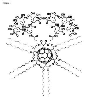
Figure 1 shows a particular substituted fullerene (which may be referred to as
an
"amphifullerene") of the present invention. The use of gray to represent three
>C(C(=O)O(CH2)1iCH3)2 moieties indicates these three moieties are bonded to
the fullerene
core at regions of the fullerene which are not directly visible to the
putative observer in this
orientation.
Figure 2 shows a cryogenic transmission electron microscopy ("cryo-TEM") image
of
a vesicle comprising the amphifullerene represented by Figure 1. The vesicle
has a diameter
of about 80 nm and a thickness of the bilayer of about 7 nm. The dark regions
in the bilayer
represent the C60-core of the amphifullerene.
Figure 3 shows the pressure as a function of a pH for a titration isotherm of
a
monolayer formed from the amphifullerene represented by Figure 1.
Figure 4 shows the UV/Vis spectrum of the Texas Ree derivative of the
amphifullerene represented by Figure 1. The Texas Ree derivative is referred
to as
compound 2 in Scheme 1, Example 3.
Figure 5 shows the UV/Vis spectrum of a partially labeled dendrofullerene
(compound 2) with 2.0 % fluorophore.
Figure 6 shows a particular substituted fullerene comprising a functional
group,
according to the present invention. The use of gray follows that of Figure 1.
The functional
group is a biotin-containing moiety and includes a linker moiety.
CA 02697093 2010-02-19
WO 2009/026315 PCT/US2008/073639
-5-
Figure 7 shows the scheme for synthesis of an amphifullerene labeled with the
fluorescence marker Texas Red . The use of gray follows that of Figure 1 and
Figure 6.
Figure 8 shows one embodiment of a method of delivering a therapeutic compound
by
the use of a carbon nanotube.
DESCRIPTION OF ILLUSTRATIVE EMBODIMENTS
In one embodiment, the present invention relates to a vesicle having an
interior, an
exterior, and a wall, wherein the wall comprises one or more layers, wherein
each layer
comprises a substituted fullerene having structure I:
(I) (B)b-Cri (A)a
wherein Cõ is a fullerene moiety comprising n carbon atoms, wherein n is an
integer
and 60 <_ n<_ 240;
B is an organic moiety comprising from 1 to about 40 polar headgroup moieties;
b is an integer and 1<_ b<_ 5;
each B is covalently bonded to the Cõ through 1 or 2 carbon-carbon, carbon-
oxygen,
or carbon-nitrogen bonds;
A is an organic moiety comprising a terminus proximal to the Cõ and one or
more
termini distal to the C, wherein the termini distal to the Cõ each comprise -
CXHy, wherein x is
an integer and 8<_ x<_ 24, and y is an integer and 1<_ y<_ 2x+1;
a is an integer, 1<_ a<_ 5;
2 <_ b+a <_ 6; and
each A is covalently bonded to the Cõ through 1 or 2 carbon-carbon, carbon-
oxygen,
or carbon-nitrogen bonds,
wherein the vesicle wall comprises at least about 50 mol% the substituted
fullerene,
and wherein the interior of the vesicle, a portion of the wall between two
layers, or both
comprise a therapeutic agent.
A "vesicle," as the term is used herein, is a collection of amphiphilic
molecules, by
which is meant, molecules which include both (a) hydrophilic ("water-loving")
regions,
typically charged or polar moieties, such as moieties comprising polar
headgroups, among
others known to one of ordinary skill in the art, and (b) hydrophobic ("water-
hating") regions,
CA 02697093 2010-02-19
WO 2009/026315 PCT/US2008/073639
-6-
typically apolar moieties, such as hydrocarbon chains, among others known to
one of
ordinary skill in the art. In aqueous solution, the vesicle is formed when the
amphiphilic
molecules form a wall, i.e., a closed three-dimensional surface. The wall
defines an interior
of the vesicle and an exterior of the vesicle. Typically, the exterior surface
of the wall is
s formed by amphiphilic molecules oriented such that their hydrophilic regions
are in contact
with water, the solvent in the aqueous solution. The interior surface of the
wall may be
formed by amphiphilic molecules oriented such that their hydrophilic regions
are in contact
with water present in the interior of the vesicle, or the interior surface of
the wall may be
formed by amphiphilic molecules oriented such that their hydrophobic regions
are in contact
io with hydrophobic materials present in the interior of the vesicle.
The amphiphilic molecules in the wall will tend to form layers, and therefore,
the wall
may comprise one or more layers of amphiphilic molecules. If the wall consists
of one layer,
it may be referred to as a "unilayer membrane" or "monolayer membrane." If the
wall
consists of two layers, it may be referred to as a "bilayer membrane." Walls
with more than
is two layers, up to any number of layers, are also within the scope of the
present invention.
The vesicle may be referred to herein as a "buckysome."
"Cõ" refers to a fullerene moiety comprising n carbon atoms.
Buckminsterfullerenes,
also known as fullerenes or, more colloquially, buckyballs, are cage-like
molecules consisting
essentially of sp2 -hybridized carbons. Fullerenes are the third form of pure
carbon, in
20 addition to diamond and graphite. Typically, fullerenes are arranged in
hexagons, pentagons,
or both. Most known fullerenes have 12 pentagons and varying numbers of
hexagons
depending on the size of the molecule. Common fullerenes include C60 and C70,
although
Ci60
fullerenes comprising up to about 400 carbon atoms are also known. Herein, " "
is
used as a representation of a C60 molecule or a C60 moiety in a molecule.
25 Fullerenes can be produced by any known technique, including, but not
limited to,
high temperature vaporization of graphite. Fullerenes are or are expected to
be commercially
available from MER Corporation (Tucson, AZ) and Frontier Carbon Corporation,
among
other sources.
The naming of specific substituted C60 isomers is complex. Within the present
30 specification, the so-called Hirsch Scheme (Hirsch, Angew. Chem. Intl. Ed.
(1994) 33(4):437-
438) will be used.
CA 02697093 2010-02-19
WO 2009/026315 PCT/US2008/073639
-7-
Methods of substituting fullerenes with various substituents are well known in
the art.
Methods include 1,3-dipolar additions (Sijbesma et al., J. Am. Chem. Soc.
(1993) 115:6510-
6512; Suzuki, J. Am. Chem. Soc. (1992) 114:7301-7302; Suzuki et al., Science
(1991)
254:1186-1188; Prato et al., J. Org. Chem. (1993) 58:5578-5580; Vasella et
al., Angew.
Chem. Int. Ed. Engl. (1992) 31:1388-1390; Prato et al., J. Am. Chem. Soc.
(1993) 115:1148-
1150; Maggini et al., Tetrahedron Lett. (1994) 35:2985-2988; Maggini et al.,
J. Am. Chem.
Soc. (1993) 115:9798-9799; and Meier et al., J. Am. Chem. Soc. (1994) 116:7044-
7048),
Diels-Alder reactions (Iyoda et al., J. Chem. Soc. Chem. Commun. (1994) 1929-
1930; Belik
et al., Angew. Chem. Int. Ed. Engl. (1993) 32:78-80; Bidell et al., J. Chem.
Soc. Chem.
Commun. (1994) 1641-1642; and Meidine et al., J. Chem. Soc. Chem. Commun.
(1993) 1342-
1344), other cycloaddition processes (Saunders et al., Tetrahedron Lett.
(1994) 35:3869-
3872; Tadeshita et al., J. Chem. Soc. Perkin. Trans. (1994) 1433-1437; Beer et
al., Angew.
Chem. Int. Ed. Engl. (1994) 33:1087-1088; Kusukawa et al., Organometallics
(1994)
13:4186-4188; Averdung et al., Chem. Ber. (1994) 127:787-789; Akasaka et al.,
J. Am.
Chem. Soc. (1994) 116:2627-2628; Wu et al., Tetrahedron Lett. (1994) 35:919-
922; and
Wilson, J. Org. Chem. (1993) 58:6548-6549); cyclopropanation by
addition/elimination
(Hirsch et al., Agnew. Chem. Int. Ed. Engl. (1994) 33:437-438 and Bestmann et
al., C. Tetra.
Lett. (1994) 35:9017-9020); and addition of carbanions/alkyl lithiums/Grignard
reagents
(Nagashima et al., J. Org. Chem. (1994) 59:1246-1248; Fagan et al., J. Am.
Chem. Soc.
(1994) 114:9697-9699; Hirsch et al., Agnew. Chem. Int. Ed. Engl. (1992) 31:766-
768; and
Komatsu et al., J. Org. Chem. (1994) 59:6101-6102); among others.
The synthesis of substituted fullerenes is reviewed by Murphy et al., U.S.
Pat. No.
6,162,926.
It has been found that fullerenes, especially C60, readily receive up to six
adducts in an
octahedral addition pattern (an octahedron having six vertices) (Brettreich et
al., Angew.
Chem. Int. Ed. (2000) 39:1845-1848).
B is chosen from any organic moiety comprising from 1 to about 40 polar
headgroup
moieties. A "polar headgroup" is a moiety which is polar, by which is meant
that the vector
sum of the bond dipoles of each bond within the moiety is nonzero. A polar
headgroup can
be positively charged, negatively charged, or neutral. The polar headgroup can
be located
such that at least a portion of the moiety can be exposed to the environment
of the molecule.
Exemplary polar headgroup moieties can include, but are not limited to,
carboxylic acid,
alcohol, amide, and amine moieties, among others known in the art. Preferably,
B has from
CA 02697093 2010-02-19
WO 2009/026315 PCT/US2008/073639
-8-
about 6 to about 24 polar headgroup moieties. In one embodiment, B has a
structure wherein
the majority of the polar headgroup moieties are carboxylic acid moieties,
which are exposed
to water when the substituted fullerene is dissolved in an aqueous solvent. A
dendrimeric or
other regular highly-branched structure is suitable for the structure of B.
The value of b can be any integer from 1 to 5. In one embodiment, if more than
one B
group is present (i.e., b> 1), that all such B groups are adjacent to each
other. By "adjacent"
in this context is meant that no B group has only A groups, as defined below,
and/or no
substituent groups at all the nearest neighboring points of addition. In the
case of an
octahedral addition pattern when b> 1, "adjacent" means that the four vertices
of the
octahedron in closest proximity to the B group are not all A groups or null.
In one embodiment, B comprises 18 polar headgroup moieties and b = 1.
The polar headgroup moieties of B tend to make the B group or groups
hydrophilic.
Each B is bonded to Cõ through a covalent bond or bonds. Any covalent bond
which
a fullerene carbon is capable of forming and will not disrupt the fullerene
structure is
contemplated. Examples include carbon-carbon, carbon-oxygen, or carbon-
nitrogen bonds.
One or more atoms, such as one or two atoms, of the B group can participate in
bonding to
C,,. In one embodiment, one carbon atom of the B group is bonded to two carbon
atoms of
C, wherein the two carbon atoms of Cõ are bonded to each other.
In one embodiment, B has the amide dendron structure
>C(C(=O)OC3H6C(=O)NHC(C2H4C(=O)NHC(C2H4C(=O)OH)3)3)2.
In structure I, A is an organic moiety comprising a terminus proximal to the
Cõ and
one or more termini distal to the C,,. In one embodiment, the organic moiety
comprises two
termini distal to C,,. By "terminus proximal to Cõ" is meant a portion of the
A group that
comprises one or more atoms, such as one or two atoms, of the A group which
form a bond
or bonds to C,,. By "terminus distal to Cõ" is meant a portion of the A group
that does not
comprise any atoms which form a bond or bonds to C,,, but that does comprise
one or more
atoms which form a bond or bonds to the terminus of the A group proximal to
C,,.
Each terminus distal to the Cõ comprises -CXHy, wherein x is an integer and
8<_ x<_
24, and y is an integer and 1<_ y<_ 2x+1. The -CXHy can be linear, branched,
cyclic, aromatic,
or some combination thereof Preferably, A comprises two termini distal to C,
wherein each
-CXHy is linear, 12 <_ x<_ 18, and y = 2x+1. More preferably, in each of the
two termini, x
12 andy=25.
The termini distal to C. tend to make the A groups hydrophobic or lipophilic.
CA 02697093 2010-02-19
WO 2009/026315 PCT/US2008/073639
-9-
The value of a can be any integer from I to 5. Preferably, a is 5. In one
embodiment,
if more than one A group is present (i.e., a> 1), all such A groups are
adjacent to each other.
By "adjacent" in this context is meant that no A group has only B groups, as
defined below,
and/or no substituent groups at all the nearest neighboring points of
addition. In the case of
an octahedral addition pattern, when a> 1, "adjacent" means that the four
vertices of the
octahedron in closest proximity to the A group are not all B groups or null.
Each A is bonded to Cõ through a covalent bond or bonds. Any covalent bond
which
a fullerene carbon is capable of forming and will not disrupt the fullerene
structure is
contemplated. Examples include carbon-carbon, carbon-oxygen, or carbon-
nitrogen bonds.
One or more atoms, such as one or two atoms, of the A group can participate in
bonding to
C. In one embodiment, one carbon atom of the A group is bonded to two carbon
atoms of
C, wherein the two carbon atoms of Cõ are bonded to each other.
In one embodiment, A has the structure >C(C(=O)O(CH2)11CH3)2.
The number of B and A groups is chosen to be from 2 to 6, i.e., 2<_ b+a <_ 6.
In one
embodiment, b+a = 6. The combination of hydrophilic B group(s) and hydrophobic
A
group(s) renders the substituted fullerene amphiphilic. The number and
identity of B groups
and A groups can be chosen to produce a fullerene with particular amphiphilic
qualities
which may be desirable for particular intended uses.
In one preferred embodiment, the substituted fullerene has structure II:
X, C60 X
5
lI~
wherein X' is >C(C(=O)OC3H6C(=O)NHC(C2H4C(=O)NHC(C2H4C(=O)OH)3)3)2
and each X is >C(C(=O)O(CH2)11CH3)2. A structural representation of a
substituted fullerene
having structure II is given in Figure 1, wherein each X is
>C(C(=O)O(CH2)11CH3)2.
In one embodiment, the substituted fullerene has the structure shown in Figure
1.
The substituted fullerene can further comprise one or more functional groups
covalently linked to one or more B groups, one or more A groups, or both. In
one
CA 02697093 2010-02-19
WO 2009/026315 PCT/US2008/073639
-10-
embodiment, the one or more functional groups are covalently linked to one or
more B
groups.
By "functional group" is meant a group that binds to a specific compound, and
thus
allows the substituted fullerene to be associated with the specific compound.
In one embodiment, the functional group is biotin or a biotin-containing
moiety, i.e., a
moiety which will bind to streptavidin.
In another embodiment, the functional group is an antigen-binding moiety, by
which
is meant a moiety comprising the antigen-recognition site of an antibody.
Examples of a
moiety comprising the antigen-recognition site of an antibody include, but are
not limited to,
monoclonal antibodies, polyclonal antibodies, Fab fragments of monoclonal
antibodies, Fab
fragments of polyclonal antibodies, Fab2 fragments of monoclonal antibodies,
and Fab2
fragments of polyclonal antibodies, among others. Single chain or multiple
chain antigen-
recognition sites can be used. Multiple chain antigen-recognition sites can be
fused or
unfused.
The antigen-binding moiety can be selected from any known class of antibodies.
Known classes of antibodies include, but are not necessarily limited to, IgG,
IgM, IgA, IgD,
and IgE. The various classes also can have subclasses. For example, known
subclasses of
the IgG class include, but are not necessarily limited to, IgGI, IgG2, IgG3,
and IgG4. Other
classes have subclasses that are routinely known by one of ordinary skill in
the art.
The antigen-binding moiety can be selected from an antibody derived from any
species. "Derived from," in this context, can mean either prepared and
extracted in vivo from
an individual member of a species, or prepared by known biotechnological
techniques from a
nucleic acid molecule encoding, in whole or part, an antibody peptide
comprising invariant
regions which are substantially identical to antibodies prepared in vivo from
an individual
member of the species or an antibody peptide recognized by antisera
specifically raised
against antibodies from the species. Exemplary species include, but are not
limited to,
human, chimpanzee, baboon, other primate, mouse, rat, goat, sheep, and rabbit,
among others
known in the art. In one embodiment, the antibody is chimeric, i.e., comprises
a plurality of
portions, wherein each portion is derived from a different species. A chimeric
antibody,
wherein one of the portions is derived from human, can be considered a
humanized antibody.
Antigen-recognition moieties are available that recognize antigens associated
with a
wide variety of cell types, tissues, and organs, and a wide variety of medical
conditions, in a
wide variety of mammalian species. Exemplary medical conditions include, but
are not
CA 02697093 2010-02-19
WO 2009/026315 PCT/US2008/073639
-11-
limited to, cancers, such as lung cancer, oral cancer, skin cancer, stomach
cancer, colon
cancer, nervous system cancer, leukemia, breast cancer, cervical cancer,
prostate cancer, and
testicular cancer; arthritis; infections, such as bacterial, viral, fungal, or
other microbial
infections; and disorders of the skin, the eye, the vascular system, or other
cell types, tissues,
or organs; among others.
Exemplary antigen-recognition moieties known in the art include, but are not
limited
to, those derived from antibodies against vascular endothelial growth factor
receptor
(VEGF-r) (available from Imclone, New York, NY), antibodies against epidermal
growth
factor receptor (EGF-r) (available from Abgenix, Fremont, CA), antibodies
against
polypeptides associated with lung cancers (available from Corixa Corporation,
Seattle, WA),
antibodies against human tumor necrosis factor alpha (hTNF-a) (available from
BASF A.G.,
Ludwigshafen, Germany), among others known in the art.
Antigen-recognition moieties can be prepared by various techniques known in
the art.
These techniques include, but are not limited to, the immunological technique
described by
Kohler and Milstein in Nature 256, 495-497 (1975) and Campbell in "Monoclonal
Antibody
Technology, The Production and Characterization of Rodent and Human
Hybridomas" in
Burdon et al., Eds., Laboratory Techniques in Biochemistry and Molecular
Biology, Volume
13, Elsevier Science Publishers, Amsterdam (1985); as well as by the
recombinant DNA
techniques described by Huse et al in Science 246, 1275-1281 (1989); among
other
techniques known to one of ordinary skill in the art.
In a further embodiment, the functional group is a tissue-recognition moiety,
by which
is meant a moiety that recognizes cells of a particular tissue by binding
specifically with one
or more proteins expressed by cells of the tissue and present on the exterior
of the cells.
Examples of such moieties include, but are not limited to, peptides, among
other classes of
moieties. The term "peptides," as used herein, encompasses any peptide
comprising 1 or
more amino acids. Exemplary peptides include, but are not limited to, VEGF,
EGF, other
growth factors, and other ligands for receptors (such as cell surface
receptors, cytoplasmic
receptors, and nuclear receptors), among others.
In one embodiment, wherein the polar headgroups are carboxylic acid moieties
and
the functional group is a peptide, a functional group can be linked to a polar
headgroup via an
amino linkage between a carboxylic acid in the polar headgroup and an amine in
the peptide.
Tissue-recognition moieties can be derived from any species or plurality of
species,
and can be selected to target any cell type, tissue, or organ, or treat any
disease.
CA 02697093 2010-02-19
WO 2009/026315 PCT/US2008/073639
-12-
The inclusion of functional groups will enhance targeting of a substituted
fullerene to
a particular tissue. The inclusion of functional groups in at least some of
the substituted
fullerene molecules of the vesicle membrane will enhance the targeting of the
vesicle to a
particular tissue.
The functional group can also comprise a linker or linkers, i.e., moieties
which are
covalently bonded to both (a) the biotin-containing moiety, the antigen-
binding moiety, or the
tissue-recognition moiety, as defined above, and (b) the substituted
fullerene, as defined
above. In one embodiment, wherein the polar headgroups are carboxylic acid
moieties, the
linker can be an ester.
If some of the substituted fullerene molecules in the vesicle membrane
comprise a
functional group, from about 0.01 mole% to about 100 mole% of the substituted
fullerene
molecules of the vesicle membrane comprise the functional group. In the
interest of reduced
expense, and in light of the observation that many functional groups are
highly sensitive to
the specific compounds which they bind, preferably from about 0.01 mole% to
about 1
mole% of the substituted fullerene molecules of the vesicle membrane comprise
the
functional group.
In one embodiment, the vesicle wall comprises at least about 50 mol% of the
substituted fullerene. The balance of the vesicle membrane comprises other
amphiphilic
compounds. By "amphiphilic compound" in this context is meant a compound whose
molecules each comprise hydrophobic and hydrophilic regions. Such amphiphilic
compounds include, but are not limited to, commercially-available lipids, such
as dimethyl
dioctadecyl ammonium bromide, phosphatidylcholine, and
dioleoyltrimethylammonium
phosphate, among others.
In one embodiment, the vesicle wall comprises at least about 75 mol% a
substituted
fullerene having structure I. In another embodiment, the vesicle wall consists
essentially of a
substituted fullerene having structure I.
In one embodiment, the vesicle wall is a bilayer membrane. The bilayer
membrane
comprises two layers, an interior layer formed from substituted fullerene and
other
amphiphilic compound or compounds, if any, wherein substantially all
substituted fullerene
and other amphiphilic molecules are oriented with their hydrophobic portions
toward the
exterior layer, and an exterior layer formed from substituted fullerene and
other amphiphilic
compound or compounds, if any, wherein substantially all substituted fullerene
and other
amphiphilic molecules are oriented with their hydrophobic portions toward the
interior layer.
CA 02697093 2010-02-19
WO 2009/026315 PCT/US2008/073639
-13-
As a result, the hydrophilic portions of substantially all molecules of each
of the interior and
exterior layers are oriented towards aqueous solvent in the vesicle interior
or exterior to the
vesicle.
Because the hydrophilicity of the hydrophilic portions of the molecules may
change if
the pH or other parameters of the solvent are changed (e.g., if the pH is
increased above the
pKa of the polar headgroup moieties of the B groups of the substituted
fullerenes, the
substituted fullerenes will readily separate from the vesicle membrane and
enter the aqueous
phase), the pH and other parameters of the solvent can be adjusted as a matter
of routine
experimentation by one of ordinary skill in the art in order to allow vesicle
formation.
io Because the vesicle comprises an interior, and the interior comprises an
aqueous
solvent, the vesicle can further comprise a therapeutic agent in the interior
of the vesicle.
Typically, such a compound is introduced to the interior of the vesicle as
part of the process
of forming the vesicle, e.g., by introducing the therapeutic agent, the
substituted fullerene,
and other amphiphilic compounds, if any, into an aqueous solvent under pH and
other
conditions whereby the substituted fullerene and other amphiphilic compounds,
if any, self-
assemble a vesicle, with molecules of the therapeutic agent being sequestered
in the vesicle
during vesicle self-assembly. To facilitate self-assembly, preferably the pH
of the solvent is
less than about 8Ø However, other techniques of including a therapeutic
agent in the interior
of the vesicle known in the art can be used.
In one embodiment, when the interior of the vesicle comprises water and
substantially
does not comprise a hydrophobic solvent, the therapeutic agent is a water-
soluble drug or
other compound which, upon administration to a mammal, can alleviate a medical
condition
from which the mammal suffers. In one embodiment, the therapeutic agent is
selected from
the group consisting of water-soluble anti-cancer drugs.
In one embodiment, the vesicle wall is a monolayer membrane, in which
molecules of
the substituted fullerene and other amphiphilic compound(s), if any, are
substantially all
oriented such that their hydrophilic regions are adjacent to a polar or
aqueous phase, either in
the vesicle interior or exterior to the vesicle, and their hydrophobic regions
are adjacent to an
apolar phase, either in the vesicle interior or exterior to the vesicle. In
this context, "polar"
and "apolar" are relative terms, in that a phase with greater hydrophilicity,
miscibility with
water, etc. is more polar than a phase with poorer solubility in water. In one
embodiment, in
the vesicle the hydrophobic regions of substantially all the molecules of the
monolayer
membrane are oriented toward the interior of the vesicle.
CA 02697093 2010-02-19
WO 2009/026315 PCT/US2008/073639
-14-
In one embodiment, when the interior of the vesicle comprises a hydrophobic
solvent
or other apolar material and substantially does not comprise water, the
therapeutic agent is a
hydrophobic drug or other compound which, upon administration to a mammal, can
alleviate
a medical condition from which the mammal suffers. The terms "hydrophobic" and
"lipophilic" are synonyms wheresoever they appear herein.
Any hydrophobic compound can be included in the vesicle interior, typically by
providing the substituted fullerene, other amphiphilic compounds, if any, and
the
hydrophobic compound in an aqueous solvent under pH and other conditions
wherein a
monolayer membrane will form, and allowing the vesicle to self-assemble,
during which
process the hydrophobic compound will be sequestered in the interior of the
vesicle. To
facilitate assembly of the vesicle, preferably the pH of the solvent is less
than about 8Ø
The vesicle can be unilamellar (having a single bimolecular membrane),
multilamellar
(having a plurality of bimolecular membranes, "onion-like") or hemilamellar
(having a single
unimolecular membrane). The vesicle can have a size from about 50 Angstroms to
about 10
microns. The size, number and nature of membranes, and other parameters of the
vesicle can
be adjusted as a matter of routine experimentation.
In one embodiment, the therapeutic agent is associated with a fullerene, such
as a
substituted fullerene comprising a functional group, or a carbon nanotube,
among others. The
association can be a covalent link between the fullerene core or carbon
nanotube and the
therapeutic agent; a covalent link between a substituent of the fullerene or
carbon nanotube, if
any, and the therapeutic agent; an ionic association between a positively- or
negatively-
charged group on the substituent of the fullerene, the substituent of the
carbon nanotube, or
the carbon nanotube, and an oppositely-charged group on the therapeutic agent;
hydrogen
bonding of the therapeutic agent to a substituent of the carbon nanotube; van
der Waals
attraction between the therapeutic agent and the carbon nanotube; or the
encapsulation of the
therapeutic agent in the fullerene core or the carbon nanotube, among others.
A covalent link can be direct or it can make use of a covalent linker linking
the
therapeutic agent and the fullerene core, substituent, if any, of the
fullerene, carbon nanotube,
or substituent, if any, of the carbon nanotube. In one embodiment, the
covalent link can be
cleavable by an appropriate cleaving technique, such as photolysis, enzymatic
cleavage, or
chemical cleavage, among others. In another embodiment, a non-covalent
association
between the therapeutic agent and the fullerene or carbon nanotube can be
dissociated by
application of an appropriate chemical, e.g., when the association is an ionic
association, the
CA 02697093 2010-02-19
WO 2009/026315 PCT/US2008/073639
-15-
association can be dissociated by application of a charged compound of the
same charge as
the charged group on the therapeutic agent. Other techniques for dissociating
a non-covalent
association between the carbon nanotube or substituted fullerene and the
therapeutic agent
will be apparent to one of ordinary skill in the art in light of the present
specification. A
chemical or enzyme used to promote dissociation can be referred to as an
"adjuvant."
Any therapeutic agent, from any source, can be used. As is known in the art,
therapeutic agents can be invented by rational drug design, combinatorial
chemistry, or
serendipitous discovery, among other known techniques. Naturally occurring
therapeutic
agents can be derived from a plant, an animal, a bacterium, a fungus, a virus,
or another
organism. Therapeutic agents can be synthesized by known chemical synthesis
techniques.
The therapeutic agent can treat any disease. Exemplary diseases include, but
are not
limited to, cancers, autoimmune diseases, infections, liver diseases, and
neurological
diseases, among many others.
In one embodiment, the therapeutic agent is an anti-cancer drug. Examples of
anti-
cancer drugs include paclitaxel (commercially available as Taxol, Bristol-
Myers Squibb),
doxorubicin (also known under the trade name Adriamycin), vincristine (known
under the
trade names Oncovin, Vincasar PES, and Vincrex), actinomycin D, altretamine,
asparaginase,
bleomycin, busulphan, capecitabine, carboplatin, carmustine, chlorambucil,
cisplatin,
cyclophosphamide, cytarabine, dacarbazine, daunorubicin, epirubicin,
etoposide, fludarabine,
fluorouracil, gemcitabine, hydroxyurea, idarubicin, ifosfamide, irinotecan,
lomustine,
melphalan, mercaptopurine, methotrexate, mitomycin, mitozantrone, oxaliplatin,
procarbazine, steroids, streptozocin, taxotere, tamozolomide, thioguanine,
thiotepa, tomudex,
topotecan, treosulfan, UFT (uracil-tegufur), vinblastine, and vindesine, among
others.
Other therapeutic agents include, but are not limited to, the following:
hydrocodone,
atorvastatin, estrogen, atenolol, levothyroxine, azithromycin, furosemide,
amoxicillin,
amlodipine, alprazolam, albuterol, loratadine, hydrochlorothiazide,
omeprazole, sertraline,
paroxetine, triamterene, lansoprazole, ibuprofen, celecoxib, simvastatin,
cephalexin,
metformin, rofecoxib, lisinopril, amoxicillin, clavulanate, propoxyphene,
progesterone,
prednisone, norgestimate, ethinyl estradiol, acetaminophen, codeine,
cetirizine, fexofenadine,
levothyroxine, amoxicillin, metoprolol, lorazepam, metoprolol, fluoxetine,
ranitidine,
zolpidem, citalopram, amitriptyline, alendronate, quinapril, sildenafil
citrate, pravastatin,
naproxen, gabapentin, warfarin, ciprofloxacin, verapamil, digoxin, albuterol,
bupropion,
lisinopril, clonazepam, tramadol, cyclobenzaprine, trazodone, fluticasone,
montelukast,
CA 02697093 2010-02-19
WO 2009/026315 PCT/US2008/073639
-16-
diazepam, isosorbide mononitrate s.a., glyburide, venlafaxine, levofloxacin,
medroxyprogesterone, amoxicillin, fluconazole, enalapril, warfarin,
carisoprodol, trimeth,
sulfameth, fluticasone propionate, benazepril, mometasone, doxycycline,
estradiol,
allopurinol, rosiglitazone maleate, clopidogrel, propranolol, amlodipine,
benazepril,
methylprednisolone, valsartan, losartan, insulin, clonidine, diltiazem,
loratidine,
pseudoephedrine, latanoprost, pioglitazone, loratidine, pseudoephedrine,
risperidone,
fexofenadine, pseudoephedrine, doxazosin, raloxifene, norethindrone, folic
acid, penicillin,
oxycodone, temazepam, diltiazem, salmeterol, fosinopril, oxycodone, ramipril,
promethazine,
terazosin, olanzapine, gemfibrozil, levothyroxine, norethindrone, sumatriptan,
hydroxyzine,
meclizine, losartan, rabeprazole, phenytoin, clarithromycin, glimepiride,
pantoprazole,
spironolactone, ipratropium, albuterol, tamsulosin, penicillin, lisinopril,
metoclopramide,
minocycline, bisoprolol, digoxin, valsartan, metronidazole, cefprozil,
triamcinolone,
glipizide, norethindrone, levonorgestrel, cefuroxime, nystatin, captopril,
promethazine,
codeine, acyclovir, norgestimate, oxycodone, irbesartan, nefazodone,
mirtazapine,
valacyclovir, methylphenidate, cerivastatin, fluoxetine, nitrofurantoin,
loratadine, glyburide,
metformin, metformin, diltiazem, desogestrel, mupirocin, 1-norgestrel,
fluvastatin, aspirin,
clarithromycin, clindamycin, esomeprazole, metaxalone, nortriptyline,
cimetidine,
fenofibrate, iprotropium bromide, tamoxifen, calcitonin salmon, felodipine,
levonorgestrel,
salmeterol, fluticasone, theophylline, tetracycline, tolterodine,
gatifloxacin, nifedipine,
diclofenac, triamcinolone acetonide, promethazine, indomethacin, benzonatate,
phenobarbital, naproxen sodium, mometasone, hydrocodone, glipizide,
divalproex,
nitroglycerin, and phenazopyridine, among others.
One or more therapeutic agents can be used in any composition or method of the
present invention.
The mode of action of the therapeutic agent can be chemotherapeutic,
radiotherapeutic, or possess another mode of action. In one embodiment, the
therapeutic
agent can mediate the application of light, heat, or other external energy in
a manner which
allows the light, heat, or other external energy to perform a therapeutic
action.
In one embodiment, the therapeutic agent is present between two layers of the
wall.
The therapeutic agent, in this embodiment, may be a water-soluble compound or
a lipophilic
compound.
In another embodiment, the vesicle can comprise a sensor molecule. A "sensor
molecule," as used herein, is a molecule which can selectively associate with
a particular
CA 02697093 2010-02-19
WO 2009/026315 PCT/US2008/073639
-17-
atom or molecule. Any sensor molecule known in the art can be used. The sensor
molecule
can be linked with a carbon nanotube, a fullerene, or anchored in a vesicle
wall by
hydrophobic, hydrophilic, or both types of interactions.
In a further embodiment, the vesicle can comprise a diagnostic agent. A
"diagnostic
agent," as used herein, is a molecule which can be readily detected by the
application of
electromagnetic radiation, heat, cooling, the measurement of radioactivity, or
other
techniques known in the art. The diagnostic agent can be linked with a carbon
nanotube, a
fullerene, or anchored in a vesicle wall by hydrophobic, hydrophilic, or both
types of
interactions.
In another embodiment, the present invention relates to a method of
administering a
therapeutic agent to a mammal, comprising:
(i) administering a solution comprising a pharmaceutically effective amount of
the
therapeutic agent, wherein the therapeutic agent is present in (a) the
interior of a vesicle
is having an interior, an exterior, and a wall, (b) a portion of the wall
between two layers, or (c)
both, to the mammal, wherein the wall comprises one or more layers and each
layer
comprises a substituted fullerene having structure I.
The vesicle, the substituted fullerene, and the therapeutic agent are as
described
above. In one embodiment, the vesicle wall comprises at least about 75 mol% a
substituted
fullerene having structure I. In another embodiment, the vesicle wall consists
essentially of a
substituted fullerene having structure I.
In one embodiment, the vesicle wall is a monolayer membrane. In another
embodiment, the vesicle wall is a bilayer membrane.
In one embodiment, from about 0.01 mole% to about 100 mole% of the substituted
fullerene molecules in the vesicle wall further comprise a functional group
covalently linked
to a B group and the functional group recognizes a tissue. In another
embodiment, the
functional group is selected from the group consisting of biotin-containing
moieties, antigen-
binding moieties, and tissue-recognition moieties, as defined above.
The pharmaceutically effective amount of the therapeutic agent will vary
depending
on the compound, the intended effect thereof, the administration regimen, and
the body
weight or other characteristics of the mammal, among others apparent to one of
ordinary skill
in the art. The dose of the therapeutic agent will typically be in the range
of from about 0.001
mg/kg body weight to about 1000 mg/kg body weight. In one embodiment, the dose
of the
CA 02697093 2010-02-19
WO 2009/026315 PCT/US2008/073639
-18-
therapeutic agent will typically be within the above range and greater than
about 0.01 mg/kg
body weight. In another embodiment, the dose of the therapeutic agent will
typically be
within the above range and less than about 100 mg/kg body weight.
In one embodiment, the therapeutic agent is an anti-cancer drug. More
preferably, the
anti-cancer drug is selected from the group consisting of paclitaxel,
doxorubicin, and
vincristine.
Any technique for incorporating the therapeutic agent in the vesicle interior
or
between layers of the vesicle wall can be used. An exemplary technique is
described above,
wherein the vesicle is formed in the presence of the therapeutic agent in an
aqueous solvent
io under pH and other conditions wherein the vesicle can form. This technique
can be
performed on either a batch or a continuous basis. Other techniques, however,
can be used,
such as microinjection of a solution of the therapeutic agent into the vesicle
interior, among
others.
In the administering step, a solution comprising the vesicle and the
therapeutic agent
in the interior thereof, between two layers of the wall thereof, or both is
introduced into the
mammal. The solution comprises a polar or aqueous solvent and the vesicle
comprising the
therapeutic agent. The solution can further comprise adjuvants, preservatives,
and other
compounds whose inclusion in light of the formation, storage, and/or use of
the solution may
be desirable.
Any mammal for which it is desired to introduce the therapeutic agent can be
the
subject of the method. In one embodiment, the mammal is a human.
The term "administering," as used herein, is intended to encompass all
techniques of
introducing a compound to a mammal. Exemplary routes of administration include
transdermal, subcutaneous, intravenous, intraarterial, intramuscular, oral,
rectal, and nasal,
among others.
In one embodiment, the vesicle comprises substituted fullerenes further
comprising a
functional group with identity and concentration as described above. A vesicle
comprising
such substituted fullerenes can be directed toward a particular tissue.
"Particular tissue" in this context is not meant to be limiting to one cell
type, but may
be meant to refer to specific bodily fluids, specific organs comprising a
variety of tissues, etc.
Particular tissues to which it may be desirable to direct the vesicles
include, but are not
limited to, gastrointestinal tissues, circulatory tissues, lymphatic tissues,
biliary tissues,
CA 02697093 2010-02-19
WO 2009/026315 PCT/US2008/073639
-19-
cerebrospinal fluid, synovial fluid, the aqueous humor of the eye, and tumors
in the foregoing
or any other tissue or cell type.
Fullerenes themselves generally have toxicological properties similar to those
of
carbon, and substituted fullerenes are generally not expected to possess toxic
activities. For
example, repeated transdermal administration of fullerenes in benzene for up
to 24 weeks
(dose = 200 g/day) to mice did not result in either benign or malignant skin
tumor formation
(Nelson et al., Toxicology & Indus. Health (1993) 9(4):623-630). Further, no
effect on either
DNA synthesis or ornithine decarboxylase activity in dermal cells was observed
over a 72-hr
time course after treatment. Zakharenko et al., Doklady Akademii Nauk. (1994)
335(2):261-
262, have shown that C6o did not produce chromosomal damage at relatively high
doses.
In one embodiment, the functional group interacts directly with the particular
tissue.
For example, if the functional group is an antigen-binding moiety, and the
antigen recognized
by the moiety is a protein present on the surface of cells of a particular
tissue, then the
functional group will bind the protein and direct the vesicle to the
particular tissue. In
another example, if the functional group is a tissue-recognition moiety, then
the functional
group will recognize a particular tissue and direct the vesicle to the
particular tissue.
Antigen-binding and tissue-recognition moieties, and the antigens they bind
and tissues they
recognize, are well known in the art. Other direct interactions between the
functional group
and a particular tissue are possible and are contemplated as part of the
present invention.
In another embodiment, the functional group interacts indirectly with a
particular
tissue. By this is meant that the functional group interacts with an adjuvant,
and the adjuvant
interacts with a particular tissue. "Adjuvant" as used herein refers to any
molecule, whether
occurring in vivo or introduced by administration to the mammal, which
provides a beneficial
function. In one embodiment, the adjuvant comprises an antigen-binding moiety
or a tissue-
recognition moiety and a streptavidin moiety, and the functional group of the
substituted
fullerene comprises a biotin-containing moiety. The vesicle interacts with the
adjuvant
through the biotin-containing moiety of the substituted fullerene and the
streptavidin moiety
of the adjuvant, and the adjuvant interacts with a particular tissue through
the antigen-binding
moiety or tissue-recognition moiety as described above.
Adjuvants can provide other or additional beneficial functions. In one
embodiment,
an adjuvant facilitates union of the vesicle with the membrane of a cell of a
tissue. The union
of the vesicle with the membrane will lead to introduction of the therapeutic
agent contained
within the vesicle into the cytoplasm of the cell.
CA 02697093 2010-02-19
WO 2009/026315 PCT/US2008/073639
-20-
Many useful adjuvants are not present in vivo in the mammal. Therefore, in one
embodiment, the method further comprises administering an adjuvant to the
mammal,
wherein the adjuvant facilitates recognition of the tissue by a functional
group, union of the
vesicle with the membrane of a cell of the tissue, or both. The adjuvant is as
described
above, and can be administered via any route of administration, as described
above. The
adjuvant can be administered before, after, or simultaneously with the
solution comprising
the vesicle. Typically, the adjuvant is administered via the same route as the
solution
comprising the vesicle, but the adjuvant can be administered by a different
route, if desired.
In one embodiment, the method allows the systemic distribution of the
therapeutic
agent to the mammal, through in vivo disaggregation of the vesicle. The method
allows the
direction of the vesicle comprising the therapeutic agent to a particular
tissue. In many cases,
it is desirable to release the therapeutic agent when the vesicle is in close
proximity to the
particular tissue.
One technique by which the therapeutic agent can be released in close
proximity to
the tissue involves union of the vesicle with the membrane of a cell of the
tissue. Union can
be facilitated by use of particular adjuvants, as described above.
Another set of techniques by which the therapeutic agent can be released in
close
proximity to the tissue involves disaggregation of the vesicle when the
vesicle is in close
proximity to the tissue. One such technique involves raising the pH of bodily
fluids in which
the vesicle is present. By raising the pH of such bodily fluids, the number of
charged
carboxyl groups (-COO-) on the B group of the substituted fullerene will
increase and,
depending on the pH and the precise structure of the substituted fullerene,
molecules of the
substituted fullerene may find it more favorable in free energy terms to enter
the aqueous
solution than to remain in the vesicle membrane. This leads to disaggregation
of the vesicle
and release of the therapeutic agent. Therefore, in one embodiment, the method
further
comprises raising the pH of bodily fluids in which the solution comprising the
vesicle and the
therapeutic agent in the interior thereof is present to a pH at which the
vesicle disaggregates.
In one embodiment, the pH is raised to greater than about 11Ø
Any technique appropriate for raising the pH can be used. Typically, raising
the pH
can be performed by administering a solution to the mammal comprising a
compound that
will raise the pH of bodily fluids, e.g., a basic solution. Such a solution
can be administered
via any route described above or known in the art.
CA 02697093 2010-02-19
WO 2009/026315 PCT/US2008/073639
-21-
Another technique for disaggregating the vesicle lies in the observation that
some
substituted fullerenes, such as some substituted fullerenes formed from Diels-
Alder
cycloaddition reactions, readily lose their substituent groups at temperatures
slightly above
room temperature depending on the diene structure of the substituent, and that
some other
substituted fullerenes, such as aldehyde-derived adducts, readily lose their
substituent groups
under moisture or heat. The B groups, A groups, or both of the substituted
fullerene can be
chosen to be added to the fullerene through such reactions. As a result,
depending on the
nature of the substituent groups and other parameters apparent to one of
ordinary skill in the
art, and as a matter of routine experimentation, the substituted fullerene can
be designed such
that it loses B groups, A groups, or both upon or soon after administration.
The loss of B
groups, A groups, or both would reduce the amphiphilic character of the
substituted fullerene,
and as a result, would reduce its ability to form or maintain the vesicle
membrane. This
would be expected to disaggregate the vesicle.
A further technique for disaggregation of the vesicle is natural
disaggregation when
the vesicle is in contact with a bodily fluid. This process may be accelerated
by the presence
of factors (proteins, lipids, salts, etc.) which may be present in the bodily
fluid. This process
may occur without further intervention by the operator of the method.
Another technique for disaggregation of the vesicle involves the use of
photocleavable polar headgroups. Photocleavable moieties, such as -Ar(N02)CH2-
, wherein
Ar is an aromatic moiety, can be used to link the fullerene core with the
polar headgroups,
and vesicles can be formed from such substituted fullerenes. Upon irradiation
of the vesicle
of this embodiment by electromagnetic radiation of an appropriate wavelength,
the
photocleavable moiety can be cleaved and the resulting removal of polar
headgroups from the
substituted fullerene can lead to disaggregation of the vesicle.
A further technique for disaggregation of the vesicle involves the use of
ultrasonic
energy. Upon exposing a region of a mammalian body to sufficient ultrasonic
energy,
vesicles present in the region can disaggregate and release a therapeutic
agent, if any,
associated with the vesicle. The vesicles could be present in the region as a
result of targeting
to a specific cell type, tissue, or organ, or could be present in the region
as a result of
systemic circulation.
Another technique for disaggregation of the vesicle involves the use of
biological
sensor molecules associated with the vesicle, such as sensor moieties
covalently linked with a
fullerene molecule in the vesicle wall or sensor molecules anchored by
hydrophobic,
CA 02697093 2010-02-19
WO 2009/026315 PCT/US2008/073639
-22-
hydrophilic, or both types of interactions with the vesicle wall. A particular
sensor molecule
can detect atoms and molecules present in bodily fluids, such as blood.
Exemplary atoms and
molecules present in blood include glucose; minerals, such as calcium,
potassium, or sodium,
among others; hormones, such as insulin, thyroid hormone, testosterone,
estrogen, or growth
factors, among others; peptides; enzymes; or blood constituents, such as red
blood cell
surface molecules, white blood cell surface molecules, platelets, or
extracellular hemoglobin,
among others; among others. In one embodiment, the sensor molecule can be
chosen such
that, upon encountering a bodily region wherein the bodily fluid has an excess
of the atom or
molecule detectable by the sensor molecule, the sensor molecule binds the atom
or molecule
and leaves the vesicle or the sensor molecule undergoes a conformational
change. In either
case, if the sensor molecule, the vesicle, and other features are properly
chosen, the vesicle
can disaggregate. Alternatively, the sensor molecule can be chosen such that
disaggregation
of the vesicle occurs upon encountering a bodily region wherein the bodily
fluid has a
shortage of the atom or molecule.
In one embodiment, the present invention relates to a method of diagnosing a
medical
condition in a mammal, comprising:
(i) administering a solution comprising a pharmaceutically effective amount of
a
diagnostic agent, wherein the diagnostic agent is present in (a) the interior
of a vesicle having
an interior, an exterior, and a wall, (b) a portion of the wall between two
layers, or (c) both,
wherein the vesicle is as described above and comprises fullerenes substituted
with a
functional group, as described above, to the mammal; and
(ii) detecting the diagnostic agent.
Any agent which is detectable by any means can be the "diagnostic agent." In
one
embodiment, the diagnostic agent is a fluorophore, which can be made to
fluoresce upon
exposure to a particular wavelength of electromagnetic radiation. In another
embodiment, the
diagnostic agent is a radionuclide, which can be detected by known techniques.
Other
diagnostic agents are known in the art.
The method is similar to the method of treatment described above, except that
instead
of releasing a therapeutic agent to a particular cell type or tissue, a
diagnostic agent is
directed to the vicinity of the cell type or tissue by the use of a vesicle
comprising fullerenes
substituted with functional groups such as antigen-binding moieties or tissue-
recognition
moieties.
CA 02697093 2010-02-19
WO 2009/026315 PCT/US2008/073639
-23-
In one embodiment, the present invention relates to a method of administering
a
therapeutic agent to a mammal, comprising:
(i) administering a solution comprising (a) a substituted fullerene,
comprising (a-i) a
fullerene moiety comprising n carbon atoms, wherein n is an integer and 60 <_
n<_ 240, and
(a-ii) a functional group selected from the group consisting of biotin-
containing moieties,
antigen-binding moieties, and tissue-recognition moieties, and (b) a
pharmaceutically
effective amount of the therapeutic agent, wherein the therapeutic agent is
associated with the
substituted fullerene.
The substituted fullerene and the therapeutic agent are as described above. In
one
embodiment, the therapeutic agent is an anti-cancer drug.
In one embodiment, the method further comprises (ii) administering an adjuvant
to
the mammal, wherein the adjuvant facilitates dissociation of the therapeutic
agent from the
substituted fullerene. An appropriate adjuvant for a particular substituted
fullerene and a
particular therapeutic agent can be selected in light of the discussion above.
The ability of vesicles, alternatively referred to as liposomes, to function
as drug
delivery vehicles is well known in the art, as discussed above and as known
from work by
Alza Corporation (Mountain View, CA), the University of California, and Sequus
Pharmaceuticals, among other entities.
In another embodiment, the present invention relates to a carbon nanotube
composition, comprising a carbon nanotube and at least one therapeutic agent
associated with
the carbon nanotube.
As is well known, carbon has not only the propensity to self-assemble from a
high
temperature vapor to form perfect spheroidal closed cages (fullerenes), but
also, with the aid
of a transition metal catalyst, to assemble into single-wall cylinders with
may be sealed at one
or both ends with a semifullerene dome. Single-wall carbon nanotubes, also
known as single
wall tubular fullerenes, are cylindrical molecules consisting essentially of
sp2 -hybridized
carbons. These tubes may be thought of as two-dimensional single crystals of
carbon. Multi-
wall cylinders, comprising nested single-wall cylinders, have also been
observed. Both
multi-wall and single-wall cylinders are encompassed by the term "carbon
nanotube," as used
herein.
CA 02697093 2010-02-19
WO 2009/026315 PCT/US2008/073639
-24-
In defining carbon nanotubes, it is helpful to use a recognized system of
nomenclature. Herein will be used the carbon nanotube nomenclature described
by
Dresselhaus et al., Science ofFullerenes and Carbon Nanotubes, Ch. 19. Single
wall tubular
fullerenes are distinguished from each other by a double index (x, y), where x
and y are
integers that describe how to cut a single strip of hexagonal graphite such
that its edges join
seamlessly when the strip is wrapped onto the surface of a cylinder. When x =
y, the
resultant tube is said to be of the "arm-chair" or (x, x) type, since when the
tube is cut
perpendicularly to the tube axis, only the sides of the hexagons are exposed
and their pattern
around the periphery of the tube edge resembles the arm and seat of an arm
chair repeated n
times. When y = 0, the resultant tube is said to be of the "zig zag" or (x, 0)
type, since when
the tube is cut perpendicular to the tube axis, the edge is a zig zag pattern.
Where x:~ y and y
~ 0, the resulting tube has chirality. The electronic properties of the
nanotube are dependent
on the conformation, for example, arm-chair tubes are metallic and have
extremely high
electrical conductivity. Other tube types are metallic, semi-metals or semi-
conductors,
depending on their conformation. Regardless of tube type, all single-wall
nanotubes have
extremely high thermal conductivity and tensile strength.
Single-wall carbon nanotubes (SWNTs) are much more likely to be free of
defects
than are multi-wall carbon nanotubes. This is believed to be because multi-
wall carbon
nanotubes can survive occasional defects, whereas SWNTs have no neighboring
walls to
compensate for defects by forming bridges between unsaturated carbon valences.
Since
single-wall carbon nanotubes have fewer defects, they are generally stronger,
more
conductive, and typically more useful than multi-wall carbon nanotubes of
similar diameter.
However, both SWNTs and multi-wall carbon nanotubes may be used within the
scope of the
present invention. The precise structure of an SWNT or a multiwall carbon
nanotube is not
crucial and is a matter of routine experimentation to one of ordinary skill in
the art. SWNTs
often have a diameter of about 0.3 nm to about 8 nm. In one embodiment, the
SWNT has a
diameter of about 1.2 nm. Multi-wall carbon nanotubes often have a diameter of
about 30 nm
to about 200 nm.
In one embodiment, the carbon nanotube is a single-wall carbon nanotube
wherein
m+n = 2 to 20. In one embodiment, the carbon nanotube is a single-wall
nanotube with
(10,10) structure.
The single wall carbon nanotube can be a cylinder with two open ends, a
cylinder
with one closed end, or a cylinder with two closed ends. Generally, an end of
a single wall
CA 02697093 2010-02-19
WO 2009/026315 PCT/US2008/073639
-25-
carbon nanotube cylinder can be closed by a hemifullerene, e.g., a (10, 10)
carbon nanotube
can be closed by a 30-carbon hemifullerene. If the single wall carbon nanotube
has one or
two open ends, the open ends can have any valences unfilled by carbon-carbon
bonds within
the single wall carbon nanotube filled by bonds with hydrogen, hydroxyl
groups, carboxy
groups, or other groups. In one embodiment, the unfilled valences are filled
by bonds
with -COOH, or a salt or ester thereof
In one embodiment, the carbon nanotube can be a fragment of a single wall
carbon
nanotube. A fragment of a single wall carbon nanotube can be generated by
fluorination and
subsequent pyrolysis, and a fragment of a desired size can be isolated by size
separation by
io any appropriate technique, such as size-exclusion chromatography, nanopore
filtration,
capillary electrophoresis, or centrifugation, among others. In one embodiment,
the fragment
of a single wall carbon nanotube is about 20 nm in length.
The carbon nanotube can be derivatized by techniques similar to those used for
derivatizing fullerenes, as discussed above.
The carbon nanotube can itself be therapeutic. For example, fullerenes are
known to
have strong antioxidant properties, and thus, carbon nanotubes can be
contemplated to treat
oxidative stress diseases, by which is meant diseases that involve reactions
by free radicals,
such as reactive oxygen species, on subcellular structures, cells, tissues,
organs, or organ
systems. Exemplary oxidative stress diseases include neurodegenerative
diseases, such as
Alzheimer's disease, Parkinson's disease, and ALS; proliferation of T-lymphoid
leukemia, at
least some other cancer cells, and smooth muscle cells; atherosclerosis;
ischemia reperfusion;
and acute pancreatitis, among others.
In addition to the carbon nanotube, the composition can further comprise one
or more
other compounds. In one embodiment, the composition can further comprise a
pharmaceutically-acceptable carrier. A "pharmaceutically-acceptable carrier"
is a compound
in which the carbon nanotube can be dissolved, suspended, emulsified, mixed,
or otherwise
combined with. Further, the carrier is generally safe when administered to a
mammal.
Exemplary pharmaceutically-acceptable carriers include, but are not limited
to, water,
buffered aqueous solutions, sucrose, and gelatin, among others. In one
embodiment, the
pharmaceutically-acceptable carrier is polyethylene glycol (PEG).
As stated above, the carbon nanotube itself, or combined with a
pharmaceutically-
acceptable carrier, can be therapeutically beneficial to a mammal suffering
from a disease,
such as an oxidative stress disease wherein the carbon nanotube can scavenge
free radicals.
CA 02697093 2010-02-19
WO 2009/026315 PCT/US2008/073639
-26-
Regardless of whether the carbon nanotube itself provides therapeutic benefit
in treating a
particular disease, the composition further comprises a therapeutic molecule
associated with
the carbon nanotube.
A "therapeutic molecule" or "therapeutic agent," as used herein, is any
molecule that
has a therapeutic or diagnostic benefit or a benefit in performing a
therapeutic or diagnostic
function. As used herein, "therapeutic molecule" can include molecules
containing metal
atoms, but does not include such molecules wherein the metal atoms are
substantially
radioisotopic ("substantially radioisotopic" refers to a population of metal
atoms wherein
more than about 1 mol% of the metal atoms are radioisotopes). Any therapeutic
molecule,
from any source, can be used. In one embodiment, therapeutic molecules can be
derived
from a plant, an animal, a bacterium, a fungus, a virus, or another organism.
In one
embodiment, therapeutic molecules can be synthesized by known chemical
synthesis
techniques.
The therapeutic molecule can treat any disease. Exemplary diseases include,
but are
not limited to, cancers, autoimmune diseases, infections, liver diseases, and
neurological
diseases, among many others.
In one embodiment, the therapeutic molecule is an anti-cancer drug. Examples
of
anti-cancer drugs include paclitaxel (commercially available as Taxol, Bristol-
Myers
Squibb), doxorubicin (also known under the trade name Adriamycin), vincristine
(known
under the trade names Oncovin, Vincasar PES, and Vincrex), actinomycin D,
altretamine,
asparaginase, bleomycin, busulphan, capecitabine, carboplatin, carmustine,
chlorambucil,
cisplatin, cyclophosphamide, cytarabine, dacarbazine, daunorubicin,
epirubicin, etoposide,
fludarabine, fluorouracil, gemcitabine, hydroxyurea, idarubicin, ifosfamide,
irinotecan,
lomustine, melphalan, mercaptopurine, methotrexate, mitomycin, mitozantrone,
oxaliplatin,
procarbazine, steroids, streptozocin, taxotere, tamozolomide, thioguanine,
thiotepa, tomudex,
topotecan, treosulfan, UFT (uracil-tegufur), vinblastine, and vindesine, among
others.
Other drugs include, but are not limited to, the following: hydrocodone,
atorvastatin,
estrogen, atenolol, levothyroxine, azithromycin, furosemide, amoxicillin,
amlodipine,
alprazolam, albuterol, loratadine, hydrochlorothiazide, omeprazole,
sertraline, paroxetine,
triamterene, lansoprazole, ibuprofen, celecoxib, simvastatin, cephalexin,
metformin,
rofecoxib, lisinopril, amoxicillin, clavulanate, propoxyphene, progesterone,
prednisone,
norgestimate, ethinyl estradiol, acetaminophen, codeine, cetirizine,
fexofenadine,
levothyroxine, amoxicillin, metoprolol, lorazepam, metoprolol, fluoxetine,
ranitidine,
CA 02697093 2010-02-19
WO 2009/026315 PCT/US2008/073639
-27-
zolpidem, citalopram, amitriptyline, alendronate, quinapril, sildenafil
citrate, pravastatin,
naproxen, gabapentin, warfarin, ciprofloxacin, verapamil, digoxin, albuterol,
bupropion,
lisinopril, clonazepam, tramadol, cyclobenzaprine, trazodone, fluticasone,
montelukast,
diazepam, isosorbide mononitrate s.a., glyburide, venlafaxine, levofloxacin,
medroxyprogesterone, amoxicillin, fluconazole, enalapril, warfarin,
carisoprodol, trimeth,
sulfameth, fluticasone propionate, benazepril, mometasone, doxycycline,
estradiol,
allopurinol, rosiglitazone maleate, clopidogrel, propranolol, amlodipine,
benazepril,
methylprednisolone, valsartan, losartan, insulin, clonidine, diltiazem,
loratidine,
pseudoephedrine, latanoprost, pioglitazone, loratidine, pseudoephedrine,
risperidone,
fexofenadine, pseudoephedrine, doxazosin, raloxifene, norethindrone, folic
acid, penicillin,
oxycodone, temazepam, diltiazem, salmeterol, fosinopril, oxycodone, ramipril,
promethazine,
terazosin, olanzapine, gemfibrozil, levothyroxine, norethindrone, sumatriptan,
hydroxyzine,
meclizine, losartan, rabeprazole, phenytoin, clarithromycin, glimepiride,
pantoprazole,
spironolactone, ipratropium, albuterol, tamsulosin, lisinopril,
metoclopramide, minocycline,
bisoprolol, digoxin, valsartan, metronidazole, cefprozil, triamcinolone,
glipizide,
norethindrone, levonorgestrel, cefuroxime, nystatin, captopril, promethazine,
codeine,
acyclovir, norgestimate, oxycodone, irbesartan, nefazodone, mirtazapine,
valacyclovir,
methylphenidate, cerivastatin, fluoxetine, nitrofurantoin, loratadine,
glyburide, metformin,
metformin, diltiazem, desogestrel, mupirocin, 1-norgestrel, fluvastatin,
aspirin,
clarithromycin, clindamycin, esomeprazole, metaxalone, nortriptyline,
cimetidine,
fenofibrate, iprotropium bromide, tamoxifen, calcitonin salmon, felodipine,
levonorgestrel,
salmeterol, fluticasone, theophylline, tetracycline, tolterodine,
gatifloxacin, nifedipine,
diclofenac, triamcinolone acetonide, promethazine, indomethacin, benzonatate,
phenobarbital, naproxen sodium, mometasone, hydrocodone, glipizide,
divalproex,
nitroglycerin, and phenazopyridine, among others.
The therapeutic molecule can be a diagnostic agent, such as an MRI contrast
agent
(e.g., a magnetic metal particle), a CT contrast agent (e.g., a hyperpolarized
gas), an X-ray
contrast agent, a nucleoscan contrast agent, or an ultrasonic contrast agent,
among others.
The therapeutic molecule can be a molecule that assists in the perfomance of a
therapeutic or diagnostic technique. In one embodiment, the therapeutic
molecule is a
sedating drug.
The term "therapeutic molecule" encompasses salts or esters of any of the
compounds
listed above.
CA 02697093 2010-02-19
WO 2009/026315 PCT/US2008/073639
-28-
The therapeutic molecule can be a molecule resulting from the covalent linkage
of a
therapeutic molecule (which may be called an "original therapeutic molecule")
with another
molecule, such as a pharmaceutically-acceptable carrier molecule, such as
polyethylene
glycol (PEG), provided the therapeutic effect of the original therapeutic
molecule is not
impaired by the covalent linkage.
One or more therapeutic molecules can be used in any composition or method of
the
present invention.
In one embodiment, the therapeutic molecule is selected from the group
consisting of
paclitaxel, doxorubicin, and salts or esters thereof
The therapeutic molecule can be associated with the carbon nanotube by one or
more
routes of association. The association can be a covalent link between the
carbon nanotube
and the therapeutic molecule; a covalent link between a substituent of the
carbon nanotube, if
any, and the therapeutic molecule; a non-covalent favorable van der Waals
interaction; an
ionic association between a positively- or negatively-charged group on a
substituent of the
carbon nanotube and an oppositely-charged group on the therapeutic molecule;
or the
encapsulation of the therapeutic molecule in the carbon nanotube, among
others.
A covalent link can be direct or it can make use of a covalent linker linking
the
therapeutic molecule and the carbon nanotube or substituent, if any, of the
carbon nanotube.
In one embodiment, the covalent link can be cleavable by an appropriate
cleaving technique,
such as photolysis, enzymatic cleavage, or chemical cleavage, among others. In
another
embodiment, a non-covalent association between the therapeutic molecule and
the carbon
nanotube can be dissociated by application of an appropriate chemical, e.g.,
when the
association is an ionic association, the association can be dissociated by
application of a
charged compound of the same charge as the charged group on the therapeutic
molecule.
Other techniques for dissociating a non-covalent association between the
carbon nanotube
and the therapeutic molecule will be apparent to one of ordinary skill in the
art in light of the
present specification. A chemical or enzyme used to promote dissociation can
be referred to
as an "adjuvant." In another embodiment, the therapeutic molecule is
covalently linked to the
carbon nanotube and is not dissociated therefrom.
The carbon nanotube can further comprise other moieties. In one embodiment,
the
carbon nanotube further comprises a functional group selected from the group
consisting of
biotin, biotin-containing moieties, antigen-binding moieties, and tissue-
recognition moieties,
as described above.
CA 02697093 2010-02-19
WO 2009/026315 PCT/US2008/073639
-29-
In another embodiment, the present invention relates to a method of delivering
a
therapeutic agent to a mammal, comprising
(i) administering to the mammal a derivatized carbon nanotube, comprising a
carbon nanotube and at least one therapeutic agent, wherein each therapeutic
agent is
covalently attached to the carbon nanotube.
The derivatized carbon nanotube is as described above. In one embodiment, the
derivatized carbon nanotube further comprises a functional group selected from
the group
consisting of biotin, biotin-containing moieties, antigen-binding moieties,
and tissue-
recognition moieties. Such a functional group can enhance the direction of the
derivatized
carbon nanotube to a desired tissue of the mammal.
The composition can be administered either systemically or locally.
Appropriate
routes for systemic administration include, but are not limited to,
intravenously, orally,
nasally, or rectally, among others. Appropriate routes for local
administration include, but
are not limited to, subcutaneously or transdermally, among others. In certain
embodiments,
the derivatization of the carbon nanotube with an antigen-binding moiety
allows a systemic
administration to provide localization to cells, tissues, organs, or organ
systems wherein the
antigen recognized by the antigen-binding site of the derivatized carbon
nanotube comprising
an antigen-binding moiety is present.
The composition can be administered at any appropriate dosage which provides
to the
mammal an amount of the therapeutic molecule effective to treat the disease.
In one
embodiment, the composition is administered at a dosage of from about 0.001 mg
therapeutic
molecule per kg body weight per day to about 1 g therapeutic molecule per kg
body weight
per day.
The composition can provide therapeutic benefit both by delivering a
therapeutic
molecule to a particular cell, tissue, organ, or organ system and by
scavenging reactive
oxygen and other free radical species by antioxidant reactions at the carbon
nanotube.
In one embodiment, the method further comprises:
(ii) administering to the mammal an adjuvant which promotes disruption of the
association between the carbon nanotube and the at least one therapeutic
agent, thereby
delivering the at least one therapeutic agent to the tissue.
The second administering step can be performed via any appropriate route as
described above. The adjuvant administered in the second administering step
can be any
CA 02697093 2010-02-19
WO 2009/026315 PCT/US2008/073639
-30-
compound that promotes disruption of the association between the at least one
therapeutic
agent and the carbon nanotube.
In one embodiment, the method further comprises administering an adjuvant that
enhances the action of the therapeutic agent in any other way. The adjuvant is
typically in an
s aqueous solution, and the solution can further comprise preservatives and
other compounds
known in the art.
One embodiment of this method, wherein the association is a covalent bond, is
shown
in Figure 8.
The following examples are included to demonstrate particular embodiments of
the
invention. It should be appreciated by those of skill in the art that the
techniques disclosed in
the examples which follow represent techniques discovered by the inventors to
function well
in the practice of the invention. However, those of skill in the art should,
in light of the
present disclosure, appreciate that many changes can be made in the specific
embodiments
is which are disclosed and still obtain a like or similar result without
departing from the spirit
and scope of the invention.
EXAMPLES
The following examples show that the amphiphilic lipofullerene 1(Structure
III) is
able to form stable liposomes (Example 1). The size of the liposomes can be
influenced by
controlling the pH value of the solution (Example 2). Furthermore, it is
possible to
functionalize the acid-units of amphiphilic lipofullerenes with a fluorescence-
marker or an
anchor molecule (Example 3).
Experimental Techniques
Cryo-TEM
A droplet (5 1) of freshly prepared amphifullerene solution (0.2% (w/v)) in
sodium-
phosphate buffer(pH=6.84) was placed on a hydrophilized holey carbon filmed
grid, exposed
to 60s plasma treatment at 8W using a BALTEC MED 020 device, and excess fluid
was
blotted off to create an ultrathin layer of the solution spanning the holes of
the carbon film.
The grids were immediately vitrified in liquid ethane at its freezing point
(89K) using a
standard plunging device. The vitrified samples were transferred under liquid
nitrogen into a
Philips CM12 transmission electron microscope using the Gatan cryoholder and -
stage
CA 02697093 2010-02-19
WO 2009/026315 PCT/US2008/073639
-31-
(Mode1626). Microscopy was carried out at -175 C sample temperature using the
microscopes low dose protocol to avoid unnecessary irradiation. The primary
magnification
was 58,300x and the defocus was chosen to be 0.9 m, corresponding to a first
zero of the
CTF (contrast transfer function) at 18 A (Cs=2mm).
Dynamic light scattering
The amphifullerene was dissolved in dust-free Milli-Q-water and filtered once
with
either a Millex-GS-Filter (Millipore, pore-size 22 m) or a Millex-HA-filter
(Millipore, pore-
size 0.45 m). All cuvettes and flasks were made dust-free in an acetone
fountain. Dynamic
lightscattering measurements were carried out with the following apparatus:
Stabilite 2060-
KR-R krypton ion laser (a,=647.1 nm) from Spectra-Physics, goniometer SP-86
from ALV,
and digital correlator ALV-3000 from ALV.
Monola. eperiments
The amphifullerene was dissolved in chloroform at a concentration of 0.2 nmol/
L.
For all monolayer experiments at the air/water interface, the subphase
contained 0.25mM
EDTA (ethylenediamine tetra-acetic acid) and was buffered with either 25mM
phosphate or
20mM HEPES (N-2-Hydroxyethylpiperazine-N'-2-ethanesulfonic acid). If not
indicated
otherwise, the pH was adjusted by NaOH (approximately 13 mM for the phosphate
buffer,
approximately 4 mM for the HEPES buffer) to pH 7Ø Buffers were prepared
using either
Millipore purified water. Monolayers of the amphiphilic fullerene-derivative
were spread
from the organic solution using a microsyringe and afterward compressed to the
desired
surface pressure.
Film balance experiments at different pH values were carried out on a Langmuir
trough with a maximum surface area of 422 cm2 and a subphase temperature of
20.0 0.2 C
for all experiments. The pK-value of the monolayer was determined employing a
dedicated
film balance equipped with an inner "titration compartment" as follows: A
monolayer of the
amphifullerene was first compressed to 4.5 mN/m on a subphase of pH 4Ø After
this, the
channel link between the trough and the titration compartment was pressure
tightly closed
and the pH of the compartment subphase was successively increased by injecting
a total of
500 L of 1.00 M NaOH into the subphase through the injection hole and the
change of R was
recorded after appropriate equilibration.
CA 02697093 2010-02-19
WO 2009/026315 PCT/US2008/073639
-32-
Example 1. Formation of a variety of stable aggregates based on the
amphiphilic
lipofullerene 1 (Figure 1)
The amphiphilic lipofullerene 1 has the structure shown in Figure 1. The
amphiphilic
lipofullerene ("amphifullerene") 1 was observed to spontaneously aggregate in
aqueous
solution at pH 7.4. The aggregates were stable at least during several days
and they did not
disaggregate down to a cmc (critical micelle-building constant) of at least
4x10-' mol/L.
With light scattering methods and electron microscopy, the size and form of
the
liposomes were determined. The amphiphilic lipofullerene 1 tended to form
unilamellar
(bilayer) vesicles and cylindrical micelles. The diameter of these vesicles
varied from 50 nm
to about 400 nm. The cylindrical micelles had a thickness of 7 nm, which is
roughly
equivalent to the size of two molecules, and the cylindrical micelles showed
different lengths
varying from 50 to 200 nm. A cryo-TEM of a typical vesicle is shown in Figure
2. This
vesicle had a diameter of about 80 nm and a thickness of the bilayer of about
7 nm. The dark
regions in the bilayer represent the C6o-core of the amphifullerene.
Example 2. Adjusting the size of liposomes by variation of the pH value
Due to the pKa values of the amphifullerene 1, a variation of the degree of
protonation
is possible and has been observed in a pH range from 6 to 11. Changes in the
pH drastically
influence the charge density on the surface of liposomes comprising the
amphifullerene.
Therefore, it was considered possible to change the aggregation properties by
differing the
pH. This behavior was demonstrated by pH titration experiments on a monolayer
of the
amphifullerene and by the dependence of light scattering measurements on pH.
Monolgyer periments
Even if pH titration of the monolayer does not provide immediate information
about
the aggregation-behavior in solution, information about the electrostatic
interaction at the
vesicle surface is readily available. The pressure at the monolayer is
directly related to the pH
value of the solution and also to the electrostatic interaction between the
acid units. With
increasing pH, the surface charge increases and with this increase the
propensity to form
aggregates would be expected to decrease. The dependence of pH on the surface
pressure is
given in Figure 3.
Light scattering experiments in solution
CA 02697093 2010-02-19
WO 2009/026315 PCT/US2008/073639
-33-
The pH-dependent altering of the vesicle size in a solution of liposomes was
shown
with light-scattering experiments. Table 1 shows that an increase of pH from
about 7 to
about 11 led to a reduction in the hydrodynamic radius of the vesicle from
about 50 nm to
about 19 nm. We expect the use of other amphiphilic lipofullerenes would lead
to similar
adjustments of these values.
CA 02697093 2010-02-19
WO 2009/026315 PCT/US2008/073639
-34-
Table 1: Dependence of vesicle size on pH. (Dapp: apparent diffusion
coefficient).
pH concentration [g/L] Dapp(q -* 0) Rh (hydrodynamic radius) [nm]
7.2 0.6 4.30 x 10⁻⁸ 49.7
8.4 0.6 4.87 x 10⁻⁸ 43.9
11 0.6 1.14 x 10-' 18.7
Example 3. Functionalization of Amphifullerene 1 with a fluorescent molecule
or an anchor molecule
In order to attach biomolecules to amphifullerene 1, we used an anchor
molecule.
The fluorescence marker Texas Ree (sulforhodamine, commercially available from
Molecular Probes, Inc., Eugene, OR) was used in this study.
In another experiment, biotin, which is able to bind to biomolecules (avidin,
streptavidin), was attached to amphifullerene 1.
Attachment of Texas Red
Texas Ree is a fluorophore which is derived from rhodamine and emits at a
longer
wavelength than other rhodamine derivatives. The preconditions for the
labeling were set to
1- 2% (maximum of 5%) of labeled amphifullerenes and only one fluorophore
moiety per
dendrimer as far as possible. The following statistical labeling of the
carboxylic end groups
was performed in absolute chloroform with freshly prepared standard solutions
of
carbonyldiimidazole (CDI) in absolute chloroform and of Texas Red in
N,N-dimethylformamide (DMF). The amphifullerene 1 was first partially
activated at the
carboxylic acid groups by CDI. After 1 hour, 5 mol% of the amino derivative of
the
fluorophore was added. The coupling was followed by thin layer chromotography
(TLC).
The solution was stirred for 1 day and subsequently diluted with chloroform
and washed with
water. The orange-colored chloroform phase was then transferred to a TLC plate
and
separated by preparative TLC (on a silica TLC plate). After repeated solvation
of the
amphifullerene-containing fraction with ethanol and precipitation of dissolved
silica with
methylenechloride, the solution was rotary evaporated and the product was
dried in vacuo.
Figure 7 shows the synthesis of a dendrofullerene hexakisadduct labeled with
the
fluorescence marker Texas Ree.
The total yield was 78 %. Use of a TLC control verified the purity of the
product.
The ratio of 1 and 2 in the mixture was determined by UV/Vis spectroscopy. The
strong
CA 02697093 2010-02-19
WO 2009/026315 PCT/US2008/073639
-35-
absorption band of Texas Ree at 589 nm rendered molecules 2 capable of
detection and
calibration for the small proportion of fluorophore units (Figure 4). In
literature, an
extinction coefficient for Texas Red in DMF of 81 x103 at 591 nm has been
reported.
Because 4-(dicyanomethylene)-2-methyl-6-(4-dimethylaminostyryl)-4H-pyran (DCM)
was
s used for the characterization of 2, the extinction coefficient for Texas Ree
in DCM
(presolvated in a small amount of DMF) was determined. The value of 62 ( 0.3)
x103 was
obtained.
The extinction coefficients of the amphifullerene 1 in DCM were determined.
The
values of s= 77 x103 at 271 nm, 79 x103 at 282 nm, 52 x103 at 318 nm and 41
x103 at 334
nm are in good agreement with the reported values.
The ratio of the extinction coefficients of amphifullerene 1 and fluorophore
Texas
Ree was used to determine the amount of labeled molecules. With an appropriate
baseline
correction in the fluorophore region of the UV/Vis spectrum shown in Figure 5,
a labeling
ratio of 2.0 % was determined.
Attachment of biotin
In a different attempt to functionalize the amphifullerene, the anchor
molecule biotin,
which is able to bind to biomolecules (avidin, streptavidin), was attached.
Based on the
coupling-experiments with the fluorophore a biotin-spacer-molecule was
attached to the
amphifullerene 1 to give the anchor-functionalized amphifullerene 3 (see
Figure 6).
All of the compositions and methods disclosed and claimed herein can be made
and
executed without undue experimentation in light of the present disclosure.
While the
compositions and methods of this invention have been described in terms of
preferred
embodiments, it will be apparent to those of skill in the art that variations
may be applied to
the compositions and methods and in the steps or in the sequence of steps of
the method
described herein without departing from the concept, spirit and scope of the
invention. More
specifically, it will be apparent that certain agents which are both
chemically and
physiologically related may be substituted for the agents described herein
while the same or
similar results would be achieved. All such similar substitutes and
modifications apparent to
those skilled in the art are deemed to be within the spirit, scope and concept
of the invention
as defined by the appended claims.