Note: Descriptions are shown in the official language in which they were submitted.
CA 02697372 2010-02-22
WO 2009/029639 PCT/US2008/074405
BALLOON CANNULA SYSTEM FOR ACCESSING AND VISUALIZING SPINE AND
RELATED METHODS
CROSS-REFERENCE TO RELATED APPLICATIONS
[0001] The present application claims priority under 35 U.S.C. 119(e) to U.S.
Provisional Application Ser. No. 60/968,086, filed August 27, 2007, and U.S.
Provisional
Application Ser. No. 61/045,919, filed April 17, 2008, all of which are hereby
incorporated by
reference in their entirety.
BACKGROUND OF THE INVENTION
[0002] Injured intervertebral discs are generally treated with bed rest,
physical
therapy, modified activities, and pain medications for substantial treatment
durations. There are
also a number of treatments that attempt to repair injured intervertebral
discs and to avoid
surgical removal of injured discs. For example, disc decompression is a
procedure used to
remove or shrink the nucleus, thereby decompressing and decreasing the
pressure on the annulus
and nerves. Less invasive procedures, such as microlumbar discectomy and
automated
percutaneous lumbar discectomy, remove the nucleus pulposus of a vertebral
disc by aspiration
through a needle laterally inserted into the annulus. Another procedure
involves implanting a
disc augmentation device in order to treat, delay, or prevent disc
degeneration. Augmentation
refers to both (1) annulus augmentation, which includes repair of a herniated
disc, support of a
damaged annulus, and closure of an annular tear, and (2) nucleus augmentation,
which includes
adding material to the nucleus. Many conventional treatment devices and
techniques, including
open surgical approaches, involve muscle dissection or percutaneous procedures
to pierce a
portion of the disc under fluoroscopic guidance, but without direct
visualization. Several
treatments also attempt to reduce discogenic pain by injecting medicaments or
by lysing
adhesions in the suspected injury area. However, these devices also provide
little in the form of
tactile sensation for the surgeon or allow the surgeon to atraumatically
manipulate surrounding
tissue. In general, these conventional systems rely on external visualization
for the approach to
the disc and thus lack any sort of real time, on-board visualization
capabilities.
[0003] Furthermore, accurately diagnosing back pain is often more challenging
than many people expect and often involves a combination of a thorough patient
history and
1
CA 02697372 2010-02-22
WO 2009/029639 PCT/US2008/074405
physical examination, as well as a number of diagnostic tests. A major problem
is the
complexity of the various components of the spine, as well as the broad range
of physical
symptoms experienced by individual patients. In addition, the epidural space
contains various
elements such as fat, connective tissue, lymphatics, arteries, veins, blood,
and spinal nerve roots.
These anatomical elements make it difficult to treat or diagnose conditions
within the epidural
area because they tend to collapse around any instrument or device inserted
therein. This may
reduce visibility in the epidural space, and may cause inadvertent damage to
nerve roots during
device insertion. Also, the insertion of a visualization device may result in
blocked or reduced
viewing capabilities. As such, many anatomical elements within the epidural
space may limit
the insertion, movement, and viewing capabilities of any access,
visualization, diagnostic, or
therapeutic device inserted into the epidural space.
BRIEF SUMMARY OF THE INVENTION
[0004] Some embodiments relate to balloon cannula systems for accessing and
visualizing the spine and related methods of treatment, including a forward-
looking balloon
system for creating a working space and the balloon system having atraumatic
dissection
capability to allow visualization in spine. The devices and methods described
herein may be
used, for example, to perform annulus repair, herniated disc excision, and
denervation of
neurological tissue. The devices and methods may also be used to dispense
pharmacological
agents and/or cell or tissue therapy agents, to diagnose disc degeneration and
bony degeneration,
spinal stenosis, and nucleus decompression, and to perform disc augmentation.
[0005] In one embodiment, there is provided a method of accessing a portion of
the spine including percutaneously approaching a portion of the spine with an
instrument having
direct visualization capability, providing an access to a portion of the spine
using the instrument,
and delivering a device into the access provided using the instrument. In a
further embodiment,
the method includes delivering an expandable structure adjacent a portion of
the spine to be
accessed and expanding the expandable structure. In another embodiment, the
expandable
structure is a balloon or an expandable atraumatic element and may contain a
material or marker
to enhance visualization of the structure using an imaging modality outside of
the body. In
another embodiment, the device to be delivered is a monitor, a therapy
delivery device, a
stimulation device or a pharmacological therapy device or, alternatively, the
device comprises an
2
CA 02697372 2010-02-22
WO 2009/029639 PCT/US2008/074405
electrode, and wherein providing an access to a portion of the spine comprises
providing an
access to the spinal epidural space. In another embodiment, the method
includes implanting the
device using the direct visualization capability of the instrument. In still
another embodiment,
expanding the expandable structure comprises atraumatically deforming a
portion of the spinal
dura mater to create a sufficient working space. In still other embodiments, a
method includes
providing an access to a portion of the spine, such as, providing an access to
the spinal epidural
space, the annulus, the layers of annulus, the disc nucleus, the facet joints,
the foramen, or the
muscle. In still another embodiment, the method also includes receiving
visualization
information from an imaging modality outside of the body such as, for example,
from
fluoroscopy, magnetic resonance imaging, and/or computer tomography. In still
other
embodiments, the method includes using the direct visualization capability of
the instrument to
maneuver the instrument between a spinal nerve root, the spinal dura and nerve
tissue and other
soft tissue, to atraumatically manipulate the spinal nerve root or other soft
tissue and/or
advancing the instrument while using a portion of the instrument to
atraumatically manipulate
the spinal nerve root or other soft tissue. In yet another embodiment, the
method includes using
the subject devices to deliver disc augmentation devices or nucleus
augmentation devices or disc
excision devices. In another embodiment, the balloon cannula device may be
used for diagnostic
purposes.
[0006] In one embodiment, a balloon cannula system (access system) is fitted
with
an extrusion (e.g. deflated balloon material) that is terminally bonded.
Following positioning of
the balloon cannula system at the targeted site to be treated, the balloon may
be inflated and may
be used as an atraumatic tool for dissection and/or an atraumatic tool to
create working space,
thereby enhancing visualization of the surrounding structures. In one
embodiment, the balloon
is a forward-looking structure so that the distal tip of the balloon may push
obstructive tissue
away from the scope, and the distal tip of the balloon may provide a depth of
view between the
scope and the targeted sites to be treated.
[0007] One embodiment is directed to a balloon cannula device comprising a
multi-lumen elongated shaft, a balloon attached at its distal end of the
shaft, wherein the
proximal end of the balloon and distal end of the balloon are attached to the
outer surface of the
same elongated shaft, and wherein the balloon is constructed such that
following inflation of the
3
CA 02697372 2010-02-22
WO 2009/029639 PCT/US2008/074405
balloon, said balloon is forward-looking to create a working space distally to
the viewing scope
to enhance direct visualization. In another embodiment, the balloon of the
balloon cannula
system includes at least one portion that is elastically expandable. The
expandable balloon may
be inflated with air, sterile saline, contrasting agent, or other agents that
may be delivered via a
syringe or a pump. In some embodiments, the balloon is able to simultaneously
undergo radial
expansion and keep the forward-looking feature of the balloon cannula system.
In one or more
of the embodiments described herein, the distance between the points of
attachment of the
balloon to the same outer shaft of the elongated shaft is between about 1 mm
and about 15 mm.
In another embodiment, one end of the balloon is attached to a distal end of
the balloon catheter
in a flipped manner (e.g. everted or inverted), such that the internal surface
of the balloon is in
contact with the elongated shaft distally, and the outer surface of the
balloon is in contact with
the same elongated shaft proximally. In yet another embodiment, the balloon
includes at least
one elastically deformable portion. In yet another embodiment, the deformable
portion is
constructed of polyurethane.
[0008] Some embodiments also provide a balloon catheter having a proximal
portion and a distal portion and one or more lumens, wherein said proximal
portion contains 3
separate lumens, one of said lumens being suitable for allowing the passage of
an endoscope,
one of said lumens being suitable for inflation of a balloon, and the other
lumen being suitable
for allowing passage of therapeutic instruments or injection of medications.
The distal portion
of the balloon catheter may be a dual-lumen conduit, comprising an inflation
lumen in
communication with said lumen in the proximal portion and suitable for
inflation of the balloon,
and a common lumen in communication with the lumens of the proximal portion
suitable for
passage of the endoscope and the lumen suitable for allowing passage of
therapeutic instruments
or injection of medications. The balloon may comprise a balloon material
attached at its distal
end to the outer surface of said distal portion of the balloon catheter,
and/or wherein at least part
of said distal portion of the balloon catheter is constructed such that
following inflation of the
balloon, said balloon is forward-looking to create a working space distally to
the viewing scope
to enhance direct visualization. In another embodiment, the distal portion of
the balloon catheter
includes at least one portion that is elastically deformable when inflated. In
yet another
embodiment, at least one elastically deformable portion comprises
polyurethane.
4
CA 02697372 2010-02-22
WO 2009/029639 PCT/US2008/074405
[0009] In another embodiment, an apparatus and method for treating spinal
disorders in a patient in need of such treatment, includes introducing a
balloon cannula device
having direct visualization capability into the patient, steering the balloon
cannula device to a
position adjacent an outer surface of the spinal targeted site using
visualization information
provided by an endoscope or other visualization device in combination with the
balloon cannula
device, dissecting and/or displacing tissue with the balloon on the balloon
cannula device to
create a working space, and using the balloon cannula device to deliver a disc
augmentation
device for treating disc degeneration.
[0010] In one embodiment, a method for treating intervertebral disc
degeneration
in a spine includes introducing a balloon cannula device that permits direct
visualization
capability into a portion of the spine, inflating the balloon cannula to
create a forward-looking
capability to enhance visualization and displacement of tissues, and
introducing a therapy device
into the balloon cannula device to treat disc degeneration.
[0011] In another embodiment, a method for treating intervertebral disc
degeneration in a spine of a body includes making an incision into a skin of
the body,
introducing a balloon cannula device that permits direct visualization into a
portion of the spine,
inflating the balloon cannula to create a forward-looking capability that
enhances visualization
and displacement of tissues, introducing a therapy device into balloon cannula
device to treat
disc degeneration, and treating the disc degeneration.
[0012] In another embodiment, a method for treating intervertebral disc
degeneration includes introducing a balloon cannula device that permits direct
visualization
capability into a portion of the spine, steering the balloon cannula device to
a position adjacent
to an outer surface of the disc or nervous tissues using visualization
information provided by a
visualization system, displacing the nervous tissues or other tissues with a
portion of the balloon
cannula device to create a working area, using the balloon cannula device to
deliver a therapy
device for treating intervertebral disc degeneration, and treating the disc
degeneration. The
visualization system may be used in conjunction with the balloon cannula
device or may be
integrated with the balloon cannula device. In some embodiments, the therapy
device is a
nucleus decompression device configured to remove a portion of the nucleus,
the annulus, or one
or more fragmented segments of the vertebral disc. In some embodiments, a
therapy device
CA 02697372 2010-02-22
WO 2009/029639 PCT/US2008/074405
shrinks a portion of the nucleus or the annulus. Treating the disc
degeneration may also
comprise repairing a herniated disc, supporting a damaged annulus, adding
material to the
nucleus or annulus, and/or sealing an annulus. In one embodiment, displacing
the tissues
comprises expanding an expandable structure of the balloon cannula device.
[0013] In another embodiment, a system for intervertebral disc augmentation
includes a balloon cannula device configured to deliver a disc augmentation
device to an
intervertebral disc. In one embodiment, the balloon cannula device includes an
elongate body,
an expandable structure, a direct visualization device, and at least one
working channel. The
expandable structure may be a mesh, a balloon, an atraumatic element, or a
combination thereof.
In one or more of the embodiments, the expandable structure may be configured
to deform a
portion of the spinal dura mater or tissues in the spinal area, and to create
a working area. In one
or more of the embodiments, the expandable structure comprises a forward-
looking balloon. A
direct visualization device inserted into the balloon cannula device or
integral with the balloon
cannula device may provide visualization information from an image generated
by a sensor
located on the direct visualization device. In some embodiments, the
augmentation device
comprises at least one mesh, cage, barrier, patch, scaffold, sealing means,
hydrogel, silicone,
growth factor, or combination thereof. In some embodiments, the augmentation
device is an
ablation device, a grasper or forceps, or a temperature-controlled energy
element, for example.
The energy element may be a thermal energy device that delivers resistive
heat, radiofrequency,
coherent and incoherent light, microwave, ultrasound or liquid thermal jet
energies to the
nucleus.
[0014] In another embodiment, a method of diagnosing disc degeneration in a
patient includes introducing a balloon cannula device permitting direct
visualization capability
into a portion of the spine, steering the balloon cannula device using
visualization information
provided by the balloon cannula device, displacing the nervous tissues or
other tissues with a
portion of the balloon cannula device to create a working area, and assessing
the condition of
targeted site. The balloon cannula device may comprise a material or marker to
enhance
visualization of the structure using an imaging modality outside of the body.
The method may
include receiving visualization information from an imaging modality outside
of the body. The
imaging modality may comprise fluoroscopy and/or magnetic resonance imaging.
The
6
CA 02697372 2010-02-22
WO 2009/029639 PCT/US2008/074405
visualization information may be provided from an image generated by a sensor
located on the
visualization device. The balloon cannula device may also include a sensor for
collecting
diagnostic data.
[0015] In another embodiment, a kit for augmenting the intervertebral disc may
include at least one disc augmentation device, a balloon cannula device having
a forward-
looking balloon at its distal tip, an endoscopic mechanism having direct
visualization
capabilities, and instructions for locating the at least one disc augmentation
device using the
balloon cannula device. The kit for decompressing the nucleus of an
intervertebral disc may also
include at least one nucleus decompression device, a balloon cannula device
having a forward-
looking balloon at its distal tip that permits direct visualization using an
endoscope or other
visualization device, and instructions for decompressing the nucleus of an
intervertebral disc
using the balloon cannula device.
[0016] In another embodiment, a method for treating intervertebral disc
degeneration includes introducing a balloon cannula device permitting direct
visualization into a
portion of the spine using a visualization mechanism, displacing the spinal
column matter with a
portion of the balloon cannula device to create a working area, and using the
balloon cannula
device to deliver a stimulation electrode device for treating intervertebral
disc degeneration. In
one or more of the embodiments, the balloon cannula device may be steered to a
position within
the spinal column using the direct visualization of a visualization mechanism
positioned within
the balloon cannula device. The method may also include introducing a balloon
cannula device
permitting direct visualization into a portion of the spine using a
visualization mechanism,
steering the balloon cannula device using visualization information provided
by the visualization
mechanism, displacing the tissues in spinal area with a portion of the balloon
cannula device to
create a working area, and using the balloon cannula device to deliver a
stimulation electrode
device for treating intervertebral disc degeneration. The visualization
mechanism, such as an
endoscope, may be placed into the balloon cannula device or may be integrally
formed with the
balloon cannula device.
[0017] In another embodiment, a balloon cannula device for assessing a target
site
within the body may include a multi-lumen elongated shaft and a balloon
attached at a distal end
of the shaft, wherein the proximal end and distal end of the balloon are
attached to the outer
7
CA 02697372 2010-02-22
WO 2009/029639 PCT/US2008/074405
surface of the elongated shaft and wherein the balloon is constructed such
that following
inflation of the balloon, the balloon is forward-looking and create a working
space distally to the
viewing scope to enhance direct visualization.
[0018] In another embodiment, a balloon cannula device for visualizing a
target
site within body may include a proximal portion and a distal portion, at least
three lumens
positioned within the proximal portion, wherein at least one lumen is suitable
for allowing the
passage of endoscope, at least one lumen is suitable for inflation of a
balloon, and at least one
lumen is suitable for allowing passage of therapeutic instruments or injection
of medications. In
some embodiments, at least two lumens may be positioned within the distal end,
and at least one
of the lumens permits visualization of therapeutic instruments or injected
medications. A
balloon may be attached to an outer surface of the distal portion of the
balloon cannula device,
and at least part of the distal portion of the balloon cannula device may be
constructed such that
following inflation of the balloon, the balloon is forward-looking to create a
working space
distally to enhance direct visualization. In one or more of the embodiments
described herein, the
balloon is constructed of polyurethane, but in other embodiments may be
constructed of a
polymer material other than polyurethane.
[0019] In another embodiment, a balloon cannula device for visualizing a
target
site within body may include an elongated shaft having a proximal portion and
a distal portion,
wherein the proximal portion contains four separate lumens, one of said lumens
being suitable
for allowing the passage of the endoscope and/or irrigation therethrough, one
of said lumens
being suitable for allowing the passage for therapeutic instruments and/or
aspiration, one of the
said lumens being suitable for inflation of the balloon, and one of said
lumens for additional
aspiration or irrigation. The distal portion of the balloon cannula device may
contain three
lumens, with one of said lumens being the continuation of the lumen for the
endoscope and/or
irrigation, one of said lumens being the continuation of the lumen for
therapeutic instruments
and/or aspiration, and one of said lumens being the continuation of lumen for
additional
aspiration or irrigation. The balloon may be attached at its distal end of the
balloon cannula
device, in such a way that one end of the balloon is attached at its distal
end in an flipped
manner, with the internal surface of said balloon in contact with the catheter
shaft distally, and
the outer surface of said balloon in contact with the same catheter shaft
proximally. The distal
8
CA 02697372 2010-02-22
WO 2009/029639 PCT/US2008/074405
portion of the device may be constructed such that following inflation of the
balloon, the balloon
is able to simultaneously undergo radial expansion and keep the forward-
looking aspect. The
use of any one lumen need not be limited to a particular instrument or
procedure, and may be
used differently from the exemplary embodiments disclosed herein. In some
embodiments, two
or more lumens may be used for the same purpose during a procedure.
[0020] In one embodiment, a minimally invasive spinal endoscopy system is
provided,
comprising a tubular shaft with a slotted flexion zone, at least two slidable
control wires, a
proximal end, a distal end, at least two irrigation channels, an inflation
channel, at least one non-
circular instrument channel, and a visualization channel. In some examples,
the tubular shaft
may have an average diameter of less than about 3.5 mm. The system may further
comprise a
movable actuator attached to at least two slidable control wires, a housing
enclosing the
proximal end of the tubular shaft and at least a portion of the movable
actuator, and an inflatable
balloon. The balloon may comprise an extruded tubular polymeric material
comprising a folded
section and post-extrusion reoriented polymer chains, a proximal attachment to
the tubular shaft
and a distal attachment to the tubular shaft, wherein the spacing between the
proximal
attachment and the distal attachment has a fixed distance, a proximal end that
is proximal to the
distal end of the shaft, a distal end that is distal to the distal end of the
tubular shaft, an open-
ended common balloon lumen between the distal end of the shaft and the distal
end of the
inflatable balloon, wherein the open-ended common balloon lumen has a length
of at least 1 mm
and is in communication with at least two irrigation channels and at least two
non-circular
instrument channels, and a balloon cavity in communication with the inflation
channel of the
tubular shaft, wherein the inflatable balloon has a substantially cylindrical
uninflated
configuration and a substantially toroidal inflated configuration having a
diameter that is about
three to about six times greater than the longitudinal length of the open-
ended common balloon
lumen when the inflatable balloon is inflated to at least about 60 psi,
wherein the average
diameter of the open-ended common balloon lumen decreases by less than about
15% from the
uninflated configuration to the inflated configuration at a pressure of at
least about 60 psi. The
minimally invasive spinal endoscopy system may further comprise an endoscope
with a shaft
having an average diameter of less than about 1 mm and configured for
insertion into the
visualization channel. In some examples, the visualization channel may have a
smaller cross-
sectional area than at least one instrument channel. The minimally invasive
spinal endoscopy
9
CA 02697372 2010-02-22
WO 2009/029639 PCT/US2008/074405
system may also further comprise a guidewire, a dilator, an introducer sheath,
a tissue debrider, a
grasper, a coagulation probe, and an infusion cannula configured for insertion
into at least one
instrument channel.
[0021] In another embodiment, a minimally invasive device for use in a body is
provided, comprising a tubular body comprising a proximal end, a distal end, a
first lumen
therebetween, and an inflation lumen, and an inflatable member with an
inflation chamber in
communication with the inflation lumen of the inflatable member, a proximal
end, a distal end, a
balloon lumen therebetween that is in communication with the first lumen of
the tubular body.
The proximal end of the inflatable member may be proximal to the distal end of
the tubular body
and the distal end of the inflatable member may be distal to the distal end of
the tubular body.
The inflatable member may also have a base unexpanded configuration and an
expanded
configuration. The inflatable member may be configured to return toward the
unexpanded
configuration when deflated from the expanded configuration. In some examples,
the inflatable
member may comprise a biaxially oriented material, including an extruded
polymeric material
with post-extrusion reoriented polymer chains. The system may be configured
such that the
average cross-sectional area of the second lumen changes less than 10 percent
between the
unexpanded configuration and the expanded configuration. The device may also
further
comprise a second lumen between the proximal end and the distal end of the
tubular body,
wherein the second lumen is in communication with the balloon lumen of the
inflatable member.
The first lumen may also have a non-circular shape. In certain steerable
embodiments, the
tubular body further comprises at least one deflection wire and a flexion
plane. The first lumen
of the tubular body comprises a first central axis, the second lumen of the
tubular body may
comprise a second central axis, and the first central axis and the second
central axis are located
generally along a plane perpendicular to the flexion plane of the tubular
body. The inflatable
member may also comprise a toroidal shape. In some further embodiments, the
distance
between the proximal end of the inflatable member and the distal end of the
tubular body may be
about three times to about seven times greater than a distance between the
distal end of the
inflatable member and the distal end of the tubular body, but in other
embodiments, may be
about four times to about six times greater than a distance between the distal
end of the inflatable
member and the distal end of the tubular body. The device may also comprise a
cannula with a
distally extending inflatable member with a through lumen, wherein the
inflatable member is
CA 02697372 2010-02-22
WO 2009/029639 PCT/US2008/074405
sealed to the cannula to withstand an inflation pressure of at least about 40
psi, or even at least
about 60 psi.
[0022] In one embodiment, a kit for performing a medical procedure may be
provided, comprising a cannula comprising a cannula lumen and a distally
extending inflatable
member with a through lumen, and a rotatable tissue removal device configured
for insertion
through the cannula and into the through lumen of the distally extending
inflatable member. The
kit may also further comprise an endoscope configured for insertion into the
cannula.
[0023] In another embodiment, a method for minimally invasively accessing a
body site is provided, comprising providing a tubular body with an inflatable
member located at
a distal end of the tubular body and protruding distally from the distal end
of the tubular body,
wherein the inflatable member has a common lumen, an unexpanded configuration
and an
expanded configuration, inserting the tubular body toward a non-vascular
target site in a body,
inflating the inflatable member to the expanded configuration while in the
body, and visualizing
the non-vascular target site from the tubular body and through the common
lumen of the distally
protruding inflatable member. The method may also optionally comprise
inserting an
endoscopic device into the tubular body. The endoscopic device may or may not
be inserted into
the through lumen of the inflatable member. The method may also include
advancing the distal
end of the tubular body toward a neural structure in contact with a non-neural
structure,
displacing the neural structure from the non-neural structure using the
inflatable member, and/or
orienting the common lumen of the inflatable member with the non-vascular
target site.
[0024] In another embodiment, a method of manufacturing a medical component is
provided, comprising providing a first tubular body comprising a proximal end
and a distal end,
providing a second tubular body comprising a proximal end, a distal end and an
intermediate
section therebetween, attaching the proximal end of the second tubular body at
a first attachment
site proximal to the distal end of the first tubular body while positioning
the distal end of the
second tubular body distal to the distal end of the first tubular body, and
attaching the distal end
of the second tubular body to the first tubular body so that at least a
portion of the intermediate
section of the second tubular body is distal to the distal end of the first
tubular body. In some
embodiments, the second tubular body may be cylindrical and/or may be an
extruded tubular
body. In some examples, the method may further comprise pressurizing the
second tubular body
11
CA 02697372 2010-02-22
WO 2009/029639 PCT/US2008/074405
while the second tubular body is heated (e.g. to a temperature of at least 110
degrees F), which
may be followed by cooling the second tubular body while the second tubular
body is
pressurized. In other examples, the second tubular body may be inserted into a
third tubular
body before pressurizing the second tubular body. In some instances, the
proximal end and the
distal end of the second tubular body may be sealed to the first tubular body
to withstand an
inflation pressure of at least about 40 psi without significant separation of
the second tubular
body from the first tubular body. The attachment of the distal end of the
second tubular body
may occur before or after attaching the proximal end of the second tubular
body.
[0025] Another embodiment comprises a method for treating intervertebral disc
degeneration in a spine, which may involve introducing a balloon cannula
device having direct
visualization capability into a portion of a spine, inflating the balloon
cannula to create a forward
looking capability to enhance visualization and displacement of tissues, and
introducing a
therapy device into the balloon cannula device to treat disc degeneration. The
therapy device
may have a variety of configurations, including to configuration that provide
structural support
to a disc annulus of the spine, those that can seal a torn annulus, and/or
those that add or remove
additional material to the nucleus.
[0026] In some embodiment, a method for treating intervertebral disc
degeneration
in a spine of a body may comprise making an incision into a skin of the body,
introducing a
balloon cannula device having direct visualization component into a portion of
the spine,
inflating the balloon cannula to create a forward looking capability to
enhance visualization and
displacement of tissues, introducing therapy device into balloon cannula
device to treat disc
degeneration, and treating the disc degeneration.
[0027] In another embodiment, a method for treating intervertebral disc
degeneration may comprise introducing a balloon cannula device having direct
visualization
capability into a portion of the spine, steering the balloon cannula device to
a position adjacent
an outer surface of the disc or nervous tissues using visualization
information provided by the
balloon cannula device, displacing the nervous tissues or other tissues with a
portion of the
balloon cannula device to create a working area, using the balloon cannula
device to deliver a
therapy device for treating intervertebral disc degeneration, and treating the
disc degeneration.
The therapy device may be a nucleus decompression device to remove a portion
of the nucleus,
12
CA 02697372 2010-02-22
WO 2009/029639 PCT/US2008/074405
annulus, or fragmented segments, or a therapy device shrinks a portion of the
nucleus or annulus,
for example. More than one therapy device may be provided or used with the
balloon cannula
device. Treatment of the disc degeneration may comprise repairing a herniated
disc, supporting
a damaged annulus, sealing an annulus, adding material or removing material
with respect to the
nucleus or annulus, and/or expanding an expandable structure of the balloon
cannula device.
BRIEF DESCRIPTION OF THE DRAWINGS
[0028] The invention is best understood from the following detailed
description
when read in conjunction with the accompanying drawings. It is emphasized
that, according to
common practice, the various features of the drawings may or may not be to-
scale. On the
contrary, the dimensions of the various features may be arbitrarily expanded
or reduced for
clarity. Included in the drawings are the following figures:
[0029] FIG. 1 is a perspective view of a balloon cannula device, wherein the
balloon inflated.
[0030] FIG. 2 is a perspective view of a distal portion of the balloon cannula
device, wherein the balloon is inflated.
[0031] FIG. 3 is a perspective view of an alternative view of the distal
portion of
the balloon cannula device.
[0032] FIG. 4 is a cross-sectional view of the balloon in a stowed condition
(uninflated).
[0033] FIG. 5 is a cross-sectional view of the balloon in a deployed condition
(inflated).
[0034] FIG. 6 is a cross-sectional view of a balloon formed in the deployed
condition.
[0035] FIG. 7 is a cross-sectional view of the balloon (inflated) attached to
the
shaft of the balloon cannula device. One end of the balloon is attached at its
distal end in a
flipped manner, such that the internal surface of the balloon is in contact
with the catheter shaft
13
CA 02697372 2010-02-22
WO 2009/029639 PCT/US2008/074405
distally, and the outer surface of the balloon is in contact with the same
catheter shaft
proximally.
[0036] FIG. 8 is a cross-sectional view of a multi-lumen extrusion for a disc
augmentation application.
[0037] FIG. 9 is a cross-sectional view of a multi-lumen extrusion for a
thermal
denervation application.
[0038] FIG. 10 is a cross-sectional view of a multi-lumen extrusion for a
selective
nerve block application.
[0039] FIGS. IIA and 11B are cross-sectional views of one embodiment of a
balloon cannula tip with a non-expandable distal segment, before and after
inflation,
respectively.
[0040] FIGS. 12A and 12B are cross-sectional views of another embodiment of a
balloon cannula tip with a non-expandable distal segment, before and after
inflation,
respectively.
[0041] FIG. 13 is a cross-sectional view of a cannula tip with a non-
expandable
atraumatic tip.
[0042] FIG. 14 is a schematic cut-away view of the housing of one embodiment
of
a balloon cannula device.
[0043] FIGS. 15A to 15C are detailed views of various embodiments of a cannula
device with a steering mechanism.
[0044] FIG. 16 depicts one embodiment of a flex region of a cannula device.
[0045] FIG. 17A depicts another embodiment of a flex region of a cannula
device;
FIG. 17B is a detailed schematic view of a flex region during flexion.
[0046] FIG. 18 depicts another embodiment of a flex region of a cannula
device.
14
CA 02697372 2010-02-22
WO 2009/029639 PCT/US2008/074405
[0047] FIGS. 19A and 19B are schematic cross-sectional views of a balloon
cannula device with an inserted endoscope in a neutral and a flexed position,
respectively.
[0048] FIG. 20 is a schematic representation of one embodiment of a tubular
shaft
of a cannula device in a neutral position and in various flexed positions
within a bending plane
(depicted with dashed lines).
[0049] FIG. 21 is a schematic representation of one embodiment of a cannula
device with two channels centered along a plane perpendicular to a bending
plane of the cannula
device.
[0050] FIGS. 22A and 22B are cut-away and side elevational views of one
embodiment of a balloon cannula device with an endoscopic coupling port,
respectively
[0051] FIG. 23 is a cut-away view of the balloon cannula device of FIG. 14
with
tubes connected to the tubular shaft.
[0052] FIG. 24 is a side elevational view of the balloon cannula device of
FIG. 23.
[0053] FIG. 25 is a side elevational view of another embodiment of a balloon
cannula.
[0054] FIG. 26 is a schematic side cut-away view of one approach to the
vertebrae.
[0055] FIG. 27 is a schematic superior cut-away view of one approach to the
vertebrae.
[0056] FIG. 28 is an isometric view of another embodiment of a balloon cannula
device with a conical balloon.
[0057] FIG. 29 is a cross-sectional view of another embodiment of a balloon
cannula device.
[0058] FIG. 30 is a cross-sectional view of an embodiment of a balloon cannula
device comprising multiple balloons.
CA 02697372 2010-02-22
WO 2009/029639 PCT/US2008/074405
DETAILED DESCRIPTION OF THE INVENTION
[0059] Conventional systems often rely on external visualization such as
fluoroscopy and CT scanning for the approach to the disc, and thus lack any
sort of real time, on-
board visualization capabilities. Also, existing devices provide little in the
form of tactile
sensation for the surgeon and do not allow the surgeon to atraumatically
manipulate surrounding
tissue.
[0060] There is a need, therefore, for minimally invasive techniques and
systems
that provide the capability to diagnose or repair the spine using direct
visualization while
minimizing damage to surrounding anatomical structures and tissues. There is
also a need for a
method and device that allows a physician to effectively enter the epidural
space of a patient,
clear an area within the space to enhance visualization and use the
visualization capability to
diagnose and treat the disc injury.
[0061] The embodiments disclosed herein will be more clearly understood and
appreciated with respect to the following Detailed Description, when
considered in conjunction
with the accompanying Drawings.
[0062] FIGS. 1 to 3 are different views of one embodiment of a balloon cannula
device 100, which may comprise a tubular shaft 102 with a proximal end 104 and
a distal end
106. The proximal end of the shaft 102 may be associated with one or more
ports 108, 110, 112,
and 114, and the distal end 106 may be coupled to a distal expandable member,
including but not
limited to an inflatable balloon 116. The balloon 116 may be used to create
working space,
dissect or deform or manipulate surrounding tissue, structure, or anatomical
features, for
example. The balloon 116 may also be used to provide a forward-looking or
forward-separating
feature for the endoscope to effectively visualize targeted sites. The
atraumatic separation of the
surrounding structures from the endoscope may increase the angle of view of
the surrounding
structures and may also improve the focus of the endoscope. Ports 108, 110,
112, and 114 may
be configured for any of a variety of usages, including but not limited to
infusion/drainage/suction of fluids or materials, insertion/removal or
supporting an endoscope or
fiber-optic device, inflation/deflation of the inflatable balloon 116, and for
insertion/removal or
support of other instruments or tools. An optional housing 118 or a handle
structure may also be
16
CA 02697372 2010-02-22
WO 2009/029639 PCT/US2008/074405
provided at the proximal end 104 of the shaft 102. The housing 118 may
facilitate manipulation
of the balloon cannula device by the user, in addition to optionally
supporting the ports 108, 110,
112, and 114 and an optional steering mechanism 120 or steering assembly. The
steering
mechanism 120 may be manipulated using one or more actuators located on the
housing 118. In
the particular embodiment depicted in FIG. 1, the actuator comprises a lever
122 projecting from
the housing 120, but in other embodiments, any of a variety of actuators may
be provided.
These and other components of the balloon cannula device 100 are described in
greater detail
below.
[0063] The shaft 102 of the balloon cannula device 100 may include one or more
working channels. In FIG. 3, the shaft 102 is depicted with three channels
126, 128, and 130
that terminate at the distal end 106 of the shaft 102. One or more channels
may have a
longitudinal length that substantially spans the length of the tubular shaft
102, but other channels
may have a length shorter than the tubular shaft 102, and may terminate
proximal to the distal
end 106. For example, FIG. 7 depicts the shaft 102 comprising an
inflation/deflation channel
132 which ends proximal to the distal end 106 of the shaft 102, and may be
used to control the
inflation and deflation of the balloon 116. Communication between the
inflation/deflation
channel 132 and the balloon cavity 156 of the balloon 116 is provided by a
balloon
channel/cavity opening 134. Other embodiments may comprise a fewer or a
greater number of
channels. Other channels may also be used, for example, to control bending or
other movements
of the cannula device. One or more channels may comprise a layer or coating to
facilitate
sliding of instruments within the channel, including PTFE and any of a variety
of biocompatible
lubricious coating materials. In some embodiments, the shaft may comprise a
rigid or semi-rigid
material, but in other embodiments, may comprise a flexible material.
[0064] Proximally, one or more of the lumens or channels 126, 128, 130 and 132
of the tubular shaft 102 may be in communication with one or more ports 108,
110, 112 and 114.
In the embodiment depicted in FIGS. 1 to 3, for example, one of the channels
128 of the balloon
cannula device 100 is in communication with an endoscopic port 114, while
another channel 126
is in communication with an instrumentation port 112, and still another
channel 130 is in
communication with an irrigation/aspiration port 108. In some embodiments, a
separate
irrigation port and aspiration port may be provided, which may permit
simultaneous infusion and
17
CA 02697372 2010-02-22
WO 2009/029639 PCT/US2008/074405
aspiration. Simultaneous infusion and aspiration may expedite clearing of the
working field
when compared to alternating infusion and aspiration using a single channel.
[0065] Distally, the visualization channel 128 may terminate about the distal
end
106 of the shaft 102. The visualization channel 128 may be used as a passage
for
insertion/removal of illumination, visualization, and/or imaging components to
provide direct
visualization capabilities at the distal end 106 of the balloon cannula device
100. In some
embodiments, a visualization channel 128 may house or may be integral with one
or more
illumination, visualization, analytical, and/or imaging components, including
but not limited to
one or more fiber-optic strands used to transmit light from a light source or
to optically visualize
the anatomy about the distal end 106 of the shaft 102.
[0066] As noted in the embodiment depicted in FIG. 3, the visualization
channel
128 provides access to the target area for endoscopic imaging and/or medical
imaging
components. In some embodiments, imaging capabilities may be augmented by one
or more
structures located about the distal end 106 of the tubular shaft 102. For
example, a distal
standoff structure may be provided to maintain some separation or spacing
between the imaging
device and the target region, and/or between the distal end 106 of the shaft
102 and the target
region. In some embodiments, the field of view of the endoscope may be
characterized by the
diameter of the balloon cannula shaft plus two times the longitudinal length
of the common
lumen and/or balloon lumen times the tangent of half of the maximum viewing
angle from
endoscope and through the common balloon lumen. Thus, by increasing the
distance between
the endoscope and the target object, the field of view may be increased. In
another example, a
distal lumen segment may comprise a greater cross-sectional area, which may
widen the field of
view or viewing angle of the working field. In some embodiments, the field of
view may be
increased by increasing the length of the common balloon lumen. However, in
some
embodiments, increases in the common balloon lumen length may be offset by
reductions in the
viewing angle. This may be due to inward bulging of the balloon lumen with
increases in
balloon lumen length. In other embodiments, the distal end of the balloon may
be configured to
outwardly expand or flare upon inflation. Referring to FIG. 28, in some
embodiments, the
balloon 410 may be configured such that the ratio of the expanded lateral
diameter 412 of the
balloon lumen 414 and the length 416 of the balloon 410is between about 1/2 to
about 2,
18
CA 02697372 2010-02-22
WO 2009/029639 PCT/US2008/074405
sometimes about 2/3 to about 3/4, and other times about 0.9 to about 1.2. In
some
embodiments, the diameter 412 of the balloon lumen 414 may also be
characterized as the total
expanded diameter 418 of the balloon 410 minus the diameter 420 of the tubular
shaft 422. In
some embodiments, balloons with ratios less than about 0.5 may have balloon
lumens that
exhibit a tendency to collapse inward upon expansion, while balloons with
ratios greater than
about 2 may have balloon lumens that exhibit a tendency to flare or expand
outwardly upon
inflation.
[0067] In the particular embodiment depicted in FIG. 3, the balloon 116
comprises
a balloon working lumen 136 that contains the distal end 106 of the tubular
shaft 102. The
balloon working lumen 132 has a greater cross-sectional area than the
visualization channel 128,
and in the particular embodiment in FIG. 3, provides a distal common lumen for
all of the
channels 126, 128 and 130 that terminate at the distal end 106 of the shaft
102. In use, an
endoscopic or other type of imaging or sensing component may be positioned at
with respect to
the most distal opening 132 of the balloon 116. As described in greater detail
below, tissue
differentiating sensors or their functional equivalent may also be provided
through the ports.
[0068] Embodiments of the balloon cannula device 100 may facilitate the
positioning of an instrument in a targeted area. For example, the instrument
may be steered
using information, such imaging or physiological data, generated by a data
device located on the
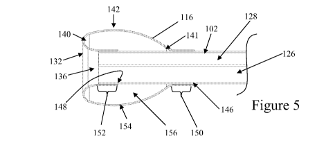
instrument. The image may come from a data device such as a camera placed on
the distal end
of the instrument, or from a sensor or combination of sensors. In one
embodiment, the sensor
utilizes light to generate the image. In another embodiment, the sensor is
adapted to see through
the bloody field as presented in the spinal region by selecting at least one
infrared wavelength
transparent to blood or other bodily fluids. In some embodiments, at least one
infrared
wavelength transparent to blood presented in the spinal field may have a
wavelength of about 1
micron to about 15 microns. In another embodiment, the at least one infrared
wavelength
transparent to blood presented in the spinal field has a wavelength between
about 1.5 micron to
about 6 microns. In yet another embodiment, the at least one infrared
wavelength transparent to
blood presented in the spinal field has a wavelength between about 6 microns
to about 15
microns. In yet another embodiment, the at least one infrared wavelength
transparent to blood
presented in the spinal field has a wavelength between about 1.0 microns to
about 1.5 microns,
19
CA 02697372 2010-02-22
WO 2009/029639 PCT/US2008/074405
about 1.5 microns to about 1.9 microns, about 2.0 microns to about 2.4
microns, about 3.7
microns to about 4.3 microns, or about 4.6 microns to about 5.4 microns. In
yet another
embodiment, the wavelength is selected or adapted for use in distinguishing
nervous tissue from
surrounding tissue and/or minimally vascularized nervous tissue. In yet
another embodiment,
the wavelength is selected to distinguish nervous tissue from muscle.
Wavelength selection
information and characterization and other details related to infrared
endoscopy are found in US
Patent 6,178,346; US Patent Application Publication No. 2005/0014995, and US
Patent
Application Publication No. 2005/0020914, each of which is hereby incorporated
by reference in
its entirety for all purposes.
[0069] The visualization channel 128 or the distal end 106 of the device 100
may
include a sensor used to generate images or identify tissue. In one example,
the sensor utilizes
acoustic energy to generate the image, similar to diagnostic ultrasound. In
another example, the
sensor utilizes an electrical characteristic to generate the image or other
types of structural or
physiological information. In yet another example, the sensor distinguishes
the type of tissue
adjacent to the sensor. Some properties used by the sensor to differentiate
adjacent structures or
tissue include resistance, capacitance, impedance, membrane voltage, acoustic,
and optical
characteristic of tissue adjacent the sensor or probe. Additionally, the
sensor or image may be
used to distinguish different types of tissue to identify neurological tissue,
collagen, or portions
of the annulus, for example. It is to be appreciated that the sensor may be a
multi-modal or
multi-sensor probe that can distinguish bone, muscle, nerve tissue, fat, etc.
to help position the
probe in the proper place.
[0070] In some embodiments, a trocar may be guided using fluoroscopic or other
external imaging modality to place the trocar in proximity to a treatment
area. In contrast to
conventional procedures that attempt to fluoroscopically navigate a trocar tip
around nerves and
other tissue, the trocar may remain safely positioned away from sensitive
structures and features.
In one embodiment, the trocar tip remains about 1 to about 2 cm or more from
vulnerable nerve
tissue. In another embodiment, the last about 1 to about 2 cm of travel to a
therapy site is
performed using direct visualization provided by a visualization mechanism in
the balloon
cannula device.
CA 02697372 2010-02-22
WO 2009/029639 PCT/US2008/074405
[0071] In some embodiments, the trocar is removed and the balloon cannula
device
100 is inserted into the pathway formed by the trocar. In other embodiments, a
tubular trocar
may be used. From the final trocar position, the balloon cannula device 100
may be passed
through a channel or lumen of the trocar and along the remaining distance to
the therapy or
treatment site using the onboard visualization capabilities. The onboard
visualization may be
used alone or in combination with the balloon 116 or other type of atraumatic
tip to identify,
atraumatically displace, and/or maneuver around nerves and other tissue as
needed. An optional
steering mechanism may be provided on the balloon cannula device 100 to
manipulate
surrounding tissue and structures, and/or to traverse the remaining distance
to one or more
therapy or treatment sites. In other embodiments, the balloon cannula device
100 may have a
rigid or fixed configuration, and may be manipulated by optionally
manipulating the trocar to
reach a desired location. In an alternative embodiment, the trocar may house
the balloon
cannula device during trocar insertion and thus utilize the direct
visualization capabilities of the
visualization mechanism within the balloon cannula device to guide trocar
positioning. In still
another embodiment, the trocar may be provided with a separate imaging system
from the
imaging device or component provided in the balloon cannula device for use
during trocar
insertion. In still another embodiment, the trocar may be configured with a
lumen to house only
the imaging component from the balloon cannula device 100. After the desired
trocar position is
reached, the trocar is removed and the imaging component is removed from the
trocar and
reinserted into the balloon cannula device 100. In yet another alternative
embodiment, both
external imaging may be used to position the trocar distal end, either alone
or in combination
with direct imaging.
[0072] As mentioned previously, one or more embodiments of the balloon cannula
device may be provided with any of a wide variety of steering configurations,
such as the
steering mechanism 120 depicted in FIG. 1. In one embodiment, the balloon
cannula device 100
is steerable in one or more axes, including a device with two axes. In some
embodiments, one
axis may be a rotation axis. In another embodiment, the balloon cannula device
is non-steerable.
In yet another alternative embodiment, the balloon cannula device may be pre-
formed into a
shape that is adapted to access a portion of the spinal region or other region
of the body. The
shape may include any of a variety of angled and/or curved segments to access
a particular body
site. In yet another embodiment, the balloon cannula device is situated within
the trocar in such
21
CA 02697372 2010-02-22
WO 2009/029639 PCT/US2008/074405
a way that the balloon cannula may have steering capability up to about 360
inside the spinal
space. The steering mechanism 120 may include one or more flexible bodies or
flex regions 124
on the balloon cannula device 100. The flexible body may be bent by
manipulating a control
such as lever 1221ocated on the housing 118. Various examples of the steering
mechanism and
the flex region 124 and are described in greater detail below.
[0073] The dimensions of the balloon cannula device may be sized and selected
based on the particular therapy being provided. For example, one embodiment of
the balloon
cannula device may be dimensioned for navigation to a spinal region for
diagnostic evaluation
and/or to apply a therapy thereto. In another embodiment, the balloon cannula
device may be
sized to fit within the epidural space. Other embodiments may be configured
for use in the chest
cavity (e.g. pleural biopsy or pleuracentesis) or abdominal-pelvic cavity
(e.g. bladder neck
suspension), or for non-spinal procedures such as breast biopsy and
transvaginal oocyte retrieval,
for example. In some embodiments, the balloon cannula device 100 may have a
diameter of
about 5 mm or less, while in other embodiments, the diameter may have a
diameter of about 3
mm or less, or even 2.5 mm or less. In another embodiment, one or more of the
working
channels 124, 126 and 128 of the balloon cannula device 100 may have a
diameter of about 5
mm or less, about 3 mm or less, about 2 mm or less, about 1 mm or less, or
about 0.8 mm or
less.
[0074] As mentioned previously, the cannula device may comprise a balloon or
other type of structure that may be used as an atraumatic tip structure to
reduce the risk of
inadvertent injury to surrounding structures during a procedure. The
atraumatic tip may be
configured to provide tactile feedback to the user of the rigidity, pliability
or feel of the tissue or
structures in contact with the tip. In one embodiment, the atraumatic tip also
provides dissection
or retraction capabilities and/or the ability to displace surrounding tissue.
The overall shape of
the atraumatic tip may allow manipulation of the nerves as the balloon cannula
device is
advanced without harming the nerve or causing pain. In one embodiment, the
atraumatic tip
may have a curved shape and no sharp edges, burrs or other features that may
pierce, snag, tear
or otherwise harm tissue that comes into contact with the atraumatic tip. The
shape, surface
contours and/or overall finish of the atraumatic tip may be selected to reduce
or minimize impact
22
CA 02697372 2010-02-22
WO 2009/029639 PCT/US2008/074405
forces when the tip comes into contact with structures such as nerves, muscle
and the spinal
dura, among others.
[0075] As mentioned previously, the atraumatic tip may also be controllably
inflatable or expandable, or otherwise comprise two or more surfaces or
structures that are
independently controllable. One potential use of such an embodiment comprises
contacting the
tip against tissue and then inflating, expanding or separating to deform or
move the tissue. The
tip may be a balloon that is inflated to create a working space in the
surrounding tissue as well as
provide a clearing for improved visibility. The balloon cannula device may
then be advanced
into the working space. The balloon may be inflated again to create another
working space and
so forth to advance the balloon cannula device in a spinal space. In addition,
the balloon cannula
device may be used to provide saline or another type of cleaning solution to
the working area for
enhancing visualization. In another embodiment, the distal balloon 116 may be
moveable or
articulated such that it may be used to displace, nudge or prod surrounding
tissue or structures.
The nudge action is felt by the user and also provides a more tactile sense of
tissue movement.
The nudge may result from active movement of the tip under control of the
user, movement
caused by releasing the tip from a bias position or from other conventional
techniques for
manipulation of surgical implements.
[0076] FIGS. 4 and 5 illustrate the balloon 116 of the balloon cannula device
100
in the stowed condition and deployed condition, respectively. As shown, a
portion of the distal
end 140 of the balloon 116 is located distal to the distal end or tip 106 of
the tubular shaft 102.
In one embodiment, the balloon 116 is distally located about 5 mm or less past
the shaft tip 106,
sometimes about 3 mm or less past the shaft tip 106; other times about 2 mm or
less past the
shaft tip 106. The net longitudinal length of the balloon 116, as mounted on
the tubular shaft
102, may be in the range of about 3 mm to about 20 mm, sometimes about 4 mm to
about 10
mm, and other times about 5 mm to about 8 mm, for example. In one embodiment,
the distal
end 140 of the balloon 116 is located about 1 mm to about 1.3 mm In the
uninflated state, the
balloon 116 may have an outer diameter of about 4 mm or less, sometimes about
3.6 mm or less,
and other times about 3 mm or less. In the inflated state, the balloon 116 may
have a maximum
outer diameter of about 4 mm or more, sometimes about 5 mm or more, and other
times about 6
mm or more. The outer diameter of the balloon 116 may vary, depending upon on
the particular
23
CA 02697372 2010-02-22
WO 2009/029639 PCT/US2008/074405
balloon configuration and the degree of inflation. In some embodiments, the
balloon cannula
device 100 may be configured to withstand inflation pressures up to about 60
psi or less, while
in other embodiments, the balloon cannula device 100 may be configured to
withstand inflation
pressures up to about 80 psi or less, or sometimes about 100 psi or less, and
other times up to
about 200 psi or more. In some particular embodiments, the balloon cannula
device 100 may be
configured to provide a diameter change of about 2 mm or more across a
pressure change of
about 50 psi (e.g. from about 30 psi to about 80 psi), sometimes about 2.5 mm
or more, and
other times about 3 mm or about 4 mm or more. In some embodiments, the balloon
116 may be
configured such that the peak ratio of diameter change to pressure change
occurs in the range of
about 30 psi to about 120 psi, sometimes about 40 psi to about 100 psi, and
other times about 60
psi to about 80 psi. In some embodiments, inflation of the balloon cannula
device 100 to a
pressure of at least about 45 psi or more may reduce the degree of balloon 116
deformation
during use, which may improve the tactile feedback of the balloon cannula
device 100. While
balloons inflated to semi-rigid or rigid pressures may exhibit less
deformation upon contact with
structures such as nerves, the shape of the balloon 116 with a curved distal
tip and a tapered
proximal end may also provide the balloon 116 with a shape that by
atraumatically displaces
away such structures upon contact.
[0077] In some embodiments, the balloon component may be formed such that its
base configuration is an uninflated shape, an inflated shape, or an
intermediate shape
therebetween. In some embodiments, a balloon with its base configuration in
the uninflated
shape may lie flatter against the tubular shaft of the balloon cannula device
in comparison to a
balloon with its base configuration in the inflated shape. In other
embodiments, balloons having
a base configuration in their inflated shape may have ripples, wrinkles or
folds when collapses or
constricted for insertion into the body. In other embodiments, such as the
balloon 160 in FIG. 6,
balloons having a base configuration in their inflated state may have more
controllable or
predictable conformation in their inflated state, when compared to balloons
having a base
configuration in their uninflated state. In the particular embodiment depicted
in FIGS. 4 and 5,
the balloon 116 comprises a tubular structure 144 having a proximal end 146
and a distal end
148. The material comprising the tubular structure 144 may have a uniform or
non-uniform
thickness, and a uniform or non-uniform axial cross-sectional area or shape.
The proximal end
146 is attached at a proximal mounting site 150 that is proximal to the distal
end 106 of the
24
CA 02697372 2010-02-22
WO 2009/029639 PCT/US2008/074405
tubular shaft 102, while the distal end 148 is attached at a distal mounting
site 152 and is folded
under a middle portion 154 of the balloon 116 such that at least a portion of
the middle portion
154 is located distal to the distal end 106 of the tubular shaft 102. As may
be seen in FIGS. 4
and 5, the tubular structure 144 may be characterized as an inverted or
everted configuration,
where the inner surface of the tubular structure is attached to the proximal
mounting site 150
while the opposite or outer surface of the tubular structure is attached to
the distal mounting site
152. During manufacture of the balloon cannula device, an inner diameter of
one end of the
tubular structure 144 may be bonded to the distal end 106 of the tubular shaft
102, while the
other end of the tubular structure 144 is free and distal to the distal end
106 of the shaft 102. The
free end of the tubular structure 144 may then be everted and pulled back over
the attached end
of the tubular structure 144 and attached to the tubular shaft 102 proximal to
the attached end of
the tubular structure 144. The proximal attachment site may be selected so
that at least a portion
of the tubular structure 144 is distal to the distal end 106 of the tubular
shaft 102. Alternatively,
the proximal attachment of the tube 144 may be made and then the distal end of
the tube 144
may be inverted and attached to the shaft 102.
[0078] Although both the proximal and distal mounting sites 150 and 152 may be
located on the same tubular shaft 102, in some embodiments the mounting sites
150 and 152
may be located on different tubular shafts have a coaxial sliding
relationship. In this latter
embodiment, the two tubular shafts may be manipulated to alter the balloon
shape. For example,
the proximal and distal mounting sites 150 and 152 may be brought closer
together to permit a
greater radial expansion range. In another example, the proximal mounting site
150 may be
moved more proximally, which in some embodiments, may shift the balloon
proximally to
elongate the balloon configuration and/or to reduce the degree of forward
positioning of the
balloon.
[0079] In some embodiments, the balloon or tubular structure may be attached
to
the tubular shaft by adhesives or by heat bonding, for example. In some
embodiments, bonding
structures or processes may be used, which may improve the sealing between the
balloon and the
tubular shaft to support the use of higher inflation pressures without
separating the balloon from
the tubular shaft. For example, crimp rings or heat shrink tubing may be used
to augment the
bonding or attachment process. In some embodiments, the crimp rings or shrink
tubing may be
CA 02697372 2010-02-22
WO 2009/029639 PCT/US2008/074405
applied temporarily to facilitate setting of other bonding processes, and are
later removed. In
other embodiments, the crimp rings or shrink tubing may be incorporated into
the final
assembled product.
[0080] In some embodiments, after inflation, the balloon may not fully revert
to its
uninflated state upon relief the inflation pressure. Due to stretching or
other types of
deformation, the balloon may fold, wrinkle or crease upon deflation, which may
affect the ability
to withdraw the balloon cannula device from an introducer or guide into which
the balloon
cannula device was inserted. In some embodiments, where the balloon material
comprises a
polymeric material, the deflation characteristics of the balloon may be
improved providing at
least some polymer chains that are circumferentially oriented with respect to
the tubular shaft.
In embodiments where the balloon 116 originates as a thermoplastic tube, the
thermoplastic tube
may be an extruded polymeric tube, which typically provides longitudinally
oriented polymer
chains due to the extrusion process used to form the tube. In some
embodiments, some of the
longitudinally oriented polymer chains may be reoriented toward a
circumferential orientation
by heating the tube while in an expanded state.
[0081] In one example, a thermoplastic tube is provided, comprising an outer
diameter that is less then the final assembled diameter of the balloon, and a
thickness that is
greater than the final thickness of the balloon. The thermoplastic tube is
placed into a molding
tube or cavity having an inner diameter that is approximate to the final
assembled balloon outer
diameter. The thermoplastic tube is pressured and expanded until the outer
surface of the tube is
constrained by the inner diameter of the modeling tube or cavity for a period
of time at an
elevated temperature. The thermoplastic tube is then cooled while pressurized
to set the new
diameter and to set the circumferentially reoriented polymer chains. The
temperature, pressure
and treatment period of the thermoplastic material may vary, depending upon
the particular
balloon material and the balloon configuration. In one particular example
wherein the
thermoplastic material comprises polyurethane with a durometer in the range of
about 80A to
about 95A and a thickness of about 0.005" to about 0.01", sometimes about
0.006" to about
0.009", and other times about 0.007" to about 0.008". The setting temperature
may be about
120 F to about 250 F, depending upon the particular material used. The
setting period may be
26
CA 02697372 2010-02-22
WO 2009/029639 PCT/US2008/074405
in the range of about 5 seconds to about 2 hours or more, sometimes about 30
seconds to about
30 minutes, and other times about 1 minute to about 2 minutes.
[0082] The balloon may be made of a flexible material such that the balloon is
inflatable upon introduction of a fluid or gas. In one embodiment, the
flexible material has
sufficient rigidity such that it may effectively maintain the tubular shape
when uninflated. As
shown in FIG. 5, the distal end 140 of the balloon 116 may remain extended
past the distal end
106 of the shaft 102 before and after inflation. Embodiments of the atraumatic
balloon 116 may
also be used to assist with or perform therapy or treatment, shield
surrounding tissue or provide
access for other devices. The atraumatic balloon 116 may be positioned at the
surgical or
treatment site in a compact or stowed condition (see, e.g., FIG. 4) and then
deployed according
to the type of device used (see e.g., FIG. 5).
[0083] The atraumatic balloon 116 may be used to manipulate surrounding tissue
in one or more ways. First, by transitioning the balloon 116 from a stowed to
deployed
configuration, the outer walls 142 of the balloon 116 will be urged outward
against the
surrounding tissue. Second, whether or not the device 100 is deployed or
stowed, the balloon
cannula device 100 may be maneuvered using the steering mechanism 120 to
manipulate tissue.
Third, the atraumatic balloon 116 may cycled between the stowed and deployed
configuration to
assist in the advancement of the steerable balloon cannula device 100. For
example, the balloon
116 may be deflated to facilitate insertion of the device 100 through a wall
of tissue, and may be
reinflated after traversing the wall. Fourth, the practitioner may advance the
balloon cannula
device 100 and manipulate surrounding tissue and push tissue away by creating
space under
direct visualization. As the balloon 116 of the balloon cannula device 100
expands, a work
space or opening may be created in the surrounding tissue, thereby easing the
advancement or
atraumatic maneuverability of the balloon cannula device 100. Thereafter, the
atraumatic
balloon 116 may be deployed or otherwise used to deform surrounding tissue
and/or to make
space available for the balloon cannula device 100 or other therapy or
treatment device provided
by working channel 126 (e.g., FIG. 3). It is contemplated that one or more of
these methods
may be used in combination to manipulate the surrounding tissue. Any of a
variety of other
methods for utilizing the balloon cannula device 100 are also contemplated.
27
CA 02697372 2010-02-22
WO 2009/029639 PCT/US2008/074405
[0084] In the embodiment illustrated in FIG. 7, the atraumatic balloon 116 is
an
inflatable structure. The balloon 116 may be adapted for delivery via trocar
or introducer. As
shown in FIG. 4, in one embodiment, the balloon 116 may be folded, compressed
or stowed in
such a manner that the balloon 116 is deliverable with an embodiment of the
balloon cannula
device via the trocar or introducer. Additionally, the balloon 116 may be held
by a sheath to
retain expandable structures in a constrained configuration. Once the balloon
116 is positioned
where desired, the sheath may be removed to allow the device to transition to
a deployed
configuration.
[0085] Exemplary embodiments of the tip structure(s) may include any of a
variety
of balloons or other shaped inflatable structures. There are a great many
different shapes, sizes
and functionality readily available in such balloons and many are well suited
and easily
adaptable for use in endoscopic spinal procedures. In one embodiment, the
balloon, when in a
stowed configuration, is dimensioned to translate through a lumen, working
channel, trocar or
introducer in an embodiment of the balloon cannula device described herein.
The atraumatic
balloon may be shaped in virtually any shape desired to further balloon
cannula. For example,
the balloon may be elongated, rounded, or other pre-formed shape. In one
specific embodiment,
the balloon has an elongate shape that follows the shape of an adjacent spinal
structure. In one
specific embodiment, the balloon is adapted to follow a portion of the dura
mater. In another
specific embodiment, the balloon is adapted to follow a portion of the
annulus. In another
embodiment, the atraumatic balloon includes a marker or other feature(s)
making all or a portion
of the balloon perceptible using external imaging modalities. In another
embodiment, the
atraumatic balloon is donut shaped to create a forward-looking design. In one
embodiment, the
marker or feature is a radio opaque marker. In other embodiments, including
the balloon
cannula device 430 in FIG. 30, multiple balloons 432 and 434 may be provided.
The multiple
balloons may be arranged serially along the balloon cannula device 430, and/or
in a parallel
fashion (e.g. two semi-cylindrical or semi-tubular balloons located at the
distal end of a balloon
cannula system). The multiple balloons need not have the same size and/or
configuration.
Embodiments comprising multiple balloons may comprise independently
controllable balloons,
or balloons that inflate in a coordinated fashion.
28
CA 02697372 2010-02-22
WO 2009/029639 PCT/US2008/074405
[0086] Atraumatic balloon embodiments are not limited to closed surface,
inflatable embodiments. Open surface structures such as mesh, scaffold
structures, polymer
stent-like structures, for example, may also be used to atraumatically deform
spinal tissues. One
example of an open surface structure is a coronary stent. Many of the delivery
techniques used
to deliver stents into the vasculature are applicable here for delivery into
the spinal space. The
stent may also be a polymer stent or a stent with a coating to improve the
atraumatic qualities of
the stent to spinal tissues and structures. In another embodiment, a suitable
scaffold includes the
collapsible scaffold structures used to deform and support tissue, and to
maintain spacing
between a radioactive source and the tissue being treated prior to and during
brachytherapy.
[0087] In one embodiment, the surfaces of the atraumatic balloon are
expandable.
For example, the atraumatic balloon might be expanded using mechanical
mechanisms,
pneumatic mechanisms, or hydraulic mechanisms. In addition, the atraumatic
balloon 116 may
also contain sensing and/or monitoring devices such as a temperature thermo-
couple. In an
alternative embodiment, the atraumatic balloon may include multiple layers and
provide
insulation or shielding to surrounding tissue by changing thermal and/or
insulating properties
either alone or in combination with expansion and contraction between the
multiple layers. The
change in properties could be accomplished by electrical, chemical, or
mechanical properties of
the layers, spaces between layers or through the use of a liquid, gas or other
material inserted
between layers or into a layer.
[0088] In some embodiments, the atraumatic balloon may not be circular in
cross
section or may not be generally cylindrical as the balloons suited for use in
the vasculature. In
one embodiment, the atraumatic balloon may be irregular in shape and may be
designed to
accommodate tissues, endoscopes, and therapeutic devices to avoid potential
injury to the tissues
during treatment options. In one embodiment, the atraumatic balloon is adapted
to conform to a
portion of the spinal anatomy when in a deployed configuration. In another
embodiment, the
atraumatic balloon is sized and adapted to conform to the shape of the
annulus. In another
specific embodiment, the atraumatic balloon may have a preformed shape, such
as a rounded
shape, an elongated shape or combinations thereof. In one specific embodiment,
the folded wall
thickness of the atraumatic balloon is about 6 to about 40 thousandths of an
inch. In another
specific embodiment, the folded wall thickness of the atraumatic balloon may
be about 12 to
29
CA 02697372 2010-02-22
WO 2009/029639 PCT/US2008/074405
about 30 thousandths of an inch. In another specific embodiment, the folded
wall thickness of
the atraumatic balloon is about 20 to about 30 thousandths of an inch. Other
sizes are possible
and may be selected based on the channel size of the balloon cannula device as
well as the
physical parameters of the patient's spinal area.
[0089] FIG. 7 is another cross-sectional view of the atraumatic balloon 116
depicting the structures relating to the inflation of the balloon 116. The
atraumatic balloon 116
not only provides the capabilities of the balloon 116 but may also include
working channels to
further assist in performing procedures. The balloon 116 is capable of both
stowed and deployed
configurations and is illustrated in a deployed configuration in FIGS. 4 and
5. The access
lumens 126, 128, and 130 may run the length of the device 100 and may be sized
to allow
passage of the catheters, endoscopes, and instruments/devices, respectively.
The balloon lumen
132 may be adapted to inflate the balloon 116. As shown, the distal portion of
the lumen 132
may include a port 134 in fluid communication with the balloon 116. The
balloon 116 may be
filled with contrast solution in order to improve fluoroscopic visualization
of the device balloon
or device. In other embodiments, the balloon may not require an
inflation/deflation port 132.
Instead, the balloon cavity may optionally be filled with compressible or non-
compressible gas
or liquid, which may be redistributed or compressed out of the balloon using a
sheath or other
constraining structure.
[0090] In some embodiments wherein the atraumatic tip comprises a balloon
structure, the balloon structure may be inflated with an optically transparent
fluid that is selected
to reduce distortions through the balloon and/or cavity, and/or reflections at
the balloon/cavity
interface. An optional bubble removal filter may be provided with the cannula
system or the
fluid injection system to reduce the number of bubbles inflated into the
balloon. In some
embodiments, visibility through the balloon structure may be different between
the uninflated
state and inflated state. In some embodiments, where the curvature of the
inflated balloon may
reduce the visual clarity through the balloon, the balloon may be configured
to limit the amount
of curvature or expansion in one or more areas. In FIGS. I I A and IIB, for
example, the distal
segment 200 of a forward-mounted balloon 202 may comprise two or more
attached, fused or
adhered balloon surfaces 204 and 206 so as to resist expansion or separation.
These
embodiments may improve visual clarity through the distal segment 200 while
providing an
CA 02697372 2010-02-22
WO 2009/029639 PCT/US2008/074405
expandable segment 208. In other embodiments, the balloon may be configured to
permit at
least some inflation without substantially changing the relative spacing
between the two balloon
surfaces, e.g. where the balloon surfaces remain longitudinally straight and
parallel. Such
embodiments may permit distal expansion while maintaining some visualization
through the
distal segment of the balloon in its expanded state. The distal segment 200 of
the forward-
mounted balloon 202 may have a length that is smaller, greater or similar to
the forward balloon
length 210 that is distal to the distal end 212 of the tubular shaft 214.
[0091] Although the embodiment depicted in FIGS. 11A and 11B comprises a bi-
layer distal segment 200, as illustrated in FIGS. 12A and 12B, for example, a
forward-mounted
balloon 216 with a single-layer distal segment 218 may be provided. A single-
layer distal
segment 218 may further augment visual clarity by eliminating one interface
between the layers,
inflation fluids, and/or the use of any adhesives, if any. The thickness of
the single-layer distal
segment 218 may or may not be about equal to the thickness of a layer
comprising the
expandable segment. In other embodiments, for example, the distal segment may
comprise three
or more layers. The average thickness of the distal segment may vary from
about 10 microns to
about 500 microns or more, sometimes about 11 microns to about 200 microns,
and other times
about 12 microns to about 150 microns. In some embodiments, the non-expandable
distal
segment may have a longitudinal length of about 1 mm to about 10 mm or more,
sometimes
about 2 mm to about 8 mm, and other times about 4 mm to about 6 mm.
[0092] In some embodiments, the distal segment may comprise a different
material
from the proximal balloon material. The distal segment may be attached to the
balloon in any of
a variety of ways, including but not limited to the use of adhesives or heat
bonding. In other
embodiments, such as the cannula device 230 depicted in FIG. 13, a non-
inflatable atraumatic tip
232 may be attached directly to the tubular shaft 234 in a forward position.
Here, no expandable
segment is provided, but in other embodiments, a non-forward mounted
expandable segment
may be provided on the tubular shaft 234, in addition to the non-inflatable
atraumatic tip 232.
The non-forward mounted may be flush mounted or proximally mounted with
respect to the
distal end 236 of the tubular shaft 234.
[0093] The distal segment or tip 232 in FIG. 13 may be selected from a
material
that is transparent to the operation of the port components, such as
visualization channel. The
31
CA 02697372 2010-02-22
WO 2009/029639 PCT/US2008/074405
atraumatic tip 232 maintains a working space about the distal end 238 about
the visualization
channe1240 while leaving the other working channels open for the introduction
of instruments
and/or endoscopes. In some embodiments, the atraumatic tip 232 may be formed
from rigid,
clear plastic, while in other embodiments, the atraumatic tip may comprise a
flexible,
deformable material. In some embodiments, the tip comprises an opaque
material, but in other
embodiments may be translucent or transparent, which may facilitate the
visualization of the
tissue or structures adjacent the tip. The tip material may be stainless
steel, cobalt chromium,
titanium, nickel-titanium, polycarbonate, acrylic, nylon, PEEK, PEK, PEKK,
PEKEK, PEI, PES,
FEP, PTFE, polyurethane, polyester, polyethylene, polyolefin, polypropylene,
glass, diamond,
quartz, or combination thereof, for example. In some embodiments, the tip
materials may
include the addition of one or more radiographic markers or materials.
[0094] In other embodiments, the forward-mounted balloon or expandable
structure may be provided with one or more support elements. The support
elements may be
oriented longitudinally, radially, and/or circumferentially along the balloon
to support the
uninflated and/or inflated balloon configurations. The configuration of a
support element may
be complementary to the shape or configuration of the balloon. In one
embodiment, the support
element may comprise a helical configuration, for example. In some
embodiments, the support
elements may be located about the balloon lumen. Support elements about the
balloon lumen
may resist involution of the balloon lumen in the inflated configuration,
particularly at higher
inflation pressures. The support elements may comprise any of a variety of
materials, including
but not limited to a metal and/or polymeric material. The support element
maybe rigid, semi-
rigid or flexible, and at least a portion of the support element may be
attached or coupled to the
shaft, the inner or outer surface of the balloon, and/or embedded in the
balloon wall.
[0095] Although the balloon 116 may be generally circumferentially symmetrical
about the longitudinal axis of the shaft 102, in other embodiments, the
balloon may be
asymmetrical. In FIG. 5, the balloon 116 comprises a toroidal configuration
wherein the distal
end 140 of the balloon 156 has a curved or rounded configuration when inflated
and the
proximal end 141 of the balloon cavity 156 has a tapered configuration. Other
balloon
configurations may also be used, and slits or windows may be optionally
provided to increase
visualization. For example, the balloon configuration may be altered using
different balloon
32
CA 02697372 2010-02-22
WO 2009/029639 PCT/US2008/074405
shapes, variable wall thickness and/or by pre-forming curves or fold along one
or more regions
of the balloon material.
[0096] In some embodiments, the balloon may have semi-circular, hood-like
configuration that is open along at least one perimeter of the balloon working
space. FIG. 28
illustrates an alternate embodiment of a balloon cannula device 300,
comprising a shaft 302 and
a conical balloon 304 with a conical balloon working space 306 that opens to
the surrounding
structures. In some embodiments, the conical balloon 306 may be configured to
extend and
retract into the distal end 308 of the shaft 302 while expanding and contract
its diameter,
respectively. In other embodiments, the conical balloon 304 and/or conical
balloon working
space 306 may controllably expand or contract without requiring extension or
retraction of the
balloon 304. This may be performed by providing two or more separately
inflatable/deflatable
compartments with respect to the balloon to provide non-uniform inflation
forces within the
balloon. In other embodiments, contractile electropolymers in or on the
balloon which may be
manipulated by applying a current or voltage using wires and electrodes
attached or contacting
the electropolymers.
[0097] FIG. 30 depicts still another embodiment of a balloon cannula device
430,
comprising multiple balloons 432 and 434. In this embodiment, one balloon 432
may be located
distally on the cannula 436 while a second balloon 434 is located more
proximally on the
cannula 436. The cannula segment 438 between the two balloons 432 and 434 may
be a single-
lumen, transparent material. This may permit an endoscope or other direct
visualization
component to view the structures adjacent to the cannula segment 438 while the
two balloons
432 and 434 provide some separation between the structures and the cannula
segment, which
may broaden the viewing angle.
[0098] Referring back to FIG. 3, the tubular shaft of the balloon cannula
device
100 may include a visualization channel 128, a larger working channel 126, and
an additional
irrigation/aspiration port 130. The channels and/or ports of the balloon
cannula device 100 may
be configured to accept wide variety of therapy devices suited to the type of
therapy being
performed. The therapy device may be configured and used to apply energy to
surrounding
tissue. The therapy device may also be a surgical instrument used to cut,
pierce or remove
tissue. Moreover, it is to be appreciated that the therapy device may be any
conventional
33
CA 02697372 2010-02-22
WO 2009/029639 PCT/US2008/074405
endoscopic instrument. The therapy device may include ultrasonic devices,
motor driven
devices, laser-based devices, RF energy devices, thermal energy devices,
cryotherapy-based
devices, or other devices selected based on the spinal therapy being
performed. For example, the
therapy device may also be a mechanical device adapted to remove tissue such
as a debrider or
an aspirator. Other examples are described in greater detail below. Moreover,
it is to be
appreciated that the balloon cannula device 100 may be used to inject
pharmacological agents
into the spinal area. The size, number and arrangement of the working channels
are readily
adaptable for different configurations, depending upon the type of procedures
performed. A
greater or a fewer number of working channels may be provided, and the working
channels need
not have the same size and shape. In addition, the working channels may also
be configured to
perform auxiliary functions. In one example the channels or ports may be used
to provide
irrigation to assist in tissue dissection as the atraumatic tip is advanced in
the spinal space. An
irrigating working channel may be in communication proximally with a fluid
source, such as a
syringe or intravenous infusion system, and in communication distally with the
distal end of the
balloon cannula device so that the fluid exiting the irrigation working
channel is directed to the
distal portion of the balloon cannula device. In another example, the
irrigation working channel
or another working channel may be used to rinse the atraumatic tip or keep
clear other portions
of the balloon cannula tool. In the particular embodiment depicted in FIG. 3,
the working
channel 126 and the visualization port 128 are configured with non-circular
cross-sectional
shapes. In some embodiments, the non-circular shape permits the placement of
an instrument
with a circular cross-sectional shape within the channel or port while
providing still providing
flow paths for fluids and material through the channel 126 and the port 128.
Shared or eccentric
flow paths along non-circular shaft channels and ports may also otherwise take
advantage of
unused sections of the cannula shaft. Unlike shafts with only circular
channels or ports, the flow
paths may be provided without having to increase the overall cross-sectional
area of the cannula
shaft. Channels or ports having non-circular cross-sectional shapes may also
be used with
instruments having a complementary non-circular cross-sectional shape. For
example,
complementary non-circular cross-sectional shapes may be used to control or
limit the amount of
instrumentation rotation within the channel or port.
[0099] FIGS. 8, 9, and 10 illustrate various embodiments of the balloon
cannula
device. As shown in FIG. 8, the balloon cannula device 100 may comprise a
shaft 102 with a
34
CA 02697372 2010-02-22
WO 2009/029639 PCT/US2008/074405
non-circular visualization or irrigation channel 128, a non-circular working
channel 126 which
may be used to provide therapy device or as aspiration port, a balloon
inflation lumen 132, and
additional port 130 for irrigation or aspiration. The shaft 102 also
optionally comprises one or
more structures 162 on its outer surface 164. These structures 162 may
comprise recessed or
protruding configurations and may be used, for example, to maintain alignment
with respect to
introducer or guide member, or to reduce the amount of frictional resistance
from any
manipulation of the balloon cannula device 100. As depicted in FIG. 9, the
shaft 166 of the
balloon cannula device 168 may have a non-circular visualization or irrigation
port 170, a
circular therapy device or aspiration port 172, a circular balloon inflation
lumen 174, and
additional circular port 176 for additional irrigation or additional
aspiration having a greater. As
demonstrated in FIG. 9, the circular ports 172, 174 and 176 need not have the
same diameter. In
FIG. 10, the shaft 178 of the balloon cannula device 180 has a visualization
or irrigation port
182, an injection port or therapy device or aspiration port 184, and a balloon
inflation lumen
186, wherein no port or lumen has a circular cross-sectional shape. It is
contemplated that
functions of various lumens in a cannula device may be suitably interchanged.
[0100] During use, the balloon cannula device may be moved or may remain in
place while an inserted therapy device is manipulated to perform the desired
function. Once the
working or therapy area has been created or accessed using the atraumatic
balloon, the
atraumatic balloon may be removed thereby allowing working channel or trocar
or introducer to
be used for another instrument or therapy device or to provide support for a
procedure. For
example, the therapy device may comprise a mechanical debrider or other type
of tissue
disrupting device that may be introduced via the working channel to assist in
removal of tissue.
Various examples of mechanical tissue disrupting devices that may be used with
a balloon
cannula device are described in U.S. Pat. No. 12/035,323, filed February 21,
2008, which was
previously by incorporated by reference in its entirety. In yet another
example of the flexibility
of the balloon cannula device, one or more the working channels or ports may
be used to provide
access for the delivery of pharmacological agents to the access site either
for application onto or
injection into tissue. In some embodiments, the therapeutic agents may be
directed injected into
the channel or port, but in other embodiments, an infusion catheter may be
inserted into a
channel or port and used to provide additional control of the therapy. The
infusion catheter may
have any of a variety of configurations and features, including but not
limited to its own optional
CA 02697372 2010-02-22
WO 2009/029639 PCT/US2008/074405
steering mechanism separate from the balloon cannula device, and a needle tip
for injecting
therapeutic agents into the tissues or structures. In some embodiments, the
needle tip may be
retractable and extendable to protect against inadvertent puncture of the
balloon cannula device
and/or the tissues or structures accessible from the balloon cannula device.
Examples of
injection catheters that may be used with embodiments of the balloon cannula
device include
U.S. Patent Serial No. 10/820,183, which is hereby incorporated by reference
in its entirety.
[0101] The therapy device may be supplied with energy from a source external
using a suitable transmission mode. For example, laser energy may be generated
external to the
body and then transmitted by optical fibers for delivery via an appropriate
therapy device.
Alternately, the therapy device may generate or convert energy at the therapy
site, for example
electric current from an external source carried to a resistive heating
element within the therapy
device. If energy is supplied to the therapy device, transmission of energy
may be through any
energy transmission means, such as wire, lumen, thermal conductor, or fiber-
optic strand.
Additionally, the therapy device may deliver electromagnetic energy, including
but not limited
to radio waves, microwaves, infrared light, visible light, and ultraviolet
light. The
electromagnetic energy may be in incoherent or laser form. The energy in laser
form may be
collimated or defocused. The energy delivered to a disc may also be electric
current, ultrasound
waves, or thermal energy from a heating element. Moreover, it is to be
appreciated that
embodiments of the balloon cannula devices described herein may also be used
to dispense a
compound, compounds or other pharmacological agents to reduce, diminish or
minimize
epidural neural tissue scarring.
[0102] The balloon cannula device may also be used to perform denervation
procedures using direct visualization from the balloon cannula device. The
denervation
procedure may be physical, chemical or electrical denervation, for example.
The approaches
used may be similar those described herein to access the posterior or
posterolateral annulus. It is
to be appreciated that the denervation procedures may be performed to relieve
discogenic pain
and/or before the disc damage has progressed to a herniated disc or torn
annulus.
[0103] Referring back to FIG. 1, as noted previously this embodiment of a
balloon
cannula device 100 further comprises a steering mechanism 120. During use, the
balloon
cannula device 100 may be advanced through the working channel of a trocar or
introducer and
36
CA 02697372 2010-02-22
WO 2009/029639 PCT/US2008/074405
into the working area. In some embodiments, the working area or space may be
created by
separating structures or tissue using the atraumatic balloon 116, either alone
or in combination
with the steering mechanism 120. The steering mechanism 120 may be configured
to provide
any of a variety of steering features, including various bending planes,
various bending ranges,
extension and retraction ranges, and rotations ranges, for example. As
mentioned previously, in
the embodiment depicted in FIG. 1, the actuator comprises a lever 122 with
both ends 188
projecting from the housing 118, but in other embodiments, any of a variety of
actuators and
actuator configurations may be used, including but not limited to dials,
knobs, sliders, buttons
and the like, as well as electronic touch controls, for example. In some
embodiments, only one
end 188 of the lever 122 may project from the housing 118. The controls used
to manipulate the
steering mechanism 120 may be manually manipulated by the user or by a
mechanical control
system comprising various motors. In still other embodiments, actuators such
as the lever 122
may be omitted and the balloon cannula device 100 may be directly coupled to a
motor control
system.
[0104] Referring still to FIG. 1, the steering mechanism 120 is configured to
cause
bending of the shaft 102 at one or more bending regions 124. In FIG. 14, the
steering
mechanism 120 is depicted with the port tubing and a portion of the housing
118 of the balloon
cannula device 100 removed. The steering mechanism comprises a lever 122 that
is configured
to rotate or pivot at a lever axle 190. The lever 122 is attached to two
control members 192 that
are slidable located along the length of the shaft 102 and are attached at a
distal location of the
shaft 102. One or more posts 191 may be provided against the control members
192. In some
embodiments, the posts 191 may be facilitate changes in the orientation of the
control members
192, smooth sliding of the control members 192, and/or to protect other
components of the
balloon cannula device from cutting or other damage caused by the movement of
the control
members 192. In some embodiments, the ends of the control members 192 are
secured to the
lever 122 in one or more retaining channels or retaining structures, but in
other embodiments,
the control members may be proximally attached to form a control member loop
that may be
secured to a lever by placing the loop within a retaining channel of the
lever. In some
embodiments, one or more control members 192 or the control loop may be
crimped, wound,
sutured and/or embedded into the lever. The movement range and force may be
augmented by
one or more bias members 198 acting upon the lever 122. The bias members 198
may comprise
37
CA 02697372 2010-02-22
WO 2009/029639 PCT/US2008/074405
helical springs as depicted in FIG. 14, but may also comprise leaf springs or
any other type of
bias member configuration. The movement range of the lever 122 may also be
affected by the
size and/or configuration of the lever openings 199 provided in the housing
118. In some
embodiments, an optional locking mechanism may be provided to substantially
maintain the
lever in one or more positions.
[0105] The control members 192 may comprise wires, threads, ribbons or other
elongate structures. The flexibility and/or stiffness of the control member
192 may vary
depending upon the particular steering mechanism. In further embodiments, the
characteristics
of the control member 192 may also vary along its length. In embodiments
comprising two or
more control members, the control members need not be configured
symmetrically, e.g. having
the same length, cross-sectional area or shape, or opposite attachment sites
with respect to the
longitudinal axis of the tubular shaft. Also, individual control members need
not have the same
configuration along their lengths. For example, although the proximal end of
the control
members 192 depicted in FIG. 14 comprises wire-like members, the distal ends
250 of the
control members 252, illustrated in FIG. 15A, comprises a ribbon structures
254. In some
embodiments, the greater surface area of the ribbon structures may reduce the
risk of damage to
the flex region 256 of the cannula device 258. In the particular embodiment
depicted in FIG.
15A, the ribbon structures 254 have a U-shaped configuration that forms a
mechanical and/or
interference fit with the flex region 256 or other distal or flexible region
of the tubular shaft.
The flex region 256 may comprise one or more notches 260, recesses or openings
262
configured to accept the ribbon structure 254. In FIG. 15A, notches 260 are
provided to resist
slippage of the ribbon structure 254 along the lip 264 of the flex region 256,
while the openings
262 are provided to permit insertion of the ribbon ends 264 to further augment
the interfit of the
ribbon structures 254 and the flex region 256. FIG. 15B illustrates another
embodiment where
in the ribbon structure 266 inserts through the opening 262. In this
particular embodiment, the
ribbon structure 266 may also be welded or soldered back onto itself to form a
loop to further
secure the ribbon structure 266 to the flex region 256. In other embodiments,
as depicted in FIG.
15C, the tip 269 of the ribbon structure 268 may be bonded or soldered to the
flex region 256 or
the tubular shaft, depending upon the material of the ribbon structures and
the flex region or the
tubular shaft.
38
CA 02697372 2010-02-22
WO 2009/029639 PCT/US2008/074405
[0106] The bending range of tubular shaft may vary depending upon the
particular
design. The cannula device may be configured with a one-sided or a two-sided
bending range
with respect to the neutral position of the tubular shaft. The bending range
is from about 0
degrees to about 135 degrees, while in other embodiments, the bending range is
from about 0
degrees to about 90 degrees, and sometimes about 0 degrees to about 45
degrees, and still other
times about 0 degrees to about 15 or about 20 degrees. The bending range of
the other side, if
any, may be less than, equal to, or greater than the first side. In some
embodiments, increased
bending angles may cause creasing or telescoping of the tubular shaft, which
may obstruct one
or more channels within the tubular shaft.
[0107] In some embodiments, to enhance the bending range of the tubular shaft,
one or more flexion slots may be provided on the shaft. FIG. 16 depicts one
embodiment of
tubular shaft 270, comprising a plurality of slots 272. The slots 272 may have
a generally
circumferential orientation, but may alternatively have a helical orientation
or other orientation.
The slots 272 may be equally or unequally spaced along the longitudinal length
of the shaft 270.
In one example, the slots that are located about the ends of the flex region
may be spaced farther
apart than the slots located about the middle of the flex region. The slots
272 may have a similar
configuration or a heterogeneous configuration. The slots 272 depicted in FIG.
15 also have a
generally constant width, but in other embodiments, the width may vary along
the length of the
slot. The spacing between the slots ends 274 of a slot 272 may be
substantially similar or
different among the slots 272 comprising the flex region.
[0108] As noted in FIG. 16, the slot ends may comprise a rounded
configuration,
or any other configuration, including but not limited to an oval end, square
end, triangular end,
or any other polygonal shape for example. In some embodiments, such as the
example depicted
in FIG. 17A, the rounded ends 276 may have a larger transverse dimension than
the width of the
rest of the slot 278. In some embodiments, a rounded end may better distribute
the flexion stress
along the edges of the slot compared to squared or angled ends. Also, ends
that are larger than
the slots, such as the enlarged rounded ends 276 in FIG. 17A, may reduce the
degree of
compression or contact between the slot edges during flexion, which may also
reduce the risk of
cracking at the slot end. FIG. 17B depicts the enlarged rounded slot ends 276
of FIG. 17A in
39
CA 02697372 2010-02-22
WO 2009/029639 PCT/US2008/074405
flexion. In some embodiments, the slot end may have a more complex
configuration, such as the
T-shaped slot end 280 as depicted in FIG. 18.
[0109] In some embodiments, the number of slots per slot region may be
anywhere
from about 1 slot to about 100 slots or more, sometimes about 12 slots to
about 50 slots, and
other times about 24 slots to about 48 slots. In some embodiments, the length
of the flex region
may be anywhere from about 1 inch to about 20 inches, sometimes from about 4
inches to about
inches, and other times about 5 inches to about 8 inches in length. In some
embodiments, the
outer diameter of the flex region may be about 0.05 inches to about 0.3
inches, sometimes about
0.08 inches to about 0.15 inches, and other times about 0.1 inches to about
0.12 inches. The
wall thickness of the flex region may be in the range of about 0.001 inches to
about 0.01 inches,
sometimes about 0.002 inches to about 0.006 inches, and other times about
0.003 inches to about
0.004 inches. The slots 272 may have an average slot width in the range of
about 0.004 inches
to about 0.02 inches, some times in the range of about 0.005 inches to about
0.015 inches, and
other times about 0.006 inches to about 0.008 inches. The spacing between the
slots 272 may be
in the range of about 0.015 inches to about 0.1 inches, sometimes about 0.020
inches to about
0.050 inches, and other times about 0.025 inches to about 0.04 inches. The
spacing between the
ends of the slots may be in the range of about 0.004 inches to about 0.05
inches, sometimes
about 0.006 inches to about 0.02 inches, and other times about 0.004 inches to
about 0.01 inches.
The maximum transverse dimension of a slot end may be in the range of about
0.004 inches to
about 0.008 inches, other times about 0.004 inches to about 0.03 inches, and
other times about
0.01 inches to about 0.04 inches.
[0110] The flexion of the cannula system may facilitate access to the target
site
and/or reduce the degree of tissue disruption in achieving access to the
target site. For example,
in some procedures, the angle for approaching the target site through the skin
may be different
from the angle that provides the visibility or viewing angle to treat or
diagnose a particular
abnormality. Referring to FIG. 26, in some embodiments, a cannula system 340
may be inserted
to a target site 342 by utilizing longer or indirect access pathways 344 in
order to achieve the
desired approach angle to a target site, and/or to avoid interference from
structures such as the
transverse spinal processes 346. By using a steerable cannula system 348 as
depicted in FIG. 27,
however, a shorter or a more direct insertion pathway 350 may be taken to a
target site 352,
CA 02697372 2010-02-22
WO 2009/029639 PCT/US2008/074405
which may reduce the aggregate degree of tissue disruption compared to a
longer insertion
pathway. By taking advantage of the steerability of the cannula system 348,
the desired
approach angle to a target site may be achieved.
[0111] In some embodiments, during bending, one or more components inserted a
channel of the balloon cannula device may exhibit different degrees of
relative displacement.
The degree of relative displacement may be affected by the degree of bending,
the fixation or
coupling site, if any between the component and the balloon cannula device,
and/or the degree of
displacement from the neutral position of the balloon cannula device.
Referring to FIG. 19A, a
balloon cannula device 100 from FIG. 5 is shown in neutral position (e.g.
straight, but may be
angled or curved in other embodiments) with an endoscope 2821ocated in the
visualization port
128. The tip 284 of the endoscope 282 is in proximity to the end 286 of the
visualization port
128. As the balloon cannula device 100 is flexed as shown in FIG. 19B, the tip
284 of the
endoscope 282 may exhibit a relative distal displacement with respect to the
end 286 of the
visualization port 128, particularly in embodiments where the endoscope 282 is
coupled to the
balloon cannula device 100 at a proximal location (e.g. about the housing).
When the balloon
cannula device 100 is flexed in the opposite direction, in some instances the
endoscope 282 may
exhibit a proximal retraction. To compensate for the displacement, the user
may manually adjust
the position of the endoscope 282 as desired.
[0112] In some embodiments, the steering mechanism may also be coupled to an
endoscope adjustment mechanism so that manipulation of the steering mechanism
also provides
at least some position adjustment which may reduce if not eliminate the degree
of displacement.
In other embodiments, the endoscope may be coupled to the balloon cannula
device about a
distal region of the tubular shaft so that, during flexion, the proximal
portions of the endoscope
exhibit the displacement rather than the distal portions. In still other
embodiments, a spring or
other type of bias member may bias the endoscope distally against an
interference structure (not
shown) located at the distal end of the tubular shaft to maintain the
endoscope position during
flexion. In some further embodiments, the interference structure may be
rotated or moved out of
its interfering position to permit endoscope positioning more distally, as
desired.
[0113] FIG. 20 is a schematic representation of a tubular shaft 320 of one
embodiment of a cannula device 322 configured for two-sided flexion within a
bending plane.
41
CA 02697372 2010-02-22
WO 2009/029639 PCT/US2008/074405
In some embodiments, one or more channels of the tubular shaft 320 may be
configured and
positioned to reduce the degree of endoscope or instrument displacement during
flexion. In FIG.
21, for example, the tubular shaft 320 comprises a visualization channe1324
and a working
channe1326 wherein the centers 328 and 330 of the channels 324 and 326,
respectively, are
located along a plane 332 that is perpendicular to a bending plane 334 of the
cannula device 322.
Plane 332 may be located, for example, between the midpoint of the two distal
attachments of
the steering mechanism. The relative position of the plane 332 and the bending
plane 334 may
vary depending upon the particular manner in which the steering mechanism is
anchored to the
flexion region. In other embodiments, the centers 328 and 330 need not be
located on the plane
332, but the central location of the optics or working instruments inserted
into the channels 324
and 326 are located on the plane 332. For example, a channel may be configured
such that the
optical center of an endoscope is substantially aligned with the plane 332,
even through the
weighted center of the channel and/or endoscope may not be located on the
plane 332 (e.g.
where the lens of the endoscope is asymmetrically located, or where the
central viewing angle In
embodiments comprising circular channels, the center of the channel may be the
center of the
circle. In other embodiments comprising non-circular channels, the center of a
channel may be
characterized as being coaxial with the center of the largest circular object
that may be inserted
into the channel.
[0114] Although the embodiment shown in FIG. 21 is directed to a cannula
device
having a single bending plane, in other embodiments, the cannula device may be
configured with
two or more bending planes. With these latter embodiments, one or more
channels may be
aligned with one bending plane but not another bending plane. In some
embodiments, a central
channel may be provided that is aligned with two or more bending planes.
[0115] As mentioned previously, an endoscope or working instrument (e.g.
graspers or tissue debrider) may be inserted into one or more channels of the
cannula device
through a proximal port. The proximal port, endoscope, and/or working
instrument may be
optionally configured with one or more features to lock and/or adjust the
position of the inserted
component. In other embodiments, one or more components of the endoscope or
working
instrument may be an integrally formed component of the cannula device and is
not configured
for removal.
42
CA 02697372 2010-02-22
WO 2009/029639 PCT/US2008/074405
[0116] For example, in FIGS. 22A and 22B, a balloon cannula device 340 is
configured with a scope port 342 in communication with the visualization
channel (not shown)
with a segment of tubing 344. The scope port 342 may comprise a lumen with a
viscoelastic or
friction surface material that is configured to slidably grip an inserted
endoscope. The slidably
grippable materials may include but are not limited to silicone, a urethane,
including viscoelastic
urethanes such as SORBOTHANE (Kent, OH) and any of a variety of styrenic
block
copolymers such as some made by KRATON Polymers (Houston, TX). The scope port
342
thus need not have any particular clamp or locking mechanism to secure the
endoscope or
working instrument to the scope port 243, nor any particular adjustment
mechanism. In other
embodiments, however, the scope port may comprise a releasable lock or clamp
mechanism
designed to couple to the endoscope or working instrument, with an optional
adjustment
assembly that may be used to modify the spacing between the lock or clamp
mechanism and the
housing.
[0117] Referring now to FIG. 23, the proximal end 360 of the tubular shaft 362
may be coupled to one or more tubing segments 364, 366, 368, 370, 372 that
correspond to one
or more channels and connectors 374, 376, 378, and 380 of the balloon cannula
device 382,
respectively. As noted in FIG. 23, a tubing segment 370 may be in
communication with another
tubing segment, such as the tubing segment 368, which connected to the working
channel of the
device 382. This particular tubing segment 370 may be used, for example, to
flush or aspirate
fluid or material inserted into the working channel of the device 382 that is
accessed through the
middle port 378 and tubing segment 368. The particular design features of a
tubing segment
may vary, depending upon the particular function. In one example, a tubing
segment used for
aspiration may be rigid or reinforced to resist collapse during application of
a suction source,
while another tubing segment used to inflate the balloon may be configured to
withstand higher
positive pressures. The connector coupled to a particular tubing segment may
comprise any of a
variety of connectors or instrument interfaces. In some embodiments, for
example, one or more
connectors may comprise a standardized connector such as Luer lock, while in
other
embodiments, the connector may be a proprietary connectors. Depending upon the
particular
channel, in some embodiments, a check valve, septum, or a hemostasis valve may
be provided to
resist retrograde flow of fluid out of the device. The characteristics of a
particular channel,
including its dimensions and flexibility or rigidity, may depend upon its
particular use. In FIG.
43
CA 02697372 2010-02-22
WO 2009/029639 PCT/US2008/074405
24, for example, a balloon cannula device 384 comprises five ports 386, 388,
390, 392 and 394,
wherein the longer, flexible ports 388 and 392 may be used for infusion or
aspiration. Such
ports may be beneficial to facilitate the attachment of a bulky item such as a
syringe. A rigid
port, such as port 390, may be provided for instruments that may otherwise be
damaged or are
difficult to pass through tubing that may exhibit greater frictional
resistance. FIG. 25 depicts
another embodiment of a balloon cannula device 396 with a branched tube 398
with two
connectors 400 and 402. This particular balloon cannula device 398 may
facilitate the infusion
or injection of multiple medicaments or substances that are mixed together to
activate the
compositions (e.g. certain adhesive-, sealing- or coating compositions).
[0118] The balloon cannula devices may be used, for example, in systems for
treating disc degeneration that include nucleus decompression devices. The
balloon cannula
device may be used for accessing the nucleus and delivering a nucleus
decompression device.
For example, a decompression device may be advanced from one of the working
channels of the
balloon cannula device and into the nucleus of a disc. A nucleus decompression
device may be
used to removed the disc nucleus tissue either by dissection, suction,
dissolving, or by shrinking
the nucleus. Various types of thermal energy are known to shrink the nucleus
such as resistive
heat, radiofrequency, coherent and incoherent light, microwave, ultrasound or
liquid thermal jet
energies. Mechanical tissue removal devices may also be used. Decompression of
the disc
nucleus may result in the protruded disc material collapsing toward the center
of the disc. This
may reduce the pressure on the spine nerve roots, thereby minimizing or
reducing the associated
pain, weakness and/or numbness in the lower extremities, upper extremities, or
neck region.
One or more devices that may be used to strengthen and/or support the weakened
disc wall may
also be used with a balloon cannula device.
[0119] The combination of a balloon cannula device and a decompression device
results in increased tactile sensation for the surgeon thereby allowing the
surgeon to
atraumatically manipulate surrounding tissue to accurately deliver the
decompression device.
The decompression device may be any of a wide variety of devices suited for
decompressing the
nucleus. By utilizing the subject balloon cannula device, nucleus
decompression devices well-
known in the art may be improved as a result of the real time, on-board
visualization capabilities
and the creation of a working area.
44
CA 02697372 2010-02-22
WO 2009/029639 PCT/US2008/074405
[0120] In additional to spinal applications, the atraumatic cannula system may
also
be used for a variety of other procedures. The atraumatic cannula system,
including the balloon
cannula systems, may be used to provide direct visualization to a variety of
both bedside and
surgical procedures that were previously performed blind and/or with indirect
visualization.
Such procedures include but are not limited to pleural biopsy, pleuracentesis,
paracentesis, renal
biopsy, and joint aspiration, for example. In another example, the cannula
system may be used
in the emergency room or trauma centers to perform peritoneal taps to
diagnosis blunt abdominal
trauma.
[0121] In some embodiments, the balloon cannula device may be used for
diagnostic purposes. Because of the complexity of the spine, it may be more
difficult to
diagnose an injury than for other medical conditions. As such, the direct
visualization
capabilities of the subject devices may be able to accurately identify any
instability or deformity
in the spine. For example, the subject device may offer direct visualization
of any tumors,
fractures, nerve damage, or disc degeneration. In addition, the subject
devices may include
sensors for collecting diagnostic data, for example, sensors that measure
flow, temperature,
pressure, or oxygen concentration. The subject devices may also be used to
remove fluid, tissue
or bone samples to be used for external diagnostic tests. Additionally, the
subject devices may
deliver testing reagents or additional instruments for diagnosing disc
degeneration and bony
degeneration, for example, the subject devices may deliver electrodes for
diagnosis and
treatment.
[0122] In one embodiment, the balloon cannula device may be used to perform
discectomy. In this particular embodiment, the patient is prepped and draped
in usual sterile
fashion and in a lateral decubitis or prone position. General, regional, or
local anesthesia is
achieved and a rigid guidewire may be inserted percutaneously to the epidural
space. Guidewire
placement may be performed under fluoroscopic guidance or other types of
indirect visualization
including ultrasound. In some instances, a small skin puncture or incision is
made about 2 to 5
inches from the midline of the patient's lumbar region to facilitate guidewire
insertion. A needle
may also be used to facilitate guidewire passage through some tissues. The
guidewire may
introduced on the ipsilateral side from which the nerve impingement has been
identified and at
an angle of about 25 degrees to about 45 degrees to the patient's back, but in
other procedures, a
CA 02697372 2010-02-22
WO 2009/029639 PCT/US2008/074405
contralateral approach and/or a different angle may be used. After
confirmation of the
guidewire location, a dilator may be inserted over the guidewire to enlarge
the guidewire path to
the epidural space. An introducer with a releasable lock may be inserted over
the dilator to
maintain access so that the dilator and guidewire may be removed. An endoscope
or other type
of direct visualization may be inserted into the scope channel of the balloon
cannula device. An
irrigation fluid source is connected to the irrigation port on the balloon
cannula and activated to
provide continuous flushing. A passive or active aspiration port or outlet
port is checked for
patency. The balloon cannula is inserted into the introducer and advanced
toward the epidural
space. Direct visualization of the epidural space may be performed with the
endoscope as the
balloon cannula nears the epidural space. As the balloon cannula enters the
epidural space, the
balloon may be manipulated (e.g. flexed and/or rotated) to orient the user and
to identify the
spinal nerve and for any disc or foraminal pathology. The balloon cannula
device may then be
inflated with about 0.5cc of contrast material and then advanced closer to the
treatment site.
Where the treatment site is abutting or impinging upon a nerve, the inflated
balloon may be used
to separate the treatment site and the nerve and to create a working space at
the treatment site. In
some embodiments, a guidewire may be reinserted into a channel of the balloon
cannula and
advanced past the tip of the balloon toward the treatment site. For example,
the guidewire may
be inserted into a bulging region of the annular wall at the site of
impingement. Insertion may
occur before or after balloon inflation, and before or after a nerve is
separated from a bulging
disc surface. A tissue disrupting instrument is then inserted in the balloon
cannula device and
activated to mince or disrupt the tissue at the treatment site. The disrupted
material may be
swept away by the continuous irrigation and flush system, or may be removed
from the
treatment site by an aspiration assembly on the tissue disrupting instrument.
A coagulation
probe, if needed, may be inserted into the balloon cannula to achieve
hemostasis and/or to shrink
tissue. In some embodiments, the treated disc surface may self-seal due to the
small size of the
tissue disrupting instrument and/or the reduced pressure in that portion of
the disc following
removal of disc material. In other embodiments, the treated disc may be
further treated to reduce
any extrusion of disc material from the treatment site. A forceps or grasper
instrument may also
be used with the balloon cannula device to remove any extra-discal fragments.
In some
instances where fragments may have migrated through a foramen of the
vertebrae, the size of the
balloon cannula may permit advancement of the balloon cannula into or even
through the
46
CA 02697372 2010-02-22
WO 2009/029639 PCT/US2008/074405
foramen. Thus, the balloon cannula may be inserted into the central spinal
canal from the
foramen to retrieve any migrated fragments.
[0123] In another embodiment, a balloon cannula system may be utilized for any
of a variety of cardiothoracic procedures, including but not limited to
bronchoscopy, pleural
biopsy, pleuracentesis pericardiocentesis, and pericardial biopsy. Pericardial
biopsy, for
example, is indicated for the investigation of a pericardial effusion. The
procedure may be
performed under fluoroscopic guidance or using endoscopic instruments, but is
still associated
with substantial morbidity, including but not limited to risks of a
pneumothorax and myocardial
rupture. A minimally invasive, direct visualization alternative may improve
the risk/benefit
profile of the procedure. In one particular embodiment, the patient is prepped
and draped in
usual sterile fashion. Local anesthesia is achieved in the subxiphoid region
of the patient. In
other embodiments, other entry points into the thoracic cavity may be used
instead. In other
embodiments, regional or general anesthesia may be used instead. In some
embodiments where
a pericardial drainage catheter was already in place, the guidewire may be
inserted into the
catheter and the catheter may be removed, leaving the guidewire in place. The
guidewire may
be a straight guidewire or a J-tip guidewire, for example. In embodiments
where an initial entry
into the pericardial space is made by the guidewire, a catheter may be
inserted over the
guidewire and one or more pericardial fluid samples may be taken for
chemistry, histology,
and/or culture, for example, before continuing the procedure. One or more
dilators may be
inserted over the guidewire and removed to widen the tissue pathway from the
skin to the
pericardial space. After widening the guidewire pathway, the balloon cannula
system may be
inserted over the guidewire. In some embodiments, as the balloon cannula
system is inserted, a
sampling of the parietal pericardial tissue (i.e. the outer pericardial
surface) may be taken before
or after the placement of the balloon cannula system into the pericardial
space. In some
embodiments, the balloon may be inflated and pressed against the parietal
pericardial surface. A
grasper may be used to take one or more tissue biopsies of the parietal
pericardial surface. A
coagulation probe may be used to provide hemostasis following the biopsy or
biopsies. The
balloon cannula may be deflated and advanced distally over the guidewire
toward the pericardial
space. Once in the pericardial space, the guidewire is optionally removed from
the balloon
cannula system. The pericardial fluid may be drained and replaced with saline
or a gas to
facilitate viewing. In patients with a hemorrhagic effusion, additional
irrigation and/or drainage
47
CA 02697372 2010-02-22
WO 2009/029639 PCT/US2008/074405
may be used to improve the clarity of the viewing field. The balloon may be
inflated and the
pericardial space may be explored by flexing and/or rotating the balloon
cannula device. In
some embodiments, the balloon cannula may be flexed in a retrograde fashion
and the inflated
balloon tip of the balloon cannula is used to atraumatically tent up the
pericardial tissue to
reduce the tissue laxity and increase the success of the biopsy. Unlike
traditional endoscopic
procedures, which are sometimes contraindicated when there is insufficient
fluid or loculated
fluid in the pericardial sac, use of the balloon cannula system may facilitate
tissue separation
between the pericardium and the epicardium to safely perform the biopsy in
those situations.
Tissue biopsies of the visceral pericardium and/or the epicardium may be taken
using graspers or
other endoscopic biopsy tools. Using a tissue debrider and/or a coagulation
probe, one or more
windows or fenestrations may be formed in the pericardium to provide ongoing
drainage of the
pericardial effusion. Pericardial windows or fenestrations, if any, may be
performed before or
after entry into the pericardial space. The balloon cannula may then be
removed and an x-ray
may be taken to check for a pneumothorax. If needed, chest tube drainage may
be provided until
the pneumothorax has resolved.
[0124] In another embodiment, the balloon cannula system may be used to
perform
any of a variety of genitourinary and OB/GYN procedures, including but not
limited to
cystoscopy (with or without bladder biopsy), renal biopsy, prostate biopsy and
surgery,
fetoscopy (including optional fetal blood draws), and bladder neck suspension
procedures. In
one particular example, cystoscopy may be performed using a flexible balloon
cannula system
with a forward-positioned inflatable balloon, but in other embodiments, a
rigid balloon cannula
system may also be used. In one embodiment, a cystoscopy procedure may be
performed by
draping a patient in the usual fashion and prepping the urethral orifice with
a sterilizing agent
and a topical anesthetic. In patients where ureteroscopy may be performed in
addition to
cystoscopy, regional or general anesthesia may be used instead. A topical
anesthetic is
optionally applied to the exterior of the balloon cannula system as the
balloon cannula system is
inserted into the urethral orifice and advanced to the bladder cavity. In some
embodiments, the
bladder may be filled with a gas or a liquid to expand the bladder wall for
viewing. Once in the
bladder, the balloon cannula system may be flexed and rotated to view the
bladder cavity.
Biopsies may be taken as indicated by inserting a biopsy instrument (e.g.
graspers) into a
channel of the balloon cannula device, actuating the biopsy instrument and
withdrawing the
48
CA 02697372 2010-02-22
WO 2009/029639 PCT/US2008/074405
biopsy instrument. The ureteral orifice may be identified and the balloon
cannula may be
inserted into the ureter. A guidewire may be optionally inserted through the
balloon cannula
system and into the ureteral orifice to facilitate passage of the balloon
cannula system into the
ureter. In some embodiments, the balloon of the balloon cannula system may be
at least partially
expanded during entry and/or advancement of the device, to reduce the risk of
ureteral
perforation. Depending upon the length of the balloon cannula system, the
balloon cannula
system may also be advanced into the intrarenal collecting system. If a stone
is encountered
during the procedure, a basket or other type of capturing instrument may be
inserted into the
balloon cannula system to remove the stone. For stones that are too large to
be withdrawn
through a channel of the balloon cannula system, a burr or other type of
disrupting structure may
be used to break up the stone. Once the biopsies and/or stone break-up or
removal is completed,
the balloon cannula system may be withdrawn.
[0125] It is to be understood that this invention is not limited to particular
exemplary embodiments described, as such may, of course, vary. It is also to
be understood that
the terminology used herein is for the purpose of describing particular
embodiments only, and is
not intended to be limiting, since the scope of the present invention will be
limited only by the
appended claims.
[0126] Where a range of values is provided, it is understood that each
intervening
value, to the tenth of the unit of the lower limit unless the context clearly
dictates otherwise,
between the upper and lower limits of that range is also specifically
disclosed. Each smaller
range between any stated value or intervening value in a stated range and any
other stated or
intervening value in that stated range is encompassed within the invention.
The upper and lower
limits of these smaller ranges may independently be included or excluded in
the range, and each
range where either, neither or both limits are included in the smaller ranges
is also encompassed
within the invention, subject to any specifically excluded limit in the stated
range. Where the
stated range includes one or both of the limits, ranges excluding either or
both of those included
limits are also included in the invention.
[0127] Unless defined otherwise, all technical and scientific terms used
herein
have the same meaning as commonly understood by one of ordinary skill in the
art to which this
invention belongs. Although any methods and materials similar or equivalent to
those described
49
CA 02697372 2010-02-22
WO 2009/029639 PCT/US2008/074405
herein can be used in the practice or testing of the present invention, some
potential and
preferred methods and materials are now described. All publications mentioned
herein are
incorporated herein by reference to disclose and describe the methods and/or
materials in
connection with which the publications are cited. It is understood that the
present disclosure
supersedes any disclosure of an incorporated publication to the extent there
is a contradiction.
[0128] It must be noted that as used herein and in the appended claims, the
singular
forms "a", "an", and "the" include plural referents unless the context clearly
dictates otherwise.
Thus, for example, reference to "a blade" includes a plurality of such blades
and reference to
"the energy source" includes reference to one or more sources of energy and
equivalents thereof
known to those skilled in the art, and so forth.
[0129] The publications discussed herein are provided solely for their
disclosure.
Nothing herein is to be construed as an admission that the present invention
is not entitled to
antedate such publication by virtue of prior invention. Further, the dates of
publication
provided, if any, may be different from the actual publication dates which may
need to be
independently confirmed.
[0130] The preceding merely illustrates the principles of the invention. It
will be
appreciated that those skilled in the art will be able to devise various
arrangements which,
although not explicitly described or shown herein, embody the principles of
the invention and
are included within its spirit and scope. Furthermore, all examples and
conditional language
recited herein are principally intended to aid the reader in understanding the
principles of the
invention and the concepts contributed by the inventors to furthering the art,
and are to be
construed as being without limitation to such specifically recited examples
and conditions.
Moreover, all statements herein reciting principles, aspects, and embodiments
of the invention as
well as specific examples thereof, are intended to encompass both structural
and functional
equivalents thereof. Additionally, it is intended that such equivalents
include both currently
known equivalents and equivalents developed in the future, i.e., any elements
developed that
perform the same function, regardless of structure. The scope of the present
invention, therefore,
is not intended to be limited to the exemplary embodiments shown and described
herein. Rather,
the scope and spirit of present invention is embodied by the appended claims.
For all the
embodiments described herein, the steps of the method need not be performed
sequentially.