Note: Descriptions are shown in the official language in which they were submitted.
CA 02697829 2010-02-24
WO 2009/049266 PCT/US2008/079656
APPARATUS AND METHODS FOR THE MEASUREMENT OF CARDIAC OUTPUT
FIELD OF THE INVENTION
[0001] The invention provides an apparatus for measuring cardiac output in a
mammalian
subject. The apparatus includes a tube and an inflatable cuff and is
configured so that electrodes
on the inflatable cuff located in close proximity to the subject's aorta
measure voltage changes
following stimulation of the tissue with a current delivered by an electrode.
The electrodes are
printed on the tube and the cuff with a positive displacement dispensing
system to improve the
durability of the apparatus.
BACKGROUND OF THE INVENTION
[0002] Cardiac output is a calculation of the volume of blood being pumped by
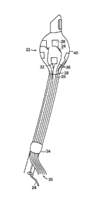
the heart, for
example a ventricle, per minute. Cardiac output is equivalent to the heart
rate multiplied by the
stroke volume. Understanding of a subject's cardiac output is important in
care of acutely injured
or ill subjects, as well as individuals with chronic cardiac pathology. Until
recently the standard of
cardiac output measurement has been pulmonary artery catheterization. See,
e.g., U.S. Patents
3,915,155; 3,726,269 and 3,651,318.
[0003] Bioelectrical impedance analysis ("BIA") has been developed to measure
physiological
and pathological properties, including cardiac output. In performing BIA, a
low level electrical
alternating current is introduced into a tissue being monitored electrically
by multiple electrodes,
such that the voltage difference between multiple locations on the tissue is
determined. From this
determination, the electrical impedance (electrical resistance plus reactance)
of the stimulated
tissue is calculated. Previously, both external (U.S. Patent 4,870,578) and
internal (U.S. Patents
4,852,580 and 4,836,214) electrodes have been employed to measure electrical
resistance
representing blood flow in the aorta. While these internal electrodes were
mounted on esophageal
catheters, it was later determined that endotracheal tubes could be adapted by
the addition of
electrodes on the inflatable cuff, which was perceived to be a more accurate
measurement of
cardiac output. See U.S. Patents 6,095,987 and 6,292,689.
[0004] The process of inserting an endotracheal tube is called intubation, and
is performed\\\
when the inflatable cuff is deflated. The presence of electrodes on the
inflatable cuff and electrode
leads on the external surface of the endotracheal tube results in a more
complex and riskier
CA 02697829 2010-02-24
WO 2009/049266 PCT/US2008/079656
intubation process. Further, the electrodes are attached to the inflatable
cuff when inflated,
resulting in irregularities (e.g., sharp edges of the electrode, broken
electrode leads) when the cuff
is deflated prior to insertion.
[0005] In view of the foregoing, it would be desirable to provide an apparatus
and methods for
safely, accurately, efficiently and continuously determining cardiac output by
measuring electrical
impedance.
SUMMARY OF THE INVENTION
[0006] In general, aspects of the present invention relate to detection of
cardiac output, and
diseases characterized by abnormal cardiac function, using a novel apparatus
that is placed in such
a manner that a portion of the apparatus contacts the tracheal tissue in close
proximity to the aorta.
[0007] In one aspect, the invention provides an apparatus that includes a tube
having a proximal
portion and a distal portion, an inflatable cuff, a ground electrode, a
plurality of sense electrodes,
and a current electrode. Generally, each sense electrode and the current
electrode contains an
electrode patch operably linked to a generally linear electrode runner; the
sense electrodes and the
current electrode are disposed on the inflatable cuff and the distal portion
of the tube, and the
portion of each of the electrode runners disposed on the inflatable cuff forms
a beam-like
structure. In certain embodiments, the portion of the electrode runner
disposed on the tube
extends in a generally linear proximal-distal direction along the tube and is
not substantially
curved. Additionally, the circumferential distance between adjacent beam-like
structures is
greater at the region of the inflated inflatable cuff wherein maximal outer
diameter of the cuff is
achieved than at the region where the inflatable cuff contacts the distal
portion of the tube. In
some embodiments the apparatus also includes a tubule for inflating the
inflatable cuff.
[0008] The apparatus includes sense electrodes and a current electrode that
are separated from
the inflatable cuff by a polymeric underlayer that is applied to the
inflatable cuff and the tube prior
to application of the sense electrodes and the current electrode. The
apparatus also includes a
polymeric overlayer that contacts a portion of the electrode patch and the
entirety of the electrode
runner, and the polymeric overlayer is applied to the electrode after
application of the electrode to
the tube and cuff. In certain embodiments the polymeric underlayer contains a
medical grade
adhesive, such as a urethane oligomer/acrylate monomer blend (e.g., Dymax 1-
20323 resin,
Torrington, CT) the electrode contains electrically conductive silver
particles suspended in a resin
2
CA 02697829 2010-02-24
WO 2009/049266 PCT/US2008/079656
and a volatile solvent that forms a polymeric matrix material once cured (such
as Creative
Materials - CMI 101-59), or the polymeric overlayer contains a medical grade
adhesive, such as a
urethane oligomer/acrylate monomer blend (e.g., Dymax 1-20323 resin).
[0009] The apparatus includes a collection of at least three sense electrodes,
and may be three,
four, five or more than five sense electrodes. For example, the combination of
the five sense
electrodes provides three orthogonal pairs of sense electrodes. The tube of
the apparatus may be
an endotracheal tube. Also, the current electrode is disposed on the distal
portion of the tube
between the termini of the electrode runners (near the middle of the tube in a
proximal-distal
direction) and the inflatable cuff. In certain embodiments, the current
electrode is at least one
centimeter in length (e.g., one, two or more centimeters) as measured in the
proximal-distal
dimension of the tube. In some embodiments the current electrode extends over
about 90 , 120 ,
or about 180 of the circumference of the tube. Optionally, the ground
electrode is also placed on
the tube.
[0010] In another aspect, the invention provides a method of fabricating an
apparatus by
providing an apparatus having a first portion and a second portion that is
capable of being inflated,
at least partially inflating the second portion, imaging the inflated second
portion so as to obtain
imaging data, directing a positive displacement dispensing system to apply to
a region of the
inflated second portion a polymeric underlayer based upon the imaging data,
applying to at least a
portion of the polymeric underlayer a conductive material based upon the
imaging data, and
applying to a portion of the conductive material a polymeric overlayer based
upon the imaging
data. In certain embodiments, no polymeric overlayer is applied to a plurality
of regions of the
conductive material, thereby forming a plurality of electrode patches. The
imaging step comprises
capturing images, such as dynamic video images. The imaging step includes in
certain
embodiments the capturing of a plurality of images that are used to identify
one or more contours
of the second portion.
[0011] In certain embodiments, one or more of the polymeric underlayer, the
conductive
material, or the polymeric overlayer are applied by a positive displacement
dispensing system.
The positive displacement dispensing system includes a pen tip that is kept
substantially
perpendicular to the surface of the second portion during application of the
polymeric underlayer,
the conductive material, or the polymeric overlayer.
[0012] In some embodiments, the apparatus is mounted on a stage having at
least three
3
CA 02697829 2010-02-24
WO 2009/049266 PCT/US2008/079656
independent axes of motion relative to the pen tip. For example, the apparatus
is mounted on a
stage having at least four independent axes of motion relative to the pen tip:
motion along a
direction perpendicular to the pen tip, motion along a direction towards or
away from the pen tip,
rotational motion along an axis perpendicular to the pen tip, and rotational
motion along an axis
parallel to the pen tip. In other embodiments, the positive displacement
dispensing system
includes a MicroPen (MicroPen Technologies Honeoye Falls, NY).
[0013] In a further aspect, the invention provides a method of fabricating an
endotracheal tube
by providing a tube having a proximal portion and a distal portion and an
inflatable cuff disposed
on the distal portion of the tube, at least partially inflating the cuff,
imaging the inflated cuff so as
to obtain imaging data, directing a positive displacement dispensing system to
apply a polymeric
underlayer to a region of the inflated cuff and a region of distal portion of
the tube based upon the
imaging data, applying to at least a portion of the polymeric underlayer a
conductive material to
form a plurality of electrodes where at least a portion of the region to which
the conductive
material is applied is based upon the imaging data, and applying to a portion
of the conductive
material a polymeric overlayer where at least a portion of the region to which
the polymeric
overlayer is applied is based upon the imaging data, and where no polymeric
overlayer is applied
to a plurality of regions of the conductive material to form a plurality of
electrode patches. In some
embodiments, the positive displacement dispensing system comprises a MicroPen
.
[0014] In yet another aspect, the invention provides an apparatus produced by
a process
containing the steps of providing a tube having a proximal portion and a
distal portion, and an
inflatable cuff disposed on the distal portion of the tube, at least partially
inflating the cuff,
imaging the inflated cuff so as to obtain imaging data, directing a positive
displacement dispensing
system to apply to a region of the inflated cuff a polymeric underlayer based
upon the imaging
data, applying to at least a portion of the polymeric underlayer a conductive
material to form a
plurality of electrodes based upon the imaging data, and applying to a portion
of the conductive
material a polymeric overlayer based upon the imaging data, and where no
polymeric overlayer is
applied to a plurality of regions of the conductive material so as to form a
plurality of electrode
patches.
[0015] In another aspect, the invention provides a method of applying a
material to a non-
repeatably formed substrate by providing a positive displacement dispensing
system containing a
pen tip, mounting the substrate on a stage having four independent axes of
motion relative to the
4
CA 02697829 2010-02-24
WO 2009/049266 PCT/US2008/079656
pen tip, imaging the substrate by capturing still or video images so as to
obtain imaging data, and
directing the positive displacement dispensing system to apply to at least a
region of the substrate
a material based upon the imaging data. The independent axes of motion include
motion along a
direction perpendicular to the pen tip, motion along a direction towards or
away from the pen tip,
rotational motion along an axis perpendicular to the pen tip, and rotational
motion along an axis
parallel to the pen tip.
[0016] In certain aspects, the imaging data are processed so as to generate a
three-dimensional
representation of the substrate, and the pen tip is kept at an angle
substantially perpendicular to the
region of the substrate to which the material is being applied. Unless
otherwise defined, all
technical and scientific terms used herein have the same meaning as commonly
understood by one
of ordinary skill in the art to which this invention belongs. Although methods
and materials
similar or equivalent to those described herein can be used in the practice or
testing of aspects of
the present invention, suitable methods and materials are described below. All
publications, patent
applications, patents, and other references mentioned herein are incorporated
by reference in their
entirety. In the case of conflict, the present specification, including
definitions, will control. In
addition, the materials, methods, and examples are illustrative only and are
not intended to be
limiting.
DESCRIPTION OF THE DRAWINGS AND FIGURES
[0017] The present invention may be further appreciated with reference to the
appended drawing
sheets wherein:
[0018] FIG. 1 is a schematic illustration demonstrating one embodiment of the
endotracheal tube
of the present invention;
[0019] FIG. 2 is a partial sectional illustration of the endotracheal tube of
the present invention.
[0020] FIG. 3 is a partial sectional illustration of an electrical assembly of
the present invention.
[0021] FIG. 4 is a schematic illustration of a positive displacement
dispensing system used in the
present invention.
[0022] Other objects, features, and advantages of the present invention will
become apparent
from the following detailed description. It should be understood, however,
that the detailed
description and the specific examples, while indicating preferred embodiments
of the invention,
are given by way of illustration only, since various changes and modifications
within the spirit and
CA 02697829 2010-02-24
WO 2009/049266 PCT/US2008/079656
scope of the invention will become apparent to those skilled in the art from
this detailed
description.
DETAILED DESCRIPTION OF THE INVENTION
[0023] In some embodiments described herein, the present invention relates
generally to an
apparatus useful as an endotracheal tube (also known as an intratracheal tube
or ET tube). The
endotracheal tube is useful in measuring physiological characteristics of a
mammalian subject,
particularly human subjects suffering from acute or chronic injury or illness.
For example, the
endotracheal tube is used to measure cardiac output in a mammalian subject.
The endotracheal
tube is inserted into the trachea, generally via the mouth, but sometimes
through the nares of the
nose or even through a tracheostomy.
[0024] The apparatus 10 for measuring a mammalian subject's cardiac output
shown in FIG. 1
contains tube 12 having proximal portion 14 and distal portion 16. The tube 12
is generally
formed of a medically approved synthetic polymeric material such as silicone
rubber, polyvinyl
chloride or polypropylene. See, U.S. Patents 3,599,642 and 4,593,690, the
contents of which are
incorporated herein by reference in their entireties. The distal portion 16 is
inserted into the subject
during the intubation, and generally has a beveled end with a smooth, curved
tip 18 to facilitate
insertion. The proximal portion 16 of the tube contains a coupler member 20
that is suited to be
connected to medical equipment such as a ventilator. Connected to the distal
portion 16 is an
inflatable cuff 22 that, when inflated, causes occlusion of the airway
surrounding the apparatus 10,
thereby fixing the tube in correct position while allowing the ventilator to
completely regulate the
patient's respiration. Generally the inflatable cuff 22 is fully deflated when
it is inserted into the
subject's airway in order to reduce the risk of injury to the subject during
intubation. Inflation and
deflation of the cuff 22 are controlled through a small secondary tubule 24
that is inserted at the
proximal end of the tube.
[0025] The apparatus 10 also includes several electrodes 24 operably joined to
the distal portion
of the tube and the inflatable cuff. The electrode contains two principal
features: an electrode
patch 26 that is generally rectangular and is disposed on the outer surface of
the inflatable cuff,
and an electrode runner 28 that is extends in a proximal-distal orientation
between the electrode
patch and the point where the electrode runners terminate and are attached to
the bundle or sheath
of external wires 30. The collection of external wires 30 is also termed a
flexible circuit or flex
6
CA 02697829 2010-02-24
WO 2009/049266 PCT/US2008/079656
circuit. Generally the electrode runners terminate near the middle of the tube
in a proximal-distal
orientation. In certain embodiments the electrode patch 26 has a rectangular
(e.g., square) shape,
but it should be recognized that the present invention provides for any shape
of electrode patch
that can be fabricated using the methods described herein and the teachings of
the art (e.g., circle,
oval, or any polyhedra such as triangle, pentagon, hexagon, heptagon, or
octagon). The electrode
patch 26 is connected to the electrode runner 28 at a corner or side of the
electrode patch 26. The
electrode patch can include a triangularly-shaped conductive material 32 that
interfaces with the
electrode patch 26 and electrode runner 28.
[0026] In certain embodiments of the invention the electrode runners 28 and
the external wires
30 are connected using a conductive compound. An exemplary embodiment of the
connection
between the electrode runners 28 and the external wires 30 is schematically
depicted in FIGS. 3A
and 3B. The external wires 30 terminate at an end 68 not surrounded by any
insulating sheath, but
is connected to a traced conductive circuit material 70 operably linked to a
flexible support
material 72. The flexible support material 72 is any suitable material having
the properties of
being thin and flexible, such as a polyimide or polyamide material (e.g., a
Kapton polyimide
film by DuPont). The traced conductive circuit material 70 and the flexible
support material 72
contain a series of holes 74. After the electrode runners 28 are printed on
the tube 12 the traced
conductive circuit material 70 and the flexible support material 72 are
applied over the termini of
the electrode runners 28, such that the holes 74 align with the proximal end
of each electrode
runner 28. A conductive polymeric material 34 (such as Conductive Compounds
EP-600 epoxy
resin, Londonderry, NH) is applied so as to fill or partially fill holes 74
and thereby form an
electrical connection between electrode runner 28 and external wires 30.
Optionally, the
conductive polymeric material 34 is cured, such as by placing the apparatus in
a container heated
to a temperature of about 110 C for a period of time from about ten minutes to
about two hours.
An insulating material 78 is applied over the connection between the electrode
runner 28 and the
external wires 30. Insulating material 78 is a sealing tape, a molded sealing
collar or any
medically-acceptable polymeric material, such as a two-stage medical epoxy
(e.g., Loctite M-
121HP epoxy, Henkel Corporation) that protects the electrodes from bodily
fluids and thereby
increases the durability of the device.
[0027] The spacing between the ends of the electrode runners 28 to which the
external wires 30
are connected is a consideration. It is generally preferable to have a space
of at least about one
7
CA 02697829 2010-02-24
WO 2009/049266 PCT/US2008/079656
millimeter between adjacent ends of the electrode runners 28. This spacing
prevents the build-up
of any capacitance between adjacent ends. Also, this spacing also reduces the
risk of a high
potential electrical failure.
[0028] In several embodiments of the invention, a plurality of electrodes is
disposed on the
inflatable cuff 22. The placement of the electrode patches 26 is dictated to
some extent by the
opportunity to maximize the detection and measurement of voltages caused by
the current flowing
in the tissue. An exemplary placement of multiple electrode patches 26 on the
outer surface of the
inflated cuff is shown in FIG. 2. In certain embodiments, there is a plurality
of sense electrodes
that includes at least two sense electrodes, and preferably includes three,
four or five sense
electrodes. The combination of five sense electrodes provides three orthogonal
pairs of sense
electrodes.
[0029] The portion of the electrode runner 28 on the region of the inflatable
cuff 22 that does not
contact the tube is fabricated such that it forms a beam-like structure 36. By
this is meant that the
electrode runners on the inflatable cuff remain substantially linear and rigid
when the inflatable
cuff is 22 deflated. These beam-like structures 36 are important to the
functionality of the
apparatus 10 when it is inserted into a subject because they increase
electrode durability and
facilitate deflation of the inflatable cuff 22. When the distal portion 16 of
the tube is placed in a
subject's trachea and the inflatable cuff 22 is inflated to secure the
apparatus in position, the
electrode patches 26 come into tight contact with the subject's tracheal
walls. During the
breathing cycle, the pressure on the inflated cuff 22 rhythmically increases
and is then relaxed.
The beam-like structures 36 do not appreciably change shape during this cycle,
but there is
substantial change in the shape of the portions of the inflatable cuff between
the beam-like
structures 36, which decreases the force on the beam-like structures 36 and
increases durability of
the electrodes.
[0030] The regions of the electrode runners 28 that are positioned on the
inflatable cuff 22 are
arranged in an array so as to increase the ability of the cuff 22 to collapse
when deflated, such that
the electrode patches and beam-like structures 36 lay roughly flat against the
portion of the tube
underlying the inflatable cuff 22. In certain embodiments, multiple beam-like
structures 36 extend
from the point where the proximal end of the inflatable cuff 38 contacts the
tube to the electrode
patch.
[0031] The electrode runners 28 are placed generally parallel to adjoining
electrode runners
8
CA 02697829 2010-02-24
WO 2009/049266 PCT/US2008/079656
along the distal portion of the tube. The width of the electrode runner 28 can
by adjusted. For
example, the width of the electrode runner 28 can range from about 0.1
millimeters to about two
millimeters; in a preferred embodiment, the electrode runners 28 are about one
millimeter in width
along the distal portion of the tube 12. The electrode runners 28 diverge from
adjoining electrode
runners 28 at a point on the tube proximal to the inflatable cuff 22; in other
words, the distance
between adjacent runners 28 is generally uniform along the length of the
distal 16 portion of the
tube 12, but increases as the electrode runners 28 near the inflatable cuff
22. The electrode
runners 28 extend generally linearly along the surface of the inflatable cuff
22 to form beam-like
structures 36 and separate from adjacent beam-like structures 36. This
separation increases as the
electrode runners 28 approach the electrode patches 26, which are in proximity
to the point of the
inflatable cuff 22 at which the maximal circumference 40 is obtained. The
result of this separation
is that the circumferential distance between adjacent beam-like structures 36
is greater at the
region of the inflated inflatable cuff wherein maximal outer diameter 40 of
the cuff 22 is achieved
than at the region where the inflatable cuff 22 contacts the distal portion of
the tube 38. This
separation increases the ability of the electrode patches 26 and beam-like
structures 36 to fold flat
against the tube 12 during deflation of the inflatable cuff 22.
[0032] The apparatus 10 also includes a current electrode 42. The current
electrode 42 has an
electrode patch 44 of generally rectangular shape that is positioned between
the distal end 18 of
the tube and the midpoint 46 of the apparatus. Preferably, the current
electrode 42 is located on
the outer radius of the curve formed by the tube. This orientation provides
for better contact
between the current electrode 42 and the subject's trachea. The current
electrode 42 is of an area
sufficient to function as a current electrode. For example, the electrode
patch 44 of the current
electrode 42 is at least 28 millimeters in length as measured in a proximal-
distal orientation. The
current electrode 42 also includes an electrode runner 48 extending distally
from the flex circuit 30
of the apparatus to the electrode patch 44 of the current electrode 42. In
some embodiments the
current electrode extends over about 90 , 120 , or about 180 of the
circumference of the tube. As
described herein, the current electrode runner 48 is fabricated from a
conductive material, and is
separated from the tube by a polymeric underlayer that is applied to the tube
prior to application of
the conductive material. The electrode patch of the current electrode 44 may
be separated from the
tube by the polymeric underlayer. Furthermore, the current electrode runner 48
is covered by a
polymeric overlayer applied to the conductive material.
9
CA 02697829 2010-02-24
WO 2009/049266 PCT/US2008/079656
[0033] When fully inflated, the cuff 22 is of sufficient size to fix the
position of the endotracheal
tube such that there is not substantial movement either downward or upward
relative to the
subject's trachea. For example, the cuff 22 has a maximal outer diameter of at
least twenty
millimeters.
[0034] In certain embodiments the apparatus also includes a tubule 24 for
inflating the inflatable
cuff 22. For example, the tubule 24 has a proximal 50 and distal end 52, the
distal end 52
extending from the cuff 22 in the internal space of the distal portion 16 of
the tube and exiting the
tube 12 in the proximal portion near the midline 46. The proximal end 50 of
the tubule has an
inlet 54 for air or another gas under pressure for inflating the cuff 22. At
the proximal end 50 of
the tubule 24 is a valve housing provided with an air inlet bore 56 and valve
means 58 in the bore
56 such that the inlet is 54 normally closed, but air is admitted under
pressure through the inlet
bore 56 to inflate the cuff 22.
[0035] In certain embodiments, the apparatus 10 is operably connected to a
bioelectrical
impedance recorder, where the impedance recorder is electrically coupled to
the sense electrodes.
Bioelectrical impedance analysis of blood flow using electrode sensors arrayed
within or external
to the trachea is well known in the art. See, e.g., U.S. Patents 5,791,349 and
6,095,987, the
contents of which are incorporated herein by reference in their entireties.
[0036] FABRICATION OF ELECTRODES ON ENDOTRACHEAL TUBES USING A
POSITIVE DISPLACEMENT DISPENSING SYSTEM
[0037] In certain embodiments the present invention provides an apparatus 10
arrayed with
electrodes 24 disposed on an inflatable cuff 22. These electrodes 24 are
applied to the tubes 12
using a novel printing methodology that uses a positive displacement
dispensing system 60. While
this methodology is specifically described herein as useful for applying
materials onto the surface
of the apparatus 10 and the associated inflatable cuff 22, one of skill in the
art will recognize that
the printing methods described herein are also useful for applying a material
to any non-repeatably
formed substrate (e.g., a dilation balloon used in a medical device).
[0038] The printing methodology generally involves two steps: imaging the non-
repeatably
formed substrate and applying one or more materials thereon. The application
step can be termed
"writing", "printing" or any other equivalent term known to those skilled in
the art. These two
general steps are discussed in turn.
[0039] The inflatable cuff 22 is at least partially inflated prior to printing
the electrodes 24 on its
CA 02697829 2010-02-24
WO 2009/049266 PCT/US2008/079656
outer surface. Due to inherent variations in the three dimensionality of the
inflated cuff 22 one
must have an understanding of the shape of the inflated cuff 22 prior to
positioning the electrodes
24. For this reason, the inflated cuff is 22 imaged by capturing either video
or still images. In
certain embodiments video images of the inflated cuff 22 and the adjacent
regions of the distal
region 16 of the tube are collected and sent to a processing system, such as a
computer 62 that
generates a map showing the contours of the inflated cuff 22. In other
embodiments, one or more
still images are captured and reproduced (such as by digital printing) in
order to generate the
contour image map. Generally three or more still images are captured to
generate the contour
image map. In preferred embodiments, eight images are captured.
[0040] Information from the contour map obtained above is provided to a
positive displacement
dispensing system 60 capable of responding to the contour map by altering one
or more printing
dimensions. The displacement dispensing system contains a writing head 64
(such as a pen tip)
and a substrate stage 66 capable of moving the substrate in at least three
independent dimensions.
The writing head is 64 capable of movement relative to the substrate stage 66.
The writing head
64 applies to the substrate any liquid or semi-solid materials, including the
polymeric underlayer
and overlayers, and the conductive material used to form the electrodes 24.
[0041] An exemplary positive displacement dispensing system 60 is shown in
FIG. 4. A writing
head 64 is mounted on an axis capable of moving in one dimension only, shown
in FIG. 4 as the
y-axis. In contrast, the substrate stage 66 is capable of moving in at least
three independent
dimensions, shown in FIG. 4 as the x-axis, ~(clockwise or counter-clockwise
rotation along the z-
axis, and 0 (clockwise or counter-clockwise rotation along the x-axis). In
certain embodiments, the
substrate stage 66 is capable of moving in a fourth independent direction,
shown in FIG. 4 as the
y-axis.
[0042] Preferably, the positive displacement dispensing system 60 is used to
print the electrodes
24 in a sandwich format: the conductive material is surrounded by the
polymeric underlayer on the
bottom (i.e., the area closest to the tube) and the polymeric overlayer on the
top (i.e., the area
furthest from the tube), except for a portion of the electrode patch 26, which
is not covered by the
polymeric overlayer and therefore is able to directly contact the tracheal
mucosa when inserted
into a subject's trachea, and the end of the electrode runner 28 that contacts
the external wires 30.
As such, the writing head 64 applies to a region of the inflated cuff 22 a
polymeric underlayer.
The region to which the polymeric underlayer is applied is based upon the
imaging data obtained
11
CA 02697829 2010-02-24
WO 2009/049266 PCT/US2008/079656
from the contour map described above. Extending from the flex circuit 30 of
the apparatus 10 the
writing head 64 writes a thin, narrow layer of material directly on the distal
portion of the tube 16
and extending to the inflatable cuff 22, which is at least partially inflated.
For example, the
inflatable cuff 22 is inflated to an inflation pressure of about 10 to about
40 cm H20, e.g., about 25
cm H20. Because the course of the writing head 64 is controlled based on
information regarding
the contours of the inflated cuff 22 and the distal portion 16 of the tube,
multiple parallel lines can
be formed along the proximal-distal axis of the tube 12. As used herein,
materials useful as
polymeric underlayer include an ultraviolet (UV)-curable resin such as Dymax
1-20323 resin
and Creative Materials dielectric inks (e.g., CMI-115-30). Prior to printing
the underlayer the tube
12 and cuff 22 may be cleaned with a solvent such as ethanol or with physical
means (such as an
ionizing gun) to remove debris. After printing, the underlayer is optionally
cured, such as by
exposure to UV or visible light radiation or a similar curing agent.
[0043] Wrinkles or other deformations may exist in the cuff 22 prior to
printing. In certain
embodiments, prior to printing the underlayer the tube 12 may be heated, such
as at 30-100 C
(e.g., about 60 C) for a period of time (e.g., 1-60 minutes, preferably about
45 minutes) after
inflation of the cuff 22 to remove any wrinkles present in the tube 12 or the
inflatable cuff 22.
Alternatively, a physical force can be applied to the cuff 22 to remove any
wrinkles prior to
printing.
[0044] The shape of the inflatable cuff 22 can be modified prior to electrode
printing. For
example, a vacuum can be applied to the end of the cuff 22 closest to the
distal tip 18 of the
apparatus, resulting in a deformation of the cuff 22; this deformation
preferably results in a
decrease in the angle formed by the inflatable cuff 22 as it extends away from
the proximal end of
the tube, such that printing on the cuff becomes easier.
[0045] In certain embodiments, physical force can be applied to the proximal
end of the cuff 22
by contacting one or more regions of the cuff with projections, or "fingers",
that pull the cuff in a
distal direction along a proximal-distal axis. The printing of the electrode
patches 26 and
electrode runners 28 is performed by applying the writing head 64 with the
portions of the
inflatable cuff 22 not contacted or otherwise obscured by the projections. The
application of
physical force reduces or eliminates any wrinkles in the inflatable cuff 22
and transforms the
inflatable cuff 22 into a defined writing surface, thereby obviating the need
to image the inflated
cuff 22 prior to the writing step.
12
~
~
CA 02697829 2010-02-24
WO 2009/049266 PCT/US2008/079656
[0046] Upon completion of the printing of the polymeric underlayer, the
dispensing system 60
has a functional "road map" for where to place the conductive material that is
used to form the
electrode runners 28 and electrode patches 26. Generally, the width of the
line formed by the
conductive material will be less than that of the polymeric underlayer, such
that no conductive
material directly contacts either the distal portion of the tube 16 or the
inflatable cuff 22. As used
herein, materials useful as a conductive material include electrically
conductive inks such as CMI
101-59 (Creative Materials Inc., Tyngsboro, MA) or any other electrically
conductive particles
such as silver or gold particles that are suspended in a resin and a solvent.
After printing the
conductive material on the tube 12 is optionally cured, such as by heating the
tube. This curing
step results in the formation of a polymeric matrix surrounding the conductive
particles. By way
of non-limiting example, the tube 12 is placed in a suitable container, which
is then heated to a
temperature of 90-150 C (e.g., 120 C) for a period of time (e.g., 30 minutes
to five hours or
more). The temperature of the container may be gradually increased, such as
increasing the
temperature by 0.1-5 C per minute.
[0047] A polymeric overlayer 76 is written over the conductive material the
length of the
electrode runner 28, which is acceptable because no signals are directly
measured from the
electrode runner 28 itself. The overlayer is written over the outer periphery
of the electrode patch
26 on the inflated cuff 22. For example, the outer one millimeter of each side
of the electrode
patch 26 is covered with the polymeric overlayer. The purpose of this
overlayer is to increase
durability of the electrodes 24 and prevent errors in signal processing.
Additionally, in one
embodiment the overlayer extends from the periphery of the electrode patch 26
over the polymeric
underlayer and onto the surrounding material of the inflatable tube 22. In
another embodiment, the
overlayer extends beyond the periphery of the electrode patch 26 but does not
extend beyond the
polymeric underlayer. This extension results in a seal that strengthens the
attachment of the
electrode patch 26 to the inflatable tube 22, thereby decreasing the
probability that physical strain
on the electrode patch 26 will cause its separation from the inflatable tube
2.
[00481 As used herein, materials useful as polymeric overlayers include an
ultraviolet (UV)-
curable resin such as Dymax 1-20323 resin, or Creative Materials dielectric
ink (e.g., CMI-115-
30). Prior to printing the overlayer, the tube 12 may be cleaned with a
solvent such as ethanol or
with physical means (such as an ionizing gun) to remove debris. After printing
the overlayer the
apparatus 10 is optionally cured, such as by exposure to UV or visible light
radiation or a similar
13
CA 02697829 2010-02-24
WO 2009/049266 PCT/US2008/079656
curing agent.
[0049] MEASUREMENT OF CARDIAC OUTPUT
[0050] Endotracheal tubes bearing electrodes have been previously described as
useful for
measuring cardiac function, including cardiac output. See U.S. Patents
6,095,987 and 6,292,689.
The endotracheal tubes as described herein are useful to measure physiological
functions in
mammalian subjects. For example, cardiac output is measured, and any
pathological situation
identified, using the electrodes arrayed on the inflatable cuff. Thus, the
invention provides a
method of measuring the cardiac output of a mammalian subject by providing an
endotracheal
tube substantially as described herein. The endotracheal tube includes a
current electrode
connected thereto and an inflatable cuff containing an array of electrodes
including a plurality of
sense electrodes and a ground electrode, and is positioned in the trachea in
the vicinity of the aorta
so that inflating the cuff results in the cuff contacts the tracheal mucosa.
Once the inflatable cuff
is positioned, a current is injected into the subject's trachea through the
current electrode, a
voltage is established between the current electrode and the ground electrode
so that a current
flows through the tissue disposed between the current electrode and the ground
electrode. With
one or more sense electrodes the voltages caused by the current flowing in the
tissue is detected,
wherein the voltages vary in accordance with changes in the bioelectrical
impedance of the tissue.
Generally, the tube is adapted to be inserted in the trachea of the subject
through the mouth, a
nasal passageway, or a tracheotomy port.
[0051] The present invention is not limited to the particular methodologies,
protocols, constructs,
formulae and reagents described but further include those known to the skilled
artisan. It is also to
be understood that the terminology used herein is for the purpose of
describing particular
embodiments only, and is not intended to limit the scope of the present
invention.
[0052] Unless defined otherwise, all technical and scientific terms used
herein have the same
meaning as commonly understood to one of ordinary skill in the art to which
this invention
belongs. Any methods, devices, and materials similar or equivalent to those
described herein can
be used in the practice or testing of the invention. All publications and
patents mentioned herein
are incorporated herein by reference.
14