Note: Descriptions are shown in the official language in which they were submitted.
CA 02698004 2016-10-06
DEVICES AND METHODS FOR PROVIDING ACCESS INTO A BODY CAVITY
CROSS-REFERENCE TO RELATED APPLICATIONS
[0001] The present application claims priority to U.S. Provisional Application
No. 61/165,080
entitled "Access Device" filed April 1, 2009.
FIELD OF THE INVENTION
[0002] The present invention relates to methods and devices for providing
surgical access into a
body cavity.
BACKGROUND OF THE INVENTION
[0003] Abdominal laparoscopic surgery gained popularity in the late 1980's,
when benefits of
laparoscopic removal of the gallbladder over traditional (open) operation
became evident.
Reduced post-operative recovery time, markedly decreased post-operative pain
and wound
infection, and improved cosmetic outcome are well established benefits of
laparoscopic surgery,
derived mainly from the ability of laparoscopic surgeons to perfon-n an
operation utilizing
smaller incisions of the body wall.
[0004] Laparoscopic procedurcs typically involve inserting a surgical access
device, such as a
straight tubular cannula or trocar sleeve, into the abdominal cavity.
Insufflation of the abdominal
cavity with carbon dioxide gas to a pressure of around 15 mm Hg is generally
used to increase
the interior space for the surgical procedure. Accordingly, various sealing
elements are used
within the trocar sleeve to seal its working channel both before and after a
surgical instrument is
inserted through the trocar sleeve to seal the body cavity from the outside in
order to achieve and
maintain insufflation. Suitable laparoscopic instruments (graspers,
dissectors, scissors,
retractors, etc.) can be placed through the one or more trocar sleeves
depending on the procedure
and needs of the surgeon. Surgeons can then perform a variety of diagnostic
procedures, such as
visual inspection or removal of a tissue sample for biopsy, or treatment
procedures, such as
removal of a polyp or tumor or restructuring tissue.
[0005] Because of the rise in popularity of minimally invasive surgeries,
there has been
significant development with respect to the procedures and the instruments
used in laparoscopic
- 1 -
CA 02698004 2010-03-30
procedures. For example, in some procedures a single incision at the navel can
be sufficient to
provide access to a surgical site. This is because the umbilicus can be a
preferred way to access
an abdominal cavity in a laparoscopic procedure. The umbilical incision can be
easily enlarged
without significantly compromising cosmesis and without significantly
increasing the chances of
wound complications, thus allowing multiple instruments to be introduced
through a single
access device placed in an incision.
[0006] Current devices used in single site laparoscopic procedures generally
provide a plurality
of seals in order to simultaneously accommodate a plurality of surgical
instruments. Seals are
typically disposed within the access device at the level of the abdomen wall
or are fixed to the
access device well above the body wall. As a result, the range of motion of
the seals is limited
by the access device, thereby vastly restricting the quadrant-to-quadrant
reach of surgical
instruments inserted therethrough. Seals that extend below the access device
but fail to extend
through the abdomen wall are subject to collapse as the incision itself closes
in around the seal
and prevents insertion of a surgical instrument through the seal.
[0007] Accordingly, there remains a need for methods and devices that provide
instrument
range-of-motion without subjecting the seal to collapse by the incision.
SUMMARY OF THE INVENTION
[0008] The present invention provides mcthods and devices for accessing a body
cavity. In
general, a surgical access device is provided that can include a retractor
that forms a working
channel through tissue, a seal housing for sealing the working channel and/or
forming a seal
around an instrument inserted therethrough, and a suspension member or tether
configured to
suspend the seal housing within the body cavity.
[0009] In one exemplary embodiment, the seal housing can be suspended within
the working
channel of the retractor by a flexible tether coupled to the retractor such
that at least a portion of
the seal housing can extend distally beyond the distal end of the retractor
and can be angularly
oriented relative to the retractor. The seal housing can form a seal across
the working channel of
the retractor, and can include one or more sealing elements disposed therein
that seal the working
channel and/or form a seal around an instrument that is inserted through the
sealing element.
- 2 -
CA 02698004 2010-03-30
[0010] The suspension member or tether can have various configurations. In one
embodiment,
the flexible tether can include a proximal housing that is rotatably coupled
to a retractor housing
on the flexible retractor. In another embodiment, the flexible tether can
include a proximal
flange that rests against the proximal end of the retractor. A distal portion
of the retractor
extending from the housing or flange can be flexible and it can include
features for seating the
seal housing. For example, the distal portion of the flexible tether can
include an engagement
feature, such as an annular rim or flange, or a sleeve, configured to
removably engage the seal
housing.
[0011] In another embodiment, the surgical access device can include a
retractor having
proximal and distal ends with a working channel extending therethrough. The
proximal end can
be configured to be positioned adjacent to an external surface of a patient's
tissue and the distal
end can be configured to extend into a body cavity such that the working
channel provides a
pathway through the tissue. The access device can also include a suspension
member having a
proximal portion configured to couple to the proximal end of the retractor and
a distal portion
that extends through the working channel of the retractor. A seal housing can
be disposed within
the distal portion of the suspension member and it can have at least one
sealing element disposed
therein and configured to form a seal around an instrument disposed
therethrough. At least a
portion of the suspension member can be flexible to allow the seal housing to
extend beyond the
distal end of the retractor.
[0012] The retractor can also have any number of configurations, shapes, and
sizes. In one
embodiment, the retractor can be a hollow flexible cylindrical member having a
mid-portion with
a maximum diameter that is less than a maximum diameter of the proximal and
distal ends of the
retractor. The mid-portion can be configured to be positioned within an
opening in tissue and the
proximal and distal ends can be configured to engage the tissue therebetween.
[0013] The suspension member or tether can have any number of configurations,
shapes, and
sizes and can be formed of any number of materials. In one embodiment, the
proximal portion
of the suspension member or tether can include a radially-outward extending
flange that is
configured to rest against the proximal end of the retractor. In another
embodiment, the
proximal portion of the suspension member or tether can include a collar that
is mated to a
- 3 -
CA 02698004 2010-03-30
housing on the proximal end of the retractor. The distal portion of the
suspension member or
tether can also have various configurations, but in one embodiment is in the
form of a flexible
sleeve. The distal end of the flexible sleeve can include various features for
engaging the seal
housing. In one embodiment, the distal portion of the suspension member can
include an
engagement feature that can removably engage the seal housing. The engagement
feature can
be, for example, an annular member or rim that can engage the seal housing or
retain the seal
housing within the distal portion of the suspension member.
[0014] The seal housing can also have any number of configurations, shapes,
and sizes and it
can be formed of any number of materials. In an exemplary embodiment, the seal
housing is
configured to form a seal across the working channel of the retractor. The
seal housing can
contain one or more sealing elements that are configured to form a seal around
an instrument
and/or seal the working channel.
[0015] In anothcr embodiment, a method for accessing a body cavity is provided
that can
include positioning a flexible retractor within tissue such that a working
channel of the flexible
retractor forms a pathway through the tissue and into a body cavity. The
method can also
include inserting a surgical instrument through a sealing element in a seal
housing suspended
within the body cavity by a flexible tether that extends between the seal
housing and a proximal
portion of the flexible retractor to position a distal end of the surgical
instrument in the body
cavity. The surgical instrument can be manipulated to cause the seal housing
to move relative to
the flexible retractor, and thereby cause the flexible tether to flex. The
flexible tether and seal
housing can be removed from the retractor such that the retractor is left
disposed within the
tissue. A second surgical instrument can also be inserted through a second
sealing element in the
seal housing to position a distal end of the second surgical instrument in the
body cavity.
BRIEF DESCRIPTION OF DRAWINGS
[0016] This invention will be more fully understood from the following
detailed description
taken in conjunction with the accompanying drawings, in which:
[0017] FIG. 1 is an exploded perspective view of one exemplary embodiment of a
surgical
access device;
- 4 -
CA 02698004 2010-03-30
[0018] FIG. 2 is a perspective cross-sectional view of thc assembled device of
FIG. 1;
[0019] FIG. 3 is a side cross-sectional view of the device of FIG. 2 disposed
in a tissue opening;
[0020] FIG. 4 is a side cross-sectional view of anothcr exemplary embodiment
of a surgical
access device disposed in an opening formed in tissue;
[0021] FIG. 5 is a side cross-sectional view of the device of FIG. 4 with a
surgical instrument
disposed therethrough;
[0022] FIG. 6 is an exploded perspective view of another exemplary embodiment
of a surgical
access device;
[0023] FIG. 7A is an exploded view of one embodiment of a seal housing;
[0024] FIG 7B is a bottom perspective view of the inner housing of the device
of FIG. 7A;
[0025] FIG. 7C is a top perspective view of the inner housing of the device of
FIG. 7A;
[0026] FIG. 7D is a top perspective view of the instrument channel member of
the device of
FIG. 7A;
[0027] FIG. 8 is a top view of another exemplary embodiment of a surgical
access device;
[0028] FIG. 9 is a side cross-sectional view of the device of FIG. 7 disposed
in an opening
formed in tissue; and
[0029] FIG. 10 is a side cross-sectional view of another exemplary embodiment
of a surgical
access device.
DETAILED DESCRIPTION
[0030] Certain exemplary embodiments will now be described to provide an
overall
understanding of the principles of the structure, function, manufacture, and
use of the devices
and methods disclosed herein. One or more examples of these embodiments are
illustrated in the
accompanying drawings. Those of ordinary skill in the art will understand that
the devices and
methods specifically described herein and illustrated in the accompanying
drawings are non-
- 5 -
CA 02698004 2010-03-30
limiting exemplary embodiments and that the scope of the present invention is
defined solely by
the claims. The features illustrated or described in connection with one
exemplary embodiment
can be combined with the features of other embodiments. Such modifications and
variations are
intended to be included within the scope of the present invention.
[0031] Various exemplary methods and devices are provided for accessing a body
cavity. In
general, the access devices and methods described herein can be used in
minimally-invasive
surgeries such as laparoscopic surgeries, and can provide improved range of
motion of surgical
instruments used therewith. The devices can have a number of different
configurations, but in
certain exemplary embodiments, an access device can generally include a
retractor configured to
form a working channel through tissue and into a body cavity, a seal housing
having at least one
seal therein for sealing the working channel and/or forming a seal around an
instrument inserted
therethrough, and a suspension member or tether configured to suspend the seal
housing within
the body cavity. In use, one or more surgical instruments can be inserted
through the seal(s) in
the seal housing, and thus through the working channel of the retractor, to
position a distal end of
the surgical instrument(s) in the body cavity. The suspension member can allow
the seal housing
to move and/or angularly diverge relative to the retractor. Such free floating
movement of the
suspension member allows a sealing engagement to be maintained between a seal
and an
instrument inserted through the seal. A person skilled in the art will
appreciate that the access
devices can be used in any surgical procedure, including open procedures, and
can have a variety
of other configurations and include various other features known in the art.
Moreover, the
suspension members disclosed herein can be used in a variety of other devices
to suspend a
housing to allow for free angular orientation of the housing.
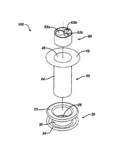
[0032] FIGS. 1-3 illustrate one exemplary embodiment of a surgical access
device 100 having a
retractor 20, seal housing 60, and suspension member or tether 40. In general,
the retractor 20 is
configured to be disposed within tissue to form a working channel 28 through
the tissue and into
the body cavity, and the suspension member 40 can be seated within the working
channel 28 of
the retractor 20 for retaining the seal housing 60 at a location distal of a
distal end of the retractor
20 such that the seal housing 60 is suspended within the body cavity. At least
a portion of the
suspension member 40 can be flexible to allow the seal housing 60 to be
oriented at various
angular orientations relative to the retractor 20.
- 6 -
CA 02698004 2010-03-30
[0033] One skilled in the art will appreciate that the retractor 20 can have
any number of
configurations, shapes, and sizes depending at least in part on the size of
the incision or opening
in which the retractor will be disposed, the surgical components with which it
will be used, and
the type of surgical procedure with which it will be used. Although generally
referred to as a
retractor herein, the retractor 20 of the various surgical access devices and
methods described
herein can be a wound protector, cannula, ring retractor, or any other member
configured to form
a pathway through tissue. The retractor 20 can provide access to an interior
surgical site within a
body cavity and can include proximal and distal ends and a working channel 28
extending
therebetween. In an exemplary embodiment, the retractor 20 is positioned
within an opening in
tissue such that the distal end of the retractor 20 extends into a patient's
body cavity or is
adjacent to an inner surface of the tissue and the proximal end is positioned
adjacent to the
patient's skin on an exterior surface of the patient's body. The working
channel 28 provides a
pathway through the tissue through which surgical instruments can be inserted
from outside the
body to the interior body cavity. The retractor 20 can be placed in any
opening within a patient's
body, whether a natural orifice or an opening made by an incision. For
example, the retractor 20
can be placed through the umbilicus, vaginally, or percutaneously.
[0034] In one exemplary embodiment, as depicted in FIGS. 1-3, the retractor 20
includes a
proximal flange 22, a distal flange 24, and a cylindrical mid-portion 26
extending therebetween.
The proximal and distal flanges 22, 24 can extend radially-outward relative to
a longitudinal axis
of the mid-portion 26 such that the flanges 22, 24 have an increased diameter
relative to the mid-
portion. As shown in FIG. 3, when the retractor 20 is positioned in tissue,
the proximal flange 22
can be disposed external to a body wall 10 and can engage the outer surface 12
of thc paticnt's
skin, the distal flange 24 can be disposed within the patient, such as within
the patient's
abdominal cavity and can engage an inner surface 14 of the patient's body wall
10 when
positioned during surgery, and the mid-portion 26 can be disposed within the
tissue wall.
[0035] The proximal and distal ends of the retractor 20 can have any suitable
configuration that
allow the retractor 20 to be secured within the incision. The proximal and
distal flanges 22, 24
are depicted as having an annular shape, but they can have any configuration
including and
without limitation, a circular, oval, elliptical, square, and rectangular
configuration.
- 7 -
CA 02698004 2010-03-30
Additionally, the proximal and distal flanges 22, 24 need not be closed or
continuous but can, for
example, include a plurality of circumferentially-spaced, radially-extending
tabs.
[0036] As shown in FIGS. 1-3, the cylindrical mid-portion 26 extending between
the proximal
flange 22 and the distal flange 24 defines a working channel 28 having a
circular cross-sectional
shape. The external surface of the cylindrical mid-portion 26 can engage at
least a portion of the
patient's body wall 10 when the retractor 20 is positioned in tissue. The
skin's natural elasticity
can result in compression of the skin against the external surface of the mid-
portion 26 and can
further assist in the retention of the retractor 20 in the body opening or
incision. Although the
working channel 28 can have any cross-sectional shape including and without
limitation,
circular, oval, elliptical, square, and rectangular, a circular working
channel can provide the
maximum area per unit of perimeter length. A circular working channel 28 can
also provide ease
of rotation of the suspension member 40 and/or the seal housing 60 relative to
the retractor 20.
[0037] The retractor 20 can have a variety of sizes. For non-limiting example,
the retractor 20
can have a longitudinal length of between about 2 cm to about 7 cm, a maximum
diameter
corresponding to diameters of the proximal and distal ends of about 40 mm to
about 80 mm, and
a working channel diameter of between about 15 mm to about 40 mm. A surgeon
can select an
appropriately-sized retractor depending on, e.g., thc procedure to be
performed and the size of
the incision. For non-limiting example, a surgeon can select a retractor 20
having a length
approximately equal to the thickness of the body wall 10 to assist in
maintaining an air-tight seal
between the retractor 20 and the body wall 10. It should also be understood
that the diameter of
the proximal and distal ends can differ such that the distal end of the
retractor 20 can have a
greater diameter than the proximal end, or vice versa. It should also be
understood that the
diameter of the working channel 28 need not be constant and can vary along its
longitudinal
length. In one embodiment, a working channel 28 smaller than that required to
permit passage
of a user's hand and generally less than about 50 mm can be desirable, so as
to provide access for
multiple instruments without requiring a relatively large incision.
Alternatively, the retractor 20
can have a working channel 28 of sufficient diameter to permit passage of a
user's hand.
[0038] The retractor 20 can be rigid, semi-rigid, or flexible. More than one
material can be
used to form the retractor 20, and the retractor 20 can include some portions
that are more rigid
- 8 -
CA 02698004 2010-03-30
than others. For example, the retractor 20 can be formed of a resiliently
deformable material,
such as natural rubber, silicone, or a suitable deformable elastomer or
elastomeric material. The
retractor 20 can also include some portions that are formed of a stiffer
material, such as
polyethylene, polycarbonate, polyester, polyetherimide material, or stainless
steel. For example,
the distal flange 24 of the retractor 20 can be resiliently deformable to ease
insertion through an
incision while the proximal flange 22 can be relatively stiff to maintain the
working channel 28
in a predetermined shape or size. Once the distal flange 24 of the retractor
=20 has been inserted
into the body cavity, the distal flange 24 can be configured to resiliently
return to its undeformed
configuration.
[0039] One of skill in the art will appreciate that the retractor 20 can
include additional features
to help secure the retractor 20 within an opening in the body and provide
access to an internal
body cavity. In some embodiments, at least a portion of the retractor 20 can
be configured to
form an air-tight seal with a surface of the patient's body wall 10 such that
insufflation of the
body cavity can be maintained. Additionally, although the distal-facing
surface of the proximal
flange 22 and the proximal-facing surface of the distal flange 24 are depicted
as being
substantially flat in FIGS. 1-3, these surfaces can include surface features
to help securely
engage the retractor 20 to the patient's body wall 10. The retractor 20 can
additionally include
mating features that allow the suspension member 40 to be fixedly, releasably,
and/or movably
coupled to the retractor 20. Such mating features can be formed in or extend
from the proximal
end, distal end, or mid-portion of the retractor 20.
[0040] The suspension member 40 can be configured to suspend the seal housing
60 within a
body cavity and can generally include a proximal portion configured to couple
to the retractor 20
and a distal portion that is configured to extend through the working channel
28 of the retractor
20 and couple to the seal housing 60. The suspension member 40 can have a
variety of
configurations, but in the embodiment shown in FIGS. 1-3, the proximal portion
of the
suspension member 40 includes an annular flange 42 that extends radially-
outward the distal
portion of the suspension member 40, which in the illustrated embodiment is in
the form of a
cylindrical sleeve 44. The annular flange 42 can have a diameter less than,
equal to, or greater
than the proximal flange 22 of the retractor 20 and it can have a diameter
greater than the
diameter of the working channel 28 such that the annular flange 42 abuts or
otherwise contacts
- 9 -
CA 02698004 2010-03-30
the proximal end of the retractor 20 when the cylindrical sleeve 44 of the
suspension member 40
is inserted through the proximal end of thc working channel 28. The flange 42
can act as a stop
to prevent the suspension member 40 from passing into or through the working
channel 28.
[0041] The flange 42 of the proximal portion of the suspension member 40 is
depicted having
an annular shape but it can have any shape including and without limitation, a
circular, oval,
elliptical, square, and rectangular shape. Additionally, the flange 42 need
not be closed or
continuous, but can, for non-limiting example, include a plurality of
circumferentially-spaced,
radially-extending tabs. One of skill in the art will appreciate that the
proximal portion of the
suspension member 40 can have any configuration such that the proximal portion
of the
suspension member 40 can support the distal portion of the suspension member
during surgery.
[0042] The suspension member 40 can be integral with the retractor 20 (e.g.,
formed as a single
unitary component) or it can be fixedly, removably, and/or movably mated to
the retractor 20.
As shown in FIGS. 1-3, the distal-facing surface of the flange 42 is
configured to abut the
proximal flange 22 of the retractor 20 when the suspension member 40 is fully
inserted into the
retractor 20. The opposing surfaces can be configured to slide relative to one
another such that
the suspension member 40 can be rotated relative to the retractor 20, or the
proximal portion of
the suspension member 40 can engage the proximal end of the retractor through
surface features
or engagement features formed on or extending from either or both of the
proximal ends of the
suspension member 40 or the retractor 20 to prevent relative rotation. For non-
limiting example,
the opposing surfaces can be textured to discourage or prevent rotation of the
suspension
member 40 relative to the retractor 20. One skilled in the art will appreciate
that any number of
engagement mechanisms (e.g., snap-fit couplings, threading, etc.) can be used
to fixedly or
removably couple the suspension member 40 to the retractor 20. One skilled in
the art will also
appreciate that the engagement mechanism can be formed in or extend from any
portion of the
suspension member 40 or the retractor 20 such that the suspension member 40
can couple to the
proximal or distal end of the retractor 20 or any location therebetween.
[0043] For non-limiting example, in another embodiment depicted in FIGS. 4 and
5, the
suspension member or tether of surgical access device 400 does not abut or
couple to the
proximal end of the retractor 420 as in the embodiment illustrated in FIGS. 1-
3, but instead
- 10 -
CA 02698004 2010-03-30
includes a tubular housing 442 that can be configured to be disposed within a
working channel of
the retractor 420. The tubular member 442 can be coupled to a flexible sleeve
444 which in turn
can be coupled to a seal housing 460. The external surface of tubular housing
442 can include an
engagement mechanism to engage the retractor 420, e.g., an annular flange 447
of the tubular
housing 442 can snap-fit within a corresponding groove 430 formed on an
internal surface of the
retractor 420. Alternatively, or in addition, a shoulder 450 of the tubular
housing 442 can rest on
or engage an annular rim 432 formed on an internal surface of the retractor
420. The sleeve 444
can extend through the working channel of the retractor 420 and suspend the
seal housing 460
within a body cavity when the surgical access device 400 is positioned in
tissue.
[0044] Again referring to FIGS. 1-3, the distal portion of the suspension
member 40 can include
a cylindrical sleeve 44 that extends distally from the flange 42. An inner
surface of the sleeve 44
can define a passageway 46 through which surgical instruments can be passed.
The sleeve 44
can have a variety of configurations, shapes, and sizes. In the illustrated
embodiment, the sleeve
44 has a generally elongate cylindrical shape. However, the sleeve 44 can have
various cross-
sectional shapes, such as square, ovular, triangular, etc. The size of the
sleeve can also vary. In
an exemplary embodiment as depicted in FIG. 3, the sleeve 44 can have a length
that is sufficient
to extend through tissue when disposed in an opening therein, and can have a
longitudinal length
L that is greater than a height H of the retractor 20 such that the seal
housing 60, when disposed
within the sleeve 44, can be suspended at a location adjacent or distal to the
distal end of the
retractor 20. The sleeve 44 can also have an outer diameter that allows the
sleeve 44 to be
disposed through the working channel 28 of the retractor 20 but is sufficient
to retain the seal
housing 60 therein. The outer diameter of the sleeve 44 can also be selected
such that the outer
surface of the sleeve 44 forms a gas-tight seal with at least a portion of the
retractor 20 when the
sleeve 44 is inserted into the working channel 28. In an exemplary embodiment,
the length of
the sleeve 44 can be in the range of about 4 cm to about 9 cm and the outer
diameter of the
sleeve 44 can be in the range of about 60 mm to about 100 mm. The diameter of
sleeve 44 need
not be constant and can vary along the length of the sleeve 28.
[0045] The suspension member 40 can be rigid, semi-rigid, or flexible
depending on, for
example, the procedure to be performed and the size of the incision. The
suspension member 40
can be formed of a resiliently deformable material, such as natural rubber,
silicone, or a suitable
- 11 -
CA 02698004 2010-03-30
deformable elastomer or elastomeric material. In some embodiments, more than
one material
can be used to form the suspension member 40, and the suspension member 40 can
include some
portions that are more rigid than others. For non-limiting example, the sleeve
44 can be formed
from a highly flexible and resilient material such that the distal portion of
the suspension
member 40 can stretch and angularly diverge relative to the longitudinal axis
of the working
channel 28. The sleeve 44 can also be formed of a relatively low friction,
puncture resistant
material that allows for relatively high elongation before tearing. The sleeve
44 can also be
relatively resistant to degradation by silicon or other lubricants. The rim 42
can be formed of the
same material as the sleeve 44 or can be formed of a different material.
Forming the rim 42 from
a relatively stiff material, such as polyethylene, polycarbonate, polyester,
polyetherimide
material, or stainless steel, can help prevent the suspension member 40 from
passing through the
working channel 28.
[0046] The distal portion of the suspension member 40 can be integral with the
proximal
portion of the suspension member 40, as shown in FIGS. 1-3. The distal portion
of the
suspension member 40 can also be fixedly or movably coupled to the proximal
portion of the
suspension member 40 using any number of engagement mechanisms (e.g., snap-fit
couplings,
threading, etc.) known in the art. For non-limiting example, in another
embodiment depicted in
FIG. 6, a surgical access device 600 includes a retractor 620, seal housing
660, and suspension
member having a proximal portion 642 and a distal portion 644. The proximal
portion 642
includes a first tubular housing 642a having an annular rim 642b that extends
radially-outward
from its proximal end. The first tubular housing 642a can be of any size or
shape but as shown
in FIG. 6 can have an outer diameter that is approximately equal to or
slightly less than the inner
diameter of the working channel 628 of the retractor 620 and a shape that
corresponds to the
shape of the working channel of the retractor 620. The first tubular housing
642a can include an
annular flange 643 formed on its inner surface. The distal portion 644 of the
suspension member
or tether can include a second tubular housing 644a and a flexible sheath 644b
that extends
distally from the distal end of the second tubular housing 644a. The distal
end of the flexible
sheath 644b can be configured to couple to seal housing 660. The second
tubular housing 644a
can have an outer diameter approximately equal to or slightly less than the
inner diameter of the
first tubular housing 642a. The second tubular housing 644a can include an
annular groove 645
formed on its outer surface. The annular flange 643 can be configured to snap-
fit into the
- 12 -
CA 02698004 2010-03-30
annular groove 645 when the second tubular housing 644a is inserted into the
first tubular
housing 642a such that the proximal portion 642 of the suspension member
couples to the distal
portion 644 of the suspension member.
[0047] The distal portion of the suspension member can also include various
engagement
features for retaining the seal housing therein. One skilled in the art will
appreciate that any
number of engagement mechanisms can be used to couple the seal housing to the
suspension
member. In one embodiment, as shown in FIGS. 2 and 3, an annular rim 48 formed
on an inner
surface of the distal end of the cylindrical sleeve 44 prevents the seal
housing 60 from being
pressed distally beyond the distal end of the suspension member 40. The
annular rim 48 can
have an inner diameter DR less than a maximum outer diameter Ds of the seal
housing 60 such
that the seal housing 60 can rest upon the annular rim 48, as shown in FIG. 3.
The annular rim
48 need not be closed or continuous, but can be, for non-limiting example, a
plurality of
circumferentially-spaced, radially-inward extending tabs. Although the seal
housing 60 rests
upon the annular rim 48 in this illustrated embodiment, the suspension member
40 can couple to
or mate with the seal housing 60 in any manner known in the art, e.g., annular
grooves formed in
an external surface of the seal housing can snap-fit with corresponding
flanges formed on an
inner surface of the distal portion of the suspension member 40, or flanges
formed on an external
surface of the seal housing 60 can snap-fit with corresponding grooves formed
on an inner
surface of the distal portion of the suspension member 40. The seal housing
can also, for non-
limiting example, slidingly or threadingly engage the suspension member or can
couple by way
of an interference fit. Any other engagement mechanism known in the art, e.g.,
adhesives, can
be utilized to fixedly retain or releasably couple the seal housing 60 to the
suspension member
40. Further, the seal housing 60 can engage any portion of the suspension
member 40 such that
the seal housing 60 can extend into the body cavity when the access device 100
is positioncd
within a tissue opening.
[0048] One skilled in the art will also appreciate that the engagement
mechanism can be formed
in or extend from any portion of the suspension member 40 or the retractor 460
such that the
suspension member 40 can couple to the proximal or distal end of the seal
housing 460 or any
location therebetween. For non-limiting example, in another embodiment
depicted in FIGS. 4-5,
the suspension member of surgical access device 400 includes an annular ring
448 that is
- 13 -
CA 02698004 2016-10-06
configured to mate with or couple to an annular shoulder 468 formed in the
seal housing 460.
Although the annular ring 448 of the suspension member is shown engaging the
suspension
member at a mid-portion of the outer surface of the seal housing 460, one
skilled in the art will
appreciate that the suspension member can couple to any portion of the seal
housing 460
including, for example, the proximal end of the seal housing 460.
[0049] One skilled in the art will appreciate that the suspension member or
tether can include
additional features to help suspend a seal housing within a body cavity and
provide access to an
internal body cavity. For non-limiting example, the interface between the
suspension member
and the retractor and the suspension member and the seal housing can be
configured to form an
air-tight seal such that insufflation of the body cavity can be maintained.
The suspension
member can also include anti-inversion features to prevent the sleeve 44 from
being inverted
when surgical instruments disposed through the seal elements 62a, 62b, and 62e
are pulled
proximally. For non-limiting example, the sleeve 44 can include one or more
longitudinal ribs
that prevent the suspension member from turning inside-out during withdrawal
of an instrument
from the seal elements.
[0050] Any and all of the surgical access devices described herein can also
include various
other features, such as one or more ventilation ports to allow evacuation of
smoke during
procedures that utilize cautery, and/or onc or more insufflation ports through
which the surgeon
can insufflate the abdomen to cause pneumoperitenium, as described by way of
non-limiting
example in U.S. Patent Application No. 2006/0247673 entitled "Multi-port
Laparoscopic Access
Device" filed November 2, 2006. The
insufflation port can be located anywhere on the device, can have any size,
and can accept a leur
lock or a needle, as will be appreciated by those skilled in the art. As will
be appreciated by
those skilled in the art, any and all of the retractor, suspension member,
seal housing, and seal
element embodiments disclosed herein can be configured to maintain
insufflation, e.g., can be
formed of a material impermeable to gases. Additionally, any couplings between
any
components can be configured to prevent leakage of insufflation gas from a
body cavity.
[0051] In order to maintain insufflation within the body cavity, a surgical
access device can
include at least one seal disposed therein to prevent air and/or gas from
escaping therefrom.
- 14 -
CA 02698004 2016-10-06
Various sealing elements are known in the art, but typically the surgical
access device can
include at least one instrument seal that forms a seal around an instrument
disposed therethrough,
but otherwise does not form a seal when no instrument is disposed
therethrough; at least one
channel seal or zero-closure seal that seals the working channel created by
the seal element when
no instrument is disposed therethrough to thus prevent the leakage of
insufflation gases delivered
through the surgical access device to the body cavity; or a combination
instrument seal and
channel seal that is effective to both fon-n a seal around an instrument
disposed therethrough and
to form a seal in the working channel when no instrument is disposed
therethrough. A person
skilled in the art will appreciate that various seals known in the art can be
used including, e.g.,
duckbill seals, cone seals, flapper valves, gel seals, diaphragm seals, lip
seals, iris seals, etc. A
person skilled in the art will also appreciate that any combination of seals
can be included in any
of the embodiments described herein, whether or not the seal combinations are
specifically
discussed in thc corresponding description of a particular embodiment.
Exemplary instrument
seal configurations are described in more detail in U.S. Patent Publication
No. 2004/0230161
entitled "Trocar Seal Assembly," filed on March 31, 2004, and U.S. Patent
Application No.
10/68'7,502 entitled "Conical Trocar Seal," filed on October 15, 2003.
[0052] The surgical access device 100 can include the seal housing 60 that can
contain one or
more seal elements 62a, 62b, and 62c that can maintain a seal between a
surgical site and an
outside environment. The seal housing 60 can generally have a proximal and
distal end and can
be configured to couple to the suspension member 40 such that the proximal end
and/or the distal
end can extend distally into the body cavity. A person skilled in the art will
appreciate that the
illustrated seal housing 60 is one embodiment, and that the seal housing can
have a variety of
configurations, shapes, and sizes. Various other exemplary seal housings are
described in more
detail in U.S. Patent Application No. 12/399,482 entitled "Methods And Devices
For Providing
Access Into A Body Cavity" filed March 6, 2009, U.S. Patent Application No.
12/399,547
entitled "Surgical Access Devices And Methods Providing Seal Movement In
Predefined Paths"
filed March 6, 2009, and U.S. Patent Application No. 12/399,625 entitled
"Methods And
filed March 6, 2009, and U.S. Patent Application No. 12/399,625 entitled
"Methods And
Devices For Providing Access Into A Body Cavity" filed March 6, 2009. In the
embodiment
depicted in FIGS. 1-3, the seal housing 60 is generally cylindrical body
having a cylindrical
outer surface and a proximal end
- 15 -
CA 02698004 2010-03-30
with one or more openings and a distal end with one or more openings. One or
more bores or
ports can extend through or be formed in the cylindrical body and align with
the one or more
openings of the proximal and distal ends such that each bore or port can be
configured to contain
a seal element 62a, 62b, and 62c. The bores or ports can have any shape, size,
and configuration
that allow a seal element 62a, 62b, and 62c to be disposed therein and a
surgical instrument to
pass therethrough.
[0053] The shape, size, number, and purpose of the seal elements 62a, 62b, and
62c can vary.
As depicted in FIGS. 1-3, the seal housing 60 contains three seal elements
62a, 62b, and 62c. As
discussed above, the seal elements 62a, 62b, and 62c can each include at least
one instrument
seal and/or at least one channel seal, and can generally be configured to
contact an instrument
inserted through the seal element. While each of the seal elements 62a, 62b,
and 62c can have a
different size and/or shape, the illustrated embodiment depicts two seal
elements 62b, 62c of
approximately equal size and one seal element 62a that is relatively larger.
Seal elements of
different sizes and shapes can be mixed and matched to allow a surgeon to
configure a desired
set-up for use with a particular surgical procedure on a particular patient.
The seal elements used
in the surgical access device can also be removable, replaceable, and
interchangeable. The seal
elements can be fixed relative to the seal housing or can be rotatable or
movable.
[0054] The seal elements 62a, 62b, and 62c can be made of a variety of
materials, but can
generally be configured to be flexible such that surgical instruments can be
moveable within the
seal element without breaking the seal. Examples of flexible materials that
can be used to form
the seal elements 62a, 62b, and 62e include polyisoprene, polyurethane, and
silicone. In some
embodiments, the seal elements 62a, 62b, and 62c can be made of rigid or semi-
rigid materials to
help protect any instruments disposed therethrough and to maintain the general
location of the
seal elements 62a, 62b, and 62c within the seal housing 60.
[0055] FIGS. 7A-7D illustrate one embodiment of a seal housing insert assembly
2000 that can
be used with the present invention. The insert assembly 2000 can include an
outer body portion
2100, a bearing member 2200, an inversion constraint member 2300, a spacer
2400, an
elastomeric instrument channel member 2500, a membrane seal 2600, and an inner
housing
2700, as described more fully below.
- 16 -
CA 02698004 2010-03-30
[0056] Outer body portion 2100 is shown in the form of a generally cylindrical
shell having a
generally cylindrical outer surface 2110, an inner surface 2112, a distal
ledge 2120 extending
radially inwardly from surface 2112, and an internal surface feature, such as
circumferentially
extending protrusions 2114. The outer body portion 2100 can be a generally
rigid, hard shell
formed of a suitable material, such as polyethylene or other suitable medical
grade materials, so
that when the insert 2000 is inserted into a flexible retractor, the outer
body portion 2100 does
not deform to any significant degree, but instead can act to radially or
circumferentially stretch or
otherwise expand the working channel of a flexible retractor to a desired
shape and size. The
outer body portion 2100 can also be sized and shaped to pass through the
working channel and
remain retained within the suspension member. For instance, the outer body
portion 2100 can
have a generally cylindrical outer surface 2110 having an outer diameter
smaller than the
diameter of the working channel and larger than the diameter of an annular rim
formed on an
inner portion of a suspension member or tether. Although the insert 2000 and
outer body portion
2100 are shown having a generally circular cross-sectional shape, the insert
2000 and outer body
portion 2100 can have any shape, for example and without limitation, circular,
ovular,
rectangular, and triangular. A circular cross-sectional shape can ease
rotation of the insert 2000
with respect to a suspension member in which it is retained.
[0057] The inner housing 2700 of the insert 2000 can include an outer
proximally facing top
surface 2702 through which one or more instrument openings 2014 can extend.
The inner
housing 2700 can also have a generally cylindrical outer side surface 2710
extending distally
from the top surface 2702. The protrusions 2114 formed in the inner surface of
outer body
portion 2100 can operatively engage a feature of the inner housing 2700, such
as a
circumferentially extending groove 2714 formed the outer surface 2710 of the
inner housing
2700. The protrusions 2114 can engage the groove 2714 to restrain the inner
housing 2700
axially (i.e. in the proximal and distal directions) with respect to the outer
body portion 2100,
while permitting rotation of the inner housing 2700 with respect to the outer
body portion 2100.
Alternatively, the body portion 2100 can include a groove, and the inner
housing 2700 can
include a protrusion for engaging such a groove.
[0058] An instrument channel member 2500 can be supported within the inner
housing 2700.
The instrument channel member 2500 can include base 2510 and one or more
instrument
- 17 -
CA 02698004 2010-03-30
channels 2550, with each channel 2550 generally aligned with and extending
from a proximal
opening 2514 in the base to a distal exit 2516. The instrument channel member
2500 can be
formed as a unitary structure formed from a deformable, resilient material
such as polyisoprene,
Kraton, or Sanoprene, so that each instrument channel 2550 is independently
deformable with
respect to the housing 2700 and to the other instrument channels. Accordingly,
surgical
instruments inserted into the instrument channels 2550 can be angled and/or
pivoted with respect
to each other, allowing for increased freedom of motion of each instrument
relative to the others.
A seal or other constriction can be provided within each instrument channel
2550 for providing
sealing about an instrument positioned within the instrument channel 2550. As
shown in FIG.
7A, a seal element 2544 can be provided at the distal end of each instrument
channel 2550. As
will be discussed below, any seal known in the art can form the seal element
2544.
[0059] A membrane seal 2600 can be captured between the inner housing 2700 and
the
instrument channel member 2500. The membrane seal 2600 can be a generally
cylindrical
member that can include a thin membrane extending across its upper surface.
The thin
membrane can be formed of a flexible material which can be punctured or
otherwise pierced by
a surgical instrument to can prevent loss of insufflation through an
instrument opening 2014
prior to insertion of an instrument through the opening. In one embodiment,
the membrane seal
2600 can include, for non-limiting example, a membrane formed of polyurethane
having a
thickness of less than about 0.010 inch, and in particular a thickness of
about 0.006 inch.
Alternatively, a zero closure seal such as a duckbill seal or other suitable
seal for sealing in the
absence of instrument can be employed in association with the instrument
channels 2550.
[0060] The distal ledge 2120 of the outer body portion 2100 can provide an
axial thrust support
surface on which a bearing member 2200 can be rotatably supported. Thc bearing
member 2200
can be an annular member and can provide rotational support for the inner
housing 2700 and the
instrumcnt channel member 2500, such that the inner housing 2700 and channel
member 2500
can rotate relative to the outer body portion 2100 about a longitudinal axis
of the working
channel of a retractor when the insert 2000 is retained by a suspension member
or tether. The
bearing member 2200 can be formed of any suitable material, such as high
density polyethylene.
Rotation of the channel member 2500 can permit rotational positioning of the
instrument
- 18 -
CA 02698004 2016-10-06
openings 2014 and channels 2550 to provide desired positioning of one or more
instruments
extending through insert 2000.
[0061] Inversion constraint member 2300 can be provided to prevent the
instrument channels
2550 from becoming "inverted" (e.g. in the manner of a shirt sleeve being
pulled inside out)
when an instrument is withdrawn from the channel 2550. The inversion
constraint member 2300
can have a generally disc shaped body 2320 having one or more apertures 2340
extending
therethrough. Each aperture 2340 can be sized to fit over the distal end of a
corresponding
instrument channel 2550. The inversion constraint member 2300 can be formed of
any suitable
material, including for instance polyisoprene, Sanoprene, or Kraton. The
flexibility of the
member 2300 can be tailored with respect to the flexibility of more proximal
portions of the
insert 2000. For instance, if inversion constraint member 2300 is made
relatively more flexible
than a proximal portion of the insert 2000 (such as for instance the top
surface of the housing
2700), then instruments inserted in the instrument channels will tend to pivot
about a fulcrum
associated with the more proximal portion of the insert. Alternatively, if the
inversion constraint
member 2300 is made relatively more rigid with respect to the more proximal
portions of the
insert 2000, then the instruments will tend to pivot about a fulcrum
associated with the member
2300. The inversion constraint member 2300 can be positioned axially between
the bearing
member 2200 and spacer 2400, and the inversion constraint member 2300 can be
positioned
radially inward of the distal portion of the inner housing 2700. The spacer
2400 can maintain the
channels 2550 at a preferred height and can consist of a generally cylindrical
member that can
extend between the inversion constraint member and the base 2510 of the
instillment channel
member 2500.
[0062] One skilled in the art will appreciate that the seal housing can have a
variety of
configurations, shapes, and sizes. Other exemplary seal housing configurations
are described in
more detail in U.S. Application Serial No. 12/399,473 entitled "Methods and
Devices for
Providing Access into a Body Cavity," filed on March 6, 2009, and U.S.
Application Serial No.
12/399,547 entitled "Surgical Access Devices and Methods Providing Seal
Movement in
Predefined Paths," filed on March 6, 2009, and U.S. Application Serial No.
12/399,625 entitled
"Methods and Devices for Providing Access into a Body Cavity," filed on March
6, 2009.
- 19 -
CA 02698004 2010-03-30
[0063] Referring back to FIGS. 1-3, in use the surgical access device 100 can
be inserted into
an incision or opening in the body wall to provide surgical access to an
internal body cavity. In
particular, after an incision is made, the retractor 20 can be inserted
through the incision and
positioned such that the distal flange 22 extends into the patient's body
cavity and engages an
inner surface of the body wall. In one embodiment, at least a portion of the
retractor 20 can be
sufficiently flexible such that the retractor 20 can be easily maneuvered
through the incision.
Upon insertion, the retractor 20 can return to its original configuration such
that the retractor 20
provides the working channel 28 through which surgical instruments can be
inserted into the
body cavity. The suspension member 40 can then be inserted into the retractor
20, e.g., by
pressing the sleeve 44 into the working channel 28 until the rim 42 abuts the
proximal flange 22
of the retractor 20. The proximal end of the seal housing 60 can extend
distally beyond the distal
end of the retractor 20 such that the seal housing is not radially restrained
by the working
channel 28. The seal housing 60 can be pre-loaded in the suspension member 40
or it can be
inserted into the passageway 46 after the retractor 20 is implanted and such
that the seal housing
60 rests on the rim 48 and is suspended within the body cavity. Surgical
instruments can then be
passed through the passageway 46 and inserted through a seal element 62a, 62b,
and 62c of the
seal housing 60. The surgical instruments can then be manipulated as required
by the surgical
procedure. As the instruments are obliquely oriented relative to the working
channel 28, the
sleeve 44 can flex, e.g., stretch, bend, deform, contort, or otherwise move,
to allow the seal
housing 60 to be obliquely oriented relative to the retractor 20. The seal
housing 60 can also
move axially along the longitudinal axis of the working channel and can move
radially relative to
the longitudinal axis of the working channel. In other words, the seal housing
60 will move with
the surgical instrument and out of alignment with the longitudinal axis of the
working channel 28
so that a seal is maintained around the instrument. If a specimen needs to be
removed, or other
access is needed, the surgical instruments can be removed from the seal
elements 62a, 62b, and
62c and the suspension member 40 can be removed from retractor 20. The
specimen can then be
removed through the working channel 28 of the retractor 20. One skilled in the
art will
appreciate that the surgical access device 100 can be assembled in any order.
For non-limiting
example, the seal housing 60 can be coupled to the suspension member 40 before
or after the
suspension member is coupled to the retractor 20.
- 20 -
CA 02698004 2010-03-30
[0064] Another exemplary embodiment of a surgical access device of the present
invention is
shown in FIGS. 8-9. Like the surgical access device 100 of FIGS. 1-3, the
surgical access device
800 can include a retractor for providing a working channel through tissue, a
suspension member
or tether having a proximal portion 842 and a distal portion 844, and a seal
housing 860 having
at least one seal element 862a, 862b, and 862c disposed therein.
[0065] The retractor of the embodiment depicted in FIGS. 8-9 can have proximal
and distal
ends and a working channel extending therethrough. The proximal and distal
ends can have any
suitable configuration that allow the retractor to be secured within the
incision. As shown in
FIGS. 8-9, the proximal end of the retractor can include a retractor housing
822 that can be
disposed outside the patient's body and that can engage an outer surface of
the patient's body
wall 810 when positioned during surgery. The retractor housing 822 can form
the proximal end
of retractor or can be a separate component that is fixedly or removably
coupled to the proximal
end of the retractor using any method known in the art. The retractor housing
822 can be
generally annular and can have any shape such as a closed or substantially
closed configuration
having, for non-limiting example, a circular, oval, elliptical, square, or
rectangular shape. The
retractor housing 822 can additionally include engagement features for
coupling to the proximal
end of the suspension member. One skilled in the art will appreciate that any
known engagement
mechanisms can be used to couple the suspension member to the retractor
housing 822.
[0066] The distal end of the retractor of FIGS. 8-9 can also have any suitable
configuration that
allows the retractor to be secured within the incision. For example, the
distal end of the retractor
can be formed as an annular flange as discussed above in reference to FIGS. 1-
3. Alternatively,
the retractor can include a flexible skirt 824 that can help maintain the
working channel in an
open configuration. The skirt 824 can flare radially outward as the retractor
extends distally
through the body wall 810 when the retractor is placed in an incision. The
external surfaces of
the skirt 824 can include surface features to help securely engage the
retractor 824 to the
patient's body wall 810.
[0067] The retractor of the embodiment depicted in FIGS. 8-9 can be rigid,
semi-rigid, or
flexible and can be formed of any suitable material. In some embodiments, more
than one
material can be used to form the retractor housing 822 and the skirt 824, and
the retractor can
- 21 -
CA 02698004 2010-03-30
include some portions that are more rigid than others. For non-limiting
example, the skirt 824
can be formed of a resiliently deformable material, such as natural rubber,
silicone, or a suitable
deformable elastomer or elastomeric material while the retractor housing 822
can be formed of a
stiffer material, such as polyethylene, polycarbonate, polyester,
polyetherimide material, or
stainless steel. For example, the skirt 824 can be deformed to ease insertion
through an incision
in the body wall 810. Once the skirt 824 is fully inserted through the
incision, the skirt 824 can
be released and can resiliently return to its undeformed configuration to
maintain open the
working channel 828.
[0068] The suspension member or tether of the embodiment depicted in FIGS. 8-9
can have a
variety of configurations, but in one exemplary embodiment, the proximal
portion of the
suspension member can include a collar 842 that is configured to couple to the
retractor housing
822. The collar 842 can define a passageway through which instruments can be
passed into the
working channel 828. The collar 842 can have any shape but generally has the
same shape as the
retractor housing 822 to which it couples. One skilled in the art will
appreciate that any number
of engagement mechanisms can be used to fixedly or removably couple the collar
842 to the
retractor housing 822. As shown in the embodiment depicted in FIG. 9, an
annular flange 843
can extend from an external surface of the collar 842 for snap-fitting into a
corresponding
annular groove 830 formed on an internal surface of the retractor housing 822
when the collar
842 is inserted into the retractor housing 822. The collar 842 can also
slidingly or threadingly
engage the retractor housing 822 or can couple in any other way, e.g., by way
of an interference
fit. The collar 842 can also be coupled to the retractor housing 822 such that
the collar 842 can
be rotated relative to thc retractor housing 822. The collar 842 can be rigid,
semi-rigid, or
flexible and can be formed of any suitable material. In some embodiments, more
than one
material can be used to form the collar 842, and the collar 842 can include
some portions that are
more rigid than others.
[0069] One skilled in the art will appreciate that any number of coupling
mechanisms can be
used to fixedly or removably couple a sleeve 844 to the collar 842. For
example, the sleeve 844
can be coupled by snap-fit or interference fit or with an adhesive to the
collar 842. The sleeve
844 can also be integral with the collar 842. The sleeve 844 can be formed of
any highly flexible
and resiliently deformable material, such as natural rubber, silicone, or a
suitable deformable
- 22 -
CA 02698004 2010-03-30
elastomer or elastomeric material such that the sleeve 844 can stretch and
angularly diverge
relative to a longitudinal axis of the working channel. The sleeve 844 can
also be formed of a
relatively low friction, puncture resistant material that allows for
relatively high elongation
before tearing. The sleeve 844 can also be formed of a material that is
relatively resistant to
degradation by silicon or other lubricants. The sleeve 844 can have any length
but preferably has
a fully-extended length greater than or equal to the height of the retractor
such that the seal
housing 860 coupled to the distal end of the sleeve 844 can distally extend
into the body cavity
beyond the distal end of the retractor when the retractor is positioned in
tissue. Further, the
sleeve 844 can be configured to extend distally beyond the distal end of the
retractor a sufficient
distance such that the proximal end of the seal housing 860 is disposed
distally beyond the distal
end of the retractor. The sleeve 844 can additionally include anti-inversion
features to prevent
the sleeve 844 from being inverted or pulled proximally through the proximal
end of the working
channel.
[0070] The sleeve 844 can be coupled to the seal housing 860 and can suspend
the seal housing
860 at a first position within the working channel. One skilled in the art
will appreciate that any
number of coupling mechanisms can be used to fixedly or removably couple the
sleeve 844 to
the seal housing 860. Alternatively, the sleeve 844 can be integral with the
seal housing 860
such that the seal housing 860 and the sleeve 844 are formed as a single
component. In one
exemplary embodiment, as shown in FIG. 9, the distal end of the sleeve 844 can
include an
elastomeric molded sleeve 848. The molded sleeve 848 can be configured to
surround at least a
portion of the external surface of the seal housing 860. The molded sleeve 848
can have a
resting diameter less than a diameter of the seal housing 860 such that the
molded sleeve 848 can
be stretched to surround the seal housing 860. The resilient elastomeric
molded sleeve 848 can
compress against an outer surface of the seal housing 860 to form an air-tight
seal. Any other
engagement mechanism known in the art can also be utilized to fixedly retain
or releasably
couple the seal housing 860 to the sleeve 844. Further, any portion of the
suspension member
can engage any portion of the seal housing 860 such that the seal housing 860
is capable of
extending into the body cavity.
[0071] One skilled in the art will appreciate that the seal housing 860 can
have a variety of
configurations, shapes, and sizes. As depicted in FIGS. 8-9, the seal housing
860 can contain
- 23 -
CA 02698004 2010-03-30
three seal elements 862a, 862b, and 862c. While each of the seal elements
862a, 862b, and 862c
can have a different size and/or shape than the other seal elements, the
illustrated embodiment
depicts three parallel slit seals formed in the seal housing 860. The seal
housing 860 can be
formed of a resilient and compressible material such that the walls of the
slit seal elements 862a,
862b, and 862c can conform to a surgical instrument inserted therethrough to
maintain the seal
between the body cavity and the outside environment. When the instrument is
removed, the
resilient material of the seal housing 860 can reseal itself.
[0072] In use, the surgical access device 800 of FIGS. 8-9 can be similarly
inserted within an
incision as discussed above in reference to the surgical access device 100 of
FIGS. 1-3. The seal
housing 860 can be coupled to the distal end of the sleeve 844 and can be
suspended at a first
position within the working channel 828 of the retractor. Surgical instruments
can then be
passed through the seal elements 862a, 862b, and 862c disposed in the seal
housing 860 and can
be manipulated such that the sleeve 844 extends distally through working
channel 828. The
sleeve 844 can flex, e.g., stretch, bend, deform, contort, or otherwise move
to allow the seal
housing 860 to be distally extended beyond the distal end of the retractor
and/or to be angularly
oriented relative to the retractor. For non-limiting example, sleeve 844 can
flex similar to the
movement of sleeve 444 of surgical access device 400 depicted in FIGS. 4 and
5.
[0073] Another exemplary embodiment of a surgical access device 1000 is shown
in FIG. 10.
The surgical access device 1000 is similar to the surgical access device 400
of FIG. 4 and can
include a seal housing 1060 having one or more interchangeable seal elements
1062a, 1062b, and
1062c. The surgical access device 1000 can include a suspension member or
tether that suspends
seal housing 1060 from the retractor 1020. A tubular housing 1042 can be
configured to be
disposed within the working channel of the retractor 1020. The external
surface of the tubular
housing 1042 can include an engagement mechanism to engage the retractor 1020.
The tubular
housing 1042 can be coupled to a flexible sleeve 1044 which in turn can be
coupled to the seal
housing 1060. Each of the seal elements 1062a, 1062b, and 1062c can include a
mating element
that allows the seal elements 1062a, 1062b, and 1062c to couple to each other
to form a seal
between a surgical site within a body cavity and an outside environment. For
example, each of
the seal elements 1062a, 1062b, and 1062c can include a single rail 1064a,
1064b, and 1064c and
a single guide 1066a, 1066b, and 1066c, respectively, that can be
complimentary to adjacent
- 24 -
=
CA 2698004 2017-04-07
guides and rails of the other seal elements 1062a, 1062b, and 1062c and an
engagement
mechanism 1048 for coupling to the sleeve 1044. The seal elements 1062a,
1062b, and 1062e
can be formed of a flexible material such that the seal elements 1062a, 1062h,
and 1062c can
conform around a surgical instrument when an instrument is inserted
therethrough. The seal
elements 1062a, 1062b, and 1062e can be integral with or can be coupled to the
rails 1064a,
1064b, and 1064c and guides 1066a, 1066b, and 1066c. The rails 1064a, 1064b,
and 1064c and
guides 1066a, 1066b, and 1066c can be formed of the same material as the
sealing elements
1062a, 1062b, and 1062c, or can be formed of a stiffer material, such as
polyethylene or stainless
steel. Accordingly, surgical acce8s device 1000 can allow a surgeon to adapt
the seal housing
1060 and interchange the seal elements 1062a, 1062b, and 1062c. Further
discussion of
interchangeable seal elements that can be used in conjunction with the
disclosed systems,
devices, and methods, are described in greater detail in U.S. Application No.
12/479096.
[0074] One skilled in the art will appreciate further -features and advantages
of the invention
based on the above-described embodiments. Accordingly, the invention is not to
be limited by
what has been particularly shown and described, except as indicated by the
appended claims.
[0075] Further, any and all of the various embodiments of the retractor,
suspension member or
tether, seal housing, and seal element embodiments disclosed herein can be
interchangeable with
one another as needed. For example, a kit can include multiple retractors,
suspension members
or tethers, and seal housings having a variety of seal elements. A surgeon can
select the
appropriate size, shape, and configuration of each component.
[0076] As surgical instruments are inserted through the surgical access device
embodiments
described herein, a risk can exist that a particularly sharp instrument can
tear or puncture a
portion of the retractor, suspension member or tether, seal housing, seal
element or nearby tissue.
Accordingly, in any and all of the embodiments described herein, a safety
shield can optionally
be included to reduce the risk of tearing or puncture by a surgical
instrument. In general the
- 25 -
CA 02698004 2010-03-30
shield can be of a material that is relatively smooth to allow ease of passage
of instruments, but
resistant to tearing and puncture. For example, the shield can be formed of
silicone, urethane,
thermoplastic elastomer, rubber, polyolefins, polyesters, nylons,
fluoropolymers, and any other
suitable materials known in the art. The shield can generally provide a liner
for the retractor or
tissue and can be detachable from a surgical access device so it can be used
as needed in a
particular procedure. The shield can also be integral with the any of the
surgical access device
embodiments or any of the components described herein. The components
themselves can also
act as shields.
[0077] In any and all of the surgical access device embodiments disclosed
herein, an
engagement and/or release mechanism can be included to allow one component to
be separated
from another component or to allow one portion of a component to be separated
from another
portion of a component. For example, a seal element can be separable from the
seal housing.
The engagement or release mechanism can be a latch, switch, c-clamp, tabs,
push button, or any
other mechanism known in the art that can be configured to release one portion
of a device from
another.
[0078] There are various features that can optionally be included with any and
all of the
surgical access device embodiments disclosed herein. For example, a component
of the device,
such as the retractor, suspension member or tether, or seal housing, can have
one or more lights
formed thereon or around a circumference thereof to enable better
visualization when inserted
within a patient. As will be appreciated, any wavelength of light can be used
for various
applications, whether visible or invisible. Any number of working channels,
suspension
members or tethers, seal housings, and seal elements can be included on and/or
through the
retractor to enable the use of various surgical techniques and devices as
needed in a particular
procedure. For example, openings and ports can allow for the introduction of
pressurized gases,
vacuum systems, energy sources such as radiofrequency and ultrasound,
irrigation, imaging, etc.
As will be appreciated by those skilled in the art, any of these techniques
and devices can be
removably attachable to the surgical access device and can be exchanged and
manipulated as
needed.
- 26 -
CA 02698004 2010-03-30
=
[0079] The devices disclosed herein can be designed to be disposed of after a
single use, or they
can be designed to be used multiple times. In either case, however, the device
can be
reconditioned for reuse after at least one use. Reconditioning can include any
combination of the
steps of disassembly of the device, followed by cleaning or replacement of
particular pieces, and
subsequent reassembly. In particular, the device can be disassembled, and any
number of the
particular pieces or parts of the device can be selectively replaced or
removed in any
combination. Upon cleaning and/or replacement of particular parts, the device
can be
reassembled for subsequent use either at a reconditioning facility, or by a
surgical team
immediately prior to a surgical procedure. Those skilled in the art will
appreciate that
reconditioning of a device can utilize a variety of techniques for
disassembly,
cleaning/replacement, and reassembly. Use of such techniques, and the
resulting reconditioned
device, are all within the scope of the present application.
[0080] Preferably, the invention described herein will be processed before
surgery. First, new
or used surgical instruments and access devices are obtained and cleaned, if
necessary. The
surgical equipment can then be sterilized. Any number of sterilization
techniques known to
those skilled in the art can be used to sterilize the equipment including beta
or gamma radiation,
ethylene oxide, steam, and a liquid bath (e.g., cold soak). In one
sterilization technique, the
equipment is placed in a closed and sealed container, such as a plastic or
TYVEK bag. The
container and equipment are then placed in a field of radiation that can
penetrate the container,
such as gamma radiation, x-rays, or high-energy electrons. The radiation kills
bacteria on the
equipment and in the container. The sterilized equipment can then be stored in
the sterile
container. The sealed container keeps the equipment sterile until it is opened
in the medical
facility.
[0081] What is claimed is:
- 27 -