Note: Descriptions are shown in the official language in which they were submitted.
CA 02698318 2010-02-26
WO 2009/026920 PCT/ K2008/000303
COMPOSITIONS AND MEANS FOR DIAGNOSING MICROBIAL INFECTIONS
Technical field of the invention
The present invention relates to compositions, platforms, kits and methods for
diagnosing, detecting and/or characterising a microbial infection or
contamination.
In particular the present invention relates to such compositions, platforms,
kits
and methods for diagnosing, detecting and/or characterising a urinary tract
infection.
Background of the invention
Urinary Tract Infections (UTI):
Urinary tract infection is one of the most common infections in general
practice as
well as the most common nosocomial infection in the western world. The overall
prevalence is 3-5% and the incidence 18/1,000 inhabitants per year 1.
Infections are most prevalent in females (six-fold higher risk than in men),
which
is usually explained by the fact that the opening of the urethra lies in
direct
contact with the vaginal flora and the the urethra is fairly short in women in
contrast to the 4-5 times longer and therefore protected urethra in men.
Regarding age groups, infections are most common early, i.e. below the age of
one, and late in life, i.e. > 60 years of age.
Although most infections are uncomplicated lower UTI, i.e. bladder infections,
which often cure themselves without antibiotic treatment, UTI can also be
complicated with ascending renal infections which can spread to the blood
leading
to septicaemia with septic shock. Around 50% of Escherichia coli bacteraemias
have the urinary tract as focus, and the mortality is around 20% for these
infections in spite of effective antibiotics. Renal impairment with uraemia
and
dialysis treatment is caused by chronic urinary tract infections in more than
a
third of cases. Operative procedures and other iatrogenic manipulations of the
urinary tract such as cystoscopy, catheterisation etc. results in a
significant
amount of infections, e.g. bladder catheterisation leads to bacteriuria in 30-
50%
of patients after a week and almost 100% after a month.
Pathogenesis and Etiology:
UTI can be caused by virus such as adenovirus and by flagellates such as
Trichomonas vaginalis but the majority of infections (>98%) are caused by
bacteria, which in most cases stem from the patients' own rectal flora. Due to
chance or low hygiene bacteria can spread from the rectum and anus via the
perineum to the vagina, where they can colonize the vagina and especially the
1
CONFIRMATION COPY
CA 02698318 2010-02-26
WO 2009/026920 PCT/ K2008/000303
area around meatus urethrae externae. From this point they gain access to the
urethra helped by factors such as e.g. low temperatures inhibiting local
immune
function or by physical factors such as intercourse. If the bacteria contain
specific
virulence factors, which help them to adhere to the mucous membranes in the
urethra and the bladder, they can penetrate into and through the epithelium
and
into the tissue underneath. An infective process ensues with intracellular
micro-
colony formation, apoptosis of the epithelial cells and release of cell
material and
bacteria into the bladder lumen with risk of renewed infection. In many cases
the
immune system will by and large remove the bacteria and restore normal
function
of the bladder wall tissue, but the bacteria in the bladder lumen can given
time
enough also ascend via the ureters to the renal pelvis, where they can spread
further to the medulla and the cortex of the kidneys causing pyelonephritis,
peri-
or intrarenal abscess or other calamities.
The immune reaction can be seen as increasing numbers of leucocytes, mostly
granulocytes, in the urine and in the tissues involved. Specific antibodies
against
the intruder can also be measured after 2-3 weeks in most persistent cases.
E. coli is the main culprit causing 60 - 90% of UTIs since this bacterium
often
holds the virulence factors necessary for causing UTI, i.e. adherence
properties
(fimbriae or pili with special predilection for receptors on the bladder
epithelium),
cell movement (flagellae) and a vast amount of other virulence factors, which
enables the bacteria to circumvent the immune function and penetrate into
human tissue. The rest of the infections are caused by other
Enterobacteriaceae
(Klebsiella and Enterobacter spp., Proteus spp.) and some Gram-positive
bacteria
(Enterococci, Staphylococcus saprophyticus, Aerococcus spp.) and more rarely
Candida spp. The possible role of Mycoplasma and Ureaplasma spp in UTI is
still
under debate.
Bacteria may be found in the urine of a patient who does not have symptoms or
other signs (e.g. leucocyturia), i.e. so-called asymptomatic bacteriuria. This
is
particularly common in elderly patients and a prevalence of 10-15% has been
found in several studies.
Clinical presentation:
The bladder infection leads to local pain, which can be felt behind the pubic
bones
or perhaps in the loins - but this is often a sign of renal involvement, as
well as
pain during voiding. Also, the irritation in the bladder will lead to frequent
voiding.
Fever can evolve although this is more common in case of pyelonephritis. The
urine will change colour and turbidity to dark, cloudy and some times with
hematuria, i.e. presence of erythrocytes due to minor bleeding from the
scarred
bladder epithelium. If bacteremia ensues the patient will develop signs of
sepsis
with general pain and malaise, high fever and shivering.
2
CA 02698318 2010-02-26
WO 2009/026920 PCT/ K2008/000303
Even with uncomplicated UTI the patient is so invalidated by the condition
that
she (or he) will stay home from work and seek medical attention.
Diagnosis of UTI
Since the urinary tract is usually sterile with low numbers of leucocytes, the
presence of bacteria and increased numbers of leucocytes is indicatory of
infection. The diagnosis of UTI is based on the typical symptoms as well as
presence of bacteria and increased numbers of leucocytes in a sterilely
obtained
urine sample. Due to the common colonisation of the external part of the
urethra,
it is difficult to obtain a sample during voiding without contamination of the
sample. To avoid this contamination the best way to obtain a sterile urine
sample
is by suprapubic puncture, or via a bladder catheter or in case of renal
pelvis
infection via a percutaneous nephrostomy. But since these latter methods are
rather cumbersome and often painful for the patient in the large majority of
cases
and also demands hospital admission, in most cases the urine is collected as a
Mid
Stream Urine (MSU), i.e. the meatus is cleaned with a cotton swab wetted with
sterile saline, the patient then voids a small first part of the urine to
cleanse the
urethra and then voids - mid-stream - a sample collected in a sterile
container -
to end by voiding the rest of the urine volume in the toilet.
Other samples such as swabs from the urethra or blood cultures are also taken
on
,
,
indication of urethritis (e.g. gonorrhoea) or bacteraemia.
Quantitative criteria for diagnosis of UTI:
Due to the problems with contamination of the urine sample when taken as MSU
and the subsequent evaluation of the results, and the fact that some patients
may
have asymptomatic bacteriuria it was in the 1950' ies found not the least in
studies by Edward Kass2, that in order to discern between an asymptomatic
patient and a patient with pyelonephritis, at least 105 bacteria/ml urine of
one
potential urinary pathogen (see above) must be present in two urine samples
taken at least 24 h apart.
Later, it was found that counts down to 103 bacteria/ml of urine of a typical
urinary pathogen (see above) is indicative of infection in patients, who have
typical symptoms of UTI 3, i.e. the Kass-criteria2 are used for patients with
asymptomatic bacteriuria.
Currently used methods of diagnosing bacteria in the urine:
a) Direct methods
1. Microscopy: Bacteria can be visualized in the urine sample by either phase
contrast microscopy of a wet smear, or by simple light microscopy of a
Gram-coloured preparation. In the wet smear, the form and possible
3
CA 02698318 2010-02-26
WO 2009/026920 PCT/ K2008/000303
movement of the bacteria can be seen but naturally not the Gram-type of
the bacteria. This can be discerned by the light microscopy of the Gram-
stain, and in both cases leucocytes can be seen and roughly quantified. The
problem with microscopy is the lack of specificity, i.e. only a presumptive
bacterial diagnosis can be given, and the sensitivity, i.e. bacteria can only
be visualized when present in numbers of at least 105 bacteria/ml of urine.
Furthermore, microscopy will not reveal the antibiotic susceptibility of the
bacteria. Phase contrast microscopy has a positive predictive value of 58%
as related to quantitative culture with > 105 bacteria/ml as criteria for UTI
4.
2. Culture: The gold standard of diagnosis of UTI. In the laboratory this is
performed by a quantitative culture, i.e. a standardized loop applying 1 or
10 pl urine on an agar plate. This allows quantification of bacteria by
counting the number of colonies (co(ony forming units, CFU) on the
incubated plate. If low numbers of bacteria are anticipated (e.g. suprapubic
puncture or catheter sample) up to one ml of urine may be cultured
allowing the counts of down to 1-2 CFU/m1 urine. The culture can lead to
further diagnosis of the bacteria by biochemical and other types of
laboratory workup. Furthermore, a susceptibility test can be performed.
Sensitivity of the method is by definition 100%, but in some cases bacteria
may not grow if special media, atmosphere or temperature conditions are
not used (some Aerococcus strains may only grow on blood agar and in
CO2 ), or if the patient has started antibiotic treatment bacteria may not
grow due to antibiotic suppression. The specificity is not 100%, since the
positive culture may still contain contaminants - the result should be
combined with the symptoms and signs as well as the presence of
increased numbers of leucocytes in the urine. The quantitative loops have a
variation of +/- 1/2 log CFU/ml, which means that only a rough estimate of
the counts can be made5. The disadvantage of quantitative cultures in the
laboratory is that the urine must be kept in a condition, where the bacteria
do not multiply prior to culture; this can be obtained by cooling the urine
during transportation (i.e. < 4 C) or by the use of transport media, which
preserve the bacteria without promoting growth, e.g. by using boric acid.
Boric acid containing tubes for transportation have therefore become
popular in recent years, but this method carries the inherent problem of
boric acid being toxic, e.g. carcinogenic to humans.
Other culture methods: Dipslides with a plastic plate skeleton carrying agar
media on one or both sides that are dipped into the urine have been used
for 20-30 years (e.g. Uricult (Orion)). The advantage of these is the ease of
inoculation and that the direct inoculation can be quantified by comparing
4
CA 02698318 2010-02-26
WO 2009/026920 PCT/ K2008/000303
the bacterial growth with pictures of dipslides used for cultures with known
quantities of bacteria. The dipslide can also be used as a transport medium.
A susceptibility test has been developed, where antibiotic containing discs
are placed on the agar surfaces after inoculation (e.g. Sensicult (Orion)).
The positive predictive value (PPV) of the susceptibility test has in some
cases been as low as 0,6 , which can be explained by the lack of
standardized inoculum and difficulty in interpreting a zone around the disc
on a rounded surface, as the agar plate is not completely flat. Also, the
small surface of the dipslide (i.e. approx. 2 x 5 cm) makes it difficult to
evaluate the growth characteristics of bacteria, and especially whether
there are more than one species. Furthermore, evaluation of susceptibility
is difficult without the knowledge of the bacterial species. A dipslide with
chromogenic agar on one of the sides (DipStreak) has recently been
marketed by Novamed in Israel.
3. Other methods: During the later years several methods have been tried for
quantifying bacteria in urine such as turbidity measurements by
spectrophotometry, cyto-centrifugation, ATP-measurement and others. So
far, to our knowledge, none of these have been applied in routine
laboratories and especially not so in primary care due to their cost and
demand for technical skill.
b) Indirect methods:
1. Dipstick for nitrite and leucocyte-esterase: Determination of nitrite in
the
urine is used for diagnosis of UTI, since this substance can only be present
if nitrate-reductase producing bacteria are present in the urine. Nitrate
stems from protein-metabolism and is found in urine in varying
concentrations depending on the intake of protein the day up to the
sampling. Nitrite can be removed, however, if there are bacteria present,
which produce nitrite-reductase (e.g. E. coli can contain both types of
enzymes). The test is not very sensitive, since the reaction needs about 3
hours of incubation, some bacteria produce nitrite-reductase, and some
urinary pathogens do not produce nitrate-reductase at all, e.g.
staphylococci. The leucocyte-esterase test is more relevant, since it is the
easiest way to prove the presence of leucocytes; the enzyme can only stem
from leucocytes, and the amount of enzyme is correlated to the numbers of
leucocytes. Whole leucocytes lyse easily and rapidly, which means that the
urine microscopy for leucocytes must be performed within an hour after
sampling in order to achieve a relevant quantitative microscopy, while the
enzyme test can be performed several hours later due to the stability of the
enzyme. The presence of leucocytes, however, is only predictive of
5
CA 02698318 2010-02-26
WO 2009/026920 PCT/ K2008/000303
infection in 50% of patients, since leucocyturia can be found in many
patients without infection. Together, the test for the two enzymes
combined (i.e. either one or both positive) has a rather low PPV (60-80%)
due to the above mentioned factors.
2. Symptoms alone: In many cases in general practice, the general
practitioner (GP) will initiate treatment based on symptoms alone. This can
be due to reservations against diagnostic workup due to cost, geography or
tradition, knowledge of susceptibility of the pathogens and/or use of broad
spectrum antibiotics suspected to cover all possible pathogens.
Treatment of UTI:
In 30% of cases of uncomplicated UTI, the infection will be self-curable, i.e.
disappear without antibiotic treatment 6. But in most cases and especially in
complicated cases i.e. all other patients than women in the age group 14-60
years
of age, antibiotic treatment is the standard. Depending on the condition and
the
antibiotic used the uncomplicated infection will be cured in 3-7 days, while
pyelonephritis needs 10-14 days of treatment, and the more chronic cases
longer
duration of treatment, which can in some cases be months to years. The effect
depends upon the susceptibility of the bacterial pathogens6'7 . The standard
test
performed in a laboratory takes time, especially when the disc susceptibility
test is
performed, since this test is based on several conditions being kept within
certain
limits (inoculum, incubation time, reading of the test, incubation atmosphere
etc.). The most important factor is the inoculum, which must be within certain
narrow limits to ensure quality of the test, why it is difficult to perform a
meaningful disc diffusion susceptibility test directly on the primary urine
sample,
since the exact number of bacteria is unknown.
WO 99/18232 (by Chen et al) provides a multi-compartment assay device based
on the combined use of medium capable of sustaining growth of total microbial
organisms, a medium which is selective for the particular target organism and
a
medium which comprises an antimicrobial susceptibility interpretation medium.
The application teaches the use of liquid medium and does not suggest a set-up
which allows differentiation between the presence of multiple groups and/or
strains of micro-organisms in the same sample and determination of the
antimicrobial susceptibility of each group or strain of said micro-organisms.
Later Chen et al in W003106696 discloses methods and devices for the detection
of pathogenic microorganisms and their antimicrobial susceptibility. The use
of a
fluoresceent or chromogenic substrate can be included to get a visual signal
of the
6
CA 02698318 2010-02-26
WO 2009/026920 PCT/ K2008/000303
presence of the microorganism, but the use of several different substances to
determine different species is not mentioned.
W00106000 discloses a test media for identification and differentiation of
enterobacteriaceae. The medium comprises an antibiotic to prevent growth of
other microorganisms than enterobacteriaceae.
US6750038 describes a rapid antibiotic susceptibility test. The use of a
chromogene is not used to identify the microorganisms.
US6251624 discloses an apparatus and method for detecting, quantifying and
characterizing microorganisms. Antibiotic susceptibility is tested by growth
zone
inhibiton.
W02004050675 discloses a multichamber growth plate with selective broth for
identification of particular microorganisms and with antibiotics in the media
for
testing susceptibility
Hence, an improved technology for diagnosis, detection and characterisation of
microbial infections or contamination, offering the possibility of
differentiating
between such multiple groups and/or strains of micro-organisms would be
advantageous. In particular, such a technology would be advantageous if
provided
in the form of a platform which is efficient, reliable and possible to
manufacture at
a reasonable cost.
Summary of the invention
Thus, an object of the present invention relates to compositions, platforms,
kits
and methods for diagnosing, detecting and/or characterising a microbial
infection
or contamination.
Thus, one aspect of the invention relates to a composition comprising a semi-
solid
microbial growth medium, two or more chromogenic or fluorescent substances (or
substrates), and an antimicrobial.
Another aspect of the present invention relates to a platform comprising a
composition according to the invention.
7
CA 02698318 2010-02-26
WO 2009/026920 PCT/ K2008/000303
Yet another aspect of the present invention provides a kit comprising a
composition according to the invention.
Further aspects of the invention provide:
A kit comprising a platform according to the invention.
Use of a compositions according to the invention, in the detection and
or/identification of pathogenic, in particular uropathogenic microorganisms.
Use of a composition according to the invention, in detection and/or diagnosis
of
infections selected from the group consisting of urinary tract infections,
skin and
soft tissue infections, infections with S. aureus (including methicillin
resistant S.
aureus), infections with meningococci, infections with gonococci, infections
with
streptococci including infections with pneumococci.
A composition according to the invention, for use in detection and/or
diagnosis of
infections selected from the group consisting of urinary tract infections,
skin and
soft tissue infections, infections with S. aureus (including methicillin
resistant S.
aureus), infections with meningococci, infections with gonococci, infections
with
streptococci including infections with pneumococci.
A method of diagnosing, detecting and/or characterising a microbial infection
or
contamination comprising the steps of:
i) providing a sample with a possible microbial infection or
contamination; and
ii) contacting said sample with a platform according to the invention.
A method of diagnosing, detecting and/or characterising a microbial infection
or
contamination comprising the steps of:
i) providing a sample with a possible a microbial infection or
contamination; and
ii) contacting said sample with a test composition (such as two or more
test compositions), comprising a semi-solid microbial growth medium,
two or more chromogenic or fluorogenic substances (or substrates),
and an antimicrobial, and with a control composition, comprising said
semi-solid growth medium and said two or more chromogenic or
fluorogenic substances (or substrates) but not comprising any
antimicrobial/said antimicrobial.
8
CA 02698318 2010-02-26
WO 2009/026920 PCT/ K2008/000303
A method of manufacturing the composition according to the invention,
comprising the step of combining a semi-solid microbial growth medium, two or
more chromogenic substances (or substrates), and an antimicrobial.
A method of manufacturing the platform according to the invention, comprising
the step of combining a semi-solid microbial growth medium, two or more
chromogenic substances (or substrates), and an antimicrobial.
A method of manufacturing the diagnostic kit according to the invention,
comprising the step of combining a semi-solid microbial growth medium, two or
more chromogenic substances (or substrates), and an antimicrobial.
Brief description of the figures
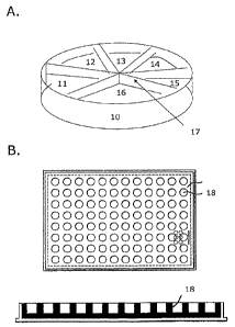
Figure 1 shows two presently preferred embodiments relating to the platform
according to the present invention: according to figure 1A the platform
comprises
an indentation (10) being capable of acting as a receptacle for a sample with
a
possible a microbial infection or contamination. The indentation is divided
into
separate compartments (11, 12, 13, 14, 15, and 16), by means of one or more
integrated dividing members (17). Each compartment contains a composition
comprising a semi-solid microbial growth medium, two or more chromogenic or
fluorogenic substances or substrates and an antibiotic; or a composition that
comprises a semi-solid microbial growth medium, two or more chromogenic or
fluorogenic substances or substrates, but does not comprise an antimicrobial.
According to figure 1B the platform comprises multiple indentations (18), each
indentation being capable of acting as a receptacle for a sample with a
possible
microbial infection or contamination. Each indentation contains a composition
comprising a semi-solid microbial growth medium, two or more chromogenic or
fluorogenic substances or substrates and an antibiotic; or a composition that
comprises a semi-solid microbial growth medium, two or more chromogenic or
fluorogenic substances or substrates, but does not comprise an antimicrobial.
Figure 2 illustrates one of the presently preferred embodiments of the
invention
according to which the platform comprises a composition according to the
inventions comprising Trimethoprim (19), a composition according to the
inventions comprising Sulphamethizole (20) a composition according to the
inventions comprising Ampicillin (21), a composition according to the
inventions
comprising Mecillinam (22) and a composition according to the inventions
comprising Nitrofurantoin (23). The platform further comprises a composition
according to the invention in which there is no antimicrobial (24). Finally,
the
9
CA 02698318 2010-02-26
WO 2009/026920 PCT/
K2008/000303
figure illustrates yet a preferred embodiment according to which the
indentation
or compartment containing said composition not comprising an antimicrobial has
a
size which is twice the size of any of the other indentations or compartments.
Figure 3 shows a standard for determining the quantity or titre of E. coli in
a
sample analysed using the platform according a preferred embodiment of the
invention (illustrated in figure 2). The photos show growth of E. coli at
different
quantities of bacteria/ml urine. The illustrated E. coil is resistant to
sulfamethizole
and ampicillin since there is growth in these two antibiotic compartments but
it is
susceptible to trimethoprim, nitrofurantoin and mecillinam.
Figure 4 shows a standard for determining the different colony types of
urinary
pathogens in a sample analysed using the platform according a preferred
embodiment of the invention. Growth conditions and colours for ordinary
urinary
tract pathogenic bacteria and their susceptibility/resistance to the five
antibiotics
in the Flexicult plate.
Bacterium Colony Colony Agar Susceptibility = S, or
size colour colour Resistance = R
Trime Sulfa Ampi Mecil Nitro
tho methi cillin lina furan
prim zole m
toin
E.coli Large Red/
brownis
Klebsiella Large, Dark
spp. Fat blue
Enterobacter Large Dark
spp blue
Proteus Large Light Brown S
mirabilis (swarm) brown/
Brown
P.vulgaris Large Greenis Brown S S R/S
R R/S
(swarm) h brown
Morganella Large Light Brown S S R R
R/S
spp. (swarm) brown
CA 02698318 2010-02-26
WO 2009/026920 PCT/ K2008/000303
Citrobacter Large Green/ -
spp. greenish
blue
P.aureginosa Small Greyish Greeni R
white/ sh
greenish
E.faecalis Small Green/ Dark
greenish ring
blue around
colony
E.faecium Small Greenis Dark
ring
around
colony
S.saprophytic Small White/ -
us Reddish
Candida spp. Large/ White
small
The present invention will now be described in more detail in the following.
Detailed description of the invention
Definitions
Prior to discussing the present invention in further details, the following
terms and
conventions will first be defined:
Microbial
In the present context the term "microbial" is to be interpreted broadly as
meaning "pertaining to a microbe". The term "microbe" refers collectively to
bacteria, fungi, archaea and protists.
11
CA 02698318 2010-02-26
WO 2009/026920 PCT/ K2008/000303
Semi-solid growth medium
The expression "semi-solid growth medium" as used herein refers to a growth
medium which allows micro-organisms to form colonies on its surface, such as a
medium which has a gel-like appearance or is in the form of a gel, a gel being
a
colloidal system in which a porous network of interconnected particles spans
the
volume of a liquid medium. It is further understood that a gel is mostly
liquid in
composition and thus exhibit densities similar to that of the particular
liquid,
however have the structural coherence of a solid. Preferably, the semi-solid
growth medium as used herein is prepared by adding to a liquid growth medium a
sufficient amount of a substance which melts when heated and solidifies when
cooled again, such as gelatin or agar. It will be understood that the porous
network of interconnected particles in the medium will allow nutrients and
antimicrobial to diffuse through the medium to become available to the micro-
organisms.
Growth medium
In the context of the present invention the term "growth medium" refers to a
substance in or on which microbes can grow. The term in particular comprises
nutrient broth (liquid nutrient medium) or Lysogeny Broth (L-B medium) and
agar, which are the most common growth media for microbes.
Likewise, the term covers liquid medium in which microbes may grow in
suspension, as well as semi-solid medium as defined above, allowing microbes
to
form colonies on its surface. The term "growth medium" also comprises
specialized media which are sometimes required for growth of certain
microorganism including fastidious organisms, requiring specialized
environments
due to complex nutritional requirements.
In general, a growth medium will comprise a carbon source such as glucose or
succinate for bacterial growth, water, various salts provide essential
elements
needed for microbial growth, such as magnesium, nitrogen, phosphorous, and
sulfur to allow the bacteria to synthesize protein and nucleic acid, and a
source of
amino acids and nitrogen (e.g., beef, yeast extract).
It is also to be understood that the term "growth medium" includes defined
media, also known as chemical defined media, as well as undefined media, also
known as basal or complex media. A defined medium for microbes will have
known quantities of all ingredients, including trace elements and vitamins
required
by the microbe and especially a defined carbon source such as glucose or
glycerol,
and a defined nitrogen source such as an ammonium salt or a nitrate. An
undefined medium on the contrary, has some complex ingredients, such as yeast
12
CA 02698318 2010-02-26
WO 2009/026920 PCT/ K2008/000303
extract or casein hydrolysate, which consist of a mixture of many chemical
species
in unknown proportions.
Chromogenic substrate
In the context of the present invention the terms "chromogenic substrate",
"chromogenic substance" and "chromogen" are used interchangeably referring to
a precursor of a biochemical pigment. It is to be understood that, in
particular,
the chromogen may be a substrate, compound or substance, which when
metabolized by a microbe produces a characteristic colour or pigment that is
useful as a means of detection and/or identification of said microbe.
Fluorogenic substrate
The term "fluorogenic substrate" is used interchangeably with "fluorogenic
substance", referring to a precursor of a fluorescent compound. A fluorescent
compound is a compound in which the molecular absorption of a photon triggers
the emission of another photon with a longer wavelength and wherein the energy
difference between the absorbed and emitted photons ends up as molecular
vibrations or heat. In particular, the absorbed photon is in the ultraviolet
range,
and the emitted light is in the visible range. Hence detection of fluorescent
compounds may typically be performed by exposing them to ultraviolet light (UV
light) and then subsequently registering the light (often visible light) which
is
emitted by the fluorescent compound. It is to be understood that, in
particular,
the fluorescent substrate may be a substrate, compound or substance, which
when metabolized by a microbe emits light that is useful as a means of
detection
and/or identification of said microbe.
Antimicrobial
In the present application the terms "antibiotic" and "antimicrobial" are used
interchangeably to define a chemical compound that inhibits or abolishes the
growth of microorganisms, such as bacteria, fungi or protozoans, that is, a
chemical compound with anti-bacterial, anti-fungal, and/or anti-parasitical
activity. The term includes antibiotic or antimicrobial compounds produced and
isolated from living organisms, for example, the penicillin-class produced by
fungi
in the genus Penicillium or streptomycin from bacteria of the genus
Streptomyces.
The terms also include antibiotic or antimicrobial compounds obtained by
chemical
synthesis, such as sulfonomide drugs.
The terms in particular include anti-bacterial antibiotics, which are
antibiotics that
do not have activity against viruses, fungi and other non-bacterial microbes.
The
anti-bacterial antibiotics include bactericidal antibiotics, which destroy
bacteria,
13
CA 02698318 2010-02-26
WO 2009/026920 PCT/ K2008/000303
and bacteriostatic antibiotics which prevent bacteria from multiplying. The
anti-
bacterial antibiotics further include "narrow-spectrum" antibiotics which
target
particular types of bacteria, such as Gram negative or Gram-positive bacteria,
and
broad spectrum antibiotics which affect a wide range of bacteria. Likewise,
the
anti-bacterial antibiotics include antibiotics for ingestion as well as
antibiotics for
intravenous administration which are often used to treat serious infections
such as
deep-seeded systemic infections, and antibiotics for topical administration.
The
anti-bacterial antibiotics comprise antibiotics within the following presently
recognised classes: Arninoglycosides, Ansamycins, Beta-lactam antibiotics,
(including the carbacephem, carbapenems, cephalosporins (first, second, third
and fourth generations), monobactams and penicillins) Glycopeptides,
Macrolides,
lincosamides, Polypeptides, Quinolones, Sulphonamides, Tetracyclines, Cyclic
lipopeptides, Glycylcyclines, Oxazolidinones, diaminopyrimidines, Nitrofurans,
Rifamycins, antibiotic peptides, amphenicols, nitroimidazoles, streptogramins
and
phosphomycins.
Aspects and embodiments of the invention
In a first and broadest aspect, the invention provides a composition
comprising a
semi-solid microbial growth medium, two or more chromogenic or fluorescent
substances or substrates, and an antimicrobial. The fact that a semi-solid
microbial growth medium is employed according to the present invention has the
advantage that microbial growth can be observed as single colonies on the
surface
of said medium. The combined use of chromogenic or fluorescent substances or
substrates offers the possibility of distinguishing multiple types or species
of
microorganisms growing on the medium by the colour of the pigment or
fluorescence produced in the colonies. Simultaneously, it is possible to
determine
the sensitivity of each microbial type or species against the antimicrobial
present
in the composition according to the invention.
Another advantage of the present invention is that when the platform is used
to
test urine for infections the number of colonies formed on the semi-solid
microbial
growth medium directly correlates with the number of bacteria per volume of
urine added to the platform. Thereby making it easy to determine the
concentration of bacteria in the urine.
In a preferred embodiment of the present invention, the semi-solid medium
comprises tryptophan. The advantage of including tryptophan is that it enables
easy detection of Enterobacteria of the Proteus-Morganella-Providencia and at
the
same time tryptophan does not interfere with determination of antibiotic
susceptibility.
14
CA 02698318 2010-02-26
WO 2009/026920 PCT/ K2008/000303
In another preferred embodiment, the semi-solid medium in the composition
according to the invention comprises a galactose polymer (Agar-agar) or
Gelatin.
Preferably the composition according to the invention comprises a microbial
growth medium selected from the group consisting of: Iso-sensitest agar,
Danish
Blood Agar, Discovery medium and Mueller-Hinton medium. For those media
which do not inherently comprise tryptophan it may be a further advantage to
include tryptophan in these media. The concentration of tryptophan in the
medium may be between 0.25 - 3.0 g/liter.
The Iso-sensitest agar is currently available from Oxoid and its composition
is
specified in The Oxoid Manual 1998. The Iso-sensitest agar is composed by 11
g/I
Hydrolysed casein, 3 g/I Peptones, 2 g/I Glucose, 3 g/I Sodium Chloride, 1 g/I
soluble starch, 2 g/I Disodium hydrogen Phosphate, 1 g/I Sodium Acetate, 0.2
g/I
Magnesium glycerophosphate, 0.1 g/I Calcium gluconate, 0.001 g/I cobaltous
sulphate, 0.001 g/I Cupric sulphate, 0.001 g/I Zinc sulphate, 0.001 g/I
Ferrous
Sulphate, 0.002 g/I Magnesium Chloride, 0.001 g/I Menadione, 0.001 g/I
Cyanobalamin, 0.02 g/I L-Cysteine hydrochloride, 0.02 g/I Tryptophan, 0.003
g/I
pyridoxine, 0.003 g/I pantothenate, 0.003 g/I nicotinamide, 0.0003 g/I Biotin,
0.00004 g/l Thiamine, 0.01 g/I Adenine, 0.01 g/I Guanine, 0.01 gil Xanthine,
0.01
g/I Uracil, 8 g/I agar, in distilled water.
As for the Danish Blood Agar this is known by the skilled person to have the
following composition: 2 g Na2HPO4 = 12 H20, 625 g tryptone, 250 g starch,
833.6 g Potassium Chloride, 2.5 g detergent, 74.8 g meat broth (Oxoid CM975K),
800g D(+)Glucose-monohydrate, 1.75 g Xanthin, 1.75 g Guanin, 17.5 g
Magnesium Sulphate 7 H20, 19.2 g CaCl2 = 2 H20, 2,720 g Agar, 5 N HCI to pH
7.4, solution of vitamins, and 12.5 I horse blood per 250 liter distilled
water.
The Discovery medium is manufactured by Oxoid (product code CM 1087).
According to the manufacturer the medium has the following composition: 14.5
g/I Peptone, 2 g/I glucose, 5.5 g/I salt mix, 1 g/I Soluble starch, 1.5 g/I
chromogenic mix, and 8 g/I Agar.
The Mueller-Hinton medium comprises 2 g/I Beef extract powder/beef extract,
17.5 g/I Acid Digest of Casein, 1.5 g/I starch and 17 g/I Agar.
It is to be understood that some variation in the amount of each component in
said medium will be tolerated; in general a variation of 20% will be
tolerated.
However, as the skilled person will know certain components may be varied to
an
even larger extent: the amount of agar may be varied substantially such as
from
4-25 g/I without significantly altering the performance of the medium. It is
thus
CA 02698318 2010-02-26
WO 2009/026920 PCT/ K2008/000303
generally preferred that the medium in the composition according to the
invention
comprises from 4-25g/I of a galactose polymer.
These media, together with other suitable media are characteristic in offering
great reliability when used for determining sensitivity towards
antimicrobials.
Such reliability is not seen for all media as some media comprise compounds
which interfere with the antibiotics and thereby affects the ability to use
these
media for testing for antibiotic susceptibility. The great reliability is
witnessed for
instance by fact that on these media:
i) the addition of 16 mg/I arnpicillin causes a reduction in the growth
of/numbers of colonies formed by reference strain Escherichia coli ATCC
25922 of at least 5 logCFU/ml, as determined after contacting the
composition with a suspension containing 105 CFU/ml and incubating the
composition for 18-24 hours at 370C and at ambient atmosphere;
ii) the addition of 32 mg/I nitrofurantoin causes a reduction in the growth
of/numbers of colonies formed by reference strain Staphylococcus
saprophyticus ATCC 49907 of at least 4 log CFU/ml, as determined after
contacting the composition with a suspension containing 105 CFU/ml and
incubating the composition for 18-24 hours at 370C and at ambient
atmosphere;
iii) the addition of 700 mg/I sulphamethizole causes a reduction in the
growth of/numbers of colonies formed by reference strain Escherichia coli
ATCC 25922 of at least 3 log CFU/ml, as determined after contacting the
composition with a suspension containing 105 CFU/ml and incubating the
composition for 18-24 hours at 370C and at ambient atmosphere
iv) the addition of 16 mg/I trimethoprim causes a reduction in the growth
of/numbers of colonies formed by reference strain Escherichia coli ATCC
25922 of at least 3 log CFU/ml, as determined after contacting the
composition with a suspension containing 105 CFU/ml and incubating the
composition for 18-24 hours at 370C and at ambient atmosphere
v) the addition of 16 mg/I mecilinam causes a reduction in the growth
of/numbers of colonies formed by reference strain Escherichia coli ATCC
25922 of at least 5 log CFU/ml, as determined after contacting the
composition with a suspension containing 105 CFU/ml and incubating the
composition for 18-24 hours at 370C and at ambient atmosphere.
16
CA 02698318 2010-02-26
WO 2009/026920 PCT/ K2008/000303
In further embodiments the microbial growth medium is a selective medium
capable of applying a selective pressure to organisms growing on it, such as a
medium which is selective for Gram-negative bacteria or for Gram-positive
bacteria.
The semi-solid media may further comprise sulpha inhibitors and/or metal ions
as
they may affect the antibiotic susceptibility. For example variations in the
concentrations Mg2+ or Ca2+, may affect results of aminoglycoside and
tetracycline
tests with Ps. aeruginosa. Furthermore, excess zinc ions may reduce zone sizes
of
carbapenems. Excessive cation content will reduce antibiotic activity, whereas
low
cation content may result in enhanced activity. The Ca2+ and Mg2+ ions may in
particular be present in the semi-solid medium in the form of soluble salts.
Sulfonamide is inhibited by thymidine, which bacteria can use and therefore
grow
in spite of sulfonamide. The presence of thymidin-phosphorylase will inhibit
thymidine and thus restore the function of sulfonamide. The concentration
needed
of thymidine-phosphorylase will depend on the concentration of thymidine in
the
medium.
Another parameter of the semi-solid media is the concentration of thymidine as
this may affect testing of trimethoprim and methicillin-resistant
staphylococci.
Most agar media contain small amounts of sulphonamide and trimethoprim
antagonists that may affect the results of susceptibility testing (especially
if blood
is not added) with low antibiotic content in the medium. Hence in a particular
embodiment Susceptibility test media should contain less than 0.03 mg/I
thymidine, otherwise small colonies are seen on the trimethoprim agar. If the
medium contains slightly more thymidine than recommended, it is possible to
reduce the concentration by adding thymidine-phosphorylase: 0.025 to 0.1 IU
enzyme/ml medium or 5% haemolysed horse blood, which contains the same
enzyme.
Whereas the presence of only one antibiotic in the composition according to
the
invention may be desirable in respect of some antibiotics and for some
purposes,
the composition according to the invention may also comprise 2 or more
antimicrobials, such as 3 or more antimicrobials, such as 4 or more
antimicrobials,
or such as 5 or more antimicrobials.
17
CA 02698318 2010-02-26
WO 2009/026920 PCT/ K2008/000303
The composition according to the invention may be characterised in that the
antimicrobial or, if more than one antimicrobial is present, that at least one
of the
antimicrobials, such as 2, 3, 4, 5 or more of the antimicrobials or all of the
antimicrobials are selected from the group consisting of: Amikacin,
Amoxicillin,
Amoxicillin-clavulanic acid, Amphothericin-B, Ampicillin, Ampicllin-sulbactam,
Apramycin, Azithromycin, Aztreonam, Bacitracin, Benzylpenicillin, Caspofungin,
Cefaclor, Cefadroxil, Cefalexin, Cefalothin, Cefazolin, Cefdinir, Cefepime,
Cefixime,
Cefmenoxime, Cefoperazone, Cefoperazone-sulbactam, Cefotaxime, Cefoxitin,
Cefpirome, Cefpodoxime, Cefpodoxime-clavulanic acid, Cefpodoxime-sulbactam,
Cefprozil, Cefquinome, Ceftazidime, Ceftibutin, Ceftiofur, Ceftobiprole,
Ceftriaxon,
Cefuroxime, Chloramphenicole, Florfenicole, Ciprofloxacin, Clarithromycin,
Clinafloxacin, Clindamycin, Cloxacillin, Colistin, Cotrimoxazol
(Trimthoprim/sulphamethoxazole), Dalbavancin, Dalfopristin/Quinopristin,
Daptomycin, Dibekacin, Dicloxacillin, Doripenem, Doxycycline, Enrofloxacin,
Ertapenem, Erythromycin, Flucloxacillin, Fluconazol, Flucytosin, Fosfomycin,
Fusidic acid, Garenoxacin, Gatifloxacin, Gemifloxacin, Gentamicin, Imipenem,
Itraconazole, Kanamycin, Ketoconazole, Levofloxacin, Lincomycin, Linezolid,
Loracarbef, Mecillnam (amdinocillin), Meropenem, Metronidazole, Mezlocillin,
Mezlocillin-sulbactam, Minocycline, Moxifloxacin, Mupirocin, Nalidixic acid,
Neomycin, Netilmicin, Nitrofurantoin, Norfloxacin, Ofloxacin, Oxacillin,
Pefloxacin,
Penicillin V, Piperacillin, Piperacillin-sulbactam, Piperacillin-tazobactam,
Rifampicin, Roxythromycin, Sparfloxacin, Spectinomycin, Spiramycin,
Streptomycin, Sulbactam, Sulfamethoxazole, Teicoplanin, Telavancin,
Telithromycin, Temocillin, Tetracyklin, Ticarcillin, Ticarcillin-clavulanic
acid,
Tigecycline, Tobramycin, Trimethoprim, Trovafloxacin, Tylosin, Vancomycin,
Virginiamycin and Voriconazole.
A complete list of antimicrobials which could be incorporated into the
composition
according to the invention either alone or in combinations is shown in Table
1.
The concentrations used in the agar is preferably related to the S/R
breakpoint for
the particular drug, however, it will be within the capacity of a skilled
person to
determine the exact concentration needed for a particular purpose.
18
CA 02698318 2010-02-26
WO 2009/026920 PCT/
K2008/000303
Table I. List of antibiotics, IUPAC codes and concentration range for possible
concentrations used in agar.
Antibiotic Chemical name (IUPAC) Range of
concentra
tions
covered
Mg/I
Amikacin 2S)-4-amino-N-[(2S,3S,4R,5S)-5-amino-2- 2 - 128
[(2S,3R,4S,5S,6R)-4-amino-3,5-dihydroxy-
6-(hydroxymethyl)oxan-2-ylloxy-4-[(2R,3R,
4S,5R,6R)-6-(aminomethyl)-3,4,5-trihydroxy-
oxan-2-ylioxy-3-hydroxy-cyclohexyl]-2-
hydroxy-butanamide
Amoxicillin 7-[2-amino-2-(4-hydroxyphenyl) - 0.1 - 32
acetyl]amino-3,3-dimethy1-6-oxo -2-thia-5-
azabicyclo[3.2.0]heptane -4-carboxylic acid
Amoxicillin- Amoxicillin:742-amino-2-(4-hydroxyphenyl) 0.1 - 32
clavulanic acid -acetyl]amino-3,3-dimethy1-6-oxo -2-thia-5-
azabicyclo[3.2.0]heptane -4-carboxylic acid -
Clavulanic acid:(2R,5R,Z)-3-(2-
hydroxyethylidene)-
7-oxo-4-oxa-1-aza-bicyclo[3.2.0]
heptane-2-carboxylic acid
Amphothericin-B (1R- 0.1 - 64
1R*,3S*,5R*,6R*,9R*,11R*,15S*,16R*,17R*,
18S*,19E,21E,23E,25E,27E,29E,31E,33R*,
35S*,36R*,37S*))-33-((3-Amino-3,6-dideoxy-
beta-D-mannopyranosyl)oxy)-
1,3,5,6,9,11,17,37-octahydroxy-15,16,18-
trimethyl -13-oxo-14,39-
dioxabicyclo(33.3.1)nonatriaconta-
19,21,23,25,27,29,31-heptaene-36-
carboxylic acid
Ampicillin 7-(2-amino-2-phenyl-acetyl)amino-3,3 0.1 - 32
-dimethy1-6-oxo-2-thia-5-azabicyclo
[3.2.0]heptane-4-carboxylic acid
Ampicllin 7-(2-amino-2-phenyl-acetyl)amino-3,3 0.1 - 32
-dimethy1-6-oxo-2-thia-5-azabicyclo
[3.2.0]heptane-4-carboxylic acid
sulbactam (2R,5R)-3,3-dimethy1-4,4,7-trioxo-4A6-thia-1-
azabicyclo[3.2.0Theptane-2-carboxylic acid
Apramycin (2R,3R,4R,5S,6R)-5-amino-2- 0.5 - 128
[((1R,2R,3R,4R,6R,8R)-8-amino-
9[(1R,2S,3R,4R,6R)-4,6-diamino-
2,3-dihydroxy-cyclohexyl]oxy-2-hydroxy-
3-methylamino-5,10dioxabicyclo[4.4.0]dec-4-
yl)oxy]-6-(hydroxymethyl)oxane-3,4-diol
Azithromycin 9-deoxy-9a-aza-9a-methyl-9a- 0.03 - 64
homoerythromycin A
19
CA 02698318 2010-02-26
WO 2009/026920 PCT/
K2008/000303
Aztreonam 342-(2-azaniumy1-1,3-thiazol-4-y1)-2-(1- 0.25 -
16
hydroxy-2- methy1-1-oxo-propan-2-
yl)oxyimino- acetyl]amino-2-methy1-4-oxo-
azetidine-1-sulfonate
Bacitracin 1 - 128
Benzylpenicillin 4-Thia-1-azabicyclo(3.2.0)heptane-2- 0.03 -
256
carboxylic acid, 3,3-dimethyI-7-oxo-6-
((phenylacetyl)amino)- (2S-
(2alpha,5alpha,6beta))-
Caspofungin 1-[(4R,5S)-5-[(2-aminoethyl)amino] -N2- 0.1 - 64
(10,12-dimethy1-1-oxotetradecy1)-4-hydroxy-
L-ornithine]-5-[(3R) -3-hydroxy-L-ornithine]
pneumocandin BO
Cefaclor 7-[(2-amino-2-phenyl-acetyl)amino]- 3-chloro- 0.05 - 256
8-oxo-5-thia-1-azabicyclo [4.2.0] oct-2- ene-
2- carboxylic acid
Cefadroxil 8-[2-amino-2-(4-hydroxypheny1)-acetyl]a 1 - 256
mino-4-methyl- 7-oxo-2-thia-6-azabicyclo
[4.2. 0] oct-4-ene-5-carboxylic acid
Cefalexin 8-(2-amino-2-phenyl-acetyl)amino-4-methyl- 4 - 256
7-oxo-2-thia-6-azabicyclo [4.2.0]oct-4-ene-5-
carboxylic acid
Cefalothin (6R,7R)-3-(acetoxymethyl)- 1 - 256
8-oxo-7-(2-(thiophen-2-yl)acetamido)-5-thia-
1-aza-bicyclo[4.2.0]oct-2-ene-2-carboxylic
acid
Cefazolin 3-[(5-methy1-1,3,4-thiadiazol-2- 2 - 32
yl)sulfanylmethyI]-
8-oxo-7-([2-(tetrazol-1-yl)acetyl]amino)- 5-
thia-1-azabicyclo[4.2.0] oct-2-ene-2-
carboxylate
Cefdinir 8-[2-(2-amino-1,3-thiazol-4-y1)-1-hydroxy-2- 0.1 -
128
nitroso-ethenyl]amino-4-etheny1-7-oxo-2-thia-
6-azabicyclo[4.2.0]oct-4-ene-5-carboxylic acid
Cefepime (6R,7R,Z)-7-(2-(2-aminothiazol-4-y1)-2- 0.1 - 16
(methoxyimino)acetamido)-3-((1-
methylpyrrolidinium-1-yOmethyl)-8-oxo-5-
thia-1-aza-bicyclo[4.2.0]oct-2-ene-2-
carboxylate
Cefixime (6R,7R)-7-{[2-(2-amino-1,3-thiazol-4-y1)-2- 0.01 -
256
(carboxy methoxyimino)acetyl]amino}-3-
etheny1-8-oxo-5-thia-1-azabicyclo[4.2.0]oct-2-
ene-2-carboxylic acid
Cefmenoxime (6R,7R)-7-{[(2E)-2-(2-amino-1,3-thiazol-4- 0.1 -
256
yI)- 2-methoxyimino-acetyl]amino}-3-[(1-
methyltetrazol-5-yl)sulfanylmethyl]-8-oxo-5-
thia-1-azabicyclo[4.2.0] oct-2-ene-2-carboxylic
acid
Cefoperazone (6R,7S)-7-{[2-[(4-ethyl-2,3-dioxo-iperazine-1- 0.1 - 520
carbonyl)amino]-2-(4-
hydroxyphenypacetylIamino]-
3-[(1-methyltetrazol-5-yl)sulfanylmethyl]-8-
CA 02698318 2010-02-26
WO 2009/026920 PCT/
K2008/000303
oxo-5-thia-1-azabicyclo[4.2.0]oct-2-ene-2-
carboxylic acid
Cefoperazone (6R,7S)-7-{[2-[(4-ethy1-2,3-dioxo-piperazine- 0.1 -
520
1-carbonyl)amino]-2-(4-
hydroxyphenypacetyllamino]-
3-[(1-methyltetrazol-5-yl)sulfanylmethyl]-8-
oxo-5-thia-1-azabicyclo[4.2.0]oct-2-ene-2-
carboxylic acid
sulbactam (2R,5R)-3,3-dimethy1-4,4,7-trioxo-4X6-thia-1-
azabicyclo[3.2.0]heptane-2-carboxylic acid
Cefotaxime (6R,7R,Z)-3-(acetoxymethyl)-7-(2-(2- 0.03 -
32
aminothiazol-4-y1)-2-(methoxyirnino)
acetamido)-8-oxo-5-thia-1-azabicyclo[4.2.0]
oct-2-ene-2-carboxylic acid
Cefoxitin (6S,7R)-4-(carbamoyloxymethyl)-7-methoxy- 4 - 64
8-oxo-7-[(2-thiophen-2-ylacetyl)amino1-5-
thia-1-zabicyclo[4.2.0]oct-2-ene-2-carboxylic
acid
Cefpirome 1-[[(6R,7R)-7-[[(2Z)-2-(2-amino-4-thiazoly1)- 0.01 -
256
2-(methoxyimino)acetyl]amino]-2-carboxy-8-
oxo-5-thia-1-azabicyclo[4.2.0]oct-
2-en-3-yl]methyI]-6,7-dihydro-5H-
Cyclopenta[b]pyridinium
Cefpodoxime (6R,7R)-7-{[(2Z)-2-(2-amino-1,3-thiazol-4- 0,05 -
16
y1)-2-methoxyimino-acetyl]amino}-3-
(methoxymethyl)-8-oxo-5-thia-1-
azabicyclo[4.2.0]oct-2-ene-2-carboxylic acid
Cefpodoxime 2R,5R,Z)-3-(2-hydroxyethylidene)- 0.05 -
16
7-oxo-4-oxa-1-aza-bicyclo[3.2.0]
heptane-2-carboxylic acid
clavulanic acid (2R,5R,Z)-3-(2-hydroxyethylidene)-
7-oxo-4-oxa-1-aza-bicyclo[3.2.0]
heptane-2-carboxylic acid
Cefpodoxime (6R,7R)-7-{[(2Z)-2-(2-amino-1,3-thiazol-4- 0.05 -
16
y1)-2-methoxyimino-acetyllaminol-3-
(methoxymethyl)-8-oxo-5-thia-1-
azabicyclo[4.2.0]oct-2-ene-2-carboxylic acid
sulbactam (2R,5R)-3,3-dimethy1-4,4,7-trioxo-4A6-thia-1-
azabicyclo[3.2.0]heptane-2-carboxylic acid
Cefprozil 8-[2-amino-2-(4-hydroxypheny1)-acetyl] 1 - 512
amino-7-oxo-4-prop-1-eny1-2-thia-6-
azabicyclo [ 4.2.0]oct-4-ene-5-carboxylic acid
Cefquinome Pharmacotherapeutic group: Cephalosporins 1 - 512
and related substancesATCvet code:
Q351DA92
Ceftazidime (6R,7R,Z)-7-(2-(2-aminothiazol-4-y1)- 0.1 - 32
2-(2-carboxypropan-2-yloxyimino)acetamido)-
8-oxo-3-pyridinium-1-ylmethyl)-5-thia-1-aza-
bicyclo[4.2.0] oct-2-ene-2-carboxylate
21
CA 02698318 2010-02-26
WO 2009/026920 PCT/
K2008/000303
Ceftibutin (+)-(6R,7R)-7-[(Z)-2-(2-Amino-4-thiazolyI)-4- 0.5 - 32
carboxycroton-amido]-8-oxo-5-thia-1-
azabicyclo[4.2.0]oct-2-ene-2-carboxylic acid
Ceftiofur 6r-[6a,7b(z)]]-7-[[(2-amino-4- 0.1 - 128
thiazoly1)(methoxyimino)acetyllaminol- 3-[[2-
furanylcarbonyl) thio] methyl]-8-oxo-5-thia- 1-
azabicyclo [4.2.0] oct-2-ene-2- carboxylic
acid; (6R-7R)-7- [[2-amino -4-thiazolyI)-z-
methoxyimino)acetyl]amino]-3- [[(2-
furanylcarbonyl) thio] methyl]- 8-oxo-5-thia-1-
azabicyclo [4.2.0]oct-2-ene-carboxylic acid
Ceftobiprole Ceftobiprole medocaril 0.03 -
512
Ceftriaxon (6R,7R,Z)-7-(2-(2-aminothiazol-4-y1)- 0.01 - 16
2-(methoxyimino)acetamido)-3-((6-hydroxy-2-
methy1-5-oxo-2,5-dihydro-1,2,4-triazin-3-
ylthio)methyl)-8-oxo-5-thia-1-aza-
bicyclo[4.2.0]oct-2-ene-2-carboxylic acid
Cefuroxime 4-(carbamoyloxymethyl)-8- [2-(2-fury1)-2- 0.1 - 64
methoxyimino-acetyllamino -7-oxo-
2-thia-6-azabicyclo[4.2.0]oct -4-ene-5-
carboxylic acid
Chloramphenicol 2,2-dichlor-N- [(aR,bR)-b-hydroxy-a- 0.5 - 128
hydroxymethyl- 4-nitrophenethyl] acetamide
Florfenicole 2 - 128
Ciprofloxacin 1-cyclopropy1-6-fluoro-4-oxo-7-piperazin-1-yl- 0.01 - 32
quinoline-3-carboxylic acid
Clarithromycin 6-(4-dimethylamino-3-hydroxy- 6-methyl- 0.03 - 64
etrahydropyran-2-y1) oxy-14-ethy1-12,13-
dihydroxy-
4-(5-hydroxy-4-methoxy-4,6- dimethyl-
tetrahydropyran-2-y1) oxy-7-methoxy-
3,5,7,9,11, 13-hexamethy1-1-
oxacyclotetradecane-2,10-dione
Clinafloxacin 7-(3-amino-1-pyrrolidinyI)-8-chloro-1- 0.01 - 64
cyclopropy1-6-fluoro-1,4-dihydro-4-oxo-3-
Quinolinecarboxylic acid,
Clindamycin (2S,4R)-N-((1R)-2-chloro-1-((3R,4R,5S,6R)- 0.05 - 32
3,4,5-trihydroxy-6-(methylthio)-tetra hydro-
2H-pyran-2-yl)propy1)-1-methyl-4-
propylpyrrolidine-2-carboxamide
Cloxacillin (2S,5R,6R)-6-{[3-(2-chlorophenyI)-5-methyl- 0.5 - 128
oxazole-4-carbonyl]amino}-3,3-dimethyl-7-
oxo-4-thia-1-azabicyclo[3.2.0]heptane-2-
carboxylic acid
Colistin colistin sulfate and colistimethate sodium 0.5 - 16
(colistin methanesulphonate sodium, colistin
sulfomethate sodium
Cotrimoxazol Trimethoprim:5-(3,4,5- 1 - 128
(Trimthoprim/sul trimethoxybenzyl)pyrimidine-2,4-
phamethoxazole) diamine/Sulphamethoxazole:4-amino-N-(5-
methylisoxazol-3-y1)-benzenesulfonamide
Dalbavancin 5,31-dichloro-38-de(methoxycarbony1)-7- 0.05 - 16
22
CA 02698318 2010-02-26
WO 2009/026920 PCT/
K2008/000303
demethyl-19-
deoxy-56-0-[2-deoxy-2-[(10-methy1-1-
oxoundecyl)amino]-b-D-
glucopyranuronosy1]-38-[[[3-
(dimethylamino)propyl]amino]carbony1]-42-0-
a-D-mannopyranosyl-N15-methyl-
Ristomycin A aglycone
Dalfopristin/Quin quinupristine N- 0.25 -
256
opristin [(6R,9S,10R,13S,15aS,18R,22S,24aS)-22-
[p-(dimethylamino)benzy1}-6-
ethyldocosahydro-
10,23-dimethy1-5,8,12,15,17,21,24-heptaoxo-
13-pheny1-18-[["(3S)-3-
quinuclidinylthio]methyl]- 12H-pyrido[2,1-
f]pyrrolo-[2,1-/][1,4,7,10,13,16]
oxapentaazacyclononadecin-9-y1]-3-
hydroxypicolinamide
dalfopristin
(3R,4R,5E,10E,12E,14S,26R,26aS)-26-[[2-
(diethylamino)ethyl]sulfony1]-
8,9,14,15,24,25,26,26a- octahydro-14-
hydroxy-3-
isopropy1-4,12-dimethy1-3H-21,18-nitrilo-
1H,22H-pyrrolo[2,1-c][1,8,4,19]-
dioxadiazacyclotetracosine-1,7,16,22(4H,17H)-
tetrone
Daptomycin N-decanoyl-L-tryptophyl-L-asparaginyl-L- 0.1 - 32
aspartyl-L-threonylglycyl-L-ornithyl-L-aspartyl-
D-alanyl-L-aspartylglycyl-D-seryl-threo -3-
methyl-L-glutamy1-3-anthraniloyl-L-
alanine[egr]1-lactone
Dibekacin D-Streptamine, 0-3-amino-3-deoxy-alpha-D- 1 - 128
glucopyranosyl-(1-6)-0-(2,6-diamino-2,3,4,6-
tetradeoxy-alpha-D-erythro-hexopyranosyl-(1-
4))-2-deoxy
Dicloxacillin (2S,5R,6R)-6-{[3-(2,6-dichloropheny1)-5- 0.2 -
128
methyl-oxazole-4-carbonyl]amino}-3,3-
dimethy1-7-oxo-4-thia-1-
azabicyclo[3.2.0Theptane-2-carboxylic acid
tupi
Doripenem 0.01 -
256
u 4 '11.3.117,Cre'smi,
it II
carbapenem ",c
Doxycycline (2-(amino-hydroxy-methylidene)-4- 0.1 - 16
dimethylamino
-5,10,11,12a-tetrahydroxy-6-methy1-
4a,5,5a,6-tetrahydro-4H-tetracene-1,3,12-
trione
Enrofloxacin 1-Cyclopropyl- 7-(4-ethylpiperazin-1-y1)- 6- 0.01 -
32
fluor-4-oxo- 1,4-dihydrochinolin- 3-carboxylic
acid
23
CA 02698318 2010-02-26
WO 2009/026920 PCT/
K2008/000303
Ertapenem 3-[5-[(3-carboxyphenyl) carbamoyl]pyrrolidin- 0.01 - 128
3-yl] sulfany1-7-(1-hydroxyethyl)- 2-methyl-
6-oxo-5-azabicyclo[3.2.0] hept-3-ene-4-
carboxylic acid
Erythromycin 6-(4-dimethylamino-3-hydroxy-6-methyl- 0.03 - 64
oxan-2-yl)oxy-14-ethyl-7,12,13-trihydroxy- 4-
(5-hydroxy-4-methoxy-4,6-dimethyl-oxan-2-
yl)oxy-3,5,7,9,11,13-hexamethyl- 1-
oxacyclotetradecane-2,10-dione
Flucloxacillin 6-((S)-3-(2-chloro-6-fluorophenyI)-5- 0.5 - 128
methylisoxazole-4-carboxamido)-3,3-dimethyl-
7-oxo-4-thia-1-azabicyclo
[3.2.0]heptane-2-carboxylic acid
Fluconazol 2-(2,4-difluoropheny1)- 0.25 -
512
1,3-bis(1H-1,2,4-triazol-1-yl)propan-2-ol
Flucytosin 4-amino-5-fluoropyrimidin-2(1H)-one J. - 512
Fosfomycin [(2R,3S)-3-methyloxiran-2-yl]phosphonic acid 1 - 64
Fusidic acid 2-(16-acetyloxy-3,11-dihydroxy-4,8,10,14- 0.05 - 16
tetramethyl- 2,3,4, 5,6,7, 9,11,12, 13,15,16-
dodecahydro- 1H-cyclopenta [a]phenanthren-
17-ylidene) -6-methyl- hept-5-enoic acid
Garenoxacin fluoroquinolone 0.01 - 32
Gatifloxacin 1-cyclopropy1-6-fluoro- 8-methoxy-7-(3- 0.01 - 32
methylpiperazin-1-yI)- 4-oxo-quinoline-3-
carboxylic aci
Gemifloxacin 7-[(4Z)-3-(aminornethyl)- 4-methoxyimino- 0.01 - 32
pyrrolidin-1-yI]- 1-cyclopropy1-6-fluoro-4-oxo-
1,8-naphthyridine-3-carboxylic acid
Gentamicin 2-[4,6-diamino-3- [3-amino-6-(1- 0.5 - 128
methylaminoethyl) tetrahydropyran-2-yl] oxy-
2-hydroxy- cyclohexoxy]-5-methyl- 4-
methylamino-tetrahydropyran-3,5-diol
Imipenem (5R,6S)-3-[2- 0.01 -
128
(aminomethylideneamino)ethylsulfanyI]-
6-(1-hydroxyethyl)-7-oxo-1-
azabicyclo[3.2.0]hept-
2-ene-2-carboxylic acid
Itraconazole 4-[4-[4-[4-[ [2-(2,4-dichlorophenyI)- 2-(1H- 0,1 - 256
1,2,4-triazol-1-ylmethyl)- 1,3-dioxolan-4-
yl]methoxy]phenyl] piperazin-1-yl]pheny1]- 2-
(1-methylpropy1)-2,4-dihydro-1, 2,4-triazol- 3-
one
Kanamycin 2-(aminomethyl)- 6-[4,6-diamino-3- [4- 2 - 256
amino-3,5-dihydroxy-6-(hydroxymethyl)
tetrahydropyran-
2-yl]oxy-2-hydroxy-cyclohexoxyl-
tetrahydropyran-3,4,5-triol
Ketoconazole 1-[4-[4-[[(2S,4R)-2-(2,4-dichlorophenyI)- 0.03 -
512
2-(imidazol-1-ylmethyl)-1,3-dioxolan-4-
yl]methoxy] phenyllpiperazin-1-yl]ethanone
Levofloxacin (-)-(S)-9-fluoro-2,3-dihydro-3-methyl-10-(4- 0.01 - 32
24
CA 02698318 2010-02-26
WO 2009/026920 PCT/
K2008/000303
methyl-1-piperaziny1)-7-oxo-7H-pyrido
[1,2,3-de]-1,4-benzoxazine-6-carboxylic acid
Lincomycin HO .,, 1 - 32
, 0 /,-- OH
N S N>OH
H 0
)----"oH
SCH3
Linezolid N-[[3-(3-fluoro-4-morpholinophenyI)- 1 - 32
2-oxooxazolidin-5-yl]methyl]acetamide
Loracarbef 8-(2-amino-2-phenyl-acetyl)a mino-4-chloro- 0.1 -
128
7-oxo- 6-azabicyclo[4.2.0] oct-4-ene-5-
carboxylic acid
Mecillnam 6-Amidinopenicillanic acid derivatives 0.25 -
254
(amdinocillin) 6-[[(hexahydro-1H-
azepin-1-yl)methylene]amino]-3,3-
dimethy1-7-oxo-, (2S,5R,6R)- 4-Thia-1-
azabicyclo[3.2.0]heptane-2-carboxylic acid,
Meropenem 3-[5-(dimethylcarbamoyl) pyrrolidin-2-yl] 0.03 -
256
sulfany1-6- (1-hydroxyethyl)-4-methy1-7-oxo-
1-azabicyclo[3.2.0] hept-2-ene-2-carboxylic
acid
Metronidazole 2-(2-methyl-5-nitro-1H-imidazol-1-y1)ethanol 1 - 512
Mezlocillin (2S,5R,6R)-3,3-dimethy1-6-[[(2R)-2-[(3- 0.5 -
512
methylsulfony1-2-oxo-imidazolidine-1-
carbonyl)amino]-2-phenyl-acetyl]amino]-7-
oxo-4-thia-1-azabicyclo[3.2.0]heptane-2-
carboxylic acid
Mezlocillin- Mezlocillin:(2S,5R,6R)-3,3-dimethy1-6- 0.5 -
512
sulbactam [[(2R)-2-[(3-methylsulfony1-2-oxo-
imidazolidine-1-carbonyl)amino]-2-phenyl-
acetyl]amino]-7-oxo-4-thia-1-
azabicyclo[3.2.0]heptane-2-
carboxylic acid Sulbactam:(2R,5R)-3,3-
dimethy1-4,4,7-trioxo-4A6-thia-1.-
azabicyclo[3.2.0]heptane-2-carboxylic acid
Minocycline 2-(amino-hydroxy-methylidene)-4,7- 0.1 -
256
bis(dimethylamino)-10,11,12a-trihydroxy-
4a,5,5a,6-tetrahydro-4H-tetracene-1,3,12-
trione
(synonym 7-Dimethylamino-6-demethy1-
6-deoxytetracycline)
Moxifloxacinõ 1-cyclopropy1-7-[(1S,6S)-2,8-diazabicyclo 0.01 -
32
[4.3.0]non-8-y1]-6-fluoro-8-methoxy-4-oxo-
quinoline-3-carboxylic acid
Mupirocin 9-[[(2Z)-3-methyl-1-oxo-4-[(2S,3R,4R,5S)- 0.1 -
256
tetrahydro-3,4-
dihydroxy-5-[[(2S,3S)-3-[(1S,2S)-2-
hydroxy-1-methylpropy1]-2-
oxiranyl]methy1]-2H-pyran-2-y1]-2-buten-
1-yl]oxy]- Nonanoic acid
CA 02698318 2010-02-26
WO 2009/026920 PCT/
K2008/000303
Nalidixic acid 1-ethy1-7-methyl-4-oxo-[1,8]naphthyridine-3- 4 - 1024
carboxylic acid
Neomycin C23H46N6013(MW 614.644 g/mol) 0.5 -
256
Netilmicin 0-3-deoxy-4-C-methyl-3-(methylamino)-b-L- 0.5 -
512
arabinopyranosyl-(1->6)-0-[2,6-diamino-
2,3,4,6-tetradeoxy-a-D-
glycero-hex-4-enopyranosyl-(1->4)]-2-
deoxy-N1-ethyl- D-Streptamine, C21H41N507
(MW 475.58 g/mol)
Nitrofurantoin 1-[(5-nitro-2-furyl)methylideneamino] 1 - 1028
imidazolidine-2,4-dione
Norfloxacin 1-ethyl-6-fluoro-4-oxo-7-piperazin-1-y1-1H- 0.5 -
128
quinoline- 3-carboxylic acid
Ofloxacin (+/-)-9-fluoro-2, 3-dihydro-3-methyl-10-(4- 0.01 -
32
methyl-1-piperaziny1)- 7-oxo-7H-pyrido [1,2,3-
de]-1, 4-benzoxazine-6-carboxylic acid
Oxacillin (2R,5R,6S)-3,3-dimethy1-6-[(5-methyl-3- 0.25 - 128
phenyl- 1,2-oxazole-4-carbonyl)amino]-7-oxo-
4-thia-1- azabicyclo[3.2.0]heptane-2-
carboxylic acid
Pefloxacin 1-ethyl-6-fluoro-7-(4-methylpiperazin-1-y1)-4- 0.01 -
32
oxo-quinoline-3-carboxylic acid
Penicillin V Phenoxymethylpenicillin 0.1 -
256
Piperacillin (2S,5R,6R)-6-{[(2R)-2-[(4-ethy1-2,3-dioxo- 2 - 1024
piperazine-1-carbonyl)amino]-2-phenyl-
acetyl]amino}-3,3-dimethy1-7-oxo-4-thia-1-
azabicyclo[3.2.0]heptane-2-carboxylic acid
Piperacillin- Piperacillin:(2S,5R,6R)-6-{[(2R)-2-[(4-ethyl- 2 - 1024
sulbactam 2,3-dioxo-piperazine-1-carbonyl)amino]-2-
phenyl-acetyllamino}-3,3-dimethyl-7-oxo-4-
thia-1-azabicyclo[3.2.0]heptane-2-carboxylic
acid Sulbactam:(2R,5R)-3,3-dimethy1-4,4,7-
trioxo-4A6-thia-1-
azabicyclo[3.2.0]heptane-2-carboxylic acid
Piperacillin- Piperacillin: as above; Tazobactam: 2 - 1024
tazobactam (2S,3S,5R)-3-methyl-4,4,7-trioxo-3- (triazol-
1-ylmethyl)-4$1^{6}-thia-1-azabicyclo[3.2.0]
heptane-2-carboxylic acid
Rifampicin 5,6,9,17,19,21-Hexahydroxy-23-methoxy- 0.005 -
2,4,12,16,18,20,22-heptamethyl- 128
8-[N-(4-methy1-1-piperazinyl)formimidoy1]-
2,7-(epoxypentadeca[1,11,13]trienimino)-
naphtho[2,1-b]furan-1,11(2H)-dione 21-
acetate
Roxythromycin Erythromycin-[0-[(2-methoxyethoxy)- 0.03 -
64
methyl]oxime]
Sparfloxacin 5-amino-1-cyclopropy1-7-[(3R,5S)3,5- 0.01-
128
dimethylpiperazin-
1-y1]-6,8-difluoro-4-oxo-quinoline-3-carboxylic
acid
Spectinomycin (2R,4aR,5aR,6S,7S,8R,9S,9aR,10aS)- 32 - 512
26
CA 02698318 2010-02-26
WO 2009/026920 PCT/
K2008/000303
4a,7,9-trihydroxy-2-methy1-6,8-
bis(methylamino) decahydro-4H-pyrano[2,3-
b][1,4]benzodioxin-4-one
Spiramycin C43H74N2014, (MW 843.053 g/mol) 0.05 - 8
Streptomycin 5-(2,4-diguanidino-3,5,6-trihydroxy- 2 - 1024
cyclohexoxy)- 444,5-dihydroxy-6-
(hydroxymethyl)-3-methylamino-
tetrahydropyran-2-yl] oxy-3-hydroxy-2-
methyl-tetrahydrofuran-3-carbaldehyde
Sulbactam (2R,5R)-3,3-dimethy1-4,4,7-trioxo-4X6-thia-1-
azabicyclo[3.2.0]heptane-2-carboxylic acid 2 - 1024
Sulfamethoxazol 4-amino-N-(5-methylisoxazol-3-y1)- 16 -
2050
benzenesulfonamide
Teicoplanin Glycopeptide - no IUPAC-code 0.05 -
128
Telavancin N3"-[2-(decylamino)ethy1]-29- 0.25 -
128
[[(phosphonomethyl)amino]methyI]-
Vancomycin
Telithromycin (1S,2R,5R,7R,8R,9S,11R,13R,14R)-8- 0.01 -
64
[(2S,3R,4S,6R)-4-dimethylamino-3-hydroxy-6-
methyl-oxan-2-yl]oxy-2-ethy1-9-methoxy-
1,5,7,9,11,13-hexamethy1-15-[4-(4-pyridin-3-
ylimidazol-1-yl)butyl]-3,17-dioxa-15-
azabicyclo[12.3.0]heptadecane-4,6,12,16-
tetrone
Temocillin (2S,5R,6S)-6-[(Carboxy-3- 0.1 -
1024
thienylacetyl)amino]- 6-methoxy-3,3-
dimethy1-7-oxo-4-thia-1-azabicyclo[3.2.0]
heptane-2-carboxylic acid,
Tetracyklin 2-(amino-hydroxy-methylidene)-4- 0.1 - 32
dimethylamino-6,10,11,12a-tetrahydroxy-6-
methy1-4,4a,5,5a-tetrahydrotetracene-1,3,12-
trione
OR
4-(dimethylamino)-1,4,4a,5,5a,6,11,12a-
octahydro-3,6,10,12,12a-pentahydroxy-
1,11dioxo-naphthacene-2carboxamide
Ticarcillin 2S,5R,6R)-6-[[(2R)-2-carboxy-2-thiophen-3- 2 - 512
yl-acetyl]amino]-3,3-dimethy1-7-oxo-4-thia-1-
azabicyclo[3.2.0]heptane-2-carboxylic acid
Ticarcillin- Ticarcillin:2S,5R,6R)-6-[[(2R)-2-carboxy-2- 2 - 512
clavulanic acid thiophen-3-yl-acetyl]amino]-3,3-dimethy1-7-
oxo-4-thia-1-azabicyclo[3.2.0]heptane-2-
carboxylic acid- Clavulanic acid:(2R,5R,Z)-3-
(2-hydroxyethylidene)-7-oxo-4-oxa-1-aza-
bicyclo[3.2.0]heptane-2-carboxylic acid
Tigecycline N-[(5aR,6aS,7S,9Z,10aS)-9-(amino-hydroxy- 0.1 -
256
methylidene)-4,7-bis(dimethylamino)-
1,10a,12-trihydroxy-8,10,11-trioxo-5a,6,6a,7-
tetrahydro-5H-tetracen-2-y1]-2-(tert-
, butylamino)acetamide
Tobramycin 4-amino-2-[4,6-diamino-3- [3-amino-6- 0.5 -
128
(aminomethyl) -5-hydroxy-etrahydropyran-2-
27
CA 02698318 2010-02-26
WO 2009/026920 PCT/
K2008/000303
yl]oxy- 2-hydroxy-cyclohexoxy]-6-
(hydroxymethyl) tetrahydropyran-3,5-diol
Trimethoprim 5-(3,4,5-trimethoxybenzyl)pyrimidine-2,4- 0.25
- 64
diamine
Trovafloxacin 7-(6-amino-3-azabicyclo[3.1.0]hex-3-y1)- 1- 0.1 -
32
(2,4-difluorophenyI)- 6-fluoro-4-oxo-[1,8]
naphthyridine-3-carboxylic acid
Tylosin macrolide 0.1 -
32
Vancomycin C66H75C12Ng02.4 0.5 -
1024
Virginiamycin C71H84N10017 0.25
- 256
Voriconazole 2-(2,4-difluorophenyI)-3- (5-fluoropyrimidin-4- 0.004
-
y1)-1- (1H-1,2,4-triazol-1-y1) butan-2- ol 128
The prevalence of antibiotic resistance has been and is continuing to increase
in
all bacteria including E. coli, and it is now becoming increasingly necessary
to
combine urine culture with a susceptibility test even in primary care.
In a recent study surveying resistance rates in E. coif from North America
(USA
and Canada)8, of the 1142 E. coli collected, 75.5% (862) were collected from
the
USA and 280 (24.5%) were from Canada. Overall, resistance to ampicillin was
37.7%, followed by SMX/TMP (21.3%), nitrofurantoin (1.1%), ciprofloxacin
(5.5%) and levofloxacin (5.1%). This study reported higher rates of antibiotic
resistance in US versus Canadian outpatient urinary isolates of E. coli and
demonstrated the continuing evolution of resistance to antimicrobial agents.
In a
European survey from 2002-3, the Eco-Sens study8, antibiotic resistance rates
were determined in E. coli from a range of European countries. The results are
shown in table 2.
28
CA 02698318 2010-02-26
WO 2009/026920 PCT/
K2008/000303
Table 2. Antimicrobial resistance of E. coli in European countries in the Eco-
Sens
study9.
___________________________________________ _
Antimicrobial agenta (resistance in per cent)
Country n AMP AMC MEC CFR TMP SUL SXT NAL CIP NIT FOF GEN
l Austria 126 17.5 2.4 1.6 0.8 9.5 25.4 9.5 2.4 0 0.8 0
0.8
I Belgium 137 30.7 2.9 1.5 0.7 13.9 32.8 14.6 6.6 2.9 0.7 0.7 0.7
Canada 166 29.5 3.6 1.2 1.8 10.8 25.3 12.0 0.6 0 1.2
0.6 0.6
Denmark 85 22.4 1.2 1.2 1.2 10.6 21.2 8.2 3.5 0 1.2
1.2 0
Finland 182 19.8 4.4 0.5 1.6 5.5 15.4 4.9 1.6 0.5 0.5 1.1 0.5
France 199 27.6 1.5 1.5 1.0 15.6 31.7 15.1 3.5 2.0 1.0 1.0 0
Germany 138 29.0 2.2 2.2 1.4 22.5 34.8 21.0 3.6 2.2 0.7 0 0.7
Greece 132 22.0 0.8 0.8 3.0 13.6 1.9.7 11.4 6.8 1.5 3.0 1.5 0.8
Ireland 154 44.8 5.8 0.6 0.6 22.1 40.3 20.8 1.9 0 0 1.3
0.6
Luxembourg 24 41.7 0 0 0 16.7 25.0
16.7 8.3 4.2 4.2 0 0
The
I Netherlands 195 28.7 2.6 1.5 4.6 12.3 25.6 10.3 5.1 2.1 1.0 0.5 0.5
Norway 168 23.8 3.6 0 2.4 13.1 25.0 11.3 1.2 0 0 1.2 0
Portugal 86 45.3 9.3 2.3 2.3 26.7 44.2 26.7 11.6 5.8 5.8 0 3.5
Spain 191 53.9 4.2 1.0 3.1 25.1 48.7 25.7 26.7 14.7 4.2 0.5 4.7
Sweden 193 15.5 5.7 1.6 5.2 8.8 16.6 8.3 2.6 0 0 0.5 0
Switzerland 122 27.0 2.5 0 0.8
18.9 31.1 18.9 6.6 2.5 0.8 0.8 3.3
United
Kingdom 180 37.2 2.8 1.7 1.7 13.3 31.7 12.2 2.2 0.6 0 0 0
Total 2478 29.8 3.4 1.2 2.1 14.8 29.1 14.1. 5.4 2.3 1.2 0.7 1.0
aAMP, ampicillin; AMC, co-amoxiclav; MEC, mecillinam; CFR, cefadroxil; TMP,
trimethoprim; SUL, sulfamethoxazole; SXT, trimethoprim/sulfamethoxazole; NAL,
nalidixic acid; CIP, ciprofloxacin; NIT, nitrofurantoin; FOF, fosfomycin; GEN,
gentamicin.
29
CA 02698318 2010-02-26
WO 2009/026920 PCT/ K2008/000303
As can be deducted from these data, the resistance rates for commonly used
antibiotics such as ampicillin, sulfonamides and trimethoprim are now so high
(i.e.
1.5 - 45%), that these drugs cannot be used empirically without a
susceptibility
test. The use of fluoroquinolones, which are effective broad-spectrum drugs in
some countries still covering most urinary pathogens, is directly related to
development of resistance, which on even short-term scale is problematic due
to
the importance of these drugs for treating serious infections in hospitals.
Since
the resistance rates vary between geographic areas different antimicrobials
may
be preferred depending on the purpose for which the composition according to
the
invention is used.
In a presently preferred embodiment compositions according to the invention
comprises one or more antimicrobials, wherein the antimicrobial/at least one
of
the antimicrobials is selected from the group consisting of: aminoglycosides,
piperacillin/tazobactam, carbapenems, cephalosporins, glycopeptides,
lipopeptides
and antimicrobial peptides (e.g. polymycin or colistin) and combinations of
these.
In an equally preferred embodiment, compositions according to the invention
are
developed for use in diagnosing urinary tract infections in Denmark and the
Scandinavian countries. In these compositions the antimicrobial or
antimicrobials
are preferably selected from the group consisting of: Ampicillin/amoxicillin,
Sulfonamide, Trimethoprim, Nitrofurantoin, Mecillinam, and Ciprofloxacin (or
other
fluoroquinolone), and combinations of these.
For the same purpose it may be preferred that the antimicrobial is selected
from
the group consisting of: trimethoprim, sulfamethizole, ampicillin,
nitrofurantoin
and mecillinam (amdinocillin) and combinations of these.
For similar reason compositions have been developed for use in UTI diagnostics
in
Europe outside Scandinavia. In these compositions the antimicrobial or
antimicrobials is/are preferably selected from the group consisting of:
Amoxicillin,
cluvulanic acid/ampicillin, sulbactam, Ciprofloxacin (or other
fluoroquinolone),
Sulphamethoxazole, trimethoprim, Cefalexin/cefuroxime/cefadroxil (or other
oral
cephalosporin), Nitrofurantoin and Fosfomycin (fosfomycin-trometerole) and
combinations of these.
For the purpose of diagnosing UTI in the United Stated it may be preferred
that
antimicrobial or antimicrobials is/are selected from the group consisting of:
Amoxicillin, cluvulanic acid/ampicillin, sulbactarn, Ciprofloxacin (or other
fluoroquinolone), Sulphamethoxazole, trimethoprim,
Cefalexin/cefuroxime/cefadroxil/cefaclor (or other oral cephalosporin),
CA 02698318 2010-02-26
WO 2009/026920 PCT/ K2008/000303
Nitrofurantoin and Fosfomycin (fosfomycin-trometerole) and combinations of
these.
It is to be understood in particular that amoxicillin may be included either
alone or
in combination with cluvulanic acid. Similarly, ampicillin may be included
alone or
in combination with sulbactam, and Sulphamethoxazole amy be used alone or in
combination with trimethoprim.
For purposes other than diagnosing UTIs the antimicrobial or antimicrobials
may
also be selected from the group consisting of: Amoxicillin/clavulanic acid (or
sulbactam), Phenoxymethyl-penicillin, Cephalosporin (e.g. cefuroxime axetil,
cefalexin), Ciprofloxacin, levofloxacin, ofloxacin, fleroxacin,
Sulphametoxazole and
trimethoprim (optionally in combination as used for oral treatment),
Tetracyclin
(doxycycline or any other tetracyclin group), Chloramphenicol, Fosfomycin,
Macrolide/clindamycin and Rifampicin, and combinations of these.
If the test would be used in a hospital laboratory, antibiotics for
intravenous use
might be considered also, including antimicrobials selected from the group
consisting of: Aminoglycosides, Cephalosporins (cefotaxime, ceftazidime,
cefepime, cefpodoxime, ceftriaxone and others), Piperacillin/tazobactam,
Carbapenems, cephalosporins, Glycopeptides, Lipopeptides, Linezolid and
antimicrobial peptides (e.g. polymycin or colistin) and combination of these.
Important additives to the selective medium
Sulfa inhibitors and Metal ions: Variation in Mg and Ca, will affect results
of
aminoglycoside and tetracycline tests with Ps. aeruginosa. Excess zinc ions
may
reduce zone sizes of carbapenems. Excessive cation content will reduce
antibiotic
acitivty, whereas low cation content may result in enhanced activity Ca and Mg
should be available in the medium in the form of soluble salts.
The thymidine content of the medium affects testing of trimethoprim and
methicillin-resistant staphylococci. Most agar media contain small amounts of
sulphonamide and trimethoprim antagonists that may affect the results of
susceptibility testing (especially if blood is not added) with low antibiotic
content
in the medium. Susceptibility test media should contain less than 0.03 mg/I
thymidine, otherwise small colonies are seen on the trimethoprim agar. If the
medium contains slightly more thymidine than recommended, it is possible to
31
CA 02698318 2010-02-26
WO 2009/026920 PCT/ K2008/000303
reduce the concentration by adding thymidinephosphorylase: 0.025 to 0.1 IU
enzyme/ml medium or 5% haemolysed horse blood, which contains the same
enzyme.
Prior to adding the antibiotic to the semi-solid media it may be an advantage
to
dissolve the antibiotic.
In particular solvents used to dissolve the antimicrobial agent(s) may include
one
or more of the following:
Water
Physiological saline 0.85%
Phosphate buffer, pH 6,0, 0.1 mol/L
Phosphate buffer, pH 8.0, 0.1 mol/L
Phosphate buffer, pH 7.2, 0.01 mol/L
Saturated solution sodium bicarbonate
Aqueous sodium bicarbonate 0.1%
Ethanol 95%
Methanol
Glacial acetic acid
Dimethylformamide (DMF)
Dimethyl sulfoxide (DMSO)
DMSO 1/10 vol
DMSO plus glacial acetic acid
1/2 volume of water plus drops of 1 mol/L Sodium hydroxide
1/2 volume of water plus drops of 0.1 mol/L Sodium hydroxide
1/2 volume of water plus drops of 2.5 mol/L Sodium hydroxide
Hydrogen chloride acid 0.04 mol/L
Polysorbate-80 (0.002%) in water
Stability of the antimicrobial in the semi-solid media may depend on the pH-
value.
The pH-value must not exceed certain limits as this will influence the
antimicrobial
effect. The stability of Ampicillin and Mecillinam has been improved by
adjusting
the pH-value of the semi-solid media to 6Ø
According to the invention the two or more chromogenic substances present in
the
composition is preferably selected from the group consisting of: 5-Bromo-4-
32
CA 02698318 2010-02-26
WO 2009/026920 PCT/ K2008/000303
chloro-3-indoly1 phosphate, 5-Bromo-6-chloro-3-indoly1 phosphate p-toluidine,
3,3'-(3,3'-dimethoxy-4,4'-biphenylylene)-bis-2-(p-nitropheny1)-5-pheny1-2H-
tetrazolium chloride, 5-Bromo-4-chloro-3-indolyl-p-D-galactopyranoside, 5-
Bromo-6-chloro-3-indolyl-3-D-galactopyranoside, 5-Bromo-3-indolyl-3-D-
galactopyranoside, 6-Bromo-2-naphthyl-p-D-galactopyranoside, 6-Chloro-3-
indolyl-f3-D-galactopyranoside, 6-Bromo-3-indolyl-3-D-galactopyranoside, 1-
Methy1-3-indolyl-p-D-galactopyranoside, o-Nitrophenyl-P-D-galactopyranoside, p-
Nitrophenyl-p-D-galactopyranoside, 3,4-cyclohexenoesculetin-f3-D-galactoside,
8-
hydroxychinoline-p-D-galactoside, 5-Bromo-4-chloro-3-indolyl-a-D-
galactopyranoside, 5-Bromo-4-chloro-3-indolyl-P-D-glucopyranoside, 5-Bromo-4-
chloro-3-indolyl-p-D-glucuronide, 5-Bromo-6-chloro-3-indolyl-P-D-glucuronide,
8-
hydroxyquinoline-p-D-glucuronide, 2,2'-Azino-bis(3-ethylbenzothiazoline-6-
sulfonic acid), 4-Chloro-1-naphthol, 3,3'-Diaminobenzidine tetrahydrochloride,
o-
Phenylenediamine, 3,3',5,5'-Tetramethylbenzidine, 4-[2-(4-octanoyloxy-3,5-
dimethoxypheny1)-viny1]-quinolinium-1-(propan-3-yl-carboxylic-acid)-bromide, 5-
Bromo-6-chloro-3-indolyl-caprylate and 5-bromo-4-chloro-3-indoxyl-myo-inositol-
1-phosphate and combinations of these.
As the skilled person will know, the components listed above are normally used
in
an amount of 0.001-1.0 gil.
In table 3 below, these chromogenes are listed according to the enzyme for
which
they serve as substrate:
33
CA 02698318 2010-02-26
WO 2009/026920 PCT/
K2008/000303
Enzyme Substrate Concentration
Color
Alkaline 5-Bromo-4-chloro-3-indoly1 phosphate 0.001 to 1.0 g/I
Blue
Phosphatase
5-Bromo-6-chloro-3-indoly1 phosphate p- 0.001 to 1.0 g/I
Red
toluidine
3,3'-(3,3'-dimethoxy-4,4'-biphenylyiene)-bis- 0.001 to 1.0 g/I Red
2-(p-nitropheny1)-5-phenyl-2H-tetrazolium
chloride
6-galactosidase 5-Bromo-4-chloro-3-indolyl-p-D- 0.001 to 1.0 g/I
Blue
galactopyranoside
5-Bromo-6-chloro-3-indolyI-6-D- 0.001 to 1.0 g/I
Magenta
galactopyranoside
5-Bromo-3-indoly1-6-D-galactopyranoside 0.001 to 1.0 g/I
Blue
6-Bromo-2-naphthy1-6-D-galactopyranoside 0.001 to 1.0 g/I
6-Chloro-3-indolyI-6-D-galactopyranoside 0.001 to 1.0 g/I
Salmon
6-Bromo-3-indolyI-6-D-galactopyranoside 0.001 to 1.0 g/I
1-Methyl-3-indolyI-6-D-galactopyranoside 0.001 to 1.0 g/I
Green
o-Nitropheny1-6-D-galactopyranoside 0.001 to 1.0 g/1
Yellow
p-Nitropheny1-6-D-galactopyranoside 0.001 to 1.0 g/I
Yellow
3,4-cyclohexenoesculetin-6-D-galactoside 0.001 to 1.0 g/I
Brown/Black
8-hydroxychinoline-6-D-galactoside 0.001 to 1.0 g/I
a-galactosidase 5-Bromo-4-chloro-3-indolyl-a-D- 0.001 to 1.0 g/I
Blue
galactopyranoside
p-glucosidase 5-Bromo-4-chloro-3-indolyI-6-D-
0.001 to 1.0 g/I Blue
glucopyranoside
6-glucuronidase 5-Bromo-4-chloro-
3-indolyl-p-D-glucuronide 0.001 to 1.0 g/I Blue
5-Bromo-6-chloro-3-indolyI-6-D-glucuronide 0.001 to 1.0 g/I Magenta
8-hydroxyquinoline-6-D-glucuronide 0.001 to 1.0 g/I
Black
Peroxidase 2,2'-Azino-bis(3-ethylbenzothiazoline-6- 0.001 to 1.0 g/I
Green
sulfonic acid)
4-Chloro-1-naphthol 0.001 to 1.0 g/I
Blue
3,3'-Diaminobenzidine tetrahydrochloride 0.001 to 1.0 g/1
Brown
o-Phenylenediamine 0.001 to 1.0 g/I
3,3',5,5'-Tetramethylbenzidine 0.001 to 1.0 g/I
Blue
Esterase 442-(4-octanoyloxy-3,5-dimethoxypheny1)- 0.001 to 1.0 g/I
Burgundy
vinyll-quinolinium-1-(propan-3-yl-carboxylic-
acid)-bromide
5-Bromo-6-chloro-3-indolyl-caprylate 0.001 to 1.0 g/I
Magenta
-Phosphatidylinositol 5-bromo-4-chloro-3-indoxyl-myo-inosito1-1- 0.001 to
1.0 g/I Blue
phospholipase C phosphate
Table 3. Chromogenic enzyme substrates
For the purpose of detecting or diagnosing urinary tract infections at least
one of
said two or more chromogenic substance is preferably selected from the group
consisting of: 5-Bromo-4-chloro-3-indolyl-p-D-galactopyranoside, 6-Chloro-3-
indolyl-p-D-galactopyranoside, 5-Bromo-4-chloro-3-indolyl-p-D-glucopyranoside,
34
CA 02698318 2010-02-26
WO 2009/026920 PCT/ K2008/000303
5-Bromo-4-chloro-3-indoly1-13-D-glucuronide, and 5-Bromo-6-chloro-3-indolyl-
phosphate or combinations of these. It may be preferred that the composition
comprises 2, 3, 4, or 5 chromogenic substances, all selected from this group.
The fluorogenic substance of the composition according to the invention may be
selected from the group consisting of: 4-Methylumbelliferyl phosphate, 4-
Methylumbellifery1-13-D-galactopyranoside, 4-Methylumbelliferyl (3-D-
lactopyranoside and 4-Methylumbelliferyl b-D-glucuronide or combinations of
these.
As described previously the semi-solid media may in a particular embodiment
comprise tryptophan, such as L-tryptophan. L-tryptophan present in the semi-
solid media will be degraded by bacteria expressing tryptophanase to indole,
pyruvate and ammonia. Enterobacteria of the Proteus-Morganella-Providencia
group forms a pigment from L-tryptophan colouring the agar surrounding the
colony reddish brown to brown. Bacteria belonging to the Proteus group:
Proteus
mirabilis, P. vulgaris, P. penneri and P. myxofaciens. Bacteria belonging to
the
Morganella group: Morganella morganii. Bacteria belonging to the Providencia
group: Providencia rettgeri, P. stuartii, P. alcalifaciens, P. rustigianii and
P.
heimbachae. Proteus spp., Morganella spp. and Providencia spp. producing
tryptophan deaminase will deaminate the amino acid L-Tryptophan
to indolepyruvic acid and ammonia. Indolepyruvic acid, ferric ions and
hydrazine
compounds in the culture media produces a reddish to brown colour in the
media.
Bacteria degrading L-tryptophan to indole can further be detected by addition
of a
paper disc to the lid of the petri dish. The disc prepared with Ehrlich-
Bohme's
reagent (Paradimethylaminobenzaldehyd dissolved in ethylalkohol /NCI) will
give a
red colour with indol vapour formed during incubation.
In a further embodiment the semi-solid media of the present invention may
comprise on or more inducers which are substances that positively regulate the
expression of one or more genes. Induction is common in metabolic pathways
that result in the catabolism of a substance and the inducer is normally the
substrate for the pathway. The effects of inducers can be critical to the
function
and sensitivity of many routine assays. Adding inducers to the medium can
increase the coloration. Common inducers are shown in table 4 below:
CA 02698318 2010-02-26
WO 2009/026920 PCT/ K2008/000303
Enzyme Inducers
Concentration
4-Aminophenyl-P-D-galactopyranoside
Isopropyl-p-D-thiogalactopyranoside
B-galactosidase 0.001 to 1.0 g/I
1-0-Methyl-p-D-galactopyranoside
Methyl-p-D-thiogalactopyranoside
A-galactosidase 1-0-Methyl-a-D-galactopyranoside
0.001 to 1.0 g/I
Isopropyl-P-D-thioglucopyranoside
B-glucosidase 0.001 to 1.0 g/I
1-0-Methyl-P-D-glucopyranoside
Isopropyl-p-D-thioglucuronic acid, sodium
salt
B-glucuronidase
0.001 to 1.0 g/I
1-0-Methyl-P-D-glucuronic acid, sodium
salt
Table 4 Inducers
As the skilled person will know, the components listed above are normally used
in
an amount of 0.001-1.0 g/I.
The composition according to the invention may comprise 3 or more chromogenic
or fluorescent substrates, such as 4 or more chromogenic or fluorescent
substrates, 5 or more, 6 or more, 7 or more, 8 or more, 9 or more or 10 or
more
chromogenic or fluorescent substrates. In particular the composition according
to
36
CA 02698318 2010-02-26
WO 2009/026920 PCT/ K2008/000303
the invention may comprise 2, 3, 4, 5, 6, 7, 8, 9, 10 or 11 chromogenic or
fluorescent substrates.
A particular advantage of the composition according to the present invention
is its
great stability. In preferred embodiments the composition is stable for a
period of
up to 12 months, such as for a period of up to 11 months, a period of up to 11
months, up to 9 months, up to 8 months, up to 7 months, up to 6 months, up to
5
months, up to 4 months, up to 3 months, up to 2 months or such as for a period
of up to 1 month when stored at a temperature of 40C or less.
A further aspect of the invention provides a platform for diagnosing,
detecting
and/or characterising a microbial infection or contamination comprising a
composition as defined above.
In particular, the platform according to the invention may comprise a multiple
of
compositions as defined above, such as 2, 3, 4, 5, 6, 7, 8, 9, 10, 11, 12, 13,
14,
15, 20, 50, or 90 compositions, wherein at least 2 of the compositions
comprises
different antimicrobials, such as wherein at least 3, at least 4, at least 5,
at least
6, at least 7, at least 8, at least 9, at least 10, at least 11, at least 12,
at least 13,
14, 15, 20, 50 of the compositions comprises different antimicrobials or
wherein
each composition comprises a different antimicrobial.
In further embodiments the platform according to the invention comprises
i) a test composition that comprises a semi-solid microbial growth
medium as defined above, and two or more chromogenic or fluorogenic
substances (or substrates), such as a chromogenic or fluorogenic
substance (or substrate) as defined above, and one or more antibiotics,
such as one or more antibiotics as defined above;
ii) a control composition that comprises a semi-solid microbial growth
medium as defined above, and two or more chromogenic or fluorogenic
substances (or substrates), as defined above, but does not comprise an
antimicrobial, such as an antimicrobial as defined above.
The control composition which does not comprise any antimicrobial, or which at
least does not comprise any of the antimicrobials defined above (i.e. an
antibiotic
which is different from the antibiotic(s) present in the test composition), is
useful
for determining the number of micro-organisms as well as the species and/or
groups of micro-organisms in a given sample. The test composition that
comprises
one or more antibiotics is useful for determining the susceptibility of these
species
37
CA 02698318 2010-02-26
WO 2009/026920 PCT/ K2008/000303
and/or groups of micro-organisms to any antibiotic present in the test
composition.
For the sake of convenience the platform according to the invention comprises
a
solid support, rendering the compositions according to the invention easy to
handle.
In particular, the platform according to the invention may be based on a solid
support, wherein said solid support comprises:
i) an indentation (10) being capable of acting as a receptacle for a sample
with a possible microbial infection or contamination, said indentation
being divided into two or more separate compartments (11- 16), each
compartment containing a test composition as defined above or a control
composition as defined above, that is a medium which does not comprise
any of the antimicrobials mentioned above or which does not comprise
any antimicrobials at all; and one or more integrated dividing members
(17) for dividing said indentation into said separate compartments; or
ii) multiple indentations (18), each indentation being capable of acting as
a
receptacle for a sample with a possible microbial infection or
contamination, each containing a test composition as defined above or a
control composition as defined above.
The numbers refer to the illustrations in figure 1, which are representative
examples of platforms according to the present invention.
According to a preferred embodiment of the invention, the platform comprises
test
compositions containing antimicrobials selected from the group consisting of:
Ampicillin/amoxicillin, Sulfonamide, Trimethoprim, Nitrofurantoin, Mecillinam,
Ciprofloxacin (or other fluoroquinolone). According to this embodiment the
platform may comprise one test composition comprising Ampicillin/amoxicillin,
one comprising Sulfonamide, one comprising Trimethoprim, one comprising
Nitrofurantoin, one comprising Mecillinam, and/or one comprising Ciprofloxacin
(or
other fluoroquinolone). Such a platform may be preferred for the purpose of
diagnosing UTIs in Scandinavia.
Likewise the platform may comprise one test composition comprising
rimethoprim, one comprising sulfamethizole, one comprising ampicillin, one
comprising nitrofurantoin and one comprising mecillinam (amdinocillin).
38
CA 02698318 2010-02-26
WO 2009/026920 PCT/ K2008/000303
For the purpose of diagnosing UTIs in the non-Scandinavian European countries
a
platform may be preferred comprising test compositions containing Amoxicillin,
cluvulanic acid/ampicillin, sulbactam, Ciprofloxacin (or other
fluoroquinolone),
Sulphamethoxazole, trimethoprim, Cefalexin/cefuroxime/cefadroxil (or other
oral
cephalosporin), Nitrofurantoin and Fosfomycin (fosfomycin-trometerole).
Finally, for the purpose of diagnosing UTIs in North America/the United States
a
platform may be preferred comprising test compositions containing Amoxicillin,
cluvulanic acid/ampicillin, sulbactam, Ciprofloxacin (or other
fluoroquinolone),
Sulphamethoxazole, trimethoprim, Cefalexin/cefuroxime/cefadroxil/cefaclor (or
other oral cephalosporin), Nitrofurantoin and Fosfomycin (fosfomycin-
trometerole)
In order to serve its purpose, the indentation or the said multiple
indentations of
the platform must have a volume sufficiently large to accommodate a suitable
volume of sample. In particular embodiments indentation or the said multiple
indentations has/have a depth of from 1-40 mm, such as from 1-35 mm, from 1-
30 mm, from 1-25 mm, from 5-40 mm, from 5-35 mm, from 5-30 mm, from 5-25
mm, from 10-40 mm, from 10-35 mm, from 10-30 mm, from 10-25 mm, from
15-40 mm, from 15-35 mm, from 15-30 mm or such as from 25-25 mm.
For urinary tract infections as for many other infections particular policies
apply
concerning the use of diagnostics in general practice. General Practitioner
treat
the majority of patients experiencing UTI's and the antibiotic consumption for
treatment of UTI amounts to about 25% of the total antibiotic use outside
hospitals - and since 90% of the total amount of antibiotics used in a western
society is used in the community, the antibiotic use for UTI is substantial.
For the
patient there is ample evidence that it is of benefit to receive the right
diagnosis
including bacterial susceptibility as early as possible. There are therefore
many
good reasons for the GP to perform the diagnosis of UTI at the clinic: It
shortens
the time to treatment by decreasing the transport time to a distant, central
laboratory, and avoiding the transport improves the quality of the culture.
Since
the GP has little experience in microbiology, the method of culture should be
easy
to inoculate and read, and the susceptibility test should be independent of
the
inoculum and also easy to read, i.e. the GP or his assistant has little time
to
measure an inhibition zone and translate it into a susceptibility group. At
the
same time, the culture system should be easy to transport to the laboratory,
if
more sophisticated bacterial diagnostic workup is needed. Other important
factors: The test should be able to measure and read counts down to 103
CFU/ml,
and it should be able to discern between pathogens and contaminants. All these
factors are incorporated into platform according to the invention.
39
CA 02698318 2010-02-26
WO 2009/026920 PCT/ K2008/000303
In order to meet these requirements the platform according to of the invention
preferably has compartments with test compositions each of which has an area
of
from 4-9 cm2, such as from 5-8 cm2, from 6.5-7.5 cm2. Likewise it is preferred
that the compartment or compartments with control composition has an area of
from 15-25 cm2, such as from 17-23 cm2, or from 19.5-21.5 cm2.
The currently most preferred version of the platform has indentations or
compartments with test compositions having an area of 6.93 cm2 and a volume of
6.24 cm3, and a compartment or an indentation with a control composition
having
an area of 20.78 cm2 and a volume of 18.7 cm3. This particular design is
adapted
to the use in detection, diagnosing and/characterising urinary tract
infections
based on the experience that these areas and volumes are sufficient if the
platform is to be used for detection of microbial infections, including
urinary tract
infections, in which the micro-organisms/bacteria are present in amounts of
103
cfu/ml of sample/urine.
The said test and control compositions may be present in the respective
indentations or compartments in an amount corresponding to from 5-75% of the
volume of the indentation or compartment, such as from 10-75%, from 10-65%,
from 10-55% from 10-45%, from 10-35%, from 20-75%, from 20-65%, from 20-
55% from 20-45%, from 20-35% such as from 25-75%, from 25-65%, from 25-
55% from 25-45%, from 25-35%, or such as in an amount corresponding to from
25-35% of the volume of the indentation or compartment.
In the platform according to the invention said indentation may, according to
certain embodiments be divided into 3 or more compartments, such as 4 or more,
5 or more, 6 or more, 7 or more 8 or more, 9 or more, 10 or more, 15 or more,
20, or more, 40 or more, 60 or more or 90 or more compartments.
Further the platform according to the invention may have 3 or more
indentations,
such as 4 or more, 5 or more, 6 or more, 7 or more 8 or more, 9 or more, 10 or
more, 15 or more, 20, or more, 40 or more, 60 or more or 90 or more
indentations.
According to certain promising embodiments, the platform according to the
invention comprises dividing members for dividing said indentation into
separate
compartments wherein said dividing members have been treated to prevent
diffusion of an antimicrobial between the compartments. It is contemplated
that
surface tension present in the compositions according to the invention when
they
are poured into the indentations or wells of the platform is causing diffusion
of
antimicrobials between the compartments if great care is not taken. In order
to
avoid this said one or more dividing members may be polished so as to have a
CA 02698318 2010-02-26
WO 2009/026920 PCT/ K2008/000303
smooth surface. Other means for reducing the surface tension and diffusion af
antimicrobials involve the addition of a detergent such as Tween to the
compositions according to the invention.
Whereas many useful designs may be contemplated the said solid support is
preferably in the form of a closed or open container, such as a tray, a test
tube, a
bottle, a multi-well plate, a microtiter plate, a stick (dipstick) or a slide.
The solid support may in particular be manufactured from a plastic/polymer
substrate, such as a polyvinyl chloride substrate, a polyethylene substrate, a
polypropylene substrate, a polycarbonate substrate, an acrylonitrile butadiene
styrene substrate, a polymethyl metacrylate substrate or a polystyrene
substrate,
from a glass substrate or from a metal substrate.
According to presently preferred embodiments the platform according to the
invention comprises a solid support, which is in the form of a Petri dish,
such as a
Petri dish having a diameter of from 50 to 150 mm, from 60-130 mm, from 70-
110 mm or from 80-100 mm. According to one particular embodiment, the
support is in the form of a 90 mm Petri dish.
The platform of the invention may be adapted to use in particular in the
detection
and/or diagnosis of infections selected from the group consisting of urinary
tract
infections, skin and soft tissue infections, infections with S. aureus
(including
methicillin resistant S. aureus), infections with meningococci, infections
with
gonococci and infections with streptococci.
Other tests may be incorporated into the platform, for instance glued to the
internal surface of a lid covering said support, to the internal side of the
support
or onto a central part of the support, for instance a part where said dividing
members coalesce. For use in such tests the platform may comprise an enzyme,
such as a lecocyte esterase. When incorporated into the platform the lecocyte
esterase enables the concomitant diagnosis of leucocyturia (could also be done
for
nitrite test). In accordance herewith the enzyme may be contained within a
separate or an integrated member of said support. For example as previously
described bacteria degrading L-tryptophan to indole can further be detected by
addition of a paper disc to the lid of the petri dish. The disc prepared with
Ehrlich-
Bohme's reagent (Paradimethylaminobenzaldehyd dissolved in ethylalkohol /HCI)
will give a red colour with indol vapour formed during incubation.
Another aspect of the invention provides kit comprising a composition as
described above.
41
CA 02698318 2010-02-26
WO 2009/026920 PCT/ K2008/000303
A related aspect provides a kit comprising a platform as described above.
Such kits may in a particular be for diagnosing, detecting and/or
characterising a
microbial infection or contamination.
According to either aspect the kit may further comprise a standard
illustrating the
amount of growth on a platform as defined above, which results from contacting
said platform with a suspension having a predetermined titre of a microbial
reference strain. A representative standard is illustrated in figure 3 of the
present
application.
The kit may also comprise a standard illustrating the development of
biochemical
pigment and/or fluorescence on a platform as defined above, which results from
growth on the platform of one or more microbial reference strains. A
representative standard is illustrated in figure 4 of the present application.
For the sake of convenience the standard is preferably a photographic or
printed
reproduction of a platform as defined above.
According to a particular embodiment the said standard illustrating the amount
of
growth on the platform has been generated by contacting a platform according
to
the invention with a reference strain of E. coli bacteria, such as E. coli
(ATCC
29522) bacteria, and/or a reference strain of Staphylococcus aureus, such as
Staphylococcus aureus, ATCC 25913.
According to a further particular embodiment the said standard illustrating
the
development of biochemical pigment and/or fluorescence on a platform has been
generated by contacting a platform according to the invention with one or more
reference strains selected from the group consisting of: E. coli, such as E.
coli
strain ATCC 25922, K. pneumoniae, such as K. pneumoniae strain ATCC 10031, E.
cloacae such as E. cloacae strain ATCC 13047, P. mirabilis such as P.
mirabilis
strain ATCC 12453, P. vulgaris such as P. vulgaris strain ATCC 13315, M.
morganii
such as ATCC 25830, C. freundii such as C. freundii strain 8090, P. aeruginosa
such as P. aeruginosa strain ATCC 27853, E. faecalis such as E. faecalis
strain
ATCC 29212, E. faecium such as E. faecium strain ATCC 35667, S. saprophyticus
such as S. saprophyticus strain 49907, C. albicans, such as C. albicans strain
ATCC 200955.
A standard which is useful in the context of the present invention is shown in
figure 4.
42
CA 02698318 2010-02-26
WO 2009/026920 PCT/ K2008/000303
The standards illustrating the amount of growth may be generated by a process
comprising:
i) providing a suspension at a density of 0.5 McFarland/1+E08 CFU/mL
of
said reference strain or reference strains in 0.9% brine;
ii) providing a series of 10-fold dilutions of said suspension in i), down
to
a density of +E03 CFU/mL;
iii) contacting each of said dilutions in ii) with a platform according to
the
invention for 2-3 seconds and subsequently decanting the solutions;
iv) incubating each platform over night at 35 C at ambient atmosphere.
According to further embodiments, the kit comprises one or more separately
packaged antimicrobials. Such one or more antibiotics may be included for on-
site
addition by the user to one or more compartments of the platform.
According to a currently preferred embodiment, the kit according to the
invention
is a kit for point-of-care diagnosis and susceptibility testing of urinary
tract
pathogens. The kit was developed for urine culture in the primary health care
setting. It is designed preferably as a nine cm Petri dish divided into six
compartments: One larger compartment for quantitative analysis and five
smaller
compartments for susceptibility testing. The agar in each small compartment
contains one of five antimicrobials (for Denmark and Scandinavia preferably:
trimethoprim, sulfamethizole, ampicillin, nitrofurantoin and mecillinam
(amdinocillin)) at a concentration adjusted to the breakpoint, such that
growth in
these compartments indicates resistance, and no growth indicates
susceptibility.
The Petri dish preferably has higher sides, approximately 2.48 cm, than the
usual
dish, i.e. ca. 1.48 cm. This allows a larger amount, maximum 100 ml, of urine
to
be poured onto the dish for inoculation.
The agar in the plate is preferably composed of:
Chromogenic substances,
Isosensitest agar,
Antibiotics mixed in agar in five compartments:
Sulfamethizole
Trimethoprim
Ampicillin
Nitrofurantoin
Mecillinam
43
CA 02698318 2010-02-26
WO 2009/026920 PCT/ K2008/000303
An illustration of this embodiment is found in figure 2.
Inoculation occurs by pouring urine (5-10 ml is sufficient) over the plate and
rotating the plate with one hand such that the fluid covers all six field.
Hereafter
the urine is poured off; presence of urine for 2-3 seconds is sufficient for
inoculation. The plate is now incubated, preferably at 35-37 C for 18-24 h
(room
temperature can be sufficient for most urinary pathogens, but growth will be
slower). Reading of the plate falls in three phases:
1) evaluation of quantity of growth: If any growth is present it is compared
with a
picture scheme showing the different quantities of CFU's for E. coli at 103,
104,
105, 106, and 107 CFU/ml.
2) The type of growth may now be evaluated, by comparing again with
pictures/colour schemes showing the different types of urinary pathogens. Also
the number of different colony types is noted, and the quantities of different
colony types according to the above scheme. The bacterial families/species can
be
discerned by the colour and the colony type as illustrated in Table 5:
Table 5: Colour codes for colonies and agar of the different urinary pathogens
on
the Flexicult plate:
Species/group Colour
Agar Colony (diameter in mm)
E. coli Salmon-red/pink
Citrobacter sp Greenish-blue
Klebsiella/Enterobacter/Citrobacter spp Dark-blue
Proteus mirabilis/Morganella morganii Brown Light brown
Proteus vulgaris Brown Dark green
Enterococcus faecalis Green-blue Greenish/blue (1-2 mm)
Enterococcus faecium Green-blue Greenish/blue (1/2-1 mm)
Staphylococcus saprophyticus Red/salmon-red
Staphylococcus, other White/yellow
Pseudomonas aeruginosa Colour-less (white/yellow - greenish
after 24-36 h incubation
Candida spp. White (hyphae after 48 h)
3) Reading the susceptibility of the bacterial growth: Each bacterium (if more
than
one) is read individually: The growth in each of the antibiotic fields is
compared
44
CA 02698318 2010-02-26
WO 2009/026920 PCT/ K2008/000303
with the growth on the control agar. If there is any growth on an antibiotic
agar,
and the amount of growth is similar to the control agar, then the bacterium is
considered to be resistant to the antibiotic in the agar. If there is no
growth (or
substantially less than the control agar), then the bacterium is considered to
be
susceptible to the antibiotic. If there is more than no growth and less than
substantially less growth, it is necessary to consider which antibiotic and
which
bacteria: For sulfamethizole and trimethoprim, which are bacteriostatic drugs,
scanty minute colonies may cover the entire agar, but the growth is
substantially
reduced as compared to the control agar: this is recorded as susceptible to
the
antibiotic in question. When nitrofurantoin and mecillinam are used as
antibiotics,
a few single colonies may be seen of the same type as the bacterium on the
control agar: These are not considered to be resistant mutants but
"persisters",
i.e. if retested they will appear susceptible. This is commonly seen also by
disc
diffusion especially for mecillinam.
4) The susceptibility/resistance of the different bacteria can also be used to
diagnose the different pathogenic species, since some bacteria are "born"
resistant to some antibiotics; i.e. they are inherently resistant to one or
more
antibiotics in contrast to resistance bacteria acquire because they are
exposed to
antibiotics. These resistance-"pattern" can be seen in Table 6.
Table 6: Normal susceptibility/resistance of common urinary pathogens. Most of
the bacteria can become resistant to the antibiotics in question by mutation
or
transfer of genes harbouring resistance-mechanisms
Bakterie Susceptible = S, or Resistant = R
Trime Su!fon Ampi Nitro Mecil
Toprim amide cillin furan linam
-toin
E.coli
Citrobacter
Klebsiella S S R S/R
Entero-bacter S S R S/R
P. mirabilis S S S S/R S/R
P.vulgaris
Morganella
S.saprophyticus S S S S S/R
E.faecalis
E.faecium
CA 02698318 2010-02-26
WO 2009/026920
PCT/ K2008/000303
S. agalactiae
Pseud. Aeruginosa R
Candida sp.
Yet another aspect of the invention provides the use of a composition
according to
the invention and as defined hereinbefore in the detection and/or diagnosis of
infections selected from the group consisting of urinary tract infections,
skin and
soft tissue infections, infections with S. aureus (including rnethicillin
resistant S.
aureus), infections with meningococci, infections with gonococci, infections
with
streptococci including infections with pneumococci. In particular the
invention
provides the use of a said composition in the detection and or/identification
of an
uropathogenic microorganism.
Still another aspect provides a composition according to the invention and as
defined hereinbefore, for use in detection and/or diagnosis of infections
selected
from the group consisting of urinary tract infections, skin and soft tissue
infections, infections with S. aureus (including methicillin resistant S.
aureus),
infections with meningococci, infections with gonococci, infections with
streptococci including infections with pneumococci.
The invention further provides a method of diagnosing, detecting and/or
characterising a microbial infection or contamination comprising the steps of:
i) providing a sample with a possible microbial infection or
contamination; and
ii) contacting said sample with a platform as defined above.
According to the invention is also provided a method of diagnosing, detecting
and/or characterising a microbial infection or contamination comprising the
steps
of:
i) providing a sample with a possible a microbial infection or
contamination; and
ii) contacting said sample with a test composition (such as two or more,
3 or more, 4 or more, 5 or more, 6 or more, or 7 or more test
compositions) as defined herein before and with a control composition
as also defined hereinbefore.
As it will be understood, the test composition, comprises a semi-solid
microbial
growth medium, two or more chromogenic or fluorogenic substances or
substrates, and an antimicrobial. The control composition as explained above,
comprises said semi-solid growth medium and said two or more chromogenic or
fluorogenic substances (or substrates) but does not comprise any antimicrobial
or
46
CA 02698318 2010-02-26
WO 2009/026920
PCT/ K2008/000303
comprises an antimicrobial different from the antimicrobial(s) present in the
test
composition.
The sample which is analysed in the method and by use of the composition,
platform or kit according to the invention may be selected from the group
consisting of: a sample of body fluid, a faecal sample, a mucous sample, a
skin
sample, a soft tissue sample, a sample of a food or food ingredient, a sample
of
an animal feed and a microbial (e.g. bacterial) pure culture.
In preferred embodiments in particular embodiments relating to diagnosing,
detecting and/or characterising urinary tract infections, the sample is a
urine
sample.
The method so provided may further comprise a step of incubating said platform
for a period of 10 hours or more, such as of 11 hours or more, 12 hours or
more,
13 hours or more, 14 hours or more, 15 hours or more, 16 hours or more, 17
hours or more, or 18 hours or more preferably at a temperature of 15-390C, and
preferably at ambient atmosphere.
The method may further be characterised by comprising a step of visually
inspecting the platform for microbial growth. This step of visually inspecting
the
compositions for microbial growth may in particular comprise:
i) evaluating a quantity of any microbial growth on said control
composition, optionally by reference to a standard showing different
quantities of colony forming units; and
ii) evaluating a type of any microbial growth on said control composition
comprising said semi-solid growth medium and said two or more
chromogenic or fluorogenic substances (or substrates) but not
comprising any antimicrobial/antimicrobial different from the
antimicrobial(s) present in the test composition, optionally by
reference to a standard (such as a picture and/or colour scheme)
illustrating growth of different groups or strains of microorganisms;
and
- 30 iii) determining antimicrobial susceptibility of said microbial growth
by
comparing the amount of growth on said test composition (such as
two or more, 3 or more, 4, 5, 6, 7, 8, 9, 10, 11, 12, 13, 14, 15, 20,
30, 50 or 90 or more test compositions) with the amount of growth on
said control composition.
Suitable standards showing different quantities of colony forming units and
standards illustrating growth of different groups or strains of micro-
organisms are
described above.
47
CA 02698318 2014-11-12
The step of visually inspecting the compositions for microbial growth may
further
comprise determining the number of different colony types and optionally the
quantity of colonies of each type on said test composition (such as on two or
more, 3
or more, 4 or more, 5 or more, 6 or more or 7 or more compositions) as defined
hereinbefore or on said control composition.
The method may further comprise determining whether a micro-organism in said
sample is susceptible to an antimicrobial in said test composition,
susceptibility being
indicated by:
i) microbial growth being absent on said test composition or on one or more of
said test compositions, while being present on said control composition; or
ii) microbial growth being present on said test composition or on one or more
of
said test compositions as well as on said control composition, the number of
colonies/area on said test or on one or more of said test composition being
at least 100 fold less than the number/area on said control composition.
In a further aspect the invention provides a method of manufacturing the
composition according to the invention, comprising the step of combining a
semi-
solid microbial growth medium, two or more chromogenic or fluorogenic
substances
(or substrates), and an antimicrobial.
Likewise the invention disclosed a method of manufacturing a platform
according to
the invention, comprising the step of combining a semi-solid microbial growth
medium, two or more chromogenic or fluorogenic substances (or substrates), and
an
antimicrobial.
Finally the invention provide a method of manufacturing the diagnostic kit
according
to the invention, comprising the step of combining a semi-solid microbial
growth
medium, two or more chromogenic or fluorogenic substances (or substrates), and
an
antimicrobial.
It should be noted that embodiments and features described in the context of
one of
the aspects of the present invention also apply to the other aspects of the
invention.
48
CA 02698318 2010-02-26
WO 2009/026920 PCT/ K2008/000303
The invention will now be described in further details in the following non-
limiting
examples.
Examples
Example 1: Preparation of a platform based on a 6-compartment Petri dish
(Flexicult)
Iso-sensitest agar was produced and combined with 1.5 g/I of a mixture of
chromogenic substrate comprising 5-Bromo-4-chloro-3-indolyI-8-D-
galactopyranoside, 6-Chloro-3-indolyI-8-D-galactopyranoside, 5-Bromo-4-chioro-
3-indoly1-8-D-glucopyranoside, 5-Bromo-4-chloro-3-indolyI-8-D-glucuronide, 5-
Bromo-6-chloro-3-indolyl-phosphate.
Alternatively Discovery agar (Oxoid, CM 1087) was used.
Antibiotic solutions were produced: trinnethoprime, sulphamethizole,
ampillicine,
nitrofurantoine, and mecilliname.
Preparation of hot plates and temperature regulation via PT-100 sensor:
1. The bottles with Iso-sensitest/Discovery agar were melted at 105 C for 60
min.
After melting, the bottles were placed in a water bath at 56 C for approx. 60
min.
2. The equipment for the filling of Flexicult was prepared: Variomag
(Multitherm
Stirring Block Thermostat and Telemodul 40 CT were interconnected and then
connected to a socket. Telemodul 40 CT is the unit that controls both the
temperature and the stirring speed of the heat block. The temperature was
adjusted by means of a PT-100 sensor, which was installed at the back of the
Telemodul 40 CT.
3. The bottles with Iso-sensitest/Discovery agar were taken from the water
bath
and placed on the Multitherm Stirring Block; each bottle was provided with a
sterile magnet.
4. The cable for the PT-100 sensor was installed at the back of the Telemodul
40
CT, the contact point was singed and is then thoroughly wiped with a sterile
cloth
wetted with surgical spirits. The contact point was then placed in the flask
with
mecilliname. Telemodul 40 CT now adjusted the temperature in the agar to the
49
CA 02698318 2010-02-26
WO 2009/026920 PCT/DI(2008/000303
required set point approx. 50.0 C 2.0 C. The speed of the magnetic stirrer
was adjusted in relation to the contents of the flask (start): 235/min.
Preparation of dispensers:
The dispensers were prepared (Fill-Master 251 and Fill-Master 311). To the
Fill-
Master 251 was attached one 4 mm sterile silicone tube and to the Fill-Master
311
were attached five 2 mm sterile silicone tubes. The respective dispensers were
set
at the right tube size (2 mm and 4 mm, respectively).
The filling volume was set so that the filled volume + the weight of the dish
are in
the range of 31 - 33 g. This was checked regularly.
Addition of the antibiotic solutions:
Note: The 5 antibiotic solutions should not be added to the agar until the
temperature is below 54.0 C. The temperature of the agar was checked in the
display of the Telemodul CT 40.
Trimethoprime (16 pg/mL):
The trimethoprime stock solution was added with a graduated pipette to the Iso-
sensitest/Discovery agar.
The solution was thoroughly mixed on the Multitherm hot plate. The suction
hose
was placed in the bottle and was connected to the correct filling needle.
Sulphamethizole (700 pg/mL):
The sulphamethizole stock solution was added with a graduated pipette to the
Iso-
sensitest/Discovery agar.
The solution was thoroughly mixed on the Multitherm hot plate. The suction
hose
was placed in the bottle and is connected to the correct filling needle.
Ampicilline (32 pg/mL):
The ampicilline stock solution was then added with a graduated pipette to the
Iso-
sensitest/Discovery agar.
The solution was thoroughly mixed on the Multitherm hot plate. The suction
hose
was placed in the bottle and was connected to the correct filling needle.
Nitrofurantoine (32 pg/mL):
The nitrofurantoine stock solution was asses with a graduated pipette to the
Iso-
sensitest/Discovery agar.
The solution was thoroughly mixed on the Multitherm hot plate. The suction
hose
was placed in the bottle and is connected to the correct filling needle.
CA 02698318 2010-02-26
WO 2009/026920 PCT/ K2008/000303
Mecilliname (16 pg/mL):
The mecilliname stock solution was then added with a graduated pipette to the
Iso-sensitest/Discovery agar.
The solution was thoroughly mixed on the Multitherm hot plate. The suction
hose
was placed in the bottle and was connected to the correct filling needle.
Iso-sensitest/Discovery agar, without antibiotics:
The suction hose is placed in the bottle and was connected to the correct
filling
needle.
Filling of the Petri dish in 6 parts:
The 6 filling needles were placed in the dispenser head ensuring that the
dosage
corresponds to figure 2.
Example 2: Stability test
The stability of the chromogenic agar in Flexicult has been determined by
testing
Flexicult agars kept up to 4 months in refrigerator (< 4 C) and tested for
growth
conditions (quantity, colony size, colour of colonies and agar) at 1, 2, 3 and
4
months after production for clinical strains of all the bacteria mentioned in
Table
2, and the same results were seen for up to 4 months after production (as
present
at day 1 after production) regarding:
Agar stability (water content/dryness, form)
Colourisation of bacterial colonies
Susceptibility/resistance towards the antibiotics (sulfannethizole,
trimethoprim,
ampicillin, nitrofurantoin and mecillinam.
Example 3: Clinical validation
The Flexicult agar kit has been tested in two GP clinics. Both clinics have 9 -
10
GP's working in the clinic and perform routine urine culture (either by
dipslide or
agar plate with subsequent antibiotic disc diffusion susceptibility in certain
cases),
performed by clinical biochemistry technician staff.
For the study, the Flexicult plate was inoculated with urine from patients
with
suspected UTI as based on history and symptoms in parallel with the routine
culture systems for each clinic. The plates were incubated at 35-37 C in
ambient
atmosphere for 18 - 24 h (tests performed Friday, which could not be read
51
CA 02698318 2010-02-26
WO 2009/026920
PCT/ K2008/000303
Saturday, were not included). Plates were read by the technicians and the
results
recorded (by the use of material provided together with the plates from the
Statens Serum Institut: A picture scheme showing the quantities of bacteria on
the Flexicult plate, see Figure 3, and a picture scheme showing the different
colony types of urinary pathogens, see Figure 4). Hereafter the plates were
transported to the National Center for Antimicrobials and Infections Control,
Statens Serum Institut, where the plates were read again, and from plates with
relevant growth of one or two potential urinary pathogens colonies were
processed for diagnosis using API-20E, Vitec or biochemical tests according to
laboratory routine. Further, for all relevant bacteria the MIC towards
sulfamethizole, trimethoprim, ampicillin, nitrofurantoin and mecilinam were
determined by agar dilution in Mueller-Hinton agar according to CSLI (former
NCCLS).
Results: The diagnoses of the bacteria are recorded in Table 7, showing the
tentative and the actual diagnosis, and the percentage correct diagnoses (to
the
group or species level)
Table 7. Results of clinical validation trial, tentative diagnosis at reading
of the
test as compared with the subsequent final diagnosis of the bacteria.
52
CA 02698318 2010-02-26
WO 2009/026920 PCT/ K2008/000303
Total Wrong Correct Correct
diagnosis diagnosis diagnosis
No. isolates No. isolates % of total
E.coli 205 16 189 92,2
Klebsiella/ 23 0 23 100,0
Enterobacter
sp
Prot/Morg 19 4 15 78,9
Enterobacte- 4 4 0
riaceae, other
Ps.aeruginosa 6 0 6 100,0
E. faecalis 67 1 66 98,5
Gr B strep 12 9 3 25,0
S.saprophyti- 6 0 6* 100,0
cus.
A.viridans 2 1 1 50,0
other 22 12 10 45,4
Candida 23 0 23 100,0
Total 389 47 342 88
Regarding the determination of susceptibility, i.e. comparing the measured
MIC's
with reading of the test, of the bacteria, Tables 7 and 8 show the results for
the
Gram-negative bacteria in Table 8 and the Gram-positive bacteria in Table 9.
The
results have been analysed according to whether the reading and reporting of
the
result could be classified as an error (i.e. the result being S for I, I for
R, R for I
or I for S), a major error (i.e. recorded as R but was actually S, which means
that
the patient could be treated with another drug) and very major error (i.e.
reported as S but was R, which could carry the risk that the patient was
treated
with an antibiotic that did not have effect).
Table 8: Results of reading the susceptibility/resistance of Gram-negative
bacteria growing on the Flexicult agar as compared to the subsequently
53
CA 02698318 2010-02-26
WO 2009/026920 PCT/ K2008/000303
determined MIC by agar-dilution. Numbers indicate number of strains with
recorded result (oh of total).
Trime- Sulfa- Ampi- Nitro- Mecil-
thoprim metizol cillin furantoin linam
N= 248 N= 23'1 N= 247 N= 183 N= 246
Error
1 2 3 11
S/R > 0
(0.4%) (0.8%) (1.6%) (4.5%)
< I
Major error
2 7 5 4 17
R > S (0.8%) (3.0%) (2.0%) (2.2%) (6.9%)
<
Very major
2 1 1 8* 1
error
S >< R (0.8 %) (0.5 %) (0.4 %) (4.4 %) (0.4 %)
Table 9: Results of reading the susceptibility/resistance of Gram-Positive
bacteria
growing on the Flexicult agar as compared to the subsequently determined MIC
by
agar-dilution. Numbers indicate number of strains with recorded result ( /0 of
total).
Trime- Sulfa- Ampicilin Nitro- Mecil-
thoprim metizol furantoin linam
N=26 N=26
Error
0 0
S/R > I (0%) (0%)
<
Major error
1 0
R > S (3.8%) (0%)
<
Very major
error 0 0
S >< R (0%) (0%)
_______________________________________________________________
Conclusion of clinical validation: The results show a high degree of precision
of the
Flexicult regarding diagnosis to the group or species level of urinary
pathogens.
54
CA 02698318 2010-02-26
WO 2009/026920
PCT/ K2008/000303
Also, the result of the susceptibility test incorporated in the Flexicult test
is
comparable to a disc-diffusion test by having < 5% very major errors.
Certainly,
the test can be used to diagnose UTI in a patient by finding the quantity of
the
bacteria at all relevant counts, to diagnose the urinary pathogen (up to
almost
90% of the pathogens were correctly diagnosed to the group or species level).
A
retest of a patient with former UTI would help showing whether the same or a
new type of pathogen was present i.e. it would within certain limits be
possible to
discern between a relapse or a new infection. When including the
susceptibility of
the isolate in question, this could help in the diagnosis of a possible
relapse/reinfection by viewing the "resisto-type" of the isolates found.
CA 02698318 2010-02-26
WO 2009/026920
PCT/ K2008/000303
Example 4: Flexicult vs. other diagnostic methods in primary care.
Comparing the Flexicult with other available methods for diagnosis in general
practice and susceptibility testing of urinary pathogens:
Table 10: Comparison of different diagnostic tests for UTI in primary care.
Test Level of Speed of Diagnosis Suscepti- Comment
CFU/ml result for of pathogen bility
Possible test directly
diagnosis
and sus-
ceptibility
Microscopy 105 NA* NA NA
Dipstick 105 NA NA NA
(nitrite/
Leucocyte)
Dipslide 103 3 days Yes,Some NA Poor PPV
dipslides use of susc.
Chromo- test
Genic agar
Agar Depend 3 days NA NA
Plate with on loop
Subs. Disc
diffusion
Flexicult 103 2 days Yes Yes
* NA, not applicable
56
CA 02698318 2010-02-26
WO 2009/026920 PCT/
K2008/000303
References
(1) Laupland KB et al. Infection, 2007, 35: 150-3.
(2) Kass EH Ann Intern Med. 1962, 56:46-53.
(3)Stamm WE et.al. N Engl J Med 1982, 307(8):463-468.
(4) Frimodt-Mdller N et al. Ugeskr Lger 1989, 151: 3062-4
(5) Frimodt-Mdller N et al APMIS 2000, 108: 525-30.
(6) Mabeck CE. Postgraduate Medical Journal 1972, 48:69-75.
(7) Ferry S et al Scand J Prim Health Care 1989, 7: 123-128
(8) Zhanel GG et al. Int J Antimicrob Agents. 2006, 27: 468-75.
(9) Kahlmeter G et al. J Antimicrob Chemother. 2003, 51: 69-76.
25
57