Note: Descriptions are shown in the official language in which they were submitted.
CA 02698599 2010-03-04
WO 2009/042375 PCT/US2008/075548
1
INTERVERTEBRAL IMPLANT
Background
The present application relates generally to vertebral implants and methods of
use,
and more particularly to implants that include gaps around a periphery of the
implant
body.
The spine is divided into four regions comprising the cervical, thoracic,
lumbar,
and sacrococcygeal regions. The cervical region includes the top seven
vertebral members
identified as C l-C7. The thoracic region includes the next twelve vertebral
members
identified as Tl-T12. The lumbar region includes five vertebral members Ll-L5.
The
sacrococcygeal region includes nine fused vertebral members that form the
sacrum and the
coccyx. The vertebral members of the spine are aligned in a curved
configuration that
includes a cervical curve, thoracic curve, and lumbosacral curve.
Intervertebral discs are
positioned between the vertebral members and permit flexion, extension,
lateral bending,
and rotation.
Various conditions may lead to damage of the intervertebral discs and/or the
vertebral members. The damage may result from a variety of causes including a
specific
event such as trauma, a degenerative condition, a tumor, or infection. Damage
to the
intervertebral discs and vertebral members can lead to pain, neurological
deficit, and/or
loss of motion.
Various procedures include replacing the entirety or a section of a vertebral
member, the entirety or a section of an intervertebral disc, or both. One or
more
replacement implants may be inserted to replace the damaged vertebral members
and/or
discs. The implants may further include bone growth material to facilitate
fusion of the
implant to one or both adjacent vertebral members.
Summary
The present application is directed to implants that fit within an
intervertebral
space formed between first and second vertebral members. The implant may
include a
body with a central web and first and second outwardly-extending flanges. The
first and
second flanges may be spaced apart along the central web. The body may include
an
exterior surface formed by one or more of the central web and the flanges. The
body may
CA 02698599 2010-03-04
WO 2009/042375 PCT/US2008/075548
2
further include a height defined by a superior surface that contacts the first
vertebral
member and an inferior surface that contacts the second vertebral member.
First and
second spaces may be formed within an interior of the exterior surface and may
extend the
height of the body. A first gap may be formed in the exterior surface on a
first side of the
central web, and the first gap may be in communication with the first space
and extend. A
second gap may be formed in the exterior surface on a second side of the
central web. The
second gap may be in communication with the second space and extend the height
of the
body.
Brief Description of the Drawings
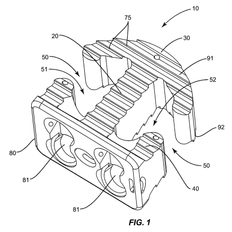
Figure 1 is a perspective view of a implant according to one embodiment;
Figures 2 - 10 are top views of implants according to various embodiments.
Figure 11 is a side view of an implant according to one embodiment.
Figure 12 is a side view of an implant according to one embodiment.
Figure 13 is a side view of an implant according to one embodiment.
Figure 14 is a side view of an implant according to one embodiment.
Figure 15 is a side view of a mount according to one embodiment.
Figure 16 is a perspective view of a cover according to one embodiment.
Figure 17 is a top view of an implant with a cover according to one
embodiment.
Figure 18 is a perspective view of an implant with a cover according to one
embodiment.
Detailed Description
The present application is directed to implants for positioning within an
intervertebral space formed between first and second vertebral members. Figure
1
illustrates one embodiment of an implant 10 with a superior surface 91 to
contact against a
first vertebral member, and an inferior surface 92 to contact against a second
vertebral
member. The implant 10 includes a central web 20 that extends between first
and second
flanges 30, 40. The flanges 30, 40 are shaped to form gaps 50 that extend the
height of the
implant 10. Spaces 51, 52 in communication with the gaps 50 are formed in an
interior of
the implant 10 to hold bone growth material.
CA 02698599 2010-03-04
WO 2009/042375 PCT/US2008/075548
3
The web 20 extends between the flanges 30, 40 and forms a central section of
the
implant 10. Web 20 may include a variety of shapes and sizes. Figures 1 and 2
include
the web 20 with a substantially constant width, with Figure 3 including a web
20 with a
variable width. The web 20 of Figure 3 reduces to a neck 26 at an intermediate
point
between the flanges 30, 40. The width increases on each side of the neck 26
with the
width being larger at the second flange 40 than at the first flange 30.
Web 20 may also include multiple different sections. Figure 4 illustrates an
embodiment with the web 20 formed by multiple sections 21, 22, 23 that are
spaced apart
with spaces 24 formed therebetween. The multiple sections 21, 22, 23 and
spaces 24 may
include the same or different shapes and sizes. Figure 5 illustrates another
embodiment
including a web 20 with a brace 25 branching off and connecting with the first
flange 30.
In another embodiment, web 20 is constructed of a single section that includes
an aperture
that extends between the superior and inferior surfaces 91, 92.
Web 20 may further be positioned at various lateral locations within the
central
section of the implant 10. Figures 1 and 2 illustrate an embodiment with the
web 20
laterally centered within the implant 10 and positioned at a middle of each of
the flanges
30, 40. In other embodiments as illustrated in Figure 5, the web 20 is
laterally offset away
from a center of the implant 10. In some embodiments as illustrated in Figures
1, 2, and 5,
the web 20 is laterally spaced the same distance along the first and second
flanges 30, 40.
Alternatively as illustrated in Figure 6, the web 20 is spaced at laterally
different positions
along the first and second flanges 30, 40.
The first and second flanges 30, 40 are space apart along the web 20. The
flanges
30, 40 may be the same shape and size, or may include different shapes and
sizes. Figure
7 includes an embodiment with each of the flanges 30, 40 being the same shape
and size.
Figure 2 is an embodiment with the first flange 30 including a different shape
than the
second flange 40. Figure 8 includes an embodiment with the flanges 30, 40
including
different shapes and sizes.
As illustrated in Figure 2, the first flange 30 may include a first arm 31
that extends
outward from a first side of the web 20, and a second arm 32 that extends
outward from a
second side of the web 20. Each arm 31,32 includes an end 33 that forms a side
of a gap
50. The arms 31, 32 may include the same shape and size as illustrated in
Figure 2, or
may include different shapes and/or sizes as illustrated in Figures 5 and 6.
Likewise, the
CA 02698599 2010-03-04
WO 2009/042375 PCT/US2008/075548
4
second flange 40 may include first and second arms 41, 42 as illustrated in
Figure 2. Each
arm 41, 42 includes an end 43 that forms a side of a gap 50. Arms 41, 42 may
include the
same or different shapes and/or sizes.
In one embodiment, one of the flanges 30, 40 includes a single arm. Figure 8
illustrates an embodiment with the first flange 30 including a single arm 31.
The first gap
50a is formed between an end 33 of the single arm 31 and end 43 of arm 41. The
second
gap 50b is formed between the web 20 and the end 43 of arm 42. In one
embodiment as
illustrated in Figure 9, the implant 10 includes a single flange 30 (i.e.,
there is no second
flange 40). Each gap 50 is formed between arm ends 33 and the web 20.
In one embodiment as illustrated in Figure 10, one or both of the flanges 30,
40
include ends 34, 44 with enlarged widths. The enlarged ends 34, 44 are
adjacent to the
gaps 50. The enlarged ends 34, 44 include a larger surface area adjacent to
the gaps 50 to
distribute the forces applied to the implant 10 from the first and second
vertebral members
and alleviate specific stresses from occurring at the ends 33, 43 of the
flanges 30, 40.
As illustrated in Figure 11, the implant 10 includes a height H measured
between
the superior surface 91 and the inferior surface 92. In one embodiment, the
height H is
substantially the same across the implant 10. In other embodiments, the height
H may
vary across the implant. Figure 12 illustrates an embodiment with a portion of
the first
flange 30 including a smaller height H'. This smaller height facilitates
insertion of the
implant 10 between the vertebral members with the section with the smaller
height H'
being initially inserted into the intervertebral space. Various other sections
of the implant
may include larger or smaller heights depending upon the context of use.
The gaps 50 extend the height H of the implant 10 between the superior and
inferior surfaces 91, 92. In one embodiment, the gaps 50 are formed between
the arm ends
33, 43 on opposing sides of the web 20. In one embodiment, the gaps 50 are
formed
between the arm ends 33, 43. In other embodiments, the gaps 50 are formed by
an arm
end 33 or 43, and the web 20. Figure 9 includes an embodiment with each of the
gaps
50a, 50b formed between ends 33 of the arms 31,32 and the web 20.
The gaps 50 may include the same or different widths W. Figure 2 includes each
of the gaps 50 with the same width W. Figure 8 includes a first gap 50a
including a width
Wl that is larger than the width W2 of the second gap 50b.
CA 02698599 2010-03-04
WO 2009/042375 PCT/US2008/075548
The widths W of the gaps 50 may be constant or may vary along the height.
Figure 11 includes an embodiment with the width W being constant along the
height H
between the superior and inferior surfaces 91, 92. Figure 13 includes an
embodiment with
a variable width with the end 33 of the first flange 30 being straight and the
end 43 of the
second flange 40 including a curved shape. The ends 33, 43 cause the width W
to be
larger in the middle of the gap 50 than at the outer ends at the superior and
inferior
surfaces 91, 92. Figure 14 includes an embodiment with the gap 50 formed
between the
first flange 30 and the web 20. The width W is smallest at the superior
surface 91 and
largest at the inferior surface 92.
Interior spaces 51, 52 are in communication with the gaps 50 and function to
contain bone growth material. The spaces are bounded on the lateral sides and
are
unbounded on the superior and superior sides. The spaces 51, 52 may include
the same or
different shapes and sizes. Figure 2 includes one embodiment with each of the
spaces 51,
52 including the same shape and size. Space 51 is positioned inward from the
gap 50a and
is bounded by the inner edges of arm 31, arm 41, and web 20. Space 52 is
positioned
inward from gap 50b and is bounded by the inner edges of arm 32, arm 42, and
web 20.
Figure 5 includes an embodiment with space 51 being smaller than space 52.
A plate 80 may be formed on a lateral side of the implant 10. The plate 80 may
be
a separate piece that is attached to the implant, or it may be integrally
formed with the
body (i.e., the body plate 80 include a unitary construction), Mount 80
extends above one
or both of the superior and inferior surfaces 91, 92 to contact against the
lateral sides of
the vertebral members to maintain the implant 10 positioned within the
intervertebral
space. Mount 80 also prevents the implant 10 from being over-inserted into the
intervertebral space. One or more apertures 81 may extend through the mount
80.
Apertures 81 are sized to receive fasteners (not illustrated) to attach the
implant 10 to the
vertebral members. In one embodiment as illustrated in Figure 15, apertures 81
a are
positioned at a central height of the mount 80 and angle upward towards the
superior
surface 91 for engaging the fasteners with the first vertebral member.
Apertures 8lb are
angled downward towards the inferior surface 92 to engage the second vertebral
member.
In one embodiment, as illustrated in Figure 15, the plate 80 extends across
the entire body.
In another embodiment as illustrated in Figure 12, the plate 80 extends across
a portion of
the body.
CA 02698599 2010-03-04
WO 2009/042375 PCT/US2008/075548
6
The apertures 81 may extend through the plate 80 and into one of the flanges
30,
40 and/or the web 20. Figure 7 illustrates one embodiment with the aperture 81
extending
through the flange 40 and into the web 20. Figure 2 illustrates an embodiment
with the
apertures 81 extending through the flange 40 and into the spaces 51, 52
respectively.
Teeth 75 may be positioned on one or both of the superior and inferior
surfaces 91,
92. Teeth 75 may include an angled orientation to facilitate insertion of the
implant 10
into the intervertebral space, and maintain the proper positioning within the
space. Teeth
75 may extend across an entirety or limited sections of the inferior and
superior surfaces
91, 92. Embodiments of teeth for an intervertebral implant are disclosed in
U.S. Patent
Applications Serial Nos. 11/394,452 and 11/412,330 that are each herein
incorporated by
reference.
A cover 60 may be attached to the implant 10 to extend across one or more of
the
gaps 50. Figure 16 illustrates one embodiment of the cover 60 that includes a
first section
63 with opposing, spaced-apart arms 61, 62. The cover 60 functions to extend
across one
or more of the gaps 50 and prevent inadvertent lateral removal of the bone
growth material
within the spaces 51, 52.
In one embodiment, arms 61, 62 are connected to the first section 63 to be
moveable in the directions of arrow Z. This may be caused by the cover 60
being
constructed of an elastic material. The cover 60 is sized to deform when
inserted onto the
spacer 10 with the arms 61, 62 expanding outward. Once attached, the arms 61,
62 apply
a compressive force to the exterior lateral sides of the implant 10 to
maintain attachment
of the cover 60. Apertures 64 may further be spaced about the cover 60 and
sized to
receive fasteners to further attach the cover 60 to the implant 10.
In another embodiment, arms 61, 62 are movably connected to the first section
63
such as by hinges, pivots, or other like structure. The arms 61, 62 may move
apart during
attachment of the cover to allow the arms 61, 62 to extend along each side of
the implant
10. Once positioned, the arms 61, 62 may move inward to contact against the
exterior
lateral sides of the implant 10. Apertures 64 may further be positioned around
the cover
60 to receive fasteners to attach the cover 60 to the implant 10.
In use, the implant 10 is initially inserted into the intervertebral space.
Prior to
insertion or after insertion, bone growth material is inserted into the spaces
51, 52. The
CA 02698599 2010-03-04
WO 2009/042375 PCT/US2008/075548
7
cover 60 is then attached to the exterior lateral sides of the implant 10. The
cover 60 may
extend across one or more of the gaps 50 to maintain the bone growth material.
Figure 17 includes one embodiment with the cover 60 attached to the implant
10.
The first section 63 includes a shape to substantially match the second flange
40. Arms
61, 62 substantially match the shape of the second flange 40, and include a
length to
extend across the gaps 50 and contact against the first flange 30. Cover 60
may also be
positioned against the mount 80 and extend over the apertures 81 to prevent
back-out of
the fasteners. Figure 18 includes an embodiment with a single arm 61 extending
outward
from the first section 63. The arm 61 extends across the first gap 50a. In
this
embodiment, arm 61 includes a smaller height than the gap 50a.
Cover 60 may also be attached to the implant 10 in other manners. In one
embodiment, implant 10 includes notches along the exterior lateral side and
the cover 60
includes outwardly-extending fingers. During attachment, the fingers slide
across the
exterior lateral side and mount within one of the notches to maintain the
attachment.
Other attachments may include snap fits and press fits.
The implant 10 may be inserted into the intervertebral space from a variety of
directions. In one embodiment, the implant 10 is inserted in an anterior
approach with the
mount 80 contacting against the anterior lateral sides of the vertebral
members. Other
applications contemplate other approaches, including posterior, postero-
lateral, antero-
lateral and lateral approaches to the spine, and accessing other regions of
the spine,
including the cervical, thoracic, lumbar and/or sacral portions of the spine.
Spatially relative terms such as "under", "below", "lower", "over", "upper",
and
the like, are used for ease of description to explain the positioning of one
element relative
to a second element. These terms are intended to encompass different
orientations of the
device in addition to different orientations than those depicted in the
figures. Further,
terms such as "first", "second", and the like, are also used to describe
various elements,
regions, sections, etc and are also not intended to be limiting. Like terms
refer to like
elements throughout the description.
As used herein, the terms "having", "containing", "including", "comprising"
and
the like are open ended terms that indicate the presence of stated elements or
features, but
do not preclude additional elements or features. The articles "a", "an" and
"the" are
CA 02698599 2010-03-04
WO 2009/042375 PCT/US2008/075548
8
intended to include the plural as well as the singular, unless the context
clearly indicates
otherwise.
The present invention may be carried out in other specific ways than those
herein
set forth without departing from the scope and essential characteristics of
the invention.
The present embodiments are, therefore, to be considered in all respects as
illustrative and
not restrictive, and all changes coming within the meaning and equivalency
range of the
appended claims are intended to be embraced therein.