Note: Descriptions are shown in the official language in which they were submitted.
CA 02698638 2010-03-05
WO 2009/031047
PCT/1B2008/003107
BIORESORBABLE AND BIOCO1VIPATIBLE COMPOUNDS FOR SURGICAL USE
TECHNICAL FIELD
The present disclosure relates to biocompatible and bioresorbable compounds
containing
a functionalized collagen covalently bonded directly to a glycosaminoglycan
without the use of a
chemical cross-linking agent. The present compounds and compositions
containing them are
useful for a variety of medical applications, including surgical implants.
DESCRIPTION OF THE RELATED ART
Collagen and glycosaminoglycans have been combined for the preparation of
biomaterials and surgical implants. Non-crosslinked collagen and chitosan
mixtures have weak
mechanical properties rendering their manipulation difficult and the in-vivo
biodegradation of the
collagen is often insufficient.
Cross linking of collagen and glycosaminoglycans, e.g. chitosan, using a cross
linker
agent such as glutaraldehyde is inconvenient in certain applications. For
example, the use of
glutaraldehyde in aqueous media leads to the formation of very high molecular
glutaraldehyde
polymers which are difficult to eliminate by simple washing techniques. Upon
implantation,
such glutaraldehyde polymers may hydrolyse and cause a release of
glutaraldehyde or remain in-
vivo and be liberated after the disappearance of the collagen/chitosan
components.
It would be advantageous to provide biocomposites or implants made of
functionalized
collagen and glycosaminoglycans. The formulation advantageously provides a
tailor-made, self
cross-linked glycoprotein network and may be based on' highly purified and
fully characterized
1
CA 02698638 2010-03-05
WO 2009/031047
PCT/1B2008/003107
extra-cellular matrix compounds which mimic the native extracellular matrix
and provide an
optimal support for cell differentiation and growth and for tissue
regeneration. The relative
amounts of oxidized collagen and glycosaminoglycans may be varied to optimize
the biological,
mechanical and biodegradation properties of the appropriate tissue to be
repaired and/or
regenerated. When compared to implants based only on collagen, the
formulations described
herein can also favor the repair and/or the regeneration of tissues by the
release of
glycosaminoglycan oligomers, showing interesting biological properties (eg.
angiogenic,
antibacterial properties), in a time controlled fashion. The formulations can
be also
advantageously obtained under different physical forms by itself (eg. gel,
film, sponge, yarn,
knitted textile, woven textile, non-knitted non-woven mesh) or can be easily
combined with other
components in an open fashion.
SUMMARY
Accordingly, the present disclosure relates to compounds containing a
functionalized
collagen covalently bonded directly to a glycosaminoglycan without the use of
a chemical cross-
linking agent. In particular embodiments, the collagen is functionalized by
oxidative cleavage:
for example, this oxidative cleavage converts pendant portions of the collagen
molecule into
aldehydes which are reactive with the amine groups of the glycosaminoglycan.
In embodiments,
the functionalized collagen includes one or more reactive moieties selected
from the group
consisting of aldehydes, sulfones, vinylsulfones, isocyanates, and acid
anhydrides. In
embodiments, the functionalized collagen includes one or more reactive
moieties selected from
the group consisting of ¨CO2N(COCH2)2, -CO2N(COCH2)2, -CO2H, -CHO, -CHOCH2, -
N=C=O, -S02CH=CH2, -N(COCH)2, and -S-S-(C5H4N).
2
CA 02698638 2010-03-05
WO 2009/031047
PCT/1B2008/003107
In embodiments, the glycosarninoglyan is selected from the group consisting of
dermatan
sulfate, hyaluronic acid, chondroitin sulfate, chitin, chitosan, heparin,
keratan sulfate,
keratosulfate, deacylated hyaluronic acid and derivatives and combinations
thereof. For example,
the glycosaminoglycan is chitosan.
Methods for preparing the compounds are also described. In particular, the
present disclosure
relates to a method of forming a bioresorbable compound comprising contacting
a functionalized
collagen with a glycosaminoglycan, in particular under reaction conditions
under which the
functionalized collagen covalently binds directly to the glycosaminoglycan,
without the use of a
crosslinking agent. In embodiments, a deionized water solution of
functionalized collagen is
combined to a deionized water solution of glycosaminoglycan to allow the
functionalized
collagen to mix with the glycosaminoglycan and form said compound. The pH of
the solution of
functionalized collagen may be adjusted between 2 and 7.5, for example by
addition of a suitable
acid. The pH of the solution of glycosaminoglycan may be adjusted between 2
and 7.5, for
example by addition of a suitable acid. The present disclosure also relates to
a compound
obtainable by such a method.
The present disclosure further relates to a mixture consisting in a deionized
water solution of
functionalized collagen, for example with an adjusted pH between 2 and 7.5,
combined to a
deionized water solution of glycosaminoglycan, for example with an adjusted pH
between 2 and
7.5.
The present disclosure also relates to an implant comprising a compound
containing a
functionalized collagen covalently bonded directly to a glycosaminoglycan
without the use of a
crosslinking agent. In embodiments, the implant of the present disclosure
comprises a sponge
containing said compound. In embodiments, the implant of the present
disclosure comprises a
3
CA 02698638 2015-03-31
textile containing said compound. In embodiments, the implant of the present
disclosure
comprises a hydrogel containing said compound. In embodiments, the implant of
the present
disclosure comprises threads containing said compound. In embodiments, the
implant of the
present disclosure comprises a non knitted, non woven composite containing
said compound. In
embodiments, the implant of the present disclosure comprises a film containing
said compound.
In embodiments, the implant of the present disclosure comprises a mesh coated
with a
composition containing said compound.
The present disclosure further relates to a composition comprising a compound
containing a functionalized collagen covalently bonded directly to a
glycosaminoglycan without
the use of a crosslinking agent.
The present compounds may be used to form a variety of surgical implants such
as gels,
films, sponges, fibers, woven textiles, knitted textiles, non-woven, non-
knitted textiles, and the
like. In embodiments, the compounds may be combined with a substrate to form a
coated
implant or to add additional layers to the implant.
BRIEF DESCRIPTION OF THE DRAWINGS
The embodiments of the present disclosure will be described more clearly by
means of
the description which follows and the attached drawings in which:
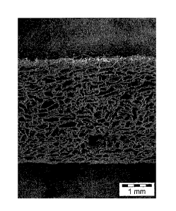
-Figure 1 represents a scanning electron microscopy image (HitachiTM S800
scanning
electron microscope with image acquisition and analysis system) of a freeze
dried sponge
according to the present disclosure, made from a 50/50 oxidized collagen
(CXN)/chitosan
mixture, from a side view;
4
CA 02698638 2010-03-05
WO 2009/031047
PCT/1B2008/003107
-Figure 2 represents a scanning electron microscopy image (Hitachi S800
scanning
electron microscope with image acquisition and analysis system) of a yarn
according to the
present disclosure, made from a wet-spinning process of an CXN/chitosan
mixture;
-Figure 3 represents a scanning electron microscopy image (Hitachi S800
scanning
electron microscope with image acquisition and analysis system) of a
multilayer implant
according to the present disclosure, including a textile made from polylactic
acid (PLA), from a
side view;
-Figure 4A represents a scanning electron microscopy images (Hitachi S800
scanning
electron microscope with image acquisition and analysis system) of a two-
dimensional textile
according to the present disclosure, the textile being knitted with
multifilaments of PLA and then
coated three times with a 50/50 CXN/chitosan mixture;
-Figure 4B represents a scanning electron microscopy images (Hitachi S800
scanning
electron microscope with image acquisition and analysis system) of a two-
dimensional textile
according to the present disclosure, at a higher magnification than for Figure
4A, the textile being
knitted with multifilaments of PLA and then coated three times with a 50/50
CXN/chitosan
mixture;
-Figure 5A represents a scanning electron microscopy images (Hitachi S800
scanning
electron microscope with image acquisition and analysis system) of a three-
dimensional textile
according to the present disclosure, the textile being knitted with both
monofilaments and
multifilaments of PLA and being coated three times with a 50/50 CXN/chitosan
mixture;
-Figure 5B represents a scanning electron microscopy images (Hitachi S800
scanning
electron microscope with image acquisition and analysis system) of a three-
dimensional textile
according to the present disclosure, at a higher magnification than for Figure
5A, the textile being
CA 02698638 2010-03-05
WO 2009/031047
PCT/1B2008/003107
knitted with both monofilaments and multifilaments of PLA and being coated
three times with a
50/50 CXN/chitosan mixture;
-Figure 6A represents a scanning electron microscopy images (Hitachi S800
scanning
electron microscope with image acquisition and analysis system) of a two-
dimensional textile
according to the present disclosure, the textile being knitted with
multifilaments of PET and then
coated three times with a 50/50 CXN/chitosan mixture;
-Figure 6B represents a scanning electron microscopy images (Hitachi S800
scanning
electron microscope with image acquisition and analysis system) of a two-
dimensional textile
according to the present disclosure, at a higher magnification than for Figure
4A, the textile being
knitted with multifilaments of PET and then coated three times with a 50/50
CXN/chitosan
mixture; and
-Figure 7 represents a viscometer reading of an oxidized collagen and chitosan
mixture
and a native collagen and chitosan mixture in accordance with the present
disclosure.
DETAILED DESCRIPTION
Compounds containing a functionalized collagen covalently bonded directly to a
glycosaminoglycan without the use of a chemical cross-linker agent are
provided in accordance
with the present disclosure. In embodiments, the compounds may be obtained by
combining a
reactive solution of collagen or gelatine, modified by a chemical reaction
(e.g. oxidative
cleavage) to functionalize a pendant portion of the collagen with moieties
which are capable of
forming a covalent bond with the reactive moieties of the glycosaminoglycan.
The compounds,
processes for preparing the compounds and design of surgical implants using
the compounds are
described in greater detail below. The methods for producing the product of
the present
6
CA 02698638 2010-03-05
WO 2009/031047
PCT/1B2008/003107
disclosure make use of steps that are recognized as effective for inactivating
viral particules and
prions. Advantageously, the collagen and glycosaminoglycan may be highly
purified and totally
free of pendant residues providing a real advantage comparatively to the
extracellular matrix
made from biological tissues such as small intestine, sub mucosa or dermis.
This gives the
product a very high safety level while eliminating the inflammatory response.
Collagen
Collagen is a naturally occurring protein featuring good biocompatibility. It
is the major
structural component of vertebrates, forming extracellular fibers or networks
in practically every
tissue of the body, including skin, bone, cartilage, and blood vessels. In
medical devices,
collagen provides a more physiological, isotropic environment that has been
shown to promote
the growth and function of different cell types, facilitating the rapid
overgrowth of host tissue
after implantation.
For the purpose of the present application, the term "collagen" is intended to
mean any
known collagen of porcine, bovine or human origin, for example natural or
recombinant
collagen, esterified collagen, for example methylated, ethylated or
alternatively succinylated
collagen, glycosylated collagen (eg. collagen glycosylated with saccharides /
polysaccharides
comprising free amino groups, collagen glycosylated with saccharides /
polysaccharides
comprising vicinal diols, collagen glycosylated with saccharides /
polysaccharides comprising ¨
CH(NH2)¨CHy(OH)¨ chemical bonds), or one of its derivatives.
The term "gelatine" here includes commercial gelatine made of collagen which
has been
denatured by heating and in which the chains are at least partially hydrolyzed
(molecular weight
lower than 100 kDa).
7
CA 02698638 2015-03-31
=
The collagen used can be of human or animal origin. Some non-limiting examples
include, type I porcine or bovine collagen, type I or type III human collagen
or mixtures in any
proportions of these types. In embodiments, the collagen or gelatine used is a
porcine collagen.
The collagen can be modified by using any method known to those skilled in the
art to
provide pendant portions of the collagen with moieties which are capable of
covalently bonding
with the reactive chemical groups of a glycosaminoglycan. Examples of such
pendant moieties
include aldehydes, sulfones, vinylsulfones, isocyanates, and acid anhydrides.
In addition,
electrophilic groups such as ¨CO2N(COCH2)2, -CO2N(COCH-)2, -CO2H, -CHO, -
CHOCH,,
-SO,CH=CH,, -N(COCH)7, -S-S-(C5H4N) may also be added to pendant chains of the
collagen to allow covalent bonding to occur with the glycosaminoglycans.
In embodiments, the collagen may be modified through the addition of an
oxidizing
agent. Contacting collagen with an oxidizing agent creates oxidative cleavage
along portions of
the collagen thereby creating pendant aldehyde groups capable of reacting with
the
glycosaminoglycans. The oxidizing agent may be, for example, iodine, peroxide,
periodic acid,
hydrogen peroxide, a periodate, a compound containing periodate, sodium
periodate, a
diisocyanate compound, a halogen, a compound containing halogen, n-
bromosuccinimide, a
permanganate, a compound containing permanganate, ozone, a compound containing
ozone,
chromic acid, sulfuryl chloride, a sulfoxide, a selenoxide,. an oxidizing
enzyme (oxidase) and
combinations thereof In embodiments, the oxidizing agent is periodic acid.
An example of the oxidative technique is described by Tardy et al. in U.S.
Patent No.
4,931,546. Briefly, this technique involves mixing the collagen in acid
solution with an
oxidizing agent, i.e., a solution of periodic acid or one of its salts, at a
concentration of between 1
and 10-5 M, in embodiments between 5 10-3 and 10-1 M, at a temperature of
between 10 and 25
8
CA 02698638 2015-03-31
C for 10 minutes to 72 hours. This process breaks down hydroxylysine and the
sugars of the
collagen, thus creating reactive sites without causing crosslinking. The
oxidative cleavage of
collagen allows moderate cross-linking later in the collagenic material.
Another technique for oxidized collagen is by oxidation of a 3% collagen
solution by
periodic acid, at a final concentration of 8mM, during 3 hours, as described
by Bayon, et al.in
U.S. Patent No. 6,596,304. Aldehyde groups are formed by oxidative cleavage on
the lateral
chains of the hydroxyl-lysine residues giving the oxidized collagen
capabilities to form covalent
bonds with amines. The oxidized collagen can be fully degraded in vivo, after
a few weeks, and
while not wishing to be bound by any theory, it is believed that the oxidized
collagen will
degrade before the glycosaminoglycan.
Glycosaminoglycans
The term "glycosaminoglycan" is intended to encompass complex polysaccharides
having repeating units of either the same saccharide subunit or two different
saccharide subunits.
Some non-limiting examples of glycosaminoglycans include dermatan sulfate,
hyaluronic acid,
the chondroitin sulfates, chitin, heparin, keratan sulfate, keratosulfate, and
derivatives thereof.
Some non-limiting examples of derivatives may include partially and fully
deacylated versions
of these compounds such as chitosan and deacylated hyaluronic acid. The
glycosaminoglycans
may be extracted from a natural source, e.g. animal tissues such as squid pens
and shrimp shells
or vegetable sources such as mushrooms (eg "champignon de Paris"), or they may
be
synthetically produced or synthesized by modified microorganisms such as
bacteria.
9
CA 02698638 2010-03-05
WO 2009/031047
PCT/1B2008/003107
In embodiments, the functionalized collagen may be combined with a
glycosaminoglycan
such as chitosan to crosslink and form covalent bonds. The glycosaminoglycan
may display a
degree of acetylation (DA) of about 0% to about 60%. In embodiments, the
glycosaminoglycan
displays a degree of acetylation (DA) of about 0.5% to about 50%. Samples of
different degrees
of acetylation can be obtained either by a heterogeneous deacetylation process
or by a
homogenous reacetylating process from a sample of a glycosaminoglycan that is
fully
deacetylated. In embodiments, the glycosaminoglycan includes a blend of
chitosan with different
degree of acetylation selected from about 0.5 to 60%.
In embodiments, the glycosaminoglycan has a molecular weight ranging from
about 100
to about 1,000,000 g/mol. In some embodiments, the glycosaminoglycan has a
molecular weight
ranging from about 164 (chitosan monomer) to about 1,000,000 g/mol. In
embodiments, the
glycosaminoglycan has a molecular weight of about 1500 to about 800,000 g/mol.
In addition,
the glycosaminoglycan may also display a low polydisperity index between about
1.2 to about
1.8. In particularly useful embodiments, the glycosaminoglycan is chitosan.
Nevertheless, the
glycosaminoglycan may be a mixture of chitosans with different degrees of
acetylation or a
mixture of chitosans and other glycosaminoglycans, e.g. hyaluronic acid, with
different degrees
of acetylation and in which all glycosaminoglycan have the capabilitiy, i.e.
have free amino
groups, to be cross-linked to the oxidized collagen.
Combining the functionalized collagen and the glycosaminoglycans
Compounds in accordance with the present disclosure are made by reacting a
functionalized collagen with a glycosaminoglycan under conditions which cause
the two
components to form covalent bonds without the use of a chemical crosslinldng
agent. The two
CA 02698638 2010-03-05
WO 2009/031047
PCT/1B2008/003107
components may take the form of any solution, suspension, emulsion, semi-
solid, or solid
material capable of allowing the two-components to interact and crosslink.
In embodiments, each component is solubilized in an acceptable solvent such as
deionized water to form two separate solutions. The two solutions may be
combined to allow the
two components to mix and form the compounds described herein. In particular
embodiments,
the glycosaminoglycan is solubilized in deionized water with a stoechiometric
amount of
hydrochloric acid with a polymer (glycosaminoglycan) concentration ranging
from about 0.5% to
about 10% (w/w). It is envisioned that the pH of the glycosaminoglycan
solution can be adjusted
if necessary between about 2 and about 7.5 depending on the degree of
acetylation. The
functionalized collagen is also solubilized in an acceptable solvent such as
deionized water to a
concentration ranging from about 0.5% to about 10% (w/w). It is also
envisioned that the pH of
the functionalized collagen solution may be adjusted between about 2 and about
7.5. The two
components in solution are mixed to a final concentration of polymer (compound
functionalized
collagen/glycosaminoglycan) ranging from 0.5% to 20% (w/w). In embodiments,
different
proportions between the functionalized collagen and the glycosaminoglycan may
be used. In
particular embodiments, the glycosaminoglycan may be composed of a mixture of
chitosans
with different degrees of acetylation (DA). The chitosan having a degradation
time in function
with its degree of acetylation (K.Kurita et al,Carbohydrate polymers. Vol 42
pp.19-21,200;
K.Tomihata et al, Biomaterials. Vol 18 n 7 pp.567-575,1997), the combination
of slow and fast
biodegradable chitosan is an important issue for the awaiting properties of
the implant, i.e.,
progressive cell colonization of the sponge. In fact, the degradation of the
slow biodegradable
oxidized collagen and chitosan with high DA, i.e. 35<DA<50, in vitro in the
presence of viable
cells and in vivo, helps to increase the interconnected porosity which is a
key parameter for the
11
CA 02698638 2010-03-05
WO 2009/031047
PCT/1B2008/003107
regeneration of healthy native like tissue in the full thickness of the
implant and the extent of
tissue integration. In embodiments, molecules released from the controlled
degradation of the
biocomposite, for example oxidized collagen/chitosan, may advantageously
confer to the implant
highly interesting biological activities e.g. antimicrobial, anticancer,
antioxidant, and
immunostimulant effects, especially in the case of chitosan (S-K. Kim et al,
Carbohydrate
Polymers, Vol. 62, Issue 4, pp.357-368,2005) and may bring, in complement of
the
biocompatibility and biodegradability, bioactive properties to the medical
devices. The
biological properties of released chitosan oligopolymers enhance the tissue
regeneration and
extend the use of the implant, e.g. to surgical sites with a high risk of
contamination.
In embodiments, a combination of two solutions comprising an acidic solution
of
oxidized collagen and an acidic solution of chitosan with one or a mix of
several degree of
acetylation may be used. The collagen is oxidized by the addition of periodic
acid as the
oxidizing agent and the chitosan solution is made acidic by the addition of
hydrochloric acid.
The mixture can be neutralized either with an alkaline vapour/solution or
buffer solution with a
pH greater than 7, leading to a cross-linked scaffold compatible for cell
adhesion and
proliferation. This combination is particularly advantageous compared to a
combination of
oxidized collagen and glutaraldehyde cross-linked collagen, because the latter
makes a
suspension which is difficult to incorporate in a homogeneous fashion to an
implantable surgical
device such as a three-dimensional mesh.
Viscosity of the mixture of native collagen/glycosaminoglycan versus
functionalized
collagen/glycosaminoglycan
12
CA 02698638 2010-03-05
WO 2009/031047
PCT/1B2008/003107
_
The reaction between the functionalized collagen and glycosaminoglycan is
characterized
by a rapid increase of the viscosity of the reaction mixture when the two
components are mixed.
Viscosity measurements were performed on a viscosimeter Lamy type TVe-05. The
solutions of
oxidized collagen and chitosan were equilibrated at the temperature of 25 C
for 1 hour and then
mixed. A sample of 5m1 was poured into the chamber of the viscosimeter and the
evolution of
viscosity against time was studied. The viscosity of the solution composed of
oxidized collagen
and chitosan and a solution composed of native collagen and chitosan were
compared to
highlight the type of interactions between the oxidized collagen and the
chitosan.
The tests were performed at 25 C with biopolymers having characteristics
described
below:
-Chitosan DA = 2.5% and Mw = 500,000 g/mol
-Oxidized collagen (CXN) prepared from native collagen by oxidative cleavage.
-Native collagen (CPP) without telopeptide and with helicoidal structure
preserved. The average
molecular weight is about 300,000 g/mol.
The solution prepared for the tests had a final polymer
(collagen/glycosaminoglycan)
concentration of 1% (w/w), a proportion close to 50/50 respectively of
collagen and chitosan.
The pH measured was close to 4.7 and 4.89, respectively, for the CXN/chitosan
and
CPP/chitosan mixtures.
As shown in Figure 7, there is an increase of viscosity in the case of the
oxidized
collagen/chitosan mixture wherein the area of stability was reached for 30
minutes at a pH of 4.7.
On the other hand, only a slight increase of viscosity is observed in the case
of the native
collagen/chitosan solution. Therefore, the difference of the viscosity
evolution against time can
be attributed, partly, to the formation of a chemical crosslink between
oxidized collagen and
13
CA 02698638 2015-03-31
chitosan. In fact, the low viscosity increase in the case of chitosan/native
collagen mixture is due
to the ionic complex because the two components exhibit a high charge of
density.
Surgical implant design using the oxidized collagen and chitosan mixture
The cross-linked mixture of functionalized collagen and a glycosaminoglycan,
ie the
compound functionalized collagen/glycosaminoglycan of the present disclosure,
can be used to
form a variety of surgical implants such as sponges, films, hydrogels, non-
woven non-knitted
meshes, three-dimensional structures such as tubular and spherical structures,
microbeads,
threads, rods, filaments, yarns, meshes, slings, sutures and other composite
materials such as
pledgets, buttresses, adhesion barriers and the like. The mixture can also be
combined with or
used to coat surgical implants, such as two-dimensional meshes, three-
dimensional meshes,
vascular prostheses, patches, slings and the like.
The surgical implants which may be combined or coated with compositions which
include the compounds of the present disclosure may be made from bioabsorbable
or non-
bioabsorbable materials. Some non-limiting examples of suitable non-absorbable
materials
which may be utilized included polyolefins, such as polyethylene,
polypropylene, copolymers of
polyethylene and polypropylene, and blends of polyethylene and polypropylene.
Other non-
absorbable materials which may be utilized include polyesters such as
polyethylene terephthalate
(PET), polyamides, aramides, expanded polytetrafluoroethylene, polyurethane,
polyvinylidene,
difluoride (PVDF), polybutester, copper alloy, silver alloy, platinum, medical
grade stainless
steels such as 316L medical grade stainless steel, combinations thereof, and
the like. Examples
of commercially available polypropylene-based textile supports which may be
utilized include
those sold under the brand name PARIETENE from SofradimTM.
14
CA 02698638 2010-03-05
WO 2009/031047
PCT/1B2008/003107
Suitable absorbable materials include, but are not limited to, trimethylene,
carbonate,
caprolactone, dioxanone, glycolic acid, lactic acid, glycolide, lactide,
hornopolymers thereof,
copolymers thereof, and combinations thereof Specific absorbable materials
which may be
suitable include, for example, chitosan, cellulose, oxidized cellulose,
combinations thereof, and
the like.
In embodiments, a solution of the present compounds may be freeze-dried to
form a
porous sponge material capable of allowing tissue in growth and induce a
progressive cell
colonization of the sponge by mixing several glycosaminoglycans with different
degrees of
acetylation and with different degradation properties. In embodiments, the
solutions described
herein may include additional polymeric materials which allow the solution to
form a non-porous
film useful in preparing adhesion barriers. In particular, the compounds of
the present disclosure
may be combined with polyethylene glycol, and glycerol to form a non-porous
film. In still other
embodiments, the sponges or films or hydrogel materials as described herein
may be used to add
a coating layer on an existing surgical implant or to form a multilayer
surgical implant. Such
combination implants may be useful in forming surgical implants which prevent
adhesions and
the in-growth of tissue one side of the implant and encourage the in-growth of
tissue and
formation of adhesions on the other side of the implant. Some non-limiting
examples include
multilayer pledgets, buttresses, surgical meshes, slings and adhesion
barriers.
In embodiments, a solution of the present compounds may be used to form yarn
by a wet
spinning process as described in the patent EP0328050A2 by Bisento de rutsuka
et al. The
biological composite yarns are fully biocompatible and biodegradable with a
wide range of
degradation times due to the mix of several glycosaminglycans with different
degrees of
acetylation. The composite yarns of the present disclosure may be used to knit
textiles with
CA 02698638 2010-03-05
WO 2009/031047
PCT/1B2008/003107
different patterns in 2 or 3 dimensions and these yarns may be used alone or
combined with other
biocompatible yarns such as yarns made from polylactic acid (PLA). The
textiles may be
employed as implants or as a part of an implant to improve the mechanical
properties of the
implant. Moreover with the functionalized collagen/glycosaminoglycan
composition of the
present disclosure, the textile may have high biocompatibility and good
mechanical properties in
a wide range of degradation times, ranging from about 2 weeks to several
months.
Advantageously, the molecules released from the degradation of the
biocomposite or compound
of the present disclosure, for example oxidized collagen/chitosan, give
biological activities of
particular interest, i.e., antimicrobial, anticancer, antioxidant, and
immunostimulant effects,
especially in the case of chitosan.
Optional Bioactive Agents
In embodiments, at least one bioactive agent may be included in compositions
containing
the present compounds and thereby incorporated into a medical device. In these
embodiments,
the implant can also serve as a vehicle for delivery of the bioactive agent.
The term "bioactive
agent", as used herein, is used in its broadest sense and includes any
substance or mixture of
substances that have clinical use. Consequently, bioactive agents may or may
not have
pharmacological activity per se, e.g., a dye, or fragrance. Alternatively a
bioactive agent could be
any agent which provides a therapeutic or prophylactic effect, a compound that
affects or
participates in tissue growth, cell growth, cell differentiation, an anti-
adhesive compound, a
compound that may be able to invoke a biological action such as an immune
response, or could
play any other role in one or more biological processes. It is envisioned that
the bioactive agent
may be applied to the medial device in any suitable form of matter, e.g.,
films, powders, liquids,
gels and the like.
16
CA 02698638 2015-03-31
Examples of classes of bioactive agents which may be utilized in accordance
with the
present disclosure include anti-adhesives, antimicrobials, analgesics,
antipyretics, anesthetics,
antiepileptics, antihistamines, anti-inflammatories, cardiovascular drugs,
diagnostic agents,
sympathomimetics, cholinomimetics, antimuscarinics, antispasmodics, hormones,
growth
factors, muscle relaxants, adrenergic neuron blockers, antineoplastics,
immunogenic agents,
immunosuppressants, gastrointestinal drugs, diuretics, steroids, lipids,
lipopolysaccharides,
polysaccharides, and enzymes. It is also intended that combinations of
bioactive agents may be
used.
Anti-adhesive agents can be used to prevent adhesions from forming between the
to implantable medical device and the surrounding tissues opposite the
target tissue. In addition,
anti-adhesive agents may be used to prevent adhesions from forming between the
coated
implantable medical device and the packaging material. Some examples of these
agents include,
but are not limited to poly(vinyl pyrrolidone), carboxymethyl cellulose,
hyaluronic acid,
polyethylene oxide, poly vinyl alcohols and combinations thereof.
Suitable antimicrobial agents which may be included as a bioactive agent in
the bioactive
coating of the present disclosure include triclosanTM, also known as 2,4,4'-
trichloro-2'-
hydroxydiphenyl ether, chlorhexidine and its salts, including chlorhexidine
acetate, chlorhexidine
gluconate, chlorhexidine hydrochloride, and chlorhexidine sulfate, silver and
its salts, including
silver acetate, silver benzoate, silver carbonate, silver citrate, silver
iodate, silver iodide, silver
lactate, silver laurate, silver nitrate, silver oxide, silver palmitate,
silver protein, and silver
sulfadiazine, polymyxin, tetracycline, aminoglycosides, such as tobramycin and
gentamicin,
rifampicin, bacitracin, neomycin, chloramphenicol, miconazole, quinolones such
as oxolinic
acid, norfloxacin, nalidixic acid, pefloxacin, enoxacin and ciprofloxacinTM,
penicillins such as
17
CA 02698638 2015-03-31
oxacillin and pipracilTM, nonoxynol 9, fusidic acid, cephalosporins, and
combinations thereof. In
addition, antimicrobial proteins and peptides such as bovine lactoferrin and
lactoferricin B and
antimicrobial polysaccharides such as fucans and derivatives may be included
as a bioactive
agent in the bioactive coating of the present disclosure.
Other bioactive agents which may be included as a bioactive agent in the
coating
composition applied in accordance with the present disclosure include: local
anesthetics; non-
steroidal antifertility agents; parasympathomimetic agents; psychotherapeutic
agents;
tranquilizers; decongestants; sedative hypnotics; steroids; sulfonamides;
sympathomimetic
agents; vaccines; vitamins; antimalarials; anti-migraine agents; anti-
parkinson agents such as L-
dopa; anti-spasmodics; anticholinergic agents (e.g. oxybutynin); antitussives;
bronchodilators;
cardiovascular agents such as coronary vasodilators and nitroglycerin;
alkaloids; analgesics;
narcotics such as codeine, dihydrocodeinone, meperidine, morphine and the
like; non-narcotics
such as salicylates, aspirin, acetaminophen, d-propoxyphene and the like;
opioid receptor
antagonists, such as naltrexone and naloxone; anti-cancer agents; anti-
convulsants; anti-emetics;
antihistamines; anti-inflammatory agents such as hormonal agents,
hydrocortisone, prednisolone,
prednisone, non-hormonal agents, allopurinol, indomethacin, phenylbutazone and
the like;
prostaglandins and cytotoxic drugs; estrogens; antibacterials; antibiotics;
anti-fungals; anti-virals;
anticoagulants; anticonvulsants; antidepressants; antihistamines; and
immunological agents.
Other examples of suitable bioactive agents which may be included in the
coating
composition include viruses and cells, peptides, polypeptides and proteins,
analogs, muteins, and
active fragments thereof, such as immunoglobulins, antibodies, cytokines (e.g.
lymphokines,
monokines, chemokines), blood clotting factors, hemopoietic factors,
interleukins (IL-2, IL-3, IL-
4, IL-6), interferons ((3-IFN, (a-IFN and y-IFN), erythropoietin, nucleases,
tumor necrosis factor,
18
CA 02698638 2010-03-05
WO 2009/031047
PCT/1B2008/003107
colony stimulating factors (e.g., GCSF, GM-CSF, MCSF), insulin, anti-tumor
agents and tumor
suppressors, blood proteins, gonadotropins (e.g., FSH, LH, CG, etc.), hormones
and hormone
analogs (e.g., growth hormone), vaccines (e.g., tumoral, bacterial and viral
antigens);
somatostatin; antigens; blood coagulation factors; growth factors (e.g., nerve
growth factor,
insulin-like growth factor); protein inhibitors, protein antagonists, and
protein agonists; nucleic
acids, such as antisense molecules, DNA and RNA; oligonucleotides;
polynucleotides; and
ribozymes.
Bioactive agents can also be additives, such as fucans, either native or
chemically
modified glucosaminoglycans, oxidized starch, emulsifiers, surfactants,
humectants, buffering
agents, pH modulators, chelating agents, viscosity agents and any other
products which may
enhance tissue repair, limit the risk of sepsis, and modulate mechanical
properties of the
compounds.
EXAMPLES
The following non-limiting examples show the preparation, formulation and uses
possible
of the present compounds and the tensile and swelling properties of the
oxidized collagen and
chitosan mixture compared to a native collagen and chitosan mixture.
EXAMPLE 1
Freeze dried sponge for the preparation of materials supporting cell growth
A collagen/chitosan mixture was prepared by mixing an acidic solution of
oxidized
collagen and an acidic solution of chitosan in different proportions with a
final polymer
(collagen/chitosan) concentration of 1% (w/w).
19
CA 02698638 2010-03-05
WO 2009/031047
PCT/1B2008/003107
Oxidized collagen
Oxidized collagen was obtained by the oxidation of a 3% collagen solution by
periodic
acid, at a final concentration of 8mM, at room temperature, during 3 hours, as
described by
Bayon, et al. in Example 4 of U.S. Pat. No. 6,596,304. At this step the pH of
the oxidized
collagen solution was about 3.2.
Native collagen
Solutions of native collagen were obtained by solubilizing collagen powder at
a 1% final
concentration, in sterile water. The pH measured close to 3.
Chitosan
The chitosan was solubilized in deionized water with a stoechiometric amount
of
hydrochloric acid with a polymer concentration of 1% (w/w). The pH of the
chitosan solution
was about 5, but the pH could have been adjusted to 3 to have better control
of the crosslink
kinetic between the oxidized collagen and chitosan.
Before freeze drying, if the application required it, the collagen/chitosan
mixture could
have been poured into a 3D mesh so as to fully cover the mesh and obtain a
freeze dried
sponge/mesh composite. The presence of the 3D mesh facilitates fastening the
implant to tissue
(e.g., via suturing). Moreover, the homogeneity of the oxidized
collagen/chitosan solution allows
a better penetration of the solution within a 3-dimensional structure of the
textile when compared
to collagen that has been oxidized with a cross-linking agent.
CA 02698638 2010-03-05
WO 2009/031047
PCT/1B2008/003107
Freeze dried composite
Several mixtures of various blends of oxidized collagen and chitosan as well
as native
collagen and chitosan (approximately 121 g) were poured within a 12cm by 17cm
plastic box and
freeze-dried for 24 hours. The samples were then neutralized in a 1M sodium
hydroxide bath for
1 hour and thoroughly washed in deionized water until the pH reached 7. The
freeze-dried
sponges were then calendered to obtain a material with a final thickness of
0.13mm.
Figure 1 represents a scanning electron microscopy image of one face of such a
sponge
obtained from a blend of oxidized collagen and chitosan.
Additives, such as fucans, native or chemically modified glucosaminoglycans,
which may
induce self chemical crosslink between collagen and glucosaminoglycans
(hyaluronic acid,
sulphate chondroitin, etc), oxidized starch, and any other product which may
enhance tissue
repair, limit the risk of sepsis, and modulate the mechanical properties of
the composite (swelling
rate in water, tensile strength, etc) could have been be added to the blend of
oxidized collagen
and chitosan.
Tensile tests on freeze-dried calendered sponges composed of native
collagen/chitosan and
oxidized collagen/chitosan
Tensile and suture tests were performed per Hounsfield H5KS (from Tinius
Olsen) at
room temperature on calendered freeze-dried sponges. Several blends of the
native collagen and
chitosan mixture and the oxidized collagen and chitosan mixture were prepared
as described
below in Table 1:
21
CA 02698638 2010-03-05
WO 2009/031047
PCT/1B2008/003107
Table 1: Composition of collagen (oxidized or native) and chitosan of
different freeze-dried
calendered sponges.
Batch % Oxidized % Native % Chitosan ')/0 Polymer
collagen collagen (w/w)
(collagen/chitosan)
(w/w) (w/w) (w/w)
RHF00004 / 50 50 1
RHF00006 80 / 20 1
RHF00007 50 / 50 1
RHF00008 20 / 80 1
Several amounts of various blends (121 g) were poured within 12cm by 7cm
plastic
boxes and freeze-dried for 24 hours. The samples were cut to desired
dimensions as described
below in Table 2, neutralized, and washed in deionized water. The freeze-dried
sponges were
calendered to obtain a material with a final thickness of 0.13 mm. The
mechanical tests were
perfoimed on hydrated samples at a speed of 50 mm/min.
Table 2: Dimensions of the samples for mechanical tests
Dimensions
Tensile test 2.5 x 4 cm
Suture test 4 x 4 cm
The results, summarized in Table 3, exhibit an increase in the breaking
strength of the
samples with the amount of chitosan present. Moreover, batch FHF00004 (native
collagen and
chitosan with a 50/50 blend) has lower mechanical properties than RHF00007
(oxidized collagen
22
CA 02698638 2010-03-05
WO 2009/031047
PCT/1B2008/003107
and chitosan with a 50/50 blend). The greater mechanical values of the
RHF00007 batch
confirm the chemical network, ie the covalent bonding of the oxidized collagen
and the chitosan,
in the case of a blend composed of chitosan and oxidized collagen.
Table 3: Breaking strength and deformation of the different batches of
calendered,
freeze-dried sponges determined by tensile and suture tests.
Tensile Tests Suture Tests
Batch Breaking Strength Deformation Breaking
Strength
(N) (%) (N)
R11F00004 4.78 57.1 0.68
RHF00006 0.51 32 /
RHF00007 8.34 39.4 0.67
R11F00008 7.75 61.7 1.46
Swelling properties of the freeze-dried sponges
The thickness of non-neutralized freeze-dried sponges were measured before and
after the
calendaring step, and then after hydration for 1 minute in a buffer (PBS 1X)
solution at 20 C as
shown in Table 4.
Table 4: Swelling properties of non-neutralized freeze-dried sponges in
different states.
Non-calendered Calendered Hydrated
,
Oxidized collagen & 3.38 mm 0.13 mm 0.28 mm
Chitosan (50/50)
Native collagen & 3.46 mm 0.12 mm 1.2 mm
Chitosan (50/50)
The values in Table 4, obtained in the case of the hydrated samples reveal
increased swelling in
the native collagen/chitosan composite, wherein only physical cross-linking
occurred, compared
to the oxidized collagen/chitosan composite (or compound), in which covalent
bonds were
formed.
23
CA 02698638 2010-03-05
WO 2009/031047
PCT/1B2008/003107
EXAMPLE 2
Preparation of cylindrical structures for supporting nervous cells growth
A collagen/chitosan mixture was prepared by mixing an acidic solution of
oxidized
collagen and acidic solution of chitosan in different proportions as described
above in Example
1, with a final polymer (collagen/chitosan) concentration of 2% (w/w).
The mixture was poured into cylindrical moulds of different diameters ranging
from 1
mm to 10 mm and freeze-dried for about 24 hours.
Thereafter, the cylinders were neutralized in a buffer solution of PBS 1X for
about 2
hours and then dried in a ventilated oven at 35 C overnight.
EXAMPLE 3
Preparation of tubular structures for supporting endothelial cells growth
A collagen/chitosan mixture was prepared by mixing an acidic solution of
oxidized
collagen and acidic solution of chitosan in different proportions as described
above in Example
1, with a final polymer (collagen/chitosan) concentration of 2% (w/w).
The mixture (about 40g) was poured into tubular moulds of different diameters
ranging
from 5 mm to 15 mm and freeze-dried for 24 hours.
Thereafter, the tubes were neutralized in a buffer solution of PBS 1X for 2
hours and then
dried in a ventilated oven at 35 C overnight.
24
CA 02698638 2010-03-05
WO 2009/031047
PCT/1B2008/003107
Optionally, a 20/80 mixture of the oxidized collagen and chitosan with a final
polymer
(oxidized collagen/chitosan) concentration of 0.5% (w/w) with a pH adjusted to
5 was used to
coat the external surface of the tubular structure bringing different
permeability properties to the
tubular composite material.
EXAMPLE 4
Preparation of film for preventing post-surgical adherence
A collagen/chitosan mixture was prepared as described above in Example 1, with
a final
polymer (collagen/chitosan) concentration of 2% (w/w).
A sterile concentrated solution of PEG 4000 (polyethylene glycol having a
molecular
weight of 4000 daltons) and glycerol was added to the collagen/chitosan
mixture, in order to
achieve a PEG concentration of 1% and a glycerol concentration of 0.6%. The pH
of the solution
was adjusted to 6.5 by adding concentrate sodium hydroxide solution. The
volume of the
solution was then adjusted with sterile water to obtain final concentrations
of collagen/chitosan,
PEG, and glycerol, of 2%, 0.9%, and 0.54%, respectively.
The solution wais distributed in a thin layer, having a density of 0.133
g/cm2, on a flat
hydrophobic support of PVC or polystyrene.
The surfaces were then exposed to a sterile stream of air at 35 C, leading to
complete
evaporation in about 12 hours.
Additives, such as fucans, either native or chemically modified
glycosaminoglycans,
which may induce self-chemical crosslink between collagen and
glycosaminoglycans, oxidized
starch, and any other products which may enhance tissue repair, limit the risk
of sepsis, and
CA 02698638 2010-03-05
WO 2009/031047
PCT/1B2008/003107
modulate the mechanical properties of the composite (such as the swelling rate
in water, tensile
strength, etc) may be added to the blend of oxidized collagen/chitosan.
EXAMPLE 5
Oxidized collagen/chitosan composite yarns by wet spinning process
100 ml of an oxidized collagen/chitosan mixture was prepared by mixing 20g of
an acidic
solution of oxidized collagen (pH 3.5) and 80g of an acidic solution of
chitosan (pH 3.5) leading
to a proportion of 20/80, with a final polymer (oxidized collagen/chitosan)
concentration of 2.4%
(w/w). The solution was then degassed by centrifugation for 10 min at 10 000
RPM at room
temperature. The solution was spun by a spinneret with an interior diameter of
0.8 mm in a 1N
sodium hydroxide bath. Then the yarn is washed with deionized water and dried
ballasted by a
mass of lg at room temperature.
Figure 2 represents a scanning electron microscopy image of such a yarn.
Additives, such as fucans, nanoparticles i.e. Ag+ or Cu2+ for they
antimicrobial
properties, and any other products which may enhance tissue repair, limit the
risk of sepsis, and
modulate the mechanical properties of the composite (such as the swelling rate
in water, tensile
strength, etc) may be added to the blend of oxidized collagen/chitosan.
=
EXAMPLE 6
Oxidized collagen/chitosan hydrogel
26
CA 02698638 2010-03-05
WO 2009/031047
PCT/1B2008/003107
100 ml of an oxidized collagen/chitosan mixture was prepared by mixing 20g of
an acidic
solution of oxidized collagen (pH 3.5) and 80g of an acidic solution of
chitosan (pH 3.5) leading
to a proportion of 20/80, with a final polymer (oxidized collagen/chitosan)
concentration of 3%
(w/w). After complete homogenization, an equivalent amount of alcohol, e.g.
glycerol or 1,2-
propandiol, was added to the solution and the blend was gently stirred for 1
hour. The solution
was degassed by centrifugation for 10 min at 10 000 RPM at room temperature.
Then, 60g of the
solution was poured within 12cm by 12cm Petri dishes and left to evaporate in
a ventilated oven
at 40 C for 24 hours. The alcohol gel was neutralized in a 4N NH4OH bath for 1
hour and then
thoroughly washed in deionized water until the water pH was closed to a value
of 7. The
hydrogels were conserved in sterile water at 4 C.
EXAMPLE 7
Mesh coatings with oxidized collagen/chitosan mixture
Oxidized collagen
Oxidized collagen was obtained by the oxidation of a 3% collagen solution by
periodic
acid, at a final concentration of 8mM, at room temperature, during 3 hours, as
described above in
Example 1.
Chitosan
The chitosan was solubilized in deionized water with a stoechiometric amount
of
hydrochloric acid with a polymer concentration of 1% (w/w). The pH of the
chitosan solution
27
CA 02698638 2010-03-05
WO 2009/031047
PCT/1B2008/003107
was adjusted to 3 to stop the crosslink kinetic reaction between the oxidized
collagen and
chitosan.
Mesh coating
Two-dimensional or three-dimensional meshes made of PLA or PET were soaked
once,
twice, or three times in an oxidized collagen/chitosan mixture, then dried and
neutralized with an
alkaline bath so as to cover the accessible surface of the PLA or PET fibers
of the mesh.
Figures 4 through 6B represent scanning electron microscopy images of such
meshes.
EXAMPLE 8
Preparation of composite material for repairing dural defect
The present dural repair materials may include one or two non-porous layers, a
porous
layer, and if necessary a reinforcement member e.g. textile.
Preparation of textile reinforcement member coated with oxidized
collagen/chitosan mixture
Two-dimensional meshes made of PLA or PET were soaked once, twice, or three
times in
an oxidized collagen/chitosan mixture, then dried and neutralized with an
alkaline bath so as to
cover the accessible surface of the PLA or PET fibers of the mesh.
Figures 4A, 4B, 6A, and 6B represent scanning electron microscopy images of
such two-
dimensional meshes.
Three-dimensional meshes made of monofilaments and multifilament PLA threads
were
soaked once, twice or three times in an oxidized collagen/chitosan mixture,
then dried and
28
CA 02698638 2010-03-05
WO 2009/031047
PCT/1B2008/003107
neutralized with an alkaline bath so as to cover the accessible surface of the
PLA filaments of the
mesh.
Figures 5A and 5B represent scanning electron microscopy images of such three-
dimensional meshes.
Preparation of textile reinforcement member based on oxidized
collagen/chitosan mixture
Two-dimensional meshes made of oxidized collagen/chitosan mixture were knitted
from
the yarn obtained by the process described in the Example 5.
Preparation of calendered collagen porous layer
A collagen/chitosan mixture was prepared by mixing an acidic solution of
oxidized
collagen and an acidic solution of chitosan with respectively 30% and 70%
composition in mass.
The acidic solution of chitosan is composed of two different degree of
acetylation of 2.5 % and
26 % in the respectively proportions of 30% and 70% .The final polymer
(oxidized
collagen/chitosan) concentration is about 1% (w/w).
The blend of oxidized collagen and chitosan (approximately 121 g) were poured
within a
12cm by 7cm plastic box and freeze-dried for 24 hours. The samples were then
neutralized in a
20% ammonia bath for 1 hour and thoroughly washed in deionized water until the
pH reached 7.
The freeze-dried sponges were then calendered to obtain a material with a
final thickness of
0.13mm.
Preparation of collagen non-porous layer
29
CA 02698638 2010-03-05
WO 2009/031047
PCT/1B2008/003107
To a 3.9% oxidized collagen solution, an ultra-filtered concentration of
solution of PEG
4000 (polyethylene glycol having a molecular weight of 4000 g/mol) and
glycerol was added in
order to achieve a PEG concentration of 1% and a glycerol concentration of
0.6%.
The pH of the suspension was adjusted to 7.0 by adding concentrate sodium
hydroxide
solution.
The volume of the solution was adjusted with sterile water to obtain a final
concentration
of collagen, chitosan, PEG, and glycerol of 2.7%, 0.55%, 0.9%, and 0.54%
respectively.
The oxidized collagen solution was then poured into a thin layer on a flat
hydrophobic
support of PVC or polystyrene, with a density of 0.133 g solution/cm2.
The layer is then exposed to a sterile stream of air at ambient temperature
leading to
complete evaporation in approximately 18 hours.
Assembly of a three-layer dural implant without textile
A thin layer of an oxidized collagen solution was poured on a flat hydrophobic
support of
PVC or polystyrene, with a density of 0.400 g solution/cm2.
The surfaces were then exposed to a sterile stream of air at ambient
temperature for less
than one hour.
A calendered sponge was then gently applied on the gelling layer of the
oxidized collagen
and the two layers were exposed to a sterile stream of air at ambient
temperature overnight.
A second layer of oxidized collagen solution was then distributed on the bi-
layer
composite with a reduced density of 0.133 g solution/cm2.
The three layers composite was then exposed to a sterile stream of air at
ambient
temperature, leading to complete evaporation in approximately 18 hours.
CA 02698638 2015-03-31
=
4
The composite material was then sterilized by gamma radiation.
Assembly of a three-layer dural implant with PLA or oxidized collagen/chitosan
textile
A thin layer of an oxidized collagen solution was poured on a flat hydrophobic
support of
PVC or polystyrene, with a density of 0.400 g solution/cm2.
A textile reinforcement member (based on PLA or oxidized collagen/chitosan)
was then
laid over the collagen solution, and pressed into the solution. Additional
solution was applied on
top of the original volume of solution to ensure the reinforcement member was
completely
embedded within the solution.
The surfaces were then exposed to a sterile stream of air at ambient
temperature for less
than one hour.
A calendered sponge was then gently applied on the gelling layer of the
oxidized collagen
and the two layers were exposed to a sterile stream of air at ambient
temperature overnight.
A second layer of oxidized collagen solution was then distributed on the bi-
layer
composite with a reduced density of 0.133 g solution/cm2.
The three layers composite was then exposed to a sterile stream of air at
ambient
temperature, leading to complete evaporation in approximately 18 hours.
The composite material was then sterilized by gamma radiation.
Figure 3 represents a scanning electron microscopy image of such a multilayer
implant.
While the invention has been described in connection with specific embodiments
thereof,
it will be understood that the scope of the claims should not be limited by
the preferred
embodiments set forth in the examples, but should be given the broadest
interpretation consistent
with the description as a whole.
31