Note: Descriptions are shown in the official language in which they were submitted.
CA 02699005 2010-03-09
WO 2009/022191 PCT/HU2008/000095
1
ARTICULAR CARTILAGE, DEVICE AND METHOD FOR REPAIRING CARTI-
LAGE DEFECTS
FIELD OF THE DISCLOSURE
The present invention relates to articular cartilages for repairing cartilage
defects
and a method for producing articular cartilage comprising the step of
collecting
cartilage from joints. Further object of the invention is a device for
harvesting
articular cartilage, comprising handle and cutting edge as well as another
device
for producing incisions in articular cartilages.
BACKGROUND OF THE INVENTION
Joint cartilage defects or deceases can result in progressive impairment of
life
quality. The so called biological regeneration methods are more and more ap-
plied worldwide, besides the protetical reconstructions. One of these methods
is
tissue engineering, advancing continuously. Tissue engineering offers a wide
field of applications in clinical work, and it seems that this method will be
the
revolutionary technology for healing, further to the gene-technology. The
major-
ity of people above 65 years have joint defects due to the decreased ability
of
regeneration (primary osteoarthrosis) or to the increased load (secundary os-
teoarthrosis) of the cartilage tissues. All these cartilage defects are still
curable
in initial stage. However, the simple biological reparation methods available
for
the time being (abrasion, drilling, debridement, shaving or microfracture)
proved
in long term examinations to be insufficient, as the produced fibrous
cartilage is
mechanically weak.
Recently, mosaic-plasty and autologous cell transplantation have been devel-
oped as modern cartilage replacement technologies.
In the case of mosaic-plasty, bone based bone-cartilage coloumns of 4 - 8 mm
diameter are taken from non-weight-bearing surface of the knee joint of the pa-
tient, and grafts are implanted to the affected area of the same knee joint
(Han-
gody L, Rathonyi GK, Duska Z, Vasarhelyi G, Fules P, and Modis L. 2004.
Autologous osteochondral mosaicplasty. Surgical technique. J Bone Joint Surg
Am65-72.). US 6.241.756 or US 6.358.253 disclose similar methods. Such os-
teochondral (bone based cartilage graft) replacements may be sufficient for
CA 02699005 2010-03-09
WO 2009/022191 PCT/HU2008/000095
2
reparing smaller (< 4 cm2) defects, but medium or greater surfaces can not be
treated in this way, as the amount of donor regions of the knee joint are re-
stricted. A further problem is that the integrity of the subchondral bone is
broken
during the preparation.
The method of autologous chondrocyte implantation (ACI) advanced quickly
since the first publication (Brittberg M, Lindahi A, Ohisson C, Isaksson 0,
and
Peterson L. 1994. Treatment of deep cartilage defects in the knee with autolo-
gous chondrocyte transplantation. N Engi J Med.889-895.). It is almost an eve-
ryday practice in the US and Western - Europe to gather cartilage cells from
do-
nor area, to culture them in a laboratory specified to this work and to
implant
them back to the damaged joint cartilage. The number of ACI operations ex-
ceeds 20 000. In case of first generation ACI (developed first), cells
cultured for
- 50 days are reimplanted in cell suspension, without supporting matrix, by
injecting them below a graft stitched to the cartilage. In case of second
genera-
15 tion ACI, the cells grown in the laboratory are seeded onto a supporting
matrix
(coliagen filaments or artificial degradable polymers), and only the final
graft
should be secured to the defected cartilage area.
These methods are already applicable for replacing greater defects (up to 10
cm2), however, the structure of the tissue is not the preferred hyaline-
cartilage
20 structure, i.e. the orientation of the collagen filaments does not show the
original
cartilage structure.
Object of the present invention is therefore to provide a solution to
eliminate the
problems outlined above.
SUMMARY OF THE INVENTION
According to the invention articular cartilages are provided, which are made
of
pure cartilage and have incisions on the surface facing the bone.
The distance between the incisions may be of 0,1 - 1 mm, and the incisions are
parallel with each other or are of different directions. They preferably have
a
depth leaving an intact layer of at least 50pm thickness.
According to a preferred embodiment, cartilage cells, first of all hyaline
cells
taken from joint cartilages are seeded on the surface provided with incisions.
CA 02699005 2010-03-09
WO 2009/022191 PCT/HU2008/000095
3
The method according to the invention comprises the step of collecting
cartilage
from joints, wherein pure cartilage is collected without bone, and incisions
are
made on the surface of the cartilage intended to face the bone and a distance
of
0,1 - 1 mm is left between the incisions, meanwhile an intact layer of at
least
50pm thickness is left at the outer side of the cartilage.
According to the method, cartilage cells, preferably hyaline cells taken from
joint
cartilages are seeded on the surface intended to face the bone. It may be ad-
vantageous if the articular cartilage is fresh frozen until use.
For harvesting articular cartilage, a device may be applied comprising handle
and cutting blade, wherein the cutting blade is curvilinear and is provided
with
spacer elements.
The distance between the cutting blade and the spacer elements is preferably
0,1 - 4 mm and the curvature of the edge is adjusted to that of the joint
surface.
The device for producing incisions in the articular cartilages comprises a
handle
and a bridge connected to said handle and being provided with one or more cut-
ting blade(s). The thickness of the cutting blades is 0,1 - 0,5 mm, and the
dis-
tance between the cutting blades is 0,1 - 1 mm.
The cutting blades may be arranged on discs or on plates and may be provided
with adjustable spacer elements.
During the method for applying the articular cartilage - if it is not seeded
with
cells - microfracturing is performed first at the cartilage defect and than
the ar-
ticular cartilage is fixed. If the articular cartilage is provided with
cartilage cells, it
is directly fixed at the cartilage defect.
The articular cartilage may be fixed by thin surgical yarn stitches or fibrin
glue. It
is also possible that the articular cartilage is fixed by small pieces of
surgical
yarn or small anchors introduced through the bone.
The invention is based on the recognition that thin cartilage allografts of
great
surfaces are optimal for the replacement of defected joint cartilages, and the
efficiency of their use may be improved if the side intended to face the bone
is
provided with incisions, and preferably with cartilage cells as well. It is
also rec-
ognized, that these cells transplanted into the matrix have the optimal
structure
if they are applied to a matrix gathered from a cadaver and cleared,
preferably
CA 02699005 2010-03-09
WO 2009/022191 PCT/HU2008/000095
4
completely, from cells. A device for harvesting articular cartilage and
another
device for producing incisions in articular cartilages have been developed for
this purpose.
BRIEF DESCRIPTION OF THE DRAWING
Further details of the invention will be set forth below in conjunction with
the
drawing where
Figure 1. is a schematic view of a first embodiment of the device accord-
ing to the invention for harvesting articular cartilage,
Figure 2. is a schematic view of a second embodiment of the device for
harvesting articular cartilage,
Figure 3. shows the steps of harvesting sterile articular cartilage,
Figure 4. is a schematic view of a first embodiment of the device accord-
ing to the invention for producing incisions in articular cartilages,
Figure 5. is a schematic view of a second embodiment of the device for
producing incisions in articular cartilage,
Figure 6. is an enlarged top view of a preferred embodiment of an articu-
lar cartilage according to the invention,
Figure 7. is section VII - VII of Figure 6,
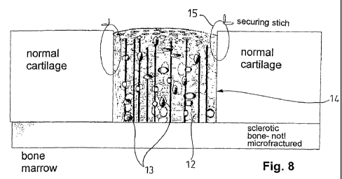
Figure 8. is the cross section of an articular cartilage provided with carti-
lage cells, after implanting and
Figure 9. is the cross section of an articular cartilage without transplanted
cells, after implanting.
DESCRIPTION OF THE PREFERRED EMBODIMENTS
Turning to Figure 1, a device for harvesting articular cartilage 1 comprises a
handle 2 provided with a sharp, curvilinear cutting blade 4 fixed in a bridge
3.
The curvature of the cutting blade 4 is adjusted to that of the joint surface
to be
harvested. At the ends of the cutting blade 4, there are spacer elements 5.
The
distance t between the edges of the spacer elements 5 and the edge of the cut-
ting blade 4 defines the depth of harvesting, i.e. the distance from the
bone/cartilage border (tidemark). This distance is in this case 0,5 mm. The
dis-
CA 02699005 2010-03-09
WO 2009/022191 PCT/HU2008/000095
tance T between the edges of the spacer elements 5 defines the width of the
harvesting.
Figure 2. illustrates another embodiment of the device for harvesting
articular
cartilage 1 according to the invention. This device also comprises a handle 2
5 with a sharp, curvilinear cutting blade 4 fixed thereon. Blade 4 is provided
with a
spacer element 5, too. This element is in this embodiment a support plate. The
distance t between the edge of the spacer element 5 and the edge of the blade
4 is in this case 0,5 mm, but can go up to 4 mm, if needed.
The thickness of the blade 4 of the device 1 according to the invention for
har-
vesting articular cartilage ranges preferably from 0,1 to 0,5 mm, and
cartilages
of rather big surfaces (6 - 10 cm2) can be harvested therewith. The steps of
har-
vesting are shown in Figures 3a - 3d.
Before implantation, the harvested articular cartilage should be provided with
incisions according to the invention, said incisions providing an indentation
on
the side of the cartilage facing the bone. The distances between the incisions
should be very small: 0,1 - 1 mm. A device 6 for producing such incisions is
shown in Figure 4 (the illustration is schematic and the proportions are not
real).
The device 6 comprises a handle 2 provided with a bridge 3 on one end, and
cutting blades 4 arranged in the bridge. The thickness of the cutting blades 4
is
0,2 mm according to this embodiment, and the distances between them is 0,4
mm.
Figure 5. shows another embodiment of the device 6 for producing incisions
(the
illustration is schematic and not scaled). Here, the cutting blades 4 in the
bridge
3 are discs arranged on a rod 7. The discs are fixed (in other embodiments
they
may be arranged rotatably) on the rod and the rod is provided with a drive 8
(preferably an electric motor). The depth of the cuts can be adjusted by legs
9
slidably arranged on the bridge 3. The legs can be fixed at the desired height
with slots 10 and nuts 11.
Other embodiments of the device 6 for producing incisions may be applied as
well. One of them may resemble to an egg cutter device: it may have a base and
then the handle 2 provided with a bridge 3 on one end, and cutting blades 4 ar-
ranged in the bridge is formed as a cutting arm tiltably connected to said
base.
CA 02699005 2010-03-09
WO 2009/022191 PCT/HU2008/000095
6
The depth of the incisions can be adjusted by changing the position of the cut-
ting blades 4 with respect to the legs 9 of the bridge 3.
For preparing the incisions, cutting arm is opened, a cartilage is arranged on
the
upper surface of the base and then the cutting arm is turned down, until legs
butt
on base.
An articular cartilage obtained in the above way is illustrated in Figures 6
and 7,
wherein Fig 6. is a top view and Fig 7. is a cross section of the cartilage.
Inci-
sions 12 produced with one embodiment of device 6 have a depth to leave an
intact layer of cartilage. The minimum thickness v of that layer is 50 pm, but
may
go up to 1000 pm. The value of v for the embodiment shown in Figures 7 and 8
is 100 pm. The incisions 12 are parallel with each other, but any other
pattern
may be used. The distances d between the incisions 12 may range from 0,1 to 1
mm, it is 0,6 mm for the embodiment shown in Figures 6 and 7.
EXAMPLES
Example 1.
Several hundred milligrams of hyaline cartilage was collected with arthroscopy
for repairing the cartilage damage of a young sportsman. The collected
cartilage
was delivered to a cell culturing laboratory.
After having obtained the required number of cells, they were suspended,
poured onto the side of the matrix provided with incision, and left for
properly
sedimenting.
The cartilage matrix had been harvested in sterile conditions from the knee
joint
of a cadaver, long before the operation, with the device shown in Figure 1.
The
matrix with a surface of 2 x 3 cm had been provided with incisions on the side
facing the bone, with the device shown in Figure 5. The incisions had been
made in two perpendicular directions, wherein the distances between the inci-
sions were 0,5 mm and the thickness of the intact collagen layer was 90 pm.
The matrix had than been provided with a sterile packing and stored on a tem-
perature of - 80 C.
The cartilage matrix obtained from a cadaver and prepared in the above
outlined
way was implanted via miniarthrotomy knee operation, as shown in Figure 9. In
the exposed knee joint, the damaged cartilage part was removed with a sharp
CA 02699005 2010-03-09
WO 2009/022191 PCT/HU2008/000095
7
spoon, up to the intact cartilage and a quadratic recipient cavity was
prepared in
the cleared surface. The graft 14 provided with cells 13 was cut to fit in the
cav-
ity and implanted in the appropriate position. It was then connected to the
edge
of the intact cartilage layer with small stitches. At last, the implant was
glued
around (sealing) with fibrin glue.
Example 2.
A patient of middle age had ankle complaints. As the result of an examination,
it
was found that he had focal cartilage defect on the upper surface of her
talus. It
was decided to perform cartilage substitution by cartilage cell
transplantation,
therefore bone marrow stem cells were collected for culturing (in cases, when
it
is not possible, joint cartilage particles may be collected for obtaining
cells). The
collected cells were delivered to a cell culturing laboratory.
Prior to the operation, cartilage sample had been harvested in sterile
conditions
from the knee joint of a cadaver, with the device shown in Figure 2. The carti-
lage had been processed with incisions on the side facing the bone, with the
device shown in Figure 5. The incisions had been made in parallel directions,
wherein the distances between the incisions were 0,8 mm and the thickness of
the intact layer was 120 pm.
The multiplied cells were centrifuged to the graft provided with incisions, to
be
captured in the incisions and were fixed therein with glue.
The cartilage had than been provided with a sterile packing and stored in
fluid
nitrogen on a temperature of - 160 C until the day of the operation, when it
was
sent to the operating room.
After having exposed the ankle joint, the cartilage defect of the talus was
cleared, the bone below was cleaned and the graft prepared and cut to proper
size and form in advance was implanted in place of the cartilage deficiency.
For
fixing, fibrin glue was applied, without stitching, on the bottom and the
sides of
the implant. Thereafter, the joint was covered and a rehabilitation protocol
of 6
weeks has been carried out with proper fractional load.
Example 3.
During arthroscopy of a women of middle age it was found that she had small
cartilage deficiency on the knee joint. Therefore, at the same time, following
a
CA 02699005 2010-03-09
WO 2009/022191 PCT/HU2008/000095
8
small joint exposure, the region of the cartilage deficiency has been cleared
up
to the healthy cartilage.
One week before the operation sound articular cartilage had been harvested in
sterile conditions from the shoulder joint of a cadaver, with the device shown
in
Figure 2. The cartilage had been provided with incisions on the side facing
the
bone. The incisions had been made in parallel directions, wherein the
distances
between the incisions were 0,1 mm and the thickness of the intact layer was
500
pm.
The cartilage had than been stored for one week in sterile conditions, without
freezing, on +4 C, until the day of the operation.
The graft was delivered to the surgeon together with the living cells therein,
for
operation. A hole was made in the bone below the cartilage (microfracture) and
the graft 14, after having been cut to proper size and form, was fixed in the
re-
gion of the cartilage deficiency, the surface provided with incisions facing
the
bone. For fixing the graft, small anchors 16 were introduced through the bone.
In this case, bone marrow cells 17 could flow to the incisions 12 through the
hole
(not shown) in the bone, and these cells produced the cellular body of the
articu-
lar cartilage by conversion to cartilage cells. The cells surviving in the
cartilage
also helped the cartilage to stick to the subchondral bone.
The drawing and the examples show, that the devices according to the invention
are simple, the use of them is safe, and they enable to prepare articular
carti-
lages of far better quality, than the ones used up to now.
The articular cartilages according to the invention offer the advantage with
re-
spect to the state of art, that the incisions considerably improve the
incorporation
of the cells cultured in laboratory or deriving from bone marrow. A further
advan-
tage is that the incisions enhance the flexibility of the cartilage and, in
this way,
the use is more simple and safe.