Note: Descriptions are shown in the official language in which they were submitted.
CA 02699451 2009-12-09
WO 2009/045581 PCT/US2008/067089
Bacteriophage with Enhanced Lytic Activity
RELATED APPLICATIONS
This application claims priority to U.S. Provisional Application 60/944,130
(filed on
June 15, 2007) which is incorporated by reference in its entirety.
GOIV'ERNMENT SUPPORT
The present invention arose in part from research funded by the Defense Threat
Reduction Agency, Department of Defense. The Government has certain rights in
the invention.
BACKGROUND OF THE INVENTION
Bacillus anthracis, a category A biothreat agent, is a spore forming Gram-
positive
bacterium of the Bacillus cereus sensu lato group. It is a zoonotic soil
bacterium that infects
animals and occasionally humans causing the disease anthrax. Bacillus
anthracis are aerobic
and spore-forming bacilli.
The notoriety of B. anthracis stems from the fact that it was successfully
used in
bioterror attacks via mail laced with anthrax spores, following the 9/11
terrorist attacks. The
prospect of biothreats using B. anthracis and the possibility of naturally
emergent or deliberately
created antibiotic resistant B. antlzracis, calls for highly integrated and
enhanced technological
platforms, capable of specifically targeting and rapidly screening for this
organism. This need is
best illustrated in case of a bacterial bioterror attack where timely
detection and intervention
with countermeasures such as antibiotic therapy are paramount in preventing
fatal consequences.
Pathology due to B. anthracis infection is primarily due to the release by the
organism
of "protective antigen" (PA) in association with lethal factor (LF) and edema
factor (EF)
(Sellman et al. (2001) Science 292: 695-7). The complete DNA and protein
sequence of PA has
been published and its three-dimensional structure is known from x-ray
crystallography (Petosa
et al. (1997) Nature 385: 833-8). The characteristics and biological functions
of the four
domains of PA are also available permitting selection of epitopes within the
domains based on
antigenic properties (Petosa et al.; Little et al. (1996) Microbiology 142:
707-15; Brossier et al.
(1999) Infect. Immun. 67: 964-7; Brossier et al. (2000) Infect. Immun. 68:
1781-6; Mogridge et
al. (2001) J. Bacteriol. 183: 2111-6). In animal studies, as well as studies
of natural human
infeetion, it was shown that individuals who survived an infection produced
antibodies to PA
suggesting its importance in protection (Brachman (1962) Am. J. Public Health
52: 632-45).
1
CA 02699451 2009-12-09
WO 2009/045581 PCT/US2008/067089
Bacillus anthracis is closely related to other members of the B. cereus group
of bacteria.
Laboratory isolates can generally be distinguished either by polymerase chain
reaction (PCR)
amplification of toxin genes and plasmids (pXOI and pXO2) and by other
clinical laboratory
analysis, especially if toxin genes are not present. An isolate of B.
anthracis typically appears as
a white or gray colony that is nonhemolytic or, at most, weakly hemolytic,
nonmotile, and is
penicillin susceptible. The ability to form capsule is also diagnostic and is
typically
demonstrated after culture on nutrient agar containing 0.7% sodium bicarbonate
incubated
overnight under CO2. Colonies of the capsulated B. anthracis appear mucoid and
the capsule
can be visualized by staining with M'Fadyean polychrome methylene blue or
India ink. An
additional important evaluation is also the susceptibility to gamma phage, a
bacteriophage.
Bacteriophages have been and still remain useful tools for bacterial species
and strain
differentiation (Hagens and Loessner (2007) Appl. Microbiol. Biotechnol.
76:513-9; McAuliffe
et al. (2007) p. 1-42. In Mc Grath and van Sinderen (eds.), Bacteriophage.
Genetics and
Molecular Biology Caister Academic Press; McKinstry and Edgar (2005) p.430-
440. In Waldor
et al. (eds.), Phages: their role in bacterial pathogenesis and biotechnology
ASM press; Petty et
al. (2007) Trends Bioteehnol. 25:7-15) although evidence for successful
application of phage
therapy is still sparse in western medicine (Sulakvelidze et al. (2001)
Antimicrob. Agents
Chemother. 45:649-59).
Recently, the inherent binding specificity and lytic action of bacteriophage
encoded
enzymes called lysins have been exploited for the rapid detection and killing
of B. anthracis
(Schuch et al. (2002) Nature 418:884-9). It was demonstrated that the PIyG
lysin, isolated from
the y phage of B. anthracis, specifically kills B. anthracis isolates and
other members of the B.
anthracis `cluster' of bacilli in vitro and in vivo. Both vegetative cells and
germinating spores
were shown to be susceptible. The lytic specificity of PlyG was also exploited
as part of a rapid
method for the identification of B. anthracis thus indicating that PlyG is a
tool for the treatment
and detection of B. anthracis (Schuch et al. (2002) Nature 418:884-9).
A well-known B. anthracis specific phage of the Tectiviridae family, AP50, was
first
isolated from soil in 1972 using B. anthracis Steme as the host (Ackermann et
al. (1978) Can. J.
Microbiol. 24:986-93; Nagy, E. (1974) Acta. Microbiol. Acad. Sci. Hung. 21:257-
63).
Originally it was thought to be an RNA phage, but later shown to contain
double stranded (ds)
DNA and phospholipid (Nagy et al. (1976) J. Gen. Virol. 32:129-32). AP50 was
also shown to
have a narrow host range; only one third of the 34 B. anthracis strains and
none of the 52 strains
belonging to 6 different Bacillus spp were susceptible to infection by AP50
(Nagy et al. (1977)
J. Gen. Microbiol. 102:215-9). Nine major structural proteins were identified
on SDS-PAGE
2
CA 02699451 2009-12-09
WO 2009/045581 PCT/US2008/067089
gels. The molecular weight of the phage DNA was estimated to be 9 X 10⁶
daltons (Nagy et al.
(1982) J. Gen. Virol 62:323-329). Treatment with organic solvents such as
chloroform (5%)
and ether (25%) for 30 minutes inactivated the phage to a survival of about 1
X 10-4 (Nagy and
Ivanovics (1982) Acta. Microbiol. Acad. Sci. Hung. 29:89-98).
Virions of the Tectiviridae family of phages possess isometric nucleocapsids
with
icosahedral symmetry and a capsid shell composed of two layers: a smooth,
rigid 3 nm thin outer
shell and a flexible, 5-6 nm thick inner lipoprotein vesicle. Virions contain
one molecule of
linear double stranded DNA with a total genome length of - 15 kb containing
inverted terminal
repeats (ITRs). A protein essential for the proposed protein primed DNA
replication process of
the phage is bound to the termini of the linear molecule (ICTV. 2002.
tnternational committee
on taxonomy of viruses-ICTVdB descriptions: 68, Tectiviridae). While phage
PRDl, infecting
Gram-negative bacteria carrying Inc P, N, W plasmids, is considered to be a
model phage for
this family (Grahn et al. (1994) J. Bacteriol. 176:3062-8; Saren et al. (2005)
J. Mol. Biol.
350:427-40), several phages belonging to this family have also been isolated
in Gram-positive
bacteria; e.g., AP50, Bam35, Gi10l, Gi116, and NSl 1(Nagy and Ivanovics (1982)
Acta.
Microbiol. Acad. Sci. Hung. 29:89-98; Ravantti et al. (2003) Virology 313:401-
14; Verheust et
al. (2005) J. Bacteriol. 187:1966-73; Verheust et al. (2003) Microbiology
149:2083-92).
Among these, phages Bam3 5, Gil01 and Gil 16 have been genetically
characterized and
their genome sequences have been determined (Ravantti et al. (2003), Verheust
et al. (2005);
Verheust et al. (2003); Stromsten et ah (2003) J. Bacteriol. 185:6985-9).
These genomes
exhibit a high degree of similarity in genetic organization to a linear
plasmid found in B. cereus
ATCC 14579, pBclinl5 (Ivanova et al. (2003) Nature 423:87-91). Unlike many
temperate
phages whose genomes are integrated into the host chromosome, some members of
this family
of phages exist as extra-chromosomal linear plasmids in the lysogenic state.
The linear ends are
protected from nucleolytic attacks by proteins (Stromsten et al. (2003)).
Although Gram-negative bacteria infecting phage PRD1 and Gram-positive
bacterium
phage Bam35 have closely related virion morphology and genome organization,
they have no
detectable sequence similarity. There is strong evidence that the Bam35 coat
protein has the
"double-barrel trimer" arrangement of PRD 1 that was first observed in
adenovirus and is
predicted to occur in other viruses with large facets. It has been suggested
that this group
includes viruses infecting very different hosts in all three domains of life:
eucarya, bacteria and
archaea suggesting a single viral lineage for this very large group of viruses
(Saren et al. (2005)
J. Mol. Biol. 350:427-40).
The standard diagnostic tests for suspected B. anthracis, recommended by the
Centers
3
CA 02699451 2009-12-09
WO 2009/045581 PCT/US2008/067089
for Disease Control and Prevention (CDC) include several procedures.
Presumptive
identification to genus level (Bacillus family of organisms) requires Gram
stain and colony
identification and presumptive identification to species level (B. antlaracis)
requires tests-for
motility, lysis by y phage, capsule production and visualization, hemolysis,
wet mount and
malachite green staining for spores. Confirmatory identification of B.
anthracis may include
lysis by y phage, capsular staining, and direct fluorescent antibody (DFA)
testing on capsule
antigen and cell wall polysaccharide. Thus, testing for 7 phage sensitivity
has been an integral
part of B. anthracis identification (CDC (2002) Center for disease control and
prevention:
Anthrax Q & A: Diagnosis). y phage exhibits a fairly narrow host range but
several B. cereus
strains (e.g., ATCC 4342) have been shown to be sensitive to infection by this
phage (Abshire et
al. (2005) J. Clin. Microbiol. 43:4780-8, Brown et al. (1955) J. Infect.
Dis.96:34-9; Davison et
al. (2005) J. Bacteriol. 187:6742-9; Schuch et al. (2002) Nature 418:884-9).
Several phages
(CP5 1, CP54 and TP2 1) isolated from B. cereus and B. thuringiensis strains
have been
successfully used for transducing chromosomal markers and plasmids between B.
anthracis
strains (Green et al. (1985) Infect Immun. 49:291-7; Ruhfel et al. (1984) J.
Bacteriol. 157:708-
11; Thome, C. B. (1968) Bacteriol. Rev. 32:358-61; 37; Walter and Aronson
(1991) Appl.
Environ. Microbiol. 57:1000-5; Yelton and Thorne (1970) J. Bacteriol. 102:573-
9). However,
their utility as B. anthracis diagnostic phages is limited because of their
broad host range.
4
CA 02699451 2009-12-09
WO 2009/045581 PCT/US2008/067089
SUMMARY OF THE INVENTION
This invention provides for an isolated Bacillus phage AP50 that has one or
more
nucleotide substitutions in the phage genome, whereby the one or more
nucleotide substitutions
increase lytic activity of the phage. The invention encompasses all 31 genes
(ORF 1-31) which
make up the genome and the proteins encoded by these genes. In addition, the
invention
provides for methods of using the phage to test for the presence of B.
anthracis.
In one embodiment of the invention, the isolated Bacillus phage AP50 has a
nucleotide
substitution at a position corresponding to nucleotide 271 of SEQ ID NO: 55
(nucleotide 271 of
ORF28). Preferably, the substitution at nucleotide 271 is a C to T
substitution. In another
embodiment, the isolated Bacillus phage AP50 has a position corresponding to
nucleotide 154
of SEQ ID NO: 63 (such as e.g. a T to C substitution).
The Bacillus phage may have the nucleotide sequence of SEQ ID NO: 63. In
another
embodiment, the Bacillus phage AP50 has the nucleotide sequence of SEQ ID NO:
6 and
nucleotide substitutions including a nucleotide substitution at a positions
corresponding to
nucleotides at position 154 and 12,881 (271 of SEQ ID NO: 55 (nucleotide 271
of ORF28)) of
SEQ ID NO: 63.
The isolated Bacillus phage AP50 according to the invention comprises various
genes
which are encoded by various open reading frames. ln one embodiment, the
Bacillus phage
genome comprises the nucleotide sequence of one or more of SEQ ID NO: 1, 3, 5,
7, 9, 11, 13,
15, 17, 19, 21, 23, 25, 27, 29, 31, 33, 35, 37, 39, 41, 43, 45, 47, 49, 51,
53, 55, 57, 59, 61 or the
complement thereof.
The isolated Bacillus phage AP50 may be part of a composition, including but
not
limited to pharmaceutical compositions, and a kit. In one embodiment, the
phage is in a
composition or kit which also contains gamma phage.
The invention further provides for nucleic acids from the isolated Bacillus
AP50 phage.
In one embodiment, the isolated nucleic acids encode protein having the amino
acid sequence
of any of SEQ ID NO: 2, 4, 6, 8, 10, 12, 14, 16, 18, 20, 22, 24, 26, 28, 30,
32, 34, 36, 38, 40,
42, 44, 46, 48, 50, 52, 56, 58, 60, or 62 (i.e. amino acid sequences of ORF1
to ORF31). In
another embodiment, the nucleic acids comprises any of SEQ ID NO: 1, 3, 5, 7,
9, 11, 13, 15,
17, 19, 21, 23, 25, 27, 29, 31, 33, 35, 37, 39, 41, 43, 45, 47, 49, 51, 53,
55, 57, 59, or 61. In yet
another embodiment of the invention, the isolated nucleic acid has at least
85% sequence
identity to any of SEQ ID NO: 1, 3, 5, 7, 9, 11, 13, 15, 17, 19, 21, 23, 25,
27, 29, 31, 33, 35, 37,
39, 41, 43, 45, 47, 49, 51, 53, 55, 57, 59, or 61. In an alternate embodiment,
the isolated nucleic
acid contains SEQ ID NO: 63. The invention also provides for recombinant
phages comprising
5
CA 02699451 2009-12-09
WO 2009/045581 PCT/US2008/067089
any of the nucleic acids. In a preferred embodiment, the recombinant phage
comprises SEQ ID
NO: 63.
The invention further provides for isolated proteins from an isolated Bacillus
phage
AP50 that has one or more nucleotide substitutions in the phage genome,
whereby the one or
more nucleotide substitutions increase lytic activity of the phage. In one
embodiment, the
isolated proteins comprises the amino acid sequence of any of 2, 4, 6, 8, 10,
12, 14, 16, 18, 20,
22, 24, 26, 28, 30, 32, 34, 36, 38, 40, 42, 44, 46, 48, 50, 52, 56, 58, 60, or
62 (i.e. the amino
acid sequences of ORFl to ORF3 1). In another embodiment of the invention, the
isolated
protein has at least 85% sequence identity to any of SEQ ID NO: 2, 4, 6, 8,
10, 12, 14, 16, 18,
20, 22, 24, 26, 28, 30, 32, 34, 36, 38, 40, 42, 44, 46, 48, 50, 52, 56, 58,
60, or 62.
The invention also provides for methods of detecting the presence of B.
anthracis. One
embodiment of the invention is a method for detecting the presence of B.
anthracis in a subject
that has at least the steps of (a) isolating a biological sample from the
subject, (b) contacting a
sample with a phage according to the invention (i.e. Bacillus phage AP50 that
has one or more
nucleotide substitutions in the phage genome, whereby the one or more
nucleotide substitutions
increase lytic activity of the phage) and (c) detecting for the presence of
bacterial lysis. In this
method, the increased presence of bacterial lysis compared to a control
indicates the presence of
B. anthracis in the sample. The step of isolating the biological sample may
also encompass
incubating biological sample under conditions sufficient to induce growth of
B. anthracis. In
one embodiment, the control is a sample which does not contain B. anthracis.
In another
embodiment, the contacting is carried out under conditions sufficient to
induce phage lysis of B.
anthracis. The method may also further comprise contacting the biological
sample with gamma
phage prior to detecting for the presence of bacterial lysis.
BRIEF DESCRIPTION OF THE DRAWINGS
The foregoing summary, as well as the following detailed description of the
invention,
will be better understood when read in conjunction with the appended figures.
For the purpose
of illustrating the invention, shown in the figures are embodiments of the
present invention. It
should be understood, however, that the invention is not limited to the
precise arrangements,
examples and instrumentalities shown.
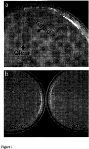
Figure 1 shows the plaque morphology of (a) mixed lysate and (b) AP50c plaques
after
overnight incubation at room temperature and at 37C.
Figure 2 shows Transmission electron micrographs of AP50 phage particles.
Figure 2A
shows Uranyl acetate staining at a magnification of 297K. Figures 2B and 2C
show AP50 after
6
CA 02699451 2009-12-09
WO 2009/045581 PCT/US2008/067089
phosphotungstate staining at a magnification of 297K. Specifically, Figures 2B
and 2C show
damaged particles (chloroform treatment) after removal of the protein capsid.
The inner
lipoprotein vesicles and a tail-like tube derived from this vesicle are seen.
The scale bar in the
figures is 100 nrii.
Figure 3 shows various features of the AP50 genome. Figure 3A shows the genome
map of AP50. Three clusters of genes based on functional grouping and
similarities to other
tectiviral phages are shown. ORF boxes are color coded to indicate the degree
of amino acid
identity with proteins of other tectiviral phages. The ORFs have between <15%
to 80% amino
acid identity with proteins of other tectiviral phages. ITR: inverted terminal
repeat; HVR: highly
variable region. Open arrow heads indicate the locations of the mutations in
AP50c phages.
Figure 3B shows a visualization summary of whole-genome nucleotide alignments
of Gram-
positive tectiviral phages. The ClustalW alignment file generated from
multifasta alignment was
visualized in Base by Base (Brodie et al. 2004, BMC Bioinformatics 5:96) In
this type of
alignment, if two sequences have insertions or deletions relative to one
another, the output looks
different depending on which of the two sequences is used as the base
sequence. White, perfect
nucleotide homology; blue, SNP; red, deletions in the indicated phage; green,
insertions in the
indicated phage. The genbank accession numbers for the sequences used in the
alignment are:
Bam35c (NC_005258), pBth35646 (NZ AAJM00000000), Gil01 (AJ536073), Gill6c
(AY701338), AP50 (EU408779), pBclinl5 (AE01878). Figure 3C shows the sequence
changes
in AP50c and AP50t genomes. The mutation in the non coding region just
upstream of ORF-1
at nt position 164 is indicated. The second mutation is in ORF 28 at position
12,881 and
changes the amino acid residue 91 (an isoleucine in AP50c to a valine in
AP50t).
Figure 4 shows ClustalW alignment of amino acid of ORF31 with similar ORFs in
Gi116c (ORF31), Bam35 (ORF31) and pBClinl5 (ORF28) genomes.
Figure 5 shows the colony morphologies of B. anthracis Steme strain 34F2 after
infection with AP50c or AP50t. Figure 5A shows uninfected 34F, cells diluted
and plated on
phage assay agar plates. Figure 5B shows AP50t infected culture, diluted and
plated; Figures
5C and 5D show AP50 t and AP50c infected cultures, respectively, plated on
phage assay agar
plates.
Figure 6 shows the morphology of AP50c resistant 34F2 mutants. Figure 6A
depicts
logarithmically grown cultures were incubated statically at room temperature
overnight. Wild
type 34F,, cells settled at the bottom of the culture tube as a pellet and the
AP50R mutant
contained a viscous material which prevented cell settling at the bottom of
the tube. Figure 6B
is a scanning electron micrographs of wild type 34F, infected with AP50. The
arrows indicate
7
CA 02699451 2009-12-09
WO 2009/045581 PCT/US2008/067089
the AP50 particels attached to the outer surface of the bacterium. Figure 6C
is a scanning
electron micrograph of 34F, AP50R mutant infected with AP50 showing the
presence of
polysaccharide material coating the outer cell surface and absence of attached
phage particles.
DETAILED DESCRIPTION
General Description
The inventors have isolated and characterized the genome of a B. anthraeis
specific
phage of the Tectiviridae family, AP50 (herein after referred to as "AP50
phage" throughout the
specification and claims). Thus, the invention encompasses a1131 genes (ORF1-
31) which
make up the genome and the proteins encoded by these genes. In addition, the
invention
encompasses a variant of AP50 which exhibits increased lytic activity.
The present invention provides AP50 phages or parts thereof that inhibit
growth of
target bacteria (e.g., B. anthracis) because of their increased bacterio-lytic
properties. The
phages are thus useful for inhibiting bacterial growth or presence in the
environment and for
treating bacterial infection in a subject in need of such treatment. In some
embodiments, the
AP50 phage are unable to replicate in a target bacteria and yet inhibit the
growth of the target
bacteria, they can be administered as a defmed dose therapeutic composition
for treatment of
bacterial infections. This provides substantial regulatory advantages, which
prevent changing
stoichiometric ratios of treatment and target entities as the bacterial
infection and bacteriophage
replication processes progress.
This invention provides that, for each pathogenic bacteria target (e.g., B.
anthracis),
phage from the Tectiviridae family, including AP50, will be useful as a defmed
dose therapeutic
agent to inhibit growth of or kill B. antlaracis.
Unless defmed otherwise, all technical and scientific terms used herein have
the same
meaning as commonly understood by one of ordinary skill in the art to which
this invention
belongs. Although any methods and materials similar or equivalent to those
described herein
can be used in the practice or testing of the present invention, the preferred
methods and
materials are described.
AP50 Phage with Enhanced Lytic Activity
As used herein "bacteriophage" is generally shortened to "phage" as is well
known in
the art. Baeteriophage typically refers to a functional phage, but in many
contexts herein may
refer to a part thereof, generally exhibiting a particular function. The AP50
phage is modified as
such to have enhanced and/or increased lytic properties. In some
circumstances, the term may
also refer to portions thereof, including, e.g., a head portion, or an
assembly of components
8
CA 02699451 2009-12-09
WO 2009/045581 PCT/US2008/067089
which provide substantially the same functional activity. The portion may be a
physical
fragment of an intact phage, a selected product from normal or abnormal
assembly of phage
parts, or even an artificial or recombinant construct, e.g., from genetic
manipulation of genes
encoding (1) phage parts, (2) critical phage assembly components, or even (3)
associated host
genes which may be useful in ensuring phage replication or production. When
referring to a
phage genome, typically the term refers to a naturally occurring phage genome
as set forth in
SEQ 1T) NO: 63, but may include fragments, artificial constructs, mutagenized
genoines
including those found in AP50c, selected genomes, and "prophage" sequences,
which are
considered to be "defective" genomes which may have had segments deleted,
inserted, or
otherwise affected to disrupt normal genome function.
Typically, phage will be morphologically identifiable, having a size which is
resolvable
by imaging methods, e.g., electron microscopy. See, e.g., Ackermann and Nguyen
(1983) Appl.
Environ. Microbiol. 45:1049-1059.
An "AP50 phage" is a phage or phage-based construct (e.g., a phage tail, tail
fragment,
phage protein, or ghost phage) that inhibits the growth, survival, or
replication of the target
bacterium (e.g., B. anthracis). ln some embodiments, the AP50 phage contains
one or more
mutations in its genome which enhance or increase lytic activity, including
but not limited to,
one or more nucleotide substitutions is at a position corresponding to
nucleotide 271 of SEQ ID
NO: 55 (i.e. nucleotide 271 of ORF 28) and/or a position corresponding to
nucleotide 154 of
SEQ ID NO: 63. In some embodiments, the AP50 phage is AP5Oc. Thus, an AP50
phage can
include a portion of a phage that can be used to inhibit growth of the target
bacterium. For
example, an AP50 phage can be a portion of an intact phage that can be
produced in a non-target
bacteria. Thus, as defmed herein, an AP50 phage can include a structural
portion of an intact
phage, e.g., a tail portion of a tailed phage; or an isolated protein
component of an intact phage.
These phage-based compositions include one or more proteins or protein domains
derived from
a natural or engineered bact,eriophage. In some embodiments, the AP50 phage is
unable to
replicate, DNA or the phage itself, or assemble in a target bacterium, but
nonetheless is capable
of infecting the target bacterium so as to inhibit the growth, survival, or
replication of the target
bacterium.
The term "recombinant" when used with reference, e.g., to a cell, or nucleic
acid,
protein, or vector, indicates that the cell, nucleic acid, protein or vector,
has been modified by
the introduction of a heterologous nucleic acid or protein or the alteration
of a native nucleic
acid or protein, or that the cell is derived from a cell so modified. Thus,
e.g., recombinant cells
express genes that are not found within the native (non-recombinant) form of
the cell or express
9
CA 02699451 2009-12-09
WO 2009/045581 PCT/US2008/067089
native genes that are otherwise abnormally expressed, under expressed, or not
expressed at all.
Certain embodiments of anti-bacterial phage include constructs which contain
less than
about 70, 50, 20, 5, 2, 1, 0.1 percent, or less of the parental phage nucleic
acid content. The
content may be either mass, or informational content, e.g., where some portion
of the
informational content is deleted.
As used herein, "target bacterium" or "target bacteria" refer to B. anthr-acis
bacterium or
bacteria whose growth, survival, or replication is inhibited by an AP50 phage.
"Growth
inhibition" can refer, e.g., to slowing of the rate of bacterial cell
division, or cessation of
bacterial cell division, and./or to death of the bacteria due to lysis by AP50
phage. In a typical
embodiment, the "target bacterium" or "target bacteria" are pathogenic forms
of B. anthracis.
Examples of B. anthracis include, but are not limited to, the strains listed
in Table 1 below and
substrains thereof.
Table 1: Exemplary B. anthracis strains
B. anthracis Strain Source Comments AP50 Sens. Gamma Sens.
ASC 004 Strain M36; used in vaccine Yes
research, U. K
ASC 006 Vollum 3b type strain. U.K. Yes
ASC 010 NCTC 2620. China. Yes
ASC 016 ATCC 937 Yes
ASC 025 U.K. bovine case presumed to be Yes Yes
caused by contaminated material
from Senegal.
ASC 027 U.K. bovine case presumed to be Yes Yes
caused by contaminated material
from Senegal.
ASC 031 U.K. bovine case presumed to be Yes Yes
caused by contaminated material
from Senegal.
ASC 032 Penicillin-resistant fatal human Yes Yes
case. U. K.
ASC 038 Fatal human case. U.K. Yes Yes
ASC 050 Zimbabwe (Human cutaneous Yes
isolate).
ASC 054 Zimbabwe (Human cutaneous Yes
isolate). Phage resistant.
ASC 061 Zebra. Etosha National Park. Yes
Namibia.
ASC 069 Human isolate. New Hampshire, Yes
U.S.A.
ASC 070 Penicillin resistant. Yes Yes
ASC 073 Zebra. Etosha N. P. Namibia. Yes Yes
ASC 074 ' Vulture feces, Etosha NP, Namibia. Yes
CA 02699451 2009-12-09
WO 2009/045581 PCT/US2008/067089
B. anthracis Strain Source Comments AP50 Sens. Gamma Sens.
ASC 120 Australia. by MLVA Yes
ASC 131 Elephant skull. Zambia. Yes
ASC 152 Giraffe bone. Namibia. Yes Yes
ASC 158 Zebra. Etosha NP Namibia Yes,No No
ASC 159 Ames. Guinea pig re-isolate from Yes
vaccine challenge studies. U.K.
ASC 161 Ames. Guinea pig re-isolate from Yes Yes
vaccine challenge studies. U.K.
ASC 165 Ames. Guinea pig re-isolate from Yes Yes
vaccine challenge studies. U.K.
ASC 206 Kruger N. P. South Africa. Yes
ASC 254 Environmental isolate. U.K. Yes
Believed to be more than 100 years
old.
ASC 285 Environmental isolate. U.K. Yes Yes
Believed to be more than 100 years
old.
ASC 330 Ames re-isolate. U.K. Yes
ASC 386 Ames re-isolate with Yes
uncharacteristic colony morphology.
U.K.
ASC 394 Ames re-isolate from guinea pig Yes
which died despite ciprofloxacin
treatment. U.K.
ASC 398 Ames re-isolate from guinea pig Yes
which died despite doxycycline
treatment. U.K.
BDRD 01 Unknown A0089 strain Yes
A 0034 Bovine. China. Yes Yes
A 0039 Bovine. Australia. Yes Yes
A 0149 Human cutaneous isolate. Turkey. Yes Yes
A 0158 Bovine. Zambia. Yes Yes
A 0174 Canada Yes Yes
A 0188 Zebra. Etosha N.P. Namibia. Yes Yes
A 0248 Human. U.S. Yes Yes
A 0256 Human. Turkey. Yes
A 0264 Human. Turkey. Yes Yes
A 0267 Bovine. U.S.A. Yes Yes
A 0293 Sheep. Ital . Yes Yes
A 0328 Pi . German . Yes Yes
A 0376 Bovine. U.S.A. Yes
A 0379 Wool. Pakistan. Yes
A 0419 South Korea (fatal human case). Yes Yes
A 0442 Kudu, Kru er N.P. South Africa. Yes No
A 0462 Ames Guinea pig re-isolate from Yes
vaccine challenge studies (Porton
Down U. K).
A 0463 Sheep. Pakistan. Yes No
A 0465 U.K. (Vollum). Yes
11
CA 02699451 2009-12-09
WO 2009/045581 PCT/US2008/067089
B. anthracis Strain Source Comments AP50 Sens. Gamma Sens.
A 0489 Bovine. Argentina. Yes
ASC 008 PCT NCTC 109 (Paddington IV) Yes
ASC 009 PCT NCTC 1328 Yes Yes
ASC 018 PCT 958G Yes Yes
ASC 019 PCT 961G Yes
ASC 020 PCT 1012G Yes
ASC 023 PCT 1011 G Yes
ASC 024 PCT NP9 Yes
ASC 026 PCT A73/77 Yes Yes
ASC 028 PCT A187/78 Yes
ASC 030 PCT A191/78 Yes Yes
ASC 033 PCT C164G Yes Yes
ASC 035 PCT C11G Yes
ASC 036 PCT C129 G Yes Yes
ASC 040 PCT M84 Yes
ASC 042 PCT Denmark 79 Yes
ASC 046 PCT St2 Yes
ASC 063 PCT Etosha 86 Yes Yes
ASC 078 PCT Q78 Yes
ASC 080 PCT L9 (1) Yes Yes
ASC 091 PCT ATX 881017002 Yes Yes
ASC 127 PCT S6U1 Yes
ASC 149 PCT CT1264/07/88 Yes No
ASC 150 PCT AM1260/7/88 Yes
ASC 187 PCT F2909/90 Yes
ASC 193 PCT Landke V 13 Yes
ASC 209 PCT RNL 440 Yes
ASC 212 PCT RNL 443 Yes
ASC 214 PCT RNL 446 Yes
ASC 228 PCT Landkey 04 Yes
ASC 236 PCT Landke R2I4 Yes
ASC 239 PCT E side North Kings Cross Yes
ASC 267 PCT Landkey sample 3 Yes Yes ASC 278 PCT C300 Yes
ASC 279 PCT C313 Yes
ASC 296 PCT C055 Yes
ASC 301 PCT C061/93 Yes
ASC 306 PCT C317 Yes
ASC 308 PCT C323 Yes
I ASC 309 PCT C325 Yes
ASC 310 PCT M8Y 040892 Yes
ASC 318 PCT DSM A74 Yes
ASC 336 PCT F Yes
ASC 338 PCT I Yes
ASC 339 PCT J Yes
ASC 340 PCT L Yes
ASC 354 PCT S 10 Yes
ASC 362 PCT 93/37 Yes Yes
12
CA 02699451 2009-12-09
WO 2009/045581 PCT/US2008/067089
B. anthracis Strain Source Comments AP50 Sens. Gamma Sens.
ASC 363 PCT 92/150 Yes Yes
ASC 369 PCT 92/123 Yes
ASC 373 PCT London 3 Yes
ASC 391 PCT AN 32/94 Yes
ASC 411 PCT 95/126 Yes Yes
As used herein, "host bacterium" or "host bacteria" refer to a bacterium or
bacteria used
to produce, replicate, or amplify a phage, sometimes referred to as a parental
phage, that is used
to produce an anti-bacterial phage. Host bacteria or bacterium are also
referred to as "host
production bacterium" or "host production bacteria" throughout. One example of
a host
bacterium is B. anthracis Sterne strain 34F, (pXO1' pXO2-). In one embodiment,
the parental
phage is a prophage, e.g., a defective or incomplete phage genome. Often the
host production
culture complements a defect in the phage, or suppresses a destructive
function encoded in the
phage. In other embodiments, the host production culture may make use of a
helper phage to
effect the capability.
AP50 phage can also include phage that comprise a mutation and cannot
efficiently
assemble into a replication competent phage in the target bacteria. Mutations
can include
mutations in genes that encode enzymes for replication of nucleic acids or
genes that encode
regulators of replication; or in genes that encode structural components of a
phage or genes that
encode regulators of the synthesis of structural components, or genes that
encode proteins
critical for assembly, e.g., assembly functions, or genes that regulate
stoichiometry of proteins
necessary for proper assembly. The mutations can be in the coding region of a
gene or in a
regulatory region of the gene, e.g., a promoter.
Nucleic Acid Molecules
The present invention further provides nucleic acid molecules that encode any
of the
proteins having SEQ ID NO: 2, 4, 6, 8, 10, 12, 14, 16, 18, 20, 22, 24, 26, 28,
30, 32, 34, 36, 38,
40, 42, 44, 46, 48, 50, 52, 56, 58, 60, 62 (herein after referred to as a
"phage protein") and the
related proteins herein described, preferably in isolated form. As used
herein, "nucleic acid" is
defmed as RNA or DNA that encodes a protein or peptide as defmed above, is
complementary
to a nucleic acid sequence encoding such peptides, hybridizes to any of SEQ ID
NO: 1, 3, 5, 7,
9, 11, 13, 15, 17, 19, 21, 23, 25, 27, 29, 31, 33, 35, 37, 39, 41, 43, 45, 47,
49, 51, 53, 55, 57, 59,
61 (herein referred to as a "phage nucleic acid" and ORF1, ORF2, ORF3, ORF4,
ORF5, ORF6,
ORF7, ORF8, ORF9, ORF10, ORFI 1, ORF12, ORF13, ORF14, ORF15, ORF16, ORF17,
ORF18, ORF19, ORF20, ORF21, ORF22, ORF23, ORF24, ORF25, ORF26, ORF27, ORF28,
13
CA 02699451 2009-12-09
WO 2009/045581 PCT/US2008/067089
ORF29, ORF30 and ORF3 1, respectively) across the open reading frame under
appropriate
stringency conditions, or encodes a polypeptide that shares at least about 85,
86, 87, 88, 89, 90,
91, 92, 93, 94, 95, 96, 97, 98, 99 or 100% sequence identity, with the entire
contiguous amino
acid sequence of any one of the phage proteins.
The "nucleic acids" of the invention further include nucleic acid molecules
that share at
least about 85, 86, 87, 88, 89, 90, 91, 92, 93, 94, 95, 96, 97, 98, 99 or 100%
sequence identity
with the nucleotide sequence of any of the phage nucleic acids, particularly
across the open
reading frame. Specifically contemplated are genomic DNA, cDNA, mRNA and
antisense
molecules, as well as nucleic acids based on alternative backbones or
including alternative bases
whether derived from natural sources or synthesized. Such nucleic acids,
however, are defmed
further as being novel and unobvious over any prior art nucleic acid including
that which
encodes, hybridizes under appropriate stringency conditions, or is
complementary to nucleic acid
encoding a protein according to the present invention.
Homology or identity at the nucleotide or amino acid sequence level is
determined by
BLAST (Basic Local Alignment Search Tool) analysis using the algorithm
employed by the
programs blastp, blastn, blastx, tblastn and tblastx (Altschul et al. (1997)
Nucleic Acids Res.
25, 3389-3402 and Karlin et al. (1990) Proc. Natl. Acad. Sci. USA 87, 2264-
2268, both fully
incorporated by reference) which are tailored for sequence similarity
searching. The approach
used by the BLAST program is to first consider similar segments, with and
without gaps,
between a query sequence and a database sequence, then to evaluate the
statistical significance
of all matches that are identified and fmally to summarize only those matches
which satisfy a
preselected threshold of significance. For a discussion of basic issues in
similarity searching of
sequence databases, see Altschul et al. (1994) Nature Genetics 6, 119-129
which is fully
incorporated by reference. The search parameters for histogram, descriptions,
alignments,
expect (i.e., the statistical significance threshold for reporting matches
against database
sequences), cutoff, matrix and filter (low complexity) are at the default
settings. The default
scoring matrix used by blastp, blastx, tblastn, and tblastx is the BLOSUM62
matrix (Henikoff et
al. (1992) Proe. Natl. Acad. Sci. USA 89, 10915-10919, fully incorporated by
reference),
recommended for query sequences over 85 in length (nucleotide bases or amino
acids).
For blastn, the scoring matrix is set by the ratios of M (i.e. , the reward
score for a pair
of matching residues) to N (i.e., the penalty score for mismatching residues),
wherein the default
values for M and N are +5 and -4, respectively. Four blastn parameters were
adjusted as
follows: Q=10 (gap creation penalty); R=10 (gap extension penalty); wink=l
(generates word
hits at every wink`h position along the query); and gapw=16 (sets the window
width within
14
CA 02699451 2009-12-09
WO 2009/045581 PCT/US2008/067089
which gapped alignments are generated). The equivalent Blastp parameter
settings were Q=9;
R=2; wink=l; and gapw=32. A Bestfit comparison between sequences, available in
the GCG
package version 10.0, uses DNA parameters GAP=50 (gap creation penalty) and
LEN=3 (gap
extension penalty) and the equivalent settings in protein comparisons are
GAP=8 and LEN=2.
"Stringent conditions" are those that (1) employ low ionic strength and high
temperature
for washing, for example, 0.015 M NaCL'0.0015 M sodium citrate/Q 1% SDS at 50
C, or (2)
employ during hybridization a denaturing agent such as formamide, for example,
50% (voL!vol)
formamide with 0.1 lo bovine serum albumin; 0.1 /a Ficoll10.1 lo
polyvinylpyrrolidone/50 mM
sodium phosphate buffer (pH 6.5) with 750 mM NaCl, 75 mM sodium citrate at 42
C. Another
example is hybridization in 50% formamide, 5x SSC (0.75 M NaC1, 0.075 M sodium
citrate),
50 mM sodium phosphate (pH 6.8), 0.1 lo sodium pyrophosphate, 5x Denhardt's
solution,
sonicated salmon sperm DNA (50 g/ml), 0.1% SDS, and 10% dextran sulfate at 42
C, with
washes at 42 C in 0.2x SSC and 0.1% SDS. A skilled artisan can readily
determine and vary
the stringency conditions appropriately to obtain a clear and detectable
hybridization signal.
Preferred molecules are those that hybridize under the above conditions to the
complement of
any of the phage nucleic acids and which encode a functional protein. Even
more preferred
hybridizing molecules are those that hybridize under the above conditions to
the complement
strand of the open reading frame of any of the phage nucleic acids.
As used herein, a nucleic acid molecule is said to be "isolated" when the
nucleic acid
molecule is substantially separated from contaminant nucleic acid molecules
encoding other
polypeptides.
The present invention further provides fragments of the encoding nucleic acid
molecule.
As used herein, a fragment of an encoding nucleic acid molecule refers to a
small portion of the
entire protein coding sequence. The size of the fragment will be determined by
the intended use.
For example, if the fragment is chosen so as to encode an active portion of
the protein, the
fragment will need to be large enough to encode the functional regions of the
protein. For
instance, fragments which encode peptides corresponding to predicted antigenic
regions may be
prepared. If the fragment is to be used as a nucleic acid probe or PCR primer,
then the fragment
length is chosen so as to obtain a relatively small number of false positives
during
probing/priming.
Fragments of the encoding nucleic acid molecules of the present invention
(i.e.,
synthetic oligonucleotides) that are used as probes or specific primers for
the polymerase chain
reaction (PCR), or to synthesize gene sequences encoding proteins of the
invention, can easily
be synthesized by chemical techniques, for example, the phosphotriester method
of Matteucci et
CA 02699451 2009-12-09
WO 2009/045581 PCT/US2008/067089
al. (1981) J. Am. Chem. Soc. 103, 3185-3191 or using automated synthesis
methods. Examples
of such probes or primers include, but are not limited to, any of SEQ ID NO:
64 to 133. In
addition, larger DNA segments can readily be prepared by well known methods,
such as
synthesis of a group of oligonucleotides that define various modular segments
of the gene,
followed by ligation of oligonucleotides to build the complete modified gene.
In a preferred
embodiment, the nucleic acid molecule of the present invention contains a
contiguous open
reading frame of at least about three-thousand and forty-five nucleotides.
The encoding nucleic acid molecules of the present invention may further be
modified
so as to contain a detectable label for diagnostic and probe purposes. A
variety of such labels
are known in the art and can readily be employed with the encoding molecules
herein described.
Suitable labels include, but are not limited to, biotin, radiolabeled
nucleotides, and the like. A
skilled artisan can readily employ any such label to obtain labeled variants
of the nucleic acid
molecules of the invention. Modifications to the primary structure itself by
deletion, addition, or
alteration of the amino acids incorporated into the protein sequence during
translation can be
made without destroying the activity of the protein. Such substitutions or
other alterations result
in proteins having an amino acid sequence encoded by a nucleic acid falling
within the
contemplated scope of the present invention.
The invention also encompasses oligonucleotides which hybridize to any region
of a
phage nucleic acid or the AP50 phage genome, including any of SEQ ID NO: 1, 3,
5, 7, 9, 11,
13, 15, 17, 19, 21, 23, 25, 27, 29, 31, 33, 35, 37, 39, 41, 43, 45, 47, 49,
51, 53, 55, 57, 59, 61,
or 134. The invention encompasses synthetic oligonucleotides having chemical
modifications
from native nucleic acids, or nucleic acid constructs that express such anti-
sense molecules as
RNA. The oligonucleotide sequence can be complementary to the phage nucleic
acids.
Oligonucleotides will generally be at least about 7, 8, 9, 10, 11, 12, 13, 14,
15, 16, 17,
18, 19, 20, 21, 22, 23, 24, 25, 26, 27, 28, 29, 30, 31, 32, 33, 34, 35, 36,
37, 38, 39, 40, 41, 42,
43, 44, 45, 46, 47, 48, 49, 50 or more nucleotides. Typical oligonucleotides
are usually not
more than about 500, more usually not more than about 50, and even more
usually not more than
about 35 nucleotides in length, where the length is governed by efficiency of
inhibition,
specificity, including absence of cross-reactivity, and the like. It has been
found that short
oligonucleotides, of from seven to eight bases in length, can be strong and
selective inhibitors of
gene expression (see Wagner et al. (1996) Nat. Biotech. 14, 840-844).
Oligonucleotides may be chemically synthesized by methods known in the art
(see
Wagner et al. (1996) Nat. Biotech. 14, 840-844). Oligonucleotides of the
invention can be
chemically modified from the native phosphodiester structure, in order to
increase their
16
CA 02699451 2009-12-09
WO 2009/045581 PCT/US2008/067089
intracellular stability and binding affmity. A number of such modifications
have been described
in the literature, which alter the chemistry of the backbone, sugars, or
heterocyclic bases.
Recombinant DNA Containing a Phage Nucleic Acid
The present invention further provides recombinant DNA molecules (rDNAs) that
contain a phage nucleic acid coding sequence. As used herein, a rDNA molecule
is a DNA
molecule that has been subjected to molecular manipulation in situ. Methods
for generating
rDNA molecules are well known in the art, for example, see Sambrook et al.
(2005) Molecular
Cloning - A Laboratory Manual, Cold Spring Harbor Laboratory Press. In the
preferred rDNA
molecules, a coding DNA sequence is operably linked to expression control
sequences andjor
vector sequences.
The choice of vector andlor expression control sequences to which one of the
protein
family encoding sequences of the present invention is operably linked depends
directly, as is
well known in the art, on the functional properties desired, e.g., protein
expression, and the host
cell to be transformed. A vector contemplated by the present invention is at
least capable of
directing the replication or insertion into the host chromosome, and
preferably also expression,
of the structural gene included in the rDNA molecule.
Expression control elements that are used for regulating the expression of an
operably
linked protein encoding sequence are known in the art and include, but are not
limited to,
inducible promoters, constitutive promoters, secretion signals, and other
regulatory elements.
Preferably, the inducible promoter is readily controlled, such as being
responsive to a nutrient in
the host cell's medium.
In one embodiment, the vector containing a coding nucleic acid molecule will
include a
prokaryotic replicon, i.e., a DNA sequence having the ability to direct
autonomous replication
and maintenance of the recombinant DNA molecule extrachromosomally in a
prokaryotic host
cell, such as a bacterial host cell, transformed therewith. Such replicons are
well known in the
art. In addition, vectors that include a prokaryotic replicon may also include
a gene whose
expression confers a detectable marker such as a drug resistance. Typical
bacterial drug
resistance genes are those that confer resistance to ampicillin or
tetracycline.
Vectors that include a prokaryotic replicon can further include a prokaryotic
or
bacteriophage promoter capable of directing the expression (transcription and
translation) of the
coding gene sequences in a bacterial host cell, such as B. anthracis Sterne
strain 34F2 (pXO1
pXO2-). A promoter is an expression control element formed by a DNA sequence
that permits
binding of RNA polymerase and transcription to occur. Promoter sequences
compatible with
17
CA 02699451 2009-12-09
WO 2009/045581 PCT/US2008/067089
bacterial hosts are typically provided in plasmid vectors containing
convenient restriction sites
for insertion of a DNA segment of the present invention.
Any prokaryotic host can be used to express a rDNA molecule encoding a protein
of the
invention. The preferred prokaryotic host is E. coli.
Transformation of appropriate cell hosts with a rDNA molecule of the present
invention
is accomplished by well known methods that typically depend on the type of
vector used and
host system employed. With regard to transformation of prokaryotic host cells,
electroporation
and salt treatment methods are typically employed, see, for example, Sambrook
et al. (2005)
Molecular Cloning - A Laboratory Manual, Cold Spring Harbor Laboratory Press.
With regard
to transformation of vertebrate cells with vectors containing rDNAs,
electroporation, cationic
lipid or salt treatment methods are typically employed, see, for example,
Graham et al. (1973)
Virol. 52, 456; Wigler et al. (1979) Proc. Natl. Acad. Sci. USA 76, 1373-1376.
Successfully transformed cells, i.e., cells that contain a rDNA molecule of
the present
invention, can be identified by well known techniques including the selection
for a selectable
marker. For example, cells resulting from the introduction of an rDNA of the
present invention
can be cloned to produce single colonies. Cells from those colonies can be
harvested, lysed and
their DNA content examined for the presence of the rDNA using a method such as
that
described by Southern (1975) J. Mol. Biol. 98, 503-504 or Berent et al. (1985)
Biotech. 3, 208-
209 or the proteins produced from the cell assayed via an immunological
method.
Production of Recombinant Proteins
The present invention further provides methods for producing a phage protein
of the
invention using nucleic acid molecules herein described. In general terms, the
production of a
recombinant form of a phage protein typically involves the following steps:
A nucleic acid molecule is first obtained that encodes a phage protein of the
invention,
such as a nucleic acid molecule comprising, consisting essentially of or
consisting of SEQ ID
NO: 1, 3, 5, 7, 9, 11, 13, 15, 17, 19, 21, 23, 25, 27, 29, 31, 33, 35, 37, 39,
41, 43, 45, 47, 49, 51,
53, 55, 57, 59, 61. If the encoding sequence is uninterrupted by introns, as
is this open reading
frame, it is directly suitable for expression in any host.
The nucleic acid molecule is then preferably placed in operable linkage with
suitable
control sequences, as described above, to form an expression unit containing
the protein open
reading frame. The expression unit is used to transform a suitable host and
the transformed host
is cultured under conditions that allow the production of the recombinant
protein. Optionally
the recombinant protein is isolated from the medium or from the cells;
recovery and purification
18
CA 02699451 2009-12-09
WO 2009/045581 PCT/US2008/067089
of the protein may not be necessary in some instances where some impurities
may be tolerated.
Each of the foregoing steps can be done in a variety of ways. For example, the
desired
coding sequences may be obtained from genomic fragments and used directly in
appropriate
hosts. The construction of expression vectors that are operable in a variety
of hosts is
accomplished using appropriate replicons and control sequences, as set forth
above. The control
sequences, expression vectors, and transformation methods are dependent on the
type of host
cell used to express the gene and were discussed in detail earlier. Suitable
restriction sites can, if
not normally available, be added to the ends of the coding sequence so as to
provide an
excisable gene to insert into these vectors. A skilled artisan can readily
adapt any
host/expression system known in the art for use with the nucleic acid
molecules of the invention
to produce recombinant protein.
The AP50 Phaze Proteins
The present invention provides isolated proteins, allelic variants of the
proteins, and
conservative amino acid substitutions of the protein comprising the amino acid
sequence of any
of SEQ ID NO: 2, 4, 6, 8, 10, 12, 14, 16, 18, 20, 22, 24, 26, 28, 30, 32, 34,
36, 38, 40, 42, 44,
46, 48, 50, 52, 54, 56, 58, 60 and 62. As used herein, the "protein" or
"polypeptide" refers, in
part, to a protein that has the amino acid sequence depicted in SEQ ID NO: 2,
4, 6, 8, 10, 12, 14,
16, 18, 20, 22, 24, 26, 28, 30, 32, 34, 36, 38, 40, 42, 44, 46, 48, 50, 52,
54, 56, 58, 60 and 62.
The terms also refer to naturally occurring allelic variants and proteins that
have a slightly
different amino acid sequence than that specifically recited above. Allelic
variants, though
possessing a slightly different amino acid sequence than those recited above,
will still have the
same or similar biological functions associated with these proteins. The
methods used to
identify and isolate other members of the family of proteins related to these
proteins are
described below.
The proteins of the present invention are preferably in isolated form. As used
herein, a
protein is said to be isolated when physical, mechanical or chemical methods
are employed to
remove the protein from cellular constituents that are normally associated
with the protein. A
skilled artisan can readily employ standard purification methods to obtain an
isolated protein.
The proteins of the present invention further include insertion, deletion or
conservative
amino acid substitution variants of any of the phage proteins. As used herein,
a conservative
variant refers to alterations in the amino acid sequence that does not
adversely affect the
biological functions of the protein. A substitution, insertion or deletion is
said to adversely
affect the protein when the altered sequence prevents or disrupts a biological
function associated
19
CA 02699451 2009-12-09
WO 2009/045581 PCT/US2008/067089
with the protein. For example, the overall charge, structure or
hydrophobic/hydrophilic
properties of the protein can be altered without adversely affecting a
biological activity.
Accordingly, the amino acid sequence can be altered, for example to render the
peptide more
hydrophobic or hydrophilic, without adversely affecting the biological
activities of the protein.
In one example, ORF28 (SEQ ID NO: 56) has a single amino acid substitution of
a isoleucine
for leucine at amino acid 91.
Ordinarily, the allelic variants, the conservative substitution variants, and
the members
of the protein family, will have an amino acid sequence having at least about
85, 86, 87, 88, 89,
90, 91, 92, 93, 94, 95, 96, 97, 98, 99 or 100 amino acid sequence identity
with the entire
sequence set forth in any of SEQ ID NO: 2, 4, 6, 8, 10, 12, 14, 16, 18, 20,
22, 24, 26, 28, 30, 32,
34, 36, 38, 40, 42, 44, 46, 48, 50, 52, 54, 56, 58, 60 and 62. Identity or
homology with respect
to such sequences is defmed herein as the percentage of amino acid residues in
the candidate
sequence that are identical with the known peptides, after aligning the
sequences and
introducing gaps, if necessary, to achieve the maximum percent homology, and
not considering
any conservative substitutions as part of the sequence identity. Fusion
proteins, or N-terminal,
C-terminal or internal extensions, deletions, or insertions into the peptide
sequence shall not be
construed as affecting homology.
Thus, the proteins of the present invention include molecules having the amino
acid
sequence disclosed in SEQ ID NO: 2, 4, 6, 8, 10, 12, 14, 16, 18, 20, 22, 24,
26, 28, 30, 32, 34,
36, 38, 40, 42, 44, 46, 48, 50, 52, 54, 56, 58, 60 and 62 and fragments
thereof having a
consecutive sequence of at least about 30, 31, 32, 33, 34, 35, 36, 37, 38, 39,
40, 41, 42, 43, 44,
45, 50, 55, 60, 65, 70, 75, 80, 85, 90, 95, 100, 105, 110, 115, 120, 125 or
more amino acid
residues of these proteins; amino acid sequence variants wherein one or more
amino acid
residues has been inserted N- or C-terminal to, or within, the disclosed
coding sequence; and
amino acid sequence variants of the disclosed sequence, or their fragments as
defmed above, that
have been substituted by at least one residue. Such fragments, also referred
to as peptides or
polypeptides, may contain antigenic regions, functional regions of the protein
identified as
regions of the amino acid sequence which correspond to known protein domains,
as well as
regions of pronounced hydrophilicity. The regions are all easily identifiable
by using commonly
available protein sequence analysis software such as MacVector (Oxford
Molecular).
Contemplated variants further include those containing predetermined mutations
by,
e.g., homologous recombination, site-directed or PCR mutagenesis, and the
alleles or other
naturally occurring variants of the family of proteins; and derivatives
wherein the protein has
been covalently modified by substitution, che,mical, enzymatic, or other
appropriate means with
CA 02699451 2009-12-09
WO 2009/045581 PCT/US2008/067089
a moiety other than a naturally occurring amino acid (for example a detectable
moiety such as an
enzyme or radioisotope).
The present invention further provides compositions comprising a protein or
polypeptide of the invention and a diluent. Suitable diluents can be aqueous
or non-aqueous
solvents or a combination thereof, and can comprise additional components, for
example water-
soluble salts or glycerol, that contribute to the stability, solubility,
activity, and/or storage of the
protein or polypeptide.
Dia2nostic Methods
The expression and activity of the AP50 phage may be used as a diagnostic
marker for
the identification of the presence of B. anthracis. For instance, a tissue
sample may be assayed
by any of the methods described above, and levels of lytic activity may be
compared to the levels
found in tissue which does not contain B. antliracis and/or does contain B.
anthracis. Such
methods may be used to diagnose or identify the presence of an infection by B.
anthracis in a
mammal, including a human.
In some embodiments, the present invention may be used to diagnose and/or
monitor the
treatment of B. anthracis infection with antibiotics. For example, at present
a combination of
several antibiotics is given to patients who have been exposed to B.
anthracis. Tissue samples
taken during treatment can be assayed for lytic activity to determine the
presence and amount of
B. anthracis present in the tissue sample. In some embodiments, the tissue
sample is used to
culture bacteria in the appropriate media, after which time the AP50 phage is
added to the
medium and lytic activity measured in the culture media. Suitable culture
media include, but are
not limited to, phage assay broth.
In one embodiment of the invention, cell cultures are grown from a sample
suspected of
containing B. anthracis and then subsequently tested for the presence of
anthrax bacteria by the
application of AP50 to cell cultures. Such a sample may be isolated from a
swab. Cell culture
isolates to be tested may be pure cultures or well-defmed single colonies in a
mixed bacterial
population. If culture integrity with respect to age or purity is in doubt,
the culture may be
subcultured to produce isolated colonies on suitable culture media, such as
e.g., 5% SBA. In
one embodiment, suspect colonies selected for testing have following
properties: nonhemolytic,
opaque, slightly raised, irregular (although round colonies can form) with
serrated edges, and
gray-white with a ground-glass appearance. Suspect colonies typically show
tenacity when the
colony is probed with an inoculation loop or needle and disturbed. Spore
suspensions with
adequate concentration to yield confluent lawns may also be tested directly.
Preferably, positive
21
CA 02699451 2009-12-09
WO 2009/045581 PCT/US2008/067089
and negative control cultures are tested concomitantly. Inoculation of test
samples and controls
may be standardized via e.g., using a 1- l loop, with which sufficient culture
growth was
removed to make an approximate 1-mm bead of cells, preferably from an
individual colony.
The growth is transferred to fresh plate such as e.g. a fresh SBA plate by
streaking a vertical line
from the edge towards the center (approximately 1 in. in length) in the first
quadrant.
A suitable amount of AP50 phage suspension (such as e.g. 5 l) is placed on
the agar
surface. The location of the where the AP50 suspension is applied is noted. In
one
embodiment, after replacing the plate lid, circles are drawn on the lid above
the sites where
phage was applied. In the same embodiment, the sides of the plate lid and
bottom are marked to
allow for realignment of the top and bottom before the plates are read
postincubation. The fresh
cultures are then grown under suitable conditions. In one embodiment, the agar
culture is
incubated at 35 C 2 C for 20 4 hours. Preferably, the acceptance criteria
for positive assay
results are that there must be a clear zone (macroplaque approximately 5 to 10
mm in diameter)
of no growth where phage was applied to the positive control in either the
first or second
quadrant. It is possible for a few colonies to emerge within the clear zone on
the positive
control, if such a control is used. A lawn of confluent growth must be present
controls and test
unknowns. A positive test yields plaque formation (which may be 5 to 10 mm in
diameter) at
the point of AP50 phage application after incubation. In one embodiment of the
invention,
positive test yields plaque fonnation 20 4 hours after incubation. Plaques
may be seen in four
to eight hours against the agar surface dulled by early bacterial growth
around the site of AP50
phage application. To decrease the detection time and increase sensitivity,
expression markers
can be inserted into AP50 phage for earlier visual detection of lytic
activity. In another
embodiment of the invention, gamma phage is in combination with AP50 phage.
The method of the present invention will be used most frequently to screen for
the
presence of B. anthracis in a mixed population of bacteria derived from a
biological sample as
described herein. The mixed bacterial populations need not be selected prior
to screening.
Preparation of the sample prior to screening will generally not provide a
homogeneous bacterial
population, although it is possible to combine the screen of the present
application with
nutritional selection as described below.
In contrast to conventional phage transduction techniques intended to produce
homogeneous colonies of transduced bacterial cells, the method of the present
invention does
not require that the transduced bacteria be isolated in any way. Instead, the
screenable
phenotype, e.g., a visually observable trait, conferred by the primary marker
gene can be
detected in a non-selected portion of the biological sample where viable,
usually proliferating,
22
CA 02699451 2009-12-09
WO 2009/045581 PCT/US2008/067089
non-target bacteria will be present. The screening can occur without selection
since there is no
need to isolate the transduced bacteria.
As described above, the assay of the present invention is useful for screening
biological
samples to determine whether B. anthracis present. The present invention is
also useful for
typing bacterial species and strains in a manner similar to conventional phage
typing which
instead relies on much slower plaque assays for determining phage infection.
For detection according to the present invention, AP50 phage is employed with
or
without gamma phage. The species and strain of the target B. anthracis may
then be determined
based on the pattern of lytic activity. Often, such tests may be run on a
single carrier, where
phage lysis are spotted in a fixed geometry or matrix on the carrier surface.
Examples of such
carriers include, but are not limited to, quantum dots. The pattern of
reactivity may then be
rapidly observed. In contrast to the previously-described screening methods,
these typing
methods will be useful in characterizing homogeneous bacterial cultures (i.e.,
contained on a
single species or strain) as well as typing target bacteria in mixed
populations.
In a specific embodiment, AP50 phage or plasmids encoding AP50 phage are
modified
to such that they contain or express a marker specific for bacterial cell
lysis. The modified (or
tagged) phage are introduced into, or mixed into, a sample environment in
which they are to be
followed. The sample environment can be any setting where bacteria exist,
including outdoors
(e.g., soil, air or water); on living hosts (e.g., plants, animals, insects);
on equipment (e.g.,
manufacturing, processing or packaging equipment); and in clinical samples.
The bacteriophage
assay of the invention can then be carried out, using AP50 bacteriophage
induced expression of
the desired marker, and the presence of the tagged bacteria can be monitored
or quantified. In
one embodiment, the marker is a strepavidin-biotin system whereby expression
of strepavidin by
the AP50 phage results in binding to a carrier surface a subsequent detection
at significantly
lower level of lysis than is detectable by visual inspection. The use of such
markers provides the
advantage of decreasing assay time by detection of initial lytic activity
which is not capable of
being determined visually.
In another embodiment, RT-PCR is used to detect lytic activity.
Oligonucleotides
specific to a lytic marker are employed to detect lysis a levels below those
that can be detected
visually. In this embodiment, the marker may be either derived from the AP50
genome (e.g.,
any of ORFI-3I) or may also be a gene exogenous to AP50 whose expression is
linked to lytic
activity. In this embodiment, detection time is also decreased by the
increased sensitivity for
detecting lysis by means other than visualization.
23
CA 02699451 2009-12-09
WO 2009/045581 PCT/US2008/067089
Treatment Methods
The method for treating B. anthracis infections comprises treating the
bacterial infection
with a therapeutic agent comprising an effective amount of AP50 phage specific
for the B.
anthracis bacteria. The phage is administered in such a way as to directly
induce lysis of the
bacteria and/or express a lytic enzyme in an environment having a pH which
allows for activity
of said lytic enzyme. The AP50 phage can be used for the treatment or
prevention of B.
anthracis infection or also commonly known as anthrax.
A "bacterial infection" refers to growth of bacteria, e.g., in a subject or
environment,
such that the bacteria actually or potentially could cause disease or a
symptom in the subject or
environment. This may include prophylactic treatment of substances or
materials, including
organ donations, medical equipment such as a respirator or dialysis machine,
or wounds, e.g.,
during or after surgery, e.g., to remove target bacteria which may cause
problems upon further
growth.
For example, if there is a B. anthracis bacterial infection of the upper
respiratory tract,
the infection can be prophylactically or therapeutically treated with a
composition comprising an
effective amount of at least one AP50 phage, and a carrier for delivering the
phage to a mouth,
throat, or nasal passage. It is preferred that the phage is in an environment
having a pH which
allows for lytic activity. If an individual has been exposed to someone with
an infection of B.
anthracis in the upper respiratory tract, the AP50 phage will reside in the
mucosal lining and
prevent any colonization of the B. anthracis infecting bacteria.
Infection of the B. anthracis bacteria by certain AP50 phage variants
including, but not
limited to AP50c, results in lysis of the bacteria. The therapeutic agent can
contain one or more
of these AP50 phage, and may also contain other phage capable of B. anthracis
lysis including,
but not limited to, gamma phage. The composition which may be used for the
prophylactic and
therapeutic treatment of B. anthracis infection includes the AP50 phage and a
means of
application (such as a carrier system or an oral delivery mode) to reach the
mucosal lining of the
oral and nasal cavity, such that the enzyme is put in the carrier system or
oral delivery mode to
reach the mucosa lining.
A "subject in need of treatment" is an animal with a bacterial infection that
is potentially
life-threatening or that impairs health or shortens the lifespan of the
animal. The animal can be a
fish, bird, or mammal. Exemplary mammals include humans, domesticated animals
(e.g., cows,
horses, sheep, pigs, dogs, and cats), and exhibition animals, e.g., in a zoo.
In some
embodiments, anti-bacterial phage are used to treat plants with bacterial
infections, or to treat
environmental occurrences of the target bacteria, such as in a hospital or
commercial setting.
24
CA 02699451 2009-12-09
WO 2009/045581 PCT/US2008/067089
Prior to, or at the time the AP50 phage is put in the carrier system or oral
delivery mode,
it is preferred that the enzyme be in a stabilizing buffer environment for
maintaining a pH range
between about 4.0 and about 9.0, and more preferably between about 5.5 and
about 7.5. The
stabilizing buffer should allow for the optimum activity of the AP50 phage.
The buffer may be a
reducing reagent, such as dithiothreitol. The stabilizing buffer may also be
or include a metal
chelating reagent, such as ethylenediaminetetracetic acid disodium salt, or it
may also contain a
phosphate or citrate-phosphate buffer.
A "pharmaceutically acceptable" component is one that is suitable for use with
humans,
animals, and/or plants without undue adverse side effects (such as toxicity,
irritation, and
allergic response) commensurate with a reasonable benefit/risk ratio.
A "safe and effective amount" refers to a quantity of a component that is
sufficient to
yield a desired therapeutic response without undue adverse side effects (such
as toxicity,
irritation, or allergic response) commensurate with a reasonable benefit/risk
ratio when used in
the manner of this invention. By "therapeutically effective amount" is meant
an amount of a
component effective to yield a desired therapeutic response, e.g., an amount
effective to slow the
rate of bacterial cell division, or to cause cessation of bacterial cell
division, or to cause death or
decrease rate of population growth of the bacteria. The specific safe and
effective amount or
therapeutically effective amount will vary with such factors as the particular
condition being
treated, the physical condition of the subject, the type of subject being
treated, the duration of
the treatment, the nature of concurrent therapy (if any), and the specific
formulations employed
and the structure of the compounds or its derivatives.
Means of application include, but are not limited to direct, indirect, carrier
and special
means or any combination of means. Direct application of the phage may be by
nasal sprays,
nasal drops, nasal ointments, nasal washes, nasal injections, nasal packings,
bronchial sprays and
inhalers, or indirectly through use of throat lozenges, or through use of
mouthwashes or gargles,
or through the use of ointments applied to the nasal nares, the bridge of the
nose, or the face or
any combination of these and similar methods of application. The forms in
which the phage
may be administered include but are not limited to lozenges, troches, candies,
injectants,
chewing gums, tablets, powders, sprays, liquids, ointments, and aerosols.
The phage may also be placed in a nasal spray, wherein the nasal spray is the
carrier.
The nasal spray can be a long acting or timed release spray, and can be
manufactured by means
well known in the art. An inhalant may also be used, so that the phage may
reach further down
into the bronchial tract, including into the lungs.
CA 02699451 2009-12-09
WO 2009/045581 PCT/US2008/067089
Any of the carriers for the AP50 phage may be manufactured by conventional
means.
However, it is preferred that any mouthwash or similar type products not
contain alcohol to
prevent deactivation andlor denaturation of the phage.
The phage may be added to these substances in a liquid form or in a
lyophilized state,
whereupon it will be solubilized when it meets body fluids such as saliva. The
enzyme may also
be in a micelle or liposome.
The effective dosage rates or amounts of the phage to treat the infection will
depend in
part on whether the lytic will be used therapeutically or prophylactically,
the duration of
exposure of the recipient to the infectious bacteria, the size, and weight of
the individual, etc.
The duration for use of the composition containing the enzyme also depends on
whether the use
is for prophylactic purposes, wherein the use may be hourly, daily or weekly,
for a short time
period, or whether the use will be for therapeutic purposes wherein a more
intensive regimen of
the use of the composition may be needed, such that usage may last for hours,
days or weeks,
and/or on a daily basis, or at timed intervals during the day. Any dosage form
employed should
provide for a minimum number of units for a minimum amount of time. The
concentration of
the active units of phage believed to provide for an effective amount or
dosage of phage may be
in the range of about 100 units/ml to about 100,000 units/ml of fluid in the
wet or damp
environment of the nasal and oral passages, and possibly in the range of about
100 units/ml to
about 10,000 units/ml. More specifically, time exposure to the active phage
units may influence
the desired concentration of active enzyme units per ml. It should be noted
that carriers that are
classified as "long" or "slow" release carriers (such as, for example, certain
nasal sprays or
lozenges) could possess or provide a lower concentration of active (phage)
units per ml, but over
a longer period of time, whereas a "short" or "fast" release carrier (such as,
for example, a
gargle) could possess or provide a high concentration of active (phage) units
per ml, but over a
shorter period of time. The amount of active units per ml and the duration of
time of exposure
depends on the nature of infection, whether treatment is to be prophylactic or
therapeutic, and
other variables.
While this product and treatment may be used in any mammalian species such as
farm
animals including, but not limited to, horses, sheep, pigs, chicken, and cows,
the preferred use of
this product is for a human.
For the prophylactic and therapeutic treatment of anthrax, the AP50 phage may
also be
applied by direct, indirect, carriers and special means or any combination of
means. Direct
application of the phage may be by nasal sprays, nasal drops, nasal ointments,
nasal washes,
nasal injections, nasal packings, bronchial sprays and inhalers, or indirectly
through use of throat
26
CA 02699451 2009-12-09
WO 2009/045581 PCT/US2008/067089
lozenges, or through use of mouthwashes or gargles, or through the use of
ointments applied to
the nasal nares, the bridge of the nose, or the face or any combination of
these and similar
methods of application. The forms in which the phage may be administered
include but are not
limited to lozenges, troches, candies, injectants, chewing gums, tablets,
powders, sprays, liquids,
ointments, and aerosols. For the therapeutic treatment of anthrax, the
bronchial sprays and
aerosols are most beneficial, as these carriers, or means of distributing the
composition, allow
the phage to reach the bronchial tubes and the lungs.
The AP50 phage of the present invention can be administered via parenteral,
subcutaneous, intravenous, intramuscular, intraperitoneal, transdermal, or
buccal routes. For
example, an agent may be administered locally to a site of injury via
microinfusion.
Alternatively, or concurrently, administration may be by the oral route. The
dosage
administered will be dependent upon the age, health, and weight of the
recipient, kind of
concurrent treatment, if any, frequency of treatment, and the nature of the
effect desired.
Without further description, it is believed that one of ordinary skill in the
art can, using
the preceding description and the following illustrative examples, make and
utilize the present
invention and practice the claimed methods. The following working examples
therefore,
specifically point out the preferred embodiments of the present invention, and
are not to be
construed as limiting in any way the remainder of the disclosure.
Examples
Example 1
Materials and Methods
Bacteria, pha . e and primers. B. anthracis and B. cereus sensu lato group
strains were obtained
from the Biological Defense Research Directorate collection (BDRD) and the
phage AP50 was
obtained from the Felix d'Herelle Reference Center for Bacterial Viruses,
University of Laval,
Quebec, Canada. Cells were grown in Luria-Bertani (LB) or phage assay
(Nutrient broth 8g; l,
NaC15g/l, MgSO4 0.2 g/l, MnSO4 0.05 g/l, CaClz 0.15 g/l, pH adjusted to 5.9
with HCl)
medium. B. anthracis Sterne strain 34F2 (pXOl- pXO2-) was used for propagation
of AP50. A
clear plaque mutant was picked and a pure line was obtained after 3 rounds of
single plaque
purification steps. B. thuringiensis strain HER 1410 was used for propagation
of phages
Bam35c and Bth35646. Primers used in this study are provided in the sequence
listing.
Preparation of phage stocks. Phage stocks were prepared by confluent lysis
method. Phages
27
CA 02699451 2009-12-09
WO 2009/045581 PCT/US2008/067089
were collected from confluent plates by pouring 5 ml of phage assay broth on
the plate and
scraping the top agar. Agar particles and cell debris were removed by
centrifugation (Beckman-
Coulter Avanti J-20 XPI centrifuge, JA14 rotor, 8 K rpm, for 30 minutes at 4
C) followed by
filtration through a 0.45 p.m filter. The resulting lysates were treated with
DNase and RNase (1
.g/ml) for 1 hour at room temperature. The phage stocks were further
concentrated by high
speed centrifugation (Beckman-Coulter Avanti J-20 XPI centrifuge, JA20 rotor,
16, 000 X g, for
2 hours at 4 C) and the pellets were resuspended in 11110'h volume of PBS or
PA broth. The titer
of the stocks were determined on 34F2 (plates were incubated overnight at 25
C) and the stocks
were stored at 4 C.
Determination of burst size. B. anthracis spores (1 X 10') were germinated by
growing in 1 ml
of phage assay broth at 37 C shaker for 1 hour and infected with AP50 at a
multiplicity of one.
The phages were allowed to adsorb to the cells without shaking at 37 C or at
room temperature
for 30 minutes or 45 minutes, respectively. The cell-phage mixture was
serially diluted and
plated with indicator bacteria (34F2) to determine the infective centers
(ICs). The dilutions were
further incubated for 2 hrs and aliquots were taken at different time points
and plated to
enumerate plaque forming units (PFU). The burst size was calculated by
dividing the PFU after
2 hours of incubation by the initial IC.
Scanning electron microscopy. Wild type B. anthracis Sterne strain 34F2 and an
AP50R mutant
derivative were infected with AP50c phage at a multiplicity of one and
incubated at room
temperature for 45 minutes, followed by the addition of 2.5% EM grade
glutaraldehyde (Ted
Pella, Inc) to fix the cells. SEM was performed at Dennis Kunkel microscopy,
Inc.
Isolation of phage DNA. Phage lysate (1 x 101 pfu/ml) in PBS was treated with
proteinase K
(266 gfml) and RNase (26.6 glml) for 30 min at 37 C followed by incubation
for 30 minutes
at 56 C. Phage particles were disrupted by adding SDS and EDTA to fmal
concentrations of 1%
and 0.05 M, respectively, and incubating the mixture for 5 minutes at room
temperature. The
solution was extracted with phenol, phenol: chloroform: isoamylalcohol and
chloroform:isoamylalcohol and the DNA was precipitated by adding sodium
acetate (fmal
concentration of 0.3M) and 2.5 volumes of ethanol. The precipitated DNA was
pelleted by
centrifuging in a microcentrifuge at 16,000 g for 30 minutes at 4 C followed
by a wash with
70% ice cold ethanol. The fmal pellet was air dried and resuspended in TE (10
mM Tris-HCl
pH 8.0, 1 mM EDTA).
28
CA 02699451 2009-12-09
WO 2009/045581 PCT/US2008/067089
DNA Seguencinxof phage genome. AP50 genome sequence was determined by
pyrosequencing method in GS20 sequencer (Roche; 454 Life Sciences). The
workflow of the
GS20 system involved generation of a single-stranded template DNA library,
emulsion-based
clonal amplification of the library by emPCR, data generation via sequencing-
by-synthesis
followed by data analysis using different bioinformatic tools. The library
consisted of a set of
random fragments that represented the entire genome. These random fragments
were generated
by nebulizing 5 g of starting DNA to an average size between 300 to 800
nucleotides.
Short adaptors (A and B), specific for both the 3' and 5' ends, were added to
each
fragment. The adaptors were used for purification, amplification, and
sequencing steps. Single-
stranded fragments with A and B adaptors composed the sample library used for
subsequent
workflow steps. The single-stranded DNA library was immobilized onto
specifically designed
DNA capture beads. The bead-bound library was emulsified with amplification
reagents in a
water-in-oil mixture. Each unique sample library fragment was amplified within
its own
microreactor. The clonally amplified fragments were enriched and loaded onto a
PicoTiter Plate
device for sequencing. Addition of one (or more) nucleotide(s) complementary
to the template
strand results in a chemiluminescent signal recorded by the CCD camera of the
Genome
Sequencer Instrument. The combination of signal intensity and positional
information generated
across the PicoTiter Plate device allows the software to determine the
sequence of more than
400,000 individual reads per 7.5-hour instrument run simultaneously. The
resulting sequence
data were assembled de novo using 454 Life Sciences newbler(I software.
Computational analyses. Preliminary identification of theopen reading frames
(ORFs) of AP50
genome was done using VectorNTIN (Invitrogen) software. Gene assignments were
made if
ribosome binding sites upstream of the putative ORFs (close match to the
sequence AGGAGG)
were present. Further, AP50 ORFs were aligned with the annotated ORFs of the
genomes of
other Gram-positive tectiviral phages: Bam35 (GenBank Accession No. AY257527),
Gill 6
(AY701338) and pBClinl5 (AE016878). Protein alignments were done using the
identity
matrix Blossum62. Possible homologies to known proteins were searched with PSI-
BLAST.
The solubility and domain prediction for each putative gene product was done
with SMART
web interface.
Identification of the mutations in clear and turbid plague variants. To
identify the mutations in
clear and turbid plaque variants of AP50, a single plaque was suspended in one
ml of water,
29
CA 02699451 2009-12-09
WO 2009/045581 PCT/US2008/067089
filtered through 0.45 m filter and 1 l of this lysate was used as template
in PCR using the
primers of SEQ ID NO: 64 to 133. The resulting PCR fragments were sequenced in
an ABI
3730 sequencer using the PCR and additional internal primers.
Example 2
Comparative analysis between of Bacillus species to lysis by modified AP50
To determine the specificity of modified AP50 a comparative analysis was
conducted.
Table 1 shows the results of a side by side comparative analysis between AP50
and y phage in B.
anthracis. As shown in Table 2, approximately 4.9% of B. anthracis colonies
were resistant to
lysis by modified AP50 while 12.2% of B. anthracis colonies were resistant to
lysis by gamma
phage. Therefore, the modified AP50 exhibits equivalent or better lytic
potential against B.
anthracis than gamma phage.
Table 2: Comparative analysis between AP50 modified and Gamma phage
Phage
AP50 (modified) Gamma phage
B. anthracis 39/41 2/41 36/41 5/41
Table 3 shows the results of a comparative analysis of lysis in various
Bacillus species after
infection by the AP50 modified phage. As illustrated in Table 3, all B. cereus
sensu lato were
resistant to lysis by modified AP50 compared to 90% for gamma phage.
Therefore, the
inventive modified AP50 is potentially more specific than gamma phage.
Table 3: Comparative analysis of lysis in various Bacillus species after
infection by the AP50 modified phage
AP 50 (modified)
Bacteria Sensitive Resistant
B. anthracis 1031`112 9/112
B. cereus sensu lato 01100 100/100
Example 3
Stability of AP50c
As seen with other tectiviral phages, AP50c is highly sensitive to chloroform
treatment
losing viability rapidly. Treatment with I lo chloroform reduced the viability
to less than < 10-8
in 1 hour at 37 C. Electron microscopic examination of chloroform treated
phage particles
CA 02699451 2009-12-09
WO 2009/045581 PCT/US2008/067089
showed collapsed empty viral heads and a pseudotail (see Figure 2A, 2C). AP50c
requires
divalent cations for stability since phage particles were found to be more
stable in phage assay
medium containing Ca+}, Mg}+ and MnF~ than in phosphate buffered saline (see
Table 4 below).
Incubation of phage particles in PBS at 37 C overnight reduced the viability
three orders of
magnitude compared to incubation in phage assay broth. A similar trend was
seen on long term
storage at room temperature. In general, AP50c was found to be more stable in
phage assay
broth at 4 C.
Table 4: Stability of modified AP50 under various conditions
Condition Efficiency of Platingb
Phage assay (PA medium) 2 x 10"'
Phosphate buffered saline (PBS) 1 x 10⁻³
PBS + PA broth salts 9 x 10⁻²
Chloroform 2 x 10"8
"overnight incubation at 37 C
b the ratio of titer on the condition examined over untreated
`1 hour at 37 C
While the invention has been described and illustrated herein by references to
various
specific materials, procedures and examples, it is understood that the
invention is not restricted
to the particular combinations of material and procedures selected for that
purpose. Numerous
variations of such details can be implied as will be appreciated by those
skilled in the art. It is
intended that the specification and examples be considered as exemplary, only,
with the true
scope and spirit of the invention being indicated by the following claims. All
references,
patents, and patent applications referred to in this application are herein
incorporated by
reference in their entirety.
31