Note: Descriptions are shown in the official language in which they were submitted.
CA 02699794 2015-04-02
60412-4252
Mitochondria-Targeted Anti-Tumor Agents
CLAIM OF PRIORITY
This application claims the benefit of U.S. Provisional Patent Application
Serial No. 60/993,195, filed on September 10, 2007.
FEDERALLY SPONSORED RESEARCH OR DEVELOPMENT
This invention was made with Government support under Grant Nos.
H154131, CA78810, and CA90917, awarded by the National Institutes of Health.
The Government has certain rights in the invention.
TECHNICAL FIELD
This invention relates to mitochondria-targeted inhibitors of molecular
chaperones, e.g., Heat Shock Protein 90 (Hsp90), Hsp60, Heat Shock 70kDa
Protein 9
(HSPA9/mortalin), or TNF Receptor-Associated Protein 1 (TRAP-1), used as anti-
tumor agents, and methods of making and using the same for the treatment of
disorders associated with unwanted cell proliferation.
BACKGROUND
Tumor cells exhibit an enhanced ability to survive and proliferate in highly
unfavorable environments. They have been shown to down-regulate many of the
cellular pathways that prevent normal (i.e., non-cancerous) cells from
dividing in a
hostile environment, and they also inactivate apoptotic pathways that bring
about cell
death in many normal tissues under adverse conditions. Tumor cells are also
believed
to up-regulate pathways required to maintain active proliferation. For
example, many
tumor cells activate the cellular stress-response pathway that allows tumor
cells to
synthesize and maintain the protein machinery they need to continue
proliferating.
The activated stress response in tumors includes up-regulation of heat-shock
proteins
(Hsps), which are ATPase-directed molecular chaperones. In particular, Hsp90
is
upregulated in many cancerous tissues. Hsp90 controls the balance between
folding/maturation and proteasomal destruction of a restricted number of
client
proteins, some of which are involved in signal transduction and cell
proliferation.
1
CA 02699794 2010-03-09
WO 2009/036092 PCT/US2008/075895
SUMMARY
The present invention is based, at least in part, on the discovery that the
molecular chaperones Hsp90, Hsp60, and TRAP-1 are found at increased levels in
mitochondria of tumor cells as compared to normal cells, and that inhibition
of
molecular chaperones in tumor cell mitochondria using mitochondrial-targeted
chaperone inhibitors results in tumor cell death.
In one aspect, the invention provides compositions having the formula:
A-B,
wherein A is a molecular chaperone inhibitor and B is a mitochondria-
penetrating
moiety and A and B are linked, optionally by a linking moiety; or a
pharmaceutically
acceptable salt thereof However, if A is Shepherdin or a fragment thereof,
then B is
not Antennapedia helix III homeodomain cell-penetrating peptide (ANT) or a
fragment thereof
In some embodiments, A is or includes a small molecule, e.g., an Ansamycin
class Hsp90 inhibitor; a geldanamycin analogue; a purine-scaffold class Hsp90
inhibitor, a resorcinol; or a macrolactone-Hsp90 inhibitor; a peptide
inhibitor of
Hsp90, e.g., a Shepherdin peptide including SEQ ID NO:2 (His-Ser-Ser-Gly-Cys);
or
a peptide including a sequence that is at least 95% identical to SEQ ID NO: 1,
that
binds to and inhibits Hsp90. In some preferred embodiments, A is or includes
radicicol or an analog thereof; a purine inhibitor of Hsp90; 17-allylamino-
demethoxygeldamycin (17-AAG); 17-dimethylaminogeldanamycin; 17-GMB-APA-
GA (a maleimido derivative of geldanamycin that enables the conjugation of GA
to a
polyp eptide); 17 -(D imethylamino ethylamino)-17 -demethoxygeldanamyc in (17-
DMAG); 17[2 -(Pyrrolidin-1 -yl)ethyl] aminno-17 -demethoxygeldanamyc in (17-
AEP-
GA); or 17 -(D imethylaminopropylamino)-17-demethoxygeldanamyc in (17-DMAP-
GA).
In some embodiments, the cationic mitochondrial-penetrating moiety, B,
includes:
N N
R1
H H n , where Ri
is H, alkyl, alkenyl, alkynyl, haloalkyl, aryl, arylalkyl, or RaRbReSi; Ra,
Rb, and Re are
independently selected from alkyl or aryl; and n can be 0, 1, 2, 3, 4, 5, or
6.
2
CA 02699794 2010-03-09
WO 2009/036092
PCT/US2008/075895
In some embodiments, the cationic mitochondrial-penetrating moiety, B,
includes
S 5
N N N N
, where, Ra,
Rb, and Re are independently selected from alkyl or aryl; and n can be 1, 2,
or 3.
In some embodiments, B is a mitochondria penetrating peptide, e.g., a
mitofusin peptide, a mitochondrial targeting signal peptide, Antennapedia
helix III
homeodomain cell-penetrating peptide (ANT) (e.g., comprising SEQ ID NO:18),
HIV-1 Tat basic domain (e.g., comprising SEQ ID NO:19 or 20); VP22 peptide, or
Pep-1 peptide; an RNA mitochondrial penetrating signal (e.g., comprising SEQ
ID
NO:21, 22, 23, or 24); or selected from the group consisting of guanidine-rich
peptoids, guanidine-rich polycarbamates,13-oligoarginines, and proline-rich
dendrimers. In some embodiments, B is a tetraguanidinium, triiguanidinium,
diguanidinium, or monoguanidinium compound, or a triphenylphosphonium
compound.
In some embodiments, the cationic mitochondrial-penetrating moiety, B,
includes (aryl)3P¨.
In some embodiments, the cationic mitochondrial-penetrating moiety, B,
includes Rhodamine 123:
HN 0 N.,,csss
H3CO2C
In some embodiments of the composition, the molecular chaperone inhibitor,
A, includes geldanamycin analogues:
3
CA 02699794 2010-03-09
WO 2009/036092
PCT/US2008/075895
0
R4 101 0
H I
0
R3µµ
2Me0 I
.,\OR
Me0
0
,osµ
NH2, where, R2 is H, alkyl, aryl,
or arylalkyl; R3 is H, alkyl; and R4 is H, alkyl, alkenyl, aryl, arylalkyl,
ORd, wherein
Rd is H, alkyl, or arylalkyl.
In some embodiments of the composition, R2 is H or alkyl; R3 is H, alkyl; and
R4 is H, or ORd, wherein Rd is H, alkyl.
In some embodiments of the composition, R2 is H; R3 is methyl; and R4 is H.
In some embodiments of the composition, B is a mitochondria penetrating
peptide, e.g., a mitofusin peptide, a mitochondrial targeting signal peptide,
Antennapedia helix III homeodomain cell-penetrating peptide (ANT) (e.g.,
comprising SEQ ID NO:18), HIV-1 Tat basic domain (e.g., comprising SEQ ID
NO:19 or 20); VP22 peptide, or Pep-1 peptide; an RNA mitochondrial penetrating
signal (e.g., comprising SEQ ID NO:21, 22, 23, or 24); or selected from the
group
consisting of guanidine-rich peptoids, guanidine-rich polycarbamates, 13-
oligoarginines, and proline-rich dendrimers. a phosphonium salt, e.g.,
methyltriphenylphosphonium and tetraphenylphosphonium. In some embodiments of
the composition, B is or includes ANT or a mitochondrial-penetrating fragment
thereof In some embodiments of the composition, B is a tetraguanidinium,
triiguanidinium, diguanidinium, or monoguanidinium compound, or a
triphenylphosphonium compound.
In some embodiments, the compositions include a linking moiety between A
and B, e.g., a peptide linker or a chemical linker.
In some embodiments, the linker moiety is divalent and can be selected from
the group consisting of alkylene, alkenylene, alkynylene, cycloalkylene,
arylene,
heteroarylene, and peptide linker, wherein any two adjacent carbon-carbon
bonds of
said alkylene, alkenylene, or alkynylene, can be optionally replaced with one
or more
of 0, NH, S, PRe, C(0)NR, arylene, heterocycloalkylene, or heteroarylene;
wherein
Re and Rf are independently selected from alkyl or aryl.
4
CA 02699794 2010-03-09
WO 2009/036092 PCT/US2008/075895
In some embodiments, the linker moiety is:
0
H
_st__N-1 N 41C-
0
0 .
In some embodiments, the linker moiety is alkylene
In some embodiments, the linker moiety is alkylene with six carbon atoms.
In some embodiments, the compositions include compounds of the formula:
_
N 0 0
H H
R10õ......õ..N....LN,,,,,,s N...------,..õ..--
...r.N.,....õ---..õ..õ,.N io
0
H n 0 R4 CH3
0 N 1
R3'. 0 H I
pR2 CH30 I
CH30 .' CHS.
/ 0
04
NH2
, wherein, R1 is H, alkyl, alkenyl, alkynyl, haloalkyl, aryl, arylalkyl, or
RaRbReSi; R2
is H, alkyl, aryl, or arylalkyl; R3 is H, alkyl; R4 is H, alkyl, alkenyl,
aryl, arylalkyl,
ORd, wherein Rd is H, alkyl, or arylalkyl; Ra, Rb, and Re are independently
selected
from alkyl or aryl; and n is an integer between 1 and 10, inclusive; or a
pharmaceutically acceptable salt thereof
In some embodiments, the salt is a hexafluorophosphate salt
In some embodiments, R1 is RaRbReSi, Ra, Rb, and Re are independently
selected from alkyl or aryl; R2 is H; R3 is H, alkyl; R4 is H; and n is 1, 2,
3, or 4.
In some embodiments, the compounds can be of the formula:
0
H
+
(ary1)3PN O o
a
X - N cH3
0 I
H
H3C0 I
\
H3C0 .= OH CH3
. Z 0
.s`sµ 0¨
NH2
,
wherein, q is 1, 2, 3, 4, 5, or 6.
In some embodiments, q is 3.
In some embodiments, aryl is phenyl.
In some embodiments, aryl is phenyl and q is 3.
5
CA 02699794 2010-03-09
WO 2009/036092
PCT/US2008/075895
In some embodiments, X can be hexafluorophosphate.
In some embodiments, the compound can be:
0
H
Ph3+P 0 0
PF6- N
0 H I
Me0 I
,OH
Me0 '
V
p
,
µ,=0' o¨i<
NH2 .
In a further aspect, the invention includes methods for inducing cancer cell
death or tumor cell death, e.g., in a subject, e.g., a mammal, e.g., a human
or non-
human mammal, the method comprising administering to the subject a
mitochondrial-
targeted chaperone inhibitor described herein an amount sufficient to induce
cancer
cell death.
In another aspect, the invention provides methods for inducing cancer cell
death or tumor cell death, e.g., in a subject, e.g., a mammal, e.g., a human
or non-
human mammal. The methods include identifying a subject having cancer or a
tumor,
e.g., cancer or a tumor comprising cancer cells or tumor cells; and
determining
whether cells of said cancer or tumor have increased mitochondrial
concentrations of
a chaperone, e.g., Hsp90 or Trap-1, e.g., as compared to a control, e.g., a
normal, non-
tumor, non-cancer cell. If the subject has increased mitochondrial levels of
said
chaperone, then the methods include administering to the mammal a
mitochondrial-
targeted chaperone inhibitor comprising the formula:
A-B,
wherein A is a chaperone inhibitor and B is a mitochondria-penetrating moiety
and A
and B are linked, optionally by a linking moiety, e.g., as described herein.
In yet a further aspect, the invention provides methods for identifying a
candidate agent for inhibiting chaperone activity. The methods include
providing a
sample comprising at least one chaperone and Cyclophilin D; contacting the
sample
with a test agent; and detecting binding of the Chaperone and Cyclophilin D in
the
sample in the presence and absence of the test agent. A test agent that
inhibits
binding is a candidate agent for inhibiting Chaperone activity. In some
embodiments,
the chaperone is Hsp60, HspA9, Hsp90, or TRAP-1.
6
CA 02699794 2010-03-09
WO 2009/036092
PCT/US2008/075895
"Cancer," as the term is used herein, refers to a disease characterized by
uncontrolled, abnormal growth of cells. A "cancer cell" is cell that divides
and
reproduces abnormally with uncontrolled growth. This cell can break away from
the
site of its origin (e.g., a tumor) and travel to other parts of the body and
set up another
site (e.g., another tumor), in a process referred to as metastasis. A "tumor"
is an
abnormal mass of tissue that results from excessive cell division that is
uncontrolled
and progressive, and is also referred to as a neoplasm. Tumors can be either
benign
(not cancerous) or malignant. The methods described herein are useful for the
treatment of cancer and tumor cells, i.e., both malignant and benign tumors as
well as
cancers with no solid tumors (such as hematopoietic cancers), so long as the
cells to
be treated have mitochondrial localization of the chaperones as described
herein.
Molecular chaperones are any of a group of proteins that are involved in the
correct intracellular folding and assembly of polypeptides without being
components
of the final structure. Molecular chaperones are found in bacteria,
mitochondria, and
the eukaryotic cytosol. Herein, "molecular chaperones" and "chaperones" are
used
interchangeably.
Herein, the term "mitochondriotropic" is used interchangeably with
"mitochondrial targeting" and "mitochondrial-penetrating".
Herein, the term "mitochondriotropic agent" refers to compositions having the
formula A-B as described herein, wherein the agent inhibits chaperone activity
and
localizes to mitochondria.
As used herein, "Gamitrinib" refers to a geldanamycin analogue, e.g., 17-
AAG, conjugated via an amino group at the C17 position via a linker to a
mitochondrial penetrating moiety, for example, a tetraguanidinium (G4),
triguanidinium (G3), diguanidinium (G2), monoguanidinium (G1), or a
triphenylphosphonium (TPP) moiety. Throughout this application, the
mitochondrial
penetrating moiety that is part of a particular Gamitrinib is sometimes
indicated. For
example, Gamitrinib-G4 refers to a Gamitrinib in which a tetraguanidinium
moiety is
present. For example, Gamitrinib-TPP refers to a Gamitrinib in which a
triphnylphosphonium moiety is present. Also throughout this application, the
use of
the plural form "Gamitrinibs" indicates one or more of the following:
Gamitrinib-G4,
Gamitrinib-G3, Gamitrinib-G2, Gamitrinib-G1, and Gamitrinib-TPP.
7
CA 02699794 2010-03-09
WO 2009/036092
PCT/US2008/075895
Although the following description is, at times, directed to the molecular
chaperone Hsp90, it should be understood that the description can be
generalized to
structurally related molecular chaperones that are overexpressed in the
mitochondria
of cancer cells, e.g., TRAP-1 (Song et al., J. Biol. Chem., 270:3574-3581
(1995);
Cechetto and Gupta, Experimental Cell Research, 260:30-39 (2000)); Heat Shock
60kDa Protein 1 (Hsp60/HspD1) (Singh et al., Biochem. Biophys. Res. Commun.
169
(2), 391-396 (1990)); Bross et al., J. Hum. Genet. 52 (1), 56-65 (2007)); and
Heat
shock 70kDa protein 9 (HSPA9/mortalin) (Domanico et al., Mol. Cell. Biol. 13
(6),
3598-3610 (1993); Bhattacharyya et al., J. Biol. Chem. 270 (4), 1705-1710
(1995);
Kaul et al., FEBS Lett. 361 (2-3), 269-272 (1995)).
In one aspect, the present invention provides molecular chaperone inhibitors
that are targeted to the mitochondria, i.e., that penetrate the mitochondrial
membrane
and accumulate there. Chaperone inhibitors can be targeted to the mitochondria
via
association with a mitochondrial penetrating moiety. The molecular chaperone
inhibitor may be, for example, a protein, or chaperone binding fragment
thereof, that
binds to a chaperone protein. For example, certain Inhibitors of Apoptosis
Proteins
(IAPs) can be used. IAPs are a family of antiapoptotic proteins (Schimmer,
Can. Res.
64:7183-7190 (2004)); useful IAPs include those that bind to the molecular
chaperone
Hsp90. Fragments of these and other proteins that naturally bind to molecular
chaperones are part of this invention. Peptidomimetics of these and other
peptides or
proteins that naturally bind to and inhibit molecular chaperones can also be
used.
The chaperone inhibitor can also be a small molecule, the mitochondria
targeted chaperone inhibitor described herein, e.g., a small molecule
identified
through screening methods described herein.
The molecular chaperone inhibitors are linked to a mitochondrial penetrating
moiety. The mitochondrial penetrating moiety can be, for example, a basic or
positively charged peptide sequence, e.g., from the third helix of the
Antennapedia
homeodomain (ANT). In some embodiments, the mitochondrial penetrating moiety
can be a tetraguanidium compound as described in Fernandez-Carneado et al. (J.
Am.
Chem. Soc., 127:869-874 (2005)).
The link between the molecular chaperone inhibitor and the mitochondrial
penetrating moiety can be a covalent bond, e.g., a peptide bond or a thioether
bond.
In some embodiments, e.g., one or both of the chaperone inhibitor or the
8
CA 02699794 2010-03-09
WO 2009/036092
PCT/US2008/075895
mitochondrial penetrating moiety is non-peptidic, e.g., a small molecule, the
molecular chaperone inhibitor and mitochondrial penetrating moiety are joined
through a chemical linker. In some embodiments, the link between a molecular
chaperone inhibitor and a mitochondrial penetrating moiety can be a non-
covalent
interaction, e.g., an ionic interaction. In another aspect, the present
invention also
features methods for identifying chaperone inhibitors. In particular, this
invention
features methods for identifying candidate compounds that disrupt the
interaction
between Hsp90 and Cyclophilin D (CypD), between Hsp60 and CypD, or between
TRAP-1 and CypD.
The invention provides several advantages. For example, the increased
expression of the chaperones in mitochondria, as compared to normal cells as
described herein, provide a targeted approach that can be used to kill tumor
cells
without harming normal cells, thus minimizing side effects and increasing
efficacy.
In addition, a wide variety of tumor and cancer cell types show mitochondrial
accumulation of chaperones, thus, numerous types of tumors and cancers that
can be
treated by the methods described herein.
This invention provides mitochondriotropic agents (e.g., mitochondrially
targeted chaperone inhibitors) that are advantageous over the art. These
agents are
localized to mitochondria with increased efficiency and selectively induce
mitochondrial collapse and cell death in cells that show mitochondrial
accumulation
of chaperones (e.g., cancer cells). These agents have maximal effect on the
function
of mitochondrially-localized chaperones present in transformed cells (e.g.,
cancer
cells) while having minimal effect on normal chaperone function (e.g., Hsp90
function in normal or non-transformed cells). Thus, these agents are ideal
candidates
for cancer therapy as they are expected to have lower toxicity than presently
known
chaperone inhibitors, which do not specifically inhibit mitochondrially-
localized
chaperones.
For example, Gamitrinibs as described herein are novel, small molecule
anticancer agents suitable for testing in humans. Although applicants do not
wish to
be bound by theory, the combinatorial design of Gamitrinibs efficiently
targets them
to mitochondria, thereby maximizing their cytotoxic effects on tumor cells
while
minimizing their effects on non-tumor or normal cells. This is because of the
presence of a mitochondrial pool of chaperones (e.g., Hsp90 and TRAP1) in
tumor
9
CA 02699794 2015-11-12
60412-4252
cells that is vital to tumor cell survival but that is not present in normal
cells or has negligible
effect on normal cell survival. In addition, the combinatorial design of
Gamitrinibs minimizes
their effects on non-tumor or normal cells which express chaperones
predominantly in the
cytosol. Compared to current Hsp90 inhibitors (see, e.g., Drysdale et al.,
"Targeting Hsp90 for
the treatment of cancer," Curr Opin Drug Discov Devel, 9:483-495 (2006)),
Gamitrinibs have
improved cytotoxic activity on tumor cells, supported by in vitro and in vivo
results. Another
key advantage is that these agents do not affect the general homeostatic
functions of Hsp90 in
the cytosol, and therefore do not elicit potentially compensatory survival
signals seen with
general Hsp90 antagonists, e.g. Hsp70 induction (see, e.g., Drysdale et al.,
(2006) supra).
Because mitochondrial Hsp90 chaperones are absent in most normal tissues (as
demonstrated
herein), Gamitrinibs are selective for tumor cells and have reduced toxicity
for normal cells,
making Gamitrinibs favored candidates for anti-cancer therapy. In addition,
tumor-associated
Hsp90 binds ATPase pocket antagonists with higher affinity compared to normal
cells (see,
e.g., Kamal et al., Nature, 425:407-410 (2003)), and this may further protect
normal organs
with low levels of mitochondrial Hsp90, i.e. brain, from Gamitrinib-based
therapy. Following
the paradigm of Gamitrinibs, other chaperone inhibitors, e.g., the purine
inhibitors or
resorcinol inhibitors (e.g., piperazinyl, morpholino and piperidyl derivatives
of the pyrazole-
based resorcinol Hsp90 inhibitor CCT018159), may be used to target cancer
cells (see, e.g.,
Sharp et al., Cancer Res. 67 (5):2206-16 (2007); Sharp et al., Mol Cancer
Ther. 6 (4): 1198-
1211(2007); Eccles et al., Cancer Res. 68 (8):2850-60 (2008); Strausberg et
al., Nature,
429:469-474 (2004); Butcher, Nat Rev Drug Discov 4, 461-467 (2005); Philips,
Biochem Soc
Trans, 33 :657-661 (2005)).
The invention as claimed relates to:
- a composition comprising: (i) a compound consisting of a geldanamycin
analogue covalently bonded to a linking moiety that is covalently bonded to
(aryl)3P, wherein
the linking moiety consists of an alkylene with six carbon atoms and the
geldanamycin
analogue is selected from the group consisting of: 17-allylamino-
demethoxygeldanamycin,
17-dimethylaminogeldanamycin, 17-GMB-APA-GA, 17-(dimethylaminoethylamino)-17-
demethoxygeldanamycin, 17-[2-(Pyrrolidin-1-yDethyljamino-17-
demethoxygeldanamycin,
17-(dimethylaminopropylamino)-17-demethoxygeldanamycin, and
CA 02699794 2015-11-12
60412-4252
R4 0
H
0
R3\ Me0
OR2
Me0
0
õ0.
NH2 , wherein R2 is H, alkyl, aryl, or
arylalkyl; R3 is H or alkyl; and R4 is H, alkyl, alkenyl, aryl, arylalkyl, or
ORd, wherein Rd
is H, alkyl, or arylalkyl; or a pharmaceutically acceptable salt of the
compound; and (ii) a
pharmaceutically acceptable carrier;
- use of a therapeutically effective amount of the composition as described
herein for the treatment of a subject having lung cancer, breast cancer, or
prostate cancer; and
- use of a therapeutically effective amount of a composition comprising a
compound consisting of:
(i)
0
Ph3P
N II
PF6-
0
Me0
OH
Me0
0
\.osµ 0
NH2
or a pharmaceutically acceptable salt of the compound; and (ii) a
pharmaceutically acceptable
carrier, for the treatment of a subject having lung cancer, breast cancer, or
prostate cancer.
Unless otherwise defined, all technical and scientific terms used herein have
the same meaning as commonly understood by one of ordinary skill in the art to
which this
invention belongs. Methods and materials are described herein for use in the
present
10a
CA 02699794 2015-11-12
60412-4252
invention; other, suitable methods and materials known in the art can also be
used. The
materials, methods, and examples are illustrative only and not intended to be
limiting. In case
of conflict, the present specification, including definitions, will control.
10b
CA 02699794 2010-03-09
WO 2009/036092
PCT/US2008/075895
Other features and advantages of the invention will be apparent from the
following detailed description and figures, and from the claims.
DESCRIPTION OF DRAWINGS
FIG lA is a pair of immunoblots showing that the Hsp90-like chaperone
TRAP-1 localizes to mitochondria at increased levels in tumor cells relative
to normal
cells. Immunoblots of TRAP-1 and the mitochondrial marker Cox-IV in cytosolic
or
mitochondrial extracts purified from the indicated tumor cell types (top
panel), or
mitochondria from normal mouse organs (bottom panel) are shown. 13-Actin
expression is shown as a control in the top panel.
FIGs. 1B-I are images of immunohistochemically stained primary tissue
samples showing in vivo expression of TRAP-1 in specimens of normal pancreas
(B),
breast (D), colon (F) or lung (H), or cases of adenocarcinoma of pancreas (C),
breast
(E), colon (G), or lung (I).
FIG 1J is a series of three related immunoblots of mitochondrial or cytosolic
extracts showing that Hsp90 localizes to mitochondria in tumor cells. Cox-IV
expression levels and 13-actin expression levels are shown as controls.
FIGs. 1K-L are electron micrographs showing that an antibody to Hsp90
localizes to isolated HeLa cell mitochondria (FIG 1K) but that a non-specific
antibody does not localize to isolated HeLa cell mitochondria (FIG. 1L).
FIG 1M is a series of six related immunoblots of total cytosol extracts (TCE)
or isolated mitochondria (PK-treated) or cytosolic extracts from HeLa cells
showing
expression levels of Calnexin, Lamp-1, GAPDH, Cox-IV, TRAP-1, and Hsp90.
FIG 2A is a set of two autoradiographs of extracts from purified mouse brain
mitochondria incubated with 35S-labeled, in vitro transcribed and translated
Hsp90 or
control PiC with or without valinomycin and treated with proteinase K (PK)
showing
levels of radiolabeled Hsp90 or control PiC following treatment.
FIG 2B is a pair of immunoblots of pellets (P) or supernatants (S) from PK
treated HeLa cell mitochondria incubated with varying concentrations of
digitonin
showing protein levels of Hsp90 and mt-Hsp70 as control.
FIG 2C is a set of four immunoblots of the protein content of HeLa cell
mitochondria suspended in buffer with (SHE) or without (HE) sucrose in the
presence
11
CA 02699794 2010-03-09
WO 2009/036092
PCT/US2008/075895
or absence of PK and having outer mitochondrial membrane mechanically
disrupted
by repeated pipetting showing protein levels of Hsp90, Smac, and CypD.
FIG 2D is a set of five immunoblots of total mitochondrial extracts (MTE)
from HeLa cells, further fractionated outer membrane extracts (OM),
intermembrane
space extracts (IMS), inner membrane extracts (IM) and mitochondrial matrix
extracts
(Matrix) showing protein levels of Hsp90, VDAC, Cyt c, Cox-IV, and CypD.
FIG 2E is a set of seven immunoblots of mouse organs fractionated into
mitochondria (left) or cytosol (right), showing protein levels of Hsp90, Cox-
IV, and
Cyt c. 13-actin is a cytosolic control.
FIG 2F is a pair of immunoblots of total cell extracts from tumor (HeLa,
MCF-7, Raji) or normal (HFF, HGF, WS-1) cells showing Hsp90 and TRAP-1 levels.
13-actin is a control.
FIG 2G is a set of seven immunoblots of indicated normal cell types or
control HeLa cells fractionated in mitochondria (left three blots) or
cytosolic extracts
(right four blots), showing levels of Hsp90, TRAP-1, and Cox-IV. 13-actin is a
control.
FIGs. 3A-D are fluorescence microscopy images of mitochondrial
accumulation of FITC-conjugated Shepherdin with (A, C) or without (B)
Antennapedia cell-penetrating sequence (ANT) or scrambled peptidomimetic with
ANT (D) incubated in the presence (A, B, D) or absence (C) of HeLa cell
mitochondria showing that both Shepherdin with ANT (Sheph-ANT) and cell
permeable scrambled peptidomimetic with ANT (Scram-ANT) accumulate in
mitochondria.
FIG 3E is a bar graph of the fluorescence intensity quantified in isolated
mitochondrial fractions following treatment of cells with FITC-conjugated
Sheph-
ANT or FITC-conjugated Scram-ANT, showing that Sheph-ANT and Scram-ANT
accumulate to similar levels in mitochondria.
FIG 3F is a bar graph of fluorescence intensity quantified in extracts of
mitochondria and mitochondrial fractions isolated from HeLa cell mitochondria
incubated with FITC-conjugated Shepherdin (Sheph), or FITC-conjugated
Shepherdin
with ANT (Sheph-ANT), showing that Sheph-ANT accumulates in the mitochondria,
outer membrane/inner membrane space (0M+IMS), and inner membrane/matrix
(IM+Matrix). Untreated sample (None) is a control.
12
CA 02699794 2010-03-09
WO 2009/036092
PCT/US2008/075895
FIG 3G is a pair of immunoblots of eluted fractions from Raj i mitochondrial
extracts (MTE) fractionated over Shepherdin-Sepharose (top) or scrambled
peptidomimetic-Sepharose (bottom) beads, showing that Hsp90 and TRAP-1 are
bound by Shepherdin-Sepharose and not scrambled peptidomimetic-Sepharose.
FIG 3H is a line graph of mitochondrial membrane potential over time given
increasing concentrations (1..tg) of TMRM-loaded mitochondria purified from
HeLa
cells incubated with Sheph-ANT and analyzed for changes in fluorescence
emission,
showing that Sheph-ANT induces a change in mitochondrial membrane potential
and
that the effect decreases with increasing concentrations of mitochondria. A
scrambled
peptidomimetic (Scram-ANT) was incubated with 60 jig of TMRM-loaded
mitochondria as control.
FIG 31 is a pair of immunoblots of extracts from purified mitochondria of Raji
cells treated with Sheph-ANT or Scram-ANT, showing that Sheph-ANT treatment
reduces mitochondrial Cyt c in a dose-dependent manner.
FIG 3J is a set of three immunoblots of extracts from a primary human
sarcoma sample obtained by treating sample with Sheph-ANT or Scram-ANT and
fractionating mitochondria (Mito) from supernatant (Sup), showing that Sheph-
ANT
treatment increases Cyt c release into the supernatant in a dose-dependent
manner.
FIG 3K is an immunoblot of HeLa cell mitochondria incubated with DMSO or
17-AAG, showing protein levels of inner mitochondrial membrane proteins Smac
and
Cyt c.
FIG 3L is an immunoblot of supernatants from 17-AAG-treated HeLa cell
mitochondria, showing Smac and Cyt c levels in the supernatant. Mitochondrial
extract (MTE) is used as a control.
FIG 4A are two line graphs showing mitochondrial membrane potential over
time following the addition of Sheph-ANT or Scram-ANT in primary WS-1
fibroblasts (left) or Raji lymphoblastoid cells (right).
FIG 4B shows three sets of immunoblots of total cell extracts (TCE, two blots
on the left) or extracts from isolated mitochondrial (middle set of three
blots) and
cytosolic (right hand set of four blots) fractions obtained after incubating
normal WS-
1 fibroblasts or HeLa cells in the presence (+) or absence (-) of glucose,
showing
protein levels of Hsp90, TRAP-1, or Grp94. CoxIV and 13-actin are controls.
13
CA 02699794 2010-03-09
WO 2009/036092
PCT/US2008/075895
FIG 4C is a graph of mitochondrial membrane potential over time following
the addition of Sheph-ANT or Scram-ANT to TMRM-loaded mitochondria isolated
from glucose-starved WS-1 fibroblasts.
FIG 4D is a pair of immunoblots of extracts from isolated mitchondria from
normal mouse liver incubated with Sheph-ANT or Scram-ANT.
FIG 4E is two line graphs of mitochondrial membrane potential over time
following the addition of Sheph-ANT or Scram-ANT to TMRM-loaded mitochondria
isolated from normal mouse liver (left) or normal mouse brain (right).
FIG 4F is a set of four immunoblots of mitochondrial (MTE) or cytosolic
extracts from wildtype NIH3T3 (normal) or Ras-transformed NIH3T3 fibroblasts,
showing Hsp90, TRAP-1 protein levels. Cox-IV and 13-actin are controls.
FIG 4G consists of two line graphs of mitochondrial membrane potential over
time following addition (at arrow) of Sheph-ANT or Scram-ANT to TMRM-loaded
mitochondria isolated from NIH3T3 (left) or Ras-transformed NIH3T3 (right)
fibroblasts, showing that Sheph induces loss of mitochondrial membrane
potential in
transformed cells.
FIG SA and FIG 5B are confocal microscopy images of HeLa cells doubled-
labeled with a mitochondrial stain (MitoTracker) and FITC-conjugated Sheph-ANT
(FIG SA) or FITC-conjugated Scram-ANT (FIG 5B) and analyzed by image merging.
FIG SC is a bar graph showing fluorescence intensity in total cell extracts
(TCE), cytosolic extracts, or mitochondrial extracts of HeLa cells treated
with FITC-
conjugated Sheph-ANT or FITC-conjugated Scram-ANT.
FIG SD is a line graph showing mitochondrial membrane potential over time
following the addition of Sheph-ANT (solid line), 17-AAG (dotted line), or
Scram-
ANT (broken line) in JC-1-loaded Raji cells.
FIG SE panels are immunoblots of cytosolic extracts from HeLa cells
incubated with different concentration of 17-AAG, showing cytochrome c and 13-
actin
as a control.
FIG SF panels are time-lapse video microscopy still images of HeLa cells
treated with Sheph-ANT or Scram-ANT showing cellular morphology of apoptosis
(top, right panel) or mitochonria fusion/fission (bottom, right panel).
FIG SG consists of two line graphs showing percent viabilities, as determined
by MTT (3-(4,5-dimethylthiazol-2-y1)-2,5-diphenyltetrazolium bromide)
viability
14
CA 02699794 2010-03-09
WO 2009/036092
PCT/US2008/075895
analysis, of tumor cells (left) and normal cells (right) incubated with
increasing
concentrations of Sheph-ANT (solid line) or increasing concentrations of Scram-
ANT
(broken line).
FIG 5H consists of two line graphs showing percent viabilities, as determined
by MTT, of tumor cells (left) and normal cells (right) incubated with
increasing
concentrations of 17-AAG for 4 hours (left) or 24 hours (right).
FIG 6A is a set of two immunoblots of mitochondrial TRAP-1 and CypD
immunoprecipitated using antibody to TRAP-1 or IgG (as control), from purified
Raji
mitochondrial extracts treated with cyclosporine A (CsA) or geldanamycin (GA).
Mitochondrial extracts not treated with any drug (None) is a control.
FIG 6B is a set of two immunoblots of Hsp90 and CypD immunoprecipitated
from Raji mitochondrial extracts treated with (+) or without (-) CsA using
antibody to
Hsp90 or IgG (as control).
FIG 6C is a set of three immunoblots; the upper and middle panels are
immunoblots of Hsp90 and TRAP-1 captured from Raji mitochondrial extracts
treated
with CsA or GA or no drug (None) using GST (as control) or GST-CypD. The lower
panel is the Coomassie stained gel corresponding to immunoblots in upper and
middle
panels.
FIG 6D is a line graph showing mitochondrial membrane potential over time
following addition (at arrow) of Sheph-ANT or Scram-ANT in the presence
(broken
line) or absence (solid line) of CsA to TMRM-loaded mitochondria isolated from
HeLa cells.
FIG 6E is a line graph showing percent viability, as determined by MTT, of
HeLa cells treated with increasing concentrations of Sheph-ANT (solid symbols)
or
Scram-ANT (open symbols) in the presence (circles) or absence (squares) of
CsA.
FIG 6F is a line graph showing percent viability, as determined by MTT, of
HeLa cells transfected with control siRNA (open symbols) or CypD-directed
siRNA
(solid symbols), treated with increasing concentrations of Sheph-ANT (squares)
or
Scram-ANT (circles).
FIG 6G is a bar graph showing percent viability, as determined by MTT, of
HeLa cells transfected with control siRNA or TRAP-1-directed siRNA in the
presence
or absenceof CsA.
CA 02699794 2010-03-09
WO 2009/036092
PCT/US2008/075895
FIG 6H is a set of two pairs of immunoblots of extracts of HeLa cells
transfected with control siRNA, or CypD-directed siRNA (top set of two panels)
or
TRAP-1-directed siRNA (bottom set of two panels). Non-transfected cultures
(None)
are controls. 13-actin immunoblots are controls.
FIG 61 is a bar graph of percent viability, as determined by MTT, of normal
WS-1 fibroblasts transfected with pcDNA3 (as a control) or TRAP-1 cDNA,
treated
with increasing concentrations of staurosporine.
FIG 7A is a line graph showing mitochondrial membrane potential over time
following addition (at arrow) of ANT-GA or the uncoupled mixture GA/ANT, for
varying concentrations (j_1,g) of TMRM-loaded mitochondria isolated from HeLa
cells.
The uncoupled mixture GA/ANT, incubated with 60 1.ig of isolated mitochondria,
is a
control.
FIG 7B consists of two line graphs showing mitochondrial membrane
potential over time following addition (at arrow) of ANT-GA or the uncoupled
mixture GA/ANT with or without CsA of TMRM-loaded mitochondria isolated from
HeLa cells (left) or mouse brain (right).
FIG 7C is a pair of sets of two immunoblots of cytochrome c released from
isolated HeLa cell mitochondria (top set of two panels) or isolated mouse-
liver
mitochondria (bottom set of two panels) treated with ANT-GA or the uncoupled
mixture of GA/ANT.
FIG 7D is a line graph showing percent viability, as determined by MTT, of
HeLa cells incubated with GA, ANT-GA, or GA/ANT at varying concentrations.
FIG 7E is a line graph showing percent viability, as determined by MTT, of
primary human fibroblasts WS-1 (black), HGF (purple), or HFF (green), treated
with
ANT-GA (solid squares) or GA/ANT (open circles). Prostate cancer PC3 cells
(blue)
are a control.
FIG 7F is four scatter plots, with propidium iodide staining intensity on the
y-
axis and DEVDase activity on the x-axis, of flow cytometry analysis of p53+/+
(two
plots at top) and p53-/- (two plots at bottom) HCT116 cells treated with ANT-
GA (two
plots at left) or the GA/ANT (two plots at right). The percentage of cells in
each
quadrant is indicated.
FIG 8 is a set of four immunoblots. The top panel is an Hsp90 immunoblot of
proteinase K (PK) treated mitochondria isolated from testis, lung, spleen,
kidney,
16
CA 02699794 2010-03-09
WO 2009/036092
PCT/US2008/075895
brain, and liver, showing minimal expression in testis and brain, only. Bc1-2
(middle
panels) and Cox-IV (bottom panel) immunoblots were used as negative and
positive
controls, respectively. A Bc1-2 immunoblot in brain mitochondria without PK
treatment was also used as a positive control (panel to left).
FIG 9 is a pair of immunoblots. The upper panel is a Cyt c immunoblot of
extracts prepared from mitochondria isolated from primary p53-/- mouse
lymphoma
specimen and incubated with Sheph-ANT or Scram-ANT. A Cox-IV immunoblot was
used as a positive control (lower panel).
FIG 10A is a set of two immunoblots. The top panel is a CypD immunoblot
in which GST or GST-TRAP-1 was incubated with recombinant CypD with addition
of CsA, GA, or no drug, showing that GST-TRAP1 CypD is pulled down by GST-
TRAP-1 but not GST. The bottom panel of FIG. 10A is a Coomassie stain of the
immunoblotted gel.
FIG 10B is a set of two immunoblots. The top panel is an autoradiograph of a
protein gel from electrophoresis of GST or GST-CypD pull-down of in vitro
transcribed and translated 355-labeled Hsp90. FIG. 10B, bottom panel, is a
Coomassie
stain of the gel used in the experiment.
FIG 10C is a set of two immunoblots. The top panel is an autoradiograph of a
protein gel from electrophoresis of GST or GST-Hsp90 pull-down of in vitro
transcribed and translated 355-labeled CypD. FIG 10C, bottom panel, is a
Coomassie
stain of the gel used in the experiment.
FIG 11 is a bar graph showing percent viability, as determined by MTT, of
normal HFF human fibroblasts transfected with pcDNA3 (as a control) or TRAP-1
cDNA and treated with various concentrations of staurosporine.
FIG 12A is a set of six panels of images; the top and middle rows are
fluorescent microscopy images of FITC-GA (top, left panel), FITC-ANT (top,
right
panel), or FITC-ANT-GA (middle, left panel) incubated with purified Raji cell
mitochondria or FITC-ANT-GA (middle, right panel) incubated without purified
Raji
cell mitochondria. The panels in the bottom row are FITC-ANT-GA incubated with
purified Raji cell mitochondria before (bottom, left panel) and after (bottom,
right
panel) PK treatment.
FIG 12B is a diagram showing one scheme for the coupling of 17-AAG or 17-
GMB-APA-GA to ANT by a thioether linkage to produce ANT-GA.
17
CA 02699794 2010-03-09
WO 2009/036092
PCT/US2008/075895
FIG 12C is a pair of mass spectrographs. The upper panel is a mass spectrum
of the coupling reaction performed to produce ANT-GA showing the peak for 17-
GMP-APA-GA at arrow. The lower panel is a mass spectrum of the coupling
reaction
of performed to produce ANT-GA showing the peaks for ANT and ANT-GA at
arrows.
FIG 12D is an immunoblot showing Akt expression (upper panel) in HeLa
cells treated with ANT-GA (middle lane) or the uncoupled mixture GA/ANT (right
lane). Untreated HeLa cells were used as a control (left lane). 13-actin was
used as a
control (bottom panel, lanes as in the top panel).
Fig. 13A is a pair of Western blots showing Hsp60 and COX-IV expression in
isolated cytosolic (C) or mitochondrial (M) fractions from MCF-7 or HCT116
cells
(top panel), or primary WS-1 or HFF human fibroblasts (bottom panel).
FIG 13B is a set of six photomicrographs showing primary human tissue
specimens of adenocarcinoma of breast, colon, or lung (tumor), or matched
normal
tissues (normal) stained with an antibody to Hsp60, and analyzed by
immunohistochemistry. Magnification, x200.
FIG 13C is a set of six plots showing the results of experiments in which
74INT normal epithelial cells or primary WS-1 normal fibroblasts were
transfected
with non-targeted or Hsp60-directed siRNA, and analyzed by DEVDase activity
and
propidium iodide staining by multiparametric flow cytometry. The percentage of
cells
in each quadrant is indicated. None, non transfected cells.
FIG 14 is an informal sequence listing of the sequences set forth herein.
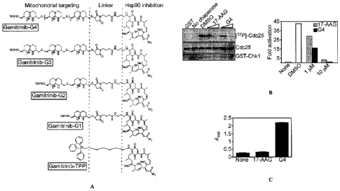
FIG. 15A is a diagram showing the combinatorial modular structure of
Gamitrinibs in which TBDPS indicates tert-butyldiphenylsilyl.
FIG. 15B left panel at top is an autoradiogram showing chaperone activity as
Chkl-dependent phosphorylation of Cdc25 in cells treated with 17-AAG or
Gamitrinib-G4 (indicated as "G4") (1-10 M) with loading controls shown at
middle
(Cdc25) and bottom (GST-Chk1). The right panel is a bar graph showing
densitometric quantification of the bands shown in the left panel and is
representative
of two experiments.
FIG. 15C is a bar graph quantifying mitochondrial accumulation of 17-AAG
or Gamitrinib-G4 (indicated as "G4") in which "None" indicates vehicle
control.
Mean SEM (n=3).
18
CA 02699794 2010-03-09
WO 2009/036092
PCT/US2008/075895
FIG. 16A is a line graph showing mitochondrial inner membrane potential
over time in TMRM-loaded mitochondria treated with Gamitrinib-Gl ("Gl"),
Gamitrinib-G2 ("G2"), Gamitrinib-G3 ("G3"), Gamitrinib-G4 ("G4"), Gamitrinib-
TPP ("TPP"), GA, or 17-AAG (at a concentration of 11.iM) and analyzed for
fluorescence emission.
FIG. 16B, left panel is a line graph showing mitochondrial inner membrane
potential over time in TMRM-loaded mitochondria incubated with 17-AAG mixed
with tetraguanidinium (17-AAG + TG-OH), 17-AAG and 11.iM Cyclosporin A ("17-
AAG + CsA"), 1.51.iM Gamitrinib-G4 ("G4"), 1.5 1..EM Gamitrinib-G4 and 11.iM
Cyclosporin A ("G4 + CsA"). The right panel is a line graph showing
mitochondrial
inner membrane potential over time in TMRM-loaded mitochondria incubated with
GA mixed with triphenylphosphonium ("GA + TPP-OH"), GA mixed with
triphenylphosphonium and 11.iM Cyclosporin A ("GA + TPP-OH + CsA"),
triphenylphosphonium by itself ("TPP") and triphenylphosphonium and 11.iM
Cyclosporin A ("TPP + CsA"). Arrows indicate point of addition.
FIG. 16C is a panel of immunoblots showing cytochrome c release in
supernatants (S) or pellets (P) showing Cox-IV as a mitochondrial marker in
tumor
mitochondria treated with Gamitrinib-Gl ("Gl"), Gamitrinib-G2 ("G2"),
Gamitrinib-
G4 ("G4"),or 17-AAG (20 minutes).
FIG. 16D is a line graph showing percent cytochrome c release over time from
mitochondria incubated with IPI-504, BIIB021, NVP-AUY922, 17-AAG, or
Gamitrinib-G4 ("G4") for 3 hours. Data are representative of two independent
experiments.
FIG. 17A consists of two line graphs showing percent viability of H460 cells,
as analyzed by MTT, treated with Gamitrinib-Gl ("Gl"), Gamitrinib-G2 ("G2"),
Gamitrinib-G3 ("G3"), Gamitrinib-G4 ("G4"), Gamitrinib-TPP ("TPP"), or 17-AAG
at different concentrations after 3 hours of treatment in the line graph to
the left and
after 24 hours of treatment in the line graph to the right. Mean SD (n=2).
FIG. 17B is a line graph showing percent viability over time of SKBr3 cells
treated with Gamitrinib-G4 ("G4"), Gamitrinib-TPP ("TPP"), or 17-AAG (at a
concentration of 10 M) and analyzed by MTT.
FIG. 17C is a bar graph showing the percentage of dead cells as analyzed by
Trypam blue staining in SKBr3 cells treated with 101.iM of Gamitrinib-G4
("G4"),
19
CA 02699794 2010-03-09
WO 2009/036092
PCT/US2008/075895
Gamitrinib-TPP ("TPP"), or 17-AAG at the indicated time intervals. Mean SEM
(n=3).
FIG. 17D consists of four scatter plots. H460 cells were treated with
Gamitrinib-G4 ("G4") or vehicle for four hours and were labeled with JC-1, and
analyzed (by multiparametric flow cytometry) for loss of mitochondrial
membrane
potential by changes in FL2/FL1 fluorescence ratio as shown in the scatter
plots at
top,or DEVDase (caspase) activity as shown in the scatter plots at bottom. The
percentage of cells in each quadrant is indicated and PI is used to indicate
propidium
iodide.
FIG. 17E are two pictures of colony formation in soft agar showing colony
formation after two weeks using H460 cells treated with vehicle (None) for 4
hours in
the top picture and colony formation after two weeks using H460 cells treated
with
Gamitrinib-G4 ("G4") at 50 uM for 4 hours in the bottom picture.
Magnification,
x200.
FIG. 17F is a line graph showing percent viability as a function of
concentration Gamitrinib-G4 (solid lines) or 17-AAG mixed with TG-OH (dashed
lines) in tumor cell lines (K562, black; MDA-MB-231, light orange; U87MG, red;
MCF-7, pink; H1975, light brown; DU145, orange; H460, blue; HCT116, purple; HL-
60, violet; Raji, dark pink; THP-1, green) as analyzed by MTT. Data are
representative of two experiments.
FIG. 17G is a line graph showing percent viability as a function of
concentration 17-AAG (circles) or Gamitrinib-G4 (squares) in H460 cells
transfected
with control (closed symbols) or CypD (open symbols) siRNA as analyzed by MTT.
Mean SEM (n=3).
FIG. 17H is a series of immunoblots showing Akt, Hsp70, Chkl, and GAPDH
protein levels in HeLa cells treated with Gamitrinib-G1 ("Gl"), Gamitrinib-G2
("G2"), Gamitrinib-G3 ("G3"), Gamitrinib-G4 ("G4"), Gamitrinib-TPP ("TPP"), or
17-AAG (at a concentration of 5 uM for 24 hours).
FIG. 18A consists of two line graphs at top and bottom. The line graph at top
shows tumor volume as a function of time in SCID/beige mice carrying H460 lung
adenocarcinoma xenograft tumors (100-150 mm3) and treated with Gamitrinib-G4
("G4") or 17-AAG. The line graph at bottom shows tumor volume as a function of
time in mice treated with a dose escalation regimen as described in Example 11
with
CA 02699794 2010-03-09
WO 2009/036092
PCT/US2008/075895
vehicle, Gamitrinib-G 1 ("G 1") or Gamitrinib-TPP ("TPP"). Tumor volume was
measured with a caliper.
FIG. 18B shows two images labeled "Vehicle" and "G4" of internucleosomal
DNA fragmentation in tumor specimens from vehicle ("Vehicle") or Gamitrinib-G4
("G4") treated tumors as visualized in situ by TUNEL. The bar graph at bottom
shows quantification of positive cells. Magnification, x400. ***, p<0.0001.
FIG. 18C shows a series of immunoblots for cytochrome c (Cyto c), Cox-IV,
and GAPDH in cytosolic fractions of H460 xenograft tumors harvested from
vehicle-
or Gamitrinib-G4 ("G4") treated animals. Two mice/group (animal #) were
analyzed.
FIG. 18D is a bar graph showing percentage weight loss in mice treated with
vehicle, 17-AAG, Gamatrinib-G 1 ("G 1"), Gamatrinib-G4 ("G4"), or Gamatrinib-
TPP
("TPP") as measured at the end of the experiment. Mean SEM.
FIG. 18E is a line graph showing percentage membrane potential over time in
TMRM-loaded mitochondria isolated from normal WS-1 fibroblasts and incubated
with uncoupled 17-AAG/TG-OH or Gamitrinib-G4 ("G4"), with or without CsA.
Arrow, point of addition.
FIG. 18F shows two immunoblots for Cyto c and Cox-IV in mitochondria
isolated from normal HFF fibroblasts or HeLa cells and treated with Gamitrinib-
Gl
("G 1"), Gamitrinib-G2 ("G2"), Gamitrinib-G3 ("G3"), Gamitrinib-G4 ("G4"),
Gamitrinib-TPP ("TPP"), or 17-AAG. Cox-IV was used as a mitochondrial marker.
FIG. 18G is a line graph showing percent viability as a function of
concentration of Gamitrinib-G4 (solid lines) or 17-AAG (dashed lines) in human
fibroblasts (HFF, black line), bovine aortic endothelial cells (medium grey),
intestinal
epithelial cells (dark grey), or human umbilical vein endothelial cells (light
grey) as
analyzed by MTT after 24 hours of incubation. Data are representative of two
experiments.
FIG 19 is a schematic diagram of chemical structures of GA (17-AAG), IPI-
504, and non-GA based (BIIB021 and NVP-AUY922) Hsp90 inhibitors used in these
studies.
FIG 20A is a line graph showing human acute leukemia HL-60 tumor volume
over time (2/mouse, 6 tumors/group) treated with vehicle or Gamitrinib-G4
("G4") at
2 mg/kg twice daily i.p. (HL-60) for the duration of treatment. Arrow, start
of
treatment.
21
CA 02699794 2010-03-09
WO 2009/036092
PCT/US2008/075895
FIG 20B is a line graph showing human breast adenocarcinoma MDA-MB-
231 tumor volume over time (2/mouse, 6 tumors/group) treated with vehicle or
Gamitrinib-G4 ("G4") with a dose escalation regimen (MDA-MB-231) starting at 2
mg/kg twice daily (day 0-2), 2.5 mg/kg twice daily (day 3-5), and 3 mg/kg
twice daily
for the duration of treatment. Arrow, start of treatment.
DETAILED DESCRIPTION
Mitochondria play a critical role in cell survival and cell death (Pandey et
al.,
EMBO J., 19:4310-4322 (2000); Green and Kroemer, Science, 305:626-629,
(2004)).
Dysfunction and loss of integrity of these organelles are molecular
prerequisites of
multiple cell death pathways, characterized by increased permeability of the
inner
mitochondrial membrane, loss of membrane potential, swelling of the matrix,
and
ultimately rupture of the outer membrane with release of apoptogenic proteins,
i.e.,
cytochrome c, in the cytosol (Green and Kroemer, Science, 305:626-629,
(2004)).
How this process, known as "mitochondrial permeability transition," is
regulated is
not completely understood (Green and Kroemer, Science, 305:626-629, (2004));
components of the permeability transition pore, including the voltage-
dependent anion
channel (VDAC-1), the adenine nucleotide translocator (ANT), or the
immunophilin
Cyclophilin D (CypD), were found to be either dispensable (Kokoszka et al.,
Nature,
427:461-1465, (2004); Krauskopf et al., Biochim. Biophys. Acta, 1757:590-595,
(2006)), or implicated in some, but not all forms of mitochondrial cell death
(Baines
et al., Nature, 434:658-662, (2005); Nakagawa et al., Nature, 434:652-658,
(2005)).
The present invention is based, at least in part, on the discovery that the
molecular chaperones Hsp60, Hsp90 and TRAP-1 are found at increased levels in
mitochondria of tumor cells, and that inhibition of molecular chaperones in
tumor cell
mitochondria using mitochondrial-targeted chaperone inhibitors results in
cancer cell
death. Without wishing to be bound by theory, the inhibition of these
mitochondrial
chaperones may result in the activation of mitochondrial permeability
transition with
collapse of mitochondrial function, including loss of mitochondrial membrane
potential and release of cytochrome c, which leads to cell death.
Thus, described herein are mitochondriotropic agents that include a chaperone
inhibitor, e.g., an HSPA9, Hsp60, Hsp90 or TRAP-1 inhibitor, and a
mitochondrial
penetrating moiety, optionally with an intervening linker, and methods of
making and
using these compositions to treat disorders associated with aberrant cellular
22
CA 02699794 2015-04-02
60412-4252
proliferation, e.g., cancer and tumors, e.g., to kill cancer and tumor cells,
e.g., in vivo
and in vitro. Also described herein are compositions containing these
mitochondriotropic agents.
I. Molecular Chaperones
Molecular chaperones, especially members of the Heat Shock Protein (Hsp)
gene family (Lindquist and Craig, Annu. Rev. Genet. 1988; 22:631-77), assist
in
protein folding quality control, protein degradation, and protein trafficking
among
subcellular compartments (Hartl and Hayer-Hartl, Science 2002; 295:1852-8).
This
involves periodic cycles of ATPase activity, recruitment of additional
chaperones, and
compartmentalization in subcellular microdomains, including mitochondria
(Young et
al., Cell 2003; 112:41-50). Molecular chaperones have often been associated
with
enhanced cell survival (Beere, J Cell Sci 2004; 117:2641-51), via suppression
of
apoptosome-initiated mitochondrial cell death (Paul et al., Mol Cell Biol
2002;
22:816-34), increased stability of survival effectors (Sato et al., Proc Natl
Acad Sci U
S A 2000; 97:10832-7), and inactivation of p53 (Wadhwa et al., J Biol Chem
1998;
273:29586-91). As described herein, the chaperone anti-apoptotic function play
a
central role in tumor cell maintenance and can be selectively targeted to kill
cancer
cells. See also: Whitesell et al., Nat Rev Cancer 2005; 5:761-72; and Isaacs
et al.,
Cancer Cell 2003; 3:213-7.
The following is a brief description of some of the molecular chaperones that
can be targeted using the present methods. In some embodiments, a molecular
chaperone polypeptide useful in the present methods (e.g., in screening
methods) is at
least about 90%, 95%, 99%, or 100% identical to an amino acid sequence
described
herein (e.g., to a human sequence). In some embodiments, a nucleic acid
encoding a
molecular chaperone useful in the present methods (e.g., in screening methods)
is at
least about 90%, 95%, 99%, or 100% identical to a nucleic acid sequence
described
herein (e.g., to a human sequence).
The comparison of sequences and determination of percent identity between
two sequences can be accomplished using a mathematical algorithm. For example,
the percent identity between two amino acid sequences can determined using the
Needleman and Wunsch ((1970) J. Mol. Biol. 48:444-453 ) algorithm which has
been
incorporated into the GAP program in the GCG software package provided by
BIO VIA, 5005 Wateridge Vista Drive, San Diego, CA 92121 USA,
23
CA 02699794 2015-04-02
60412-4252
using the default parameters, e.g., a Blossum 62 scoring
matrix with a gap penalty of 12, a gap extend penalty of 4, and a frameshift
gap
penalty of 5.
Hsp90 (Heat-Shock 90-10 Protein 1)
HSP90 is a molecular chaperone that plays a key role in the conformational
maturation of a number of proteins, including oncogenic signaling proteins. As
described herein, Hsp90 accumulates in the mitochondria of cancer cells, but
not
normal cells, and can be targeted using the compositions described herein
including a
mitochondrial-penetrating sequence.
GenBank Acc. Nos. for human Hsp90 include NM_001017963.2 (nucleic
acid) and NP 001017963.2 (protin), for heat shock protein 90kDa alpha
(cytosolic),
class A member 1 isoform 1, and NM 005348.3 (nucleic acid) NP_005339.3
(protein), for heat shock protein 90kDa alpha (cytosolic), class A member 1
isoform
2. Variant 2 differs in the 5' UTR and coding sequence compared to variant 1.
The
resulting isoform 2 is shorter at the N-terminus compared to isoform 1.
Hsp90 is also known as HSPCA; HSPC1; HSP90A; HSP89-ALPHA
(HSP89A); Lipopolysaccharide-Associated Protein 2 (LAP2); and LPS-associated
protein 2.
TRAP-1 (TNF Receptor-Associated Protein 1)
TRAP-1 has high homology to hsp90, and binds the type 1 tumor necrosis
factor receptor (see Song et al., J. Biol. Chem., 270:3574-3581 (1995)). The
deduced
661-amino acid protein is 60% similar to HSP90 family members, although it
lacks
the highly charged domain found in HSP90 proteins. See, e.g., Felts et al., J
Biol
Chem. 2000; 275(5):3305-12. As described herein, TRAP-1 accumulates in the
mitochondria of cancer cells, but not normal cells, and can be targeted using
the
compositions described herein including a mitochondrial-penetrating sequence.
GenBank Acc. Nos. for human TRAP-1 include NM 016292.2 (nucleic acid)
and NP_057376.2 (amino acid). TRAP-1 is also referred to as Heat-Shock
Protein,
75-KD (HSP75); Tumor Necrosis Factor Receptor-Associated Protein 1; TRAP 1;
and
TNFR-Associated Protein 1.
24
CA 02699794 2010-03-09
WO 2009/036092
PCT/US2008/075895
Hsp60 (Heat-Shock 60-kD Protein 1)
Hsp60, together with its associated chaperonin, Hsp10, has been recognized as
an evolutionary conserved stress response chaperone (Zhao et al., Embo J
2002;21:4411-9), largely, but not exclusively compartmentalized in
mitochondria
(Soltys and Gupta, Int Rev Cytol 2000; 194:133-96), and with critical roles in
organelle biogenesis and folding/refolding of imported preproteins (Deocaris
et al.,
Cell Stress Chaperones 2006; 11:116-28). However, whether Hsp60 also
contributes
to cell survival is controversial, with data suggesting a pro-apoptotic
function via
enhanced caspase activation (Samali et al., Embo J 1999; 18:2040-8;
Xanthoudakis et
al., Embo J 1999; 18:2049-56), or, conversely, an anti-apoptotic mechanism
involving
sequestration of Bax-containing complexes (Shan et al., J Mol Cell Cardiol
2003;
35:1135-43). A role of Hsp60 in cancer was equally uncertain, as up- (Thomas
et al.,
Leuk Res 2005; 29:1049-58; Cappello et al., BMC Cancer 2005; 5:139), or down-
regulation (Tang et al., Cell Stress Chaperones 2005; 10:46-58; Cappello et
al.,
Cancer 2006; 107:2417-24) of this chaperone has been reported in various tumor
series correlating with disease outcome. As described herein, Hsp60 is highly
expressed in tumor cells, as compared to normal cells, and targeting of Hsp60
causes
mitochondrial dysfunction and apoptosis, whereas loss of Hsp60 in normal cells
is
well tolerated, and does not result in cell death.
Hsp60 is also known as CPN60; GROEL; HSP60; HSP65; SPG13; and
HuCHA60. Exemplary GenBank Acc. Nos. for human Hsp60 include NM_002156.4
(nucleic acid) and NP 002147.2 (protein) for transcript variant 1 (the longer
variant),
and NM 199440.1 (nucleic acid) and NP 955472.1 (protein) for transcript
variant 2.
Variant 2 differs in the 5' UTR compared to variant 1. Both variants 1 and 2
encode
the same isoform.
HspA9 (heat shock 70kDa protein 9
HspA9 belongs to the heat shock protein 70 family, which contains both heat-
inducible and constitutively expressed members. The latter are called heat-
shock
cognate proteins, of which HspA9 is one. HspA9 plays a role in the control of
cell
proliferation, and may also act as a chaperone. See, e.g., Wadhwa et al., Int
J Cancer.
2006; 118(12):2973-80; Wadhwa et al., J Gene Med. 2004; 6(4):439-44.
CA 02699794 2010-03-09
WO 2009/036092
PCT/US2008/075895
HspA9 is also known as mortalin, mthsp70, and GRP75. Exemplary GenBank
Ace. Nos. for human HspA9 include NM_004134.5 (nucleic acid) and NP_004125.3
(protein), the heat shock 70kDa protein 9 precursor.
II. Inhibitors of Molecular Chaperones
The compositions and methods described herein include the use of inhibitors
of molecular chaperones, e.g., inhibitors or Hsp60, HspA9, Hsp90 and/or TRAP-
1.
The inhibitors useful in the methods and compositions described herein act
directly on
the chaperone protein itself, i.e., they do not act upstream or downstream. A
number
of such inhibitors are known in the art, e.g., peptide inhibitors and small
molecule
inhibitors. In some embodiments, the molecular chaperone inhibitors useful in
this
invention inhibit the ATPase activity of the chaperone, e.g., of Hsp60, HspA9,
Hsp90,
and/or TRAP-1. In some embodiments, the molecular chaperone inhibitors useful
in
this invention inhibit the binding of Hsp60, HspA9, Hsp90, or TRAP-1 to
Cyclophilin
D. In some embodiments, the molecular chaperone inhibitors useful in this
invention
inhibit the binding of Hsp60, HspA9, Hsp90, or TRAP-1 to survivin. In some
embodiments, molecular chaperone inhibitors bind to a chaperone, and induce
the
proteasomal degradation of the chaperone's client proteins.
In addition, there are numerous methods useful for identifying, designing, and
assaying candidate chaperone inhibitors. For example, rational screening
methods
have been used to identify additional molecules that target Hsp90, using a
computational approach using a shepherdin peptide (LFACGSSHK, all D-amino
acids, as a scaffold to screen a database of nonpeptidic structures. See,
e.g., Meli et
al., J. Med. Chem., 49:7721-7730 (2006).
Peptide Inhibitors of Molecular Chaperones
A number of peptide inhibitors of molecular chaperones, e.g., of Hsp90 and/or
TRAP-1, are known in the art. The inhibitors useful in the compositions and
methods
described herein can include the entire peptide or polypeptide (e.g., all of
an
apoptosis-inducing protein (AIP such as survivin), or an active (i.e.,
inhibitory)
fragment thereof that retains the Hsp90 inhibitory activity of the parent,
i.e., at least
40% of the activity of the parent; an active fragment preferably has at least
50%, 60%,
70%, 80%, 90%, 100% or more of the Hsp90-inhibitory activity of the parent
polypeptide.
26
CA 02699794 2010-03-09
WO 2009/036092
PCT/US2008/075895
Survivin peptides and derivatives
Survivin peptides and peptide derivatives are disclosed in U.S. Patent
Application No. 11/187,230 (herein incorporated by reference in its entirety).
Active
survivin peptides share a core Hsp90 binding sequence motif of SEQ ID NO:2
(His
Ser Ser Gly Cys), which is located in the single Baculovirus Inhibitor of
Apoptosis
(TAP) Repeat (BIR) domain of the Survivin protein. This motif corresponds to
amino
acid residues at position 80-84 of full-length Survivin (SEQ ID NO:1).
Peptides
including this motif, and peptide derivatives thereof, can (a) bind to the N-
terminal
ATPase domain of Hsp90 (the "ATP pocket") and (b) inhibit Hsp9O-Survivin
protein-
protein interactions in vitro and in vivo.
The terms Survivin peptide and Survivin peptide derivative, as used herein,
refer to peptides that include less than the complete amino acid sequence of a
functional Survivin protein that prevents cell death. Survivin peptides and
peptide
derivatives useful to this invention inhibit molecular chaperones and in
particular,
inhibit interaction between a molecular chaperone, e.g., Hsp90 or TRAP-1, and
Cyclophilin D.
The full-length human, wild type Survivin polypeptide has the following
amino acid sequence:
MGAPTLPPAWQPFLKDHRISTFKNWPFLEGCACTPERMAEAGFIHCP
TENEPDLAQCFFCFKELEGWEPDDDPIEEHKKHSSGCAFLSVKKQFE
ELTLGEFLKLDRERAKNKIAKETNNKKKEFEETAKKVRRAIEQLAAM
D (SEQ ID NO:1)
The following table (Table 1) lists some exemplary Survivin peptides that can
bind to Hsp90:
Table 1. Exemplary Survivin peptides
SEQ ID NO:2 His Ser Ser Gly Cys
SEQ ID NO:3 Lys His Ser Ser Gly Cys Ala Phe Leu Ser Val Lys
SEQ ID NO:4 Ile Asp Asp His Lys Lys His Ser Ser Gly Cys Ala Phe Leu
SEQ ID NO:5 Lys Lys His Ser Ser Gly Cys Ala Phe Leu
SEQ ID NO:6 Lys His Ser Ser Gly Cys
SEQ ID NO:7 His Ser Ser Gly Cys Ala
SEQ ID NO:8 Lys His Ser Ser Gly Cys Ala
SEQ ID NO:9 Lys Lys His Ser Ser Gly Cys
SEQ ID NO:10 His Ser Ser Gly Cys Ala Phe
SEQ ID NO: ii His Lys Lys His Ser Ser Gly Cys Ala Phe Leu Ser Val Lys Lys
SEQ ID NO:12 Lys His Ser Ser Gly Cys Ala Phe Leu
27
CA 02699794 2015-04-02
60412-4252
Variants of Survivin peptides can also be used in the methods and
compositions described herein. Conservative and non-conservative amino acid
substitutions may be made. In particular, conservative amino acid
substitutions can
be made for one or more, e.g., up to five, ten, twenty, or thirty, amino acids
outside of
the core pentamer sequence corresponding to His 80 to Cys 84 in SEQ ID NO:1
(i.e.,
SEQ ID NO:2 set forth above). Peptidomimetics of Survivin peptides are
described
by Plescia et al. (Rational design of Shepherdin, a novel anticancer agent.
Cancer
Cell. 7(5):457-68 (2005))
Other IAP peptides and derivatives
Other Inhibitors of Apoptosis Proteins (IAPs) interact with Hsp90, including
cIAP1 (Entrez Accession No.: NP 001156), cIAP2 (Entrez Accession No.:
NP_001157), and XIAP (Entrez Accession No.: NP_001158). See, e.g., Deveraux
and Reed, Genes and Dev., 13:239-252 (1999). These IAP proteins contain at
least
one Baculovirus IAP repeat domain that mediates Hsp90 interactions, as
disclosed
herein. For example, the first BIR domain of XIAP (BIR1) mediates Hsp90-XIAP
binding interactions.
TAP proteins, or Hsp90-binding and -inhibiting fragments thereof, can
therefore be used in the present compositions and methods. For example,
peptides
corresponding to one or more BIR domains in these TAP proteins, or Hsp90-
binding
fragments thereof, can be used in the compositions and methods disclosed
herein to
induce cancer or tumor cell death. IAP proteins, or Hsp90-binding fragments
thereof,
can also be screened as test compounds, e.g., to identify candidate compounds
that
inhibit binding between molecular chaperones and Cyclophilin D. In some
embodiments, IAP proteins, or Hsp90-binding fragments thereof, can be screened
as
test compounds to identify candidate compounds that induce cancer cell death.
An exemplary first BIR domain of XIAP includes the sequence:
RLKTFANFPSGSPVSASTLARAGFLYTGEGDTVRCFSCHAAVDRWQY
GDSAVGRHRKVSPNCRFIN (SEQ ID NO:14)
An exemplary first BIR domain of cIAP I includes the sequence:
RMSTYSTFPAGVPVSERSLARAGFYYTGVNDKVKCFCCGLMLDNWKR
GDSPTEKHKKLYPSCRFVQ (SEQ ID NO:15)
28
CA 02699794 2010-03-09
WO 2009/036092
PCT/US2008/075895
An exemplary first BIR domain of cIAP2 includes the sequence:
RMSTYSTFPAGVPVSERSLARAGFYYTGVNDKVKCFCCGLMLDNWKL
GDSPIQKHKQLYPSCSFIQ (SEQ ID NO:16)
Variants of peptide inhibitors
Variants of peptide inhibitors of molecular chaperones are also part of this
invention. These include sequence variants. Where a conservative amino acid
substitution is made, the substitution can be of one amino acid residue for
another in
any of the following groups: arginine, histidine, and lysine; aspartic acid
and
glutamic acid; alanine, leucine, isoleucine and valine; and phenylalanine,
tryptophan
and tyrosine. The amino acid residues listed here are naturally occurring. Non-
naturally occurring amino acid residues of like kind may also be substituted.
For
example, a negatively charged non-naturally occurring amino acid residue may
be
substituted for a negatively charged naturally occurring amino acid residue; a
hydrophobic aromatic non-naturally occurring amino acid residue may be
substituted
for a hydrophobic aromatic naturally occurring amino acid residue; and so
forth.
The degree of identity can vary and can be determined by methods well
established in the art. "Homology" and "identity" each refer to sequence
similarity
between two polypeptide sequences, with identity being a more strict
comparison.
Homology and identity can each be determined by comparing a position in each
sequence which may be aligned for purposes of comparison. When a position in
the
compared sequence is occupied by the same amino acid residue, then the
polypeptides
can be referred to as identical at that position; when the equivalent site is
occupied by
the same amino acid (e.g., identical) or a similar amino acid (e.g., similar
in steric
and/or electronic nature), then the molecules can be referred to as homologous
at that
position. A percentage of homology or identity between sequences is a function
of
the number of matching or homologous positions shared by the sequences. A
biologically active variant of a polypeptide described herein can have at
least or about
80%, 85%, 90%, 95%, 96%, 97%, 98%, or 99% identity or homology to a
corresponding naturally occurring polypeptide (e.g., a survivin fragment or a
TAP
fragment, e.g., as described herein). The nucleic acids encoding the
biologically
active variant polypeptides can be similarly described as having at least or
about 80%,
85%, 90%, 95%, 96%, 97%, 98%, or 99% identity to a corresponding naturally
occurring nucleic acid sequence. Those of ordinary skill in the art will
readily
29
CA 02699794 2010-03-09
WO 2009/036092
PCT/US2008/075895
recognize degenerate variants of nucleic acid sequences, and such variants can
be
used for the purposes described herein.
When using a peptide inhibitor and/or mitochondrial penetrating moiety in a
human subject, it will generally be desirable to use a human or humanized
sequence.
Thus, the methods described herein can include using standard molecular
biology
techniques to humanize a non-human sequence. Alternatively, human sequences
can
be used to make the construct.
Modifications of Peptide Inhibitors
Modified versions of the peptides described herein can also be used in the
compositions and methods described herein. The peptides and biologically
active
variants thereof can be modified in numerous ways. For example, agents,
including
additional amino acid residues, other substituents, and protecting groups can
be added
to either the amino terminus, the carboxy terminus, or both. The modification
can be
made for the purpose of altering the peptides' form or altering the way the
peptides
bind to or interact with one another, with non-identical peptides, or with
other
polypeptides. For example, the peptides can be modified to include cysteine
residues
or other sulphur-containing residues or agents that can participate in
disulphide bond
formation. For example, one can add at least two cysteine residues, one or
both of
which are, optionally, at the C-terminal or N-terminal of the peptide.
The peptides can be cyclized by formation of a disulfide bond between
cysteine residues (or, more generally, between two of the at least two
cysteine
residues present in the polypeptide (e.g., at the terminal regions)). While
the peptides
of the present invention may be linear or cyclic, cyclic peptides generally
have an
advantage over linear peptides in that their cyclic structure is more rigid
and hence
their biological activity may be higher than that of the corresponding linear
peptide
(see, generally, Camarero and Muir, J. Am. Chem. Soc., 121:5597-5598, (1999).
Strategies for the preparation of circular polypeptides from linear precursors
have been described and can be employed with the present peptides. For
example, a
chemical cross-linking approach can be used to prepare a backbone cyclized
version
of the peptide (Goldenburg and Creighton, J. Mol. Biol., 165:407-413, (1983)).
Other
approaches include chemical intramolecular ligation methods (see, e.g.,
Camarero et
al., Angew Chem. Int. Ed., 37:347-349, (1998); Tam and Lu, Prot. Sci., 7:1583-
1592,
(1998); Camarero and Muir, Chem. Commun., 1369-1370, (1997); and Zhang and
CA 02699794 2010-03-09
WO 2009/036092
PCT/US2008/075895
Tam, J. Am. Chem. Soc., 119:2363-2370, (1997) and enzymatic intramolecular
ligation methods (Jackson et al., J. Am. Chem. Soc., 117:819-820, (1995),
which
allow linear synthetic peptides to be efficiently cyclized under aqueous
conditions.
See also U.S. Patent No. 7,105,341.
Alternatively, or in addition, the peptide can further include a substituent
at the
amino-terminus or carboxy-terminus. The substituent can be an acyl group or a
substituted or unsubstituted amine group (e.g., the substituent at the N-
terminus can
be an acyl group and the C-terminus can be amidated with a substituted or
unsubstituted amine group (e.g., an amino group having one, two, or three
substituents, which may be the same or different)). The amine group can
include a
lower alkyl (e.g., an alkyl having 1-4 carbons), alkenyl, alkynyl, or
haloalkyl group.
The acyl group can be a lower acyl group (e.g., an acyl group having up to
four
carbon atoms), especially an acetyl group.
As used herein, the term "alkyl" is meant to refer to a saturated hydrocarbon
group which is straight-chained or branched. Example alkyl groups include
methyl
(Me), ethyl (Et), propyl (e.g., n-propyl and isopropyl), butyl (e.g., n-butyl,
isobutyl, t-
butyl), pentyl (e.g., n-pentyl, isopentyl, neopentyl), and the like. An alkyl
group can
contain from 1 to about 20, from 2 to about 20, from 1 to about 10, from 1 to
about 8,
from 1 to about 6, from 1 to about 4, or from 1 to about 3 carbon atoms.
As used herein, "alkenyl" refers to an alkyl group having one or more double
carbon-carbon bonds. Example alkenyl groups include ethenyl, propenyl, and the
like.
As used herein, "alkynyl" refers to an alkyl group having one or more triple
carbon-carbon bonds. Example alkynyl groups include ethynyl, propynyl, and the
like.
As used herein, "haloalkyl" refers to an alkyl group having one or more
halogen substituents. Example haloalkyl groups include CF3, C2F5, CHF2, CC13,
CHC12, C2C15, and the like.
As used herein, "aryl" refers to aromatic monocyclic or multicyclic groups
containing from 6 to 19 carbon atoms. Examples of aryl groups include, but are
not
limited to unsubstituted or substituted phenyl, unsubstituted or substituted
fluorenyl,
and unsubstituted or substituted naphthyl.
31
CA 02699794 2010-03-09
WO 2009/036092
PCT/US2008/075895
As used herein, "heterocycloalkyl" refers to a monocyclic or multicyclic,
saturated or unsaturated ring system, in one embodiment of 3 to 10 members, in
another embodiment of 4 to 7 members, in a further embodiment of 5 to 6
members,
where one or more, in certain embodiments, 1 to 3, of the atoms in the ring
system is
a heteroatom, that is, an element other than carbon, including but not limited
to,
nitrogen, oxygen or sulfur. In certain embodiments, one of the atoms of the
ring can
be replaced with a carbonyl or sulfonyl group.
As used herein, "alkylene," "alkenylene," "alkynylene," "cycloalkylene,"
"arylene," "heteroarylene," and "heterocycloalkylene" refer to divalent
linking
"alkyl," "alkenyl," "alkynyl," "cycloalkyl," "aryl," "heteroaryl," and
"heterocycloalkyl" groups. The divalent linkers, in some embodiments, can be
present in both directions, e.g., a C(0)NH can either be -C(0)NH- or -NHC(0)-.
As noted, the peptides can vary in length and can be or can include contiguous
amino acid residues that naturally occur in chaperone binding proteins (CBP),
e.g.,
Survivin or IAPs, or that vary to a certain degree from naturally occurring
CBP
sequences (but retain sufficient activity to be useful). Where the peptides
include, at
their N-terminus or C-terminus (or both), amino acid residues that are not
naturally
found in CBPs, the additional sequence(s) can be about 200 amino acid residues
long,
and these residues can be divided evenly or unevenly between the N- and C-
termini.
For example, both the N- and C-termini can include about 10, 20, 30, 40, 50,
60, 70,
80, 90, or 100 amino acid residues. Alternatively, one terminus can include
about 10,
20, 30, 40, 50, 60, 70, 80, 90, 100, 110, 120, 130, 140, 150, 160, 170, 180,
190, or 200
residues, and one terminus can include none (e.g., it can terminate in an
amino acid
sequence identical to a naturally occurring Survivin sequence).
More specifically, the N- or C-termini can include 1 to about 100 (e.g., 1, 2,
3,
4, 5, 6, 7, 8, 9, 10, 15, 20, 25, 30, 40, 50, 60, 70, 80, 90, or 100) amino
acid residues
that are positively charged (e.g., basic amino acid residues such as arginine,
histidine,
and/or lysine residues); 1 to about 100 amino acid residues that are
negatively charged
(e.g., acidic amino acid residues such as aspartic acid or glutamic acid
residues); 1 to
about 100 glycine residues; 1 to about 100 hydrophobic amino acid residues
(e.g.,
hydrophobic aliphatic residues such as alanine, leucine, isoleucine or valine
or
hydrophobic aromatic residues such as phenylalanine, tryptophan or tyrosine);
or 1 to
about 100 (e.g., 1-4) cysteine residues.
32
CA 02699794 2010-03-09
WO 2009/036092
PCT/US2008/075895
The peptides, including the modified peptides described above, can be
protease resistant and can include one or more types of protecting groups such
as an
acyl group, an amide group, a benzyl or benzoyl group, or a polyethylene
glycol.
More specifically, a peptide, including the modified peptides described above,
can be
N-terminally acetylated and/or C-terminally amidated.
Where non-naturally occurring or modified amino acid residues are included
they can be selected from the following or many others available in the art:
4-hydroxyproline, gamma-carboxyglutamic acid, o-phosphoserine,
o-phosphotyrosine, or delta-hydroxylysine. Other examples include
naphthylalanine,
which can be substituted for trytophan to facilitate synthesis, L-
hydroxypropyl, L-3,4-
dihydroxyphenylalanyl, alpha-amino acids such as L-alpha-hydroxylysyl and D-
alpha-methylalanyl, L-alpha-methylalanyl, beta-amino acids, and isoquinolyl.
Peptides having non-naturally occurring amino acid residues may be referred to
as
synthetic peptides and constitute one type of variant as described herein.
Other
variants include peptides in which a naturally occurring side chain of an
amino acid
residue (in either the L- or D-form) is replaced with a non-naturally
occurring side
chain.
In one embodiment, the peptides can have three extra amino acids (Met-Gly-
Ser) at either terminus (or both) (e.g., at the N-terminus) and seven to eight
extra
amino acids (Thr-Ser-His-His-His-His-His-His-Cys (SEQ ID NO:13)) at either
terminus (or both) (e.g., at the C-terminus).
In another embodiment, the peptides can be PEGylated by methods known in
the art.
For guidance on peptide modification by reduction/alkylation and/or acylation,
one can consult Tarr, Methods of Protein Microcharacterization, J. E. Silver
ed.,
Humana Press, Clifton N.J. (1986) 155-194; for guidance on chemical coupling
to an
appropriate carrier, one can consult Mishell and Shiigi, eds, Selected Methods
in
Cellular Immunology, WH Freeman, San Francisco, Calif (1980) and U.S. Pat. No.
4,939,239; and for guidance on mild formalin treatment, one can consult Marsh,
Int.
Arch. Allergy Appl. Immunol., (1971) 41:199-215.
Peptidomimetics of the inhibitory peptides can also be used. Peptide
inhibitors disclosed herein and known in the art can be modified according to
methods
known in the art for producing peptidomimetics. See, e.g., Kazmierski, W.M.,
ed.,
33
CA 02699794 2010-03-09
WO 2009/036092
PCT/US2008/075895
Peptidomimetics Protocols, Human Press (Totowa NJ 1998); Goodman et al., eds.,
Houben-Weyl Methods of Organic Chemistry: Synthesis of Peptides and
Peptidomimetics, Thiele Verlag (New York 2003); and Mayo et al., J. Biol.
Chem.,
278:45746, (2003). In some cases, these modified peptidomimetic versions of
the
peptides and fragments disclosed herein exhibit enhanced stability in vivo,
relative to
the non-peptidomimetic peptides.
Methods for creating a peptidomimetic include substituting one or more, e.g.,
all, of the amino acids in a peptide sequence with D-amino acid enantiomers.
Such
sequences are referred to herein as "retro" sequences. In another method, the
N-
terminal to C-terminal order of the amino acid residues is reversed, such that
the order
of amino acid residues from the N-terminus to the C-terminus of the original
peptide
becomes the order of amino acid residues from the C-terminus to the N-terminus
in
the modified peptidomimetic. Such sequences can be referred to as "inverso"
sequences.
Peptidomimetics can be both the retro and inverso versions, i.e., the "retro-
inverso" version of a peptide disclosed herein. The new peptidomimetics can be
composed of D-amino acids arranged so that the order of amino acid residues
from
the N-terminus to the C-terminus in the peptidomimetic corresponds to the
order of
amino acid residues from the C-terminus to the N-terminus in the original
peptide.
Other methods for making a peptidomimetics include replacing one or more
amino acid residues in a peptide with a chemically distinct but recognized
functional
analog of the amino acid, i.e., an artificial amino acid analog. Artificial
amino acid
analogs include 13-amino acids, 13-substituted 13-amino acids ("133-amino
acids"),
phosphorous analogs of amino acids, such as a-amino phosphonic acids and a-
amino
phosphinic acids, and amino acids having non-peptide linkages. Artificial
amino
acids can be used to create peptidomimetics, such as peptoid oligomers (e.g.,
peptoid
amide or ester analogues), 13-peptides, cyclic peptides, oligourea or
oligocarbamate
peptides; or heterocyclic ring molecules. Exemplary Survivin retro-inverso
peptidomimetics include LFACGSSHK (SEQ ID NO:25), CGSSH (SEQ ID NO:26),
GSSHK (SEQ ID NO:27), KKWKMRRNQFWVKVQRLFACGSSHK (SEQ ID
NO:28), KKWKMRRNQFWVKVQRCGSSH (SEQ ID NO:29), and
KKWKMRRNQFWVKVQRGSSHK (SEQ ID NO:30), wherein the sequences
34
CA 02699794 2015-04-02
60412-4252
include all D-amino acids. These sequences can be modified, e.g., by
biotinylation of
the amino terminus and amidation of the carboxy terminus.
Any of the peptides described herein, including the variant forms described
herein, can further include a heterologous polypeptide (e.g., a polypeptide
having a
sequence that does not appear in a CBP). The heterologous polypeptide can be a
polypeptide that increases the circulating half-life of the peptide to which
it is
attached (e.g., fused, as in a fusion protein). The heterologous polypeptide
can be an
albumin (e.g., a human serum albumin or a portion thereof) or a portion of an
immunoglobulin (e.g., the Fc region of an IgG). The heterologous polypeptide
can be
a mitochondrial-penetrating moiety.
Compounds mimicking the necessary conformation of the peptides described
herein are contemplated as within the scope of this invention. A variety of
designs for
such mimetics are possible. U.S. Patent No. 5,192,746; U.S. Patent No.
5,169,862;
U.S. Patent No. 5,539,085; U.S. Patent No. 5,576,423; U.S. Patent No.
5,051,448; and
U.S. Patent No. 5,559,103 describe multiple
methods for creating such compounds.
Non-peptidic compounds that mimic peptide sequences are known in the art
(Meli et al. J. Med. Chem., 49:7721-7730 (2006), describing methods of
identifying
nonpeptide small molecule mimics of shepherdin). Synthesis of non-peptide
compounds that mimic peptide sequences is also known in the art (see, e.g.,
Eldred et
al. J. Med. Chem., 37:3882, (1994); Ku et al. J. Med. Chem., 38:9, (1995);
Meli et al.
J. Med. Chem., 49:7721-7730 (2006)). Such nonpeptide compounds that mimic CBP
peptides that bind chaperones are specifically contemplated by the present
invention.
The present invention also contemplates synthetic mimicking compounds. As
is known in the art, peptides can be synthesized by linking an amino group to
a
carboxyl group that has been activated by reaction with a coupling agent, such
as
dicyclohexylcarbodiimide (DCC). The attack of a free amino group on the
activated
carboxyl leads to the formation of a peptide bond and the release of
dicyclohexylurea.
It can be necessary to protect potentially reactive groups other than the
amino and
carboxyl groups intended to react. For example, the (a-amino group of the
component
containing the activated carboxyl group can be blocked with a
tertbutyloxycarbonyl
group. This protecting group can be subsequently removed by exposing the
peptide to
dilute acid, which leaves peptide bonds intact.
CA 02699794 2010-03-09
WO 2009/036092
PCT/US2008/075895
With this method, peptides can be readily synthesized by a solid phase method
by adding amino acids stepwise to a growing peptide chain that is linked to an
insoluble matrix, such as polystyrene beads. The carboxyl-terminal amino acid
(with
an amino protecting group) of the desired peptide sequence is first anchored
to the
polystyrene beads. The protecting group of the amino acid is then removed. The
next
amino acid (with the protecting group) is added with the coupling agent. This
is
followed by a washing cycle. The cycle is repeated as necessary.
In one embodiment, the mimetics of the present invention are peptides having
sequence homology to the herein-described chaperone inhibitor peptides. These
mimetics include, but are not limited to, peptides in which L-amino acids are
replaced
by their D-isomers. One common methodology for evaluating sequence homology,
and more importantly statistically significant similarities, is to use a Monte
Carlo
analysis using an algorithm written by Lipman and Pearson to obtain a Z value.
According to this analysis, a Z value greater than 6 indicates probable
significance,
and a Z value greater than 10 is considered to be statistically significant
(Pearson and
Lipman, Proc. Natl. Acad. Sci. (USA), 85:2444-2448, (1988); Lipman and
Pearson,
Science, 227:1435-1441, (1985). More generally, the CBP peptides described
herein
and the mimetics described above can be synthesized using any known methods,
including tea-bag methodology or solid phase peptide synthesis procedures
described
by Merrifield et al., Biochemistry, 21:5020-5031, (1982); Houghten Wellings,
Proc.
Natl. Acad. Sci. (USA), 82:5131-5135, (1985); Atherton, Methods in Enzymology,
289:44-66, (1997), or Guy and Fields, Methods in Enzymology, 289:67-83,
(1997), or
using a commercially available automated synthesizer.
Small Molecule Inhibitors of Molecular Chaperones
A number of small molecule chaperone inhibitors useful in the methods and
compositions described herein are known in the art. For example, small
molecule
chaperone inhibitors that are useful in the compositions and methods described
herein
include, but are not limited to, molecules that bind to a Hsp90 ATP binding
pocket.
Small molecule Hsp90 inhibitors known in the art are described, for example,
in
Rodina et al., (Nature Chemical Biology, published online July 1, 2007).
In some embodiments, the chaperone inhibitor is an Hsp90 inhibitor selected
from one of several chemotypes. Two of these chemotypes are ansamycin and
macrolactone inhibitors. These are represented by radicicol and
cycloproparadicicol,
36
CA 02699794 2010-03-09
WO 2009/036092
PCT/US2008/075895
members of the macrolactone Hsp90 inhibitor class, and 17-
dimethylaminoethylamino-17-demethoxy-geldanamycin (17DMAG) and 17AAG,
members of the ansamycin class of Hsp90 inhibitors. The structural basis for
inhibition of Hsp90 by radicicol and geldanamycin is known, so one of skill in
the art
could readily generate and test analogs thereof that would retain Hsp90
inhibitory
activity, see, e.g., Roe et al., J. Med. Chem. 42(2):260-6 (1999). Purine
inhibitors
form a third class of compounds useful in the compositions and methods
described
herein.
Ansamycin Inhibitors of Hsp90
Further examples of molecular chaperone inhibitors that are useful in this
invention include, but are not limited to, quinine ansamycin antibiotics, such
as the
macbecins, geldanamycin, geldanamycin analogues, and herbimycin A.
Geldanamycin is an inhibitor of heat shock protein-90 (Hsp90), which is
involved in the folding, activation and assembly of a wide range of proteins
("client
proteins"), including key proteins involved in signal transduction, cell cycle
control
and transcriptional regulation. The binding of geldanamycin to Hsp90 disrupts
Hsp90-client protein interactions, preventing the client proteins from folding
correctly. Geldanamycin and geldanamycin analogues are part of this.
As used herein, "geldanamycin analogues" refers to compounds that share a
common core structure with geldanamycin but have minor chemical modifications.
Geldanamycin analogues that are variant at position 17 of geldanamycin are
known in the art and many are commercially available. Examples of commercially
available geldanamycin analogues include, but are not limited to, 17-
allylamino-
demethoxygeldamycin (17-AAG), 17-dimethylaminogeldanamycin, 17-GMB-APA-
GA (a maleimido derivative of geldanamycin that enables the conjugation of GA
to a
polypeptide), 17-(Dimethylaminoethylamino)-17-demethoxygeldanamycin (17-
DMAG), 1742-(Pyrrolidin-1-yl)ethyl]aminno-17-demethoxygeldanamycin (17-AEP-
GA), and 17-(Dimethylaminopropylamino)-17-demethoxygeldanamycin (17-DMAP-
GA). See also Sasaki et al., U.S. Pat. No. 4,261,989 (1981); Schnur et al.,
U.S. Pat.
No. 5,932,566 (1999); Schnur et al., J. Med. Chem., 38:3806-3812, (1995);
Schnur et
al., J. Med. Chem., 38:3813-3820, (1995); and Santi et al., US 2003/0114450 Al
(2003).
37
CA 02699794 2015-04-02
60412-4252
Geldanamycin analogues that are variant at position 11 are known in the art.
Examples include, but are not limited to, Muroi et al., U.S. Pat. No.
4,421,688 (1983);
Schnur, U.S. Pat. No. 5,387,584 (1995); Schnur et al., U.S. Pat. No. 5,932,566
(1999);
Welch et al., U.S. Pat. No. 6,015,659 (2000); Whitesell et al., WO 94/08578 A2
(1994); Ho et al., WO 00/03737 A2 (2000); Snader et al., WO 02/36574 Al
(2002);
Snader et al., WO 02/079167 Al (2002); Santi et al., WO 03/013430 A2 (2003);
Zhang etal., WO 03/066005 A2 (2003); Omura et al., JP 63-218620 (1988); Schnur
et
al., J. Med. Chem., 38:3806-3812, (1995); and Schnur et al., J. Med. Chem.,
38:3813-
3820, (1995) 11-0-methylgeldanamycin
compounds known in the art are described in U.S. Patent No. 6,855,705, U.S.
Patent
No. 6,887,993, and U.S. Patent No. 6,870,049.
In some embodiments of the composition, the molecular chaperone inhibitor
includes geldanamycin analogues:
H
R4 0
N
H
¨ Me0
,OR2
Me0
0
S.
NH2, where, R2 is H, alkyl, aryl,
or arylallcyl; R3 is H, alkyl; and R4 is H, alkyl, alkenyl, aryl, arylallcyl,
ORd, wherein
Rd is H, alkyl, or arylallcyl.
In some embodiments of the composition, R2 is H or alkyl; R3 is H, alkyl; and
R4 is H, or OW, wherein Rd is H, alkyl.
In some embodiments of the composition, 12.2 is H; le is methyl; and R4 is H.
Resorcinol-derived Inhibitors of Hsp9O
Compounds derived from Resorcinol are potent inhibitors of Hsp90. These
include compounds based on the 4,5-diarylisoxazole scaffold (see, for example,
Brough et al., J. Med. Chem, 2007), compounds based on the 3,4-diarylpyrazole
scaffold (see, for example, U.S. Patent No. 7,247,734 and Sharp et al., Cancer
Res. 67
(5):2206-16 (2007)), and 3,4-diaryl pyrazole resorcinol HSP90 inhibitor
(CCT018159), amide resorcinol compounds (as described, for example, in
International Publication No. WO/2006/117669), and isoxazole resorcinol
38
CA 02699794 2010-03-09
WO 2009/036092
PCT/US2008/075895
compounds. See also Sharp et al., Mol Cancer Ther. 6 (4):1198-1211 (2007)
(synthetic, potent resorcinylic pyrazole/isoxazole amide analogues, e.g., VER-
49009
and the corresponding isoxazole VER-50589); Eccles et al., Cancer Res. 68
(8):2850-
60 (2008) (NVP-AUY922, a novel resorcinylic isoxazole amide heat shock protein
90
(HSP90) inhibitor); Banil et al., Bioorg. Med. Chem. Let. 16(9):2543-2548
(2006)
(piperazinyl, morpholino and piperidyl derivatives of the pyrazole-based Hsp90
inhibitor CCT018159).
Macrolactone-Hsp90 Inhibitors
The macrocyclics radicicol and monocillin, and analogs thereof such as
cycloproparadicicol, are inhibitors that bind the ATP-binding site of Hsp90.
See, e.g.,
Turbyville et al., J. Nat. Prod., 69(2):178-184 (2006), Soga et al., Curr.
Cancer Drug
Targets. 3(5):359-69 (2003), Shiotsu et al., Blood. 96(6):2284-91 (2000), and
U.S.
Pat. No. 7,115,651. KF25706, a novel oxime derivative of radicicol, has in
vivo
antitumor activity via selective depletion of Hsp90 binding signaling
molecules (Soga
et al., Cancer Res. 59(12):2931-8 (1999)).
Chimeric inhibitors that include structural components of radicicol and
geldanamycin are also known, see, e.g., Hadden et al., Cum Top. Med. Chem.
6(11):1173-82; Shen et al., J. Org. Chem. 71(20):7618-31 (2006).
Purine Inhibitors of Hsp90
Hsp90 inhibitors of the purine-scaffold class have been reported to be potent
and selective against Hsp90 both in vitro and in vivo models of cancer, and
the
structural basis of this activity has been determined. See Wright et al.,
Chem. Biol.
11(6):775-85 (2004). Several 8-Aryl-Sulfanyl Adenine compounds have been
synthesized and shown to have Hsp90 inhibitory activity, e.g., PU-H71 and PU-
H64,
the structures of which have been solved with Hsp90. See Immormino et al., J.
Med.
Chem. 49(16):4953-60 (2006). Other purine class Hsp90 inhibitors are known in
the
art and include, for example, 3,4-diaryl pyrazoles and related analogs
(McDonald et
al., Cum Top. Med. Chem. 6(11):1193-203 (2006)); pyrazolopyrimidines and
related
analogs (U.S. Patent No. 7,148,228), pyrrolopyrimidines and related analogs
(U.S.
Patent No. 7,138,402), and 2-aminopurine analogs (U.S. Patent No. 7,138,401).
Hsp60 Inhibitors
Several Hsp60 inhibitors are known in the art, including epolactaene (Nagumo
et al., Biochem. J. 387:835-840 (2005); Tan and Negishi, Org. Lett. 8(13):2783-
2785
39
CA 02699794 2010-03-09
WO 2009/036092
PCT/US2008/075895
(2006); and Nagumo et al., Bioorganic & Medicinal Chemistry Letters 14:4425-
4429
(2004)), and mizoribine (bredinin) (Itoh et al., J. Biol. Chem. 274:35147-
35151
(1999)).
HspA9 (Monalin) Inhibitors
MKT-077, a cationic rhodacyanine dye analogue with selective toxicity to
cancer cells, binds to HspA9/mortalin, and abrogates its interactions with the
tumor
suppressor protein, p53. See, e.g., Wadhwa et al., Cancer Res. 2000;
60(24):6818-21.
Other Inhibitors
Molecular chaperone inhibitors that are useful in this invention also include
molecules that inhibit interaction between Hsp60 and Cyclophilin D, Hsp90 and
Cyclophilin D, or TRAP-1 and Cyclophilin D. These inhibitors may be identified
from molecules known in the art, or present in chemical libraries by the
methods
described herein; see, e.g., Meli et al., J. Med. Chem. 49:7721-7730 (2006),
and
Howes et al., Anal. Biochem., 350(2):202-213 (2006). For example, the non-
peptidic
small molecule 5-aminoimidazole-4-carboxamide-1-beta-D-ribofuranoside (AICAR)
was identified as a structurally novel inhibitor of Hsp90 (see Meli et al.,
2006, supra),
and can be used in the methods described herein. See also Blagg et al., Med.
Res.
Rev. 26(3):310-338 (2005).
III. Mitochondrial-Penetrating Moieties
Described herein are mitochondrial penetrating molecular chaperone
inhibitors. Any of the molecular chaperone inhibitors described herein can be
modified by association with mitochondrial-penetrating moieties using methods
known in the art, with the proviso that if the chaperone inhibitor is
Shepherdin or an
active fragment thereof, the mitochondrial-penetrating moiety is not
Antennapedia or
a fragment thereof Examples are given below.
As used herein, a mitochondrial-penetrating moiety is a chemical group, e.g.,
a
peptide, peptidomimetic, or other compound, that increases mitochondrial
localization
of an associated, e.g., chemically conjugated, molecular chaperone inhibitor,
as
compared to the molecular chaperone inhibitor alone.
Peptide Mitochondrial-Penetrating Moieties
In the compositions described herein, a chaperone inhibitor (as described
herein) can be attached to a peptide mitochondrial-penetrating moiety. For
example,
an Antennapedia carrier sequence, corresponding to a sequence found on the
third a-
CA 02699794 2015-04-02
60412-4252
helix of the Antennapedia (Gratton etal., Cancer Cell, 4:31, (2003)), can be
used. An
exemplary sequence of such a peptide is RQIKIWFQNRRMKWKK (SEQ ID
NO:18), herein ANT. Other examples of targeting peptides to which the
chaperone
inhibitors disclosed herein can be attached include, but are not limited to,
e.g., the
TAT protein sequence from HIV-1 (Chen et al., Proc. Natl. Acad. Sci. USA,
96:4325,
(1999); Kelemen et al., J. Biol. Chem., 277:8741-8748, (2002)), e.g.,
RKKRRQRRR
(SEQ ID NO:19) (Brooks et al., Adv. Drug Del. Rev., 57(4):559-577 (2005)), or
a
modified TAT having the sequence RKKRRORRRGC (SEQ ID NO:20) (Barnett et
al., Inv. Ophth. Vis. Sci., 47:2589-2595 (2006)). Yet other examples include
VP22
protein from Herpes Simplex virus (Lundberg and Johansson, Biochem. Biophys.
Res. Comm., 291:367-371, (2002)), and the Pep-1 peptide carrier (Morris et
al.,
Nature Biotech., 19:1173-1176, (2001)). In some embodiments, the peptides
comprise D-isomer amino acids or other modifications, e.g., to improve uptake
or
reduce cellular degradation.
Polypeptides that include peptide mitochondrial-penetrating moieties can be
produced by standard techniques, such as chemical synthesis, or expressed from
a
nucleic acid that encodes the polypeptide.
Other fragments that may be useful as mitochondrial-penetrating moieties
include, but are not limited to, mitoehondrial-targeting sequences that are
found in
proteins that localize to mitochondria. Non-limiting examples of mitochondrial-
targeting sequences include the N-terminal region of human cytochrome c
oxidase
subunit VIII, the N-terminal region of the P1 isoform of subunit c of human
ATP
synthase, or the N-terminal region of the aldehyde dehydrogenase targeting
sequence
as described in U.S. Pat. App. 20040072774 . For
example, fragments of mitofusins (human mitofusin 1 sequence is at GenBank
Ace.
No. NP_284941.2; human mitofusin 2 sequence is at GenBank Acc. No.
NP 055689.1), e.g., amino acids 97-757 of human mitofusin 2 (see U.S. Pat. No.
6,953,680, are useful as mitochondrial-targeting
moieties in this invention.
Peptidominietic Mitochondrial-Penetrating Moieties
Peptidomimetic mitochondrial penetrating moieties can also be used in the
compositions and methods disclosed herein. A general description of
peptidomimetics, and methods for making them, can be found above.
41
CA 02699794 2015-04-02
60412-4252
For example, non-hydrolyzeable tetraguanidinium compounds as described in
Fernandez-Carneado et al. J. Am. Chem. Soc. 127(3):869-74, (2005),
incorporated
herein, can be used in the present compositions and methods.
Mitochondrial Targeting Signal Peptides
Fragments of that direct proteins to the mitochondria can also be used.
Examples include RRIVVLHGYGAVKEVLLNHK (SEQ ID NO:41), amino acids
74-95 of Rat Cytochrome P450 2E1 (CYP2E1) (Neve and Ingelman-Sundberg, J.
Biol. Chem., 276(14):11317-11322 (2001); the cleavable prepiece from the yeast
cytochrome c oxidase IV precursor (MLSLRQDIRFFKPATRTLCSSR (SEQ ID
NO:42), see Maarse et al., EMBO J. 3(12):2831-2837 (1984) and Hurt et al.,
FEBS
178(2) 306-310 (1984)); mitochondrial-targeting signal from the PB2 protein of
influenza viruses (Carr et al., Virology, 344(2):492-508, (2006); import
signal
contained within heme lyases (Diekert et al., Proc. Natl. Acad. Sci. U. S. A.
96(21):11752-11757, (1999); the leader peptide of the mitochondrial matrix
enzyme
ornithine transcarbamylase (OTC) (Horwich et al., EMBO J. 4(5):1129-1135,
(1985).
Hay et al., Biochim. Biophys. Acta. 779(1):65-87, (1984); Fujiwara et al.,
Genome
Inform. Ser. Workshop, Genome Inform. 8:53-60, (1997).
Nucleic Acid Mitochondrial-Penetrating Moieties
Nucleic acids that act as mitochondrial penetrating moieties (such as those
described in U.S. Patent No. 5,569,754, herein incorporated by reference,
e.g.,
CCGCCAAGAAGCG (SEQ ID NO:21); GCGTGCACACGCGCGTAGACTTCC
CCCGCAAGTCACTCGTTAGCCCGCCAAGAAGCGACCCCTCCGGGGCGAGC
TGAGCGGCGTGGCGCGGGGGCGTCAT (SEQ ID NO:22);
ACGTGCATACGCACGTAGACATTCCCCGCTTCCCACTCCAAAGTCCGCCA
AGAAGCGTATCCCGCTGAGCGGCGTGGCGCGGGGGCGTCATCCGTCAGCT
C (SEQ ID NO:23); or ACTTCCCCCGCAAGTCACTCGTTAGCCCGCCAAGAAG
CGACCCCTCCGGGGCGAGCTG (SEQ ID NO:24) can also be used in the
compositions and methods described herein. Methods for linking nucleic acids
to
peptides are known in the art.
Lipophilic Cation Mitochondrial-Penetrating Moieties
Lipophilic cations that act as mitochondrial penetrating moieties are
described
in Smith et al., Proc. Natl. Acad. Sci. U.S.A., 100(9):5407-12 (2003).
Lipophilic cations that are useful to this
42
CA 02699794 2010-03-09
WO 2009/036092 PCT/US2008/075895
invention include, for example, Rhodamine 123 and phosphonium salts, e.g.,
methyltriphenylphosphonium and tetraphenylphosphonium.
In some embodiments, the cationic mitochondrial-penetrating moiety includes:
R10
H H n , where Ri
is H, alkyl, alkenyl, alkynyl, haloalkyl, aryl, arylalkyl, or RaRbReSi; Ra,
Rb, and Re are
independently selected from alkyl or aryl; and n can be 0, 1, 2, 3, 4, 5, or
6.
In some embodiments, the cationic mitochondrial-penetrating moiety includes
N N
RcIRbRaSi NAN''''S"'"---NANSf"1-
H H n
, where, Ra,
Rb, and Re are independently selected from alkyl or aryl; and n can be 1, 2,
or 3.
In some embodiments, the cationic mitochondrial-penetrating moiety includes
(aryl)3P¨.
In some embodiments, the cationic mitochondrial-penetrating moiety includes
Rhodamine 123:
HN 40LO H el N y
H3CO2C 0
IV. Linking Moiety
In some embodiments, the mitochondrial penetrating moiety is linked to a
molecular chaperone inhibitor as described herein via a linker. As used
herein, to
"link" means to associate a mitochondrial-penetrating moiety and a chaperone
inhibitor via a covalent or non-covalent bond or association.
A number of linkers can be used to link the chaperone inhibitor, to the
mitochondrial-penetrating moiety. For example, a peptide linker can be used,
e.g., a
peptide linker including one, two, three, four, five, six, seven, eight, or
more amino
acids. In some embodiments, the peptide linker is flexible, i.e., contains
amino acids
43
CA 02699794 2010-03-09
WO 2009/036092
PCT/US2008/075895
that adopt flexible conformations, e.g., comprising glycine, alanine, and/or
glutamine
residues.
In embodiments where the mitochondrial-penetrating moiety and the
chaperone inhibitor are both peptides, it will generally be desirable to
produce the
mitochondrial-targeted chaperone inhibitor as a fusion protein, with or
without an
intervening linker, e.g., using a nucleic acid that encodes the entire fusion
protein.
In some embodiments, the linker moiety is divalent and can be selected from
the group consisting of alkylene, alkenylene, alkynylene, cycloalkylene,
arylene,
heteroarylene, and peptide linker, wherein any two adjacent carbon-carbon
bonds of
said alkylene, alkenylene, or alkynylene, can be optionally replaced with one
or more
of 0, NH, S, PRe, C(0)NR, arylene, heterocycloalkylene, or heteroarylene;
wherein
Re and Rf are independently selected from alkyl or aryl.
In some embodiments, the linker moiety is:
0
H
0
0 .
In some embodiments, the linker moiety is alkylene
In some embodiments, the linker moiety is alkylene with six carbon atoms.
One type of mitochondrial-targeted chaperone inhibitor is produced by
crosslinking a chaperone inhibitor to a mitochondrial-penetrating moiety.
Suitable
crosslinkers include those that are heterobifunctional, having two distinctly
reactive
groups separated by an appropriate spacer (e.g., m-maleimidobenzoyl-N-
hydroxysuccinimide ester) or homobifunctional (e.g., disuccinimidyl suberate).
Such
linkers are available from Pierce Chemical Company, Rockford, Ill.
General methodology useful for making the compositions described herein are
known in the art. In some embodiments, the methods can include contacting a
mitochondrial-penetrating moiety, e.g., ANT as described herein, with a
linker, e.g., a
disulfide linker such as SSP, to form a reaction mixture, contacting the
reaction
mixture with a chaperone inhibitor, e.g., a geldanamycin analog, and obtaining
a
composition that includes a mitochondrial-penetrating moiety conjugated to the
chaperone inhibitor. In some embodiments, the methods include contacting the
mitochondrial-penetrating moiety with an amount of linker such that the ratio
of
linker to mitochondrial-penetrating moiety in the reaction mixture is about
1:1.
44
CA 02699794 2010-03-09
WO 2009/036092
PCT/US2008/075895
Accordingly, the invention features methods of preparing a mitochondrial-
penetrating
moiety, e.g., ANT as described herein, conjugated to a chaperone inhibitor,
e.g., a
geldanamycin analog such as 17-AAG.
The mitochondrial-penetrating moiety and chaperone inhibitor can be joined,
using recombinant methods known in the art, by a synthetic linker that enables
them
to be made as a single protein chain; see e.g., Bird et al., Science, 242:423-
426
(1988); and Huston et al. Proc. Natl. Acad. Sci. USA, 85:5879-5883 (1988)).
For example, chaperone inhibitor of the invention can be functionally linked
(by chemical coupling, genetic fusion, noncovalent association or otherwise)
to one or
more mitochondrial-penetrating moieties.
The compositions described herein include compounds of the formula:
_____S.__0 0
H H
R10 ---)..,. = s.........---------Thr-N-......-N io
N 0
H n 0 R4 CH3
0
R3N= 0 H I
.:0R2 CH30 I
cH30 .' CH3
..--- 0
S. 0¨
NH2
,
wherein, RI- is H, alkyl, alkenyl, alkynyl, haloalkyl, aryl, arylalkyl, or
RaRbReSi; R2 is
H, alkyl, aryl, or arylalkyl; R3 is H, alkyl; R4 is H, alkyl, alkenyl, aryl,
arylalkyl, ORd,
wherein Rd is H, alkyl, or arylalkyl; Ra, Rb, and Re are independently
selected from
alkyl or aryl; and n is an integer between 1 and 10, inclusive or a
pharmaceutically
acceptable salt thereof
In some embodiments, the salt is a hexafluorophosphate salt
In some embodiments, R1 is RaRbReSi, Ra, Rb, and Re are independently
selected from alkyl or aryl; R2 is H; R3 is H, alkyl; R4 is H; and n is 1, 2,
3, or 4.
CA 02699794 2010-03-09
WO 2009/036092
PCT/US2008/075895
In some embodiments, the compounds can be selected from:
N 0 0
H H
TBDPSON 1...-1.,N.,,,,S NrNN 0 o
H o
o N
H I
0,, 0
Me0 I
õOH
Me0 '
, ip
0--4(
NH2
/
H H
TBDPSONN.õ/S NrNN 0 N o
H 2 0
0
H
võ.= 0 I
Me0 I
s,OH
Me0 '
/ ip
,,,.. 0-4(
NH2
/
- -
N 0 0
H H
TBDPSONN=õ/S NrNN IS o
H 3 0
_
0 N
H
,õ.= 0 I
Me0 I
õOH
Me0 '
, ip
.s''' 0¨I<
NH2
/
and
N 0 0
H H
TBDPSONN.õ/S Nr NN is o
H 4 o
-
o N 1
H I
0õ. 0
Me0 I
õOH
Me0 '
/ ip
0-4(
NH2 ;
or a pharmaceutically acceptable salt thereof
46
CA 02699794 2010-03-09
WO 2009/036092
PCT/US2008/075895
In some embodiments, the compounds can be of the formula:
0
(ary1)3PN
X - cH3
0
H I
,õ==
H3C0 I
H3C0 -OH = CH3
0-4(
NH2 , wherein,
q is 1, 2, 3, 4, 5, or 6; and X is pharmaceutically acceptable counter-ion.
In some embodiments, q is 3.
In some embodiments, aryl is phenyl.
In some embodiments, aryl is phenyl and q is 3.
In some embodiments, X can be hexafluorophosphate.
In some embodiments, the compound can be:
0
0
PF6-
0
Me0
OH
Me0
NH2.
Methods of Synthesis
The compounds described herein can be prepared by the conjugation of
geldanamycin or the 17-GMB-APA-GA analogue. The use of the either of these
compounds allows for conjugation with nucleophilic moieties such as thiols,
amines,
or alcohols. The elaboration of the cationic mitochondrial-penetrating moiety
can be
performed to include between one and 10 of the guanidinio moieties containing
the
monomeric structure:
s
which can be abbreviated as c' . A
general iterative method of synthesis of such compositions containing
oligomeric
guanidinio structures is shown below. The mesylate G1 can be treated with
potassium
47
CA 02699794 2010-03-09
WO 2009/036092 PCT/US2008/075895
acetate (KSAc) to provide the thio ester which upon base treatment followed by
exposure to the maleimide derivative (L¨GA) of a molecular chaperone
inhibitor,
such as geldanamycin (GA), can provide the desired first generation
compositions
Gl-GA. The G1-thioester compound can in turn be treated in sequence such as
with
methanesulfonic acid; cesium carbonate in the presence of tributylphosphine;
followed by reaction with G1 and finally treating with methanesulfonic
anhydride
provides G2 which is the higher homologue of Gl. Such iterative process as can
be
seen clearly provides access to oligomeric guanidinio units. Few compounds are
elaborated below in the Examples to demonstrate the process. Although the
scheme
shown here is described for the linkers with maleimide group for facilitating
conjugation, this method can be extended to linkers with other functionalities
for
conjugation such as the N-hydroxy succinimide esters (such as 17-NHS-ALA-GA).
One of skill in the art will also recognize that this iterative scheme can be
extended to
other non-GA based Hsp90 inhibitors such as the purine based antagonists or
the
resorcinol antagonists.
KSAc base
PO 0 OMs _... pp 0 SAc ¨... pp 0 S, GA
1_
L¨GA
Cl G1-GA
a) Ms0H
b) C52CO3/PBu3
c) Cl
d) Ms20
PO 0 s 0 OMs a) KSAc
PO 0 s 0 S,,GA
b) base
G2
L¨GA G2-GA
a) Ms0H
b) C52CO3/PBu3
c) Cl
d) Ms20
PO 0 s 0 s 0 0Ms a) KSAc
PO 0 s 0 s 0 S
GA
b) base
L¨GA
G3 G3-GA
0
H H
...::(,....õ---)rN.,.........,,...,N,GA
L¨GA =
\ 0
0
V. Methods of Treatment
The compounds described herein, i.e., mitochondrial-targeted chaperone
inhibitors, are useful in the treatment of disorders associated with
uncontrolled
cellular proliferation, as occurs, for example, in tumor formation and in
cancer. In
48
CA 02699794 2010-03-09
WO 2009/036092
PCT/US2008/075895
some embodiments, tumors treated by a method described herein can be
associated
with a cancer described herein.
Generally, the methods include administering a therapeutically effective
amount of a therapeutic compound as described herein, to a subject who is in
need of,
or who has been determined to be in need of, such treatment.
As used in this context, to "treat" means to ameliorate at least one symptom
of
the disorder associated with uncontrolled cellular proliferation. Ideally, a
treatment
can result in the death of the proliferating cells, or in a decrease in the
rate of
proliferation of the cells (i.e., the cancer or tumor cells).
An "effective amount" is an amount sufficient to effect beneficial or desired
results. For example, a therapeutic amount is one that achieves the desired
therapeutic effect. This amount can be the same or different from a
prophylactically
effective amount, which is an amount necessary to prevent onset of disease or
disease
symptoms. An effective amount can be administered in one or more
administrations,
applications or dosages. A therapeutically effective amount of a composition
depends
on the composition selected.
The compositions can be administered systemically, locally, or both, using
methods known in the art, e.g., parenteral, oral, mucosal, or other routes of
administration. As one of skill in the art will appreciate, the route of
administration
should be selected based on suitability for the treatment of the specific
condition, and
the formulation of the composition.
The compositions can be administered from one or more times per day to one
or more times per week; including once every other day. The skilled artisan
will
appreciate that certain factors may influence the dosage and timing required
to
effectively treat a subject, including but not limited to the severity of the
disease or
disorder, previous treatments, the general health and/or age of the subject,
and other
diseases present. Moreover, treatment of a subject with a therapeutically
effective
amount of the compositions described herein can include a single treatment or
a series
of treatments.
The compounds described herein are useful in the treatment of tumors and
cancer. The compounds described herein can be administered to a patient
diagnosed
with cancer, e.g., any of the types of cancers referred to herein. For
example, the
mitochondrial-targeted chaperone inhibitor disclosed herein can be used,
without
49
CA 02699794 2010-03-09
WO 2009/036092
PCT/US2008/075895
limitation, to treat a subject suffering from one or more of a cancer or tumor
of the
lung, breast, epithelium, large bowel, rectum, testicle, gallbladder, bile
duct, biliary
tract, prostate, colon, stomach, esophagus, pancreas, liver, uterus, ovary, or
brain. In
certain embodiments, the compounds described herein are useful in the
treatment of
chronic myelogenoeous leukemia, B lymphoblastoid leukemia, breast
adenocarcinoma, lung adenocarcinoma, prostate adenocarcinoma, gliobastoma,
colon
adenocarcinoma, and cervical carcinoma. In other examples, the mitochondrial-
targeted chaperone inhibitor disclosed herein can be used to treat, without
limitation, a
subject suffering from haemangioma, Hodgkin's disease, large cell non-
Hodgkin's
lymphoma, malignant lymphoma, leukemia, polycythemia vera, neuroblastoma,
retinoblastoma, myelodysplastic syndrome with refractory anemia,
neuroblastoma,
glioma, pheochromocytoma, soft tissue sarcoma, maxillary cancer, lingual
cancer, lip
cancer, mouth cancer, melanoma, or non-melanoma skin cancer. In general,
cancers
that can be treated by the compounds and candidate compounds described herein
include but are not limited to carcinomas, sarcomas, lymphomas, leukemias, or
germ
cell tumors. In preferred embodiments, the compounds described herein can be
administered to a patient diagnosed with cervical cancer, breast cancer,
prostate
cancer, lung cancer, epithelial carcinoma, colorectal cancer, Burkitt
lymphoma,
myeloid leukemia, and leukemic monocyte lymphoma.
Administration and Dosing
Toxicity and therapeutic efficacy of such compounds can be determined by
standard pharmaceutical procedures in cell cultures or experimental animals,
e.g., for
determining the LD50 (the dose lethal to 50% of the population) and the ED50
(the
dose therapeutically effective in 50% of the population). The dose ratio
between toxic
and therapeutic effects is the therapeutic index and it can be expressed as
the ratio
LD50/ED50. Compounds which exhibit high therapeutic indices are preferred.
While compounds that exhibit toxic side effects may be used, care should be
taken to
design a delivery system that targets such compounds to the site of affected
tissue,
e.g., bone or cartilage, in order to minimize potential damage to uninfected
cells and,
thereby, reduce side effects.
The data obtained from cell culture assays and animal studies can be used in
formulating a range of dosage for use in humans. The dosage of such compounds
lies
preferably within a range of circulating concentrations that include the ED50
with
CA 02699794 2010-03-09
WO 2009/036092
PCT/US2008/075895
little or no toxicity. The dosage may vary within this range depending upon
the
dosage form employed and the route of administration utilized. For any
compound
used in the method of the invention, the therapeutically effective dose can be
estimated initially from cell culture assays. A dose may be formulated in
animal
models to achieve a circulating plasma concentration range that includes the
IC50
(i.e., the concentration of the test compound which achieves a half-maximal
inhibition
of symptoms) as determined in cell culture. Such information can be used to
more
accurately determine useful doses in humans. Levels in plasma may be measured,
for
example, by high performance liquid chromatography.
The skilled artisan will appreciate that certain factors influence the dosage
and
timing required to effectively treat a patient, including but not limited to
the type of
patient to be treated, the severity of the disease or disorder, previous
treatments, the
general health and/or age of the patient, and other diseases present.
Moreover,
treatment of a patient with a therapeutically effective amount of a protein,
polypeptide, antibody, or other compound can include a single treatment or,
preferably, can include a series of treatments.
If the compound is a small molecule, exemplary doses include milligram or
microgram amounts of the small molecule per kilogram of subject or sample
weight
(e.g., about 1 microgram per kilogram to about 500 milligrams per kilogram,
about
100 micrograms per kilogram to about 5 milligrams per kilogram, or about 1
microgram per kilogram to about 50 micrograms per kilogram. It is furthermore
understood that appropriate doses of a small molecule depend upon the potency
of the
small molecule with respect to the expression or activity to be modulated.
When one
or more of these small molecules is to be administered to an animal (e.g., a
human) to
modulate expression or activity of a polypeptide or nucleic acid of the
invention, a
physician, veterinarian, or researcher may, for example, prescribe a
relatively low
dose at first, subsequently increasing the dose until an appropriate response
is
obtained. In addition, it is understood that the specific dose level for any
particular
animal subject will depend upon a variety of factors including the activity of
the
specific compound employed, the age, body weight, general health, gender, and
diet
of the subject, the time of administration, the route of administration, the
rate of
excretion, any drug combination, and the degree of expression or activity to
be
modulated.
51
CA 02699794 2010-03-09
WO 2009/036092
PCT/US2008/075895
Identifying Subjects for Treatment
In some embodiments, the methods include (i) identifying and selecting an
individual suffering from cancer, and optionally (ii) determining if the
individual's
cancer cells express high levels of Hsp90 chaperones in the mitochondria. If
these
cells express high levels of Hsp90 chaperones in the mitochondria, then the
individual
is a candidate for, i.e., can be selected for, treatment with a mitochondrial-
targeted
chaperone inhibitor, and the method further includes (iii) administering to
the
individual a pharmaceutical composition including a mitochondrial-targeted
chaperone inhibitor.
Individuals with cancer can be identified using methods known in the art,
e.g.,
because they display symptoms or as a result of screening. Additional clinical
tests
can be performed and include, but are not limited to, blood tests, X-rays, CT
scans,
endoscopy, and histological examination of biopsy tissue, to confirm the
diagnosis.
Symptoms of cancer in an individual include, but are not limited to, unusual
lumps or swelling, hemorrhage, pain and/or ulceration, enlarged lymph nodes,
cough
and hemoptysis, hepatomegaly (enlarged liver), bone pain, fracture of affected
bones
and neurological symptoms, weight loss, poor appetite and cachexia (muscle
wasting), excessive sweating, and anemia.
Screens for identifying individuals with cancer are known in the art.
Screening methods include, but are not limited to, self-examination,
mammograms,
fetal occult blood testing, cervical cytology (e.g., Pap smear), digital
rectal exam,
prostate specific antigen (PSA) blood testing, sigmoidoscopy, which looks for
visual
abnormality in the rectum and lower part of the colon, and colonoscopy, which
allows
visualization of the rectum and entire colon, and double contrast barium enema
(DCBE), which allows radiographic examination of the rectum and colon.
A number of methods are known in the art for detecting high levels of
chaperones in the mitochondria, including immunoassays, e.g., using an
antibody to
Hsp90. For example, the detection of chaperones in mitochondria can be
achieved by
obtaining mitochondrial and submitochondrial fractions, followed by the use of
known detection methods, such as Western blotting, immunoelectron microscopy
with an antibody to Hsp90, and matrix-assisted laser desorption/ionization
(MALDI)
proteomics (e.g., mass spectroscopy and time-of-flight analysis) of
mitochondrial
fractions.
52
CA 02699794 2010-03-09
WO 2009/036092
PCT/US2008/075895
Additional methods of identifying individuals who are candidates for
treatment with a chaperone inhibitor are disclosed herein. In these methods, a
cancer
cell from an individual is (i) exposed to a mitochondrial-targeted chaperone
inhibitor
and (ii) assayed for the presence of one or more of the following activities:
increased
cell death, loss of cell viability, loss of mitochondrial membrane potential,
loss of
mitochondrial membrane integrity (e.g., Smad or cytochrome c release), and
loss of
Hsp90 chaperone activity (e.g., degradation of Akt kinase). Methods for
performing
such assays are known in the art and include flow cytometry, the MTT assay,
gel
electrophoresis, and western blotting. Exemplary methods are also described in
the
Examples herein.
If the cancer cell exhibits one or more of these activities, then the
individual is
classified as a candidate for treatment with a mitochondrial-targeted
chaperone
inhibitor. In other new methods, cancer cells from the same individual are
placed in
culture media. Some of the cancer cells are contacted with a mitochondrial-
targeted
chaperone inhibitor, and cultured under conditions that allow the cells to
proliferate.
If the mitochondrial-targeted chaperone inhibitor inhibits proliferation
and/or induces
apoptosis of the contacted cancer cells, e.g., relative to cells that are not
contacted
with an inhibitor, then the individual is a candidate for treatment with
mitochondrial-
targeted chaperone inhibitor.
VI. Pharmaceutical Compositions
The mitochondrial-targeted chaperone inhibitors described herein (all of
which can be referred to herein as "active compounds") can be incorporated
into
pharmaceutical compositions. Such compositions typically include the active
compound and a pharmaceutically acceptable carrier. A "pharmaceutically
acceptable
carrier" can include solvents, dispersion media, coatings, antibacterial and
antifungal
agents, isotonic and absorption delaying agents, and the like, compatible with
pharmaceutical administration. Supplementary active compounds can also be
incorporated into the compositions. Also included are the pharmaceutical
compositions themselves, and pharmaceutically acceptable salts of the
compounds
described herein. It is well known in the pharmacological arts that nontoxic
addition
salts of pharmacologically active amine compounds do not differ in activities
from
their free base.
53
CA 02699794 2010-03-09
WO 2009/036092
PCT/US2008/075895
Pharmaceutically acceptable salts include both acid and base addition salts.
"Pharmaceutically acceptable salt" refers to those salts which retain the
biological
effectiveness and properties of the free bases and which are not biologically
or
otherwise undesirable. Suitable pharmaceutically acceptable acid addition
salts can
be formed with inorganic acids such as hydrochloric acid, hydrobromic acid,
sulfuric
acid, nitric acid, phosphoric acid, and the like, and organic acids such as
acetic acid,
propionic acid, glycolic acid, pyruvic acid, fumaric acid, tartaric acid,
citric acid,
benzoic acid, cinnamic acid, mandelic acid, methanesulfonic acid, and p-
toluenesulfonic acid, and the like.
Pharmaceutically acceptable base addition salts include those derived from
inorganic
bases such as sodium, potassium, lithium, ammonium, calcium, magnesium, iron,
zinc, copper, manganese, aluminum salts and the like. Particularly preferred
are the
ammonium, potassium, sodium, calcium and magnesium salts. Salts derived from
pharmaceutically acceptable organic non-toxic bases include salts of primary,
secondary, and tertiary amines, substituted amines, including naturally
occurring
substituted amines, cyclic amines and basic ion exchange resins, such as
isopropylamine, tripropylamine, ethanolamine, 2-diethylaminoethanol, 2-
dimethylaminoethanol, dicyclohexylamine, lysine, arginine, histidine,
caffeine,
procain, hydrabamine, choline, betaine, ethylenediamine, glucosamine,
methylglucamine, theobromine, purines, piperazines, piperidine, polyamine
resins and
the like. Particularly preferred organic non-toxic bases are isopropylamine,
diethylamine, ethanol-amine and dicyclohexylamine.
A pharmaceutical composition is generally formulated to be compatible with
its intended route of administration. Examples of routes of administration
include
parenteral, e.g., intravenous, intradermal, subcutaneous, oral (e.g.,
inhalation),
transdermal (topical), transmucosal, and rectal administration. Solutions or
suspensions used for parenteral, intradermal, or subcutaneous application can
include
the following components: a sterile diluent such as water for injection,
saline solution,
fixed oils, polyethylene glycols, glycerine, propylene glycol or other
synthetic
solvents; antibacterial agents such as benzyl alcohol or methyl parabens;
antioxidants
such as ascorbic acid or sodium bisulfite; chelating agents such as
ethylenediaminetetraacetic acid; buffers such as acetates, citrates or
phosphates and
agents for the adjustment of tonicity such as sodium chloride or dextrose. pH
can be
54
CA 02699794 2010-03-09
WO 2009/036092
PCT/US2008/075895
adjusted with acids or bases, such as hydrochloric acid or sodium hydroxide.
The
parenteral preparation can be enclosed in ampoules, disposable syringes, or
multiple
dose vials made of glass or plastic.
Pharmaceutical compositions suitable for injectable use include sterile
aqueous solutions (where water soluble) or dispersions and sterile powders for
the
extemporaneous preparation of sterile injectable solutions or dispersion. For
intravenous administration, suitable carriers include physiological saline,
bacteriostatic water, Cremophor ELTM (BASF, Parsippany, NJ) or phosphate
buffered
saline (PBS). In all cases, the composition must be sterile and should be
fluid to the
w extent that easy syringability exists. It should be stable under the
conditions of
manufacture and storage and must be preserved against the contaminating action
of
microorganisms such as bacteria and fungi. The carrier can be a solvent or
dispersion
medium containing, for example, water, ethanol, polyol (for example, glycerol,
propylene glycol, and liquid polyetheylene glycol, and the like), and suitable
mixtures
thereof The proper fluidity can be maintained, for example, by the use of a
coating
such as lecithin, by the maintenance of the required particle size in the case
of
dispersion and by the use of surfactants. Prevention of the action of
microorganisms
can be achieved by various antibacterial and antifungal agents, for example,
parabens,
chlorobutanol, phenol, ascorbic acid, thimerosal, and the like. In many cases,
it will
be preferable to include isotonic agents, for example, sugars, polyalcohols
such as
mannitol, sorbitol, sodium chloride in the composition. Prolonged absorption
of the
injectable compositions can be achieved by including an agent which delays
absorption, e.g., aluminum monostearate or gelatin, in the composition.
Sterile injectable solutions can be prepared by incorporating the active
compound in the required amount in an appropriate solvent with one or a
combination
of ingredients enumerated above, as required, followed by filtered
sterilization.
Generally, dispersions are prepared by incorporating the active compound into
a
sterile vehicle which contains a basic dispersion medium and the required
other
ingredients from those enumerated above. In the case of sterile powders for
the
preparation of sterile injectable solutions, the preferred methods of
preparation are
vacuum drying and freeze-drying which yields a powder of the active ingredient
plus
any additional desired ingredient from a previously sterile-filtered solution
thereof
CA 02699794 2010-03-09
WO 2009/036092
PCT/US2008/075895
Oral compositions generally include an inert diluent or an edible carrier. For
the purpose of oral therapeutic administration, the active compound can be
incorporated with excipients and used in the form of tablets, troches, or
capsules, e.g.,
gelatin capsules. Oral compositions can also be prepared using a fluid carrier
for use
as a mouthwash. Pharmaceutically compatible binding agents, and/or adjuvant
materials can be included as part of the composition. The tablets, pills,
capsules,
troches and the like can contain any of the following ingredients, or
compounds of a
similar nature: a binder such as microcrystalline cellulose, gum tragacanth or
gelatin;
an excipient such as starch or lactose, a disintegrating agent such as alginic
acid,
PRIMOGEL, or corn starch; a lubricant such as magnesium stearate or STEROTES;
a
glidant such as colloidal silicon dioxide; a sweetening agent such as sucrose
or
saccharin; or a flavoring agent such as peppermint, methyl salicylate, or
orange
flavoring.
For administration by inhalation, the compounds are delivered in the form of
an aerosol spray from pressured container or dispenser that contains a
suitable
propellant, e.g., a gas such as carbon dioxide, or a nebulizer.
Systemic administration can also be by transmucosal or transdermal means.
For transmucosal or transdermal administration, penetrants appropriate to the
barrier
to be permeated are used in the formulation. Such penetrants are generally
known in
the art, and include, for example, for transmucosal administration,
detergents, bile
salts, and fusidic acid derivatives. Transmucosal administration can be
accomplished
through the use of nasal sprays or suppositories. For transdermal
administration, the
active compounds are formulated into ointments, salves, gels, or creams as
generally
known in the art. Although applicants do not wish to be bound by theory, any
non-
specific cytotoxic effects of a systemically administered mitochondrial-
targeted
chaperone inhibitor as described herein are expected to be minimal, for at
least the
following reasons: levels of mitochondrial Hsp90 and TRAP 1 are low in most
normal tissue; as demonstrated herein, mitochondrial localization of Hsp90 and
TRAP-1 is generally tumor cell-specific, so that the inhibitors will
preferentially
accumulate in the mitochondria of tumor cells; in those normal tissues that
have
mitochondrial-localized Hsp90 and TRAP-1, the activity of Hsp90 and TRAP-1 is
decreased relative to the activity in tumor cells; and the blood-brain barrier
is
expected to protect the brain.
56
CA 02699794 2010-03-09
WO 2009/036092
PCT/US2008/075895
The compounds can also be prepared in the form of suppositories (e.g., with
conventional suppository bases such as cocoa butter and other glycerides) or
retention
enemas for rectal delivery.
In one embodiment, the active compounds are prepared with carriers that will
protect the compound against rapid elimination from the body, such as a
controlled
release formulation, including implants and microencapsulated delivery
systems.
Biodegradable, biocompatible polymers can be used, such as ethylene vinyl
acetate,
polyanhydrides, polyglycolic acid, collagen, polyorthoesters, and polylactic
acid.
Methods for preparation of such formulations will be apparent to those skilled
in the
art. The materials can also be obtained commercially from Alza Corporation and
Nova Pharmaceuticals, Inc. Liposomal suspensions (including liposomes targeted
to
infected cells with monoclonal antibodies to viral antigens) can also be used
as
pharmaceutically acceptable carriers. These can be prepared according to
methods
known to those skilled in the art, for example, as described in U.S. Patent
No.
4,522,811.
It is advantageous to formulate oral or parenteral compositions in dosage unit
form for ease of administration and uniformity of dosage. Dosage unit form as
used
herein refers to physically discrete units suited as unitary dosages for the
subject to be
treated; each unit containing a predetermined quantity of active compound
calculated
to produce the desired therapeutic effect in association with the required
pharmaceutical carrier.
Nucleic acid molecules encoding a polypeptide described herein can be
inserted into vectors and used as gene therapy vectors. Gene therapy vectors
can be
delivered to a subject by, for example, intravenous injection, local
administration (see,
e.g., U.S. Patent 5,328,470) or by stereotactic injection (see, e.g., Chen et
al., Proc.
Natl. Acad. Sci. USA, 91:3054-3057, (1994)). The pharmaceutical preparation of
the
gene therapy vector can include the gene therapy vector in an acceptable
diluent, or
can comprise a slow release matrix in which the gene delivery vehicle is
imbedded.
Alternatively, where the complete gene delivery vector can be produced intact
from
recombinant cells, e.g., retroviral vectors, the pharmaceutical preparation
can include
one or more cells that produce the gene delivery system.
Modifications such as lipidation can be used to stabilize proteins and to
enhance uptake and tissue penetration. A method for lipidation is described by
57
CA 02699794 2010-03-09
WO 2009/036092
PCT/US2008/075895
Cruikshank et al., J. Acquired Immune Deficiency Syndromes and Human
Retrovirology, 14:193, (1997).
The pharmaceutical compositions can be included in a container, pack, or
dispenser together with instructions for administration.
VII. Nucleic acids
Also included within the present disclosure are nucleic acids that encode
peptide-based mitochondrial-targeted chaperone inhibitors as described herein.
Nucleic acids that are part of this invention can encode any of the peptides
identified by the methods disclosed herein that bind to and inhibit
mitochondrial
Hsp90 chaperones, e.g., Hsp90 and TRAP-1. The nucleic acids disclosed herein
also
include nucleic acids encoding modified versions of peptides that bind to and
inhibit
mitochondrial Hsp90 chaperones, e.g., retro peptides, peptides linked to a
heterologous polypeptide sequence, peptides linked to a mitochondrial-
penetrating
sequence, peptides linked to a cellular internalization sequence, and retro
peptides
linked to a mitochondrial-penetrating sequence.
In some embodiments, the nucleic acids encode mitochondrial-targeted
chaperone inhibitors for use in gene therapy.
Nucleic acids disclosed herein include both RNA and DNA, including
recombinant DNA isolated from a cell and synthetic (e.g., chemically
synthesized)
DNA. Nucleic acids can be double-stranded or single-stranded. Nucleic acids
can be
synthesized using oligonucleotide analogs or derivatives (e.g., inosine or
phosphorothioate nucleotides). Such oligonucleotides can be used, for example,
to
prepare nucleic acids with increased resistance to nucleases.
Also included in the invention are genetic constructs (e.g., vectors and
plasmids) that include a nucleic acid encoding a mitochondrial-targeted
chaperone
inhibitor described herein operably linked to a transcription and/or
translation
sequence that enables expression of the mitochondrial-targeted chaperone
inhibitor,
e.g., expression vectors. A selected nucleic acid, e.g., a DNA molecule
encoding a
peptide described herein, is "operably linked" to another nucleic acid
molecule, e.g., a
promoter, when it is positioned either adjacent to the other molecule or in
the same or
other location such that the other molecule can direct transcription and/or
translation
of the selected nucleic acid.
58
CA 02699794 2010-03-09
WO 2009/036092
PCT/US2008/075895
Also included in the invention are various engineered cells, e.g., transformed
host cells, which contain, and optionally express, a nucleic acid disclosed
herein.
Prokaryotic and eukaryotic cells, e.g., mammalian cells (e.g., tumor cells),
yeast,
fungi, and bacteria (such as Escherichia coli), and primary and transformed
cells, can
be host cells. A number of suitable cells are known in the art.
VIII. Methods of Screening
Described herein are methods for identifying candidate compounds, e.g., small
organic or inorganic molecules (e.g., having a M.W. less than 1,000 Da),
oligopeptides, oligonucleotides, carbohydrates, and antibodies that are useful
in the
methods of treatment described herein. In some methods, a candidate compound
is
screened for its ability to bind a chaperone, e.g., Hsp90 or TRAP-1. In some
methods, a candidate compound is screened for its ability to bind Cyclophilin
D. In
some methods, candidate compounds are screened in silico by computational
methods
(as described, for example, in Example 12, in order to identify candidate
compounds
that are expected to bind to Hsp90, e.g., the apo-open form of Hsp90).
Libraries of
chemical structures are known in the art.
These candidate compounds can optionally be linked (via covalent or non-
covalent interactions) to the mitochondrial-penetrating moieties described
herein. In
some methods, a candidate compound is screened for its ability to inhibit an
interaction between Cyclophilin D and a chaperone, e.g., Hsp90 or TRAP-1. In
some
methods, a candidate compound is screened for its ability to localize to
mitochondria.
In some methods, a candidate compound is screened for its ability to induce
cell
death.
Libraries of Test Compounds
In certain embodiments, screens for candidate compounds that can be used to
treat cancer cells use libraries of test compounds. As used herein, a "test
compound"
can be any chemical compound, for example, a macromolecule (e.g., a
polypeptide, a
protein complex, glycoprotein, or a nucleic acid) or a small molecule (e.g.,
an amino
acid, a nucleotide, an organic or inorganic compound). A test compound can
have a
formula weight of less than about 10,000 grams per mole, less than 5,000 grams
per
mole, less than 1,000 grams per mole, or less than about 500 grams per mole.
The test
compound can be naturally occurring (e.g., an herb or a natural product),
synthetic, or
can include both natural and synthetic components. Examples of test compounds
59
CA 02699794 2010-03-09
WO 2009/036092
PCT/US2008/075895
include peptides, peptidomimetics (e.g., peptoids), amino acids, amino acid
analogs,
polynucleotides, polynucleotide analogs, nucleotides, nucleotide analogs, and
organic
or inorganic compounds, e.g., heteroorganic or organometallic compounds.
Test compounds can be screened individually or in parallel. An example of
parallel screening is a high throughput drug screen of large libraries of
chemicals.
Such libraries of candidate compounds can be generated or purchased, e.g.,
from
Chembridge Corp., San Diego, CA. Libraries can be designed to cover a diverse
range of compounds. For example, a library can include 500, 1000, 10,000,
50,000,
or 100,000 or more unique compounds. In some cases libraries include classes
of
compounds with enhanced potential for having anti- cancer activity. Classes of
compounds with enhanced potential include known chaperone inhibitors and
structurally similar compounds. For example libraries can include ansamycin
antibiotics, geldanamycin analogs, and pyrazolopyrimidines and related
analogs. A
library can be designed and synthesized to cover such a class of chemicals.
The synthesis of combinatorial libraries has been reviewed (see, e.g., Gordon
et al., J. Med. Chem., 37:1385, (1994); DeWitt and Czamik, Acc. Chem. Res.,
29:114,
(1996); Armstrong et al., Acc. Chem. Res., 29:123-131, (1996); Ellman, J. A.,
Acc.
Chem. Res., 29:132, (1996); Gordon et al., Acc. Chem. Res., 29:144, (1996);
Lowe,
G. Chem. Soc. Rev., 309, (1995), Blondelle et al., Trends Anal. Chem., 14:83,
(1995);
Chen et al., J. Am. Chem. Soc., 116:2661, (1994); U.S. Patents Nos. 5,359,115,
5,362,899, and 5,288,514; and PCT Publication Nos. W092/10092, W093/09668,
W091/07087, W093/20242, and W094/08051).
Libraries of compounds can be prepared according to a variety of methods,
some of which are known in the art. For example, a split-pool strategy can be
implemented in the following way: beads of a functionalized polymeric support
are
placed in a plurality of reaction vessels; a variety of polymeric supports
suitable for
solid-phase peptide synthesis are known, and some are commercially available
(for
examples, see, e.g., Bodansky, Principles of Peptide Synthesis, 2nd edition,
Springer-
Verlag, Berlin (1993)). To each aliquot of beads is added a solution of a
different
activated amino acid, and the reactions are allowed to proceed to yield a
plurality of
immobilized amino acids, one in each reaction vessel. The aliquots of
derivatized
beads are then washed, pooled (i.e., recombined), and the pool of beads is
again
divided, with each aliquot being placed in a separate reaction vessel. Another
CA 02699794 2010-03-09
WO 2009/036092
PCT/US2008/075895
activated amino acid is then added to each aliquot of beads. The cycle of
synthesis is
repeated until a desired peptide length is obtained. The amino acid residues
added at
each synthesis cycle can be randomly selected; alternatively, amino acids can
be
selected to provide a biased library, e.g., a library in which certain
portions of the
inhibitor are selected non-randomly, e.g., to provide an inhibitor having
known
structural similarity or homology to a known peptide capable of interacting
with an
antibody, e.g., the an anti-idiotypic antibody antigen binding site. It will
be
appreciated that a wide variety of peptidic, peptidomimetic, or non-peptidic
compounds can be readily generated in this way.
The split-pool strategy can result in a library of peptides, e.g., modulators,
which can be used to prepare a library of test compounds for use in the
screens
described herein. In another illustrative synthesis, a diversomer library is
created by
the method of DeWitt et al., Proc. Natl. Acad. Sci. U.S.A., 90:6909, (1993).
Other
synthesis methods, including the "tea-bag" technique, described in Houghten et
al.,
Nature, 354:84, (1991), can also be used to synthesize libraries of compounds
according to the subject invention.
Libraries of compounds can be screened to determine whether any members of
the library have chaperone, e.g., Hsp90 or TRAP-1, inhibitory activity, and,
if so, to
identify the inhibitor. Methods of screening combinatorial libraries have been
described. See, e.g., Gordon et al., J. Med. Chem., supra. Soluble compound
libraries can be screened to isolate inhibitors of chaperones, e.g., Hsp90 or
TRAP-1,
followed by identification of the isolated ligands by conventional techniques
(e.g.,
mass spectrometry, NMR, and the like). Screens are described herein.
Screens
Provided herein are methods for identifying candidate compounds for the
treatment of tumors or cancer. Although applicants do not intend to be bound
by any
particular theory as to the biological mechanism involved, such compounds are
thought to inhibit chaperones in mitochondria thereby inhibiting chaperone-
mediated
antagonism of Cyclophilin D (CypD) function. Cyclophilin D is an immunophilin
that induces mitochondrial cell death, and chaperones are thought to
antagonize CypD
function via protein folding/refolding mechanisms. Disabling this pathway
using
novel Hsp90 ATPase antagonists directed to mitochondria causes sudden collapse
of
mitochondrial function and selective tumor cell death. Thus, chaperones are
novel
61
CA 02699794 2010-03-09
WO 2009/036092
PCT/US2008/075895
regulators of mitochondrial integrity, and their organelle-specific
antagonists may
provide a novel class of potent anticancer agents.
In certain embodiments, screening for compounds capable of inhibiting
chaperones in mitochondria can include identifying from a group of test
compounds
those that (i) inhibit and/or bind to a molecular chaperone, (ii) inhibit
interaction
between a molecular chaperone and Cyclophilin D, and/or (iii) decrease levels
of
chaperones in tumor cell mitochondria. Test compounds that exhibit one or more
of
activities (i), (ii), or (iii) are referred to herein as "candidate
compounds." Screening
assays can optionally include further testing candidate compounds for their
ability to
modulate proliferation of cancer cells in vitro or in vivo. Screening assays
of the
present invention may be carried out in whole cell preparations and/or in ex
vivo cell-
free systems. In some embodiments, test compounds or candidate compounds are
linked to a mitochondrial-penetrating moiety.
Binding of a test compound to a cell-free sample that includes a chaperone
protein can be detected, for example, in vitro by reversibly or irreversibly
immobilizing the chaperone protein on a substrate, e.g., the surface of a well
of a plate
(e.g., 96-well polystyrene microtitre plate). For example, microtitre plates
can be
coated with the chaperone protein, or a fragment thereof, washed and blocked
(e.g.,
with BSA) to prevent non-specific binding of test compounds to the plates. The
chaperone protein is then cross-linked to the plate. Test compounds are added
to the
coated plate under a number of conditions (e.g., at 37 C for 0.5 - 12 hours).
The plate
can then be rinsed and binding of the test compound to the chaperone protein
can be
detected by any of a variety of art-known methods. For example, an antibody
that
specifically binds to the chaperone protein can be used in an immunoassay. If
desired, the antibody can be labeled (e.g., fluorescently or with a
radioisotope) and
detected directly (see, e.g., West and McMahon, J. Cell Biol., 74:264,
(1977)).
Alternatively, a second antibody can be used for detection (e.g., a labeled
antibody
that binds to the anti-chaperone protein antibody). Test compounds that bind
to the
chaperone protein can be detected by their ability to inhibit binding of
antibody to
immobilized chaperone protein. In an alternative detection method, the test
compound is labeled (e.g., with a radioisotope, fluorophore, chromophore, or
the
like), and the binding of a test compound to the chaperone protein is detected
by
detecting label that is immobilized on the substrate.
62
CA 02699794 2010-03-09
WO 2009/036092
PCT/US2008/075895
In still another embodiment, test compounds are immobilized on a substrate,
e.g., to a microtitre plate as described above, incubated with a cell free
sample that
includes a chaperone protein (or a fragment thereof), washed, and the ability
of the
chaperone protein to bind to an immobilized test compound is detected. For
example,
Hsp90 (or a fragment thereof) can be produced as a fusion protein with a
protein that
can be detected optically, e.g., green fluorescent protein or a variant
thereof (which
can be detected under UV light), and the ability of the fusion protein to bind
the test
compound is detected. Alternatively, a chaperone can be produced as a fusion
protein
with an enzyme having a detectable enzymatic activity, such as horseradish
peroxidase, alkaline phosphatase, P-galactosidase, or glucose oxidase. Genes
encoding all of these enzymes have been cloned and are available for use by
skilled
practitioners. If desired, the fusion protein can include an antigen, which
can be
detected and measured with a polyclonal or monoclonal antibody using
conventional
methods. Suitable antigens include enzymes (e.g., horse radish peroxidase,
alkaline
phosphatase, and Vgalactosidase) and non-enzymatic polypeptides (e.g., serum
proteins, such as BSA and globulins, and milk proteins, such as caseins). In
these
methods, the ability of the chaperone fusion protein to bind to a test
compound is
detected.
To identify polypeptides that bind to a chaperone protein a two-hybrid assays
of protein/protein interactions can be used (see, e.g., Chien et al., Proc.
Natl. Acad.
Sci. USA, 88:9578, (1991); Fields et al., U.S. Pat. No. 5,283,173; Fields and
Song,
Nature, 340:245, (1989); Le Douarin et al., Nucleic Acids Research, 23:876,
(1995);
Vidal et al., Proc. Natl. Acad. Sci. USA, 93:10315-10320, (1996); and White,
Proc.
Natl. Acad. Sci. USA, 93:10001-10003, (1996)). Kits for practicing various two-
hybrid methods are commercially available (e.g., from Clontech; Palo Alto,
CA).
In certain other embodiments, the interaction of a chaperone protein, or
fragment thereof, and test compound is detected by fluorescence resonance
energy
transfer (FRET) between a donor fluorophore covalently linked to either the
chaperone protein or the test compound and an acceptor fluorophore covalently
linked
to either the chaperone protein or the test compound, wherein the acceptor and
donor
fluorophore are not both linked to the chaperone protein or the test compound,
and
there is suitable overlap of the donor emission spectrum and the acceptor
excitation
spectrum to give efficient nonradiative energy transfer when the fluorophores
are
63
CA 02699794 2010-03-09
WO 2009/036092
PCT/US2008/075895
brought into close proximity through the chaperone protein-test compound
interaction.
In some methods, test compounds that are candidate compounds for the
treatment of tumors or cancer can be identified by contacting a test compound
to a
sample that includes one or more chaperone proteins and CypD, and then
screening
for decreased interaction between a chaperone and CypD. In one embodiment, a
cell-
free system is used to determine if recombinant TRAP-1 or recombinant Hsp90 co-
immunoprecipitate with recombinant CypD in the presence of a test compound.
In some methods, test compounds that are candidate compounds for the
treatment of tumors or cancer are contacted with one or more tumor cells and
are
evaluated for decreased expression of the chaperone. In a related method, one
or
more test compound is contacted to a tumor cell that expresses a recombinant
chaperone, and the cells are evaluated for decreased expression of the
recombinant
chaperone. Expression of a chaperone can be measured, for example, by Northern
blot, RT-PCR analysis, RNAse protection analyses, Western blot, enzyme-linked
immunosorbent assay (ELISA), radioimmunoassay (RIA) and fluorescent activated
cell sorting (FACS). The level of expression in the presence of the test
molecule,
compared with the level of expression in its absence, will indicate whether or
not the
test compound inhibits the expression of the chaperone. In one embodiment, the
test
compound is a small interfering RNA (siRNA).
Having identified a test compound as a candidate compound, the candidate
compound can be further tested, e.g., in proliferation assays of tumor cells
using in
vitro or in vivo model systems. In vitro proliferation assays include
contacting a
candidate compound to a culture of tumor cells, e.g., Raji cells, and
evaluating the
ability of the candidate compound to induce apoptosis in and/or prevent
proliferation
of the cultured cells. In vivo tumor assays include administering a candidate
compound to an animal model, e.g., a rodent, with a tumor or a predisposition
to
develop a tumor, and subsequently evaluating the candidate compound's ability
to
inhibit tumor development or tumor proliferation in the animal. Exemplary
animal
models of cancer include animals with xenografted cancer cells. Other animal
models
include rodents with a genetic predisposition to develop tumors, e.g., mice
bearing
mutant forms of (i) adenomatous polyposis coli (APC) gene (e.g., a multiple
intestinal
neoplasia (APCivim) mouse (see, e.g., Haigis et al., Proc. Nat'l. Acad. Sci.
USA,
64
CA 02699794 2010-03-09
WO 2009/036092
PCT/US2008/075895
101:9769-9773, (2004)), (ii) mut-s homologue-2 (Msh2) gene (see, e.g., Kohonen-
Corish et al., Cancer Research, 62:2092-2097, (2002)), and/or (iii) MutL
homologue-
1 (M1h1) gene (see, e.g., Cohen etal., Cell, 85:1125-1134, (1996)). The
C57BL/6J-
Apcmln mouse is available from Jackson Harbor Labs (Bar Harbor, ME).
Alternatively, an animal model can be exposed to carcinogenic chemicals such
as
dimethylhydrazine derivatives or heterocyclic amines, such as 2-amino-l-methy1-
6-
phenylimidazo[4,5-b]pyridine (PhIP), that have been reported to induce tumors
in
animal models.
In some methods, candidate compounds for the treatment of tumors or cancer
can be further tested for apoptosis-inducing activity by contacting the
candidate
compound to a sample that includes one or more tumor cells, and then screening
for
decreased cell viability. In one embodiment, decreased cell viability is
measured
using an MTT (3-(4,5-dimethylthiazol-2-y1)-2,5-diphenyltetrazolium bromide)
reduction assay. The colorimetric MTT assay, developed by Mossman (J. Immunol.
Methods 65:55-63 (1983)), is based on the conversion of the water-soluble MTT
to an
insoluble purple formazan. The formazan is then solubilized, and its
concentration
determined by optical density at 570 nm. The methods can be performed, e.g.,
as
described in Plescia et al. (Cancer Cell. 2005 May;7(5):457-68). Other
viability
assays can also be used.
In some methods, candidate compounds for the treatment of tumors or cancer
can be further tested by contacting a test compound to a sample that includes
one or
more tumor cells, and then screening for increased apoptosis. In one
embodiment,
increased apoptosis is evident as increased caspase activity as determined by
DEVDase hydrolysis. Methods for measuring apoptosis are well known in the art.
In some methods, candidate compounds for the treatment of tumors or cancer
can be further tested for their ability to disrupt mitochondrial membrane
integrity. For
example, candidate compounds can be further tested for their ability to induce
a
change in mitochondrial membrane potential, increase cytochrome c release, or
increase Smad release. In one embodiment, cells are treated with a candidate
compound and further treated with a mitochondrial membrane potential-sensitive
fluorescent dye JC-1, and analyzed for changes in green/red fluorescence ratio
by
flow cytometry.
CA 02699794 2010-03-09
WO 2009/036092
PCT/US2008/075895
In some methods, candidate compounds for the treatment of tumors or cancer
can be further tested for their ability to inhibit chaperone activity. For
example,
candidate compounds can be further tested for their ability to induce
degradation of
Akt, an Hsp90 client protein.
Medicinal Chemistry
Once a compound (or agent) of interest has been identified, standard
principles
of medicinal chemistry can be used to produce derivatives of the compound.
Derivatives can be screened for improved pharmacological properties, for
example,
efficacy, pharmaco-kinetics, stability, solubility, and clearance. The
moieties
responsible for a compound's activity in the assays described above can be
delineated
by examination of structure-activity relationships (SAR) as is commonly
practiced in
the art. A person of ordinary skill in pharmaceutical chemistry can modify
moieties
on a candidate compound or agent (i.e., a lead compound) and measure the
effects of
the modification on the efficacy of the compound or agent to thereby produce
derivatives with increased potency. For an example, see Nagarajan et al., J.
Antibiot.,
41:1430-1438, (1988). Furthermore, if the biochemical target of the compound
(or
agent) is known or determined, the structure of the target and the compound
can
inform the design and optimization of derivatives. Molecular modeling software
is
commercially available (e.g., from Molecular Simulations, Inc.) for this
purpose.
EXAMPLES
The invention is further described in the following examples, which do not
limit the scope of the invention described in the claims.
Example 1: Hsp90 Chaperones in Mitochondria
The experiments described in this example were designed to determine
subcellular localization of the chaperones TRAP-1 and Hsp90.
An antibody to TRAP-1 detected an abundant ¨75 kDa immunoreactive band
in purified mitochondria isolated from various tumor cell types (Fig. 1A).
This
localization was selective because TRAP-1 was found at very low levels in
mitochondria isolated from normal mouse tissues (Fig. 1A), and was absent in
the
cytosol of tumor or normal cells (Fig. lA and not shown) (Chen et al., Mol.
Cell.
Biol., 16:4691-4699, (1996)). Differential TRAP-1 expression in primary tumor
specimens and their respective normal tissues, in vivo, were examined. By
immunohistochemistry, TRAP-1 was intensely expressed in the tumor cells of
66
CA 02699794 2010-03-09
WO 2009/036092
PCT/US2008/075895
adenocarcinoma of the pancreas (Fig. 1C), breast (Fig. 1E), colon (Fig. 1G),
and lung
(Fig. 1I). Conversely, epithelia of normal pancreas (Fig. 1B), breast (Fig.
1D), colon
(Fig. 1F), and lung (Fig. 1H) contained very low levels of TRAP-1, and IgG did
not
stain normal or tumor tissues (not shown).
In addition to its known localization in cytosol, an abundant pool of Hsp90
was detected in mitochondria of various tumor cell types, by Western blotting
(Fig. 1J). Accordingly, an antibody to Hsp90 labeled purified mitochondria
isolated
from HeLa cells (26.6 4.1 gold particles/mitochondria, n=13), by electron
microscopy (Fig. 1K), whereas IgG did not significantly stain mitochondria
(1.1 0.3 3
gold particles/mitochondria, n=13; p<0.0001) (Fig. 1L). Mitochondria were
characterized as follows. Mitochondrial fractions purified from HeLa cells
contained
TRAP-1 and Hsp90, but not proteins of the endoplasmic reticulum (calnexin), or
cytosol (GAPDH), and very low amounts of Lamp-1, a lysosomal marker (Fig. 1M).
Hsp90 was actively imported in isolated mitochondria. 355-labeled in vitro
transcribed and translated Hsp90 proteins readily accumulated inside isolated
brain
mitochondria after treatment with proteinase K, and this reaction was
completely
inhibited by the uncoupler, valinomycin (Fig. 2A). Similar results were
obtained with
355-labeled mitochondrial phosphate carrier, PiC, as a control for
mitochondrial
import (Fig. 2A).
To determine if Hsp90 localized to mitochondria in vivo, immunoblots were
performed on mitochondrial extracts obtained from primary testis, lung,
spleen,
kidney, brain and liver cells. These immunoblots showed that Hsp90 is
expressed at
low levels in the mitochondria of primary cells (Fig. 8). Proteinase K
degradation of
outer membrane proteins, including Bc1-2, did not reduce Hsp90 reactivity in
isolated
mitochondria (Fig. 8), suggesting that it was protected from proteolysis.
Conversely, permeabilization of the outer membrane with digitonin resulted in
concentration dependent release of Hsp90 from mitochondrial pellets into the
supernatant, whereas matrix associated mt-Hsp70 was unaffected (Fig. 2B). In
the
absence of sucrose, mechanical disruption of the outer membrane completely
depleted
Smac from the mitochondrial intermembrane space, without affecting matrix-
associated Cyclophilin D (CypD) (Fig. 2C). Although reduced by this treatment,
substantial Hsp90 reactivity remained associated with mitochondria (Fig. 2C),
suggesting that Hsp90 localized to both the matrix and the mitochondrial
67
CA 02699794 2010-03-09
WO 2009/036092
PCT/US2008/075895
intermembrane space. A submitochondrial fractionation protocol that allows
analysis
of individual organelle compartments, including the outer membrane (OM),
intermembrane space (IMS), inner membrane (IM) and the matrix was used to
examine localization of Hsp90. Hsp90 localized to both the intermembrane space
and
the mitochondrial matrix (Fig. 2D).
Similar to TRAP-1, the mitochondrial localization of Hsp90 was selective,
and, except for brain and testis, no expression of mitochondrial Hsp90 was
found in
other normal mouse tissues surveyed (Fig. 2E). Further, levels of
mitochondrial
Hsp90 in brain and testis was significantly lower than mitochondrial Hsp90
levels
observed for tumor cell types (Fig. 1J). Conversely, the cytosolic pool of
Hsp90 was
ubiquitously present in normal and tumor cell types (Fig. 2E). In human cells,
both
Hsp90 and TRAP-1 were expressed at high levels in various tumor cell lines,
but
expressed at low levels in three normal primary fibroblast cell types (Fig.
2F). This
differential localization was not due to a globally reduced expression of
Hsp90
chaperones in normal versus tumor cells. The cytosolic amount of Hsp90 in
normal
cells was comparable to that of a representative tumor cell type, whereas its
mitochondrial pool was considerably reduced, and TRAP-1 levels in mitochondria
were also decreased (Fig. 2G).
Example 2: Targeting Mitochondrial Hsp90 Chaperones Causes
Mitochondrial Permeability Transition and Cell Death
In the experiments described in this example, two ATPase pocket antagonists
of Hsp90 chaperones were used, the small molecule GA derivative, 17-AAG
(Isaacs
et al., Cancer Cell, 3:213-217, (2003)), and the peptidomimetic Shepherdin
(Sheph),
which is made cell permeable by the addition of an Antennapedia helix III
homeodomain cell-penetrating sequence ("ANT", Plescia et al., Cancer Cell,
7:457-
468, (2005)). Both molecules inhibit Hsp90 chaperone activity by competing
with
ATP binding and inhibiting Hsp90 ATPase activity (Neckers and Ivy, Curr. Opin.
Oncol., 15:419-424, (2003); Plescia et al., Cancer Cell, 7:457-468, (2005)).
Purified mitochondria were isolated from HeLa cells as described below.
Fluorescein-conjugated Sheph-ANT accumulated inside the purified mitochondria
(Fig. 3A), whereas no fluorescence signal was detected for Shepherdin lacking
the
Antennapedia cell-penetrating sequence, Sheph (Fig. 3B), or in the absence of
mitochondria (Fig. 3C). A fluorescein-conjugated cell-permeable scrambled
68
CA 02699794 2010-03-09
WO 2009/036092
PCT/US2008/075895
peptidomimetic, Scram-ANT, also accumulated inside isolated mitochondria
(Fig. 3D), and quantification of fluorescence intensity showed that both Sheph-
ANT
and Scram-ANT sequence were indistinguishable for efficiency of
intramitochondrial
penetration (Fig. 3E) in the isolated mitochondria.
The submitochondrial distribution of Shepherdin with (Sheph-ANT) or
without (Sheph) Antennapedia cell-penetrating peptide was quantified. Cell
permeable Sheph-ANT was present in unfractionated mitochondria, as well as in
all
submitochondrial compartments, including the intermembrane space, the inner
membrane and the matrix (Fig. 3F). This localization was entirely dependent on
the
Antennapedia peptide (ANT), as Sheph (without ANT) was not found in
mitochondria
or any sub-mitochondrial compartment (Fig. 3F). To determine whether
Shepherdin
directly bound Hsp90 molecules in mitochondria in vivo, we next coupled
Shepherdin
or scrambled peptidomimetic to Sepharose beads. Fractionation of Raj i
mitochondrial
extracts over Shepherdin-Sepharose resulted in the specific elution of both
TRAP-1
and Hsp90, by Western blotting (Fig. 3G, top). In contrast, no association of
Hsp90
molecules with immobilized scrambled peptidomimetic was demonstrated (Fig. 3G,
bottom).
Under these experimental conditions, addition of Sheph-ANT to purified HeLa
cell mitochondria caused sudden loss of mitochondrial membrane potential (Fig.
3H).
This response was progressively attenuated at increasing mitochondria
concentrations
(Fig. 3H), suggesting a requirement for compound accumulation inside
mitochondria.
Conversely, Sheph-ANT did not affect mitochondrial membrane potential of
normal
cells over background levels (see below, Figs. 4A, C, E, G), and a scrambled
peptidomimetic (Scram-ANT) had no significant effect on mitochondrial membrane
potential of tumor or normal cells (Fig. 3H and see below, Figs. 4A, C, E, G).
In
addition, Sheph-ANT-induced concentration-dependent release of cytochrome c
from
mitochondria isolated from Raji lymphoblastoid cells (Fig. 31), and a primary
human
sarcoma sample, in vivo (Fig. 3J), whereas Scram-ANT had no significant effect
(Figs. 31, J). At variance with these data, the Hsp90 antagonist 17-AAG did
not
induce cytochrome c or Smac release from isolated tumor mitochondria (Fig.
3K), and
only at high concentrations (20 !LEM), it caused a small discharge of
mitochondrial
cytochrome c, but not Smac, in the supernatant (Fig. 3L).
69
CA 02699794 2010-03-09
WO 2009/036092
PCT/US2008/075895
Example 3: Differential Regulation of Mitochondrial Homeostasis in
Tumor Versus Normal Cell Types
Sheph-ANT did not cause loss of membrane potential of mitochondria isolated
from WS-1 normal human fibroblasts (Fig. 4A, left), whereas it readily
depolarized
mitochondria purified from B-lymphoblastoid Raji cells (Fig. 4A, right). Scram-
ANT
did not have significant effect on normal or tumor mitochondria (Fig. 4A).
Hsp90
levels can be modulated by cellular stress. Glucose deprivation of WS-1
fibroblasts
increased the levels of the endoplasmic reticulum Hsp90 homolog, Grp94, used
as a
control (Fig. 4B, left). However, there were minimal changes in endogenous
TRAP-1
and Hsp90 expression in WS-1 mitochondria after glucose deprivation (Fig. 4B,
right). Consistent with this, Sheph-ANT did not significantly affect membrane
potential of glucose-deprived WS-1 mitochondria, as compared with Scram-ANT
(Fig. 4C).
Sheph-ANT-induced cytochrome c release from mitochondria isolated from a
p53-/- mouse lymphoma specimen (Fig. 9); this treatment had no effect on
cytochrome
c or Smac levels of normal mouse liver mitochondria (Fig. 4D), and did not
affect
membrane potential of normal mouse liver (Fig. 4E, left), or brain (Fig. 4E,
right)
mitochondria. A control scrambled peptidomimetic, Scram-ANT, did not have a
significant effect on normal or tumor mitochondria (Fig. 9, Fig. 4D, E).
To examine the basis for the differential recruitment of Hsp90 molecules to
tumor versus normal mitochondria, the effect of oncogene expression on Hsp90
localization and expression levels were examined. Normal NIH3T3 fibroblasts
exhibited low levels of mitochondrial Hsp90 (Fig. 4F). However, retroviral
transduction of these cells with a mutant Ras oncogene resulted in increased
recruitment of Hsp90 to mitochondria, whereas TRAP-1 expression increased less
prominently (Fig. 4F). Conversely, cytosolic Hsp90 levels did not change in
normal
or Ras-transformed NIH3T3 cells (Fig. 4F). Sheph-ANT had no effect on
mitochondria isolated from normal NIH3T3 cells (Fig. 4G, left), whereas it
readily
depolarized Ras-transformed NIH3T3 mitochondria (Fig. 4G, right), similarly to
established tumor cells.
CA 02699794 2010-03-09
WO 2009/036092
PCT/US2008/075895
Example 4: Differential Regulation of Tumor Cell Killing by Inhibition of
Mitochondrial Hsp90 Molecules
Sheph-ANT was shown to selectively kill tumor cells. Fluorescein-conjugated
Sheph-ANT accumulated in the perinuclear area of tumor cells, and co-localized
with
the reactivity of a mitochondrial marker, MitoTracker, by confocal microscopy
(Fig. 5A). Although Scram-ANT accumulated inside isolated, purified
mitochondria
(Fig. 3D, E), Scram-ANT did not colocalize with MitoTracker in intact, living
cells
(Fig. 5B). These results suggest that while Scram-ANT is competent for
penetrating
mitochondria (as indicated by accumulation in isolated mitochondria), Scram-
ANT
does not accumulate to levels detectable by confocal microscopy in
mitochondria in
situ (in cells). Although applicants do not wish to be bound by theory, the
inability to
detect accumulation of Scram-ANT in mitochondria in situ may reflect non-
specific
penetration of Scram-ANT in other cytosolic membranous compartments, e.g., an
equilibrium distribution of the Scram-ANT throughout the compartments of the
cell,
thereby reducing steady state levels of mitochondrially-localized Scram-ANT
beyond
the limit of detection of confocal microscopy. Again, not wishing to be bound
by
theory, the accumulation of the Sheph-ANT is likely to be due to tight binding
of the
Sheph moiety to Hsp90 or TRAP-1 localized inside the mitochondria. Consistent
with this prediction, quantification of fluorescence intensity revealed that
Scram-ANT
accumulation in mitochondrial extracts of treated cells was reduced, as
compared to
Sheph-ANT (Fig. 5C). Conversely, both sequences comparably accumulated in
total
cell extracts and cytosol fractions of treated cells (Fig. 5C).
Within five minutes of addition, Sheph-ANT-induced loss of mitochondrial
membrane potential in tumor cells (Fig. 5D), and discharge of mitochondrial
cytochrome c in the cytosol (not shown). In contrast, a cell permeable
scrambled
peptidomimetic (Scram-ANT) was without effect (Fig. 5D). In parallel
experiments,
17-AAG did not affect mitochondrial membrane potential in HeLa cells (Fig.
5D),
and had no effect on cytochrome c release over a wide range of concentrations
(Fig. 5E). When analyzed by time-lapse videomicroscopy, tumor cells exposed to
Sheph-ANT, but not Scram-ANT, exhibited within minutes morphological features
of
apoptosis, including cell shrinkage, membrane blebbing, and fusion/fission of
mitochondria around the perinuclear area (Fig. 5F). Accordingly, a 1 hour
exposure to
Sheph-ANT was sufficient to kill disparate tumor cell types in a concentration-
71
CA 02699794 2010-03-09
WO 2009/036092
PCT/US2008/075895
dependent manner, whereas a cell-permeable scrambled peptidomimetic, Scram-
ANT,
was ineffective (Fig. 5G). Consistent with selectivity of action, comparable
concentrations of Sheph-ANT did not affect the viability of various normal
human
fibroblast cell types (Fig. 5G). In contrast, 17-AAG had no effect on tumor
cell
viability within the same kinetics, and only a prolonged exposure to the drug
resulted
in partial cell killing, detectable 24 hours after treatment (Fig. 5H).
Example 5: An Hsp9O-Regulated Chaperone Network in Mitochondria
TRAP-1 immunoprecipitated from isolated Raji mitochondria was found in a
complex with endogenous CypD (Fig. 6A). The CypD inhibitor, cyclosporine A
(CsA), prevented the formation of a CypD-TRAP-1 complex, in vivo (Fig. 6A).
Treatment with GA had no effect (Fig. 6A). Similar to TRAP-1, Hsp90
immunoprecipitated from isolated mitochondrial extracts associated with
endogenous
CypD, in vivo, and this interaction was also prevented by CsA (Fig. 6B). Hsp90
was
not found in TRAP-1 immune complexes (not shown), suggesting the existence of
independent TRAP-1- and Hsp90-complexes containing CypD in mitochondria. The
interactions between Hsp90 chaperones and CypD were confirmed using a cell-
free
system. In pull down experiments, recombinant TRAP-1 or Hsp90 bound directly
to
recombinant CypD, and, vice versa, CypD directly associated with recombinant
Hsp90 molecules, in a reaction also abolished by CsA, but not GA (Fig. 10). In
contrast, GST did not associate with Hsp90 or TRAP-1, in vitro (Fig. 10).
Incubation
of GST-CypD with Raji mitochondrial extracts resulted in the isolation of both
TRAP-
1 and Hsp90, and these interactions were inhibited by CsA, but not GA (Fig.
6C).
Example 6: Molecular Requirements of Hsp9O-Directed Mitochondrial
Homeostasis
Inhibition of CypD activity with CsA completely prevented membrane
depolarization of tumor mitochondria by Sheph-ANT (Fig. 6D). Sheph-ANT had no
effect on normal liver mitochondria, with or without CsA. CsA significantly
inhibited
Sheph-ANT-induced cell death, preserving a 70% cell viability at
concentrations of
Sheph-ANT (50 p.M) that produce complete cell killing in cultures that are not
treated
with CsA (Fig. 6E). To confirm that the protective effect of CsA was specific,
we
next acutely ablated its target, CypD, by small interfering RNA (siRNA), and
quantified tumor cell killing mediated by Shepherdin. siRNA ablation of CypD
also
72
CA 02699794 2010-03-09
WO 2009/036092
PCT/US2008/075895
prevented Sheph-ANT-induced tumor cell killing, restoring a 60-70% cell
viability
over a broad range of effective concentrations (50-100 litM) of Sheph-ANT
(Fig. 6F).
In contrast, a scrambled peptidomimetic, Scram-ANT, had no effect in the
presence or
absence of CsA (Fig. 6E), or after transfection of non-targeted or CypD-
directed
siRNA (Fig. 6F).
In order to determine if mitochondrial Hsp90, TRAP-1, or both molecules
antagonize the function of CypD in permeability transition, the following
experiments
were performed. TRAP-1 expression, which is solely present in mitochondria,
was
ablated by siRNA and the effects on cell viability were determined. TRAP-1
silencing
reduced HeLa cell viability by approximately 50%, as compared with non-
targeted
siRNA (Fig. 6G). Preincubation with CsA completely inhibited cell death
induced by
TRAP-1 silencing (Fig. 6G), suggesting that Hsp90 chaperone antagonism of CypD
in
the mitochondria is required for cancer cell viability. In control
experiments, siRNA
directed to TRAP-1 or CypD reduced the expression of these two proteins in
HeLa
cells, whereas non-targeted siRNA had no effect (Figure 6H). To further
examine
whether mitochondrial Hsp90 molecules confer active protection against
apoptosis,
TRAP-1 was transfected into normal human fibroblasts, which express very low
levels of this protein (Fig. 2F, G). The transfected human fibroblasts were
subsequently tested for resistance to staurosporine-induced apoptosis.
Expression of
recombinant TRAP-1 in WS-1 (Fig. 61) or HFF (Fig. 11) normal human fibroblasts
strongly counteracted apoptosis over a broad range of staurosporine
concentrations, as
compared with controls.
Example 7: Design and Chemical Synthesis of Mitochondria-Directed GA
Although Sheph-ANT induced permeability transition in isolated mitochondria
and live cells, and triggered selective tumor cell death, a well-characterized
Hsp90
antagonist, 17-AAG (Neckers and Ivy, Curr. Opin. Oncol., 15:419-424, (2003)),
had
no effect on mitochondrial integrity (Fig. 3K, L), and exhibited modest
anticancer
activity (Fig. 5H). In order to determine if the lack of effect of 17-AAG was
due to a
failure of 17-AAG to accumulate inside mitochondria, localization studies were
performed. Fluorescein-conjugated GA failed to accumulate inside isolated
tumor
cell mitochondria (Fig. 12A). A variant of 17-AAG, 17-(3-(4-
Maleimidobutyrcarboxamido)propylamino)-demethoxygeldanamycin (17-GMB-
APA-GA) (Fig. 12B, C) was covalently coupled by a thioether linkage to the
73
CA 02699794 2010-03-09
WO 2009/036092
PCT/US2008/075895
Antennapedia cell-penetrating peptide (ANT). When conjugated with FITC, this
new
compound, termed ANT-GA (Fig. 12C), readily accumulated inside isolated tumor
mitochondria, whereas no fluorescence signal was detected in the absence of
mitochondria (Fig. 12A). Pretreatment of ANT-GA with proteinase K abolished
intramitochondrial accumulation, whereas addition of proteinase K after ANT-GA
incubation with mitochondria was ineffective (Fig. 12A), indicating that the
compound was protected from proteolysis. Overnight incubation of HeLa cells
with a
suboptimal concentration of ANT-GA resulted in the degradation of Akt, an
Hsp90
client protein (Fig. 12D), confirming its ability to inhibit Hsp90 ATPase
activity,
indistinguishably from the uncoupled mixture of ANT and GA (GA/ANT).
Example 8: Induction of Mitochondrial Permeability Transition and
Selective Tumor Cell Death by Mitochondria-Directed GA (ANT-GA)
Incubation of purified HeLa cell mitochondria with ANT-GA resulted in
sudden loss of mitochondrial membrane potential (Fig. 7A). This response was
progressively attenuated at increasing mitochondria concentrations, indicating
that
compound accumulation inside mitochondria was required for activity (Fig. 7A).
CsA
completely reversed ANT-GA-induced membrane depolarization of tumor
mitochondria (Fig. 7B), reinforcing a role of CypD in this pathway. In
addition, ANT-
GA was selective for tumor cells, and triggered concentration-dependent
release of
cytochrome c in isolated tumor mitochondria (Fig. 7C, top), but did not affect
the
membrane potential of normal brain mitochondria, with or without CsA (Fig.
7B), or
the cytochrome c content of normal liver mitochondria (Fig. 7C, bottom). In
control
experiments, the uncoupled mixture of GA/ANT, or GA alone, did not
significantly
affect normal or tumor mitochondria membrane potential (Fig. 7B), and had no
effect
on cytochrome c release (Fig. 7C). When added to tumor cells, ANT-GA, but not
GA
alone or the uncoupled ANT/GA mixture, produced rapid (¨ 2 hours), and
concentration-dependent cell killing (Fig. 7D), whereas none of the compounds
affected the viability of various normal human fibroblast cell types (Fig.
7E). Finally,
tumor cell killing induced by ANT-GA had the hallmarks of apoptosis with
increased
caspase activity, as determined by DEVDase hydrolysis, and was unaffected by
the
presence or absence of p53 (Fig. 7F). In contrast, the uncoupled mixture
GA/ANT
did not induce apoptosis in p53+/+ or p53-/- cells (Fig. 7F).
74
CA 02699794 2010-03-09
WO 2009/036092
PCT/US2008/075895
Example 9: Selective Hsp60 Cytoprotection in Tumors
To determine whether Hsp60 cytoprotection was preferentially exploited in
cancer, its expression and function in normal versus tumor cell types was
examined.
Mitochondrial and cytosolic fractions were extracted from tumor cells (6-
7x10⁷),
essentially as described (Dohi et al., J Clin Invest 2004;114:1117-27). Hsp60
was
abundantly present in mitochondrial and extramitochondrial (Soltys and Gupta,
Int
Rev Cytol 2000;194:133-96), i.e. cytosolic, fractions of Breast adenocarcinoma
MCF-
7 and colon adenocarcinoma HCT116 cells (Figure 13A, top panel). In contrast,
primary WS-1 and HFF human fibroblasts exhibited considerably reduced levels
of
Hsp60 in both subcellular compartments (Figure 13A, bottom panel). By
immunohistochemistry, Hsp60 was undetectable, or expressed at very low levels
in
normal epithelium of breast, colon, and lung, in vivo (Figure 13B). In
contrast, Hsp60
was abundantly expressed in the tumor cell population of adenocarcinoma of
breast,
colon, and lung (Figure 13B); the primary tissue specimens of breast, lung and
colon
adenocarcinoma, and normal matched tissues were obtained anonymously from the
UMass Memorial Cancer Center Tissue Bank. Tissue sections were processed for
immunohistochemistry using IgG or an antibody to Hsp60 (1:1000), as described
(Dohi et al., J Clin Invest 2004; 114:1117-27). In control experiments, IgG
did not
stain normal or tumor epithelia.
To determine whether Hsp60 cytoprotection was selectively operative in tumor
cells, Hsp60 expression was targeted in normal and tumor cell types, and cell
viability
analyzed. Gene silencing by small interfering RNA (siRNA) was carried out by
transfection of non-targeted (VIII) or Hsp60-directed double stranded (ds) RNA
oligonucleotides using oligofectamine (3 ial/well), as described (Beltrami et
al., J Biol
Chem 2004; 279:2077-84). Alternatively, cells were transfected with control or
SMART pool siRNA oligonucleotides to Hsp60 (Dharmacon), by oligofectamine. For
double transfection experiments, cells were loaded twice with control or Hsp60-
directed siRNA at 48 hour intervals between each transfection.
Transfection of 74INT normal human epithelial cells or WS-1 primary human
fibroblasts with Hsp60-directed siRNA resulted in suppression of Hsp60
expression,
whereas a non-targeted siRNA was without effect. At variance with the results
obtained with tumor cell lines, acute siRNA ablation of Hsp60 in normal cells
did not
CA 02699794 2010-03-09
WO 2009/036092
PCT/US2008/075895
result in loss of cell viability, or increased caspase activity, as compared
with control
cultures transfected with non-targeted siRNA (Figure 13C).
Thus, Hsp60 (which binds to survivin, data not shown) contributes to a broad
anti-apoptotic program that is differentially exploited in tumors, in vivo,
and can
therefore be targeted for preferentially killing tumor cells while sparing
normal cells.
Example 10: Gamitrinibs Efficiently Disrupt Mitochondrial Integrity
Gamitrinibs (GA mitochondrial matrix inhibitors) are the first class of small
molecule antagonists of Hsp90 chaperones compartmentalized in mitochondria.
The
structure of Gamitrinibs is combinatorial, and contains a benzoquinone
ansamycin
backbone derived from the Hsp90 inhibitor, 17-allylamino geldanamycin (17-AAG)
(Isaacs et al., Cancer Cell, 3:213-217 (2003)), a linker region on the C17
position, and
a mitochondrial targeting moiety, either provided by one to four tandem
repeats of
cyclic guanidinium (Fernandez-Carneado et al., J Am Chem Soc, 127:869-874
(2005)). (Gamitrinib G1-G4), or triphenylphosphonium (Armstrong, Br J
Pharmacol,
151:1154-1165 (2007)) (Gamitrinib-TPP) (Fig. 15A). The 17-AAG portion of
Gamitrinibs is predicted to make contacts with the Hsp90 ATPase pocket,
whereas the
guanidinium module is excluded from the binding interface, pointing outside of
the
ATPase pocket towards the solvent. In the predicted docking structure, the
binding
arrangement of Gamitrinibs to Hsp90 closely follows that of Geldanamycin (GA)
(Stebbins et al., Cell, 89:239-250 (1997)), with root mean square deviation of
heavy
atoms of the 17-AAG region being 0.5 A.
Gamitrinib-G4 effectively competed with GA affinity beads for binding to
Hsp90 in a tumor cell lysate and inhibited Hsp90 chaperone activity (Fig. 15B)
in a
purified client protein reconstitution assay (Arlander et al., J. Biol. Chem.,
281:2989-
2998 (2006)). Gamitrinib-G4 selectively accumulated in isolated tumor
mitochondria, whereas non-targeted 17-AAG did not penetrate or accumulate in
mitochondria (Fig. 15C)(Kang et al., Cell, 131:257-270 (2007)).
Gamitrinibs disrupted mitochondrial integrity. When added to isolated tumor
mitochondria, Gamitrinibs caused sudden loss of inner membrane potential, all
with
comparable efficiency (Fig. 16A). In contrast, non-targeted Hsp90 antagonists,
GA,
17-AAG or 17-(dimethylaminoethylamino)-17-demethoxygeldanamycin (DMAG) did
not affect mitochondrial membrane potential (Fig. 16A). Gamitrinibs (G4 or
TPP)
promptly depolarized tumor mitochondria, and this reaction was inhibited by
76
CA 02699794 2010-03-09
WO 2009/036092
PCT/US2008/075895
cyclosporine A (CsA), an inhibitor of CypD (Fig. 16B). In contrast, 17-AAG or
GA
mixed with the isolated mitochondrial-penetrating moieties, e.g., TG-OH or TPP-
OH,
had no effect on mitochondrial membrane potential, with or without CsA (Fig.
16B).
All Gamitrinibs also induced rapid (20 minutes) discharge of mitochondrial
cytochrome c, whereas 17-AAG was ineffective (Fig. 16C).
Several recently developed purine- and isoxazole resorcinol-based Hsp90
antagonists (Fig. 19) were tested for changes in mitochondrial integrity.
Gamitrinib-
G4 induced sudden and complete discharge of cytochrome c from mitochondria
(Fig.
16D). In contrast, 17-AAG, hydroquinone derivative of 17-AAG (IPI-504), purine
analog (BIIB021), or isoxazole (NVP-AUY922) Hsp90 inhibitors had no effect on
cytochrome c release (Fig. 16D).
Mitochondrial depolarization and release of cytochrome c are hallmarks of
mitochondrial permeability transition (Green et al., Science 305:626-629
(2004)),
which typically results in cell death. Consistent with this prediction, a 3
hour
exposure of lung adenocarcinoma H460 cells to Gamitrinib-G3 or ¨G4 was
sufficient
to produce a concentration-dependent (IC50 ¨0.5 M) and complete loss of cell
viability (Fig 17A, left). Within this time frame, Gamitrinib-Gl or 17-AAG had
no
effect, and Gamitrinib-G2 or Gamitrinib-TPP had intermediate activity,
reflecting
different efficiencies of intracellular accumulation (Fig. 17A, left). By 24
hours, all
Gamitrinibs had comparably killed the entire tumor cell population, whereas 17-
AAG
resulted in a partial reduction in cell viability or cell proliferation (Fig.
17A, right).
Current Hsp90 inhibitors predominantly cause cell cycle arrest in most tumor
cell types, followed by a variable degree of apoptosis by 48-72 hours. To look
for
potential mechanistic differences in anticancer activity of Gamitrinibs
compared to
non-mitochondrially targeted Hsp90 inhibitors, breast adenocarcinoma SKBr3
cells (a
model cell type that is highly sensitive to Hsp90 inhibition) was used.
Treatment of
SKBr3 cells with Gamitrinibs (G4 or TPP) or 17-AAG comparably reduced
metabolic
activity, by 48 hours and throughout a 96-hours interval (Fig. 17B). However,
most
SKBr3 cells treated with 17-AAG were still alive after 72 hours, whereas
Gamitrinibs
were cytotoxic, and caused nearly complete tumor cell killing by 24 hours
(Fig. 17C).
This cell death response was characterized by loss of mitochondrial inner
membrane
potential and caspase activity, indicative of mitochondrial apoptosis (Fig.
17D).
Consistent with their cytotoxic properties, Gamitrinibs (G4) suppressed
anchorage-
77
CA 02699794 2010-03-09
WO 2009/036092
PCT/US2008/075895
independent tumor growth in soft agar (Fig. 17E) and had cytotoxic effect on a
panel
of heterogeneous tumor cell types (including, for example, human tumor cell
types,
chronic myelogenoeous leukemia cells, B lymphoblastoid leukemia cells, breast
adenocarcinoma cells, lung adenocarcinoma cells, prostate adenocarcinoma
cells,
gliobastoma cells, colon adenocarcinoma cells, and cervical carcinoma cells).
The
cytotoxic effect of Gamitrinibs was independent of p53 status and expression
of
survival factors, e.g. Bc1-2 (Fig. 17F). Acute silencing of CypD (Green et
alScience,
305:626-629 (2004)) by siRNA partially attenuated tumor cell killing mediated
by
Gamitrinib (Fig. 17G), confirming a role of a permeability transition pore in
this
pathway.
In order to determine if Gamitrinibs are specific for the mitochondrial pool
of
Hsp90 chaperones, cervical carcinoma HeLa cells were treated with 17-AAG,
resulting in destabilization of client proteins, Chkl and Akt (Isaacs et al.,
Cancer Cell,
3:213-217 (2003)), and increased expression of the chaperone Hsp70 (Beere et
al.,
Nat Cell Biol, 2:469-475 (2000)). Decreased levels of Chkl and Akt and
increased
levels of Hsp70 are consistent with inhibition of cytosolic Hsp90 (Fig. 17H).
In
contrast, Gamitrinibs had undetectable effect on levels of Chkl, Akt, and
Hsp70
suggesting that it has minimal effect on cytosolic Hsp90 (Fig. 17H).
Example 11: Gamitrinibs are Effective in Xenograft Tumor Models
The effectiveness and toxicity of gamitrinibs was evaluated in xenograft tumor
models.
To create the models used in this example, HL60 (10 x 106) or H460 (4 x 10⁶)
cells suspended in sterile PBS (200 pl) were injected subcutaneously into both
flanks
of 10 week-old CB17 SCID/beige (Taconic Farms) immunocompromised female
mice. Alternatively, MDA-MB-231 cells (5 x 10⁶) suspended in 200 !al of 50%
Matrigel (BD Biosciences) were used for subcutaneous injection in CB17
SCID/beige
mice. When superficial tumors reached volumes of 100-150 mm3, animals were
randomized in two groups (2 tumors/mouse, 3 animals/group), and treated with
vehicle (DMSO) or Gamitrinib dissolved in 20% Cremophor EL (Sigma) in PBS by
intraperitoneal (i.p.) injection. Gamitrinib-G4 was used as sterile i.p.
injections with
the following schedules: HL60 xenografts, 2 mg/Kg twice daily; H460
xenografts, 2
mg/kg twice daily for d 0, 2.5 mg/kg twice daily for d 1, 3.0 mg/kg twice
daily for the
duration of treatment; MDA-MB-231 xenografts, 2 mg/Kg twice daily for d 0-2,
2.5
78
CA 02699794 2010-03-09
WO 2009/036092
PCT/US2008/075895
mg/Kg twice daily for d 3-5, and 3 mg/Kg twice daily throughout the rest of
the
treatment. 17-AAG was dissolved in 20% Cremophor EL and used as systemic i.p.
injections with the same dose-escalating regimen as Gamitrinib-G4 in H460
xenograft
studies. Gamitrinib-Gl was used with following schedules: 30 mg/Kg daily (d 0-
2),
and 50 mg/Kg daily for the rest of treatment. Gamitrinib-TPP was used as i.p.
injections at 10 mg/Kg daily throughout the duration of the experiment. Tumor
measurements were taken daily with a caliper, and tumor volume was calculated
with
the formula ([length in millimeters] x [width in millimeters]2)/2. Mice in the
various
treatment groups were weighed at the beginning and at the end of each
experiment.
In vivo subcellular fractionation was performed as follows. HL60 xenograft
tumors from vehicle- or Gamitrinib-treated mice were harvested when they
reached a
volume of 300-400 mm3, and cytosol fractions were prepared using a
Mitochondria
Isolation Kit (SIGMA). Cytochrome c released in the cytosol was analyzed by
Western blotting.
In situ internucleosomal DNA fragmentation (TUNEL) was performed as
follows. At the end of treatment, tumors were harvested from vehicle- or
Gamitrinib-
treated animals, fixed in formalin, embedded in paraffin, and sectioned. TUNEL
staining was performed with the ApopTag Plus Peroxidase In Situ Apoptosis
Detection kit (Chemicon), according to the instruction manual, as described
previously (Dohi et al., Mol Cell, 27:17-28 (2007)). Images were captured
using an
Olympus microscope with an on-line charge-coupled device camera at 400x
magnification (necrotic regions were excluded from the analysis). For
quantification,
TUNEL-positive cells were counted in 10 independent areas of a 400x
magnification
field (10 fields/each group).
Histology was performed as follows. Animals in the vehicle or Gamitrinib
group were euthanized at the end of the experiment, and organs, including
brain,
colon, heart, kidney, liver, lung, pancreas, small intestine, spleen, and
stomach were
collected, fixed in formalin and embedded in paraffin. Sections (5 p.m) were
put on
high-adhesive slides, stained with hematoxylin eosin and analyzed by light
microscopy.
Data were analyzed using the unpaired t-test on a GraphPad software program
(Prism 4.0). All of the statistical tests were two sided. A p-value of 0.05
was
considered to be statistically significant.
79
CA 02699794 2010-03-09
WO 2009/036092
PCT/US2008/075895
Systemic administration of Gamitrinib-G4 to mice inhibited the growth of
established human leukemia (Fig. 20A), breast tumors (Fig. 20B), and lung
tumors
(Fig. 18A) in vivo. Comparable doses of 17-AAG had no effect on human lung
cancer growth in mice (Fig. 18A, top). Gamitrinibs carrying different
mitochondriotropic moieties (mono-guanidinium (G1) or triphenylphosphonium
(TPP)) also inhibited lung cancer growth in vivo (Fig. 18A, bottom). Lung
tumors
harvested from Gamitrinib-treated animals exhibited extensive apoptosis in
situ (Fig.
18B). In addition, lung tumors harvested from Gamitrinib-treated animals
exhibited
cytosolic cytochrome c (Fig. 18C), suggesting Gamitrinib induced mitochondrial
dysfunction in vivo. Furthermore, the results suggest that Gamitrinibs could
have
minimal side effects: at the concentrations used, Gamitrinibs did not cause
significant
weight loss in animal subjects over the course of treatment (Fig. 18D), and
organs
collected from Gamitrinib-treated animals were histologically normal relative
to
tissues from animals not treated with Gamitrinib. In order to determine if
Gamitrinibs
induction of mitochondrial dysfunction is selective for tumor, but not normal
cells,
Gamitrinib was used to treat tumor and normal cells. Effective concentrations
of
Gamitrinib did not affect mitochondrial membrane potential (in the presence
and
absence of CsA (Fig. 18E)) of normal human fibroblasts. Neither did Gamitrinib
affect the cytochrome c content (Fig. 18F) of normal human fibroblasts.
Concentrations of Gamitrinibs that induce complete tumor cell killing did not
decrease the viability of normal human cell types (Fig. 18G).
These results indicate that Gamitrinibs are selective, effective, and safe.
Example 12: Computational methods for identifying mitochondriotropic
chaperone inhibitors.
The crystal structure of Hsp90 used for all docking calculations was taken
from the protein data bank with coordinates corresponding to the pdb code
1YET.pdb
(Stebbins et al., Cell, 89:239-250 (1997)). The original X-ray structure
contained the
ligand Geldanamycin (GA), which was removed from the active site to yield the
apo-
open form of Hsp90. Gamitrinib was docked into the active site of Hsp90 using
different docking procedures, different computational approaches programs, and
energy functions to define a consensus structure representative of the free
energy
minimum of the Gamitrinib-Hsp90 complex. First, the structure of Gamitrinib
was
minimized using the Macromodel program (Mohamadi et al., J. Comp. Chem.,
CA 02699794 2010-03-09
WO 2009/036092
PCT/US2008/075895
11:440-467 (1990)), the AMBER force field (Duan et al., J. Comp. Chem.,
24:1999-
2012 (2003)) and the GB/SA approach (Rami Reddy et al., J. Comp. Chem., 19:769-
780 (1998)) to take into account the effects of the water solvent.
In a first set of docking calculations, the energy minimized structure of
Gamitrinib was subjected to blind docking experiments on the putative N-
terminal
Hsp90 receptor using the program AutoDock (Morris et al., J. Comp. Chem.
19:1639-
1662 (1998)). Mass-centered grid maps were generated with 0.35 A spacing by
the
program Autogrid around the ATPase pocket of Hsp90. Lennard¨Jones parameters
12-10 and 12-6 (default parameters in the program package) were used for
modeling
H-bonding and Van der Waals interactions, respectively. The distance dependent
dielectric permittivity of Mehler and Solmajer (Mehler and Solmajer, Protein
Eng,
4:903-910 (1991)) was used for the calculation of the electrostatic grid maps.
The
Lamarckian genetic algorithm (LGA) and the pseudo-Solis and West methods were
applied for minimization using default parameters. The number of generations
was
set to 25 million in all runs, and the stopping criterion was therefore
defined by the
total number of energy evaluations. Random starting positions on the grid,
random
orientations, and torsions (flexible ligand only) were used for the ligand. A
total of
310 runs were performed. At the end of the docking runs, conformations of the
ligand
were listed in increasing energy order. Subsequently, the ligand conformation
with
lowest energy was used as a reference, and all conformations with a center of
mass to
center of mass distance of <1.5 A from the reference were taken to belong to
the first
cluster. Once a conformation was assigned to a cluster, it was not used again
for other
(energetically less favorable) clusters. Then the process was repeated for all
hitherto
unclassified conformations until all conformations were put in a cluster. Most
of the
docked structures shared common conformational characteristics which are
prototypically represented by the structure of the global minimum of the
complex.
The 17-AAG region of free energy minimum structure obtained from the Autodock
runs is well superimposible to the benzoquinone ansamicyin backbone of GA with
a
root mean square deviation (rmsd) of all heavy atoms of 0.56 A.
In a second set of docking calculations, the minimized structure of Gamitrinib
was docked onto the Hsp90 receptor using the Glide software (Friesner et al.,
J. Med.
Chem., 47:1739-1749 (2004); and Halgren et al., J. Med. Chem., 47:1750-1759
(2004)). A cubic bounding box of 14 A length on for each side was build for
the
81
CA 02699794 2010-03-09
WO 2009/036092
PCT/US2008/075895
ligand around the ATPase binding pocket. Full flexibility was allowed for the
ligand
and the docking poses were scored using the Glide standard-precision (SP)
mode.
The 17-AAG region of Gamitrinib in best docking pose obtained from this
procedure
is once again superimposible to the benzoquinone ansamicyin backbone of GA in
the
X-ray structure (Stebbins et al., Cell, 89:239-250 (1997)), and in previous
docking
calculation (rmsd of 0.51 A).
Finally, in order to evaluate the possibility that different conformations of
the
flexible ligand may determine a different complex geometry, Gamitrinib was
subjected to a preliminary conformational analysis in isolation in solution,
with an
implicit representation of water through the GB/SA method. To explore the
conformational space of Gamitrinib, a torsion-based conformational search was
run
using 10000 steps of Monte Carlo Multiple Minimum method (Chang et al., J. Am.
Chem. Soc., 111:4379-4386 (1989)) and the AMBER force field, as implemented in
Macromodel. 4223 unique conformations were identified and saved for the
ligand.
All the conformations obtained from this calculation were then used as ligands
for
docking calculations on the Hsp90 receptor using the same procedures as
described in
the simple Glide docking approach. Each the conformations docked into the
receptor
with a different score. Importantly, the top-ranked 226 poses are once more
fully
overlapping in their 17-AAG region with the to the benzoquinone ansamicyin
backbone of GA, with an average rmsd of 0.6 A.
82
CA 02699794 2010-03-09
WO 2009/036092
PCT/US2008/075895
Example 13: Synthesis of (4E,6Z,8S,9S,10E,12S,13R,14S,16R)-19-(3-(4-(3-
(a2R,8R)-8-((a2R,8R)-8-((a2R,8R)-8-((a2R,8R)-8-((tert-
butyldiphenylsilyloxy)methyl)-2,3,4,6,7,8-hexahydro-1H-pyrimido11,2-
al pyrimidin-2-yl)methylthio)methyl)-2,3,4,6,7,8-hexahydro-1H-pyrimido11,2-
al pyrimidin-2-yl)methylthio)methyl)-2,3,4,6,7,8-hexahydro-1H-pyrimido11,2-
al pyrimidin-2-yl)methylthio)methyl)-2,3,4,6,7,8-hexahydro-1H-pyrimido11,2-
al pyrimidin-2-yl)methylthio)-2,5-dioxopyrrolidin-1-
yl)butanamido)propylamino)-13-hydroxy-8,14-dimethoxy-4,10,12,16-
tetramethy1-3,20,22-trioxo-2-azabicyclo116.3.11docosa-1(21),4,6,10,18-pentaen-
9-
yl carbamate, tetrakis hexafluorophosphate salt (Gamitrinib-G4, 1)
0 0
___ZrNI'''=-="*-'"--"N 10
0 0
0
H
me0 I
2
Me0
04
NH
2
4HPF6
X
34 OEMs
SC(0)CH3
0
TBDPSON N
N 0
_ H _ 3 H 0
0
4HPF6 H
Me0OH
I
1 Me0
sss.
NH2
Step 1. ((2R,8R)-8-((((2R,8R)-8-((((2R,8R)-8-((((2R,8R)-8-((tert-
butyldiphenylsilyloxy)methyl)-2, 3,4,6,7, 8-hexahydro-1 H-pyrimido[1, 2-a_
pyrimidin-2-
yl)methylthio)methyl)-2,3,4,6,7,8-hexahydro-1H-pyrimido[1,2-alpyrimidin-2-
yOmethylthio)methyl)-2,3,4,6,7,8-hexahydro-1H-pyrimido[1,2-alpyrimidin-2-
yOmethylthio)methyl)-2,3,4,6,7,8-hexahydro-lH-pyrimido[1,2-alpyrimidin-2-
yOmethyl methanesulfonate, tetrakis hexafluorophosphate salt 4:
A solution of alcohol 3 (synthesized as described in Fernandez-Carneado et
al., J.
Am. Chem. Soc., 127:869-874 (2005), 445 mg, 0.276 mmol) in acetonitrile (5 mL)
was treated with N-methylmorpholine (0.30 mL, 2.76 mmol) and methanesulfonic
anhydride (240 mg, 1.38 mmol) at room temperature under N2. After stirred for
5
hours at room temperature, most of the volatiles were removed in vacuo. The
residue
was diluted with dichloromethane (30 mL) and washed with 0.1 M aq. NH4PF6 (20
83
CA 02699794 2010-03-09
WO 2009/036092
PCT/US2008/075895
mL). The aqueous phase was re-extracted with additional dichloromethane (30
mL).
The combined organic phase was dried over Na2SO4, filtered and concentrated.
Purification by column chromatography (2-5% Me0H in dichloromethane) afforded
4
as tetrahexafluorophosphate salt (452 mg, 97 %, pale brown foam). 1H-NMR (600
MHz, acetone-d6) 6 7.72-7.67 (m, 4H), 7.52-7.48 (m, 2H), 7.48-7.43 (m, 4H),
7.35-
7.00 (br, salt protons), 4.46 (dd, 1H, J= 4.2 Hz, 10.8 Hz), 4.28 (dd, 1H, J=
7.2 Hz,
10.2 Hz), 4.00-3.95 (m, 1H), 3.85-3.68 (m, 9H), 3.60-3.47 (m, 16 H), 3.18 (s,
3H),
3.04-2.95 (m, 6H), 2.76-2.68 (m, 6H), 2.28-2.14 (m, 8H), 2.05-1.89 (m, 8H),
1.06 (s,
9H).
Step 2. S-((2R,8R)-8-((((2R,8R)-8-((((2R,8R)-8-((((2R,8R)-8-((tert-
butyldiphenylsilyloxy)methyl)-2,3,4,6,7,8-hexahydro-1H-pyrimido[],2-
alpyrimidin-2-
yOmethylthio)methyl)-2,3,4,6,7,8-hexahydro-1H-pyrimido[1,2-alpyrimidin-2-
yOmethylthio)methyl)-2, 3,4,6,7,8-hexahydro-1 H-pyrimido [1, 2-a_ 1 pyrimidin-
2-
yl)methylthio)methyl)-2, 3,4,6, 7,8-hexahydro-1 H-pyrimido[1, 2-a_ 1 pyrimidin-
2-
yOmethyl ethanethioate, tetrakis hexafluorophosphate salt 5:
A solution of the mesylate 4 (452 mg, 0.267 mmol) and potassium thioacetate
(153 mg, 1.34 mmol) in tetrahydrofuran (THF, 8 mL)/ H20 (3 mL) was refluxed
for
16 hours. After cooling to room temperature, the reaction mixture was diluted
with
dichloromethane (30 mL) and washed with 0.1 M aq. NH4PF6 (20 mL). The aqueous
phase was re-extracted with additional dichloromethane (30 mL). The combined
organic phase was washed with 0.1 M aq. NH4PF6 (20 mL), dried over Na2SO4,
filtered and concentrated. Trituration from diethyl ether-hexanes (1:1)
afforded 5 as a
tetrahexafluorophosphate salt (420 mg, 94 %, pale brown solid). 1H-NMR (400
MHz,
acetone-d6) 6 7.73-7.66 (m, 4H), 7.53-7.42 (m, 6H), 7.22-6.92 (br, salt
protons), 3.85-
3.65 (m, 10H), 3.62-3.46 (m, 16H), 3.16 (d, 2H, J= 6.4 Hz), 3.04-2.86 (m, 6H),
2.77-
2.67 (m, 6 H), 2.37 (s, 3H), 2.29-2.14 (m, 8H), 2.04-1.88 (m, 8H), 1.06 (s,
9H); MS
(El) m/z 1087 (M+1), 1233 (M+HPF6+1).
Step 3. (4E,6Z,8S,9S,10E,12S,13R,14S,16R)-19-(3-(4-(3-(((2R,8R)-8-((((2R,8R)-8-
((((2R,8R)-8-((((2R,8R)-8-((tert-butyldiphenylsilyloxy)methyl)-2,3,4,6,7,8-
hexahydro-
1H-pyrimido[1,2-a_ lpyrimidin-2-yOmethylthio)methyl)-2,3,4,6,7,8-hexahydro-1H-
pyrimido[1,2-alpyrimidin-2-yOmethylthio)methyl)-2,3,4,6,7,8-hexahydro-1H-
84
CA 02699794 2010-03-09
WO 2009/036092
PCT/US2008/075895
pyrimido[1, 2-a 1 pyrimidin-2-yOmethylthio)methyl)-2,3,4, 6,7, 8-hexahydro-1H-
pyrimido [1, 2-a 1 pyrimidin-2-yOmethylthio)-2, 5-dioxopyrrolidin- 1-
Abutanamido)propylamino)-13-hydroxy-8, 14-dimethoxy-4, 10, 12,16-tetramethyl-
3,20,22-trioxo-2-azabicyclo [ 1 6.3. 1 ] docosa- 1(21),4, 6, 10, 18-pentaen-9-
yl carbamate,
tetrakis hexafluorophosphate salt 1:
A solution of 5 (118 mg, 0.071 mmol) in degassed Me0H (4 mL) under N2 at
room temperature was treated with potassium tert-butoxide (0.21 mL, 0.21 mmol,
1
M in THF). After 30 minutes, the reaction mixture was neutralized with 1 N aq.
HC1
(ca. 0.1 mL) and treated with 0.1 N phosphate buffer (pH 6, 3 mL). To the
buffered
solution under N2 at room temperature was added a solution of geldanamycin-
maleimide 2 (synthesized as described in Mandler, et al. Bioconjug. Chem.
13:786-
791(2002), 65 mg, 0.085 mmol) in degassed Me0H (2 mL). After 2 hours, the
reaction was concentrated to ca. 3 mL. The resulting reaction mixture was
diluted
with dichloromethane (20 mL) and washed with 0.1 M aq. NH4PF6 (30 mL). The
aqueous phase was re-extracted with dichloromethane (20 mL). The combined
organic phase was dried over Na2SO4, filtered and concentrated. Separation by
prep-
HPLC (5-50% acetonitrile in water, 0.1 % TFA) followed by concentration
afforded 1
as a trifluoroacetate (TFA) salt. The TFA salt was dissolved in
dichloromethane (3
mL) and washed successively with 0.1 M aq. NH4PF6 (2 mL x 5). Concentration
followed by trituration from diethyl ether-hexanes (1:1) afforded 1 as
tetrahexafluorophosphate salt (88 mg, 52 %, purple solid). The purity of! was
more
than 99% by HPLC at 254 nm. The measured molecular mass of 1 ([M+31-1]3+, miz
604.9698) measured by HRMS was consistent with the theoretical mass (m/z
604.9736). 1H-NMR (400 MHz, CD3CN) 6 9.25 (s, 1H), 7.70-7.60 (m, 4H), 7.53-
7.40 (m, 6H), 7.30 (br s, 1H), 7.11 (d, 1H, J= 8.4 Hz), 7.06 (s, 2H), 6.80-
6.40 (br, salt
protons), 6.74 (dt, 1H, J= 18.8 Hz, J= 6 Hz), 6.63 (t, 1H, J= 11.2 Hz), 5.83
(t, 1H, J
= 10 Hz), 5.68 (d, 1H, J= 9.6 Hz), 5.24 (br s, 2H), 5.08 (s, 1H), 4.43 (d, 1H,
J= 9.2
Hz), 3.98-3.86 (m, 1H), 3.86-3.78 (m, 1H), 3.75-3.68 (m, 1H), 3.67-3.60 (m,
1H),
3.60-3.42 (m, 14H), 3.42-3.23 (m, 21H), 3.22-3.10 (m, 3H), 3.20 (s, 3H), 3.03
(dd,
1H, J= 14 Hz, 5 Hz), 2.92-2.52 (m, 7H), 2.52-2.40 (m, 9H), 2.40-2.30 (m, 1H),
2.25-
2.00 (m, 10H), 1.97 (s, 3H), 1.86-1.70 (m, 14H), 1.71 (s, 3H), 1.05 (s, 9H),
0.95 (d,
3H, J= 6.4 Hz), 0.92 (d, 3H, J= 6.8 Hz) ; MS (El) m/z 1812.5 (M+1).
CA 02699794 2010-03-09
WO 2009/036092 PCT/US2008/075895
Example 14: Synthesis of (4E,6Z,8S,9S,10E,12S,13R,14S,16R)-19-(3-(4-
(3-(a2R,8R)-8-((tert-butyldiphenylsilyloxy)methyl)-2,3,4,6,7,8-hexahydro-1H-
pyrimido11,2-alpyrimidin-2-yflmethylthio)-2,5-dioxopyrrolidin-1-
ylbutanamido)propylamino)-13-hydroxy-8,14-dimethoxy-4,10,12,16-
tetramethy1-3,20,22-trioxo-2-azabicyclo116.3.11docosa-1(21),4,6,10,18-pentaen-
9-
yl carbamate, hexafluorophosphate salt (Gamitrinib-G1, I)
TBDPSONN,,,,,OMs _m_. TBDPS0eLN.,,,,S0
H H
HPF6 HPF6
1-1 1-2
N 0 0
H H
TBDPS0eLN.,,õS NrNI\I re) 0
0 N
HPF6 H I
S. 0
Me0 I
I \OH
Me0 's
Z 0
0-
NH2
Step 1. S42R,8R)-8-((tert-butyldiphenylsilyloxy)methyl)-2,3,4,6,7,8-hexahydro-
1H-
pyrimido[1,2-alpyrimidin-2-yl)methyl ethanethioate, hexafluorophosphate salt 1-
2:
A stirred solution of I-1 (synthesized as described in Fernandez-Carneado et
al., J. Am. Chem. Soc., 127:869-874 (2005), 2.30 g, 3.48 mmol) and potassium
thioacetate (794 mg, 6.95 mmol) in THF (40 mL)/ H20 (16 mL) was refluxed for
16
hours. After cooling to room temperature, the reaction mixture was diluted
with
dichloromethane (100 mL) and washed with 0.1 M aq. NH4PF6 (50 mL). The aqueous
phase was re-extracted with additional dichloromethane (50 mL). The combined
organic phase was washed with 0.1 M aq. NH4PF6 (30 mL), dried over Na2504,
filtered and concentrated. Purification by column chromatography (25%
hexane/ethyl
acetate to 100% ethyl acetate) and concentration afforded 1-2 as
hexafluorophosphate
salt (2.12 g, 95 %, pale yellow solid). 1H-NMR (400 MHz, acetone-d6) 6 7.72-
7.65
(m, 4H), 7.52-7.41 (m, 6H), 6.98 (br, 2H), 3.85-3.73 (m, 3H), 3.73-3.64 (m,
1H),
3.61-3.46 (m, 4H), 3.15 (d, 2H, J= 6.4 Hz), 2.36 (s, 3H), 2.23-2.10 (m, 2H),
2.08-
1.91 (m, 2H) 1.06 (s, 9H).
86
CA 02699794 2010-03-09
WO 2009/036092
PCT/US2008/075895
Step 2. (4E,6Z,8S,9S,10E,12S,13R,14S,16R)-19-(3-(4-(3-(((2R,8R)-8-((tert-
butyldiphenylsilyloxy)methyl)-2,3,4,6,7,8-hexahydro-1H-pyrimido[1,2-a_
lpyrimidin-2-
yl)methylthio)-2,5-dioxopyrrolidin-1-yl)butanamido)propylamino)-13-hydroxy-
8,14-
dimethoxy-4, 10, 12 , 16-tetramethyl-3, 20, 22-trioxo-2-azabicyclo [1 6.3. 1 ]
docosa-
1(21),4,6,10,18-pentaen-9-yl carbamate, hexafluorophosphate salt I:
To a solution of I-2 (100 mg, 0.156 mmol) in degassed Me0H (3 mL) under
N2 at room temperature was added potassium tert-butoxide (0.47 mL, 0.47 mmol,
1 M
in THF). After 30 minutes, the reaction mixture was neutralized with 1 N aq.
HC1 (ca.
0.5 mL) and treated with 0.1 N phosphate buffer (pH = 6, 2 mL). To the
buffered
solution under N2 at room temperature was added a solution of geldanamycin-
maleimide 2 (144 mg, 0.188 mmol) in degassed Me0H (1 mL). After 2 hours, the
reaction was concentrated to ca. 2 mL. The resulting reaction mixture was
diluted
with dichloromethane (30 mL) and washed with 0.1 M aq. NH4PF6 (30 mL). The
aqueous phase was re-extracted with dichloromethane (30 mL). The combined
organic phase was dried over Na2SO4, filtered and concentrated. Purification
by prep.
HPLC (5-50% acetonitrile in water, 0.1 % TFA) and concentration afforded I as
TFA
salt. The resulting TFA salt was dissolved in dichloromethane (3 mL) and
washed
with 0.1 M aq. NH4PF6 (2 mL x 5). Concentration followed by trituration from
diethyl
ether afforded I as hexafluorophosphate salt (107 mg, 50 %, purple solid). The
purity
of I was more than 98% by HPLC at 254 nm. The measured molecular mass of I
([M+H]+, 1221.6073) measured by HRMS was consistent with the theoretical mass
(m/z 1221.6090). 1H-NMR (600 MHz, acetone-d6) 6 9.40 (d, 1H, J= 5.4 Hz), 7.75-
7.60 (m, 4H), 7.53-7.40 (m, 6H), 7.40-7.23 (m, 2H), 7.11 (s, 1H), 6.93-6.85
(m, 1H),
6.66 (t, 1H, J=11 Hz), 5.85 (t, 1H, J= 10 Hz), 5.78 (d, 1H, J= 9.6 Hz), 5.12
(s, 1H),
4.55 (d, 1H, J= 9.6 Hz), 4.12-4.05 (br d, 1H), 4.06-3.98 (m, 1H), 3.98-3.93
(m, 1H),
3.85-3.45 (m, 13H), 3.39-3.15 (m, 5H), 3.32 (s, 3H), 3.20 (s, 3H), 3.05-2.95
(m, 1H),
2.87 (s, 3H), 2.77-2.68 (m, 1H), 2.64-2.58 (m, 1H), 2.53-2.39 (m, 2H), 2.30-
2.13 (m,
4H), 2.05-1.91 (m, 2H), 2.01 (s, 3H), 1.88-1.76 (m, 4H), 1.75 (s, 3H), 1.75-
1.67 (m,
2H), 1.06 (s, 9H), 1.00 (d, 3H), 0.91 (d, 3H) MS (El) m/z 1221.58 (M+1).
87
CA 02699794 2010-03-09
WO 2009/036092 PCT/US2008/075895
Example 15: Synthesis of (4E,6Z,8S,9S,10E,12S,13R,14S,16R)-19-(3-(4-(3-
(a2R,8R)-8-((a2R,8R)-8-((tert-butyldiphenylsilyloxy)methyl)-2,3,4,6,7,8-
hexahydro-1H-pyrimido11,2-al pyrimidin-2-yflmethylthio)methyl)-2,3,4,6,7,8-
hexahydro-1H-pyrimido11,2-al pyrimidin-2-yflmethylthio)-2,5-dioxopyrrolidin-1-
yflbutanamido)propylamino)-13-hydroxy-8,14-dimethoxy-4,10,12,16-
tetramethy1-3,20,22-trioxo-2-azabicyclo116.3.11docosa-1(21),4,6,10,18-pentaen-
9-
y1 carbamate, bis hexafluorophosphate salt (Gamitrinib-G2, II)
ThV
,..- = S 0
N -r
2HPF6 2HPF6
II-1 11-2
H H
0
0
0
2HPF6 H
OHMe
Me0
ssµ.. V 0
04
NH2
10 Step 1. S42R,8R)-8-((((2R,8R)-8-((tert-butyldiphenylsilyloxy)methyl)-
2,3,4,6,7,8-
hexahydro-1H-pyrimido[1,2-alpyrimidin-2-yOmethylthio)methyl)-2,3,4,6,7,8-
hexahydro-1H-pyrimido[1,2-alpyrimidin-2-yOmethyl ethanethioate, bis
hexafluorophosphate salt 11-2:
A stirred solution of II-1 (synthesized as described in Fernandez-Carneado et
al., J. Am. Chem. Soc., 127:869-874 (2005), 1.71 g, 1.70 mmol) and potassium
thioacetate (583 mg, 5.10 mmol) in THF (20 mL)/ H20 (8 mL) was refluxed for 16
hours. After cooling to room temperature, the reaction mixture was diluted
with
dichloromethane (100 mL) and washed with 0.1 M aq. NH4PF6 (50 mL). The aqueous
phase was re-extracted with additional dichloromethane (50 mL). The combined
organic phase was washed with 0.1 M aq. NH4PF6 (50 mL), dried over Na2504,
filtered and concentrated. Purification by column chromatography (100% ethyl
acetate ¨> 5 % Me0H in dichloromethane) and concentration afforded 11-2 as a
dihexafluorophosphate salt (1.45 g, 87 %, pale brown solid). 1H-NMR (400 MHz,
acetone-d6) 6 7.73-7.67 (m, 4H), 7.53-7.42 (m, 6H), 7.27 (br d, 2H), 7.11 (br
d, 2H),
3.85-3.63 (m, 6H), 3.62-3.48 (m, 8H), 3.61-3.46 (m, 4H), 3.14 (d, 2H, J= 6
Hz), 2.99
(dd, 2H, J= 14, 4.6 Hz), 2.73 (ddd, 2H, J= 14, 9, 4.6 Hz), 2.36 (s, 3H), 2.28-
2.12 (m,
4H), 2.05-1.89 (m, 4H) 1.06 (s, 9H).
88
CA 02699794 2010-03-09
WO 2009/036092
PCT/US2008/075895
Step 2. (4E,6Z,8S,9S,10E,12S,13R,14S,16R)-19-(3-(4-(3-(((2R,8R)-8-((((2R,8R)-8-
((tert-butyldiphenylsilyloxy)methyl)-2,3,4,6,7,8-hexahydro-1H-pyrimido[1,2-
alpyrimidin-2-yOmethylthio)methyl)-2,3,4,6,7,8-hexahydro-1H-pyrimido[1,2-
alpyrimidin-2-yOmethylthio)-2,5-dioxopyrrolidin-1-Abutanamido)propylamino)-13-
hydroxy-8,14-dimethoxy-4,10,12,16-tetramethyl-3,20,22-trioxo-2-
azabicyclo[1 6.3. l]docosa-1(21),4,6,10,18-pentaen-9-yl carbamate, bis
hexafluorophosphate salt II:
To a solution of II-2 (110 mg, 0.112 mmol) in degassed Me0H (4 mL) under
N2 at room temperature was added potassium tert-butoxide (0.34 mL, 0.34 mmol,
1 M
in THF). After 30 minutes, the reaction mixture was neutralized with 1 N aq.
HC1 (ca.
0.3 mL) and treated with 0.1 N phosphate buffer (pH 6, 0.5 mL). To the
buffered
solution under N2 at room temperature was added a solution of geldanamycin-
maleimide 2 (94 mg, 0.122 mmol) in degassed Me0H (2 mL). After 2 hours, the
reaction was concentrated to ca. 2 mL. The resulting reaction mixture was
diluted
with dichloromethane (30 mL) and washed with 0.1 M aq. NH4PF6 (30 mL). The
aqueous phase was re-extracted with dichloromethane (30 mL). The combined
organic phase was dried over Na2SO4, filtered and concentrated. Purification
by prep.
HPLC (5-50% acetonitrile in water, 0.1 % TFA) and concentration afforded II as
TFA salt. The resulting TFA salt was dissolved in dichloromethane (3 mL) and
washed with 0.1 M aq. NH4PF6 (2 mL x 5). Concentration followed by trituration
from diethyl ether afforded II as dihexafluorophosphate salt (55 mg, 29 %,
purple
solid). The purity of II was more than 99% by HPLC at 254 nm. The measured
molecular mass of II ([M+21-1]2+, m/z 709.8534) measured by HRMS was
consistent
with the theoretical mass (m/z 709.8577). 1H-NMR (600 MHz, CD3CN) 6 9.26 (s,
1H), 7.70-7.65 (m, 4H), 7.52-7.43 (m, 6H), 7.13-7.08 (m, 1H), 7.06 (s, 1H),
6.87 (br,
1H), 6.78-6.68 (m, 1H), 6.64-6.55 (m, 2H), 6.46 (br, 1H), 5.83 (t, 1H, J= 10
Hz),
5.72-5.65 (m, 1H), 5.21 (br, 2H), 5.07 (s, 1H), 4.46-4.42 (m, 1H), 3.82-3.78
(m, 1H),
3.73-3.69 (m, 1H), 3.66-3.62 (m, 1H), 3.62-3.41 (m, 9H), 3.41-3.25 (m, 11H),
3.29 (s,
3H), 3.22-3.03 (m, 3H), 3.20 (s, 3H), 2.93-2.71 (m, 3H), 2.70-2.58 (m, 2H),
2.56-2.39
(m, 3H), 2.38-2.32 (m, 1H), 2.25-2.03 (m, 5H), 1.98-1.95 (m, 6H), 1.85-1.70
(m, 8H),
1.71 (s, 3H), 1.05 (s, 9H), 0.97-0.93 (m, 3H), 0.92 (d, 3H) MS (El) m/z
1418.51
(M+1).
89
CA 02699794 2010-03-09
WO 2009/036092 PCT/US2008/075895
Example 16: Synthesis of (4E,6Z,8S,9S,10E,12S,13R,14S,16R)-19-(3-(4-(3-
(a2R,8R)-8-((a2R,8R)-8-((a2R,8R)-8-((tert-butyldiphenylsilyloxy)methyl)-
2,3,4,6,7,8-hexahydro-1H-pyrimido11,2-al pyrimidin-2-yl)methylthio)methyl)-
2,3,4,6,7,8-hexahydro-1H-pyrimido11,2-al pyrimidin-2-yl)methylthio)methyl)-
2,3,4,6,7,8-hexahydro-1H-pyrimido11,2-alpyrimidin-2-yl)methylthio)-2,5-
dioxopyrrolidin-1-yl)butanamido)propylamino)-13-hydroxy-8,14-dimethoxy-
4,10,12,16-tetramethyl-3,20,22-trioxo-2-azabicyclo116.3.11docosa-
1(21),4,6,10,18-
pentaen-9-yl carbamate, tris hexafluorophosphate salt (Gamitrinib-G3, III)
ii-1
OTBDPSõ,ecin õ 0Ms
TBDPSO.ID '2HPF6
TBDPS0XnCn X
N N "*".. N N .'"=""
NPF6 3NPF6
X
I-1
COOHMs
Cii1.3 SC(0)CH3
0 0
TBDPSOõ...,CAD 1111 0
0 N
3NPF6 H I
Me0 I
,NOH
III Me0
o'= r
0
04
NH2
Step 1. ((2R,8R)-8-((((2R,8R)-8-((((2R,8R)-8-((tert-
butyldiphenylsilyloxy)methyl)-
2,3,4,6,7,8-hexahydro-1H-pyrimido[1,2-alpyrimidin-2-yOmethylthio)methyl)-
2,3,4,6,7,8-hexahydro-1H-pyrimido[1,2-alpyrimidin-2-yOmethylthio)methyl)-
2,3,4,6,7,8-hexahydro-1H-pyrimido[1,2-alpyrimidin-2-yl)methanol, tris
hexafluorophosphate salt 111-1:
A stirred solution of the mesylate I-1 (545 mg, 0.82 mmol) and potassium
thioacetate (188 mg, 1.65 mmol) in THF (10 mL)/ H20 (4 mL) was refluxed for 16
hours. Methanesulfonic acid (0.27 mL, 4.12 mmol) was added and the reaction
mixture was refluxed for 24 hours. After cooling to room temperature, organic
and
aqueous phases were separated in diethyl ether (50 mL) and water (50 mL). The
aqueous phase was re-extracted with additional water (20 mL). The combined
aqueous phases were washed with diethyl ether. Then the aqueous phases were
neutralized with potassium bicarbonate (495 mg, 4.94 mmol) and the solvent was
evaporated to dryness. To this resulting solid was added Me0H (100 mL), and
the
precipitate was removed by filtration. This procedure was repeated twice with
Me0H/
CH2C12 system (Me0H/CH2C12 = 50/50 ¨> 5/95). Concentration afforded the crude
yellow foam. To a solution of this product in Me0H (10 mL) were added cesium
CA 02699794 2010-03-09
WO 2009/036092
PCT/US2008/075895
carbonate (322 mg, 0.99 mmol) and tributylphosphine (0.12 mL, 0.49 mmol) at
room
temperature. After being stirred for 40 minutes, a solution of the mesylate II-
1 (697
mg, 0.69 mmol) in THF (10 mL) was added and the reaction mixture was stirred
for
16 hours at room temperature. Then most of the volatiles were removed in
vacuo. The
residue was diluted with dichloromethane (50 mL) and washed with 0.1 M aq.
NH4PF6 (30 mL). The aqueous phase was re-extracted with additional
dichloromethane (30 mL). The combined organic phase was dried over Na2SO4,
filtered and concentrated. Purification by column chromatography (Me0H/CH2C12:
2
% to 5 %) afforded III-1 (560 mg, 64 %, white solid) as a
trihexafluorophosphate salt.
1H-NMR (400 MHz, acetone-d6) 6 7.74-7.70 (m, 4H), 7.54-7.44 (m, 6H), 4.28 (t,
1H,
J= 5.2 Hz), 3.84-3.60 (m, 8H), 3.60-3.44 (m, 14 H), 3.07-2.97 (m, 4H), 2.75-
2.58 (m,
4H), 2.27-2.14 (m, 5H), 2.14-1.74 (m, 7H), 1.06 (s, 9H).
Step 2. ((2R,8R)-8-((((2R,8R)-8-((((2R,8R)-8-((tert-
butyldiphenylsilyloxy)methyl)-
2, 3,4,6,7,8-hexahydro-1 H-pyrimido [1, 2-a_ 1 pyrimidin-2-
yl)methylthio)methyl)-
2,3,4,6,7,8-hexahydro-1H-pyrimido[1,2-alpyrimidin-2-yOmethylthio)methyl)-
2,3,4,6,7,8-hexahydro-1H-pyrimido[1,2-alpyrimidin-2-yOmethanethiol, tris
hexafluorophosphate salt 111-2:
A solution of alcohol III-1 (433 mg, 0.34 mmol) in THF (5 mL) was treated
with N-methylmorpholine (0.19 mL, 1.70 mmol) and methanesulfonic anhydride
(178
mg, 1.02 mmol) at room temperature under N2. After stirred for 2 hours at room
temperature, the reaction mixture was diluted with dichloromethane (30 mL) and
washed with 0.1 M aq. NH4PF6 (20 mL). The aqueous phase was re-extracted with
additional dichloromethane (30 mL). The combined organic phase was dried over
Na2SO4, filtered and concentrated. Purification by column chromatography
(Me0H/CH2C12: 2 % to 5 %) afforded 111-2 as trihexafluorophosphate salt (359
mg,
78 %, pale brown foam). 1H-NMR (400 MHz, acetone-d6) 6 7.80-7.65 (m, 4H), 7.55-
7.40 (m, 6H), 4.41 (dd, 1H, J= 10.4 Hz, 4.4 Hz), 4.25 (dd, 1H, J= 10.4 Hz, 7.6
Hz),
3.95-3.87 (m, 1H), 3.84-3.58 (m, 7H), 3.58-3.40 (m, 12H), 3.20 (s, 3H), 3.07-
2.94 (m,
4H), 2.76-2.57 (m, 4H), 2.28-2.10 (m, 6H), 2.05-1.86 (m, 6H), 1.06 (s, 9H).
Step 3. S-((2R,8R)-8-((((2R,8R)-8-((((2R,8R)-8-((tert-
butyldiphenylsilyloxy)methyl)-
2,3,4,6,7,8-hexahydro-1H-pyrimido[1,2-alpyrimidin-2-yl)methylthio)methyl)-
2, 3,4,6,7,8-hexahydro-1 H-pyrimido [1, 2-a_ 1 pyrimidin-2-
yl)methylthio)methyl)-
91
CA 02699794 2010-03-09
WO 2009/036092
PCT/US2008/075895
2,3,4,6,7,8-hexahydro-1H-pyrimido[1,2-alpyrimidin-2-yOmethyl ethanethioate,
tris
hexafluorophosphate salt 111-3:
A stirred solution of the mesylate 111-2 (359 mg, 0.27 mmol) and potassium
thioacetate (97 mg, 0.85 mmol) in THF (8 mL)/ H20 (3 mL) was refluxed for 16
hours. After cooling to room temperature, the reaction mixture was diluted
with
dichloromethane (50 mL) and washed with 0.1 M aq. NH4PF6 (30 mL). The aqueous
phase was re-extracted with additional dichloromethane (30 mL). The combined
organic phase was washed with 0.1 M aq. NH4PF6 (20 mL), dried over Na2SO4,
filtered and concentrated. Trituration from diethyl ether-hexanes (1:1)
afforded 111-3
as trihexafluorophosphate salt (294 mg, 83 %, pale yellow solid). 1H-NMR (600
MHz, acetone-d6) 6 8.20-7.74 (br, salt protons), 7.74-7.68 (m, 4H), 7.53-7.44
(m, 6H),
3.86-3.75 (m, 3H), 3.73-3.56 (m, 5H), 3.56-3.44 (m, 12H), 3.21 (dd, 1H, J=
13.8, 6
Hz), 3.08-2.95 (m, 5H), 2.80-2.61 (m, 4H), 2.37 (s, 3H), 2.24-2.10 (m, 6H),
2.00-1.82
(m, 6H) 1.06 (s, 9H).
Step 4. (4E,6Z,8S,9S,10E,12S,13R,14S,16R)-19-(3-(4-(3-(((2R,8R)-8-((((2R,8R)-8-
((((2R,8R)-8-((tert-butyldiphenylsilyloxy)methyl)-2,3,4,6,7,8-hexahydro-1H-
pyrimido[1,2-alpyrimidin-2-yOmethylthio)methyl)-2,3,4,6,7,8-hexahydro-1H-
pyrimido[1,2-alpyrimidin-2-yOmethylthio)methyl)-2,3,4,6,7,8-hexahydro-1H-
pyrimido[1,2-alpyrimidin-2-yOmethylthio)-2,5-dioxopyrrolidin-1-
yl)butanamido)propylamino)-13-hydroxy-8,14-dimethoxy-4,10,12,16-tetramethyl-
3, 20, 22-trioxo-2-azabicyclo [1 6.3. 1 ] docosa- 1 (21),4, 6, 10, 18-pentaen-
9-yl carbamate,
tris hexafluorophosphate salt III:
To a solution of III-1 (209 mg, 0.157 mmol) in degassed Me0H (4 mL) under
N2 at room temperature was added potassium tert-butoxide (0.47 mL, 0.47 mmol,
1 M
in THF). After 30 minutes, the reaction mixture was neutralized with 1 N aq.
HC1 (ca.
0.1 mL) and treated with 0.1 N phosphate buffer (pH 6, 3 mL). To the buffered
solution under N2 at room temperature was added a solution of geldanamycin-
maleimide 2 (133 mg, 0.173 mmol) in degassed Me0H (2 mL). After 2 hours, the
reaction was concentrated to ca. 3 mL. The resulting reaction mixture was
diluted
with dichloromethane (20 mL) and washed with 0.1 M aq. NH4PF6 (30 mL). The
aqueous phase was re-extracted with dichloromethane (20 mL). The combined
organic phase was dried over Na2SO4, filtered and concentrated. Purification
by prep.
HPLC (5-50% acetonitrile in water, 0.1 % TFA) and concentration afforded III
as
92
CA 02699794 2010-03-09
WO 2009/036092 PCT/US2008/075895
TFA salt. The resulting TFA salt was dissolved in dichloromethane (3 mL) and
washed with 0.1 M aq. NH4PF6 (2 mL x 5). Concentration followed by trituration
from diethyl ether-hexanes (1:1) afforded III as trihexafluorophosphate salt
(110 mg,
34 %, purple solid). The purity of III was more than 96% by HPLC at 254 nm.
The
measured molecular mass of III ([M+3F1]3+, m/z 539.2695) measured by HRMS was
consistent with the theoretical mass (m/z 539.2740). 1H-NMR (400 MHz, CD3CN) 6
9.26 (s, 1H), 7.70-7.63 (m, 4H), 7.52-7.40 (m, 6H), 7.15-7.08 (br, 1H), 7.06
(s, 1H),
6.81-6.68 (m, 2H), 6.65-6.57 (m, 2H), 6.45-6.15 (br, 6H), 5.83 (t, 1H, J= 10.2
Hz),
5.69 (d, 1H, J= 7.2 Hz), 5.21 (br s, 2H), 5.08 (s, 1H), 4.44 (dd, 1H, J= 9.6,
4.8 Hz),
3.83-3.78 (m, 1H), 3.73-3.70 (m, 1H), 3.66-3.61 (m, 1H), 3.61-3.42 (m, 10H),
3.42-
3.25 (m, 18H), 3.22-3.14 (m, 3H), 3.20 (s, 3H), 2.85-2.71 (m, 7H), 2.71-2.41
(m, 7H),
2.38-2.32 (m, 1H), 2.25-2.18 (m, 1H), 2.18-2.04 (m, 8H), 1.97 (s, 3H), 1.85-
1.70 (m,
10H), 1.72 (s, 3H), 1.06 (s, 9H), 1.01 (d, 3H), 0.95 (dd, 3H) MS (El) m/z
1615.51
(M+1).
Example 17: Synthsis of (4E,6Z,8S,9S,10E,12S,13R,14S,16R)-19-(6-
(triphenylphosphonio)hexylamino)-13-hydroxy-8,14-dimethoxy-4,10,12,16-
tetramethy1-3,20,22-trioxo-2-azabicyclo116.3.11docosa-1(21),4,6,10,18-oentaen-
9-
yl carbamate hexafluoroohosohate (Gamitrinib-TPP, 9)
Ph3P
HCI
BOCHNBr ________________________
BOCHNPPh3 __________________________________________________________
MeCN, Reflux Br- DCM/1,4-dioxane
6 7
0
Ph3PN a 0
p,6- N
GA H
0,, 0
CHCI3, r t
H2NPPh3 _______________________________________________ OHMe0 I
HCI 8 Me0 '
0
9 04
NH2
Step 1. tert-butyl 6-(triphenylphosphonium)hexylcarbamate, bromide salt 7:
To a solution of 6 (synthesized as described in Egbertson, et al. J. Med.
Chem., 37:2537-2551 (1994), 1.60 g, 5.71 mmol) in acetonitrile (10 mL) was
added
triphenylphosphine (1.57 g, 5.99 mmol) and the reaction was refluxed for 16
hours.
After the reaction was cooled to room temperature, excess triphenylphosphine
was
93
CA 02699794 2010-03-09
WO 2009/036092
PCT/US2008/075895
removed by extraction with n-hexane (100 mL x 3). Concentration and drying
under
vacuum gave the phosphonium salt 7 (3.09 g, 99 %, white solid). 1H-NMR (600
MHz, DMSO-d6) 6 7.89-7.84 (m, 3H), 7.80-7.71 (m, 12H), 6.72 (br t, 1H), 3.57-
3.50
(m, 2H), 2.85-2.79 (m, 2H), 1.50-1.38 (m, 4H), 1.30-1.16 (m, 4H).
Step 2. 6-(triphenylphosphonium)hexan-1-amine chloride hydrochloride 8:
A solution of the phosphonium salt 7 (1.5 g, 0.765 mmol) in dichloromethane
(100 mL) was treated with HC1 solution (4 N in 1,4-dioxane, 171 mL) at room
temperature. After being stirred for 3 hours, the reaction was concentrated.
Drying
under vacuum afforded the amine 8 (1.31 g, 99%, white solid). The amine 8 was
used
without further purification. 1H-NMR (600 MHz, DMSO-d6) 6 8.12 (br s, 3H),
7.94-
7.88 (m, 3H), 7.86-7.75 (m, 12H), 3.70-3.60 (m, 2H), 2.75-2.68 (m, 2H), 1.58-
1.44
(m, 6H), 1.38-1.30 (m, 2H).
Step 3. (4E,6Z,8S,9S,10E,12S,13R,14S,16R)-19-(6-
(triphenylphosphonio)hexylamino)-13-hydroxy-8,14-dimethoxy-4, 10,12,16-
tetramethyl-3, 20, 22-trioxo-2-azabicyclo[ 1 6.3. 1 ] docosa- 1 (21), 4, 6,
10, 18-pentaen-9-yl
carbamate hexafluorophosphate 9:
To a solution of geldanamycin (GA, 150 mg, 0.27 mmol) in chloroform (25
mL) under N2 at room temperature was added amine 8 (390 mg, 0.81 mmol) and N,N-
diisopropylethylamine (0.47 mL, 2.70 mmol). After being stirred for 3 hours,
additional amine 8 (390 mg, 0.81 mmol) was added. After 10 hours, the reaction
was
concentrated and purified by column chromatography (2-10% methanol in
dichloromethane). The resulting salt was dissolved in dichloromethane (3 mL)
and
washed with 0.1 M aq. NH4PF6 (2 mL x 5). Concentration followed by trituration
from diethyl ether afforded 9 as a hexafluorophosphate salt (173 mg, 62 %,
purple
solid). The purity of 9 was more than 98% by HPLC at 254 nm. The measured
molecular mass of 9 (M+, m/z 890.4559) measured by HRMS was consistent with
the
theoretical mass (m/z 890.4509). 1H-NMR (600 MHz, acetone-d6) 6 9.39 (s, 1H),
8.00-7.87 (m, 9H), 7.87-7.75 (m, 6H), 7.30 (d, 1H, J= 11.4 Hz), 7.10 (s, 1H),
6.66 (t,
1H, J=11 Hz), 6.58 (br, 1H), 5.85 (t, 1H, J= 10 Hz), 5.78 (d, 1H, J= 9.6 Hz),
5.11
(s, 1H), 4.55 (d, 1H, J= 9.6 Hz), 4.06 (d, 1H, J= 6 Hz), 3.66-3.55 (m, 4H),
3.54-3.47
(m, 1H), 3.37-3.33 (m, 1H), 3.31 (s, 3H), 3.21 (s, 3H), 2.78-2.68 (m, 1H),
2.59 (dd,
94
CA 02699794 2010-03-09
WO 2009/036092
PCT/US2008/075895
1H, J= 13.8, 4.2 Hz), 2.40 (dd, 1H, J= 13.8, 9.6 Hz), 2.01 (s, 3H), 1.90-1.77
(m, 3H),
1.74 (s, 3H), 1.74-1.62 (m, 6H), 1.54-1.45 (m, 3H), 0.97 (d, 3H, J= 7.2 Hz),
0.91 (d,
3H, J= 7.2 Hz) MS (El) m/z 890.08 (M+).
Example 18: Design and Chemical Synthesis of Mitochondria-Permeable
GA
A maleimido GA derivative, 17-(3-(4-Maleimidobutyrcarboxamido)
propylamino)-demethoxygeldanamycin (17-GMB-APA-GA) was purchased from
Invivogen. The cell permeable helix III Antennapedia peptide (ANT) was
synthesized
with or without an amino-terminal FITC group, and an amide (CONH2)-capped Cys
residue at the COOH-terminus with the amino acid sequence,
RQIKIWFQNRRMKWKKC (SEQ ID NO:40). The sulfhydryl group of the COOH-
terminal Cys in ANT was reacted with the maleimido group of 17-GMB-APA-GA to
generate thioether linkages. For the conjugation reaction, ANT and 17-GMB-APA-
GA were dissolved in 50 mM Hepes, pH 7.0, and DMSO, respectively, at a final
concentration of 10 mM, and mixed in a 1:1 ANT:17-GMB-APAGA ratio for 1 hours
at 22 C with gentle mixing. The resulting ANT-17-GMB-APA-GA conjugate was
analyzed by mass spectrometry, and used for analysis of mitochondrial
permeability
transition and cell viability.
General Methods
Chemical characterization. 1H-NMR spectra were obtained on either Varian
Inova 400NB (400 MHz) or Varian Inova 600 (600 MHz) spectrometers. Mass
spectra were recorded on a HP1100 series LC/MS spectrometer. The progress of
reaction was checked on TLC plates (Macherey-Nagel 0.25 mm silica gel 60 with
fluorescent indicator UV254), and the spots were visualized under UV light
(254 nm)
and/or charring after dipping the TLC plate into ninhydrin or Ce-Mo staining
solution.
Column chromatography was performed on silica gel (Merck 9385 silica gel 60).
The
final products were analyzed by HPLC (Waters alliance) equipped with YMC-Pack
Pro CHRS column (YMC) and detected at 254 nm. Chemical identity of synthesized
compounds was confirmed by high resolution mass spectrometry (HRMS) using
Waters Q-TOF Premier mass spectrometer with the [M+2FI]2T ion or singly
charged
product ions from [Glul]-fibrinopeptide B (CAS 103213-49-6) as the lock mass
reference. Theoretical molecular masses were calculated using MassLynxTM
software
CA 02699794 2010-03-09
WO 2009/036092
PCT/US2008/075895
(Waters Corp.) and compared with the measured mass. All measured masses were
within measurement error (5 amu) of the theoretical values and are consistent
with the
expected elemental compositions. All reagents and solvents (acetonitrile,
methanol,
diethyl ether and hexanes) were purchased as reagent grade, and used without
further
purifications. Tetrahydrofuran and dichloromethane were distilled from Na-
benzophenone and CaH2, respectively.
Cell lines and antibodies. Cervical carcinoma HeLa, colorectal
adenocarcinoma HCT116, breast adenocarcinoma MCF-7 and MDA-MB-231, lung
adenocarcinoma H460 and H1975, prostate adenocarcinoma PC3 and DU145,
epidermoid squamous cell carcinoma A431, and B-lymphoblastoid Raji, HL-60 and
U937 cells were obtained from the American Tissue Culture Collection (ATCC,
Manassas, VA), and maintained in culture according to the supplier's
specifications.
Human chronic myelogeneous leukemia in blast crisis K562, monocytic leukemia
THP-1, glioblastoma U87MG, cervical carcinoma HeLa, colorectal adenocarcinoma
HCT116, breast adenocarcinoma MCF-7 (ER-positive) and MDA-MB-231 (Estrogen
receptor-negative), lung adenocarcinoma H460 and H1975, prostate
adenocarcinoma
PC3 and DU145, epidermoid squamous cell carcinoma A431, and B-lymphoblastoid
Raji, myeloblastic leukemia HL-60 and U937 cells were obtained from the
American
Tissue Culture Collection (ATCC, Manassas, VA), and maintained in culture
according to the supplier's specifications. The normal human cell types, HGF,
foreskin fibroblast HFF, epithelial fibroblast WS-1, and intestinal epithelial
INT were
also obtained from ATCC. Bovine aortic endothelial cells and human umbilical
vein
endothelial cells were isolated, and maintained in culture according to
published
protocols 16 (Mesri et al., "Suppression of vascular endothelial growth factor-
mediated endothelial cell protection by Survivin targeting," Am. J. Pathol.,
158:1757-
1765 (2001)). Bax-/- and p53-/- HCT116 cells were kindly provided by Dr. Bert
Vogelstein (Johns Hopkins University). The following antibodies were used:
cytochrome c (Clontech), Cox-IV (Clontech), Hsp90 (BD Biosciences), TRAP-1 (BD
Biosciences), cyclophilin D (CypD, peptidylprolyl isomerase F, ppif,
Calbiochem),
Bc1-2 (BD Biosciences), Smac (ProSci), mt-Hsp70 (ABR), and 13-actin (Sigma-
Aldrich).
Peptides, plasmids and recombinant protein expression. HPLC-purified
cell permeable retro-inverso Shepherdin peptidomimetic (survivin sequence
Lys79-
96
CA 02699794 2010-03-09
WO 2009/036092
PCT/US2008/075895
Leu87) and its scrambled cell-permeable variant were synthesized as described
(Plescia et al., Cancer Cell, 7:457-468, (2005)), and used in analysis of cell
viability,
cytochrome c release and mitochondrial membrane potential. FITC conjugated
native
Sheph, and cell-permeable Sheph-ANT, Scram, Scram-ANT were also synthesized as
described (Plescia et al., 2005, supra). A Mammalian Gene Collection (MGC)
full-
length clone of CypD (GenBank Acc. No. BC030707) was purchased from
Invitrogen, and amplified by PCR using primers 5'
AAAAAGAATTCCTGGCGCTGCGCTGCGGCTC 3' (SEQ ID NO:31) and 5'
AAAAACTCGAGCAGATTAGCTCAACTGGCCACAGTC 3' (SEQ ID NO:32) or,
alternatively, 5' AAAAAGAATTCGGCGGCATGTGCAGCAAGGGCTCCGGCG 3'
(SEQ ID NO:33) and 5' AAAAACTCGAGCAGATTAGCTCAACTGGCCACAGTC
3' (SEQ ID NO:34).
An MGC full length clone of TRAP-1 (GenBank ACC. No. NM_004257
(protein is NP004248)) was purchased from Invitrogen and the full length clone
used
for transfection experiments and the transcript corresponding to the mature
form of
the protein starting at Ser60 were amplified using forward primers 5'
AAAAAGGATCCGTACGACATGGCGCGCGA 3' (SEQ I.D NO:35) and
5'AAAAAGGATCCAGCACGCAGACCGCCGAGG 3' (SEQ ID NO:36),
respectively, and a 3' reverse primer: 5'
AAAAACTCGAGCTAGTGTCGCTCCAGGGCCTT 3' (SEQ ID NO:37). The PCR
products were digested with EcoRI/XhoI (CypD) or BamHI/XhoI (TRAP-1), and
ligated in pGEX-4T (Pharmacia) or pcDNA3.0 (Invitrogen) for prokaryotic or
eukaryotic/in vitro translation expression, respectively. pGEX-CypD, pGEX-TRAP-
1, or pGEX-Hsp90 cDNA was transformed into BL21-CodonPlus-RIL E. Coli strain
(Stratagene).
A full length clone of mouse PiC (Solute carrier family 25 (mitochondrial
carrier, phosphate carrier), member 3 (GenBank Acc. No. AL360268 (protein is
CAI13838)) was purchased from Invitrogen. A PiC cDNA was amplified using
primers 5' AAAAAGGATCCAGGAGGATGTTCTCGTCCGTAGC 3' (SEQ ID
NO :38) and 5' AAAAACTCGAGCTACTCAGTTAACCCAAGCTTCTTCTTC 3'
(SEQ ID NO:39) and PCR products were digested with BamHI/XhoI, and subcloned
into pcDNA3 to generate pcDNA-PiC. Recombinant proteins were induced with 0.2
97
CA 02699794 2010-03-09
WO 2009/036092
PCT/US2008/075895
mM IPTG at 30 C for 5 hours, and purified from bacterial extracts, as
described
(Fortugno et al., Proc. Natl. Acad. Sci. U.S.A. 100(24):13791-6, (2003)).
Protein concentration was determined with a Protein Assay reagent (Bio-Rad)
using Bovine Serum Albumin (BSA) as standard.
Submitochondrial fractionation. Submitochondrial fractionation was
performed by phosphate swelling-shrinking as described before with minor
modifications (Bijur and Jope, J. Neurochem., 87:1427-1435, (2003); Hovius et
al.,
Biochim. Biophys. Acta, 1021:217-226, (1990)). Briefly, highly purified
mitochondrial pellets isolated by sucrose step gradient as described above
were
suspended in swelling buffer (10 mM KH2PO4, pH 7.4 and protease inhibitor) and
incubated for 20 minutes at 0 C with gentle mixing. Swelled mitochondria were
mixed with equal volume of shrinking buffer (10 mM KH2PO4, pH 7.4, 32%
sucrose,
30% glycerol, 10 mM MgC12 and protease inhibitor) and incubated for additional
20
minutes at 0 C. After centrifugation at 10,000 x g for 10 minutes, the
supernatant was
collected as containing outer membrane and inner membrane mitochondrial
fractions
(OM & IMS). The pellets were washed with 1:1 mixture of swelling/shrinking
buffer
three times, suspended in swelling buffer, and sonicated to disrupt the inner
membrane, which was collected as containing inner membrane and matrix
mitochondrial fractions (IM & MA). OM & IMS and IM & MA were further
fractionated by centrifugation at 150,000 x g for 1 hour at 4 C. The pellets
were
collected as OM and IM fractions, respectively. Supernatants were further
concentrated using Centricon 10K and Microcon 10K centrifugal filter devices
(Millipore) and collected as IMS and MA fractions, respectively.
In other experiments, mitochondria isolated from HeLa cells or mouse brain (2
ug4i1, 15 ul) were suspended in SHE buffer (250 mM sucrose in HE buffer),
diluted
in 135 ul of SHE buffer or HE buffer (10 mM Hepes, 1 mM EDTA, pH 7.2), and
incubated for 15 minutes at 0 C with mechanical disruption of the
mitochondrial outer
membrane by repeated pipetting. Samples were incubated with 50 ug/m1proteinase
K
(Roche), for 10 minutes at 0 C, mixed with 1 mM PMSF, centrifuged at 10,000 x
g
for 10 minutes, and analyzed by Western blotting. Alternatively, samples were
treated
with increasing concentrations of digitonin (0-0.4%) to permeabilize the
mitochondrial membrane and repartition of mitochondrial proteins from pellets
to
98
CA 02699794 2010-03-09
WO 2009/036092
PCT/US2008/075895
supernatants was analyzed by Western blotting, as described (Dohi et al., J.
Clin.
Invest. 114(8):1117-27, (2004)).
Isolation of mitochondria and `mitochondriotropic' property of drugs.
Mitochondria were isolated from HeLa cells, as described previously (Kang et
al.,
Cell, 131:257-270 (2007)). Briefly, HeLa cells were harvested and washed with
TD
buffer (135 mM NaC1, 5 mM KC1, 25 mM Tris, pH 7.6). The cell pellet was
suspended in CaRSB buffer (10 mM NaC1, 1.5 mM CaC12, 10 mM Tris, pH 7.5,
protease inhibitor), and incubated for 5 minutes at 0 C. Swelled cells were
homogenized in a Dounce grinder, and immediately mixed with 1.5 volume of MS
buffer (210 mM mannitol, 70 mM sucrose, 5 mM Tris, pH 7.6, 5 mM EDTA). Nuclei
and other cellular debris were removed by centrifugation at 600 x g for 15
minutes.
Samples were further incubated with 200 !LEM Gamitrinib or 17-AAG per 2600 ug
mitochondria for 5 minutes at 0 C, and treated mitochondria were re-isolated
by
centrifugation at 6,000 x g for 10 minutes. The mitochondrial pellet was
suspended in
MS buffer, and applied onto a 1 M/1.5 M sucrose step gradient in 10 mM Tris, 5
mM
EDTA, pH 7.6, 2 mM DTT, plus protease inhibitors for 1.5 hours at 110,000 x g.
The
mitochondrial bands were isolated, washed in MS buffer, and lysed in buffer
containing 150 mM NaC1, 10 mM Tris, pH 7.4, 0.5% IGEPAL CA-630, 1 mM
EDTA, plus protease inhibitors. Protein concentrations were determined using a
Bio-
Rad protein assay reagent with BSA as a standard. Absorbance on comparable
protein concentrations was determined at 338 nm using a DU530
spectrophotometer
(Beckman Coulter). Due to maximum absorption and comparable signals to 17AAG
and Gamitrinibs, absorbance at 338 nm was used for drug detection.
Fluorescence analysis of isolated mitochondria. Individual mitochondrial
subfractions (20 lag) incubated with FITC-conjugated Shepherdin were incubated
in 3
ml of 20 mM Tris buffer, and fluorescence intensity (U, arbitrary units) was
measured
at 497 nm of excitation wavelength and 525 nm of emission wavelength using a
CARY ECLIPSE Fluorescence Spectrophotometer (Varian Inc. CA, USA). In some
experiments, MCF-7 cells were treated with 20 uM of FITC-Shepherdin or FITC-
scrambled peptide for 30 minutes. Cells were harvested and mitochondria were
fractionated using a Mitochondria Isolation kit from PIERCE. Protein
concentration
was determined using a Protein assay reagent (Bio-Rad), with BSA as a
standard.
Fifty ug of protein samples were mixed with 3 ml of 20 mM Tris buffer, pH 7.4,
and
99
CA 02699794 2010-03-09
WO 2009/036092
PCT/US2008/075895
fluorescence intensity was measured at 497/525 nm excitation/emission
wavelength
on a spectrophotometer (Varian Inc. CA, USA).
In vitro mitochondrial import assay. Import of recombinant proteins in
isolated mitochondrial fractions was carried out as described (Young et al.,
Cell,
112:41-50, (2003)) with minor modifications. Briefly, isolated mouse brain
mitochondria were washed in MC buffer containing 250 mM sucrose, 80 mM
potassium acetate, 20 mM HEPES-KOH, pH 7.5, 5 mM magnesium acetate, as
described. In vitro transcribed and translated 35S-labeled proteins were
diluted with
one vol of MCS buffer (500 mM sucrose, 80 mM potassium acetate, 20 mM HEPES-
KOH (pH 7.5), 5 mM magnesium acetate), and mixed in a total volume of 50 ul
with
purified mitochondria (30 lag) for 1 hour at 30 C in the presence or absence
of 1 uM
valinomycin. Samples were cooled on ice and treated with 50 ug/m1proteinase K
for
10 minutes at 0 C. The proteolytic digestion was stopped by addition of 1 mM
PMSF, and mitochondria were re-isolated by centrifugation at 6,000xg for 10
minutes.
Differential protein import into mitochondria was determined by
autoradiography.
Immunoprecipitation, pull down assays and affinity chromatography.
Isolated Raji mitochondria were lysed in buffer containing 150 mM NaC1, 10 mM
Tris, pH 7.4, 1% Triton X-100, 0.5 % IGEPAL CA-630 plus protease inhibitors
(Roche) for 1 hour at 4 C under constant agitation. After centrifugation at
13,000 x g
for 10 minutes at 4 C, the supernatant was precleared with Protein G-agarose
beads
(Calbiochem) for 3 hours at 4 C, and 200 ug of precleared protein extracts
were
incubated with an antibody to Hsp90 or TRAP-1 for 16 hours at 4 C in the
presence or
absence of CsA (5 uM) or GA (10 uM). The precipitated immune complexes were
washed in lysis buffer and bound proteins were separated by SDS gel
electrophoresis,
and analyzed by Western blotting. For pull down experiments, GSH-bead-bound
GST-CypD, GST-TRAP-1, or GST-Hsp90 were blocked with H-buffer containing 20
mM Hepes, pH 7.7, 75 mM KC1, 0.1 mM EDTA, 2.5 mM MgC12, 0.05% NP40, 1
mM DTT plus 1 mg/ml BSA. Blocked beads were incubated with purified
recombinant proteins or 35S-labeled proteins in H-buffer for 16 hours at 4 C
in the
presence of CsA or GA. At the end of the incubation, pelleted beads were
washed in
Hbuffer and bound proteins were separated by SDS gel electrophoresis, and
analyzed
by Western blotting or autoradiography. For in vivo capture assays, GST or GST-
CypD was mixed with isolated Raji mitochondria in H-buffer for 16 hours at 4 C
in
100
CA 02699794 2010-03-09
WO 2009/036092
PCT/US2008/075895
the presence of CsA (5 uM) or (10 uM) GA. Bound proteins were washed, and
analyzed by Western blotting. In some experiments, Shepherdin or scrambled
peptidomimetic (5 mg/ml) were coupled to Sepharose beads, and used to
fractionate
purified Raji mitochondria. After washes, bound material was eluted with 0.1 M
glycine, pH 2.5, immediately neutralized, and analyzed by Western blotting.
Cytochrome c release. Tumor cell types were treated with controls or the
various Hsp90 antagonists, and cytosolic extracts were harvested at increasing
time
intervals between 5-30 minutes and analyzed by Western blotting. For
experiments in
a cell-free system, purified mitochondria (20 rig) were suspended in 500 ul of
SB
buffer (0.2 M sucrose, 10 mM Tris-MOPS, pH 7.4, 5 mM succinate, 1 mM sodium
phosphate, 10 uM EGTA-Tris, and 2 uM rotenone). Samples were treated with
controls or the various Hsp90 antagonists for 20 minutes at 22 C. At the end
of each
incubation reaction, mitochondria and supernatants were separated by
centrifugation
at 6,000 x g for 10 minutes, and analyzed by Western blotting.
Mitochondrial membrane potential. Raji cells were treated with Shepherdin
or control scrambled peptidomimetic (100 uM) or 17-AAG (5 uM), loaded with the
mitochondrial membrane potential-sensitive fluorescent dye JC-1, and analyzed
for
changes in green/red fluorescence ratio by flow cytometry. For experiments in
a cell-
free system, purified mitochondria isolated from primary normal cells, various
tumor
cells types, or normal mouse organs were suspended in SB buffer. Samples (100
ug)
were incubated with 0.1 uM tetramethylrhodamine methyl ester (TMRM) in SB
buffer, treated with Shepherdin or control scrambled peptidomimetic (0.5-1.5
uM),
17-AAG (1.5 uM), or ANT-GA (1-1.5 uM) in the presence or absence of CsA (5
uM),
and analyzed continuously at 549 nm excitation and 575 nm emission (Photon
Technology International, Inc). For these experiments, TMRM-loaded
mitochondria
in SB buffer were allowed to reach stable fluorescence, which was set as fully
polarized state (maximum membrane potential). The fluorescence intensity after
treatment with 2 mM CaC12 was set as minimum membrane potential (fully
depolarized state). Changes in fluorescence intensity after each treatment
were
plotted as a ratio between maximum and minimum membrane potential. In some
experiments, increasing concentrations (10-100 jig) of TMRM-loaded
mitochondria
isolated from HeLa or MCF-7 cells were diluted in 3 ml of SB buffer,
normalized to a
101
CA 02699794 2010-03-09
WO 2009/036092
PCT/US2008/075895
total protein concentration of 500 ug with BSA, and analyzed for changes in
membrane potential in response to control or the various Hsp90 antagonists.
Mitochondrial function. Normal or tumor mitochondria (100 ug) were
loaded with 0.1 mM tetramethylrhodamine methyl ester (TMRM), incubated with
Gamitrinibs or 17-AAG, with or without CsA, and analyzed continuously for
changes
in inner membrane potential at 549 nm excitation and 575 nm emission (Photon
Technology International, Inc.). The fluorescence intensity after treatment
with 2 mM
CaC12 corresponded to a fully depolarized state. Alternatively, H460 cells
were
labeled with the fluorescent dye JC-1 (Molecular Probes), and analyzed for
changes in
red/green (F1-2/F1-1) fluorescence ratio after treatment with the various
agents, by
multiparametric flow cytometry. Cytochrome c content in pellets or
supernatants of
drug-treated isolated mitochondria was determined by Western blotting.
Analysis of cell death. Modulation of cell viability was determined by MTT
(Kang et al., "Regulation of tumor cell mitochondrial homeostasis by an
organelle-
specific Hsp90 chaperone network," Cell, 131:257-270 (2007)). For
determination of
apoptosis, cells were analyzed for caspase activity (DEVDase activity) and
plasma
membrane integrity (propidium iodide) using CaspaTag (Intergen, Burlington,
MA),
by multiparametric flow cytometry (Kang et al., "Regulation of tumor cell
mitochondrial homeostasis by an organelle-specific Hsp90 chaperone network,"
Cell,
131:257-270 (2007)).
Analysis of Hsp90 function. For GA-bead competition experiments, SkBr3
breast cancer cells were lysed in TNESV lysis buffer (50 mM Tris¨HC1, pH
7.4,1%
Nonidet P-40, 2 mM EDTA, 100 mM NaC1, 1 mM sodium orthovanadate, 1 mM
phenylmethylsulfonyl fluoride, 20 jig of aprotinin and leupeptin per m1).
After
centrifugation to clarify the supernatant, lysates were incubated with 0, 0.5,
1, or 10
uM Gamitrinib, or 0, 0.05, 0.1, or 0.5 uM GA, on ice for 30 minutes. Lysates
(equal
protein) were then subjected to affinity purification of Hsp90 using GA-bead
precipitation and blotted for Hsp90 as previously described (Marcu et al.,
"Novobiocin and related coumarins and depletion of heat shock protein 90-
dependent
signaling proteins," J Natl Cancer Inst, 92:242-248 (2000)). The levels of
Hsp90
remaining in the lysates are determined by densitometric quantifications by
scanning
and image analysis of Hsp90 bands visualized by Western blotting. Data were
102
CA 02699794 2010-03-09
WO 2009/036092
PCT/US2008/075895
representative of two independent experiments with identical results and
indicated
that Gamitrinib is as effective as GA in competing with GA-beads for Hsp90
binding.
In other experiments, chaperone-dependent GST-Chkl reconstitution was
determined as described previously (Arlander et al., "Chaperoning checkpoint
kinase
1 (Chkl), an Hsp90 client, with purified chaperones," J Biol Chem, 281:2989-
2998
(2006)). Briefly, each sample contained 0.7 mg of resin-bound GST-Chkl
(residues
1-265), 1 mg of purified human Hsp90a, and the following amounts of other
purified
chaperone proteins: 10 mg Hsp70, 2 mg Hdjl, 2 mg p5037, 0.06 units CK2, 2.5 mg
p60H0P. Optical densities due to Chkl-dependent phosphorylation of Cdc25 in
the
presence and absence of Gamitrinib or 17-AAG were determined and plotted as
fold
activation above the sample lacking added chaperone proteins. In some
experiments,
HeLa cells were treated with Gamitrinibs (G1-G4) or 17-AAG (5 M) for 24
hours,
and isolated extracts were analyzed for modulation of Akt or Hsp70 expression,
by
Western blotting
Cellular imaging. For fluorescence labeling studies, HeLa cells were
incubated with FITC-conjugated Shepherdin or cell permeable scrambled
peptidomimetic in the presence of the mitochondrial marker, MitoTracker.
Images
were taken on an inverted microscope (Zeiss Axiovert 200) using a Perkin-Elmer
CSU-10 spinning-disc confocal scanner. Z-sections were taken every 0.3 i.tm
for the
entire cell using a Hamamatsu ORCA camera, and presented as a projection using
Metamorph 6.3r5 (Universal Imaging Corp.). For time lapse videomicroscopy,
HeLa
cells were maintained in 35 mm glass-bottom tissue-culture dishes (Mat-tek).
Prior to
imaging, cells were incubated with 400 nM CM-H2XRos (M7513, Molecular Probes)
under growth conditions for 15 minutes. After washes, fresh culture medium was
added, and cells were imaged in an environmental chamber (PDMI-2; Harvard
Apparatus) in complete medium with CO2 exchange (0.5 liters/minute) at 37 C.
Cells
were imaged every 1 minute using a 100x phase contrast lens with a green
interference filter on an inverted microscope (Olympus IX-70). Images were
captured
on a CoolSnap HQ CCD camera (Roper Scientific) and concatenated using
Metamorph software (Universal Imaging Corp.). Cells were imaged in the absence
of
any reagent for the first 10 minutes of the time lapse, at which point
Shepherdin or
cell permeable scrambled peptidomimetic was added dropwise in between
acquisitions. Phase-contrast images of mitochondria were verified by the
presence of
103
CA 02699794 2010-03-09
WO 2009/036092
PCT/US2008/075895
CM-H2XRos labeling. In some experiments, isolated mitochondria were
equilibrated
with ANT-GA, treated with 50 mg/m1proteinase K, and analyzed by fluorescence
microscopy.
Electron microscopy. Isolated HeLa cell mitochondria were fixed in 3%
formaldehyde and 0.1% glutaraldehyde (EM grade) for 10 minutes at 37 C,
incubated
in 50 mM NH4C1 in PBS, pH 7.4, for 60 minutes at 22 C to aminidate free
aldehydes,
dehydrated through a gradual series of ethanol to 100%, and transferred into a
mixture
of 50:50 (v/v) resin (Lowicryl K4M):100% ethanol overnight at 22 C. Samples
were
transferred to aliquots of fresh resin (x3) and applied to filling embedding
capsules for
24 hours at 60 C. Thin sections were cut using an ultramicrotome (Reichert-
Jung
Ultracut E), placed on gold support rids, blocked (Zymed) for 30 minutes at 22
C, and
incubated with an antibody to the N-domain of Hsp90 or control non-binding
IgG.
After addition of gold-conjugated secondary antibodies (1:20, Jackson
ImmunoResearch Laboratories), samples were washed, exposed to 0s04 vapor for 1
hour at 22 C, post-stained with uranyl acetate and lead citrate, and analyzed
on a
Philips EM10 electron microscope at 80 kV, as described (Dohi et al., J. Clin.
Invest.
114(8): 1117-27, (2004)).
Analysis of cell viability and apoptosis. Normal or tumor cell types were
treated with increasing concentrations of Hsp90 antagonists or their
respective
controls (Shepherdin, 0-150 M; 17-AAG, 0-100 M; ANT-GA, 0-100 M) for 1-2.5
hours at 37 C, and analyzed for loss of cell viability by an MTT assay
(Plescia et al.,
Cancer Cell, 7:457-468, (2005)). Alternatively, HeLa cells were treated with
the
CypD inhibitor, CsA (1 M), or transfected with control non-targeted siRNA or
SmartPool siRNA (Dharmacon) to CypD, incubated with Shepherdin or control
scrambled peptidomimetic after 48 hours, and analyzed for cell viability by
MTT. In
other experiments, HeLa cells were transfected with control non-targeted siRNA
or
TRAP-1-directed siRNA (Dharmacon), incubated in the presence or absence of CsA
(1 M), and analyzed for cell viability by MTT after 48 hours. Changes in
protein
expression in the various experiments of siRNA targeting were assessed by
Western
blotting. For analysis of TRAP-1-directed cytoprotection, primary
nontransformed
human fibroblasts HFF and WS-1 were transfected with control pcDNA3 or TRAP-1
cDNA by lipofectamine for 24 hours, exposed to increasing concentrations (0-1
M)
of the cell death stimulus staurosporine (STS), and analyzed for cell
viability after
104
CA 02699794 2010-03-09
WO 2009/036092
PCT/US2008/075895
additional 24 hours incubation by MTT. For analysis of apoptosis, p53+1+ or
p53-/-
HCT116 cells were treated with control or ANT-GA (100 M), and analyzed for
DEVDase activity (CaspaTag) and propidium iodide staining by simultaneous
multiparametric flow cytometry, as described (Dohi et al., J. Clin. Invest.
114(8):1117-
27, (2004); Plescia et al., Cancer Cell, 7:457-468, (2005)).
Tissue procurement and immunohistochemistry. Anonymous primary
surgical specimens of human breast adenocarcinoma, pancreas adenocarcinoma,
lung
adenocarcinoma, colon adenocarcinoma, and their respective normal tissues were
obtained without identifiers from the UMass Memorial Cancer Center Tissue
Bank.
Tissue specimens were fixed in buffered formalin, and embedded in paraffin.
For
tissue staining, sections were deparaffinized, rehydrated in water, and
quenched for
endogenous peroxidase. Epitope heat retrieval was carried out by steaming the
slides
in 10% sodium citrate for 60 minutes. Processed slides were rinsed in PBS, and
stained with an antibody to TRAP-1 or control IgG using standard avidin-biotin-
peroxidase technique (Histostain-plus, Zymed Laboratories). Slides were
incubated
with DAB as a chromogen and counterstained with haematoxylin. Two independent
cases per each histopathologic diagnosis, and respective normal tissues were
analyzed
with identical results.
Statistical analysis. Data were analyzed using the two-sided unpaired t test
on a GraphPad software package for Windows (Prism 4.0). A p value of 0.05 was
considered as statistically significant.
OTHER EMBODIMENTS
It is to be understood that while the invention has been described in
conjunction with the detailed description thereof, the foregoing description
is intended
to illustrate and not limit the scope of the invention, which is defined by
the scope of
the appended claims. Other aspects, advantages, and modifications are within
the
scope of the following claims.
105
CA 02699794 2016-03-16
SEQUENCE LISTING IN ELECTRONIC FORM
In accordance with Section 111(1) of the Patent Rules, this description
contains a
sequence listing in electronic form in ASCII text format (file: 60412-4252
Seq 04-MAR-16 vl.txt).
A copy of the sequence listing in electronic form is available from the
Canadian
Intellectual Property Office.
105a