Note: Descriptions are shown in the official language in which they were submitted.
CA 02699905 2015-05-13
WO 2009/039175 PCT/US2008/076679
METHOD FOR INHIBITING BONE RESORPTION
TECHNICAL FIELD OF THE INVENTION
[0001] The invention generally relates to methods of using sclerostin binding
agents to
modulate bone density.
[0002]
[0003]
BACKGROUND OF THE INVENTION
[0004] Loss of bone mineral content can be caused by a wide variety of
conditions and
may result in significant medical problems. For example, osteoporosis is a
debilitating
disease in humans and is characterized by marked decreases in skeletal bone
mass and
mineral density, structural deterioration of bone, including degradation of
bone
microarchitecture and corresponding increases in bone fragility (i.e.,
decreases in bone
strength), and susceptibility to fracture in afflicted individuals.
Osteoporosis in humans is
generally preceded by clinical osteopenia, a condition found in approximately
25 million
people in the United States. Another 7-8 million patients in the United States
have been
diagnosed with clinical osteoporosis. The frequency of osteoporosis in the
human population
increases with age. Among Caucasians, osteoporosis is predominant in women
who, in the
United States, comprise 80% of the osteoporosis patient pool. The increased
fragility and
susceptibility to fracture of skeletal bone in the aged is aggravated by the
greater risk of
1
CA 02699905 2010-03-17
WO 2009/039175 PCT/US2008/076679
accidental falls in this population. Fractured hips, wrists, and vertebrae are
among the most
common injuries associated with osteoporosis. Hip fractures in particular are
extremely
uncomfortable and expensive for the patient, and for women, correlate with
high rates of
mortality and morbidity.
SUMMARY OF THE INVENTION
[0005] The invention is directed to methods of using a sclerostin inhibitor
for inhibiting
bone resorption in humans. The method comprises administering to a human an
amount of
sclerostin inhibitor that is effective to reduce the level of a marker of bone
resorption and
optionally increase the level of a marker of bone formation. In some
embodiments, bone
resorption is inhibited and bone formation is increased for at least about 7
days, 2 weeks, 3
weeks, 4 weeks, 1 month, 5 weeks, 6 weeks, 7 weeks, 8 weeks, 2 months, 3
months or longer.
In related embodiments, the invention provides a method of increasing bone
mineral density
or treating a bone-related disorder. The invention further provides a method
of ameliorating
the effects of an osteoclast-related disorder. The method comprises
administering to a human
a sclerostin inhibitor that reduces the level of a marker of bone resorption
compared to bone
marker levels absent treatment. The sclerostin inhibitor also increases the
level of a marker
of bone formation by at least about 10% compared to bone marker levels absent
treatment.
The sclerostin inhibitor can be administered via a single dose or in multiple
doses. For
example, the sclerostin inhibitor can be administered in a short-term therapy
regimen to, e.g.,
increase bone formation, and/or can be administered long-term to prevent loss
of bone
mineral density in a maintenance therapeutic regimen.
[0006] In any of the methods disclosed herein, the level of one or more
markers of bone
resorption is reduced by at least about 5%, 10%, 15%, 20%, 30%, 40%, 50% or
more for at
least 2 weeks, 3 weeks, 30 days, 1 month, 6 weeks, 2 months or longer,
compared to pre-
treatment levels or normal levels for that patient population. By way of non-
limiting
example, the level of the marker of bone resorption by 3 weeks after treatment
is decreased
by, e.g., at least about 20% compared to pre-treatment levels or normal levels
for that patient
population. In any of the preceding methods, the level of the marker of bone
formation is
increased by at least about 10%, about 20%, about 30%, about 40%, about 50%,
about 60%,
about 70%, about 80%, about 90%, about 100% or more for at least about 2
weeks, 3 weeks,
30 days, 1 month, 6 weeks, 2 months or longer, compared to pre-treatment
levels or normal
levels for that patient population. By way of non-limiting example, the level
of the marker of
2
CA 02699905 2010-03-17
WO 2009/039175 PCT/US2008/076679
bone formation by 3 weeks after treatment is increased by, e.g., at least
about 20% compared
to pre-treatment levels or normal levels for that patient population. In one
exemplary
embodiment, the marker of bone resorption is serum level of C-telopeptide of
type I collagen
(CTX). In other exemplary embodiments, the marker of bone formation is bone-
specific
alkaline phosphatase (BSAP), osteocalcin (OstCa), and/or N-terminal extension
of
procollagen type 1 (P1NP).
[0007] The invention also provides a method of treating a bone-related
disorder, wherein
the method comprises administering to a human one or more amounts of a
sclerostin inhibitor
effective to increase bone mineral density for the total body (e.g., head,
trunk, arms, and legs)
or at the hip (e.g., total hip and/or femoral neck), spine (e.g., lumbar
spine), wrist, finger, shin
bone and/or heel by about 1%, about 2%, about 3%, about 4%, about 5%, about
6%, about
8%, about 10%, about 12%, about 15%, about 18%, about 20%, about 25%, or 30%
or more.
In some embodiments, the bone mineral density of the human before treatment is
characteristic of osteoporosis or osteopenia, and one or more doses of
sclerostin inhibitor are
administered in an amount and for a time effective to improve bone mineral
density such that
the bone mineral density is no longer characteristic of osteoporosis and/or
osteopenia. For
example, one or more doses may be administered for an initial time period to
increase bone
mineral density to within 2.5, or one, standard deviations of the density
normal for a young
adult (i.e., a T-score > -2.5 or a T-score > -1, as defined below). In
exemplary embodiments,
the initial time period is about 3 months or less, 6 months or less, 9 months
or less, 1 year or
less, 18 months or less, or longer. The method may further comprise
subsequently
administering one or more amounts of a sclerostin inhibitor effective to
maintain bone
mineral density, optionally for a maintenance time period of at least about 6
months, 1 year, 2
years or longer (e.g., over the life-time of the subject).
[0008] The invention further provides a method of treating a bone-related
disorder in a
human by administering one or more doses between about 0.1 to about 20 mg/kg,
or about
0.1 to about 12 mg/kg, or about 0.5 to about 12 mg/kg, or about 1 to about 10
mg/kg, or about
1 to about 8 mg/kg, or about 2 to about 8 mg/kg, or about 3 to about 8 mg/kg.
In some
embodiments, doses may be administered at an interval of about once 2 weeks or
longer,
once every month or longer, or once every 2 months or longer, or once every 3
months or
longer, or once every 4 months or longer, or once every 5 months or longer, or
once every 6
months or longer, or once every 9 months or longer, or once every year or
longer. The
sclerostin inhibitor may be used in the preparation of a medicament for
administration using
any of the dosing and timing regimens described herein. Optionally, the
sclerostin inhibitor
3
CA 02699905 2010-03-17
WO 2009/039175 PCT/US2008/076679
is presented in a container, such as a single dose or multidose vial,
containing a dose of
sclerostin inhibitor for administration (e.g., about 70 to about 450 mg of
sclerostin inhibitor).
In one exemplary embodiment, a vial may contain about 70 mg or 75 mg of
sclerostin
inhibitor, e.g. anti-sclerostin antibody, and would be suitable for
administering a single dose
of about 1 mg/kg. In other embodiments, a vial may contain about 140 mg or 150
mg; or
about 210 mg or 220 mg or 250 mg; or about 280 mg or 290 mg or 300 mg; or
about 350 mg
or 360 mg; or about 420 mg or 430 mg or 440 mg or 450 mg of sclerostin
inhibitor, e.g., anti-
sclerostin antibody.
[0009] Additionally, the invention provides a method of treating a bone-
related disorder in
a human suffering from or at risk of hypocalcemia or hypercalcemia, a human in
which
treatment with a parathyroid hormone or analog thereof is contraindicated, or
a human in
which treatment with a bisphosphonate is contraindicated. The method comprises
administering to the human an amount of a sclerostin inhibitor effective to
increase the level
of a marker of bone formation and/or reduce the level of a marker of bone
resorption, without
resulting in hypocalcemia or hypercalcemia (e.g., clinically-significant
hypocalcemia or
hypercalcemia).
[0010] The invention also provides a method of monitoring anti-sclerostin
therapy, i.e., the
physiological response to a sclerostin inhibitor. The method comprises the
steps of
administering one or more doses of a sclerostin inhibitor, and detecting the
level of one or
more markers of bone resorption, wherein a reduction of at least about 5%,
about 10%, about
15%, about 20%, about 30%, about 40%, about 50% or more in the level of a
marker of bone
resorption, compared to pre-treatment levels or normal levels for that patient
population, is
indicative of effective treatment. The method optionally further comprises the
step of
detecting the level of one or more markers of bone formation, wherein an
increase of at least
about 10%, about 20%, about 30%, about 40%, about 50%, about 60%, about 70%,
about
80%, about 90%, or about 100% in the level of a marker of bone formation,
compared to pre-
treatment levels or normal levels for that patient population, is indicative
of effective
treatment. In certain embodiments, the increase in bone formation marker
levels is about
20%. The method may further comprise the step of adjusting the dose of a
sclerostin
inhibitor to a different amount, e.g., higher if the change in bone resorption
and/or bone
formation is less than desired, or lower if the change in bone resorption
and/or bone
formation is more than desired.
[0011] In a different aspect, the invention provides selected sclerostin
inhibitors that
reduce the level of a marker of bone resorption by at least about 5%, about
10%, about 15%,
4
CA 02699905 2010-03-17
WO 2009/039175 PCT/US2008/076679
about 20%, about 30%, about 40%, about 50% or more and increase the level of a
marker of
bone formation by at least about 10%, about 20%, about 30%, about 40%, about
50%, about
60%, about 70%, about 80%, about 90%, about 100%, or more, for at least about
1 week,
about 2 weeks, about 1 month, about 6 weeks, about 2 months, about 10 weeks,
or about 3
months. In a related aspect, the invention provides a method of selecting such
sclerostin
inhibitors by administering a candidate sclerostin inhibitor to an animal and
selecting a
candidate sclerostin inhibitor that changes the level of a marker of bone
resorption and/or
formation to the desired extent.
[0012] In any of the preceding methods or embodiments of the invention, the
sclerostin
inhibitor may be a sclerostin binding agent. The use of sclerostin binding
agents disclosed in
U.S. Patent Publication No. 20070110747, e.g., in any of the methods disclosed
herein or for
preparation of medicaments for administration according to any of the methods
disclosed
herein, is specifically contemplated. In this regard, the invention includes
use of a sclerostin
binding agent in preparation of a medicament for inhibiting bone resorption in
an amount
from about 1 mg/kg to about 10 mg/kg, wherein the amount is effective to
reduce serum level
of C-telopeptide of type I collagen (CTX) by at least 20%, compared to pre-
treatment or
normal levels, by 3 weeks after treatment begins. The invention also includes
use of a
sclerostin binding agent in preparation of a medicament for increasing bone
mineral density
in an amount from about 1 mg/kg to about 10 mg/kg, wherein the amount is
effective to (a)
reduce serum level of CTX by at least 20% compared to pre-treatment or normal
levels, by 3
weeks after treatment begins, and (b) increase serum level of a bone formation
marker
selected from the group consisting of serum level of bone-specific alkaline
phosphatase
(BSAP), serum level of amino-terminal extension of peptide of procollagen type
1 (PINP),
and serum level of osteocalcin (OstCa), by at least 20%, compared to pre-
treatment or normal
levels, by 3 weeks after treatment begins.
[0013] The invention further includes use of a sclerostin binding agent in
preparation of a
medicament for treating a bone-related disorder in an amount from about 1
mg/kg to about 10
mg/kg for a first period of time, wherein the amount is effective to increase
bone mineral
density at the hip, spine, wrist, finger, shin bone and/or heel by at least
about 3%, followed by
an amount of from about 1 mg/kg to about 10 mg/kg for a second period of time
effective to
maintain bone mineral density. Use of a sclerostin binding agent in
preparation of a
medicament for treating a bone-related disorder in a human suffering from or
at risk of
hypocalcemia or hypercalcemia in an amount from about 1 mg/kg to about 10
mg/kg, also is
contemplated, as well as use of a sclerostin binding agent in preparation of a
medicament for
CA 02699905 2010-03-17
WO 2009/039175 PCT/US2008/076679
treating a bone-related disorder in (a) a human in which treatment with a
parathyroid
hormone or analog thereof is contraindicated or (b) a human in which treatment
with
bisphosphonate is contraindicated.
[0014] The invention also includes containers comprising anti-sclerostin
antibody or
fragment thereof. In one embodiment, the container comprises anti-sclerostin
antibody or
fragment thereof and instructions for administering the antibody or fragment
thereof in an
amount effective to (a) reduce serum level of C-telopeptide of type I collagen
(CTX) by at
least 20%, compared to pre-treatment or normal levels, by 3 weeks after
treatment begins,
and (b) increase serum level bone-specific alkaline phosphatase (BSAP), serum
level of
amino-terminal extension of peptide of procollagen type 1 (PINP), or serum
level of
osteocalcin (OstCa) by at least 20%, compared to pre-treatment or normal
levels, by 3 weeks
after treatment begins. Alternatively or in addition, the container comprises
an amount of
anti-sclerostin antibody from about 70 mg to about 450 mg. The invention
further provides a
container comprising anti-sclerostin antibody or fragment thereof and
instructions for
administering the antibody or fragment thereof for treating a bone-related
disorder in an
amount from about 1 mg/kg to about 10 mg/kg every two or four weeks. In
addition, the
invention provides a container comprising anti-sclerostin antibody or fragment
thereof and
instructions for administering the antibody or fragment thereof for treating a
bone-related
disorder in an amount from about 1 mg/kg to about 10 mg/kg for a period of
about 3 months.
BRIEF DESCRIPTION OF THE FIGURES
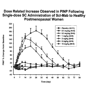
[0015] Figure 1 is a graph of percent change of N-terminal extension of
procollagen type 1
(P1NP) levels compared to baseline and placebo P1NP levels versus time (day)
post-
administration of various single doses of a sclerostin binding agent in
healthy,
postmenopausal women.
[0016] Figure 2 is a graph of percent change of bone-specific alkaline
phosphatase (BSAP)
levels compared to baseline and placebo BSAP levels versus time (day) post-
administration
of various single doses of a sclerostin binding agent in healthy,
postmenopausal women.
[0017] Figure 3 is a graph of percent change of osteocalcin levels compared to
baseline
and placebo osteocalcin levels versus time (day) post-administration of
various single doses
of a sclerostin binding agent in healthy, postmenopausal women.
[0018] Figure 4 is a graph of percent change of serum C-terminal telopeptide
of type 1
collagen (CTX) levels compared to baseline and placebo serum CTX levels versus
time (day)
6
CA 02699905 2010-03-17
WO 2009/039175 PCT/US2008/076679
post-administration of various single doses of a sclerostin binding agent in
healthy,
postmenopausal women.
[0019] Figure 5 are graphs of percent change of osteocalcin, BSAP, P1NP, and
CTX levels
compared to baseline and placebo levels versus time (day) post-administration
of a single
dose of 5 mg/kg or 10 mg/kg of sclerostin binding agent in healthy,
postmenopausal women.
[0020] Figure 6 is a graph of percent change of serum calcium levels compared
to baseline
and placebo serum calcium levels versus time (day) post-administration of
various single
doses of a sclerostin binding agent in healthy, postmenopausal women.
[0021] Figure 7 are graphs of percent change of bone mineral density compared
to baseline
and placebo versus time (day) post-administration of various single doses of
sclerostin
binding agent in healthy, postmenopausal women.
DETAILED DESCRIPTION OF THE INVENTION
[0022] The invention is predicated, at least in part, on the surprising
discovery that
blocking or inhibiting the biological activity of human sclerostin triggers
multiple
physiological responses linked to increased bone mineral density (BMD),
including
significant inhibition of bone resorption. Most currently available therapies
only inhibit bone
resorption without increasing bone formation. Some currently available
therapies for
disorders associated with reduced BMD only increase bone formation without
significantly
reducing bone resorption. For example, when bone formation is triggered by
some current
drugs, bone resorption may also increase (albeit potentially at a lower rate
than before
therapy). In contrast, agents that interfere with sclerostin activity both
enhance bone
formation and reduce bone resorption. In other words, sclerostin inhibitors
"uncouple" bone
formation and bone resorption to more effectively build bone. The materials
and methods of
the invention are superior to existing therapies whose therapeutic efficacy is
limited and
which are accompanied by potentially serious adverse side effects.
[0023] In this regard, the invention provides a method of inhibiting bone
resorption, e.g.,
bone resorption mediated by osteoclasts, bone cells that dissolve bone mineral
matrices. The
invention further provides a method of ameliorating the effects of an
osteoclast-related
disorder, i.e., a disorder caused by abnormally increased osteoclast activity
that, in some
embodiments, manifests as abnormally high bone resorption. The inventive
method
comprises administering to a human an amount of sclerostin binding agent that
reduces the
7
CA 02699905 2015-05-13
WO 2009/039175 PCT/US2008/076679
level of a marker of bone resorption and, optionally, increases the level of a
marker of bone
formation.
[0024] Activity of a sclerostin inhibitor, e.g., a sclerostin binding agent,
(further described
below) may be measured in a variety of ways. Sclerostin binding agent-mediated
increases in
bone mineral content or bone density may be measured using single- and dual-
energy X-ray
absorptometry, ultrasound, computed tomography, radiography, and magnetic
resonance
imaging. The amount of bone mass may also be calculated from body weights or
by using
other methods (see Guinness-Hey, Metab. Bone Dis. Relat. Res., 5:177-181
(1984)). Animals
and particular animal models are used in the art for testing the effect of the
pharmaceutical
compositions and methods on, for example, parameters of bone loss, bone
resorption, bone
formation, bone strength, or bone mineralization that mimic conditions of
human disease
such as osteoporosis and osteopenia. Examples of such models include the
ovariectomized
rat model (Kalu, Bone and Mineral, /5:175-192 (1991); Frost and Jee, Bone and
Mineral,
/8:227-236 (1992); and Jee and Yao, J. Musculoskel. Neuron. Interact., 1:193-
207 (2001)).
The methods for measuring sclerostin binding agent activity described herein
also may be
used to determine the efficacy of other sclerostin inhibitors.
[0025] In humans, bone mineral density can be determined clinically using dual
x-ray
absorptiometry (DXA) of, for example, the hip and spine. Other techniques
include
quantitative computed tomography (QCT), ultrasonography, single-energy x-ray
absorptiometry (SXA), and radiographic absorptiometry. Common central skeletal
sites for
measurement include the spine and hip; peripheral sites include the forearm,
finger, wrist and
heel. Except for ultrasonography, the American Medical Association notes that
BMD
techniques typically involve the use of x-rays and are based on the principle
that attenuation
of the radiation depends on thickness and composition of the tissues in the
radiation path. All
techniques involve the comparison of results to a normative database.
[0026] Alternatively, a physiological response to one or more sclerostin
binding agents can
be gauged by monitoring bone marker levels. Bone markers are products created
during the
bone remodeling process and are released by bone, osteoblasts, and/or
osteoclasts.
Fluctuations in bone resorption and/or bone formation "marker" levels imply
changes in bone
remodeling/modeling. The International Osteoporosis Foundation (I0F)
recommends using
bone markers to monitor bone density therapies (see, e.g., Delmas et al.,
Osteoporos Int.,
Suppl. 6:S2-17 (2000)= Markers indicative of bone
resorption (or osteoclast activity) include, for example, C-telopeptide (e.g.,
C-terminal
telopeptide of type 1 collagen (CTX) or serum cross-linked C-telopeptide), N-
telopeptide (N-
8
CA 02699905 2010-03-17
WO 2009/039175 PCT/US2008/076679
terminal telopeptide of type 1 collagen (NTX)), deoxypyridinoline (DPD),
pyridinoline,
urinary hydroxyproline, galactosyl hydroxylysine, and tartrate-resistant acid
phosphatase
(e.g., serum tartrate-resistant acid phosphatase isoform 5b). Bone
formation/mineralization
markers include, but are not limited to, bone-specific alkaline phosphatase
(BSAP), peptides
released from N- and C-terminal extension of type I procollagen (P1NP, PICP),
and
osteocalcin (OstCa). Several kits are commercially-available to detect and
quantify markers
in clinical samples, such as urine and blood.
[0027] Upon administration, the sclerostin binding agent preferably reduces
the level of
one or more markers of bone resorption, such as the serum level of C-
telopeptide of type I
collagen (CTX). Accordingly, the invention further provides a method of
monitoring anti-
sclerostin therapy, i.e., the physiological response to a sclerostin binding
agent or other
sclerostin inhibitor. The method comprises administering a sclerostin binding
agent, then
measuring the level of one or more markers of bone resorption. In addition,
the method can
comprise measuring the level of one or more markers of bone formation before
administration of a sclerostin binding agent. The level of bone resorption
marker during
and/or after treatment with the sclerostin binding agent may be compared to a
pre-treatment
level, or alternatively may be compared to a standard range typical of that
patient population.
One of ordinary skill in the art can readily determine a suitable standard
range by testing a
representative number of patients of like age, gender, disease level, and/or
other
characteristics of the patient population. The level of bone resorption marker
can be reduced
by at least about 5% (e.g., about 10%, about 20%, or about 30%) by a single
dose of
sclerostin binding agent. In some embodiments, the dose of sclerostin binding
agent reduces
the level of bone resorption marker at least about 40% (e.g., about 50%, about
60%, or about
70%) compared to the level of the bone resorption marker prior to
administering the
sclerostin binding agent. In addition, the bone resorption marker level may be
reduced for at
least about 3 days (e.g., about 7 days, about 2 weeks, about 3 weeks, about 1
month, about 5
weeks, about 6 weeks, about 7 weeks, about 2 months, about 9 weeks, about 10
weeks, about
11 weeks, or about 3 months) after administration of a single dose of the
sclerostin binding
agent.
[0028] In addition to decreasing the level of bone resorption markers, the
amount of
sclerostin binding agent administered to a patient also can increase the level
of one or more
markers of bone formation, such as the serum level of BSAP, the serum level of
P1NP,
and/or the serum level of OstCa. A single dose of sclerostin binding agent can
increase the
level of a bone formation marker by, for example, at least about 5% (e.g.,
about 10%, about
9
CA 02699905 2010-03-17
WO 2009/039175 PCT/US2008/076679
20%, or about 30%). In some embodiments, the dose of sclerostin binding agent
elevates the
level of a bone formation marker at least about 40% (e.g., about 50%, about
60%, or about
70%). In other embodiments, the dose of sclerostin binding agent increases the
level of one
or more bone formation markers by at least about 75% (e.g., about 80%, about
90%, about
100%, or about 110%). In yet other embodiments, the dose of sclerostin binding
agent
increases the level of a bone formation marker by at least about 120% (e.g.,
about 130%,
about 140%, about 150%, about 160% or about 170%). In alternative embodiments,
the
sclerostin binding agent increases the level of bone formation marker by least
about 180%
(e.g., about 190% or about 200%). Bone formation marker levels ideally remain
elevated
(compared to bone formation marker levels pre-treatment or to a standard range
typical of
that patient population) for at least about 3 days (e.g., about 7 days, about
2 weeks, about 3
weeks, about 1 month, about 5 weeks, about 6 weeks, about 7 weeks, about 2
months, about 9
weeks, about 10 weeks, about 11 weeks, or about 3 months) after administration
of a single
dose of the sclerostin binding agent.
[0029] The invention also provides a method of increasing bone mineral density
(BMD),
wherein an amount of sclerostin binding agent that (a) reduces the level of a
marker of bone
resorption and (b) increases the level of a marker of bone formation is
administered to a
human. BMD generally correlates with skeletal fragility and osteoporosis.
Typically, BMD
is can be measured "total body" (e.g., head, trunk, arms, and legs) or at the
hip (e.g., total hip
and/or femoral neck), spine (e.g., lumbar spine), wrist, finger, shin bone
and/or heel. In
osteoporosis diagnosis, a patient's BMD is compared to the peak density of a
30-year old
healthy adult (i.e., a "young adult"), creating the so-called "T-score." A
patient's BMD also
may be compared to an "age-matched" bone density (see, e.g., World Health
Organization
Scientific Group on the Prevention and Management of Osteoporosis, "Prevention
and
management of osteoporosis: report of a WHO scientific group." WHO Technical
Report
Series; 921, Geneva, Switzerland (2000)). The difference between a patient's
BMD and that
of a healthy, young adult is conventionally referred to in terms of the
multiple of a "standard
deviation," which typically equals about 10% to about 12% decrease in bone
density. The
World Health Organization proposed four diagnostic categories based on BMD T-
scores. A
BMD value within 1 standard deviation of the young adult reference mean (T-
score > -1) is
"normal." Low bone mass (osteopenia) is indicated by a BMD value more than 1
standard
deviation below the young adult mean, but less than 2 standard deviations (T-
score < -1 and >
-2.5). A T-score of more than 2.5 standard deviations below the norm supports
a diagnosis of
CA 02699905 2010-03-17
WO 2009/039175 PCT/US2008/076679
osteoporosis. If a patient additionally suffers from one or more fragility
fractures, the patient
qualifies as having severe osteoporosis.
[0030] The sclerostin inhibitor, e.g., a sclerostin binding agent, may be
administered to a
patient to improve bone mineral density regardless of the patient's T-score.
The sclerostin
binding agent may be administered at a dose and for a time period effective to
increase BMD
in the patient by at least about 1% (about 2%, about 3%, about 4%, about 5%,
or about 6%).
In some embodiments, BMD is increased by at least about 8% (e.g., at least
about 10%, about
12%, about 15%, or about 18%). In other embodiments, BMD is increased by the
sclerostin
binding agent at least about 20% (e.g., at least about 22%, about 25%, or
about 28%) at the
hip, spine, wrist, finger, shin bone, and/or heel. In yet other embodiments,
BMD is increased
at least about 30% (e.g., at least about 32%, about 35%, about 38%, or about
40%). In other
words, the BMD can be increased to the range of about 1 to about 2.5 standard
deviations
(preferably a range of about 0 to about 1 standard deviations) below the
normal BMD of a
healthy young adult.
[0031] Alterations in bone remodeling can lead to fluctuations in mineral
concentrations
throughout the body. Bone is one of the principal regulators of calcium levels
in the
bloodstream. Osteoclast-mediated bone resorption releases stored calcium into
the systemic
circulation, while osteoblast-mediated bone formation removes calcium from
circulation to
incorporate into bone tissue. In normal bone remodeling, these processes cycle
to maintain
healthy, strong bone and maintain free calcium levels at about 8.5 mg/dL to
about 10.5
mg/dL (e.g., about 2.2 mmol/L to about 2.6 mmol/L). Bone disorders, other
illnesses, and
even certain therapies can disrupt systemic calcium levels with dire
consequences.
Hypercalcemia is associated with high levels of calcium in the blood (e.g.,
greater than 12
mg/dL or 3 mmol/L). Extraordinarily high calcium levels leads to, for example,
fatigue,
confusion, constipation, decreased appetite, frequent urination, heart
problems, and bone
pain. Hypocalcemia is an electrolyte imbalance indicated by an abnormally low
level of
calcium in the blood (e.g., less than about 9 mg/dL or 2.2 mmol/L). Calcium
levels of < 7.5
mg/dL (< 1.87 mmol/L) or less are considered severe hypocalcemia and may be
accompanied
by clinical symptoms.
[0032] Common symptoms of hypocalcemia include nerve and muscle spasms and
cramps,
numbness, tingling in the extremities, confusion, and heart irregularities.
Extreme variations
in system calcium can lead to coma and death.
11
CA 02699905 2010-03-17
WO 2009/039175 PCT/US2008/076679
[0033] Several ailments and pharmaceutical therapies alter system calcium
levels.
Hypercalcemia and hypocalcemia can result from, for example, chronic kidney
disease, renal
failure, primary or secondary hyperparathyroidism, pseudohyperparathyroidism,
hypoparathyroidism, pseudohypoparathyroidism, magnesium depletion, alcoholism,
bisphosphonate therapy, severe hypermagnesemia, vitamin D deficiency,
hyperphosphatemia,
acute pancreatitis, hungry bone syndrome, chelation, osteoblastic metastases,
sepsis, surgery,
chemotherapy, neoplasia syndrome, familial hypocalciuric hypercalcemia,
sarcoidosis,
tuberculosis, berylliosis, histoplasmosis, Candidiasis, Coccidioidomycosis,
histiocytosis X,
Hodgkin's or Non-Hodgkin's lymphoma, Crohn's disease, Wegener's
granulomatosis,
leukemia, pneumonia, silicone-induced granulomas, immobilization, or drug
therapy, such as
administration of thiazide diuretics, lithium, estrogens, fluorides, glucose,
and insulin. In
addition, serum calcium fluctuations are a side effect of many existing bone-
related therapies,
such as bisphosphonate and parathyroid hormone therapy. Because of the
potentially life-
threatening consequences of calcium imbalance, patients susceptible to
hypocalcemia or
hypercalcemia may need to forego certain therapy options.
[0034] Remarkably, sclerostin inhibitors, e.g., sclerostin binding agents,
have been shown
to promote bone formation and inhibit (or slow) bone resorption with minimal
fluctuations in
systemic calcium levels (e.g., calcium levels fluctuate 10% or less from
baseline serum
calcium levels). Accordingly, the materials and method of the invention are
particularly
advantageous in treating patients that are susceptible or sensitive to
unstable calcium levels.
The amount of sclerostin binding agent administered to a human in the context
of this aspect
of the invention is an amount that does not result in hypocalcemia or
hypercalcemia (e.g.,
clinically-significant hypocalcemia or hypercalcemia). In addition, the
invention provides a
method of treating a bone-related disorder in a human suffering from or at
risk of
hypocalcemia or hypercalcemia or a human in which treatment with
bisphosphonate, a
parathyroid hormone, or parathyroid hormone analog is contraindicated. The
method
comprises administering to the human an amount of a sclerostin binding agent
effective to
increase the level of a marker of bone formation, such as serum levels of
BSAP, P1NP,
and/or OstCa and/or reduce the level of a marker of bone resorption, such as
CTX.
[0035] The inventive method is useful for treating or preventing bone-related
disorders,
such as bone-related disorders associated with abnormal osteoblast or
osteoclast activity.
Indeed, the sclerostin inhibitor (e.g., sclerostin binding agent) can be
administered to a human
suffering from a bone related disorder selected from the group consisting of
achondroplasia,
cleidocranial dysostosis, enchondromatosis, fibrous dysplasia, Gaucher's
Disease,
12
CA 02699905 2010-03-17
WO 2009/039175 PCT/US2008/076679
hypophosphatemic rickets, Marfan's syndrome, multiple hereditary exotoses,
neurofibromatosis, osteogenesis imperfecta, osteopetrosis, osteopoikilosis,
sclerotic lesions,
pseudoarthrosis, pyogenic osteomyelitis, periodontal disease, anti-epileptic
drug induced
bone loss, primary and secondary hyperparathyroidism, familial
hyperparathyroidism
syndromes, weightlessness induced bone loss, osteoporosis in men,
postmenopausal bone
loss, osteoarthritis, renal osteodystrophy, infiltrative disorders of bone,
oral bone loss,
osteonecrosis of the jaw, juvenile Paget's disease, melorheostosis, metabolic
bone diseases,
mastocytosis, sickle cell anemia/disease, organ transplant related bone loss,
kidney transplant
related bone loss, systemic lupus erythematosus, ankylosing spondylitis,
epilepsy, juvenile
arthritides, thalassemia, mucopolysaccharidoses, Fabry Disease, Turner
Syndrome, Down
Syndrome, Klinefelter Syndrome, leprosy, Perthe's Disease, adolescent
idiopathic scoliosis,
infantile onset multi-system inflammatory disease, Winchester Syndrome, Menkes
Disease,
Wilson's Disease, ischemic bone disease (such as Legg-Calve-Perthes disease
and regional
migratory osteoporosis), anemic states, conditions caused by steroids,
glucocorticoid-induced
bone loss, heparin-induced bone loss, bone marrow disorders, scurvy,
malnutrition, calcium
deficiency, osteoporosis, osteopenia, alcoholism, chronic liver disease,
postmenopausal state,
chronic inflammatory conditions, rheumatoid arthritis, inflammatory bowel
disease,
ulcerative colitis, inflammatory colitis, Crohn's disease, oligomenorrhea,
amenorrhea,
pregnancy, diabetes mellitus, hyperthyroidism, thyroid disorders, parathyroid
disorders,
Cushing's disease, acromegaly, hypogonadism, immobilization or disuse, reflex
sympathetic
dystrophy syndrome, regional osteoporosis, osteomalacia, bone loss associated
with joint
replacement, HIV associated bone loss, bone loss associated with loss of
growth hormone,
bone loss associated with cystic fibrosis, chemotherapy-associated bone loss,
tumor-induced
bone loss, cancer-related bone loss, hormone ablative bone loss, multiple
myeloma, drug-
induced bone loss, anorexia nervosa, disease-associated facial bone loss,
disease-associated
cranial bone loss, disease-associated bone loss of the jaw, disease-associated
bone loss of the
skull, bone loss associated with aging, facial bone loss associated with
aging, cranial bone
loss associated with aging, jaw bone loss associated with aging, skull bone
loss associated
with aging, and bone loss associated with space travel.
[0036] The inventive method need not cure the patient of the disorder or
completely
protect against the onset of a bone-related disorder to achieve a beneficial
biological
response. The method may be used prophylactically, meaning to protect, in
whole or in part,
against a bone-related disorder or symptom thereof. The method also may be
used
therapeutically to ameliorate, in whole or in part, a bone-related disorder or
symptom thereof,
13
CA 02699905 2015-05-13
WO 2009/039175 PCT/US2008/076679
or to protect, in whole or in part, against further progression of a bone-
related disorder or
symptom thereof. Indeed, the materials and methods of the invention are
particularly useful
for increasing bone mineral density and maintaining the increased BMD over a
period of
time. In this regard, the invention provides a method of treating a bone-
related disorder,
which method comprises (a) administering one or more amounts of a sclerostin
binding agent
effective to increase BMD measured for the total body (e.g., head, trunk,
arms, and legs) or at
the hip (e.g., total hip and/or femoral neck), spine (e.g., lumbar spine),
wrist, finger, shin
bone and/or heel by about 1%, about 2%, about 3%, about 6%, about 8%, about
10%, about
12%, about 15%, about 18%, about 20%, about 25%, or 30% or more. One or more
administrations of a pharmaceutical composition comprising the sclerostin
binding agent may
be carried out over a therapeutic period of, for example, about 1 month to
about 12 months
(e.g., about 2 months, about 3 months, about 4 months, about 5 months, about 6
months,
about 7 months, about 8 months, about 9 months, about 10 months, or about 11
months). The
method further includes (b) subsequently administering one or more amounts of
a sclerostin
binding agent effective to maintain bone mineral density. By "maintain bone
mineral
density" is meant that the increased BMD resulting from step (a) does not fall
more than
about 1% to about 5% over the course of step (b) (e.g., about 6 months, about
9 months about
1 year, about 18 months, about 2 years, or over the course of the patient's
life). It will be
appreciated that a patient can require alternate treatment phases for
increasing bone density
and maintaining bone density.
[0037] The sclerostin binding agent is preferably administered to a patient in
a
physiologically-acceptable (e.g., pharmaceutical) composition, which can
include carriers,
excipients, or diluents. It will be appreciated that the sclerostin binding
agents described
herein may be used in the preparation of a medicament for administration using
any of the
dosage and timing regimens disclosed herein. Pharmaceutical compositions and
methods of
treatment are disclosed in U.S. Patent Publication No. 20050106683.
"Physiologically-acceptable" refers to molecular entities and
compositions that do not produce an allergic or similar untoward reaction when
administered
to a human. In addition, the composition administered to a subject may contain
more than
one sclerostin inhibitor (e.g., a sclerostin binding agent and a synthetic
chemical sclerostin
inhibitor) or a sclerostin inhibitor in combination with one or more
therapeutics having
different mechanisms of action.
[0038] The development of suitable dosing and treatment regimens for using the
particular
compositions described herein in a variety of treatment regimens, including
e.g.,
14
CA 02699905 2010-03-17
WO 2009/039175 PCT/US2008/076679
subcutaneous, oral, parenteral, intravenous, intranasal, and intramuscular
administration and
formulation, is well known in the art and discussed in U.S. Patent Publication
No.
20070110747. For example, in certain circumstances, it will be desirable to
deliver a
pharmaceutical composition comprising a sclerostin binding agent
subcutaneously,
parenterally, intravenously, intramuscularly, or even intraperitoneally. Such
approaches are
well known to the skilled artisan, some of which are further described, for
example, in U.S.
Patent Nos. 5,543,158; 5,641,515; and 5,399,363. Illustrative pharmaceutical
forms suitable
for injectable use include sterile aqueous solutions or dispersions and
sterile powders for the
extemporaneous preparation of sterile injectable solutions or dispersions (for
example, see
U.S. Patent No. 5,466,468). In all cases the form must be sterile and must be
fluid to the
extent that easy syringability exists.
[0039] In one embodiment, for parenteral administration in an aqueous
solution, the
solution should be suitably buffered if necessary and the liquid diluent first
rendered isotonic
with sufficient saline or glucose. These particular aqueous solutions are
especially suitable
for intravenous, intramuscular, subcutaneous, and intraperitoneal
administration. For
example, one dose may be dissolved in 1 ml of isotonic NaCl solution and
either added to
1000 ml of hypodermoclysis fluid or injected at the proposed site of infusion
(see, for
example, Remington's Pharmaceutical Sciences, 15th ed., Mack Pub. Co., Easton,
PA, pp.
1035-1038 and 1570-1580). Some variation in dosage and frequency of
administration may
occur depending on the condition of the subject being treated; age, height,
weight, and overall
health of the patient; and the existence of any side effects. In addition, a
pharmaceutical
composition comprising a sclerostin binding agent may be placed within
containers (e.g.,
vials), along with packaging material that provides instructions regarding the
use of such
pharmaceutical compositions. Generally, such instructions will include a
tangible expression
describing the reagent concentration, as well as within certain embodiments,
relative amounts
of excipient ingredients or diluents (e.g., water, saline or PBS) that may be
necessary to
reconstitute the pharmaceutical composition.
[0040] The sclerostin binding agent is administered in an amount that reduces
the level of a
bone resorption marker and/or increases the level of a bone formation marker
and/or
increases bone density. The dose of sclerostin binding agent administered may
range from
about 0.5 mg/kg to about 20 mg/kg (e.g., 12 mg/kg) of body weight. For
example, the dose
of sclerostin binding agent may range from about 1 mg/kg to about 10 mg/kg
(e.g., about 2
mg/kg or about 9 mg/kg), about 1 mg/kg to about 3 mg/kg, or about 3 mg/kg to
about 8
mg/kg (e.g., about 4 mg/kg, 5 mg/kg, 6 mg/kg, or 7 mg/kg).
CA 02699905 2015-05-13
WO 2009/039175 PCT/US2008/076679
[0041] In addition, it may be advantageous to administer multiple doses of a
sclerostin
binding agent or space out the administration of doses, depending on the
therapeutic regimen
selected for a particular patient. The sclerostin binding agent can be
administered
periodically over a time period of one year or less (e.g., 9 months or less, 6
months or less, or
3 months or less). In this regard, the sclerostin binding agent can be
administered to the
human once every about 7 days, or 2 weeks, or 3 weeks, or 1 month, or 5 weeks,
or 6 weeks,
or 7 weeks, or 2 months. or 9 weeks, or 10 weeks, or 11 weeks, or 3 months, or
13 weeks, or
14 weeks, or 15 weeks, or 4 months, or 17 weeks, or 18 weeks, or 19 weeks, or
5 months, or
21 weeks, or 22 weeks, or 23 weeks, or 6 months, or 12 months.
[0042] The inventive method comprises administering an amount of a "sclerostin
inhibitor." As used herein, the term "sclerostin inhibitor" means any molecule
that inhibits
the biological activity of sclerostin on bone, as measured by changes to bone
mineralization,
bone density, effect on osteoblasts and/or osteoclasts, markers of bone
formation, markers of
bone resorption, markers of osteoblast activity, and/or markers of osteoclast
activity. Such
inhibitors may act by binding to sclerostin or its receptor or binding
partner. Inhibitors in this
category include "sclerostin binding agents," such as, e.g., antibodies or
peptide-based
molecules. "Sclerostin inhibitors" also refers to small organic chemical
compounds,
optionally of less than about 1000 Daltons in molecular weight that bind
sclerostin and inhibit
its activity. Inhibitors may alternatively act by inhibiting expression of
sclerostin. Inhibitors
in this category include polynucleotides or oligonucleotides that bind to
sclerostin DNA or
mRNA and inhibit sclerostin expression, including an antisense
oligonucleotide, inhibitory
RNA, DNA enzyme, ribozyme, an aptamer or pharmaceutically acceptable salts
thereof that
inhibit the expression of sclerostin.
[0043] A "sclerostin binding agent" specifically binds to sclerostin or
portions thereof to
block or impair binding of human sclerostin to one or more ligands.
Sclerostin, the product
of the SOST gene, is absent in sclerosteosis, a skeletal disease characterized
by bone
overgrowth and strong dense bones (Brunkow et al., Am. J. Hum. Genet., 68:577-
589 (2001);
Balemans et al., Hum. Mol. Genet., /0:537-543 (2001)). The amino acid sequence
of human
sclerostin is reported by Brunkow et al. and is disclosed in U.S. Patent
Publication No.
200701 10747 as SEQ ID NO: 1.
Recombinant human
sclerostin/SOST is commercially available from R&D Systems (Minneapolis,
Minn., USA;
2006 Catalog #1406-ST-025). Additionally, recombinant mouse sclerostin/SOST is
commercially available from R&D Systems (Minneapolis, Minn., USA; 2006 Catalog
#1589-
16
CA 02699905 2015-05-13
WO 2009/039175 PCT/US2008/076679
ST-025). Research grade sclerostin-binding monoclonal antibodies are
commercially
available from R&D Systems (Minneapolis, Minn., USA; mouse monoclonal: 2006
Catalog #
MAB1406; rat monoclonal: 2006 Catalog # MAB1589). U.S. Patent Nos. 6,395,511
and
6,803,453, and U.S. Patent Publication Nos. 20040009535 and 20050106683 refer
to anti-
sclerostin antibodies generally. Examples of sclerostin binding agents
suitable for use in the
context of the invention also are described in U.S. Patent Publication Nos.
20070110747 and
20070072797. Additional information regarding
materials and methods for generating sclerostin binding agents can be found in
U.S. Patent
Publication No. 20040158045.
[0044] The sclerostin binding agent of the invention preferably is an
antibody. The term
"antibody" refers to an intact antibody, or a binding fragment thereof. An
antibody may
comprise a complete antibody molecule (including polyclonal, monoclonal,
chimeric,
humanized, or human versions having full length heavy and/or light chains), or
comprise an
antigen binding fragment thereof. Antibody fragments include F(aW),, Fab,
Fab', Fv, Fc, and
Fd fragments, and can be incorporated into single domain antibodies, single-
chain antibodies,
maxibodies, minibodies, intrabodies, diabodies, triabodies, tetrabodies, v-NAR
and bis-scFv
(see, e.g., Hollinger and Hudson, Nature Biotechnolog)', 23(9):1126-1136
(2005)). Antibody
polypeptides, including fibronectin polypeptide monobodies, also are disclosed
in U.S. Patent
No. 6,703,199. Other antibody polypeptides are disclosed in U.S. Patent
Publication No.
20050238646. Anti-sclerostin antibodies may bind to sclerostin of SEQ ID NO:
1, or a
naturally occurring variant thereof, with an affinity of less than or equal to
1 x 10⁻⁷M, less
than or equal to 1 x 10⁻⁸M, less than or equal to 1 x 10⁻⁹M, less than or
equal to 1 x 10⁻¹ M,
less than or equal to 1 x 10⁻¹IM, or less than or equal to 1 x 10-12M.
Affinity may be
determined by an affinity ELISA assay. In certain embodiments, affinity may be
determined
by a BlAcore assay. In certain embodiments, affinity may be determined by a
kinetic
method. In certain embodiments, affinity may be determined by an
equilibrium/solution
method.
[0045] An antibody fragment may be any synthetic or genetically engineered
protein. For
example, antibody fragments include isolated fragments consisting of the light
chain variable
region, "Fv" fragments consisting of the variable regions of the heavy and
light chains,
recombinant single chain polypeptide molecules in which light and heavy
variable regions are
connected by a peptide linker (scFv proteins).
[0046] Another form of an antibody fragment is a peptide comprising one or
more
complementaiity determining regions (CDRs) of an antibody. CDRs (also termed
"minimal
17
CA 02699905 2016-06-06
,
recognition units" or "hypervariable region") can be obtained by constructing
polynucleotides
that encode the CDR of interest. Such polynucleotides are prepared, for
example, by using
the polymerase chain reaction to synthesize the variable region using mRNA of
antibody-
producing cells as a template (see, for example, Larrick et al., Methods: A
Companion to
Methods in Enzymology, 2:106 (1991); Courtenay-Luck, "Genetic Manipulation of
Monoclonal Antibodies," in Monoclonal Antibodies Production, Engineering and
Clinical
Application, Ritter et al. (eds.), page 166, Cambridge University Press
(1995); and Ward et
al., "Genetic Manipulation and Expression of Antibodies," in Monoclonal
Antibodies:
Principles and Applications, Birch et al., (eds.), page 137, Wiley-Liss, Inc.
(1995)).
[00471 In one embodiment of the invention, the sclerostin binding agent cross-
blocks the
binding of at least one of antibodies Ab-5 and Ab-23 (both of which are
described in U.S.
Patent Publication No. 20070110747) to sclerostin. Alternatively or in
addition, the sclerostin
binding agent is cross-blocked from binding to sclerostin by at least one of
antibodies Ab-5 and
Ab-23 (both of which are described in U.S. Patent Publication No.
20070110747). The terms
"cross-block," "cross-blocked," and "cross-blocking" are used interchangeably
herein to mean
the ability of an antibody or other binding agent to interfere with the
binding of other antibodies
or binding agents to sclerostin. The extent to which an antibody or other
binding agent is able to
interfere with the binding of another to sclerostin, and therefore whether it
can be said to cross-
block, can be determined using competition binding assays. In some aspects of
the invention, a
cross-blocking antibody or fragment thereof reduces sclerostin binding of a
reference antibody
between about 40% and about 100%, such as about 60% and about 100%,
specifically between
70% and 100%, and more specifically between 80% and 100%. A particularly
suitable
quantitative assay for detecting cross-blocking uses a Biacore machine which
measures the
extent of interactions using surface plasmon resonance technology. Another
suitable
quantitative cross-blocking assay uses an ELISA-based approach to measure
competition
between antibodies or other binding agents in terms of their binding to
sclerostin.
[0048] Suitable sclerostin binding agents include antibodies and portions
thereof described
in U.S. Patent Publication No. 20070110747, such as one or more of CDR-H1, CDR-
H2,
CDR-H3, CDR-L1, CDR-L2 and CDR-L3 as specifically disclosed therein. At least
one of
18
CA 02699905 2016-06-06
N
t
the regions of CDR-H1, CDR-H2, CDR-H3, CDR-L1, CDR-L2, and CDR-L3 may have at
least one amino acid substitution, provided that the binding agent retains the
binding
specificity of the non-substituted CDR. The non-CDR portion of the binding
agent may be a
non-protein molecule, wherein the binding agent cross-blocks the binding of an
antibody
disclosed herein to sclerostin and/or neutralizes sclerostin. The non-CDR
portion of the
binding agent may be a non-protein molecule in which the binding agent
exhibits a similar
binding pattern to human sclerostin peptides in a human sclerostin peptide
epitope
competition binding assay as that exhibited by at least one of antibodies Ab-5
and Ab-23 (both
of which are described in U.S. Patent Publication No. 20070110747), and/or
neutralizes
sclerostin. The non-CDR portion of the binding agent may be composed of amino
acids,
wherein the binding agent is a recombinant binding protein or a synthetic
peptide, and the
recombinant binding protein cross-blocks the binding of an antibody to
sclerostin and/or
neutralizes sclerostin. The non-CDR portion of the binding agent may be
composed of amino
acids, wherein the binding agent is a recombinant binding protein, and the
recombinant binding
protein exhibits a similar binding pattern to human sclerostin peptides in the
human sclerostin
peptide epitope competition binding assay (described in U.S. Patent
Publication No.
20070110747) as that exhibited by at least one of the antibodies Ab-5 and Ab-
23 (described in
U.S. Patent Publication No. 20070110747), and/or neutralizes sclerostin.
Preferably, the
sclerostin binding agent is Ab-5 or Ab-23 of U.S. Patent Publication No.
20070110747.
[0049] In addition, the sclerostin binding agent can comprise at least one CDR
sequence
having at least 75% identity (e.g., 100% identity) to a CDR selected from SEQ
ID NOs:
78, 79, 80, 239, 240, 241, 245, 246, 247, 269, 270 and 271 disclosed in
19 =
CA 02699905 2016-06-06
U.S. Patent Publication No. 20070110747. Preferably, the sclerostin binding
agent comprises
at least one CDR sequence having at least 75% identity to a CDR selected from
SEQ ID
NOs:245, 246, 247, 78, 79, 80, 269, 270, 271, 239, 240 and 241, all of which
is described in
U.S. Patent Publication NO. 20070110747. As described in U.S. Patent
Publication No.
20070110747, the sclerostin binding agent can comprise CDR sequences of SEQ ID
NOs:78,
79 and 80 and CDR sequences of SEQ ID NOs:245, 246 and 247, or CDR sequences
of SEQ
ID NOs:239, 240 and 241 and CDR sequences of SEQ ID NOs:269, 270 and 271.
[0050] The sclerostin binding agent also can comprise at least one CDR
sequence having
at least 75% identity to a CDR selected from CDR-H1, CDR-H2, CDR-H3, CDR-L1,
CDR-
L2, and CDR-L3 wherein CDR-111 has the sequence given in SEQ ID NO: 245 or SEQ
ID
NO: 269, CDR-H2 has the sequence given in SEQ ID NO: 246 or SEQ ID NO: 270,
CDR-H3
has the sequence given in SEQ ID NO: 247 or SEQ ID NO: 271, CDR-L1 has the
sequence
given in SEQ ID NO: 78 or SEQ ID NO: 239, CDR-L2 has the sequence given in SEQ
ID
CA 02699905 2010-03-17
WO 2009/039175 PCT/US2008/076679
NO: 79 or SEQ ID NO: 240 and CDR-L3 has the sequence given in SEQ ID NO: 80 or
SEQ
ID NO 241, all of which is described in U.S. Patent Publication No.
20070110747.
[0051] Alternatively, the sclerostin binding agent can have a heavy chain
comprising
CDR's H1, H2, and H3 and comprising a polypeptide having the sequence provided
in SEQ
ID NO: 137 or a variant thereof in which said CDR's are at least 75% identical
to SEQ ID
NO: 245, 246, and 247, respectively, and a light chain comprising CDR's L1, L2
and L3 and
comprising a polypeptide having the sequence provided in SEQ ID NO: 133 or a
variant
thereof in which said CDR's are at least 75% identical to SEQ ID NO: 78, 79,
and 80,
respectively (as described in U.S. Patent Publication No. 20070110747).
[0052] The sclerostin binding agent may have a heavy chain comprising CDR's
H1, H2,
and H3 and comprising a polypeptide having the sequence provided in SEQ ID NO:
145 or
392 or a variant thereof in which said CDR's are at least 75% identical to SEQ
ID NO: 245,
246, and 247, respectively, and a light chain comprising CDR's L1, L2, and L3
and
comprising a polypeptide having the sequence provided in SEQ ID NO: 141 or a
variant
thereof in which said CDR's are at least 75% identical to SEQ ID NO: 78, 79,
and 80,
respectively (as described in U.S. Patent Publication No. 20070110747).
[0053] The sclerostin binding agent may have a heavy chain comprising CDR's
H1, H2,
and H3 and comprising a polypeptide having the sequence provided in SEQ ID NO:
335 or a
variant thereof in which said CDR's are at least 75% identical to SEQ ID NO:
269, 270, and
271, respectively, and a light chain comprising CDR's L1, L2, and L3 and
comprising a
polypeptide having the sequence provided in SEQ ID NO: 334 or a variant
thereof in which
said CDR's are at least 75% identical to SEQ ID NO: 239, 240, and 241,
respectively (as
described in U.S. Patent Publication No. 20070110747).
[0054] Alternatively, the sclerostin binding agent has a heavy chain
comprising CDR's H1,
H2, and H3 and comprising a polypeptide having the sequence provided in SEQ ID
NO: 331
or a variant thereof in which said CDR's are at least 75% identical to SEQ ID
NO: 269, 270,
and 271, respectively, and a light chain comprising CDR's L1, L2, and L3 and
comprising a
polypeptide having the sequence provided in SEQ ID NO: 330 or a variant
thereof in which
said CDR's are at least 75% identical to SEQ ID NO: 239, 240, and 241,
respectively (as
described in U.S. Patent Publication No. 20070110747).
[0055] The sclerostin binding agent may have a heavy chain comprising CDR's
H1, H2,
and H3 and comprising a polypeptide having the sequence provided in SEQ ID NO:
345 or
396 or a variant thereof in which said CDR's are at least 75% identical to SEQ
ID NO: 269,
21
CA 02699905 2016-06-06
e =
270, and 271. respectively, and a light chain comprising CDR's LI, L2, and L3
and
comprising a polypeptide having the sequence provided in SEQ ID NO: 341 or a
variant
thereof in which said CDR's are at least 75% identical to SEQ ID NO: 239, 240,
and 241,
respectively (as described in U.S, Patent Publication No. 20070110747),
100561 Alternatively, the sclerostin binding agent has a heavy chain
comprising a polypeptide
having the sequence provided in SEQ ID NO:145 or 392, and a light chain
comprising a
polypeptide having the sequence provided in SEQ ID NO:141; or a heavy chain
comprising a
polypeptide having the sequence provided in SEQ ID NO:345 or 396, and a light
chain
comprising a polypeptide having the sequence provided in SEQ ID NO:341 (as
described in
U.S. Patent Publication NO. 20070110747).
[0057] Sclerostin binding agents for use in the inventive method preferably
modulate
sclerostin function in the cell-based assay described in U.S. Patent
Publication No.
20070110747 and/or the in vivo assay described in U.S. Patent Publication No.
20070110747
and/or bind to one or more of the epitopes described in U.S. Patent
Publication No.
20070110747 and/or cross-block the binding of one of the antibodies described
in U.S. Patent
Publication No. 20070110747 and/or are cross-blocked from binding sclerostin
by one of the
antibodies described in U.S. Patent Publication No. 20070110747.
[0058] Alternatively, the inventive method can comprise administering a
sclerostin
inhibitor other than a sclerostin binding agent described herein. Such agents
can act directly
or indirectly on SOST or sclerostin. Sclerostin inhibitors contemplated for
use in the
inventive method include those described in U.S. Patent
Publication No. 20030229041. For example,
agents useful for modulating SOST
expression and sclerostin activity include, but are not limited to, steroids
(such as those
corresponding to Formula 1 of U.S. Patent Publication No. 20030229041),
alkaloids,
terpenoids, peptoids, and synthetic chemicals. In some embodiments, the SOST
antagonist or
agonist can bind to a glucocorticoid receptor. For example, dexamethasone
tends to abolish
the stimulatory effect of BMP-4 and BMP-6 on SOST expression. Other chemical
entities
= 22
CA 02699905 2015-05-13
WO 2009/039175 PCT/US2008/076679
including glucocorticoid analogs, bile salts (such as those corresponding to
Formula 3 of U.S.
Patent Publication No. 20030229041), and prostaglandins (such as those
corresponding to
Formula 2 of U.S. Patent Publication No. 20030229041) also modulate the
effects of bone
morphogenetic proteins on SOST expression, and are contemplated for use in the
inventive
method.
[0059] The sclerostin inhibitor may also be other small molecule therapeutics
that act
directly or indirectly on SOST or sclerostin to decrease the level of at least
one bone
resorptive marker and/or increase the level of at least one bone formation
marker in vivo.
The term "small molecule" includes a compound or molecular complex, either
synthetic,
naturally derived, or paitially synthetic, and which preferably has a
molecular weight of less
than 5,000 Daltons (e.g., between about 100 and 1,500 Daltons). Agents can be
obtained
using any of the numerous approaches in combinatorial library methods known in
the art,
including spatially addressable parallel solid phase or solution phase
libraries, synthetic
library methods requiring deconvolution, the "one-bead one-compound" library
method, and
synthetic library methods using affinity chromatography selection (see, e.g.,
Lam, Anticancer
Drug Des., /2:145 (1997) and U.S. Patent Nos. 5,738,996; 5,807.683; and
7,261,892).
Methods of developing and screening sclerostin inhibitors are further
described in U.S. Patent
Publication No. 20030229041.
[0060] Sclerostin expression inhibitors that may be used according to the
methods of the
invention include inhibitor oligonucleotides or polynucleotides, including
pharmaceutically
acceptable salts thereof, e.g., sodium salts. Nonlimiting examples include:
antisense
oligonucleotides (Eckstein, Antisense Nucleic Acid Drug Dev., 10: 117-
121(2000); Crooke,
Methods Enzymol., 313: 3-45 (2000); Guvakova et al., J. Biol. Chem., 270: 2620-
2627
(1995); Manoharan, Biochirn. Biophys. Acta, 1489: 117-130 (1999); Baker et
al., J. Biol.
Chem., 272: 11994-12000 (1997); Kurreck, Eur. J. Biochem., 270: 1628-1644
(2003);
Sierakowska et al., Proc. Natl. Acad. Sci. USA, 93: 12840-12844 (1996);
Marwick, J. Am.
Med. Assoc., 280: 871 (1998); Tomita and Morishita, Cum Phann. Des., 10: 797-
803 (2004);
Gleave and Monia, Nat. Rev. Cancer, 5: 468-479 (2005) and Patil. AAPS J., 7:
E61-E77
(2005)), triplex oligonucleotides (Francois et al., Nucleic Acids Res., 16:
11431-11440 (1988)
and Moser and Dervan, Science, 238: 645-650 (1987)). ribozymes/deoxyribozymes
(DNAzymes) (Kruger et al., Tetrahymena. Cell. 31:147-157 (1982); Uhlenbeck,
Nature,
328: 596-600 (1987); Sigurdsson and Eckstein, Trends Biotechnol., 13: 286-289
(1995);
Kumar et al., Gene Ther., 12: 1486-1493 (2005); Breaker and Joyce, Chem.
Biol., 1: 223-229
(1994); Khachigian, Curr. Pharm. Biotechnol., 5: 337-339 (2004); Khachigian,
Biochem.
23
CA 02699905 2010-03-17
WO 2009/039175 PCT/US2008/076679
Pharmacol., 68: 1023-1025 (2004) and Trulzsch and Wood, J. Neurochem., 88: 257-
265
(2004)), small-interfering RNAs/RNAi (Fire et al., Nature, 391: 806-811
(1998);
Montgomery et al., Proc. Natl. Acad. Sci. U.S.A., 95: 15502-15507 (1998);
Cullen, Nat.
Immunol., 3: 597-599 (2002); Hannon, Nature, 418: 244-251 (2002); Bernstein et
al., Nature,
409: 363-366 (2001); Nykanen et al., Cell, 107: 309-321 (2001); Gilmore et
al., J. Drug
Target., 12: 315-340 (2004); Reynolds et al., Nat. Biotechnol., 22: 326-330
(2004);
Soutschek et al., Nature, 432173-178 (2004); Ralph et al., Nat. Med., 11: 429-
433 (2005);
Xia et al., Nat. Med., 10816-820 (2004) and Miller et al., Nucleic Acids Res.,
32: 661-668
(2004)), aptamers (Ellington and Szostak, Nature, 346: 818-822 (1990); Doudna
et al., Proc.
Natl. Acad. Sci. U.S.A., 92: 2355-2359 (1995); Tuerk and Gold, Science, 249:
505-510
(1990); White et al., Mol. Ther., 4: 567-573 (2001); Rusconi et al., Nature,
419: 90-94
(2002); Nimjee et al., Mol. Ther., 14: 408-415 (2006); Gragoudas et al., N.
Engl. J. Med.,
351: 3805-2816 (2004); Vinores, Curr. Opin. Mol. Ther., 5673-679 (2003) and
Kourlas and
Schiller et al., Clin. Ther., 28: 36-44 (2006)) or decoy oligonucleotides
(Morishita et al.,
Proc. Natl. Acad. Sci. U.S.A., 92: 5855-5859 (1995); Alexander et al., J. Am.
Med. Assoc.,
294: 2446-2454 (2005); Mann and Dzau, J. Clin. Invest., 106: 1071-1075 (2000)
and Nimjee
et al., Annu. Rev. Med., 56: 555-583 (2005)). The foregoing documents are
hereby
incorporated by reference in their entirety herein, with particular emphasis
on those sections
of the documents relating to methods of designing, making and using inhibitory
oligonucleotides. Commercial providers such as Ambion Inc. (Austin, TX),
Darmacon Inc.
(Lafayette, CO), InvivoGen (San Diego, CA), and Molecular Research
Laboratories, LLC
(Herndon, VA) generate custom siRNA molecules. In addition, commercial kits
are available
to produce custom siRNA molecules, such as SILENCERTM siRNA Construction Kit
(Ambion Inc., Austin, TX) or psiRNA System (InvivoGen, San Diego, CA).
[0061] Inhibitory oligonucleotides which are stable, have a high resistance to
nucleases,
possess suitable pharmacokinetics to allow them to traffic to target tissue
site at non-toxic
doses, and have the ability to cross through plasma membranes are contemplated
for use as a
therapeutic. Inhibitory oligonucleotides may be complementary to the coding
portion of a
target gene, 3' or 5' untranslated regions, or intronic sequences in a gene,
or alternatively
coding or intron sequences in the target mRNA. Intron sequences are generally
less
conserved and thus may provide greater specificity. In one embodiment, the
inhibitory
oligonucleotide inhibits expression of a gene product of one species but not
its homologue in
another species; in other embodiments, the inhibitory oligonucleotide inhibits
expression of a
gene in two species, e.g. human and primate, or human and murine.
24
CA 02699905 2010-03-17
WO 2009/039175 PCT/US2008/076679
[0062] The constitutive expression of antisense oligonucleotides in cells has
been shown to
inhibit gene expression, possibly via the blockage of translation or
prevention of splicing. In
certain embodiments, the inhibitory oligonucleotide is capable of hybridizing
to at least 8, 9,
10, 11, or 12 consecutive bases of the sclerostin gene or mRNA (or the reverse
strand thereof)
under moderate or high stringency conditions. Suitable inhibitory
oligonucleotides may be
single stranded and contain a segment, e.g. at least 12, 15 or 18 bases in
length, that is
sufficiently complementary to, and specific for, an mRNA or DNA molecule such
that it
hybridizes to the mRNA or DNA molecule and inhibits transcription, splicing or
translation.
Generally complementarity over a length of less than 30 bases is more than
sufficient.
[0063] Typically, stringent conditions will be those in which the salt
concentration is less
than about 1.5 M Na ion, typically about 0.01 to 1.0 M Na ion concentration
(or other salts) at
pH 7.0 to 8.3 and the temperature is at least about 30 C for short nucleic
acids (e.g., 10 to 50
nucleotides) and at least about 60 C for longer nucleic acids (e.g., greater
than 50
nucleotides). Stringent conditions may also be achieved with the addition of
destabilizing
agents such as formamide. Exemplary low stringency conditions include
hybridization with a
buffer solution of 30% to 35% formamide, 1 M NaCl, 1% SDS (sodium dodecyl
sulphate) at
37 C, and a wash in 1X to 2X SSC (20X SSC = 3.0 M NaCl/0.3 M trisodium
citrate) at 50 C
to 55 C. Exemplary moderate stringency conditions include hybridization in 40%
to 45%
formamide, 1.0 M NaCl, 1% SDS at 37 C, and a wash in 0.5X to 1X SSC at 55 C to
60 C.
Exemplary high stringency conditions include hybridization in 50% formamide, 1
M NaCl,
1% SDS at 37 C, and a wash in 0.1X SSC at 60 C to 65 C. Duration of
hybridization is
generally less than about 24 hours, usually about 4 hours to about 12 hours.
[0064] In some cases, depending on the length of the complementary region,
one, two or
more mismatches may be tolerated without affecting inhibitory function. In
certain
embodiments, the inhibitory oligonucleotide is an antisense oligonucleotide,
an inhibitory
RNA (including siRNA or RNAi, or shRNA), a DNA enzyme, a ribozyme (optionally
a
hammerhead ribozyme), an aptamer, or pharmaceutically acceptable salts
thereof. In one
embodiment, the oligonucleotide is complementary to at least 10 bases of the
nucleotide
sequence encoding SEQ ID NO: 1 of U.S. Patent Publication No. 20040158045. In
one
embodiment, the oligonucleotide targets the nucleotides located in the
vicinity of the 3'
untranslated region of the sclerostin mRNA.
[0065] The specific sequence utilized in design of the oligonucleotides may be
any
contiguous sequence of nucleotides contained within the expressed gene message
of the
target. Factors that govern a target site for the inhibitory oligonucleotide
sequence include
CA 02699905 2015-05-13
WO 2009/039175 PCT/1JS2008/076679
the length of the oligonucleotide, binding affinity, and accessibility of the
target sequence.
Sequences may be screened in vitro for potency of their inhibitory activity by
measuring
inhibition of target protein translation and target related phenotype, e.g.,
inhibition of cell
proliferation in cells in culture. In general it is known that most regions of
the RNA (5' and
3' untranslated regions, AUG initiation, coding, splice junctions and introns)
can be targeted
using anti sense oligonucleotides. Programs and algorithms, known in the art,
may be used to
select appropriate target sequences. In addition, optimal sequences may be
selected utilizing
programs designed to predict the secondary structure of a specified single
stranded nucleic
acid sequence and allowing selection of those sequences likely to occur in
exposed single
stranded regions of a folded mRNA. Methods and compositions for designing
appropriate
oligonucleotides may be found, for example, in U.S. Patent No. 6,251,588.
[0066] Phosphorothioate antisense oligonucleotides may be used. Modifications
of the
phosphodiester linkage as well as of the heterocycle or the sugar may provide
an increase in
efficiency. Phophorothioate is used to modify the phosphodiester linkage. An
N3'-P5'
phosphoramidate linkage has been described as stabilizing oligonucleotides to
nucleases and
increasing the binding to RNA. Peptide nucleic acid (PNA) linkage is a
complete
replacement of the ribose and phosphodiester backbone and is stable to
nucleases, increases
the binding affinity to RNA, and does not allow cleavage by RNAse H. Its basic
structure is
also amenable to modifications that may allow its optimization as an antisense
component.
With respect to modifications of the heterocycle, certain heterocycle
modifications have
proven to augment antisense effects without interfering with RNAse H activity.
An example
of such modification is C-5 thiazole modification. Finally, modification of
the sugar may
also be considered. 2'-0-propyl and 2'-methoxyethoxy ribose modifications
stabilize
oligonucleotides to nucleases in cell culture and in vivo.
[0067] Most mRNAs have been shown to contain a number of secondary and
tertiary
structures. Secondary structural elements in RNA are formed largely by Watson-
Crick type
interactions between different regions of the same RNA molecule. Important
secondary
structural elements include intramolecular double stranded regions, hairpin
loops, bulges in
duplex RNA and internal loops. Tertiary structural elements are formed when
secondary
structural elements come in contact with each other or with single stranded
regions to
produce a more complex three dimensional structure. A number of researchers
have
measured the binding energies of a large number of RNA duplex structures and
have derived
a set of rules which can be used to predict the secondary structure of RNA
(see, e.g., Jaeger et
26
CA 02699905 2010-03-17
WO 2009/039175 PCT/US2008/076679
al., Proc. Natl. Acad. Sci. USA, 86:7706 (1989); and Turner et al., Annu. Rev.
Biophys.
Biophys. Chem. 17:167 (1988)). The rules are useful in identification of RNA
structural
elements and, in particular, for identifying single stranded RNA regions which
may represent
segments of the mRNA to target for siRNA, ribozyme, or antisense technologies.
[0068] Short interfering (si) RNA technology (also known as RNAi) generally
involves
degradation of an mRNA of a particular sequence induced by double-stranded RNA
(dsRNA)
that is homologous to that sequence, thereby "interfering" with expression of
the
corresponding gene. Any selected gene may be repressed by introducing a dsRNA
which
corresponds to all or a substantial part of the mRNA for that gene. It appears
that when a
long dsRNA is expressed, it is initially processed by a ribonuclease III into
shorter dsRNA
oligonucleotides of as few as 21 to 22 base pairs in length. Accordingly,
siRNA may be
affected by introduction or expression of relatively short homologous dsRNAs.
Exemplary
siRNAs have sense and antisense strands of about 21 nucleotides that form
approximately 19
nucleotides of double stranded RNA with overhangs of two nucleotides at each
3' end.
Indeed the use of relatively short homologous dsRNAs may have certain
advantages.
[0069] Mammalian cells have at least two pathways that are affected by double-
stranded
RNA (dsRNA). In the sequence-specific siRNA pathway, the initiating dsRNA is
first
broken into short interfering RNAs, as described above. Short interfering RNAs
are thought
to provide the sequence information that allows a specific messenger RNA to be
targeted for
degradation. In contrast, the nonspecific pathway is triggered by dsRNA of any
sequence, as
long as it is at least about 30 base pairs in length.
[0070] The nonspecific effects occur because dsRNA activates two enzymes: PKR,
which
in its active form phosphorylates the translation initiation factor eIF2 to
shut down all protein
synthesis, and 2', 5' oligoadenylate synthetase (2', 5'-AS), which synthesizes
a molecule that
activates RNase L, a nonspecific enzyme that targets all mRNAs. The
nonspecific pathway
may represent a host response to stress or viral infection, and, in general,
the effects of the
nonspecific pathway are preferably minimized. Significantly, longer dsRNAs
appear to be
required to induce the nonspecific pathway and, accordingly, dsRNAs shorter
than about 30
bases pairs are contemplated to effect gene repression by RNAi (see Hunter et
al., J. Biol.
Chem., 250: 409-17 (1975); Manche et al., Mol. Cell. Biol. 12: 5239-48 (1992);
Minks et al.,
J. Biol. Chem., 254: 10180-3 (1979); and Elbashir et al., Nature, 411: 494-8
(2001)).
[0071] siRNA has proven to be an effective means of decreasing gene expression
in a
variety of cell types. siRNA typically decreases expression of a gene to lower
levels than that
27
CA 02699905 2015-05-13
WO 2009/039175 PCT/US2008/076679
achieved using antisense techniques, and frequently eliminates expression
entirely (see Bass,
Nature, 411: 428-9 (2001)). In mammalian cells, siRNAs are effective at
concentrations that
are several orders of magnitude below the concentrations typically used in
antisense
experiments (Elbashir et al., Nature, 411:494-8 (2001)).
[0072] The double stranded oligonucleotides used to effect RNAi are preferably
less than
30 base pairs in length, for example, about 25, 24, 23, 22, 21, 20, 19, 18, or
17 base pairs or
less in length, and contain a segment sufficiently complementary to the target
mRNA to
allow hybridization to the target mRNA. Optionally the dsRNA oligonucleotides
may
include 3' overhang ends. Exemplary 2-nucleotide 3' overhangs may be composed
of
ribonucleotide residues of any type and may even be composed of 2'-
deoxythymidine
resides, which lowers the cost of RNA synthesis and may enhance nuclease
resistance of
siRNAs in the cell culture medium and within transfected cells (see Elbashi et
al., supra).
Exemplary dsRNAs may be synthesized chemically or produced in vitro or in vivo
using
appropriate expression vectors (see, e.g.. Elbashir et al., Genes Dev., 15:188-
200 (2001)).
Longer RNAs may be transcribed from promoters, such as T7 RNA polymerase
promoters,
known in the art.
[0073] Longer dsRNAs of 50, 75, 100, or even 500 base pairs or more also may
be utilized
in certain embodiments of the invention. Exemplary concentrations of dsRNAs
for effecting
RNAi are about 0.05 nM, 0.1 nM, 0.5 nM, 1.0 nM, 1.5 nM, 25 nM, or 100 nM,
although
other concentrations may be utilized depending upon the nature of the cells
treated, the gene
target and other factors readily discernable to the skilled artisan.
[0074] Further compositions, methods and applications of siRNA technology are
provided
in U.S. Patent Nos. 6,278,039; 5,723,750; and 5,244,805.
[0075] Compared to siRNA, shRNA offers advantages in silencing longevity and
delivery
options. See, e.g., Hannon et al., Nature, 43/:371-378 (2004) for review.
Vectors that
produce shRNAs, which are processed intracellularly into short duplex RNAs
having siRNA-
like properties have been reported (Brummelkamp et al., Science, 296: 550-553
(2000);
Paddison et al., Genes Dev., 16: 948-958 (2002)). Such vectors provide a
renewable source
of a gene-silencing reagent that can mediate persistent gene silencing after
stable integration
of the vector into the host-cell genome. Furthermore, the core silencing
'hairpin' cassette can
be readily inserted into retroviral, lentiviral, or adenoviral vectors,
facilitating delivery of
shRNAs into a broad range of cell types (Brummelkamp et al., Cancer Cell,
2:243-247
28
CA 02699905 2010-03-17
WO 2009/039175 PCT/US2008/076679
(2002); Dirac et al., J. Biol. Chem., 278:11731-11734 (2003); Michiels et al.,
Nat.
Biotechnol., 20:1154-1157 (2002); Stegmeie et al., Proc. Natl. Acad. Sci. USA,
102:13212-
13217 (2005); Khvorova et al., Cell, 115:209-216 (2003)) in any of the
innumerable ways
that have been devised for delivery of DNA constructs that allow ectopic mRNA
expression.
[0076] A hairpin can be organized in either a left-handed hairpin (i.e., 5'-
antisense-loop-
sense-3') or a right-handed hairpin (i.e., 5'-sense-loop-antisense-3'). The
siRNA may also
contain overhangs at either the 5' or 3' end of either the sense strand or the
antisense strand,
depending upon the organization of the hairpin. Preferably, if there are any
overhangs, they
are on the 3' end of the hairpin and comprise between 1 to 6 bases. The
overhangs can be
unmodified, or can contain one or more specificity or stabilizing
modifications, such as a
halogen or 0-alkyl modification of the 2' position, or internucleotide
modifications such as
phosphorothioate, phosphorodithioate, or methylphosphonate modifications. The
overhangs
can be ribonucleic acid, deoxyribonucleic acid, or a combination of
ribonucleic acid and
deoxyribonucleic acid.
[0077] Additionally, a hairpin can further comprise a phosphate group on the
5'-most
nucleotide. The phosphorylation of the 5'-most nucleotide refers to the
presence of one or
more phosphate groups attached to the 5' carbon of the sugar moiety of the 5'-
terminal
nucleotide. Preferably, there is only one phosphate group on the 5' end of the
region that will
form the antisense strand following Dicer processing. In one exemplary
embodiment, a right-
handed hairpin can include a 5' end (i.e., the free 5' end of the sense
region) that does not
have a 5' phosphate group, or can have the 5' carbon of the free 5'-most
nucleotide of the
sense region being modified in such a way that prevents phosphorylation. This
can be
achieved by a variety of methods including, but not limited to, addition of a
phosphorylation
blocking group (e.g., a 5'-0-alkyl group), or elimination of the 5'-OH
functional group (e.g.,
the 5'-most nucleotide is a 5'-deoxy nucleotide). In cases where the hairpin
is a left-handed
hairpin, preferably the 5' carbon position of the 5'-most nucleotide is
phosphorylated.
[0078] Hairpins that have stem lengths longer than 26 base pairs can be
processed by Dicer
such that some portions are not part of the resulting siRNA that facilitates
mRNA
degradation. Accordingly the first region, which may comprise sense
nucleotides, and the
second region, which may comprise antisense nucleotides, may also contain a
stretch of
nucleotides that are complementary (or at least substantially complementary to
each other),
but are or are not the same as or complementary to the target mRNA. While the
stem of the
shRNA can be composed of complementary or partially complementary antisense
and sense
strands exclusive of overhangs, the shRNA can also include the following: (1)
the portion of
29
CA 02699905 2015-05-13
WO 2009/039175
PCT/US2008/076679
the molecule that is distal to the eventual Dicer cut site contains a region
that is substantially
complementary/homologous to the target mRNA; and (2) the region of the stem
that is
proximal to the Dicer cut site (i.e., the region adjacent to the loop) is
unrelated or only
partially related (e.g., complementary/homologous) to the target mRNA. The
nucleotide
content of this second region can be chosen based on a number of parameters
including but
not limited to thermodynamic traits or profiles.
[0079] Modified shRNAs can retain the modifications in the post-Dicer
processed duplex.
In exemplary embodiments, in cases in which the hairpin is a right handed
hairpin (e.g., 5'-S-
loop-AS-3') containing 2-6 nucleotide overhangs on the 3' end of the molecule,
2'-0-methyl
modifications can be added to nucleotides at position 2, positions 1 and 2, or
positions 1, 2,
and 3 at the 5' end of the hairpin. Also, Dicer processing of hairpins with
this configuration
can retain the 5' end of the sense strand intact, thus preserving the pattern
of chemical
modification in the post-Dicer processed duplex. Presence of a 3' overhang in
this
configuration can be particularly advantageous since blunt ended molecules
containing the
prescribed modification pattern can be further processed by Dicer in such a
way that the
nucleotides carrying the 2' modifications are removed. In cases where the 3'
overhang is
present/retained, the resulting duplex carrying the sense-modified nucleotides
can have
highly favorable traits with respect to silencing specificity and
functionality. Examples of
exemplary modification patterns are described in detail in U.S. Patent
Publication No.
20050223427 and International Patent Publication Nos. WO 2004/090105 and WO
2005/078094.
[0080] shRNA may comprise sequences that were selected at random, or according
to any
rational design selection procedure. For example, rational design algorithms
are described in
International Patent Publication No. WO 2004/045543 and U.S. Patent
Publication No.
20050255487. Additionally, it may be desirable to
select sequences in whole or in part based on
average internal stability profiles ("AISPs") or regional internal stability
profiles ("RISPs")
that may facilitate access or processing by cellular machinery.
[0081] Ribozymes are enzymatic RNA molecules capable of catalyzing specific
cleavage
of mRNA, thus preventing translation. (For a review, see Rossi, Current
Biology, 4:469-471
(1994)). The mechanism of ribozyme action involves sequence specific
hybridization of the
ribozyme molecule to complementary target RNA, followed by an endonucleolytic
cleavage
event. The ribozyme molecules preferably include (1) one or more sequences
CA 02699905 2015-05-13
WO 2009/039175 PCT/US2008/076679
complementary to a target mRNA, and (2) the well known catalytic sequence
responsible for
mRNA cleavage or a functionally equivalent sequence (see, e.g., U.S. Patent
No. 5,093,246.
[0082] While ribozymes that cleave mRNA at site-specific recognition sequences
can be
used to destroy target mRNAs, hammerhead ribozymes may alternatively be used.
Hammerhead ribozymes cleave mRNAs at locations dictated by flanking regions
that form
complementary base pairs with the target mRNA. Preferably, the target mRNA has
the
following sequence of two bases: 5'-UG-3'. The construction and production of
hammerhead ribozymes is well known in the art and is described more fully in
Haseloff and
Gerlach, Nature, 334:585-591 (1988); and International Patent Publication. No.
WO
89/05852.
[0083] Gene targeting ribozymes may contain a hybridizing region complementary
to two
regions of a target mRNA, each of which is at least 5, 6, 7, 8, 9, 10, 11, 12,
13, 14, 15, 16, 17,
18, 19, or 20 contiguous nucleotides (but which need not both be the same
length).
[0084] Hammerhead ribozyme sequences can be embedded in a stable RNA such as a
transfer RNA (tRNA) to increase cleavage efficiency in vivo (Perriman et al.,
Proc. Natl.
Acad. Sci. USA, 92:6175-79 (1995); de Feyter and Gaudron, Methods in Molecular
Biology,
Vol. 74, Chapter 43, "Expressing Ribozymes in Plants," Turner, P. C. (ed.),
Humana Press
Inc., Totowa, N.J.). In particular, RNA polymerase III-mediated expression of
tRNA fusion
ribozymes are well known in the art (see Kawasaki et al., Nature, 393:284-9
(1998);
Kuwabara et al., Nature Biotechnol., /6:961-5 (1998); and Kuwabara et al.,
Mol. Cell, 2:617-
27 (1998); Koseki et al., J. Virol.. 73:1868-77 (1999); Kuwabara et al., Proc.
Natl. Acad. Sci.
USA, 96:1886-91 (1999); Tanabe et al., Nature, 406:473-4 (2000)). There are
typically a
number of potential hammerhead ribozyme cleavage sites within a given target
cDNA
sequence. Preferably the ribozyme is engineered so that the cleavage
recognition site is
located near the 5' end of the target mRNA- to increase efficiency and
minimize the
intracellular accumulation of non-functional mRNA transcripts. Furthermore,
the use of any
cleavage recognition site located in the target sequence encoding different
portions of the
target mRNA would allow the selective targeting of one or the other target
genes.
[0085] Ribozymes for use in the inventive method also include RNA
endoribonucleases
("Cech-type ribozymes") such as the one which occurs naturally in Tetrahyrnena
thennophila
(known as the IVS, or L-19 IVS RNA) and which has been extensively described
in Zaug et
al., Science, 224:574-578 (1984); Zaug, et al., Science, 231:470-475 (1986);
Zaug et al.,
31
CA 02699905 2010-03-17
WO 2009/039175 PCT/US2008/076679
Nature, 324:429-433 (1986); International Patent Publication No. WO 88/04300;
and Been et
al., Cell, 47:207-216 (1986)). The Cech-type ribozymes have an eight base pair
active site
which hybridizes to a target RNA sequence whereafter cleavage of the target
RNA takes
place. In one embodiment, the inventive method employs those Cech-type
ribozymes which
target eight base-pair active site sequences that are present in a target gene
or nucleic acid
sequence.
[0086] Ribozymes can be composed of modified oligonucleotides (e.g., for
improved
stability, targeting, etc.) and can be chemically synthesized or produced
through an
expression vector. Because ribozymes, unlike antisense molecules, are
catalytic, a lower
intracellular concentration is required for efficiency. Additionally, in
certain embodiments, a
ribozyme may be designed by first identifying a sequence portion sufficient to
cause effective
knockdown by RNAi. Portions of the same sequence may then be incorporated into
a
ribozyme.
[0087] Alternatively, target gene expression can be reduced by targeting
deoxyribonucleotide sequences complementary to the regulatory region of the
gene (i.e., the
promoter and/or enhancers) to form triple helical structures that prevent
transcription of the
gene in target cells in the body. (See generally Helene, C., Anticancer Drug
Des., 6:569-84
(1991); Helene et al., Ann. N.Y. Acad. Sci., 660:27-36 (1992); and Maher, L.
J., Bioassays,
/4:807-15 (1992)).
[0088] Nucleic acid molecules to be used in triple helix formation for the
inhibition of
transcription are preferably single stranded and composed of
deoxyribonucleotides. The base
composition of these oligonucleotides should promote triple helix formation
via Hoogsteen
base pairing rules, which generally require sizable stretches of either
purines or pyrimidines
to be present on one strand of a duplex. Nucleotide sequences may be
pyrimidine-based,
which will result in TAT and CGC triplets across the three associated strands
of the resulting
triple helix. The pyrimidine-rich molecules provide base complementarity to a
purine-rich
region of a single strand of the duplex in a parallel orientation to that
strand. In addition,
nucleic acid molecules may be chosen that are purine-rich, for example,
containing a stretch
of G residues. These molecules will form a triple helix with a DNA duplex that
is rich in GC
pairs, in which the majority of the purine residues are located on a single
strand of the
targeted duplex, resulting in CGC triplets across the three strands in the
triplex.
[0089] Alternatively, the target sequences that can be targeted for triple
helix formation
may be increased by creating a so-called "switchback" nucleic acid molecule.
Switchback
32
CA 02699905 2010-03-17
WO 2009/039175 PCT/US2008/076679
molecules are synthesized in an alternating 5'-3', 3'-5' manner, such that
they base pair with
first one strand of a duplex and then the other, eliminating the necessity for
a sizable stretch
of either purines or pyrimidines to be present on one strand of a duplex.
[0090] Alternatively, DNA enzymes may be used to inhibit expression of target
gene, such
as the sclerostin gene. DNA enzymes incorporate some of the mechanistic
features of both
antisense and ribozyme technologies. DNA enzymes are designed so that they
recognize a
particular target nucleic acid sequence, much like an antisense
oligonucleotide. They are,
however, also catalytic and specifically cleave the target nucleic acid.
[0091] DNA enzymes include two basic types identified by Santoro and Joyce
(see, for
example, U.S. Patent No. 6,110,462). The 10-23 DNA enzyme comprises a loop
structure
which connect two arms. The two arms provide specificity by recognizing the
particular
target nucleic acid sequence while the loop structure provides catalytic
function under
physiological conditions.
[0092] Preferably, the unique or substantially unique sequence is a G/C rich
segment of
approximately 18 to 22 nucleotides. High G/C content helps insure a stronger
interaction
between the DNA enzyme and the target sequence. The specific antisense
recognition
sequence that will target the enzyme to the message may be divided between the
two arms of
the DNA enzyme.
[0093] Methods of making and administering DNA enzymes can be found, for
example, in
U.S. Patent No. 6,110,462. Additionally, one of skill in the art will
recognize that, like
antisense oligonucleotide, DNA enzymes can be optionally modified to improve
stability and
improve resistance to degradation.
[0094] Inhibitory oligonucleotides can be administered directly or delivered
to cells by
transformation or transfection via a vector, including viral vectors or
plasmids, into which has
been placed DNA encoding the inhibitory oligonucleotide with the appropriate
regulatory
sequences, including a promoter, to result in expression of the inhibitory
oligonucleotide in
the desired cell. Known methods include standard transient transfection,
stable transfection
and delivery using viruses ranging from retroviruses to adenoviruses. Delivery
of nucleic
acid inhibitors by replicating or replication-deficient vectors is
contemplated. Expression can
also be driven by either constitutive or inducible promoter systems (Paddison
et al., Methods
Mol. Biol., 265:85-100 (2004)). In other embodiments, expression may be under
the control
of tissue or development-specific promoters.
33
CA 02699905 2010-03-17
WO 2009/039175 PCT/US2008/076679
[0095] For example, vectors may be introduced by transfection using carrier
compositions
such as Lipofectamine 2000 (Life Technologies) or Oligofectamine (Life
Technologies).
Transfection efficiency may be checked using fluorescence microscopy for
mammalian cell
lines after co-transfection of hGFP-encoding pAD3 (Kehlenback et al., J. Cell
Biol., 141:863-
74 (1998)).
[0096] The delivery route will be the one that provides the best inhibitory
effect as
measured according to the criteria described above. Delivery mediated by
cationic
liposomes, delivery by retroviral vectors and direct delivery are efficient.
[0097] The effectiveness of the inhibitory oligonucleotide may be assessed by
any of a
number of assays, including reverse transcriptase polymerase chain reaction or
Northern blot
analysis to determine the level of existing human sclerostin mRNA, or Western
blot analysis
using antibodies which recognize the human sclerostin protein, after
sufficient time for
turnover of the endogenous pool after new protein synthesis is repressed.
[0098] The invention is further described in the following example. The
example serves
only to illustrate the invention and are not intended to limit the scope of
the invention in any
way.
EXAMPLE
[0099] This example describes in vivo studies wherein a sclerostin binding
agent reduced
the level of a marker of bone resorption and increased the level of one or
more markers of
bone formation.
[0100] A single-center, randomized, double-blind, placebo-controlled,
ascending single-
dose study in healthy men and postmenopausal women was conducted.
Approximately 72
subjects enrolled in one of six dose cohorts. For cohorts 1, 2, 3a, 4, 5 and
6a, eight healthy
postmenopausal women were randomized to receive a sclerostin binding agent or
placebo via
subcutaneous injection in a 3:1 ratio at dose levels of 0.1 mg/kg, 0.3 mg/kg,
1 mg/kg, 3
mg/kg, 5 mg/kg, or 10 mg/kg, respectively. In cohorts 3b and 6b, 8 healthy
males received
the sclerostin binding agent or a placebo intravenously and subcutaneously in
a 3:3:1:1 ratio
(sclerostin binding agent intravenously: sclerostin binding agent
subcutaneously: placebo
intravenously: placebo subcutaneously) at a dose level of 1 mg/kg or 10 mg/kg
(reduced to 5
mg/kg), respectively. For cohorts 3c and 6c, four healthy postmenopausal women
were
randomized to receive the sclerostin binding agent or placebo intravenously in
a 3:1 ratio at a
dose level of 1 mg/kg or 10 mg/kg (reduced to 5 mg/kg), respectively.
34
CA 02699905 2010-03-17
WO 2009/039175 PCT/US2008/076679
[0101] The anti-sclerostin therapy was monitored by measuring the levels of
bone
resorption markers and bone formation markers prior to administration, then at
least every
week for 12 weeks post-administration. P1NP and BSAP levels were monitored
following a
single-dose subcutaneous administration of sclerostin binding agent in
healthy,
postmenopausal women (see Figures 1 and 2). Subjects dosed at 0.1 mg/kg and
0.3 mg/kg
enjoyed the least elevation of P1NP or BSAP levels (e.g., levels increased
less than 20%).
[0102] P1NP levels in subjects given 1 mg/kg increased approximately 20% by
Day 10
and gradually tapered off to baseline around Day 56, while BSAP levels peaked
at Day 14 at
about 30% above baseline. P1NP and BSAP levels in subjects given 3 mg/kg
peaked at Day
21 at approximately 100% (P1NP) and 60% (BSAP) increase from baseline, and
returned to
baseline about Day 56. In subjects administered 5 mg/kg, the level of P1NP
rose to about
140% above baseline at Day 14 post-administration, and remained elevated at
Day 77. In
other words, the level of P1NP increased about 140% by two weeks post-
treatment. BSAP
rose to about 115% above baseline and remained elevated at Day 84. Similarly,
administration of 10 mg/kg triggered a 180% increase in P1NP levels at about
Day 28. P1NP
levels remained elevated throughout the monitoring period. Subjects
administered 10 mg/kg
demonstrated a peak increase of BSAP levels at Day 21(125% baseline for 3
weeks post-
administration), which also remained elevated at Day 84. The results of the
study are
illustrated in Figures 1 and 2.
[0103] Osteocalcin also was monitored following a single-dose, subcutaneous
administration of sclerostin binding agent in healthy, postmenopausal women
(see Figure 3).
Subjects given less than 1 mg/kg experienced little elevation of Osteocalcin.
Osteocalcin
levels fluctuated in patients administered 1 mg/kg, peaking at about 30% above
baseline at
Days 21 and 35. Osteocalcin levels peaked at about 100% above baseline at Day
21 in
subjects administered 3 mg/kg, and levels remained elevated until about Day
56. Likewise,
administration of 5 mg/kg sclerostin binding agent resulted in a 140% increase
in osteocalcin
levels at day 28, which levels remained at Day 84. Subjects dosed at 10 mg/kg
demonstrated
a peak osteocalcin level of about 180% above baseline at Day 35. Osteocalcin
levels
remained elevated above baseline until at least about Day 77.
[0104] Levels of the bone resorptive marker sCTx also were monitored (see
Figure 4).
Subjects administered placebo and 0.1 mg/kg demonstrated modest decreases in
sCTx levels
(e.g., less than 20%). Administration of 0.3 mg/kg of sclerostin binding agent
reduced sCTx
levels by about 20% by Day 21 (i.e., sCTX levels were reduced about 20% by two
weeks
after treatment). Levels fluctuated in subjects dosed at 1 mg/kg but reached
about 30% below
CA 02699905 2015-05-13
WO 2009/039175 PCT/US2008/076679
baseline at Days 10, 28, and 49. Levels in subjects administered 3 mg/kg, 5
mg/kg, and 10
mg/kg fell lowest at Day 14 to about 35%, 55%, and 55% below baseline,
respectively, and
levels remained below baseline when monitored thereafter. A comparison of the
levels of all
monitored biomarkers is provided in Figure 5.
[0105] Serum ionized calcium levels were monitored following a single,
subcutaneous
dose of sclerostin binding agent in healthy, postmenopausal women (see Figure
6).
Remarkably, ionized calcium levels did not fluctuate dramatically at any
dosage. Indeed, all
subjects (including those receiving placebo) experienced a modest transient
decrease in
serum ionized calcium of approximately 5% during the monitoring period.
[0106] Finally, bone mineral density was measured in the spine and hip of
healthy,
postmenopausal women receiving 1 mg/kg, 3 mg/kg, 5 mg/kg, or 10 mg/kg
sclerostin binding
agent (see Figure 7). Significant increases in BMD were observed in the spine,
for example,
at Days 28, 56, and 84, particularly in patients receiving 5 mg/kg and 10
mg/kg. BMD in the
hip increased less than that of the spine, but BMD was elevated at Day 56 in
patients
administered 3 mg/kg, 5 mg/kg, and 10 mg/kg. BMD was further elevated at Day
84 in
patients dosed at 5 mg/kg and 10 mg/kg.
[0107] This example illustrates the ability of the inventive method to reduce
levels of a
marker of bone resorption, elevate levels of markers of bone formation, and
increase bone
mineral density without dramatic alterations in serum calcium. The therapeutic
effect of a
single dose of sclerostin binding agent is long-lived, with increased bone
formation marker
levels and decreased bone resorptive marker levels continuing to be observed
at 84 days (12
weeks) post treatment. Furthermore, data described herein suggests that the
therapeutic
efficacy of the invention have significant advantages compared to other
treatments by
"uncoupling" bone formation and bone resorption to maximize bone formation and
mineralization in vivo.
[0108] While this invention has been described with an emphasis upon preferred
embodiments, it will be obvious to those of ordinary skill in the art that
variations
of the preferred compounds and methods may be used and that it is intended
that
the invention may be practiced otherwise than as specifically described
herein.
Accordingly, the scope of the claims should not be limited to the illustrative
embodiments, but should be given the broadest interpretation consistent with
the
description as a whole.
36