Note: Descriptions are shown in the official language in which they were submitted.
CA 02699967 2015-04-07
78401-29
POSITRON EMISSION TOMOGRAPHY PROBES FOR IMAGING IMMUNE
ACTIVATION AND SELECTED CANCERS
(0001] The present invention relates to Positron Emission
Tomography
(PET) probes for molecular imaging.
BACKGROUND OF THE INVENTION
[0002] The advent of molecular imaging approaches such as Positron
Emission Tomography (PET) has enabled measurements of molecular and cellular
mechanisms throughout the body in preclinical and clinical settings. Such
measurements have widespread diagnostic utility and their use for evaluation
of
treatment responses and to assist drug development is expanding rapidly.
Recent
studies in mice have documented the feasibility of using PET to visualize
immune
responses. We and others have demonstrated that anti-tumor T cell immunity can
be
monitored using PET reporter gene imaging. Similar approaches may enable
evaluation of T cell trafficking thus expanding to use in patients undergoing
cancer
immunotherapy. Nonetheless, development of novel probes that allow for direct
measurements of immune function would significantly widen the utility of PET
imaging. Various cell surface receptors and intracellular enzymes may
potentially be
imaged by PET using specialized probes. Recently, in a mouse model of
autoimmune
demyelination, we showed that a probe for glycolysis called
[18F]fluorodeoxyglucose
.([18F]FDG) enables PET-based monitoring of disease onset via distribution of
the
probe in organs and of immunosuppressive therapy. However, [18F]FDG
accumulates
in non-lymphoid tissues including the heart, brain, and liver. This invention
concerns
the development of assays and novel probes to monitor biochemical cascades
involved in fundamental cellular events such as proliferation, apoptosis,
malignant
transformation or lymphocyte activation.
1
CA 02699967 2010-03-17
WO 2009/038795 PCT/US2008/010948
SUMMARY
[0003] In an embodiment according to the invention, a PET probe
includes a
compound having a structure according to Formula IA and/or Formula IB,
A A
0 0
HO OH
R6 R3 R.
RI R2 R2 R1
Formula IA Formula 1B.
R1 can be H, OH, or F, R2 can be H, OH, or F, R3 can be H or F, and R6 can be
H or F.
A can be selected from the group consisting of
NH2 NH2
N ON
R5
and
R4 can be H, halogen, or alkyl from 1 to 6 carbons. R5 can be H, halogen, or
alkyl
from 1 to 6 carbons. One or more of RI, R2, R3, R4, R5, and R6 can be a
radioisotope,
for example, 18F, 76Br and 1241. For example, A can be
NH2
ON
1
2
CA 02699967 2015-04-07
78401-29
and R1 can be OH or fluorine, R2 can be hydrogen or fluorine, R3 can be
fluorine, R4
can be H, F, Cl, Br, I, CH3, or C2H5, and one or more of RI, R2, R3, and R4
can be 18F.
For example, A can be
NH2
N
R5
and R1 can be OH or fluorine, R2 can be hydrogen, R3 can be fluorine, R5 can
be Cl, F,
Br, I, CH3, or C2H5, and at one or more of RI, R2, R3, and R5 can be 18F. For
example,
R4 can be H, F, Cl, Br, or CH3, R5 can be Cl, RI, R3, R4, and/or R6 can be the
radioisotope 18F. R2 and R5 can be not radioisotopes other than in a naturally
occurring proportion. Some examples of PET probes according to the invention
include the following:
NH2 NH2
N
I
ON 0 N
0
HO
'SF r 18F OH
HO OH
(18ND-FXAC [18F]L-FXAC,
{D-18F-FXAC; IL-18F-FXAC;
21-deoxy-21418F]fluoro-5-halo 2'-deoxy-2'-[18F)fluoro-5-halo
-0-D-arabinofuranosylcytosine} -O-L-arabinofuranosylcytosine}
3
CA 02699967 2010-03-17
WO 2009/038795 PCT/US2008/010948
NO2 NH2
N N
0 0
0 0
HO OH
I8F 18F
HO OH
= =
5
[18F]D-FRAC [18F]L-FRAC
{D-18F-FRAC; {L-1 8F-FRAC ;
2'-deoxy-2'41 8F] fluoro-5-alkyl 2'-deoxy-2'-[ 1 8F] fluoro-5-alkyl
5 -13-D-arabinofuranosy1cytosinel 13-L-arabinofuranosy1cytosinel
NH2 NH2
N N
0 N
HO -\(), 0 No-OH
18F 18F
HO OH =
[18F]D-FAC [18F]L-FAC
{D-1 8F-FAC; 2'-deoxy-2'-[ 1 8F] fluoro {L-18F-FAC; 2'-deoxy-2'-[18F]fluoro
-13-D-arabinofuranosy1cytosinel -13-L-arabinofuranosy1cytosine}
NH2 NH2
N
IN
I >
0 N N
HO 0 4-,
' SF
OH
I8F
0 H OH
D-2-18F-CA L-2-18F-CA
{ 2-chloro-9-(2-deoxy-2-[1 8F] fluoro {2-chloro-9-(2-deoxy-2-[ 1 8F] fluoro
43-D-arabinofuranosyDadenine} -13-L-arabinofuranosyl)adenine }
4
CA 02699967 2010-03-17
WO 2009/038795
PCT/US2008/010948
NH2
NH2
N I >
C1NN
I \>
Cl N 0
HO ¨\<;)40----
I8F OH
I F
HO
01-1; =
D-3-'8F-CA L-3-'8F-CA
{2-chloro-9-(3-deoxy-3-[18F]fluoro 12-chloro-9-(3-deoxy-3418F]fluoro
-13-D-arabinofuranosyDadeninel -13-L-arabinofuranosyDadeninel
NH2
NH2
18F
18F
ON
0 N
HO 0
OH
HO
OH
D-Compound #5 L-Compound #5
{2',2'-deoxy-2',2'-difluoro {2',2'-deoxy-2',2'-difluoro
13-D-arabinofuranosyl 13-L-arabinofuranosyl
-5418F1fluorocytosine; -5[18F]fluorocytosine;
isomer of 5-['8F]fluoro isomer of 54189 fluor
-2',2'-difluorodeoxycytidinel -2',2'-difluorodeoxycytidinel
5
CA 02699967 2015-04-07
= 78401-29
NH2
NH2
N) ON
HO N
OH
I8F I8F
=
D-Compound #6 L-Compound #6
{21,3'-dideoxy-2'418F]fluoro 12',3'-dideoxy-2'418F]fluoro
-3'-fluoro-fl-D-arabinofuranosy1cytosine) -3'-fluoro-3-L-
arabinofuranosy1cytosine}
NH2 NH2
N N
0 N ON
HO
OH
I8F 18F
= =
D-Compound #7 L-Compound #7
{2',3'-dideoxy-2'-fluoro-31418F1fluoro {2',3'-dideoxy-2'-fluoro-
3'418F1fluoro
-P-D-arabinofuranosylcytosine} -fl-L-arabinofuranosylcytosinel
NH2 NH
NXCH,
j-
N I I
0 N
5HO<leF OH
HO
OH
D-18F-FMAC L-18F-FMAC
{2'-deoxy-2'4189fluoro-5-methyl {2'-deoxy-2'4189fluoro-5-methyl
-0-D-arabinofuranosylcytosine) -0-L-arabinofuranosylcytosine}
6
CA 02699967 2015-04-07
78401-29
NH2 NH2
Nj
0 3 O N
0,
HO8f
OH
HO
OH
D-18F-FBAC L-18F-FBAC
{2'-deoxy-2'418F]fluoro-5-bromo {2'-deoxy-2'418F]fluoro-5-bromo
-(1-D-arabinofuranosy1cytosine} -I3-L-arabinofuranosy1cytosine}
NH2 NH2
Lci
ii
0 N
HO o
=
HO OH
D-18F-FCAC L-18F-FCAC
{2'-deoxy-2'418F]fluoro-5-chloro {2'-deoxy-2'418F]fluoro-5-chloro
-13-D-arabinofuranosy1cytosine} -I3-L-arabinofuranosy1cytosine}
NH2
NH2
N
) 0 N
0 N
'32,:r0H
HO
OH
HO ;and
D-18F-FFAC L-18F-FFAC
(2'-deoxy-2'418F]fluoro-5-fluoro 12'-deoxy-
2'418F]fluoro-5-fluoro
-13-D-arabinofuranosy1cytosine} -P-L-arabinofuranosylcytosine}
7
CA 02699967 2010-03-17
WO 2009/038795 PCT/US2008/010948
[0004] As
evident to one of ordinary skill in the art, in addition to the
compound, the PET probe can include one or more pharmaceutically acceptable
carriers and/or excipients. One of ordinary skill in the art will be able to
selection
appropriate pharmaceutically acceptable carriers and/or excipients based on an
envisioned application.
[0005] For
example, the PET probe can be a dCK substrate. For example, the
PET probe can be resistant to deamination, for example, resistant to
deamination by
cytidine deaminase (CDA) or adenosine deaminase. For example, the PET probe
resistant to deamination can be [I8F]L-FAC, [I8F]L-FXAC, [I8F]L-FBAC, [I8F]L-
FCAC, [I8F]L-FFAC, [I8F]L-FRAC, [I8F]L-FMAC, [I8F]F-CA, D-2-' 8F-CA, L-2-
18F-CA, D-3-18F-CA, and L-3-18F-CA.
[0006] For
example, the PET probe can be used in the diagnosis and treatment
of a condition selected from the group consisting of rheumatoid arthritis,
inflammatory bowel disease, type 1 diabetes, EAE (Experimental Autoimmune
Encephalomyelitis), multiple sclerosis, atherosclerosis, an autoimmune
disorder, and
cancer. For example, the PET probe can be used to evaluate the efficacy in the
treatment of cancer of anticancer agents that are taken up into cells via
nucleoside
transporters and deoxycytidine kinase (dCK)-mediated phosphorylation.
[0007] A
method of synthesizing a PET probe according to the invention can
include the following. 2-0-
[(trifluoromethypsulfonyl]-1,3,5-tri-O-benzoyl-a-D-
ribofuranose (an isomer of 5-
benzoyloxymethy1-4,2-benzoyloxy-3-
trifluoromethylsulfonatofuran) can be reacted with [I8F]fluoride ion. 2-deoxy-
2-
[18F]fluoro-1,3,5-tri-O-benzoyl-a-D-arabinofuranose (an isomer of
5-
benzoyloxymethy1-4,2-benzoyloxy-3-18fluorofuran) can be reacted with hydrogen
bromide. 2-deoxy-2418F]fluoro-3,5-di-O-benzoyl-a-D-arabinofuranosyl bromide
(an
isomer of 5-benzoyloxymethy1-4-benzoyloxy-3-18fluoro-2-bromofuran) can be
reacted with 4-N-(trimethylsily1)-2-0-(trimethylsilyl)pyrimidine-4-amine
(i.e., N-
(trimethylsily1)-2-((trimethylsilyl)oxy)pyrimidin-4-amine). And, 1-(2'-deoxy-
2'-
[18F]fluoro-3,5-di-O-benzoy1-13-D-arabinofuranosyl)cytosine (an isomer of 1-(4-
benzoyloxymethy1-3-benzoyloxy-2-deoxy-2-18fluoroarabinofuranosyl)cytosine) can
8
CA 02699967 2010-03-17
WO 2009/038795 PCT/US2008/010948
be reacted with an alkoxide to form the PET probe. The alkoxide can be, for
example,
an alkalki methoxide, e.g., sodium methoxide.
[0008] A
method of synthesizing a PET probe according to the invention can
include the following. 2-0-
[(Trifluoromethyl)sulfonyl]-1,3,5-tri-O-benzoyl-a-L-
ribofuranose can be reacted with [18F]fluoride ion to form 2-deoxy-2-
[18F]fluoro-
1,3,5-tri-0-benzoyl-a-L-arabinofuranose as a first radiolabeled intermediate.
The
first radiolabeled intermediate can be reacted with hydrogen bromide to form 2-
deoxy-2418F]fluoro-3,5-di-0-benzoyl-a-L-arabinofuranosyl bromide as a second
radiolabeled intermediate. The second radiolabeled intermediate can be reacted
with
4-N-(trimethylsily1)-2-0-(trimethylsilyppyrimidine-4-amine to form 1-(2'-deoxy-
2'-
[18F]fluoro-3,5-di-0-benzoyl-P-L-arabinofuranosyl)cytosine as a third
radiolabeled
intermediate. The third radiolabeled intermediate can be reacted with an
alkoxide to
form the [18F]L-FAC PET probe.
[0009] A
method of synthesizing an [18F]-CA PET probe according to the
invention can include the following. 2-chloroadenosine can be reacted with
monomethoxytrityl chloride to form a first intermediate. The first
intermediate can be
reacted with trifyl chloride to form a second intermediate. And the second
intermediate can be reacted with [18F]fluoride ion to form the [18F]-CA PET
probe.
For example, an [18F]D-CA PET probe can be synthesized by reacting D-2-
chloroadenosine and monomethoxytrityl chloride to form the first intermediate,
which
can be reacted with [18F]fluoride ion to form the [18F]D-CA PET probe. For
example,
an [18F}L-CA PET probe can be synthesized by reacting L-2-chloroadenosine and
monomethoxytrityl chloride to form the first intermediate, which can be
reacted with
[18F]fluoride ion to form the [18F]L-CA PET probe..
[0010] A method of synthesizing an [18F]D-FXAC PET probe according to the
present invention can include the following. 2-0-[(Trifluoromethypsulfonyl]-
1,3,5-
tri-O-benzoyl-a-D-ribofuranose can be reacted with [18F]fluoride ion to form 2-
deoxy-2-[18F]fluoro-1 ,3,5-tri-0-benzoyl-a-D-arabinofuranose as a first
radiolabeled
intermediate. The first radiolabeled intermediate can be reacted with hydrogen
bromide to form 2-deoxy-2418F]fluoro-3,5-di-0-benzoyl-a-D-arabinofuranosyl
bromide as a second radiolabeled intermediate. The second radiolabeled
intermediate
can be reacted with 5-halo-4-N-(trimethylsily1)-2-0-(trimethylsilyppyrimidine-
4-
.
9
CA 02699967 2010-03-17
WO 2009/038795
PCT/US2008/010948
amine to form 5-
halo-1-(2'-deoxy-2'418F] fluoro-3,5-di-O-benzoyl-3-D-
arabinofuranosyl)cytosine as a third radiolabeled intermediate. And the third
radiolabeled intermediate can be reacted with an alkoxide to form the [18F]D-
FXAC
PET probe 5-halo-1-(2 ' -deoxy-2 '418F] fluoro-P-D-arabinofuranosyl)cytosine.
For
example, halo can be fluoro, chloro, or bromo.
[0011] A
method of synthesizing an [18F]L-FXAC PET probe according to the
present invention can include the following. 2-0-[(Trifluoromethyl)sulfonyl]-
1,3,5-
tri-O-benzoyl-a-L-ribofuranose can be reacted with [18F]fluoride ion to form 2-
deoxy-2418F]fluoro-1,3,5-tri-O-benzoyl-a-L-arabinofuranose as a first
radiolabeled
intermediate. The first radiolabeled intermediate can be reacted with hydrogen
bromide to form 2-deoxy-2-[18F]fluoro-3,5-di-O-benzoyl-a-L-arabinofuranosyl
bromide as a second radiolabeled intermediate. The second radiolabeled
intermediate
can be reacted with 5-halo-4-N-(trimethylsily1)-2-0-(trimethylsilyppyrimidine-
4-
amine to form 5-
halo-1-(2 ' -deoxy-2'418F] fluoro-3 ,5-di-O-benzoyl-f3-L-
arabinofuranosyl)cytosine as a third radiolabeled intermediate. And the third
radiolabeled intermediate can be reacted with an alkoxide to form the [18F]L-
FXAC
PET probe 5-halo-1-(2'-deoxy-2'-[18F]fluoro-13-L-arabinofuranosyl)cytosine.
For
example, halo can be fluoro, chloro, or bromo.
[0012] A
method of synthesizing an [18F]D-FRAC PET probe according to the
present invention can include the following. 2-0-[(Trifluoromethyl)sulfony1]-
1,3,5-
tri-O-benzoyl-a-D-ribofuranose can be reacted with [18F]fluoride ion to form 2-
deoxy-2-[18F]fluoro-1,3,5-tri-O-benzoyl-a-D-arabinofuranose as a first
radiolabeled
intermediate. The first radiolabeled intermediate can be reacted with hydrogen
bromide to form 2-deoxy-2418F]fluoro-3,5-di-O-benzoyl-a-D-arabinofuranosyl
bromide as a second radiolabeled intermediate. The second radiolabeled
intermediate
can be reacted with 5-
(lower alkyl)-4-N-(trimethylsily1)-2-0-
(trimethylsilyl)pyrimidine-4-amine to form 5-(lower alkyl)-1-(2'-deoxy-2'-
[18F]fluoro-3,5-di-O-benzoyl-P-D-arabinofuranosyl)cytosine as a third
radiolabeled
intermediate. And the third radiolabeled intermediate can be reacted with an
alkoxide
to form the [18F]D-FRAC PET probe 5-(lower alkyl)-1-(2'-deoxy-2'418F]fluoro-P-
D-
arabinofuranosyl)cytosine. For example, a lower alkyl can be an alkyl having
from 1
CA 02699967 2010-03-17
WO 2009/038795
PCT/US2008/010948
to 6 carbons. For example, the lower alkyl can be methyl, so that the
synthesized PET
probe is [I8F]D-FMAC.
[0013] A
method of synthesizing an [18F]L-FRAC PET probe according to the
present invention can include the following. 2-0-[(Trifluoromethypsulfonyl]-
1,3,5-
tri-O-benzoyl-a-L-ribofuranose can be reacted with [18F]fluoride ion to form 2-
deoxy-2418F]fluoro-1,3,5-tri-O-benzoyl-a-L-arabinofuranose as a first
radiolabeled
intermediate. The first radiolabeled intermediate can be reacted with hydrogen
bromide to form 2-deoxy-24 I8F] fluoro-3,5-di-O-benzoyl-a-L-arabinofuranosyl
bromide as a second radiolabeled intermediate. The second radiolabeled
intermediate
can be reacted with 5-(lower
alkyl)-4-N-(trimethylsily1)-2-0-
(trimethylsilyl)pyrimidine-4-amine to form 5-(lower alkyl)-1-(2'-deoxy-2'-
[18F]fluoro-3,5-di-O-benzoyl-13-L-arabinofuranosyl)cytosine as a third
radiolabeled
intermediate. And the third radiolabeled intermediate can be reacted with an
alkoxide
to form the [I8F]L-FRAC PET probe 5-(lower alkyl)-1-(2'-deoxy-2'418F]fluoro-13-
L-
arabinofuranosyl)cytosine. For example, a lower alkyl can be an alkyl having
from 1
to 6 carbons. For example, the lower alkyl can be methyl, so that the
synthesized PET
probe is [I8F]L-FMAC.
[0014] A
method of imaging according to the invention can include the
following. A PET probe can be contacted with biological material. PET imaging
can
be used to determine a local concentration of the PET probe in the biological
material.
And the local concentration of the PET probe can be correlated with a local
immune
response. The local immune response can be the accumulation of activated T
lymphocytes, and the activated T lymphocytes can take up more PET probe per
cell
than non-activated T lymphocytes. A quantity of a PET probe, for example,
[I8F]D-
FAC, can be administered to an animal or human. For example, the PET probe can
be
a dCK substrate and/or resistant to deamination by an enzyme, e.g., cytidine
deaminase (CDA) or adenosine deaminase.
[0015] PET
imaging can be used to determine a local concentration of the
PET probe in the animal or human, and the local concentration of the PET probe
can
be correlated with a local immune response or neoplastic tissue. For example,
the
local concentration of the PET probe can be correlated with abnormal activity
in an
11
CA 02699967 2010-03-17
WO 2009/038795 PCT/US2008/010948
organ or portion of the lymphatic system, for example, in a lymph node or in
the
spleen. For example, the local concentration of the PET probe can be
correlated with
a lymphoma lesion or with a malignant lymphoid disease. The animal or human
can
have a condition such as cancer, lymphadenopathy, melanoma, leukemia, glioma,
an
autoimmune disorder, a development disorder, viral infection, bacterial
infection,
parasitical infection, infection, a metabolic disease, inflammation,
rheumatoid arthritis,
inflammatory bowel disease, type 1 diabetes, Experimental Autoimmune
Encephalomyelitis (EAE), multiple sclerosis, and/or atherosclerosis. The PET
probe
can be used in the diagnosis and/or treatment of such a condition. The animal
or
human can be undergoing a therapy such as cancer immunotherapy, immunotherapy,
interferon therapy, vaccination, radiation therapy, chemotherapy, and/or
antibiotic
therapy. For example, the local concentration of the PET probe can be used to
diagnose cancer and/or monitor cancer treatment.
[0016] A method of imaging according to the invention can include
the
following. A PET probe that is a dCK substrate resistant to deamination can be
contacted with a biological material. For example, the PET probe can be
cytosine or
adenosine analog. PET imaging can be used to determine a local concentration
of the
PET probe in the biological material. The local concentration of the PET probe
can
be correlated with a local immune response or neoplastic tissue.
[0017] In a method according to the invention, the PET probe can be used to
diagnose, treat, and/or monitor treatment of a condition, such as cancer,
rheumatoid
arthritis, inflammatory bowel disease, type 1 diabetes, EAE, multiple
sclerosis, and
atherosclerosis. The PET probe can be used to evaluate the efficacy in the
treatment
of cancer of an anticancer agent, e.g., cytarabine or 2'-
difluorodeoxycytidine, that is
taken up into cells via nucleoside transporters and deoxycytidine kinase (dCK)-
mediated phosphorylation.
[0018] A method of predicting resistance to an oncolytic prodrug
according to
the invention can include the following. A PET probe, for example, [18F]D-FAC,
[18F]L-FAC, [18F]D-FXAC, [18F]L-FXAC, [18F]D-FFAC, [18F]L-FFAC, [18F]D-
FCAC, [18F]L-FCAC, [18F]D-FBAC, [18F] L-FBAC, [18F]D-FRAC, [18F]L-FRAC,
[18F]D-FMAC, [18F]L-FMAC, 18F-CA, D-2-18F-CA, L-2-18F-CA, D-3-18F-CA, or L-
3-18F-CA, can be contacted with a neoplasm. For example, the cells in the
neoplasm
can be leukemia, acute non-lymphocytic leukemia, acute lymphocytic leukemia,
blast
12
CA 02699967 2015-04-07
78401-29
phase of chronic myelocytic leukemia, meningeal leukemia, pancreatic cancer,
ovarian cancer, breast cancer, non-small cell lung cancer, B-cell chronic
lymphocytic
leukemia, hairy cell leukemia, relapsed acute lymphoblastic leukemia, or
refractory
acute lymphoblastic leukemia cells. For example, the representative neoplastic
cells
that express dCK can be L1210 murine leukemia cells and the representative
neoplastic cells that do not express dCK can be L1210-10K murine leukemia
cells.
PET imaging can be used to determine a local concentration of the PET probe in
the
neoplasm. The local concentration of the PET probe can be compared with a
baseline
level. A local concentration of the PET probe substantially lower than the
baseline
level can be correlated with low dCK expression of the neoplasm. Low dCK
expression of the neoplasm can be correlated with oncolytic nucleoside analog
resistance. The baseline level can correspond, for example, to the mean of
concentration of the PET probe in representative neoplastic cells that express
dCK
and concentration of the PET probe in representative neoplastic cells that do
not
express dCK. For example, the oncolytic prodrug can be cytosine arabinoside
(Ara-
C), fludarabine, cladribine, clofarabine, or gemcitabine.
[0019] In an embodiment according to the invention, a PET probe is
a dCK
substrate resistant to deamination by an enzyme, for example, cytidine
deaminase
(CDA) or adenosine deaminase. For example, the PET probe can be [I8F]L-FAC;
[I8F]L-FX.AC, [I8F]L-FBAC, [I8F]L-FCAC, [18F]L-FFAC, [I8F]L-FRAC, [I8F]L-
FMAC, [I8F]F-CA, D-2-8F-CA, L-2-I8F-CA, D-3-'8F-CA, and L-3-'8F-CA.
13
CA 02699967 2015-04-07
78401-29
[0019A] The present invention as claimed relates to:
- a PET probe comprising a compound having a formula comprising:
NH2
N
N
HO 0
18F
HO H
[I8F]D-FAC
ID-18F-FAC; 21-deoxy-2'418F]fluoro-P-D-arabinofuranosylcytosinel;
NH2
F
N7
ON
HO 0
HO F
{2',2'-deoxy-2',21-difluoro-3-D-arabinofuranosy1-5418F]fluorocytosine;
5418F]fluoro-2',2)-
difluorodeoxycytidinel;
NH2
N
ON
I
HO 0
18F
F H
13a
CA 02699967 2015-04-07
78401-29
12',31-dideoxy-2'-[18F]fluoro-31-fluoro-b-D-arabinofuranosylcytosinel;
NH2
N)1
0 N
HO ç4
18F H
12',31-dideoxy-2'-fluoro-3'418F]fluoro-b-D-arabinofuranosylcytosinel;
NH2
N N
CI N N
HO 0
18F
HO H
D-2-18F-CA
12-chloro-9-(2-deoxy-2-[18F]fluoro-P-D-arabinofuranosyl)adeninel;
NH, NH,
N N
ON ON
./
O
OH
HO N 18F
18F
HO OH
[189D-FXAC [18F]L-FXAC,
13b
CA 02699967 2015-04-07
78401-29
1D-18F-FXAC; {L-18F-FXAC;
2'-deoxy-2'-[18F]fluoro-5-halo 2'-deoxy-2'-[18F]fluoro-5-halo
-13-D-arabinofuranosy1cytosine} -13-L-arabinofuranosylcytosinel;
wherein X is halogen; wherein X is halogen;
NH2 NH,
N R
N
0 0
HO OH
18F 18F
HO OH
[I'M-FRAC C8FIL-FRAC
{D-"F-FRAC; {L-"F-FRAC;
2'-deoxy-2'-[18F]fluoro-5-alkyl 2'-deoxy-2'-['8F]fluoro-5-alkyl
-13-D-arabinofuranosy1cytosinel -13-L-arabinofuranosy1cytosine}
wherein R is alkyl having from 1 to 6 carbons; wherein R is alkyl having
from 1 to 6 carbons;
NH 2 NH 2
N
HO 0 N 0 Nozcif OH
18F 1 8 F
HO OH
[18F1D-FAC [18F]L-FAC
13c
CA 02699967 2015-04-07
78401-29
1D-18F-FAC; 2'-deoxy-2'-[18F]fluoro {L-18F-FAC; 2'-deoxy-2'418F]fluoro
-13-D-arabinofuranosylcytosinel ; -13-L-arabinofuranosy1cytosine} ;
NH2 NI-12
N N
N >
Cl N N
HOO.
' 8F
OH
18F
OH OH
D-2-18F-CA L-2-18F-CA
5 {2-chloro-9-(2-deoxy-2-[18F]fluoro {2-chloro-9-(2-deoxy-2{18F]fluoro
-13-D-arabinofuranosy1)adenine ; -13-L-arabinofuranosy1)adenine ;
NH2
NH2
N N >
I
Cl N CNN
HO ¨\<c).-;\ Ko
"F i8F OH
OH HO
D-3-18F-CA L-3-18F-CA
2-chloro-9-(3-deoxy-3- [18F] fluoro {2-chloro-9-(3-deoxy-3-[18F]fluoro
-P-D-arabinofuranosyDadenine} ; -13-L-arabinofuranosy1)adeninel;
13d
CA 02699967 2015-04-07
78401-29
NH2
l'F
NH2
N
18F
ON
0 N
H 0 0 0
OH
H 0 OH
D-Compound #5 L-Compound #5
12',2'-deoxy-2',2'-difluoro {2',2'-deoxy-2',2'-difluoro
-P-D-arabinofuranosyl 43-L-arabinofuranosyl
-54'8F1fluorocytosine; -5-C8Flfluorocytosine;
isomer of 5[18F]fluoro isomer of 5418F1fluoro
-2',2'-difluorodeoxycytidine}; -2',2'-difluorodeoxycytidine};
NH2
NH
N
N
0
I
0 N
HO
18F 7
OH
18F
D-Compound #6 L-Compound #6
12',3'-dideoxy-2'418F1fluoro 12',3'-dideoxy-T4C8F]fluoro
-3'-fluoro43-D-arabinofuranosy1cytosinel; -31-fluoro-3-L-
arabinofuranosy1cytosinel;
1 3e
CA 02699967 2015-04-07
, 78401-29
NH2 NH2
N N
I
0 ,- N ON
HO _____ .\/0.....j o
F OH
F
18F
18F
D-Compound #7 L-Compound #7
12',3'-dideoxy-2'-fluoro-3'[18F]fluoro {2',31-dideoxy-2'-fluoro-
3'418F]fluoro
-P-D-arabinofuranosylcytosine} ; -P-L-arabinofuranosylcytosine 1 ;
NH2 NH2
N)7. CH3 NCH3
J' I
0 Ni 0 N
(21.
HOHBF 18F OH
HO OH
D-18F-FMAC L-18F-FMAC
{2'-deoxy-2'418F]fluoro-5-methyl {2'-deoxy-2'418F]fluoro-5-methyl
-P-D-arabinofuranosylcytosine}; -13-L-arabinofuranosylcytosinel;
NH2 NH2
N,,E3r
j 1
0 N 0 N
. j
HO
18F 14) OH
HO OH
10 D-111F-FBAC L-18F-FBAC
13f
CA 02699967 2015-04-07
78401-29
{2'-deoxy-2'-[18F]fluoro-5-bromo 121-deoxy-2'418F]fluoro-5-bromo
-P-D-arabinofuranosylcytosinel ; -13-L-arabinofuranosy1cytosinel ;
NH2 NH2
N
CI N0'
0 N 0 N
0
HO'71 IBF OH
HO OH
D-18F-FCAC L-18F-FCAC
{2'-deoxy-2'-[18F1 fluoro-5-chloro {2'-deoxy-2'-[18F]fluoro-5-chloro
43-D-arabinofuranosylcytosine} ; -13-L-arabinofuranosylcytosine ;
NH2 NH2
NF
N F
1
0) NJ
0 N
_ 0J
0
HO 18F 18F OH
HO OH
D-18F-FFAC L-18F-FFAC
2'-deoxy-2'- [18F] fluoro-5-fluoro {2'-deoxy-2'418F]fluoro-5-fluoro
-P-D-arabinofuranosylcytosine ; -13-L-arabinofuranosylcytosine} ;
13g
CA 02699967 2015-04-07
, 78401-29
NH2
N
ONj
HO 0
18F
HO H
[18F]D-FAC
{D-18F-FAC; 2'-deoxy-2'-[18F]fluoro-13-D-arabinofuranosy1cytosine};
NH2
N 1
I
0,\N/
0
18F
OH
OH
[18F]L-FAC
1L-18F-FAC; 21-deoxy-2'-[18F1fluoro-13-L-arabinofuranosylcytosinel;
NH2
N-K-N
j- 7?
CI N -
HO 0-,
18F
OH,
13h
CA 02699967 2015-04-07
78401-29
D-2-18F-CA
12-chloro-9-(2-deoxy-2-[18F]fluoro-13-D-arabinofuranosyDadeninel; or
NH2
CH3
N
ON
OH
18F
OH
{[18FPFMAC
{ 2'-deoxy-2't 18F]fluoro-5-methyl-P-L-arabinofuranosylcytosine};
- a method of synthesizing a PET probe compound, comprising: reacting
2-0-[(trifluoromethyl)sulfonyl]-1,3,5-tri-0-benzoyl-a-Q-ribofuranose with
[18F]fluoride ion
to form 2-deoxy-2418F]fluoro-1,3,5-tri-O-benzoyl-u-Q-arabinofuranose as a
first radiolabeled
intermediate; reacting the first radiolabeled intermediate with hydrogen
bromide to form
2-deoxy-2418F]fluoro-3,5-di-O-benzoyl-a-Q-arabinofuranosyl bromide as a second
radiolabeled intermediate; reacting the second radiolabeled intermediate with
5-Z-4-N-
(trimethylsily1)-2-0-(trimethylsilyppyrimidine-4-amine or 2-chloroadenine to
form
5-Z-1-(2-deoxy-2-[18F]fluoro-3,5-di-O-benzoy1-13-Q-arabinofuranosyl)cytosine
or 2-chloro-9-
(4-benzoyloxymethy1-3-benzoyloxy-2-deoxy-2-[18F1fluoro-13-Q-
arabinofuranosyDadenine as a
third radiolabeled intermediate; reacting the third radiolabeled intermediate
with an alkoxide to
form the PET probe compound 5-Z-1-(2-deoxy-24189fluoro-13-Q-
arabinofuranosy1)cytosine or
2-chloro-9-(2-deoxy-2418F]fluoro-13-Q-arabinofuranosyl)adenine, wherein Z is
hydrogen,
halogen, or lower alkyl, wherein lower alkyl is an alkyl having from 1 to 6
carbons, and wherein
Q is D or L of the D,L-system for naming enantiomers; and
13i
CA 02699967 2015-04-07
78401-29
- a method of synthesizing an ['8F]-CA PET probe compound, comprising:
reacting Q-2-chloroadenosine and monomethoxytrityl chloride to form a first
intermediate;
reacting the first intermediate with trifyl chloride to form a second
intermediate; reacting the
second intermediate with [18F]fluoride ion to form 2-chloro-9-(2-deoxy-
2418F]fluoro-13-Q-
arabinofuranosyl)adenine and 2-chloro-9-(3-deoxy-3-[18F]fluoro-3-Q-
arabinofuranosyl)adenine
as the [18F]-CA PET probe compound, wherein Q is D or L of the D,L-system for
naming
enantiomers.
BRIEF DESCRIPTION OF THE DRAWINGS
[0020] Figure 1 is a diagram showing the identification of
fluorinated deoxycytidine
analogs retained in activated vs. naive T lymphocytes and incorporated into
DNA. Figure 1A
shows the relative intracellular retention of several deoxycytidine analogs;
Figure 1B shows
the chemical structure of the dFdC and D-FAC compounds; Figure 1C shows the
retention of
[3H]dFdC and [3HP-FAC by the activated mouse CD8+T cells; Figure 1D shows the
uptake
of [3H]D-FAC by cells; and Figure 1E shows incorporation of [3H]D-FAC into the
DNA of
proliferating T cells.
13j
CA 02699967 2010-03-17
WO 2009/038795
PCT/US2008/010948
[0021] Figure 2A presents the chemical structures of 15 compounds
according to embodiments of the invention.
[0022] Figures 2B, 2C, and 2D show microPET scans using [I8F]D-FAC.
[0023] Figure 3A presents the radiochemical synthesis of 1-(2'-deoxy-
2'-
[I8F]fluoroarabinofuranosyl)cytosine (herein, [I8F]D-FAC). Figure 3B presents
the
radiochemical synthesis of 2-chloro-9-(2-deoxy-2-[18F]fluoro-13-D-
arabinofuranosyDadenine (herein, 21 8F-CA).
[0024] Figure 4A through Figure 4J present the radiochemical
synthesis of
2-chloro-9-(2'-deoxy-2'418F]fluoro-13-D-arabinofuranosyl)adenine (herein,
2418F]-
CA), 3-[ '8F]-CA, [I8F]L-FAC, [18F]D-FMAC, [18F]L-FMAC, [I8F]D-FBAC, [I8F]L-
FBAC, [I8F]D-FCAC, [I8F]L-FCAC, [I8F]D-FFAC, and [I8F]L-FFAC.
[0025] Figure 5 shows the selectivity of [I8F]D-FAC for lymphoid
organs.
Figure 5A shows a digital whole-body autoradiograph (DWBA); Figures 5B and 5C
show microPET/CT scans; Figure 5D shows the [18F]D-FAC retention in thymocytes
and splenocytes; Figure 5E shows the proportion of [I8F]D-FAC retention per
cell
lineage.
[0026] Figure 6 shows the increased [I8F]D-FAC retention in spleen
and
lymph nodes at the peak of the primary anti-tumor immune response. Figure 6A
shows a microPET/CT image; Figure 6B shows the accumulation of [18F]D-FAC in
the spleen and lymph nodes; Figure 6C shows the relative retention of [I8F]D-
FAC in
CD8+T and naive cytotoxic T cells; Figures 6D and 6E show microPET/CT images
illustrating the accumulation of [I8F]FDG and [18F]FLT probes.
[0027] Figure 7 shows that [I8F]D-FAC microPET/CT allows
visualization of
increased lymphoid mass associated with systemic autoimmunity and can be used
to
monitor immunosuppressive therapeutic interventions.
[0028] Figure 8 is a diagram showing micro-PET scans performed on
BDC-
2.5 T cell receptor transgenic mice to which [I8F]D-FAC has been administered.
Figure 8A shows microPET/CT images; Figure 8B presents [I8F]D-FAC
accumulation measured in necroscopy tissue samples.
[0029] Figure 9 shows the chemical structures of some of the fluorinated
uridine, thymidine, and cytidine analogs discussed in this disclosure.
[0030] Figure 10 is a diagram showing the biodistribution of [18F]D-
FAC.
Figure 10A shows decay-corrected mean time-activity curves in various organs
of
14
CA 02699967 2010-03-17
WO 2009/038795
PCT/US2008/010948
normal mice injected with [I8F]D-FAC determined from necroscopy tissue samples
and indicative of [I8F]D-FAC accumulation. Figure 10B shows [I8F]D-FAC
biodistribution measured on necropsy tissue samples 60 min after injection.
[0031] Figure 11 is a cartoon showing potential metabolic pathways
for
[I8F]D-FAC.
[0032] Figure 12 is a shows [I8F]D-FAC microPET/CT imaging of human
and murine malignancies. Figure 12A shows the result of injecting SCID mice
with
Ba/F3 cells; Figure 12B shows the result of injecting NOD SCID mice with
retroviral
stock; Figure 12C shows the result of injecting C57BL/6 mice with B16 melanoma
cells; Figure 12D shows the result of injecting SCID mice with U87 glioma
cells.
[0033] Figure 13 is a Western blot demonstrating the expression of
deoxycytidine kinase (dCK) in the L1210 cell lines. The L1210 cell lines were
probed with anti-total dCK antibody.
[0034] Figure 14 shows that deoxycytidine kinase (dCK) expression
causes
the retention of D-FAC. Figure 14A shows the retention of [3H]D-FAC and
[3H]FLT
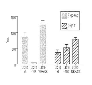
probes in L1210, L1210-10K, and L1210-10K with reintroduced dCK cell lines.
Figure 14B shows D-FAC phophorylation in the cell lines.
[0035] Figure 15 shows results of an in vivo study demonstrating
that D-FAC
can be used to predict resistance to widely used oncolytic prodrugs such as
Gemcitabine and Ara-C. Figure 15A shows accumulation of [I8F]FDG; Figure 15B
shows accumulation of [I8F]D-FAC.
[0036] Figure 16 shows the biodistribution of [I8F]D-FAC in a
healthy human
volunteer via a coronal microPET scan 48 minutes after injection of the [18F]D-
FAC.
[0037] Figure 17 demonstrates that the intracellular accumulation
(retention
and phosphorylation) of ['8F]-CA and [I8F]L-FAC requires the expression of
deoxycytidine kinase (dCK). Figure 17A shows the accumulation of [I8F]D-FAC,
[I8F]L-FAC, and ['8F]-CA probes in L1210-WT and L1210-10K cell lines; Figure
17B shows the extent of phsophorylation of probes in L1210-WT and L1210-10K
cell
lysates.
[0038] Figure 18 shows the results of biodistribution studies of [18F]L-FAC
and ['8F]-CA in C57/BL6 mice. Figure 18A shows images obtained with [I8F]L-
FAC; Figure 18B shows a [I4C]F-CA DWBA; Figure 18C shows images obtained
CA 02699967 2010-03-17
WO 2009/038795
PCT/US2008/010948
with [I8F]L-FAC and microPET/CT; Figure 18D shows images obtained with [I8F]F-
CA and microPET/CT.
[0039] Figure 19 illustrates that [18F]L-FAC is more resistant to
deamination
than [I8F]D-FAC according to in vivo studies in mice and data using human
plasma.
Figures 19A and 19B show chromatographs of [18F]D-FAC and [I8F]L-FAC in
plasma at 10 minutes and 45 minutes following injection into a mouse; Figures
19C
and 19D show chromatographs of [I8F]D-FAC and [18F]L-FAC in plasma at 10
minutes and 45 minutes after the probe was incubated with human plasma.
[0040] Figure 20 shows [I8F]L-FAC microPET images of lymphadenopathy
in an animal model of systemic autoimmunity.
[0041] Figure 21 shows [I8F]L-FAC microPET images of immune
activation
during a primary T cell mediated anti-tumor immune response.
[0042] Figure 22 illustrates that [I8F]L-FAC can be used to predict
gemcitabine resistance in vivo. Figure 22A shows [I8F]L-FAC microPET/CT scans;
Figure 22B shows [I8F]FDG microPET/CT scans.
[0043] Figure 23 illustrates that the intracellular accumulation of
[I8F]L-
FMAC requires the expression of deoxycytidine kinase.
[0044] Figure 24A shows the results of biodistribution studies of L-
FAC, D-
FAC, and L-FMAC. Figure 24B shows biodistribution [I8F]L-FMAC in necropsy
samples.
[0045] Figure 25 illustrates that [18F]L-FMAC is resistant to
deamination.
Figure 25A shows results obtained with the [I8F]L-FMAC standard; Figure 25B
shows results for [I8F]L-FMAC in plasma collected 45 minutes after injection.
[0046] Figure 26 show [I8F]L-FMAC microPET images of lymphadenopathy
in an animal model of systemic autoimmunity. Figure 26A shows results obtained
with the wild type BL/6 mouse. Figure 26B shows results obtained with the
B6.MRL-FasIP7.1 autoimmune mouse.
[0047] Figure 27 shows [I8F]L-FMAC microPET images of immune
activation during a primary T cell mediated anti-tumor immune response.
[0048] Figure 28 shows [I8F]L-FMAC microPET images of melanoma
tumors in mice.
[0049] Figures 29A and 29B present [I8F]D-FAC PET images of lymphoma
lesions in a human.
16
CA 02699967 2015-04-07
' 78401-29
[0050] Figure 30 presents PET/CT scans of a 56 year old human
male with
chronic pancreatitis. Figure 30A shows an image obtained with the L-FAC probe;
Figure 30B shows an image obtained with the FDG probe.
DETAILED DESCRIPTION
[0051] Embodiments of the invention are discussed in detail
below. In
describing embodiments, specific terminology is employed for the sake of
clarity.
However, the invention is not intended to be limited to the specific
terminology so
selected. A person skilled in the relevant art will recognize that other
equivalent parts
can be employed and other methods developed without parting from the
scope of the invention.
[0052] An objective of the work leading to the present
invention, of which
several embodiments are presented in this text, is the development of small
molecule
PET probes¨other than 2-[I8F]fluoro-2-deoxy-D-glucose (herein, FDG) and such
as,
for example, 1-(2'-deoxy-2'418F]fluoro-13-D-arabinofuranosyl)cytosine (herein,
[18F]D-FAC), ['8F]-CA, [I8F]L-FAC, [I8F]D-FMAC, [189L-FMAC, [I8F]D-FBAC,
[18F]L-FBAC, [18F]D-FCAC, [18F]L-FCAC, [I8F]D-FFAC, [18F]L-FFAC¨that
specifically target genes expressed during T lymphocyte activation or during
malignant transformation. We have identified several chemical compounds that
accumulate specifically in activated T lymphocytes and that can be labeled
with the
positron-emitting radioisotope [18F]fluorine to generate PET probes for
imaging the
activation of lymphocytes in vitro and in vivo. These probes also enable the
imaging
of selected cancers and can be used to predict resistance to certain oncolytic
nucleoside analogs.
[0053] In this text, when a compound is presented of which a
substituent is
stated to be a specific radioisotope or specified radioisotopes, it is to be
understood
that an agglomeration of more than one molecule of the compound that has one
or
more molecules in which the substituent is a different radioisotope or a
stable isotope
is encompassed. When a compound is presented of which a substituent is stated
to
not be a radioisotope, it is to be understood that an agglomeration of more
than one
molecule of the compound that has one or more molecules in which a substituent
is a
17
CA 02699967 2010-03-17
WO 2009/038795 PCT/US2008/010948
radioisotope, for example, a naturally occurring radioisotope that is
represented in the
agglomeration in a proportion found in nature, is encompassed.
[0054] In this text, when an enantiomer is discussed, the enantiomer
of
opposite handedness is also implied, unless the context indicates otherwise.
The term
[18F]-CA implies either or both of the isomers 2-18F-CA and 3-18F-CA.
[0055] In another aspect, the present invention also relates to
novel methods
of synthesizing PET probes disclosed herein. In still another aspect, the
present
invention relates to methods of using PET probes in the diagnosis and
treatment of
diseases and conditions involving inflammation, e.g., rheumatoid arthritis,
inflammatory bowel disease, type 1 diabetes, Experimental Autoimmune
Encephalomyelitis (EAE), multiple sclerosis, atherosclerosis and cancer. As
used
herein, "treatment" comprises prevention, partial alleviation, or cure of the
condition
or disorder.
[0056] In another aspect, this invention relates to methods of
evaluating the
usage efficacy of particular classes of anticancer agents in the treatment of
cancer
such as those that are taken up into cells via nucleoside transporters and
deoxycytidine kinase (dCK)-mediated phosphorylation. In an additional aspect,
the
present invention relates to methods of diagnosis and treatment of conditions
that
implicate cells with high deoxyribonucleoside salvage pathway activity, e.g.,
lymphocytes, bone marrow cells, and intestinal enterocytes. In another aspect,
the
present invention relates to compositions incorporating the compounds
disclosed
herein. In still another aspect, the present invention relates to kits
comprising any
embodiment of the present invention.
[0057] Monitoring immune function throughout the body using
molecular
imaging may significantly impact the diagnosis and treatment evaluation of
immunological disorders. Positron Emission Tomography (PET) is a molecular
imaging modality with numerous applications in cancer and other diseases.
However,
PET studies of immune function have been limited by a lack of specialized
probes.
Using a differential screening strategy, we identified PET probes for the
deoxyribonucleotide salvage pathway. By way of reminder, these are probes
other
than 2-[18F]fluoro-2-deoxy-D-glucose (herein, FDG). Examples of probes used
are 1-
(2 ' -deox y-2 ' - [18F] fluoro-f3-D-arabinofuranosyl)cytosine (herein, [18F]D-
FAC), [18F]-
18
CA 02699967 2010-03-17
WO 2009/038795
PCT/US2008/010948
CA, [18F]L-FAC, [18F]D-FMAC, [18F] L-FMAC, [18F]D-FBAC, [18F]L-FBAC, [18F]D-
FCAC, [18F]L-FCAC, [18F]D-FFAC and [18F]L-FFAC.
[0058] The PET probes disclosed herein enabled lymphoid organ
visualization by microPET that was sensitive to localized immune activation in
mouse
models of anti-tumor immunity. The PET probes disclosed herein also detected
early
changes of a lymphoid mass in systemic autoimmunity and allowed for evaluation
of
immunosuppressive therapy. These data support the use of PET probes disclosed
herein for immune monitoring and suggest a wide range of clinical
applications,
including for treatment visualization of certain types of cancer.
[0059] In order to identify candidates for PET probes that can distinguish
between activated T cells and non-activated, naïve T cells, we conducted a
radioactive
uptake assay, the results of which are shown in Figure 1. Figure 1A shows the
retention profiles for tested nucleoside analogs in activated and quiescent
(naïve) T
cells. These measurements were performed after incubating cells with
radioactive
compounds for 1 hr and performing successive washes to remove unincorporated
probes. The structures and chemical formulas of tested compounds are shown in
Figure 9. Full names of the abbreviations for the compounds are provided in
Table 1.
The largest difference in probe retention by proliferating compared to naïve T
cells
was observed for 2',2'-difluorodeoxycytidine (dFdC) and was >20 fold (Figure
1B).
The results shown in Figure 1 guided our design of [18F]fluorine-radiolabeled
PET
probes analogous to 2'-deoxycytidine. For example, we identified compounds #1
through #15 (see Figure 2A for chemical structures) as PET probe candidates
useful
for detecting activated T cells. Figure 2A presents substrates of dCK labeled
with
the 18F positron emitting radioisotope.
19
CA 02699967 2010-03-17
WO 2009/038795
PCT/US2008/010948
URIDINE ANALOGS
2'-fluoro-2'-deoxy-5-fluorouracil-3-D-arabinofuranoside FFAU
1-(2-deoxy-2-fluoro-13-D-arabinofuranosyl)-uracil FAU
2'-fluoro-2-deoxyuridine 2FdUrd
-fluoro-2-deoxyuridine 5FdUrd
THYMIDINE ANALOGS
2'-fluoro-2'-deoxythymidine 2'FLT
1 -(2-deoxy-2-fluoro-13-L-arabinofuranosyl)-5 -methyluracil L-FMAU
1 -(2-deoxy-2-fluoro-13-L-arabinofurano syl)-5-methylurac D-FMAU
3'-fluoro-3'-deoxythymidine FLT
CYTIDINE ANALOGS
5-fluoro-2'-deoxycytidine 5Fdc
2',2'-difluorodeoxycytidine dFdC
5-fluoro-2,3-dideoxycytidine 5FddC
(+f3-2,3-dideoxy-5-fluoro-3-thiacytidine FTC
2,3-dideoxy-3-fluorocytidine 3FddC
Table 1
[0060] The synthesis of 1-
(2' -deoxy-2' - [18F] fluoro-13-D-
5 arabinofuranosyl)cytosine (herein, [I8F]D-FAC) is illustrated in Figure
3A. 2-0-
[(Trifluoromethyl)sulfony1]-1,3,5-tri-0-benzoyl-a-D-ribofuranose (1) can be
reacted
with [I8F]fluoride ion to produce 2-deoxy-2-[18F]fluoro-1,3,5-tri-O-benzoyl-a-
D-
arabinofuranose (2) which can be reacted with hydrogen bromide to 2-deoxy-2-
[18F]fluoro-3,5-di-O-benzoyl-a-D-arabinofuranosyl bromide (3). The bromo
compound 3 can be reacted with 4-N-(trimethylsily1)-2-0-
(trimethylsilyppyrimidine-
4-amine (4) to produce 1-
(2'-deoxy-2'418F]fluoro-3,5-di-0-benzoy1-13-D-
arabinofuranosyl)cytosine (5). The benzoyl-groups can be removed by reacting 5
with
sodium methoxide to produce the PET probe, 1-(2'-deoxy-2'418F]fluoro-13-D-
arabinofuranosyl)cytosine (6).
[0061] The synthesis of .. 2-chloro-9-(2-
deoxy-2418F]fluoro-13-D-
arabinofuranosyDadenine (herein, 2-'8F-CA) is illustrated in Figure 3B. 2-0-
[(trifluoromethyl)sulfonyl]-1,3,5-tri-O-benzoyl-a-D-ribofuranose can be
reacted with
[I8F] fluoride ion to produce 2-deoxy-2418F]fluoro-1,3,5-tri-O-benzoyl-a-D-
arabinofuranose which can be reacted with hydrogen bromide to 2-deoxy-2-
[I8F]fluoro-3,5-di-O-benzoyl-a-D-arabinofuranosyl bromide. The bromo compound
can be reacted with 2-chloroadenine to produce 2-chloro-9-(4-benzoyloxymethy1-
3-
CA 02699967 2010-03-17
WO 2009/038795
PCT/US2008/010948
benzoyloxy-2-deoxy-2418F]fluoro-13-Q-arabinofuranosyDadenine. The benzoyl
groups can be removed by reacting 2-chloro-9-(4-benzoyloxymethy1-3-benzoyloxy-
2-
deoxy-24 I8F]fluoro-13-Q-arabinofuranosy1)adenine with sodium methoxide to
produce the PET probe, 2-
chloro-9-(2-deoxy-2-[18F]fluoro-3-D-
arabinofuranosyl)adenine.
[0062] The synthesis of 2-
chloro-9-(2'-deoxy-2 '418F] fluoro-P-D-
arabinofuranosyDadenine (herein, ['8F]-CA) is illustrated in Figure 4A. 2-
Chloroadenosine (1) upon reaction with trityl chloride provided a mixture of
alcohols
2 and 3 which were completely separated by silica gel column chromatography.
The
separated alcohols 2 and 3 were treated with triflyl chloride to yield the
corresponding
triflates 4 and 5. Reaction of the triflate 4 with [18F] fluoride ion followed
by
deprotection of the trityl groups with dilute mineral acids such as HC1 or
H2SO4 gave
2-chloro-9-(2' -deoxy-2 '118F] fluoro-P-D-arabinofuranosyl)adenine (6)
(herein,
[18F]CA or 18F-CA). Similarly, the reaction of the triflate 5 with
[18F]fluoride ion
followed by deprotection with acids gave the isomeric 3'-deoxy-3'-[I8F]fluoro
derivative 7. The synthesis of [18F]L-FAC, [I8F]D-FMAC, [18F]L-FMAC, [I8F]D-
FBAC, [18F]L-FBAC, [18F]D-FCAC, [I8F]L-FCAC, [I8F]D-FFAC, [18F]L-FFAC is
shown in Figure 4B-J.
[0063]
Figures 2B-2D show the [I8F]D-FAC microPET images of normal
mice and mice undergoing systemic immune inactivation. Figure 2B shows a naïve
BL6 mouse injected with [I8F]D-FAC 1 hr prior to imaging; the PET imaging
shows
accumulation in the spleen and the thymus, the latter of which was predicted
based on
elevated dCK expression in that tissue. In Figure 2C, a BL6 mouse was injected
with
100 micrograms of anti-CD3 antibody 24 hr prior to imaging such that a
systemic
immune response can be generated. After the 1 hr uptake of [18F]D-FAC, the
probe
accumulated in the spleen but there was less accumulation in the thymus
because of
antibody treatment. In Figure 2D, the anti-CD3-stimulated mouse is imaged 2 hr
after
[18
F]D-FAC injection and shows that the probe clears from the kidneys such that
clearer visualization of the spleen is possible.
[0064] In addition to [18F]D-FAC (Compound #1), [I8F]L-FAC, (Compound
#2), 2-chloro-9-
(2' -deoxy-2'-[18F]fluoro13-D-arabinofuranosyDadenine (herein,
[I8F]CA, Compound #3), [I8F]D-FMAC (Compound #8) and [I8F]L-FMAC,
21
CA 02699967 2010-03-17
WO 2009/038795
PCT/US2008/010948
(Compound #9), several other compounds can be useful for identifying activated
T
cells through deoxycytidine kinase-associated uptake detected by PET imaging;
examples of these additional compounds are shown in Figure 2A and Figure 4.
The
compounds [I8F]D-FRAC and [I8F]L-FRAC can be useful for identifying activated
T
cells through deoxycytidine kinase-associated uptake detected by PET imaging.
,18
[ F]D-FRAC is similar to [I8F]D-FMAC, and [I8F]L-FRAC is similar to [I8F]L-
FMAC, except that instead of a methyl group substituted at the 5-position of
the
pyrimidine ring, an alkyl group having from 1 to 6 carbon atoms can be
substituted at
this position. The compounds [I8F]D-FXAC and [I8F]L-FXAC can be useful for
identifying activated T cells through deoxycytidine kinase-associated uptake
detected
by PET imaging. [I8F]D-FXAC is similar to [I8F]D-FAC, and [I8F]L-FXAC is
similar to [I8F]L-FAC, except that instead of a hydrogen substituted at the 5-
position
of the pyrimidine ring, a halogen, for example, fluorine, chlorine, bromine,
or iodine,
can be substituted at this position.
[0065] Thus, lymphocyte activation can be non-invasively monitored by
injecting a subject animal or human with a trace amount of an [I8F]fluorine-
labeled
PET probe (e.g., such as in Figures 2-4), whereby the probe is expected to
accumulate at sites of local immune activation and can be monitored at a whole
body
level using a PET scanner. The approach of using an [I8F]fluorine-labeled PET
probe
to monitor immune activation is that this probe would be more specific and
sensitive
than with an approach using FDG3I. An [I8F]fluorine-labeled PET probe (like in
Figures 2-4) can be administered to an animal or a human for diagnostic
purposes
such as to determine the presence or extent of a disease or disorder (e.g.,
cancer,
autoimmune disease, developmental disorder, viral infection, bacterial
infection,
parasitical infection, other infections, metabolic disease, or inflammation).
For
instance, the [I8F]fluorine-labeled PET probe can be administered to monitor
the
progress of cancer or other disease-based types of immunotherapy, interferon
therapy,
vaccination, radiation therapy, and antibiotic therapy. (Notice that as used
herein,
"developmental disorder" includes immune deficiencies. Also as used herein,
"metabolic disease" includes defects in macrophage function due to problems in
enzyme storage.)
[0066] In the research context, the [I8F]fluorine-labeled PET probes
presented
in Figures 2, 3 and 4 can be administered to an animal for the purpose of
developing
22
CA 02699967 2010-03-17
WO 2009/038795
PCT/US2008/010948
a diagnostic technique, a therapy, or to develop a basic understanding of
disease or
disorder mechanisms.
[0067] We
describe the identification and validation of new PET probes for
the deoxyribonucleotide salvage pathway. PET imaging using probes (other than
FDG) such as [I8F]D-FAC allows for visualization of the thymus and spleen in
mice.
Moreover, this technology is able to monitor alterations in the lymphoid mass
and
immune status under various experimental conditions. Current PET imaging work
in
EAE shows the utility of using [ D-
FAC for measuring key metabolic pathways in
immune cells. While these probes are not exclusively retained in immune cell
lineages,
changes in probe accumulation throughout the body may be indicative of
"disease
states" and provide early biomarkers for treatment efficacy. Furthermore, the
accumulation of [I8F]D-FAC in the thymus and spleen as well as the variations
in
[I8F]D-FAC retention at lymphoid organs during immune responses may reflect a
critical role for the deoxyribonucleoside salvage pathway (as measured by the
said
probe) in T cell development and function. While the biological function of
dCK is
currently unknown, mice deficient in the dCK-related gene thyrnidine kinase 1
(TK1)
display immunological abnormalities in histology and function. Novel genetic
mouse
models of dCK deficiency and imaging via [I8F]D-FAC PET may provide unique
tools to dissect the immunological functions of the deoxyribonucleotide
salvage
pathway.
[0068] The
unique distribution pattern of [18F]D-FAC and other FAC analogs
suggests that the utility of these probes may extend beyond the FasiPr model
to several
other immune disorders. Elevated retention of the [18F]D-FAC probe in joints
and the
intestine over physiologic uptake values may reflect the presence of an active
inflammatory processes that is characteristic of rheumatoid arthritis and
inflammatory
bowel disease, respectively. [I8F]D-FAC microPET can also be used to detect
overt
autoreactive immune activation in a BDC-2.5 mouse model that is prone to type
1
diabetes (Figure 8). In Figure 8A, the 1 mm corona] sections illustrate the
pattern of
[I8F]D-FAC probe accumulation in BDC-2.5 mice. (Abbreviations: CV, cervical
LNs;
AX, axillary LNs; BR, brachial LNs; IN, inguinal LNs; THY, Thymus; GI,
Gastrointestinal tract; H, heart.) In Figure 8B, [I8F]D-FAC accumulation is
measured
in necropsy tissue samples from BL/6, BALB/c, NOD LTJ, and BDC-2.5 mice. The
23
CA 02699967 2010-03-17
WO 2009/038795 PCT/US2008/010948
data indicate that of these strains, the spleen and lymph nodes of BDC2.5 mice
accumulate the highest levels of [18F]D-FAC.
[0069] The low retention of [18F]D-FAC in the brain (Figure 5)
suggests that
this probe is superior to FDG for detection of inflammatory infiltrates
affecting the
central nervous system in EAE and multiple sclerosis (MS). While it is not
known
whether [18F]D-FAC can cross the blood-brain-barrier, the integrity of this
structure is
frequently compromised in EAE and MS. Regarding atherosclerosis, FDG has been
shown to enable visualization of carotid plaques but its high accumulation in
the
myocardium limits imaging of coronary lesions. However, it is this very aspect
of
[18F]D-FAC and in contrast to FDG, the lack of [18F]D-FAC retention in the
heart
provides the necessary low background that can enhance PET imaging of
activated
macrophages and other immune cells at coronary atherosclerotic lesions (Figure
5).
[0070] In addition to immune diseases, [18F]D-FAC may also be used
to
measure dysregulated nucleoside metabolism in cancer. To this end, we examined
the
utility of [18F]D-FAC microPET in animal models representative of leukemia or
lymphoma, melanoma, and glioma tumors (Figure 12, where the images are 1 mm
coronal sections from [18F]D-FAC microPET/CT scans 1 hr after probe
injection). In
Figure 12A, SCID mice were injected intravenously with Ba/F3 cells expressing
p210 BCR-ABL and go on to develop aggressive disease with massive splenic
infiltration that typically results in death within ¨15 days (mice shown were
imaged
on day 12). In Figure 12B, the NOD SCID mice were transplanted with wild type
total bone marrow cells infected with MSCV-GFP-IRES-P185 BCR-ABL retroviral
stocks and the leukemic mice were imaged 28 days following transplantation. In
Figure 12C, C57BL/6 was injected subcutaneously with 1 x 10⁵ B16 melanoma
cells
and imaged 7 days later. In Figure 12D, SCE) mice were injected subcutaneously
with 1 x 10⁶ U87 glioma cells and imaged 10 days later. (Abbreviations: L,
Liver; SP,
Spleen; GI, Gastrointestinal tract; BL, Bladder; Tu, Tumor.) The increased
[18F]D-
FAC retention in the spleen was observed in mouse models of oncogene-induced
leukemia using Bcr-Abl-expressing Ba/F3 cells and Bcr-Abl transformed bone
marrow. [18F]D-FAC PET also detected implanted murine B16 cells
(representative of
malignant melanoma) and human U87 cells (representative of glioma tumors).
[18F]D-FAC may additionally be used to predict tumor responses to a particular
class
of anticancer agents, which include the widely used prodrugs cytarabine (Ara-
C) and
24
CA 02699967 2010-03-17
WO 2009/038795
PCT/US2008/010948
2',2'-difluorodeoxycytidine (dFdC, Gemcitabine). Structurally, these prodrugs
are
closely related to FAC and require cellular uptake via nucleoside transporters
and
dCK-mediated phosphorylation for conversion to their active drug metabolites.
We
suggest that the availability of a PET biomarker to measure the cellular
pharmacology
of Ara-C and dFdC may assist with the stratification of susceptible and
resistant
tumors leading to a more rational clinical use of these important anticancer
drugs.
[0071]= 18
Results presented here indicate that PET imaging with [ F]D-FAC and
the other inventive compounds offers new advantages in diagnostics and
treatment
monitoring of a wide range of disorders.
EXAMPLES
[0072] We
considered the salvage pathway for DNA synthesis, in which
deoxyribonucleosides are imported into cells by the specialized nucleoside
transport
proteins that are converted to their triphosphate forms via consecutive
phosphorylation steps catalyzed by deoxyribonucleoside kinases. While the
majority
of normal tissues utilize the de novo pathway for deoxyribonucleotide
synthesis,
certain tissues¨including thymus and spleen¨rely extensively on the salvage
pathway. Thus, we carried out the following studies: (i) in vitro screening of
nucleoside analogs for retention in proliferating and quiescent T cells and
identification of D-FAC, a new PET probe candidate; (ii) gene expression and
biochemical analyses to investigate the mechanisms of elevated D-FAC retention
in
activated T cells; (iii) radiochemical synthesis of [I8F]D-FAC and in vivo
biodistribution studies; (iv) comparison of [18F]D-FAC with other PET probes
currently used to measure nucleoside metabolism and glycolysis; and (v)
evaluation
of [18F]D-FAC in mouse models of immune activation. Findings from these
studies
provide the impetus for translational [18F]D-FAC PET imaging in a wide range
of
immunological disorders in patients. According to the invention, the strategy
used to
identify and evaluate [I8F]D-FAC and its analogs is broadly applicable to the
development of new PET probes with defined specificity for various biochemical
pathways and/or immune cell lineages.
[0073] The
following nucleosides were purchased from Moravek
Biochemicals (Brea, CA): 3'-
Fluoro-3'-deoxythymidine (3'-FLT); 2'-Fluoro-2'-
deoxythymidine (2'-FLT); 1-
(2 ' -Deox y-2' -fluoro-13-D-arabinofuranosyl)-5-
CA 02699967 2010-03-17
WO 2009/038795
PCT/US2008/010948
methyluracil (D-FMAU); 1-
(2'-Deoxy-2 ' -fluoro-13-L-arabinofuranosyl)-5-
methyluracil (L-FMAU); 2',3'-Dideoxy-3'-fluorocytidine (3'-FddC); (+13-2',3'-
Dideoxy-5-fluoro-3'-thiacytidine (FTC); 5-Fluoro-2',3'-dideoxycytidine
(5FddC);
2',2!-Difluorodeoxycytidine (dFdC); 5-Fluoro-2'-deoxycytidine (5FdC); 5-Fluoro-
2'-
deoxyuridine (5FdURD); 2'-Fluoro-2'-deoxyuridine (2FdUrd); 1-(2'-Deoxy-2'-
fluoro43-D-arabinofuranosyl)-uracil (FAU); 2'-Fluoro-2'-deoxy-5-fluorouracil-
I3-D-
arabinofuranoside (FFAU).
[0074] T
lymphocytes from pmel-1 T cell receptor (TCR) transgenic mice
were stimulated ex vivo using a melanoma antigen (1 micromolar hgp10025_33).
These
cells were then cultured for radioactive uptake and kinase assays that were
performed
72 hours post-stimulation. In more detail, 1 microCi of [3H]D-FAC or [3H]dFdC
were added to a well containing 5x10⁴ cells in a 96-well tissue culture plate
and
incubated for 1 hr at 37 C and 5% CO2. The plate was then washed 5 times with
media containing 5% fetal calf serum (FCS) by using the Millipore Vacuum
Manifold
(Billerica, MA); the amount of incorporated probe was measured by
scintillation
counting using the PerkinElmer Microbeta (Waltham, MA).
[0075]
Mice were kept warm, under gas anesthesia (2% isoflurane) and
injected intravenously with 200 microCi of various PET probes; 1 hr was
allowed for
uptake. Mice were then positioned using an imaging chamber and the data was
obtained using Siemens Preclinical Solutions (Knoxville, TN) microPET Focus
220
and MicroCAT II CT systems. MicroPET data was acquired for 10 minutes and then
reconstructed via statistical maximum a posteriori probability algorithms
(MAP) into
multiple frames 3. The spatial resolution of PET is ¨1.5 mm with 0.4 mm voxel
size.
CT images provide a low dose (400 micron) resolution acquisition with 200
micron
voxel size. MicroPET and CT images were co-registered using a previously
described
method and regions were drawn using the AMIDE software (Andreas Loening,
http://amide.sourceforge.net/, v0.8.16) 4'5. Quantification was performed by
drawing
3D regions of interest (ROI).
[0076] All
the mice used in these studies were bred and maintained according
to the guidelines of the Department of Laboratory Animal Medicine (DLAM) at
the
University of California, Los Angeles. For the oncoretrovirus-induced sarcoma
model,
C57/BL6 mice were challenged intramuscularly in the right triceps with the
Moloney
murine sarcoma and leukemia virus complex (MoMSV) in a volume of 100 uL of
26
CA 02699967 2010-03-17
WO 2009/038795
PCT/US2008/010948
PBS as described previously 6. B6-MRL-FasiP7J mice used for systemic
autoimmunity
studies were purchased from the Jackson Laboratory (stock number 000482). To
monitor the immunosuppressive treatment, these mice were given intraperitoneal
injections of dexamethasone (DEX, 10 mg/kg) at 24 hr intervals and were
scanned by
microPET/CT 24 hr after the last injection. Animals were anesthetized with 2%
isoflurane, injected intravenously with 200 microCi [18F]D-FAC and then
scanned
with microPET/CT; mice were sacrificed immediately after imaging. Organs were
rapidly excised, weighed, and the radioactivity was measured in a well
counter. After
decay correction, results were expressed as percent of the injected dose of
activity per
gram of tissue (%ID/g). Other mice were anesthetized with 2% isoflurane and
injected
intravenously with 1 mCi [18F]D-FAC. After 1 hr, mice were euthanized,
embedded
in 3% carboxymethyl cellulose (CMC, Sigma), and frozen in 100% ethanol with
dry
ice for 45 min. The 50 micron sections were cut using a whole body cryostat,
(PMV,
Stockholm, Sweden); samples were exposed overnight on BAS-TR2025 plates
(Fujifilm Life Science, Stamford, CT). Imaging plates were read using a
Fujibas-5000
phosphorimager (16 bit, 25 micron resolution; Fujifilm Life Science).
[0077] The total RNA was extracted from the purified naïve CD8 T
cells and
72 hrs post activation proliferating CD8 T cells of pmel-1 TCR transgenic
mice. RNA
was pooled from 4 independent experiments and hybridized to Affymetrix Mouse
Genome 430 2.0 arrays. The absolute calls describing whether a probe set is
present
(P), marginally present (M), or absent (A) were generated using the Affymetrix
GeneChip Operating Software v1.3 (GCOS) and expression values were calculated
using the PM/MM difference model of DNA-Chip (dChip) 7. Expression values
across samples were normalized using dChip's invariant set method. As
conditions for
inclusion, a gene was considered differentially expressed if the corresponding
probe
set fit the following criteria: absolute call was P in at least half of the
samples, fold
change >1.4 between baseline (naïve CD8+ T cells) and experimental (activated
CD8+ T cells) using the lower 90% confidence bound of fold change as defined
in
dChip, and expression difference between the baseline and experimental samples
was
>100. Genes involved in the nucleoside de novo biosynthesis and salvage
pathways
were taken from the KEGG database (pathway IDs 00230 and 00240, respectively)
and the corresponding probe sets were manually extracted from Affymetrix's
NetAffx
27
CA 02699967 2010-03-17
WO 2009/038795
PCT/US2008/010948
to ensure complete coverage of all nucleoside pathway genes (239 probe sets)
plus the
SLC28 and SLC29 transporters (10 probe sets) 8,9
[0078] Total RNA was purified from tissues using the Qiagen RNeasy
Mini
kit and 1.5 fig of this RNA was then used to synthesize cDNA using the TaqMan
Reverse Transcription Reagents (Applied Biosystems). Pre-designed TaqMan
assays
were purchased from Applied Biosystems for dCK (Assay ID Mm00432794_m1),
S1c29a1 (Assay ID Mm00452176_m1), and Slc28a3 (Assay ID Mm00627874 m1).
TaqMan beta-actin (Applied Biosystems, Part: 4352341E) reagents were used as
an
endogenous control for quantification. The samples were ran out on a 48-well
StepOne Real-Time PCR System (Applied Biosystems) and were analyzed with the
StepOne Software v2.0 (Applied Biosystems) using the comparative Ct method
(AACt). The qPCR mixture (20 [IL) contained 15 ng cDNA, TaqMan buffer, 5.5 mM
MgC12, 200 tiM dATP, 200 M dCTP, 200 M dGTP, 400 M dUTP, the appropriate
TaqMan assay, 0.5U AmpliTaq Gold, and 0.2U uracil-N-glycosylase (UNG). Each
assay included cDNA template in triplicates.
[0079] Six to 8 week old mice with severe combined immunodeficiency
(NOD SCID) were sublethally irradiated (275 rads) one day prior to
reconstitution.
Whole bone marrow was isolated from the tibias and femurs of 4-8 week old
wildtype
mice and infected with MSCV-GFP-IRES-P185 BCR-ABL retroviruses. Three hours
after infection, bone marrow cells were injected intravenously by the tail
vein into
recipient NOD SOD mice. Animals were monitored daily for signs of illness
during a
period of two months as previously described
[0080] p210 BCR-ABL transfected Ba/F3 cell lines were previously
described
11. Ba/F3 cell lines were maintained in RPMI containing 10% FCS in 5% CO2 at
37 C
(with the addition of 10% WEHI conditional medium as a source of IL-3 to the
parental cell line). The spontaneous gp100+ murine melanoma B16 (H-2b) cell
line
and the U87 glioma cell line were obtained from the American Type Culture
Collection (ATCC, Rockville, MD).
[0081] Graphs were constructed using GraphPad Prism software,
version 4.02.
P-values were calculated using Student's t test and only p-values of <0.05
were
considered significant. Data are presented as means standard errors of the
mean
(SEM).
28
CA 02699967 2010-03-17
WO 2009/038795 PCT/US2008/010948
[0082] In this text (including Figures and any other information
presented),
unless otherwise specified, the presentation, mention, or discussion of a
chiral
chemical compound also implies the presentation, mention, or discussion of
each of
the enantiomers of that chemical compound and their racemic mixtures. In this
text
(including Figures and any other information presented), unless otherwise
specified,
the presentation, mention, or discussion of a chemical compound with a
specified
chirality also implies the presentation, mention, or discussion of the
enantiomer of
that chemical compound with specified chirality and racemic mixtures of these.
Example 1: Differential screening to identify potential PET probes sensitive
to
changes in nucleoside flux during T cell activation and proliferation.
[0083] An in vitro assay (Figure 1) was used to measure the
retention of 3H-
labeled nucleoside analogs (NA) in naive and proliferating primary T cells.
Selection
criteria for tested NA accounted for the known propensity of fluorine
substitutions to
significantly change the stereoelectronic and biochemical properties of
nucleosides.
Thus, only deoxyribonucleosides containing 'cold' fluorine (19F) atom
substitutions
were tested (Figure 9). Subsequent substitution of 19F with 18F for
radiochemical
synthesis of PET probes would decrease the nuclear mass by a single atomic
mass
unit, which is a change of limited, if any, biochemical consequences.
Moreover, only
NA modified at the C-2' or 3' positions on the sugar moiety or at position 5
of the
nucleobase were screened since fluorination at C-4' would be incompatible with
radiochemical synthesis while fluorination at C-5' would prevent
phosphorylation by
nucleoside kinases.
[0084] Figure 1 identifies fluorinated deoxycytidine analogs
retained in
activated versus naive T lymphocytes and incorporated into DNA. In Figure 1A,
T
lymphocytes from pmel-1 T cell receptor (TCR) transgenic mice were stimulated
ex
vivo using a melanocyte/melanoma antigen (hgp10025_33); after 72 hrs, the
proliferating T cells were incubated for 1 hr with 3H-labeled (1 microCi)
deoxyribonucleoside analogs (see Figure 9). Following successive washes,
intracellular radioactivity was measured by scintillation counting. This part
of the
figure shows the retention profiles for tested NA in activated and naïve T
cells and the
striking differences likely reflect differential expression of nucleoside
transporters and
kinases sensitive to the nucleobase structure and to fluorine substitutions of
native
29
CA 02699967 2010-03-17
WO 2009/038795
PCT/US2008/010948
hydrogen and hydroxyl groups. In Figure 1B, 1-(2'- deoxy-2'-fluoro-
arabinofuranosyl) cytosine (D-FAC) is a dFdC analog, amenable to 18F labeling.
Here,
the largest (>20 fold) difference in retention was observed for 2',2'-
difluorodeoxycytidine (dFdC) when proliferating T cells were compared to naïve
T
cells. Figure 1C shows the retention of [3H]dFdC and [3H]D-FAC by the
activated
mouse CD8+ T cells; notice that the F-ara analog 1-(2'- deoxy-2'-D-
fluoroarabinofuranosyl) cytosine (D-FAC) resembled dFdC biochemically as
indicated by their similar retention in proliferating CD8+ T cells. In Figure
1D, the
increased uptake of [3H]D-FAC was observed in NIH3T3 fibroblasts that were
engineered to overexpress nucleoside kinases (dCK) and the nucleoside
transporter
SLC29A1. Note that [3H]FLT was used as a positive control for TK1 expressing
cells.
Lastly, in Figure 1E, [3H]D-FAC is incorporated in the DNA of proliferating T
cells.
Example 2: Biochemical mechanisms of D-FAC retention in proliferating T cells.
[0085] Increased retention of D-FAC in proliferating T cells compared to
naïve T cells may reflect any one or combination of several biochemical
events: (i)
upregulation of nucleoside transporters; (ii) elevated phosphorylation by
deoxyribonucleoside kinases leading to intracellular trapping of charged
products; and
(iii) increased incorporation into the DNA. Gene expression analyses by
microarray
and qPCR were performed in T cells before and after activation (at 72 hrs) to
determine the transcriptional status of specific nucleoside transporters and
kinases. In
terms of the context for D-FAC transport, previous studies using 2'-
deoxycytidine
(dCyd) analogs suggest the involvement of members of the solute carrier (SLC)
families SLC28 and SLC29. SLC29A1 expression was upregulated by ¨4-fold in
proliferating T cells vs. naïve T cells while two other potential D-FAC
transporters
(SLC28A1 and SLC28A3) were not expressed in these cells (data not shown). D-
FAC
phosphorylation may be carried out by deoxycytidine kinase (dCK, Kcat/Km for
dCyd = 2 x 10⁵) and thymidine kinase 2 (TK2, Kcat/Km for dCyd = 3 x 10⁴).
Following T cell activation, dCK mRNA levels increased by ¨2-fold whereas TK2
expression decreased by ¨5-fold (data not shown). Collectively, these data
suggest
that [3H]D-FAC retention in proliferating T cells reflects upregulation of
SLC29A1
and that this allows increased availability of intracellular substrate and/or
of dCK,
which in turn leads to increased phosphorylation capacity. (Notably, SLC29A1
and
CA 02699967 2010-03-17
WO 2009/038795 PCT/US2008/010948
dCK were both previously described to be involved in the metabolism of dFdC, a
FAC-related nucleoside12'13.)
Example 3: 1-18F1D-FAC has greater specificity for lymphoid organs than PET
probes
currently used to measure nucleoside metabolism and glycolysis.
[0086]
Biodistribution, metabolism, and clearance of [I8F]D-FAC were
studied in C57/BL6 mice. Tissue decay-corrected mean time-activity curves
obtained
from dynamic [I8F]D-FAC microPET/CT scans suggest that [18F]D-FAC is
predominantly cleared through the kidney (Figure 10). Time on the horizontal
axis in
Figure 10A is in units of seconds. Imaging data were corroborated with
measurements of retained radioactivity in necropsy tissue samples (Figure 10B)
and
with digital whole-body autoradiography (DWBA, Figure 5). One hour after
intravenous injection of [I8F]D-FAC, the accumulated radioactivity was
detected in
the thymus, spleen, intestine, bone/bone marrow and, to a lesser extent, in
the liver.
Biochemical studies may determine whether [I8F]D-FAC biodistribution reflects
tissue trapping by dCK-mediated phosphorylation, conversion to uracil
metabolites
via deamination (Figure 11), or both. Regardless of the specific biochemical
mechanism for retention, [I8F]D-FAC microPET and DWBA data suggest that this
probe enables visualization of cells with high deoxyribonucleoside salvage
pathway
activity such as lymphocytes, bone marrow cells, and intestinal enterocytes15.
[0087]
Figure 11 shows the potential biochemical pathway measured by
[I8F]D-FAC. Putative transporters for D-FAC include SLC28A1, SLC28A3 and
SLC29A1; however, only SLC29A1 is expressed in naïve and proliferating cells.
Intracellularly, D-FAC is phosphorylated by deoxycytidine kinase (dCK) and can
also
be converted to its uracil metabolite D-FAU (1-(2'-deoxy-2'-fluoro-3-D-
arabinofuranosyOuracil) by cytidine deaminase (CDA), which can be inhibited by
3,4,5,6-tetrahydrouridine (THU). Monophosphorylated D-FAC (D-FAC-MP) is a
potential substrate for cytidylate kinase (CMPK) and deoxycytidylate deaminase
(DCTD). Diphosphorylated D-FAC (D-FAC-DP), which is a potential substrate for
nucleoside diphosphate kinases (NME1, NME2, NME4, NME6, NME7), can inhibit
ribonucleotide reductase (RRM), triphosphorylated D-FAC (D-FAC-TP), DCTD, and
cytidine triphosphate synthase (CTPS). D-FAC-TP may be incorporated into DNA
via DNA polymerase. Enzyme Commission (EC) numbers for the key enzymes
31
CA 02699967 2010-03-17
WO 2009/038795
PCT/US2008/010948
involved in the nucleoside salvage pathway are as follows: CDA (3.5.4.5); CMPK
(2.7.4.14); CTPS (6.3.4.2); dCK (2.7.1.74); DCTD (3.5.4.12); NME1, NME2, NME4,
NME6, and NME7 ( 2.7.4.6); NT5 (3.1.3.5); POL (2.7.7.7); RRM (1.17.4.1); and
TK1 (2.7.1.21).
[0088] In Figure 5A, [18F]D-FAC digital whole-body autoradiography
(DWBA) is shown along with the corresponding tissue section. The orientation
of the
sagittal, coronal, and transverse sections, 1 mm thick for each, are depicted
in the 3D
microCT image in Figure 5B. In Figures 5B and 5C, an immunocompetent mouse
(C57/BL6) was scanned by microPET/CT using probes for the dCK and TK1-
dependent segments of the deoxyribonucleoside salvage pathway (namely, [18F]D-
FAC, [18F]FLT, [18F]D-FMAU) and for glycolysis (namely, [18F]FDG). Mice were
imaged 60 min after intravenous injection of the probes. Direct comparison of
[18F]FAC with probes for nucleoside metabolism¨ [18F]FLT and [18F]D-FMAU¨
and glycolysis¨[18F]FDG¨further confirmed the ability of [18F]FAC to provide
functional imaging data that cannot be obtained using existing probes 16
(Figure 5C
and Table 1). In fact, neither [18F]FLT nor [18F]D-FMAU showed detectable
accumulation in the thymus and spleen and the high retention in the myocardium
limits the use of [18F]FDG to visualize the thymus. To gain further insight
into the
cellular specificity of [18F]D-FAC retention in the thymus and spleen, major
immune
cell types at these sites were isolated from mice injected with [18F]D-FAC,
sorted by
flow cytometry, and counted in a well counter. In Figure 5D, the [18F]D-FAC
retention per cell number in thymocytes and splenocytes is shown; here, the
highest
retention of radioactive probe per cell number was detected in double negative
thymocytes, presumably reflecting intense cellular proliferation at this stage
of T cell
development. In Figure 5E, the proportion of [18F]D-FAC retention per cell
lineage
per lymphoid organ is displayed, which is, in other words, the fractional
contribution
of various immune populations to [18F]D-FAC retention in the thymus and
spleen.
These data indicate that, in addition to T and B cells, [18F]D-FAC also labels
CD 1 1b
myeloid cells. (Abbreviations: B, bone; BL, bladder; BR, brain; GB, gall
bladder; GI,
gastrointestinal tract; H, heart; K, kidney; L, liver; LU, lung; SP, spleen;
Thy, thymus;
SC, spinal column; tissue %ID/g, the percent injected dose per gram.). Table 2
shows
that amongst PET probes for nucleoside metabolic pathways and glycolysis,
[18F]FAC
shows better selectivity for thymus and spleen than conventional probes
(values
32
CA 02699967 2010-03-17
WO 2009/038795 PCT/US2008/010948
are %ID/g per organ normalized to %ID/g muscle). Retention of [I8F]FDG in the
thymus could not be measured because of signal spillover from the heart.
Number of
mice was 3.
118F1FAC [18FIFLT [18FIFMAU 118FIFDG
Spleen 2.16 +/- 0.48 1.02 +/- 0.21 1.08
+/- 0.27 1.69 +/- 0.16
Thymus 3.29 +/- 0.48 1.22 +/- 0.23 1.33 +/- 0.24 N.D.
Table 2
Example 4: [18F]D-FAC PET imaging detects localized changes in immune status
during a primary anti-tumor immune response.
[0089] Having established that [18F]D-FAC allows for visualization of
lymphoid organs in immunologically naive mice, we investigated whether this
probe
can also be used to monitor immune responses in vivo using a well-studied
oncoretrovirus tumor model in which mice are challenged with the Moloney
murine
sarcoma and leukemia virus complex (MoMSV) develop non-metastatic sarcomas. In
this model, antigen-specific T cells primed by immunodominant epitopes encoded
by
viral gag and env genes rapidly expand in spleen and tumor draining lymph
nodes
(DLNs) and then traffic to the tumor lesion which is rejected over a period of
2-5
weeks17'18. In fact, the kinetics of T cell responses against MoMSV-induced
sarcomas
have been studied extensively using conventional ex vivo approaches and our
group
has used PET reporter gene imaging to visualize tumor rejection in this
mode117. To
analyze whether the [18F]D-FAC microPET/CT imaging can detect sites of
localized
immune activation, mice were scanned before and after the virus challenge.
Relative
to baseline scans (day 1), the scan acquired at the peak of the anti-tumor
immune
response (day 15) showed significantly increased [18F]D-FAC accumulation in
the
spleen and tumor DLNs (Figures 6A and 6B). To further investigate the cellular
basis
of elevated [18F]D-FAC retention, splenic CD8+ T cells from mice injected with
[18F]FAC were fractionated by flow cytometry into naïve (CD62LHIGH/CD44LOW)
and effector populations (CD62LLOW/CD44HIGH). Radioactivity measurements
showed that effector CD8+ T cells retained ¨4-fold more [18F]D-FAC than naïve
33
CA 02699967 2010-03-17
WO 2009/038795
PCT/US2008/010948
cytotoxic T cells (Figure 6C). These data confirm our initial observations
that were
obtained using cultured naïve and activated T cells (Figure 1).
[0090] To determine whether [I8F]D-FAC can provide unique
information
regarding localized immune activation via the afore-mentioned model, the mice
imaged by [18F]D-FAC were also scanned on consecutive days using [I8F]FDG and
[I8F]FLT. As shown previously, elevated [I8F]FDG accumulation was detected on
day
13 post-virus challenge not only at the tumor but also at tumor DLNs and the
spleen
(Figure 6D). In particular, tumor lesions accumulated high amounts of
[I8F]FDG,
namely 8.2 4.2 percent injected dose of activity per gram of tissue (%ID/g)
tumor
over background, which was defined as the contralateral muscle tissue. In
contrast,
18
[ F]D-FAC retention in the tumor was significantly lower (1.9 0.3 %ID/g tumor
over background). The preferential accumulation of [I8F]D-FAC in the tumor
DLNs
(at 13 2.7 %ID/g) compared to the tumor (at 6.7 0.3 %ID/g) suggests that
[I8F]D-
FAC is a more specific probe than [I8F]FDG for imaging anti-tumor immunity in
the
oncoretrovirus model (Figures 6A and 6D). Moreover, no detectable [I8F]FLT
accumulation was observed on day 14 at sites of immune activation that can be
clearly
visualized by [18F]D-FAC and [18F]FDG (Figure 6D). The marked difference
between these nucleoside PET probes may reflect the high level of thymidine
present
in the rodent serum that competes with [I8F]FLT and/or may reflect better
sensitivity
of [I8F]D-FAC for measuring the rate of the deoxyribonucleoside salvage
pathway in
immune cells.
Example 5: Disease and treatment evaluation using [I8F1D-FAC PET in animal
models of autoimmunity.
[0091] We also asked whether [I8F]D-FAC enables monitoring of a systemic
autoimmune disorder such as the FasiPr syndrome, which is a well-studied
animal
model resembling human systemic lupus erythematosus. In this syndrome,
deficient
apoptosis of lymphocytes carrying the FasiPr mutation results in
lymphadenopathy,
arthritis, and immune complex-mediated glomerulonephrosis. We used B6-MRL-
Fas1P7,1 mice since they show significantly slower disease progression than
the
original MRL/Mp-lpr/lpr strain. MicroPET/CT scans of 2-3 month old B6-MRL-
FasiPr/J mice revealed a significant increase in the numbers of [I8F]D-FAC
positive
axillary and brachial LNs relative to age-matched wildtype C57BL/6J controls
34
CA 02699967 2010-03-17
WO 2009/038795
PCT/US2008/010948
(Figure 7). The images were taken at 60 min after intravenous injection of
[18F]D-
FAC and show three lmm thick coronal slices from wildtype (C57BL/6J) and B6-
MRL-Fas1Pra both before and after treatment with DEX. (Abbreviations: Thy,
thymus; LN, lymph nodes; B, bone). In summary, [18F]D-FAC positive LNs were
detected only in 2 out of 19 wild type mice, whereas microPET scans of
Fasi'/.1 mice
showed the presence of enlarged LNs in 9 out of 13 mice. To determine whether
[18F]D-FAC microPET/CT enables evaluation of therapeutic interventions, B6-MRL-
Fas1l'a mice were treated with dexamethasone (DEX), a synthetic glucocorticoid
that
has pleiotropic, potent immunosuppressive effects. As shown in Figure 7, DEX
treatment (2-7 days) reduced [18F]D-FAC retention in the thymus and peripheral
LNs
to undetectable levels. These findings suggest that [18F]D-FAC microPET
imaging
allows for detection of lymphadenopathy at early stages of autoimmunity and
indicate
the utility of [18F]D-FAC as a biomarker for monitoring the effects of
immunosuppressive therapy.
[0092] Figure 8 presents the results of microPET/CT imaging performed on
BDC-2.5 T cell receptor transgenic mice injected with the [18F]D-FAC probe.
The
BDC-2.5 strain is a well-established animal model for type I (autoimmunity-
based)
diabetes. In Figure 8A, the 1 mm coronal sections illustrate the pattern of
the [18F]D-
FAC probe accumulation in BDC-2.5 mice. (Abbreviations: CV, cervical LNs; AX,
axillary LNs; BR, brachial LNs; IN, inguinal LNs; THY, Thymus; GI,
Gastrointestinal tract; H, heart.) In Figure 8B, [18F]D-FAC accumulation is
measured
in necropsy tissue samples from BL/6, BALB/c, NOD Ltj and BDC-2.5 mice. The
data indicate that among these strains, BDC-2.5 mice accumulate the highest
levels of
[18F]D-FAC in the spleen and lymph nodes. The accumulation of [18F]D-FAC probe
in the lymphoid organs may indicate the activated status of the autoreactive T
lymphocytes in the mouse.
SYNTHESIS OF COMPOUNDS
Example 6: Radiochemical synthesis of 1-(2'-Deoxy-2' 418F]fluoro-13-D-
arabinofuranosyl) cytosine ([18FID-FAC).
[0093] 2-0-[(Trifluoromethyl)sulfonyl]-1,3,5-tri-O-benzoyl-a-D-
ribofuranose
(1) (Figure 3) was prepared as reported in the literature 27. The synthesis of
the 18F-
CA 02699967 2010-03-17
WO 2009/038795
PCT/US2008/010948
fluoro analog 2 was carried out by a modification of the method reported by
Mangner
et al.2. No-carrier-added [18F]fluoride ion was produced by 11 MeV proton
bombardment of 98% enriched [180]water in a silver target body using a RDS-112
cyclotron. The aqueous [18F]fluoride ion was treated with a solution of K2CO3
(1 mg)
and Kryptofix 2.2.2 (10 mg) dissolved in water (0.04 mL) and acetonitrile
(0.75 mL)
mixture. The solution was evaporated at 115 C with a stream of nitrogen gas.
The
residue was dried by the azeotropic distillation with acetonitrile (3 X 0.5
mL);
specifically, a solution of the triflate 1 (10 mg) in 0.7 mL of acetonitrile
was added to
the dry residue before the reaction mixture was heated at 165 C for 15 mm in a
sealed
vessel. The solution was then cooled to room temperature and passed through a
small
cartridge of silica gel, from which the product was eluted with 5 mL of ethyl
acetate.
Next, the ethyl acetate solution was evaporated to dryness before 0.1 mL of
30% HBr
in acetic acid solution and then 0.4 mL of dichloroethane were added
sequentially.
This new reaction mixture was heated at 80 C in a sealed vessel for 10 min and
the
solution was concentrated to ¨ 50% of the initial volume. In the following
step, 0.7
mL of toluene was added and this solution was evaporated at 110 C to give the
bromo
(compound 3). A freshly prepared disilyl derivative of cytosine (4, 35 mg) was
dissolved in 1 mL of dichloroethane and added to the bromo compound 3. The
condensation reaction was carried out at 160 C in a sealed vessel for 30 mm
before
the reaction mixture was cooled to room temperature and then passed through a
small
column of silica gel. The product was eluted off the column using 5 mL of a
solution
mixture of 10% methanol with 90% dichloromethane. This solution was evaporated
to dryness at 100 C and then treated with 0.5 mL of a solution of 0.5 M sodium
methoxide in methanol. The reaction mixture was heated at 100 C for 5 min in
a
sealed vessel and, thereafter, the basic reaction mixture was neutralized with
0.25 mL
of 1M HC1 in water. This reaction mixture was diluted to a total volume of 3
mL with
a mixture of 1% ethanol with 99% 10 mM ammonium dihydrogen phosphate in water
and injected into a semi-preparative HPLC column (Phenomenex Gemini C-18
column; 25 cm X 1 cm). The HPLC column was eluted with a solvent mixture of 1%
ethanol with 99% 10 mM ammonium dihydrogen phosphate at a flow rate of 5.0
mL/min. The effluent from the HPLC column was monitored with a 254 nm UV
detector followed by a gamma radioactive detector. The chemically and
36
CA 02699967 2010-03-17
WO 2009/038795 PCT/US2008/010948
radiochemically pure [18F]D-FAC was eluted off the column with a retention
time of
¨15 min and the radiochemical yield ranged 20-30%.
[0094] The chemical and radiochemical purities of [18F]D-FAC were
determined by an analytical HPLC method using a Phenomenex Luna column (25 cm
X 0.46 cm, 5 particle size). The column was eluted with 10% ethanol and 90%
50
mM ammonium acetate at a flow rate of 1.0 mL/min. The effluent from the HPLC
column was passed through a UV detector (21/4, = 254 nm) followed by a gamma
radioactivity detector. The chemical and radiochemical purities of [18F]D-FAC
prepared as described above exceeded 99 %.
[0095] Analytical HPLC also was used to determine the specific activity of
[18F]D-FAC. A range of mass vs UV absorption at 254 nm wavelength for non-
radiolabeled D-FAC was determined using the analytical HPLC method described
above and the data set was used to construct a calibration graph. Using this
calibration
graph, the specific activity of [18F]D-FAC was found to be >1000 Ci/mmol.
Example 7: Radiochemical synthesis of 1-(2'-deoxy-2'418F]fluoro-13-L-
arabinofuranosyl)cytosine ([18F] L-FAC).
[0096] The title compound was synthesized via the reaction scheme
shown
above using the 2-0-[(trifluoromethypsulfony1]-1,3,5-tri-O-benzoyl-a-L-
ribofuranose
instead of the D-isomer (1) (Figure 4B) .
Example 8: Radiochemical synthesis of 2-chloro-9-(2'-deoxy-T-[18F]fluoro-fi-D-
arabinofuranosyfladenine ([18FICA) (or D-2-18F-CA) and 2-chloro-9-(3'-deoxy-3'-
[18F] fluoro-I3-D-arabinofuranosyl)adenine (3-18F-CA) (or D-3 -18F-CA).
[0097] The trityl protected chloroadenosine derivative mixture 2 and 3
(Figure 4A) was prepared by a general procedure developed previously28. 2-
chloroadenosine (1) (that is, D-2-chloroadenosine) (9.2 mmol), 4-
dimethylaminopyridine (9.2 mmol), and monomethoxytrityl chloride (32.4 mmol)
were placed in a dry 250 mL round bottom flask under argon and combined with
80
mL of dry pyridine. The mixture was stirred at 90 C for 18 hr, during which
time
pyridine evaporated in a rotary evaporator; the last traces of the mixture
were
azeotropically removed with toluene. The residue was dissolved in
dichloromethane
37
CA 02699967 2015-04-07
78401-29
and washed with water. The organic layer was dried with Na2SO4, filtered,
evaporated,
and the crude product was subjected to silica gel column chromatography with
25%
ethyl acetate in hexane as the eluent to separate and isolate the pure hydroxy
products
2 and 3. The triflates 4 and 5 were prepared from the corresponding hydroxy
derivatives 2 and 3 as follows: The hydroxy compound 2 and same for compound 3
(0.1 mmol) was dissolved in 3 mL of dichloromethane under argon before
addition of
4-dimethylaminopyridine (0.18 mmol) and the cooling of the solution in an ice
bath at
00 C for 10 min. Triflyl chloride (0.02 mL) was added and the reaction mixture
was
gradually warmed to room temperature and stirred for 3 hr; the mixture was
then
diluted with 10 mL of dichloromethane and washed with water, and the organic
layer
was dried with Na2SO4. Evaporation of dichloromethane gave an oily residue,
which
was purified by silica gel column chromatography using 30% ethyl acetate in
hexane
as eluent; this provided the pure triflate derivatives 4 and 5.
[0098] No-carrier-added [18F]fluoride ion was produced by 11 MeV
proton
bombardment of 98% enriched [180]water in a silver target body using a RDS-112
cyclotron. The aqueous [18F]fluoride ion was treated with a solution of K2CO3
(1 mg)
and Kryptofix 2.2.2 (10 mg) dissolved in water (0.04 mL) and acetonitrile
(0.75 mL)
mixture. The solution was evaporated at 115 C with a stream of nitrogen gas
and the
remaining residue was dried by the azeotropic distillation with acetonitrile
(3 X 0.5
mL). The triflate precursor 4 or 5 (10 mg) was then dissolved in 1 mL of
acetonitrile,
which was added to the dried K18F/KryptofixTMcomplex and this mixture reacted
at
110 C for 25 min. The reaction mixture was cooled to room temperature and
passed
through a small cartridge of silica gel, which was eluted with 4 X 2 mL of
ethyl
acetate. The ethyl acetate was evaporated to dryness and the residue was then
dissolved in 0.5 mL of acetonitrile. One mL of 1M HC1 was added to the
acetonitrile
solution and heated at 100 C for 5 min. The reaction mixture was diluted to a
total
volume of 3 mL with a solution of 15% ethanol with 85% 25 mM ammonium acetate
TM
in water and injected into a semi-preparative HPLC column (Phenomenex Gemini C-
18 column; 25 X 1 cm). The resulting mixture was eluted with a mobile phase of
15%
ethanol with 85% 25 mM ammonium acetate in water at a flow rate of 5.0 mUmin.
The effluent from the column was monitored with a UV detector (X = 263 nm) and
a
gamma radioactive detector. The chemically and radiochemically pure "F-labeled
38
CA 02699967 2010-03-17
WO 2009/038795
PCT/US2008/010948
products 6 and 7 with retention times of 11-13 min were thus isolated in 10-
15%
radiochemical yields.
Example 8B: Radiochemical synthesis of 2-chloro-9-(2-deoxy-2418F1fluoro-13-L-
arabinofuranosyl)adenine (L-2-18F-CA) and 2-chloro-9-(3-deoxy-3118F1fluoro-f3-
L-
arabinofuranosyfladenine (L-3-18F-CA).
[0099] The title compounds can be synthesized via the reaction
scheme shown
above using L-2-chloroadenosine (the enantiomer of D-2-chloroadenosine).
Example 9:
[00100] Radiochemical synthesis of [18F]D-FRAC, [18F]L-FRAC, [18F]D-
FMAC, [18F]L-FMAC, [18F]D-FXAC, [18F]L-FXAC, [18,D-FBAC, [18F]L-FBAC,
[18 FP-FCAC, [18F]L-FCAC, [I8F]D-FFAC, [18F]L-FFAC followed the reaction
conditions described above for [18F]D-FAC and [18F]L-FAC using appropriately
substituted silylated cytosine derivatives as shown in Figures 4C-J.
[00101] L rI8
F]D-FAC PET can be used to determine the reasons for drug
resistance of tumors to oncolytic nucleoside analogs (NAs). Oncolytic drugs
such as
gemcitabine (Gemzar) and Ara-C are widely used to treat a variety of
hematological
malignancies and solid tumors. However, primary or acquired resistance to
these NAs
and other related prodrugs represent a significant problem to cancer treatment
(Table
3). Table 3 presents prodrug nucleoside analogs that require dCK
(deoxycytidine
kinase) for activation and pharmacodynamic effects. Previous studies have
shown
that dCK deficiency is a key determinant of resistance to gemcitabine and Ara-
C 19'20.
Furthermore, clinical studies have also reported a significant correlation
between dCK
expression in pancreatic cancer patients and their response to gemcitabine
treatment;
specifically, patients with tumors expressing low levels of dCK had a
decreased
survival time compared to those with tumors expressing high levels of dCK
21'22.
Although resistance to gemcitabine and Ara-C is usually acquired over the
course of
treatment via selection of drug resistant clones, there have been reports of
polymorphisms in the dCK gene that confer inherent (primary) resistance to
26.
gemcitabine 25,Drug resistance of tumors has also been linked to decreased
expression of nucleoside transporters (e.g., SLC29A1/ENT1) and deoxycytidine
kinase (dCK); also, the upregulation of cytidine deaminase (CDA), cytidylate
39
CA 02699967 2010-03-17
WO 2009/038795 PCT/US2008/010948
deaminase (DCTD), 5' nucleotidases, and ribonucleoside reductase have caused
drug
resistance (see review 23). From these mechanisms, [18F]D-FAC and its analogs
may
be used to estimate gemcitabine and Ara-C resistance via decreased expression
of
nucleoside transporters and/or dCK. We focused on the latter mechanism since
dCK
represents the rate-limiting step of pro-drug activation 24.
Drug Structure Indications
Cytosine arabinoside NI H2 Acute non-lymphocytic
(Ara-C) leukemia, acute
Ni lymphocytic leukemia and
Ithe blast phase of chronic
oN/
myelocytic leukemia,
?- prophylaxis and treatment
rI
---, of meningeal leukemia
HO '¨OH
Gemcitabine NH2 Pancreatic, ovarian,
breast,
(Gemzar; Lilly) and non-small cell lung
cancers
1
N
0
F
0
/ F
; -"--
HO OH
Fludarabine OH B-cell chronic
(Fludara; Berlex) / lymphocytic leukemia
(CLL)
/-------?OH
o\----=,õ
i /OH
N
F\....:;;;.% "====...õ---N
I
N > N
NH2
_
CA 02699967 2010-03-17
WO 2009/038795
PCT/US2008/010948
Drug Structure Indications
Cladribine OH Hairy cell leukemia
(Leustatin; R.W. Johnson)
OH
N
NH,
Clofarabine OH Relapsed or refractory
(Evoltra /Clolar; acute lymphoblastic
Bioenvision, Genzyme) OH leukemia after at least
two
prior regimens
CN
Nf
NH,
Table 3
Example 10 - Use of [18F]D-FAC PET to determine resistance to oncolytic
nucleoside
analogs
[00102] To evaluate whether [18F]D-FAC PET can determine resistance to
gemcitabine and Ara-C, we used a previously described experimental model based
on
the L1210 murine leukemia cells and their gemcitabine/Ara-C resistant 10K
derivatives 20
. The molecular defect responsible for resistance in the L1210 10K cells
is loss of dCK expression due to a genetic modification of chromosome 5 in the
3'
region of the dCK gene. The 10K cell line was derived by exposing the parental
L1210 cells to increasing concentrations of gemcitabine 20; before
experiments, we
confirmed that L1210 10K cells lack expression of dCK at the protein level
(Figure
13). Thus, to determine whether D-FAC retention correlates with gemcitabine
resistance in these cells, we performed radioactive tracer uptake assays using
[3H]D-
FAC with [3H]-FLT used as a negative control. We observed that the gemcitabine-
resistant L1210-10K cell line retained much lower amounts of [3H]D-FAC in
contrast
to the gemcitabine-sensitive L1210 parental cell line (Figure 14A). In fact,
[31-I]D-
FAC uptake was 40-fold higher in dCK+ cells compared to cells lacking dCK
activity
41
CA 02699967 2010-03-17
WO 2009/038795
PCT/US2008/010948
(dCK + = 1288 fmols/1 x105 cells; dCK- = 34 fmols/1 x105 cells; p<0.001). In
contrast,
[3H]FLT uptake was indistinguishable between these cell lines. Figure 14B
presents
a [3H]D-FAC kinase assay with the L1210 cell lines using [3H]D-FAC as a
substrate
(1 microgram protein/reaction). In dCK positive cells shown in Figure 14B, D-
FAC
phosphorylation was 52-fold higher than in dCK deficient cells (dCK+ = 62
fmols;
dCK- = 1.2 fmols; p=0.028). Reintroduction of dCK into the L1210-10K cells
restored
[I8F1D-FAC uptake and phosphorylation (Figure 14), thus further confirming the
critical role of this nucleoside kinase in regulating D-FAC metabolism.
[00103] To investigate whether [I8F]D-FAC PET can distinguish
gemcitabine-
sensitive cancer cells from the resistant cancer cells in vivo, mice were
injected with
L1210 wild type or 10K cells to establish subcutaneous tumors; mice were
scanned
with [I8F]FDG PET to confirm that implanted cells were viable and growing in
vivo
and [I8F]D-FAC PET scans were carried out to compare D-FAC accumulation in
gemcitabine sensitive versus resistant cells. Both gemcitabine sensitive and
resistant
cells showed equivalent FDG accumulation (Figure 15A). In contrast, the
retention of
[I8F]D-FAC was clearly detectable in dCK positive tumors whereas this probe
did
not accumulate in dCK deficient tumors (Figure 15B). These data indicate that
[I8F]D-FAC PET measurements of dCK activity in tumors can be used to predict
resistance to gemcitabine and related prodrugs; i.e., [I8F]D-FAC PET/CT scans
can
distinguish gemcitabine sensitive and resistant tumors in vivo. In each of the
panels A
and B of Figure 15, the left image shows SOD mice injected subcutaneously with
L1210 WT, and the right image shows SCID mice injected subcutaneously with
L1210-10K. The mice were injected 4 days prior to imaging.
Example 11 - Preliminary evaluation of [18F]D-FAC in humans
[00104] [I8F]D-FAC was evaluated in humans to a preliminary degree,
whereby we investigated the normal biodistribution and radiation dosimetry of
[I8F]D-FAC in human subjects (Figure 16). Biodistribution data were obtained
from
attenuation-corrected whole-body PET scans of 3 healthy male subjects after a
bolus
injection of [I8F]D-FAC (8.6 2.3 mCi). Emission scans were acquired 20, 48,
and 76
min post injection and radiation dosimetry estimates were calculated using the
Olinda software. The organs with the highest accumulation of [18F]D-FAC were
the
bladder, the kidneys, the spleen, the salivary glands, and the heart. The
organs
42
CA 02699967 2010-03-17
WO 2009/038795 PCT/US2008/010948
receiving the highest absorbed doses were the urinary bladder wall (2.06E-01
rem/mCi), followed by the kidneys (1.06E-01 rem/mCi), spleen (7.36E-02
rem/mCi),
osteogenic cells (6.69E-02 rem/mCi), the heart wall (5.92E-02 rem/mCi), and
the
small intestine (5.80E-02 rem/mCi) with the effective dose overall being 5.08E-
02
rem/mCi. The radiation dosimetry estimates show high agreement with the
dosimetry
results obtained from the studies in mice. The dose-limiting organs were the
urinary
bladder wall and the kidneys. The human [18F]D-FAC scan shown in Figure 16
resembles certain aspects of the [18F]D-FAC images acquired in mice, namely
that
the probe accumulates in the spleen and to a lesser extent in the bone marrow
of
spinal column. However, in contrast to mice, the retention of [18F]D-FAC in
the GI
tract is substantially lower in humans.
Example 12 - Development and evaluation of novel dCK 18F-labeled substrates
with
improved in vivo stability and specificity
[00105] The development and evaluation of novel dCK 18F-labeled substrates
with improved in vivo stability and specificity was undertaken. Deamination-
resistant
deoxycytidine kinase (dCK) substrates as potential PET imaging probes are
shown in
Figures 2A and 4.
[00106]
Although in mice the susceptibility of D-FAC to deamination does not
affect its utility for imaging immune activation and cancer, deamination-
resistant D-
FAC analogs are needed. Deamination-resistant dCK substrates may have several
advantages over D-FAC, including improved specificity, sensitivity, and in
vivo
stability. We thus synthesized and evaluated potential probes to measure dCK
activity
by PET (Figures 2A and 4). For example, [18F]L-FAC is a novel non-natural
analog
of D-FAC developed based on the differential enantioselectivity of dCK and CDA
towards D- and L-nucleosides (note that while dCK phosphorylates both natural
D-
enantiomers and non-natural L-enantiomers, CDA has a strict requirement for D-
[18F]
deoxycytidine analogs). -CA
(Figures 2A and 4) is a purine dCK substrate
resistant to deamination by adenosine deaminase.
[00107] The observation that the intracellular accumulation of [18F]-CA and
[18F]L-FAC requires the expression of dCK is demonstrated in Figure 17
utilizing the
L1210 wild type cells and the 10K dCK deficient cells. Each cell type was
incubated
separately with each of the following: [18F]L-FAC, and [18F]-CA, and the
positive
43
CA 02699967 2010-03-17
WO 2009/038795 PCT/US2008/010948
control [I8F]D-FAC. The cells were incubated with [18F]L-FAC, ['8F]-CA, and
[I8F]D-FAC for 1 hour. Following successive washes, intracellular
radioactivity was
measured by scintillation counting to obtain the result shown in Figure 17A.
By
measuring intracellular radioactivity, it was determined that retention and
phosphorylation of [18F]L-FAC and [18F]-CA requires dCK expression. Figure 17B
shows results obtained from incubation of L1210 WT and 10K cell lysates with
I8F]L-
FAC, [18F]-CA, and [I8F]D-FAC; phosphorylated products were measured by
scintillation counting.
[00108] PET images show biodistribution studies of [I8F]L-FAC and
[18F]-CA
in mice (Figure 18). Figure 18A presents images obtained with [I8F]L-FAC.
Figure
18B presents a [I4C]F-CA DWBA with corresponding tissue sections. C57/BL6 mice
were scanned by microPET/CT using [I8F]L-FAC (Figure 18C) and [I8F]F-CA
(Figure 18D). Mice were imaged 60 minutes after intravenous injection of
probes.
Images are 1 mm thick sagittal, coronal, and transverse slices. Percent [Dig
is the
percent injected dose per gram of tissue. The labels are as follows: B, Bone
Marrow/Bone; BL, Bladder; BR, Brain; GB, Gall Bladder; GI, Gastrointestinal
tract;
H, heart; K, kidney; L, Liver; LU, Lung; SP, Spleen; Thy, Thymus; ST, Stomach.
The results of Figure 18 demonstrate that the biodistribution of [I8F]L-FAC
resembles that of [I8F]D-FAC. In contrast, ['8F] -CAdid not accumulate in
thymus and
spleen; the [I8F]L-FAC compound was thus further evaluated in C57/BL6 mice
that
were injected with the probes.
[00109] The chromatograph in Figure 19 indicates that [18F]L-FAC is
more
resistant to deamination than [I8F]D-FAC. Figure 19 presents chromatographs of
[I8F]D-FAC (Figure 19A) and [18F]L-FAC (Figure 19B) in plasma at 10 minutes
and
45 minutes following intravenous injection of the probe into a C57BL/6 mouse.
Figure 19 presents chromatographs of [18FID-FAC (Figure 19C) and [I8F]L-FAC
(Figure 19D) in plasma 10 minutes and 45 minutes after the probe was incubated
with human plasma. In vivo studies in mice and data using human plasma show
that
[I8F]L-FAC has improved stability relative to [I8F]D-FAC. In Figure 20, the
microPET/CT image uses [I8F]L-FAC to show lymphadenopathy in an animal model
with systemic autoimmunity. That is increased lymphoid mass in systemic
autoimmunity is shown. The images are 60 minutes after intravenous injection
of
[I8F]L-FAC and show three 1 mm thick coronal slices from mice. The labels are
as
44
CA 02699967 2010-03-17
WO 2009/038795
PCT/US2008/010948
follow: Thy, Thymus; LN, Lymph Nodes; BM, Bone-Marrow/Bone. Mice carrying
the Fast'''. mutation develop lymphadenopathy, arthritis and immune complex-
mediated glomerulonephrosis 29 due to defects in apoptosis of T and B
lymphocytes.
To evaluate the ability of [I8F]L-FAC PET to monitor an autoimmune phenotype,
we
used FasiPr mice on the C57BL/6J genetic background. [I8F]L-FAC PET imaging of
immune activation during a primary T cell-mediated anti-tumor immune response
is
shown in Figure 21 (the oncoretrovirus model of anti-tumor T cell-mediated
immunity was utilized30). Figure 21 shows PET/CT images of localized immune
activation in the MSV anti-tumor immunity model. Mice were challenged with the
MoMSV onco-retrovirus. Images are 60 minutes after intravenous injection of
[I8F]L-FAC and show three 1 mm thick coronal slices from mice at the peak of
immune response. The labels are as follow: B, Bone Marrow/Bone; BL, Bladder;
GI,
Gastrointestinal tract; H, Heart; SP, Spleen; TU, Tumor; LN, Lymph Node.
Figure
22 shows that [I8F]L-FAC microPET/CT can be used to visualize leukemia cells
that
are dCK positive and thus predict gemcitabine resistance in vivo in a SC1D
mouse
model. That is, [I8F]L-FAC microPET/CT allows visualization of dCK positive,
gemcitabine sensitive L1210 leukemia cells, but not visualization of the dCK
negative, gemcitabine negative L1210 10K subline. SCID mice were injected
subcutaneously with L1210 WT (left) and L1210-10K (right) cells 4 days prior
to
imaging. Figure 22A shows [18F]L-FAC microPET/CT scans; and Figure 22B
shows [I8F]FDG microPET/CT scans.
[00110]
Figures 23-28 demonstrate similar findings as above for [I8F]L-
FMAC. Figure 23 shows that retention and phosphorylation of [18F]L-FMAC
requires dCK expression. Lysates from L1210 wildtype (WT) and dCK deficient
(10K) cells were incubated for 20 minutes with [18F]L-FMAC; phosphorylated
products were measured by scintillation counting.
Figure 24A shows the
biodistribution of [18F]L-FMAC from PET measurements; Figure 24B shows the
biodistribution of [18F]L-FMAC from necroscopy data. Figure 25 shows that
[I8F]L-
FMAC is resistant to deamination in mice. Figure 25A shows results obtained
with
the [18F]L-FMAC standard; and Figure 25B shows results for [I8F]L-FMAC in
plasma collected 45 minutes after injecting the probe into mice.
[00111]
Figure 26 shows that [I8F]L-FMAC microPET/CT allows
visualization of increased lymphoid mass in systemic autoimmunity. Figure 26A
CA 02699967 2010-03-17
WO 2009/038795
PCT/US2008/010948
shows results obtained with the wild type BL/6 mouse. Figure 26B shows results
obtained with the B6.MRL-FasiPra autoimmune mouse. Images are 60 minutes after
intravenous injection of [I89L-FMAC and show three 1 mm thick coronal slices
from
mice. The labels are as follow: Thy, Thymus; LN, Lymph Nodes; BM, Bone-
Marrow/Bone; Bl, Bladder; GI, Gastrointestinal tract; GB, Gall Bladder; Sp,
Spleen;
K, Kidney; SC, Spinal Column.
[00112]
Figure 27 shows PET/CT imaging of localized immune activation in a
model of cancer immunotherapy. Figure 28 shows the results of [I8F]L-FMAC
microPET/CT imaging of B16 melanoma tumors. The images shown are 2 mm
coronal sections from [I8F]L-FMAC microPET/CT scans 1 hour after probe
injection.
C57BL/6 mice were injected subcutaneously with 1x10⁵ B16 melanoma cells and
imaged 7 days later. The
labels are as follow: L, Liver; SP, Spleen; GI,
Gastrointestinal tract; BL, Bladder; Tu, Tumor; SC, Spinal Column; K, Kidney.
[00113]
Figure 29 presents PET scan images of a human subject after injection
of [18F]D-FAC. The coronal image (Figure 29B) shows high concentration of
[I8F]D-FAC in a lymph node 294. High concentration of a PET probe, such as
[18F]D-FAC, in an organ or portion of the lymphatic system can be correlated
with
abnormal activity in the organ or portion. For example, the high concentration
of
[18F]D-FAC in the lymph node 294 in Figure 29B can be correlated with a
lymphoma
lesion. For example, high concentration of a PET probe, such as [I8F]D-FAC,
can be
correlated with a malignant lymphoid disease. The heterogeneous spleen 292 is
visible.
[00114]
Figure 30 presents PET/CT scans of a 56 year old human male with
chronic pancreatitis. The pancreas head 301 is visible. Microscopic
examination of a
biopsy sample showed a predominantly lymphocytic inflammatory infiltrate with
associated degenerative small ducts. The clear accumulation of L-FAC (Figure
30A)
in the pancreatic inflammatory lesions indicates that this novel PET probe
could be a
better alternative to FDG (Figure 30B) for diagnosis and management of such
disorders in humans.
[00115] The PET probe compounds discussed herein that are dCK
(deoxycytidine kinase) substrates can be used to predict resistance of
cancerous cells
to certain oncolytic prodrugs. As discussed above, cells that are resistant
exhibit
subnormal expression of dCK (see Figures 14 and 15). Because of the subnormal
46
CA 02699967 2015-04-07
78401-29
dCK expression, these resistant cells exhibit low uptake of the PET probe
compounds
that are dCK (deoxycytidine kinase) substrates. By contrast, cancerous cells
that are
not resistant express dCK and, therefore, exhibit higher uptake of the PET
probe
compounds that are dCK (deoxycytidine kinase) substrates. Thus, administration
of
the PET probe compounds to a subject or patient in conjunction with PET
imaging
can identify and locate oncolytic prodrug resistant cancer cells, facilitating
the design
of an appropriate course of treatment.
[00116] The PET probe compound [I8F]D-FAC discussed herein exhibits
low
retention in the brain and the myocardium. This is in contrast to the
conventional
PET probe FDG which substantially accumulates in the brain and the myocardium.
Therefore, [I8F]D-FAC is superior to FDG for the imaging of biological
processes and
states in and near to the brain and heart. The much lower background of [18F]D-
FAC
in the brain and heart allows for the visualization of biological processes
and states of
interest. For example, autoirrunune and/or inflammatory processes such as may
be
associated with multiple sclerosis and atherosclerosis can be imaged with the
use of
[18F]D-FAC.
[00117] The embodiments illustrated and discussed in this
specification are
intended only to teach those skilled in the art the best way known to the
inventors to
make and use the invention. Nothing in this specification should be considered
as
limiting the scope of the present invention. All examples presented are
representative
and non-limiting. The above-described embodiments of the invention may be
modified or varied, without departing from the invention, as appreciated by
those
skilled in the art in light of the above teachings. It is therefore to be
understood that,
within the scope of the claims and their equivalents, the invention may be
practiced
otherwise than as specifically described.
U.S. provisional patent application Serial Nos. 60/960,183, filed
September 19, 2007, and 61/064,963, filed April 4, 2008, and all benefits of
the right of
priority are claimed.
47
CA 02699967 2010-03-17
WO 2009/038795
PCT/US2008/010948
REFERENCES
1. Hamacher, K., Coenen, H. H., & Stocklin, G. (1986) J Nucl Med 27, 235-
238.
2. Mangner, T. J., Klecker, R. W., Anderson, L., & Shields, A. F. (2003)
Nuclear
Medicine and Biology 30, 215-224.
3. Qi, J., Leahy, R. M., Cherry, S. R., Chatziioannou, A., & Farquhar, T.
H. (1998) Phys
Med Biol 43, 1001-1013.
4. Chow, P. L., Stout, D. B., Komisopoulou, E., & Chatziioannou, A. F.
(2006) Phys
Med Biol 51, 379-390.
5. Loening, A. M. & Gambhir, S. S. (2003) Mol Imaging 2, 131-137.
6. Dubey, P., Su, H., Adonai, N., Du, S., Rosato, A., Braun, J., Gambhir,
S. S., & Witte,
0. N. (2003) Proc Natl Acad Sci USA 100, 1232-1237.
7. Li, C. & Wong, W. H. (2001) Proc Natl Acad Sci USA 98, 31-36.
8. Kanehisa, M. & Goto, S. (2000) Nucleic Acids Research 28, 27-30.
9. Liu, G., Loraine, A. E., Shigeta, R., Cline, M., Cheng, J., Valmeekam,
V., Sun, S.,
Kulp, D., & Siani-Rose, M. A. (2003) Nucleic Acids Research 31, 82-86.
10. Afar, D. E., Han, L., McLaughlin, J., Wong, S., Dhaka, A., Parmar, K.,
Rosenberg,
N., Witte, 0. N., & Colicelli, J. (1997) Immunity 6, 773-782.
11. Ahmed, M., Dusanter-Fourt, I., Bernard, M., Mayeux, P., Hawley, R. G.,
Bennardo,
T., Novault, S., Bonnet, M. L., Gisselbrecht, S., Varet, B., et al. (1998)
Oncogene 16,,
489-496.
12. Nakano, Y., Tanno, S., Koizumi, K., Nishikawa, T., Nakamura, K.,
Minoguchi, M.,
Izawa, T., Mizukami, Y., Okumura, T., & Kohgo, Y. (2007) British Journal of
Cancer 96, 457-463.
13. van der Wilt, C. L., Kroep, J. R., Bergman, A. M., Loves, W. J.,
Alvarez, E.,
Talianidis, I., Eriksson, S., van Groeningen, C. J., Pinedo, H. M., & Peters,
G. J.
(2000) Advances in experimental medicine and biology 486, 287-290.
14. Eriksson, S., Munch-Petersen, B., Johansson, K., & Eklund, H. (2002)
Cell Mol Life
Sci 59, 1327-1346.
15. Griffith, D. A. & Jarvis, S. M. (1996) Biochimica et Biophysica Acta
1286, 153-181.
16. Shields, A. F., Grierson, J. R., Dohmen, B. M., Machulla, H. J.,
Stayanoff, J. C.,
Lawhorn-Crews, J. M., Obradovich, J. E., Muzik, 0., & Mangner, T. J. (1998)
Nature
medicine 4, 1334-1336.
17. Schepers, K., Toebes, M., Sotthewes, G., Vyth-Dreese, F. A., Dellemijn,
T. A.,
Melief, C. J., Ossendorp, F., & Schumacher, T. N. (2002) J Immunol 169, 3191-
3199.
18. Becker, S. & Haskill, S. (1981) International Journal of Cancer 27, 229-
234.
19. Kroep, J.R., et al. (2002) Mol Cancer Ther 1, 371-376.
20. Jordheim, L.P., et al. (2004) Clin Cancer Res 10, 5614-5621.
21. Sebastiani, V., et al. (2006) Clin Cancer Res 12, 2492-2497.
22. Ueno, H., Kiyosawa, K. & Kaniwa, N. (2007) Br J Cancer 97, 145-151.
23. Jordheim, L.P. & Dumontet, C. (2007) Biochimica et Biophysica Acta
1776, 138-159.
24. Giovannetti, E., et al. (2007) Pharmacol Res 55, 343-349.
25. Shi, J.Y., et al. (2004) Pharmacogenetics 14, 759-768.
26. Lamba, J.K., et al. (2007) J Pharmacol Exp Ther 323, 935-945.
27. Tann, C.H., Brodfuehrer, P.R., Brundidge, S.P., Sapino, C.J. & Howell,
H.G. (1985)
Journal of Organic Chemistry 50, 3644-3647.
28. Pankiewicz, K.W., Krzeminski, J., Ciszewski, L.A., Ren, W.Y. &
Watanabe, K.A.
(1992) Journal of Organic Chemistry 57, 553-559.
29. Morse, H.C., 3rd, et al. (1982) J Immunol 129, 2612-2615.
48
CA 02699967 2010-03-17
WO 2009/038795
PCT/US2008/010948
30. Fefer, A., McCoy, J.L., Perk, K. & Glynn, J.P. (1968) Cancer Research
28, 1577-
1585.
31. Radu, C.G. et al. (2007) Proc Natl Acad Sci 104:6, 1937-1942
49