Note: Descriptions are shown in the official language in which they were submitted.
CA 02700073 2015-07-14
MICROFLUIDIC DEVICE AND METHOD FOR FLUID CLOTTING TIME
DETERMINATION
DESCRIPTION
Field of the invention
The invention relates to a device of the type lab-on-a-chip
and a method for determining clotting time of a fluid
medium, in particular for determining blood clotting time.
It also relates to a measuring device, such as a
coagulometer, to be used in combination with the lab-on-a-
chip of the invention.
Background of the invention
In healthy subjects, blood viscosity and thickness is
regulated by a process known as hemostasis. This mechanism
prevents loss of blood from the vascular system.
Blood coagulation is regulated by a complex process to stop
any bleeding occurring in the body. Stable clots are formed
through the interaction of coagulation protein factors,
blood vessels and platelets. The process continues after
healing, when the blood clot is dissolved.
During the first stages of clot formation, platelets
aggregate, at the same time as a phenomenon known as blood
cascade is activated. In this process, fibrinogen, a
soluble plasma protein, is converted to an insoluble fibrin
mesh or blood clot. This conversion is catalysed by
thrombin, an enzyme generally present in blood in its
inactive form, prothrombin.
CA 02700073 2010-03-18
WO 2009/037361 PCT/EP2008/062642
2
Blood disorders arise from imbalances in hemostasis. These
can be of a genetic origin, such as in hemophilia or Von
Willebrand's disease; triggered by other conditions such as
antiphospholipid antibody syndrome, irritable bowel
syndrome or cancer; or acquired through extrinsic factors:
patients taking oral anticoagulants as treatment or
prophylaxis of thrombotic disorders, cardiac or vascular
diseases.
Oral anticoagulant therapy, such as warfarin, is widely
used and need frequent monitoring because of its narrow
therapeutic index. The dosage should be adjusted
periodically, in order to avoid thrombosis or risk of
bleeding.
For these and other patients with known predisposition
conditions such as immobility, obesity, mediation, or
undergoing surgery or dental treatment, the availability of
reliable tests enabling them to regularly monitor
coagulation at their homes would represent a convenient,
fast and cheap alternative to the clinic coagulation tests
currently available. Such tests may also be employed as a
preliminary aid in the diagnosis of hemostatic disorders.
The world's most common coagulation analysis is the so-
called International Normalised Ratio (INR). This ratio is
calculated through the Prothrombin Time (PT), which is the
time elapsed from activation by the coagulating agent to
the start of blood clotting. The activation agent is a
tissue factor or thromboplastin and this mechanism is
called the "extrinsic" pathway. Because of differences
between different batches and manufacturers of tissue
factor (it is a biologically obtained product), the INR was
devised to standardise the results. The INR is the ratio of
a patient's prothrombine time to the mean prothrombin time
CA 02700073 2010-03-18
WO 2009/037361 PCT/EP2008/062642
3
(MNPT) of at least 20 healthy normal people, raised to the
power of the international Sensitivity Index (ISI)value for
the control sample used. Each manufacturer gives an ISI for
any factor tissue commercialised, indicating how the
particular batch of tissue factor compares to an
internationally standardized sample.
There is a second, but less commonly used analysis type,
which consists of an analogous coagulation mechanism,
through the "intrinsic" pathway, and it is called the
Activated Partial Prothrombin Time (APTT). Both of these
analyses are referred to as clotting times in the present
application.
Traditionally, in Europe, these analyses were carried out
in laboratories, where blood sample preparation is usually
required prior to determining the PT. In recent years an
emerging trend to employ Point-of-Care (POC) devices, or
similarly named Nearly-Patient-Testing (NPT), to be used
directly by the nurse or physician, or autonomously by the
patient, has taken place and has largely replaced
traditional methods.
The methods that were developed initially and known in the
art required extraction of large or exact volumes of blood
by venipuncture, subsequent treatment of blood prior to
running the test and expert personnel to perform the
process and interpret the results. In contrast, Point-of-
care coagulometers, also known as portable coagulometers,
require a whole blood droplet extracted by fingerpricking
and provide immediate INR results.
Patent application WO 92/21028 describes a detection method
based on ferromagnetism. The device contains a coagulation
CA 02700073 2010-03-18
WO 2009/037361 PCT/EP2008/062642
4
chamber and a control chamber, each of which is fitted with
an agitating vane, which rotates in an oscillating magnetic
field. The rotation of the vane in the coagulation chamber
slows down as the coagulation of blood starts and exerts
resistance against its movement. The coagulation time is
measured as the time at which the relative movement of the
agitation vanes in the chambers changes.
Other devices, such as those in US patent US 5,110,727
contain a blood sample with metallic particles dispersed
through it. When an oscillating magnetic field is applied,
a back and forth movement of the particles is induced that
slows down as blood coagulates. The decrease in speed
correlates to the increase of blood sample viscosity or the
start of coagulation.
Patent application WO 00/06761 and WO 02/48707 A2 describe
both a device fitted with electrodes in contact with a
stationary blood sample and measure, respectively, the
variation in electrical conductivity and current as blood
viscosity increases.
WO 2004/059316 Al describes a low cost, disposable device
for determining clotting time of blood. The device is
fitted with a microsensor, at least partially in contact
with the fluid and measures the impedance and capacitance
of the blood in the channel when blood coagulates and the
flow stops.
However, high production costs associated with these
devices restrict their use as disposable units.
CA 02700073 2010-03-18
WO 2009/037361 PCT/EP2008/062642
Therefore, there remained a need for accurate, low cost
disposable chips and detection methods for POC and/or NPT
clotting time determination.
5 There has been a development towards detection tests of
smaller size, requiring smaller and unmeasured whole blood
samples, in the microliter scale, due to the advances in
materials science and in electronic and optical methods.
Patent Application WO 2007/025559 Al discloses a multi-
layer device for the determination of coagulation in a
plasma or whole blood sample, comprising one or more
detection areas, all of them provided with at least one
coagulation stimulation reagent.
Patent application US2007/0122849A1 discloses a sample
assay structure in a microfluidic chip for quantitative
analysis and detection of analytes.
EP 0394070 B1 describes a microfluidic device of one
capillary channel, optimised for determining the APTT in a
whole blood sample, of 40 pL of volume and residence time
of 200s. The device uses as reagent a mixture of an
activated agent for activated partial thromboplastin time
measurements and a mixture of phospholipids. The detection
method employed through the capillary track is visual or
optical, such as a LED, and determines the APTT when the
blood flow stops along the device.
US 6,900,021 describes a microfluidic device to conduct in
vitro studies on the reaction and effects of various
compounds on cells. The fluid flow is controlled using
pumps, pressure differences or electrical fields, and not
CA 02700073 2010-03-18
WO 2009/037361 PCT/EP2008/062642
6
by capillarity in the microfluidic channel. There are two
inlet flow paths intersecting and merging with a main flow
path to allow the reaction to occur. Therefore, the main
flow path does not comprise an area containing a reagent.
Further, the reagents are not present in the chip, but
added at different points and times, this allows the chip
to be used for different reaction assays with different
reagents.
Despite these developments, the point of care coagulometers
being used today still have important drawbacks:
- although most of the chips or test strips used are
disposable, they include several components such as
means to collect the blood sample, means to measure
the change in conductivity or means for measuring the
change in viscosity. The presence of active
components such as electrochemical contacts or
oscillating particles in the strip makes the
production of the disposable chip complex and
expensive. Further, the size cannot be reduced
without compromising the quality of the strip.
- Although advances have been made concerning the
amount of blood sample needed for the test, the
volume is still in the range of 10 pl in the best of
cases, which is still inconvenient for the patient.
This compares unfavourably, for example, with the
amount used for other tests such as glucose
measuring,
which can be accurately done with a
sample of blood of 1 pl or less.
- The detection and measuring apparatus that are used
with the known test strips or chips are still rather
complex. In some cases they need additional means to
convey or move the blood sample, such as magnetic
CA 02700073 2015-07-14
7
fields or pumps. In others the device needs several
detection means: eletrochemical or magnetic means to
measure some property changes in the sample that
require calibration chips, and additional detection
means to read additional on-board quality control
Systems. Tnis increases the complexity and theretore
the cost of the portable device.
In view of these drawbacks, it is an object of the present
invention to provide an improved microfluidic device and
method for determining clotting time in a fluid medium such
as blood or plasma, which involves only minimal steps, has
a low cost, and can thus be used autonomously by the
patient. It is another object to provide a measuring device
to be used with the microfluidic device, such as a
coagulometer, in order to detect and monitor the clotting
time of the sample and the quality controls present in the
microfluidic device, which is simple to manufacture, is
compact and can be autonomously used by the patient.
Summary of the invention
In a first aspect the present invention provides a low cost
microfluidic device for determining clotting time in a
fluid medium such as blood or plasma.
In a second aspect, the present invention provides a
coagulometer device comprising a slot for introducing the
microfluidic device, means for detecting and/or monitoring
at least one property of a fluid medium and means for
processing the data delivered by said detecting and/or
CA 02700073 2015-07-14
8
monitoring means for the determining the clotting time of
said fluid.
In a third aspect the present invention provides a method
for determining clotting time in a fluid medium.
In a further aspect the present invention provides a method
for manufacturing a microfluidic device for determining
clotting time in a fluid medium.
The present invention thus provides an improved
microfluidic passive device of low production cost and
simple use, which therefore can be disposable, for
determining clotting time of a fluid. In addition, the
microfluidic device (test strip), measuring device
(coagulometer) and method according to the invention,
JJLOVidC ctc,:uuLaL ws f
tJLILUiL1iL1y PLuLbLumbili Tiut
with a minimal sample of blood, and thus can be easily and
autonomously used by the patient without requiring
venipuncture.
These and other aspects of the invention will be apparent
from and elucidated with reference to the embodiments
described hereinafter.
CA 02700073 2010-03-18
WO 2009/037361 PCT/EP2008/062642
9
Brief description of the drawings
The invention will be better understood and its numerous
objects and advantages will become more apparent to those
skilled in the art by reference to the following drawings,
in conjunction with the accompanying specification in
which:
Figure 1 shows an exploded perspective view of an
embodiment of the device of the present invention, showing
the two layers separately.
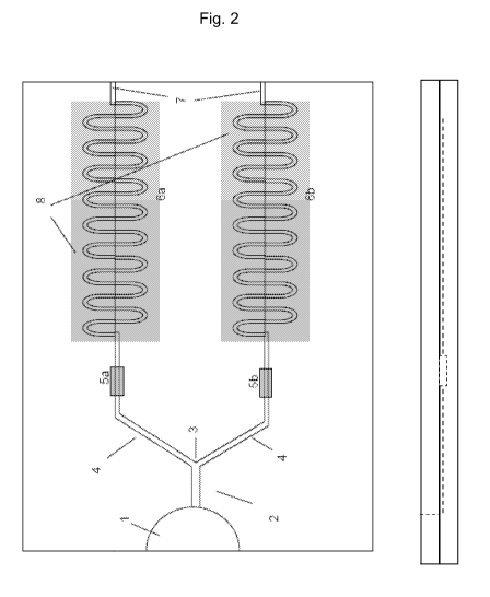
Figure 2 shows a top view (left part of the figure) and
side view (right part of the figure) of the device
according to the embodiment of Figure 1.
Figure 2A shows a top view of another embodiment of the
microfluidic device.
Figure 3 shows a graphical representation of the
superposition of the flow front positions in the clotting
and control channels.
Figure 4 shows a graphical representation of the
superposition of the flow front velocities in the clotting
and control channels.
Figure 5 shows the schematic flow front positions prior to
clotting in the embodiment according to Figure 1.
Figure 6 shows the schematic flow front positions after
clotting in the embodiment according to Figure 1.
Figure 7 shows the absorption coefficient of blood vs.
wavelength.
Figure 8 shows the emission spectrum of a LED.
Figure 9 shows the response curve of a photodiode optimized
to detect the greenish wavelengths.
CA 02700073 2010-03-18
WO 2009/037361 PCT/EP2008/062642
Figure 10 shows the detected current intensity versus time
in two chips of different size for the clotting and control
channels.
Figure 11 shows the derivatives of the current intensity
5 curves of Figure 10.
Figure 12 shows the superposition of a serpentine of the
embodiment according to Figure 1 and a CCD array with pixel
sizes 19x19pm.
Figures 13-16 show graphics of the equations used to
10 determining clotting time through theoretical curves.
Figure 17 shows typical data at step 3 from real
coagulation tests and clotting times as determined
following theoretical method 1 or 2.
Throughout the figures like reference numerals refer to
like elements.
Detailed description of the invention
The present invention provides a device in the form of a
chip or disposable test strip, for determining clotting
time of a fluid, such as blood and plasma, a measuring
apparatus to be used as portable coagulometer with the test
strip of the invention, and a method of determining
clotting time using the microfluidic device of the
invention.
A Portable coagulometer, as a Point-of-care device, is a
technology that follows four main lines of improvements:
cost reduction, blood sample reduction, quality control and
enhanced portability. All these four aspects are especially
important for economically and reliably spreading patient
self-testing.
CA 02700073 2010-03-18
WO 2009/037361 PCT/EP2008/062642
11
The present invention has significant advantages with
respect to the current state-of-the-art portable test
strips and coagulometers:
- Cost reduction: the disposable
microfluidic chip
is an extremely simple (passive) component,
manufactured with high-volume low-cost production
technologies and materials.
- Blood sample reduction: blood samples well below
5pL can be tested through the microfluidic chip
technology with the necessary quality controls and
accuracy.
- Quality control: A number of distinct on-board
quality controls can be integrated on the
disposable device of the invention and read by a
single detector means. In addition, the device
allows the use of calibrated plasmas as external
quality control.
- Enhanced portability: the detection systems are
extremely compact, low-cost and can be embedded on
thin portable devices.
The invention is based on the fact that an appropriate
microfluidic channel allows for the capillary flow of the
fluid sample, such as blood or plasma, allowing the
position or the velocity of the fluid front to be
accurately monitored with simple means, in a passive way,
without contact with the sample fluid. Rheological changes
of the sample fluid upon the initiation of the clotting
cascade (when the sample makes contact with the clotting
reagent), and in particular the apparent viscosity changes
at the clotting endpoint, have a significant effect on the
monitored dynamical parameters.
CA 02700073 2010-03-18
WO 2009/037361 PCT/EP2008/062642
12
These parameters can be monitored with the same simple
detection means, and compared either with a control sample
that does not contain a clotting reagent, or contains a
different control reagent, or alternatively with a
predicted theoretical value.
Without willing to be bound by theory, we believe that the
microfluidic system of the invention mimics in some way the
microcapillary structure of blood vessels and the dynamics
of flowing blood. Due to the complexity and high
sensitivity of blood coagulation stages (initiation,
amplification, propagation and clot formation) it is highly
favourable to reproduce as close as possible the in-vivo
hemostasis environment. According to a published report
from the University of Chicago [Kastrup, C. J. Runyon, M.
K. Shen, F. Ismagilov, R. F. Modular chemical mechanism
predicts spatiotemporal dynamics of initiation in the
complex network of hemostasis, Department of Chemistry and
institute for Biophysical Dynamics, University of Chicago,
Edited by George M. Whitesides, Harvard University.], a
microfluidic in vitro environment can mimic the actual
blood clotting behaviour in human capillaries, which they
proof is critical for the determination of the clotting
times.
In addition, this invention allows continuous monitoring of
the flow dynamics so that hemostasic molecular changes can
be detected, providing high accuracy and reproducibility.
In particular, the formation of the first insoluble fibrins
has a measurable effect on the rheological properties due
to the size of the microcapillary structure.
As shown in Figure 1, in one embodiment the microfluidic
device of the invention is a two-layer assembly comprising
a lower planar substrate and a cover layer. On the lower
CA 02700073 2010-03-18
WO 2009/037361 PCT/EP2008/062642
13
substrate a sample distribution system is patterned,
resulting in a series of channels or conducts, connected
through one end by appropriate means to a sample
introduction area.
The channels induce the flow through capillarity. The
skilled person will be able to adjust the size and form of
the channel patterned on the lower substrate to obtain a
flow position or velocity which can be monitored with
accuracy. To create the capillary flow of the fluid sample,
a hydrophilic surface is needed in the channel, so that
sufficient negative pressure is induced. This hydrophilic
surface can be present on the lower substrate or on the
cover layer.
In one embodiment the lower substrate is made of plastic.
If the plastic is hydrophobic, the hydrophilicity in the
channel has to be induced by means known to the skilled
person such as a chemical treatment, chemical coating or
plasma treatment, to obtain the desired surface energy or
contact angle.
In a preferred embodiment, the hydrophilic surface is
brought by the cover layer that seals the microfluidic
channels patterned on the lower layer. In this embodiment,
either a hydrophilic material is selected as cover layer,
or ia material which is subjected to a hydrophilic
treatment as described above.
Alternatively, in a preferred embodiment, the hydrophilic
properties are provided to the top layer by the adhesive
used to bond the two layers that form the chip. In such a
case it is important that the adhesive coating selected
does not react with the fluid sample or interferes with the
clotting reaction.
CA 02700073 2010-03-18
WO 2009/037361 PCT/EP2008/062642
14
Therefore, the cover layer may consist of adhesive polymer
films of various types, such as heat seals and pressure
sensitive adhesives. Hydrophilic formulations, with added
surfactants within the adhesive, can be employed. Hard
adhesives are preferred, to prevent channel blockage due to
adhesive flow during the sealing step or due to creep.
Figures 2 and 2A show a top view of different embodiments
of the microfluidic device of the invention, said device
comprising the components described below.
Means (1) for introducing a sample of fluid medium, mainly
consisting of an inlet port. This inlet port is coupled to
a distribution capillary channel (2), followed by a channel
bifurcation (3) which splits the distribution channel (2)
into a first (6a) and a second region (6b), which permit
said fluid medium to flow along a length of said regions.
Optionally, the distribution channel contains a cell filter
(only depicted in figure 1).
In a preferred embodiment, said first (6a) and second (6b)
regions have identical structures.
Each of said regions (6a, and 6b) comprise, in order from
the distribution channel, first an area (5a, 5b) and at
least one microfluidic channel, which will be referred to
as the scanning area (8) herein. The first area (5a)
contains a first reagent capable of reacting with said
fluid medium, and makes the microfluidic channel in region
(6a) function as a reaction channel, while the second area
(5b) is either empty or contains a different reagent, so
that the microfluidic channel in region (6b) functions as a
control channel. Preferably, said first reagent is capable
of initiating clotting of said fluid medium.
CA 02700073 2010-03-18
WO 2009/037361 PCT/EP2008/062642
In another embodiment, more than two regions are present in
the chip. One of the regions functions as the reaction
channel as explained above, and the other two or more are
5 control channels.
For on-board quality control, the blood sample can be
capillary driven along control channels where the reaction
chambers have specific compounds that provide known and
10 fixed (or narrow band) coagulation times. For example two
types of such controls can be incorporated, normalized
control and abnormal control, to provide lower and higher
references to coagulation times.
15 The control channels have a different reagent composition
from the reagent present in the reaction channel.
Therefore in one embodiment, there is a normalized control
channel, the reagent present in it can be for example at
least one Vitamin K dependent clotting factor. Such
clotting factors can come from a dried or lyophilized pool
of normal patient plasmas.
In another embodiment, there is an abnormal control
channel, which comprises a clotting factor inhibitor such
as, heparines, citrates, oxalates, EDTA and the like.
Further, it can comprise the same Vitamin K dependent
clotting factorsas in the normalized control channel.
The following are illustrative of preferred embodiments
describing the number of regions and their functionality:
= 2 regions: One reaction channel for blood sample
clotting time determination with respect to a control
CA 02700073 2010-03-18
WO 2009/037361 PCT/EP2008/062642
16
channel with no coagulant agent or with a coagulation
inhibitor agent.
= 2 regions: One reaction channel for blood sample
clotting time determination through theoretical curves
and one control channel which provides normalized
clotting times.
= 3 regions: One reaction channel for blood sample
clotting time determination with respect to a control
channel with no coagulant agent or with a coagulation
inhibitor agent. In addition, another control channel
which provides normalized clotting times.
= 3 regions: One reaction channel for blood sample
clotting time determination through theoretical curve
comparison. In addition, one control channel which
provides normalized clotting times and another control
channel which provides known abnormally high clotting
times.
All these embodiments and other variants that will be
apparent to the skilled person are emcompassed by the
present invention.
In the device of the invention, the flow is driven by
capillary forces only and thus the chip or test strip is a
passive device with no needs of external forces. The
hydrophilic channel surfaces allow the wetting meniscus to
move along the channels towards the negative capillary
pressure, while the dewetting meniscus remains at the inlet
port. The flow is stopped at stop valves by inducing a
hydrophobic surface or by designing a suitable channel
opening. In a preferred embodiment, each region (6a, 6b)
CA 02700073 2010-03-18
WO 2009/037361 PCT/EP2008/062642
17
contains means (7) for venting, most preferably a venting
port, which also functions as a stop flow valve. Although
depicted at the end of the channel in figure 2, the venting
ports (7) can be located at other positions along the
microfluidic channels. For example, connecting venting
ports (7) with flow stops at the exit of the reaction
chambers allows that capillary flow speeds up to this point
are maximized, as depicted in figure 2A. In another
embodiments each channel has more than one venting port
(7), the venting ports (7) allow to control and modulate
the velocity and the flowing properties of the fluid.
At least a property of the fluid medium, preferably the
position or the velocity of the fluid front, is monitored
as the fluid medium transits scanning areas (8) of the
first (6a), second (6b) and optional third regions.
Comparison between said properties in said different
regions enables detection of the moment when the reaction
in the first region (6a) has taken place and the
determination of the clotting time for the fluid sample.
The regions are preferably capillary channels.
The working principles of this device rely on
microfluidics, for which the governing principles radically
differ from the conventional flow theory, due to system
down-scaling.
Governing principles
The dynamic filing under Newtonian behaviour of a capillary
conduit of constant cross section can be determined through
the volumetric flow rate Q, which depends upon the
viscosity n, the total flow resistance RFR, and the
CA 02700073 2010-03-18
WO 2009/037361 PCT/EP2008/062642
18
pressure difference AP, between the wetting (front) and
dewetting (rear) meniscus:
AP
Q = 1 -- (1)
rl RFR
For a channel of length "L" and rectangular cross-section
A, width "a" and depth "b", the flow resistance RFR can be
expressed as:
-1
1"1 5a AR2 1
R =[- I+ H (2)
FR
12 6b; L
Where "RH" is the hydraulic radius and is defined as
R ____________
ab
- .
H 2(a + b)
To determine L=L(t), i.e. the flow front position against
time, the integration of equation (1) with time is
required. Thus, L and the velocity, calculated as the
derivative of L with time, are expressed as:
( (
2Ap 1 1+ 5a R2 t
_______________________________________________ H
L(t)= "\ 12 6b 0 (3)
11
1
AP 1+ 5a .2
H
dL 12 6b
¨dt =1 2n t
These are the governing flow equations prior to clotting,
as the viscosity has been assumed constant. When clotting
is initiated the viscosity is a function of time, with an
exponential increase, so that according to equation (1),
the flow rate, which is linearly inverse to viscosity, will
undergo a sudden decrease. The curves L(t) and the
derivatives shown in further sections have been numerically
determined for variable viscosity.
CA 02700073 2010-03-18
WO 2009/037361 PCT/EP2008/062642
19
With equations (1) to (3) it is possible to produce a
preliminary design of the channel lengths needed to allow
permanent flow up to the highest clotting times. The sample
volume "V" of a conduit of constant section can be
estimated as:
V=a b L(t) (4)
Thus, the device must be designed and the size of the
channels chosen according to the existing relation (4)
between geometrical parameters of the channels, a, b and L,
the volume of sample required and the maximum clotting
time.
Clotting time determination through theoretical curves
In one embodiment of the invention, taking advantage of the
flow dynamics continuous monitoring, the clotting time can
be determined or controlled through comparison of the
measured property of the sample with the theoretical
predicted value.
Since the dynamical behaviour is well predicted prior
clotting, the clotting time can be determined as the
instant when the monitored clotting curve deviates beyond a
particular threshold from the theoretical curves from
equations (3). A few mathematical operations can be applied
so that such deviation depends only on the qualitative flow
dynamic behaviour and not on the quantitative one. Two
different but analogous approaches are described as
follows:
Method 1:
CA 02700073 2010-03-18
WO 2009/037361
PCT/EP2008/062642
Step 1:
According to equation (3) for the capillary length under
Newtonian behaviour, L(t) is a power function of time.
Starting from the L(t) and t values extracted from the
5 detection system, the following curve can be constructed:
L(t)= Kt 5 (5)
The monitored curve (coagulation channel) and theoretical
10 curve are plotted on the graph depicted in figure 13.
Step 2:
15 Applying logarithms at both sides of the mentioned
expression, a linear curve of 0,5 slope is obtained (see
also the graph of figure 14):
LogL(t)= log K + 0.5 log t (6)
The quantitative term is log K and the qualitative is 0,5
log t.
Step 3:
By changing the variable (u=log t) a new function Y=Y(u)
can be defined, and differentiating it with respect to u
(see also the graph of figure 15):
Y (u)= log K +0.5u (7)
dY Ac
(8)
du
Step 4:
Second differentiation of Y with respect to u is carried
out(figure 16):
CA 02700073 2010-03-18
WO 2009/037361 PCT/EP2008/062642
21
d2Y
_____________________________________ =0 (9)
du 2
The decay from the constant value beyond a predefined
threshold in either the velocity (-.) or acceleration
du
d2Y
________ ) curves determines the clotting time. The above
du 2
mentioned operations are the mathematical basis of an
algorithm that allows the clotting time determination
through only one independent coagulation channel.
The microfluidic chip of the present invention is designed
so that flowing blood has a predominant Newtonian behaviour
prior clotting. Deviation from this behaviour is only due
to the pseudo-plastic effect, which can appear at low flow
rates. If this occurs, the method still applies and works
reasonably well because such pseudo-plastic effect is much
weaker than the clotting effect, and can be distinguished
on the acceleration curves.
Method 2:
A second and analogous mathematical approach for
theoretical clotting time determination can be briefly
described as follows. Starting from the same raw data, the
L(t) and t values obtained at step 1, the following curve
can be constructed:
L2
la-- (10)
CA 02700073 2010-03-18
WO 2009/037361 PCT/EP2008/062642
22
This curve is proportional to viscosity (n), as can be
derived from equation (3). The following steps (2, 3 and 4)
are applied identically as before (i.e. logarithm
application, first derivative and second derivative), so
that velocity and acceleration curves are constructed.
Based on real test data, both methods roughly give the same
clotting time (PT). A surprising result found in
practically all monitored curves, as the ones shown in the
graph of figure 17, was an initially unexpected behaviour
which is opposed to the coagulation effect, see the
highlighted areas in both curves under the term
"inversion". This effect is in fact a transient viscosity
decrease of about 1 or 2 seconds duration which is always
seen just prior the clotting time. This behaviour provides
an easier clotting time identification as the PT instant
thus becomes a clear inflection point, either a maximum in
method 1 or a minimum in method 2. Although the reason for
this unexpected behaviour is unknown, some evidence
suggests that this can be due to the formation of the
fibrin insoluble monomers coupled with the Fahraeus-
Lindqvist effect, which reduces the apparent viscosity
prior to the formation of fibrin polymers.
Besides the clotting time determination, the theoretical
approach described above, can also be employed for quality
control by correlating the test curves with the theoretical
predictions. Under a normal operator (i.e. no patient
misuse) and correct device conditions, the blood sample
flow prior to clotting should lie close to the mentioned
linear behaviour. Any significant deviation from such
behaviour can be detected and processed by the flow
CA 02700073 2010-03-18
WO 2009/037361
PCT/EP2008/062642
23
monitoring system and processor, providing a test
cancellation order.
According to a preferred embodiment the fluid medium is
blood, preferably capillary whole blood from patient
fingerpricking, and calibrated plasma with known clotting
times can be used for external quality control. The reagent
capable of reacting with said fluid medium is a clotting
reagent, more preferably a tissue factor or thromboplastin.
In this case, the device and method of the invention are
particularly suited to determine the Prothrombin Time, i.e.
the time elapsed between clotting activation and start of
clotting.
The device can be designed according to standard INR
values; the recommended highest INR range is about 8, which
also means PT about 100 seconds. The dimensions required
for reaching such a maximum INR are shown in Table 1. As
previously mentioned, the required dimensions and total
volumes "Vt" of different conduits designs are governed by
equation (3).
a(mm) b(mm) L(mm) Vt(iaL)
Microfluidic design 0.08 0.08 150 1.0
Microfluidic design 0.125 0.125 250 3.9
Intermediate design 0.5 0.5 500 125
Conventional design 1 1 700 700
Table 1. Required lengths and total volumes "Vt" of
different conduits designs for reaching such maximum INR
range (100 sec).
CA 02700073 2010-03-18
WO 2009/037361 PCT/EP2008/062642
24
This table demonstrates that simply, by downscaling the
fluidic design to the microscale, the standard INR range
can be achieved with just a blood droplet.
The shape and the dimensions of the channels according to
the present invention allow the determination of the
clotting time of a blood sample of no more than 15p1, and
the total volume allocated when all the circuits are filled
is less than 10p1 allowing a remaining volume within the
inlet port, necessary to fix the dewetting meniscus at the
inlet port. The microfluidic channels allow a continuous
flow, lasting from several seconds to more than a hundred
seconds, allowing the PT determination around a long time
range. Thus the chip and method of the invention allow the
measurement of accurate clotting times and INR
determination with low amounts of blood sample, preferably
below 10 pl, more preferably below 5 pl, and most
preferably with about 1 pl or less. This is very important
for the convenience of the patient.
The length of the capillary channels (6a, 6b) should be
large enough to enable the reaction of the reagent with the
fluid to be completed before the fluid front reaches the
end of the channel. In a preferred embodiment the capillary
channels (6a, 6b) are in a curved shape, most preferably
having a serpentine shaped track, in order to minimize the
area of the device while maintaining the length of the
channels.
The preferred cross-section of the channels is rectangular
due to manufacturing constrains, allowing a pure 2D
geometry, which simplifies the mould fabrication processes.
The specific dimensions have to be carefully calculated as
CA 02700073 2010-03-18
WO 2009/037361 PCT/EP2008/062642
the flow dynamic, and total volume employed is very
sensitive to channel dimensions. As shown herein, dimension
values well above 100 pm require very large channel lengths
to permit flow durations up to the highest clotting times,
5 and higher blood sample volumes are required. With a
microfluidic design, or in other words, channel cross
section dimensions about 100 pm or less, channel lengths
can be reduced with little blood usage. In addition the
size of the chip and its cost are also reduced
10 considerably.
Preferably, the reaction and control channels have a cross-
section where a=b. In this case a and b are preferably
between 30 to 125 pm, more referably beween 50 and 100 pm,
15 and even more preferably of about 80m.
Also the dimensions of the area containing the reagent,
preferably a reaction cell, must be appropriate to allow
enough volume for dispensing the reagent in liquid state.
20 Besides, the design has to be defined so the diffusion time
permits reaching enough reagent concentration in order to
maximize the activated blood volume. This can be achieved
by maximizing the surface to volume ratio within the
reaction chamber. Preferably, the footprint chamber design
25 should be circular for adapting to droplet dispensed shape,
with dimensions between 1 to 4 mm in diameter and height
between 40 to 150 pm. More preferably, the diameter is
about 1.5 mm and height is about 80 pm.
The height dimension of the distribution channel is
preferably between 150pm and 350pm, more preferably about
250 pm.
CA 02700073 2010-03-18
WO 2009/037361
PCT/EP2008/062642
26
The blood inlet port is preferably the gap left between the
cover and base substrates at the edge of the chip, on the
distribution channel, and therefore can have the height of
said distribution channel. Volume allocated on the
distribution channel should be slightly larger than the
volume allocated in the subsequent capillary structure, so
that once the distribution channel is completely filled
with fluid it can never be emptied. This volume defines the
minimum test sample volume requirement.
In order to fulfil construction requirements and
dimensional constrains, the flow rate Q can be modified
through the introduction of passive flow control valves by
modifying the cross section of the microfluidic channels,
for example, by narrowing segments of the microfluidic
channels or by introducing tapered microfluidic channels.
Operation of the microfluidic device
The present invention requires applying a sample of blood
or plasma to the inlet port, through which the blood or
plasma enters the sample distribution channel, along which
the same blood sample or plasma is split into a
reaction/clotting channel and one or more control channels.
At a time tm prior to blood clotting the flow front
positions in the channels can be represented as follows,
L=L(tm)
L'=L'(tm) (11)
Where L y L' are respectively the clotting and control
positions. The time t=0 is the instant the flow exits the
reaction cell of the clotting channel, as it is the moment
CA 02700073 2010-03-18
WO 2009/037361 PCT/EP2008/062642
27
the tissue factor or thromboplastin has solubilized and the
reaction mechanisms are initiated.
The split flows have nearly identical motion dynamics until
the coagulation is initiated in the clotting channel. This
instant, when the first blood clotting occurs, is
identified as the Prothrombin Time, and induces a sudden
increase in viscosity. At this instant the flow dynamics
along the clotting channel is decelerated with respect to
the control channel(s). By continuous monitoring (8) the
flow front position as a function of time, the derivative
of the position with time, which can be referred to as the
flow front velocity, can be calculated.
In Figure 3, it is illustrated how the flow front positions
in two channels and the Prothombin Time can be identified.
These curves have been numerically calculated with the
following assumptions, where variables a, b, ri and PT have
the meaning indicated previously herein, and y is the blood
surface tension:
y (N/r0 0,05589 Contact angle 35
0,00012544Pa:41 0,003
16 (10 0,000125 For 25s
Table 2. Assumptions for the numerical calculations.
Prior to PT the difference between the channels should be
minimal, only affected by non-uniform environmental
conditions, manufacturing tolerances and detection noise.
The derivative with time curves are preferred as it is a
more sensitive to viscosity changes, which can be referred
as the flow front velocities. Analogously at a time tm
CA 02700073 2010-03-18
WO 2009/037361
PCT/EP2008/062642
28
prior to PT, the velocities are monitored for clotting (V)
and control (V') will be:
V = V(tm)
V'= V' (tin) (12)
These curves are shown in Figure 4.
PT can be determined by defining a suitable threshold "A"
for the difference between the velocities V(tm)-V'(tm).
Prior to PT, the viscosity is constant and the flow front
positions and velocities have minor differences as
schematically shown in figure 5.
At a time tp the velocity difference has just surpassed the
threshold (see figure 6) and this instant is PT.
Detecting means
For a continuous detection or monitoring of the flow front
motion L=L(t) or v=v(t) different detection techniques can
be used:
= Detection through Photodiode
= Detection through optical sensors such as
Charged-Coupled-Device (CCD) or Complementary
Metal Oxide Semiconductor (CMOS).
The coefficient of absorption of blood is plotted in figure
7. It can be seen that it absorbs especially at 400 nm, and
also around the green (530 nm).
Detection through Photodiode
CA 02700073 2010-03-18
WO 2009/037361 PCT/EP2008/062642
29
The serpentine is illuminated with a LED and transmitted
light is detected with the photodiode. The moving flow
front linearly increases the absorption and thus the
intensity detected is accordingly reduced. With a signal
amplifier it is possible to monitor tiny flow position
increments.
In the following some calculations have been carried out to
evaluate the viability of such monitoring scheme, using
standard low cost components.
A LED and a photodiode, both low cost, from readily
available distributors have been selected.
The LED has 3 mm size and emits within a 200 angle. The
intensity is 15000 mcd = 0.0309 Watts/str, so by taking the
whole 20 solid angle (0.095 str) the total emission power
reaches 0.00294.
The emission spectrum of the LED and the response curve of
the photodiode, which is a standard Silicon one but also
optimized to detect the greenish wavelengths, can be shown
in figures 8 and 9.
Under these assumptions and by further acquiring the
scanning area (8), channel dimensions and the actual L(t)
curve from figure 3, the intensity signal detected by the
photodiode can be obtained. For simplicity reasons, it has
been also assumed that the chip is perfectly transparent
and no Fresnel reflections are taking place. The Intensity
signal, plotted in Figure 10, also contains a dark current
random noise simulation of 20picoA, as specified by the
manufacturer. This curve corresponds to a channel section
CA 02700073 2010-03-18
WO 2009/037361 PCT/EP2008/062642
of 250x250pm. By calculating the derivative of the
intensity signal with time, a signal proportional to the
flow velocity can be obtained, as shown in Figure 11.
5 With the two shown plots (figures 10 and 11) it is
demonstrated that the flow front monitoring is viable, with
a sufficiently high sensitivity, as can be deduced from the
negligible noise affecting the curves. In addition, the
time response of the photodiode is very high, which permits
10 frequency sampling as high as 10MHz and the amplifier
itself is limited to 10Khz. This values are orders of
magnitude beyond the needed frequency for accurate
monitoring, about 20Hz.
15 Detection through optical sensors
With this detection scheme, the system employs a similar
configuration but substituting the detection device. In
this case we employ CCD or CMOS sensors, so that flow front
20 position is obtained by processing the data acquired after
high frequency mapping of the scanning surface.
The LED system can be similar to the one defined in the
previous case. Interestingly, in this case no high
25 sensitivity is required, as each cell or pixel within the
CCD is to detect the presence of absence of flow in this
position. As shown in figure 12, by superposing the CCD
effective area of a standard with the serpentine, the
mapped image would allow the identification of the flow
30 front position, with enough resolution and time response
(>1KHz).
CA 02700073 2010-03-18
WO 2009/037361 PCT/EP2008/062642
31
This technique requires image data processing, so that from
a blurry image the meniscus position can be identified.
This increases the complexity of the monitoring system.
However, and in contrast with the photodiode detection
scheme, the sensitivity of each cell or pixel is less
stringent, which in this sense will favour the CCD
detection scheme.
In order to improve the detecting signal quality, optical
means, such as a lens can be integrated. Commercial rigid
blocks, integrating lens and sensor are available nowadays
at very low cost, such as the miniature cameras that are
supplied to the mobile industry. These blocks measure just
a few millimetres and thus allows very compact and thin
integration into portable systems, such as the portable
coagulometer.
The detected signal is processed by the microprocessor with
embedded software. Dynamic flow data curves are generated
and the algorithms are employed for coagulation time
determination and also for various quality controls.
As explained before, the chip (test strip) and method of
the invention have another significant advantage, in that
the same detection means can be used for monitoring the
sample fluid flow and for fulfilling various quality
control task.
When the detection means is provided through artificial
vision system, such as CCD/CMOS sensor or microcamera,
three main quality controls, usual in test strips for
coagulameters, can be performed through field of view image
processing of such a vision system:
On-board ambient condition indicators for stability
monitoring: ambient conditions such as temperature and
CA 02700073 2010-03-18
WO 2009/037361 PCT/EP2008/062642
32
humidity can be monitored through colour sensitive
compounds to these factors. The selected compounds undergo
an irreversible colour change when subjected to temperature
and humidity thresholds, signalling a deficient chip. They
can be added directly on the reaction chambers, on the base
substrate or on the cover surface, under the detector's
field of view. A combination of different sensitive
compounds can be used to this end. Examples of such
compounds as sensible temperature compounds: Leuco dyes,
Oxazines, Crystal violet lactone, phenolphthalein and the
like. Metallic salts as sensible moisture compounds: cobalt
chloride, calcium sulfate and the like. N-oxide or Nitroso
compounds as both temperature and moisture sensible
compounds.
This will allow the measuring device (such as portable
coagulometer) to inform the patient that the test strip has
not passed the quality control and should be discarded.
External quality control: calibrated plasmas with known
clotting times, commercially available for performing INR
and PT test calibrations, can be used as external quality
control, so that the whole portable coagulometer system can
be evaluated. In this embodiments the artificial vision
system is adjusted to allow detection of the flowing
plasmas. Although plasma is a nearly transparent fluid,
little adjustment of the illumination led system and image
processing is required to effectively track plasma flow,
since moving plasma is recognized like a grey shadow
advancing along bright channels.
Printed Codebar: printed code carrying among other relevant
information calibration data, traceability data and expiry
date. Standard data matrix codes of a few millimiters
CA 02700073 2010-03-18
WO 2009/037361 PCT/EP2008/062642
33
dimensions used in this kind of test strips can be printed
onto the chip's cover layer or onto a transparent label.
The suitable detecting and/or monitoring means described
above are comprised in an external device (coagulometer)
which comprises a slot for receiving the microfluidic
device of the invention and is designed to cooperate with
said microfluidic device.
Additionally, the external device comprises means for
processing the data delivered by the detecting and/or
monitoring means and produces a signal output into a
displaying means.
Manufacturing
The present microfluidic device can be easily manufactured
with current plastic replication technologies and
assembling techniques. The assembly is formed by two sealed
components: the lower substrate, where the microstructures
are patterned and the top substrate or cover lid, as
illustrated in Figure 1.
The materials suitable for both the lower substrate and the
cover layer of the device are a range of polymer, thermoset
and/or thermoplastic materials should have good optical
properties and good dimensional stability. For example,
COC, PMMA, PC, PSU, SAN, PETG, PS and PP can be used.
Most polymeric materials are hydrophobic in nature.
Therefore if a strongly hydrophobic material is chosen as
patterned substrate, a subsequent production step to render
hydrophilic some surfaces would be necessary, as explained
before. For this reason, hydrophilic or at least not
hydrophobic (contact angle < 900) plastics are recommended.
CA 02700073 2010-03-18
WO 2009/037361 PCT/EP2008/062642
34
That is the case for PMMA, Cellusose Acetate, PC, COC and
PS, among other well known materials. One material that is
particularly preferred is PMMA, in view of its good contact
angle, optical properties and dimensional stability.
The lower substrate can be easily replicated with a range
of technologies, available today, and with very high
accuracies, allowing low microfeature tolerances. The most
relevant current techniques for said patterning step are
microinjection moulding, hot embossing and soft lithography
imprinting.
The sealing step can be performed with a number of well
known techniques such as thermal compression bonding,
adhesive bonding, plasma activated bonding, ultrasonic
bonding, laser welding and others.
The cover is preferably a hydrophilic film. It is
preferably transparent, to allow accurate monitoring of the
fluid flow. As explained above hydrophilic films provide
very cost-effective means that enable both sealing and
channel hydrophilization, avoiding the surface treatment
step. In this case, the production technique consists of
standard lamination processes, which can require pressure
and temperature control. Other production techniques are
embossing or pressing processes.
As described above the Reaction chambers can allocate a
number of dry-reagent compounds for various purposes. The
main compound is thromboplastin to initiate the coagulation
cascade. Due to the tiny dimensions of the reaction chamber
high performance compounds can be added without
significantly increasing the cost of production.
CA 02700073 2010-03-18
WO 2009/037361 PCT/EP2008/062642
Human thromboplastin recombinants have extremely useful
properties in terms of solubilization and sensitivity due
to their chemical purity. The former property has been
traditionally enhanced by the use of specific additives.
5 Under the present invention's design, a fraction of a
microliter of human recombinant factor can be dispensed,
showing excellent results in terms of solubilization and
sensitivity.
10 A number of additional agents play a role in the proper
functioning of the dry reagent. They may be employed not
only for rapid solubilization, but also for control
diffusion parameters, improving fabrication steps and
reagent stability, or for addressing the following issues:
a) Modulate uptake of the liquid into the dry reagent:
simple polymers such as hydroxylpropyl cellulose, polyvinyl
alcohol, polyethylene glycol and the like.
b) Rapid solubilization, stabilizers and shortening the
drying process: albumin, glutamate, sacarides, (such as
glucose, saccharose, trehalose, etc), and the like.
c) Controlled wettability: Triton, Macol, Tetronic,
Silwet, Zonyl, Pluronic, and the like.
d) Color indicator for monitoring stability and for
dispensing control: Leuco dyes as sensible temperature
compounds (Oxazines, Crystal violet
lactone,
phenolphthalein and the like.). Metallic salts as sensible
moisture compounds such as cobalt chloride, calcium sulfate
and the like. N-oxide or Nitroso compounds as both
temperature and moisture sensible compounds.
e) Enhancing ambient conditions stability: organomercury
compounds such as Thimerosal and the like.
CA 02700073 2010-03-18
WO 2009/037361 PCT/EP2008/062642
36
f) Other compounds for various functionalities: Polybrene
(antiheparin agent) and buffers.
The dry-reagents can be applied on the reaction chamber or
alternatively onto the cover substrate, through a number of
well known techniques: liquid drop dispensing, gel
dispensing, jet dispensing, screen printing, blade coating,
selective spraying and film casting. The dispensing step is
followed by a drying step.
Preferably, dry reagent is dispensed in liquid state onto
the reaction chamber forming a droplet occupying most of
the chamber that upon drying becomes a thin dry-reagent
layer.
Advantageously, both the manufacturing method and the chip
(test strip) so fabricated are extremely simple, no
embedded components are required, such as electrodes or any
form of multilayer structures. Indeed, the presented
manufacturing techniques allow low cost production, so that
cheap disposable devices can be produced.
The current invention, through its microfluidic design,
provides very sensitive and accurate means for clotting
time determination. The clotting time (such as the
Prothrombin Time) relates to the moment when the insoluble
fibrin molecules start to polymerise that later produces a
"mesh" that forms the clot. The formation of fibrin
polymers, typically of the order of a few micrometers,
leads to an abrupt increase on the apparent viscosity of
the flowing blood, specially when the channel cross-section
becomes as tiny as in the current microfluidic design. In
CA 02700073 2010-03-18
WO 2009/037361 PCT/EP2008/062642
37
terms of accuracy and sensitivity, this device offers the
previously mentioned advantages with respect to previous
devices for clotting time determination.
In addition, the combination of chip and measuring device
of the invention provides combined advantages. The use of
single optical detection means allows to simultaneously
combine the detection of fluid flow changes and different
quality controls. This means that the portable measuring
device will be less complex and more compact, using
standard components. In fact the measuring device can have
the size of a mobile telephone. There is also a significant
improvement in precision and sensitivity from previous
devices, especially those that are based upon blood flow,
as the flow monitoring is made continuously with a high
frequency sampling. In this way, the very instant when the
clot formation has the first decelerating effect on blood
flow can be accurately determined.
As will be recognised by those skilled in the art, the
innovative concepts described in the present application
can be modified and varied over a wide range of
applications.
Accordingly, the scope of patented subject matter should
not be limited to any of the specific exemplary teachings
discussed, but is instead defined by the following claims.
Any reference signs in the claims shall not be construed as
limiting the scope thereof.