Note: Descriptions are shown in the official language in which they were submitted.
CA 02700200 2010-03-18
WO 2009/039457 PCT/US2008/077136
IDENTIFICATION OF NOVEL PATHWAYS FOR DRUG DEVELOPMENT FOR
LUNG DISEASE
CROSS REFERENCE TO RELATED APPLICATIONS
This application claims the benefit of U.S. provisional
application Serial No. 60/994,643, filed September 19, 2007,
the entire teachings of which are incorporated herein by
reference.
STATEMENT REGARDING FEDERALLY SPONSORED RESEARCH
Work described herein was supported by funding from
Grant Nos. NIH/NCI R01CA124640 and NIH/NIEHS U01ES016035.
The U.S. Government has certain rights in the invention.
BACKGROUND OF THE INVENTION
Cigarette smoke is the dominant cause of lung cancer in
the United States, accounting for an estimated 90% of all
cases [1]. However, the damage caused by cigarette smoke is
not limited solely to the lung, but rather constitutes a
`field of injury' throughout the entire respiratory tract
[2-6]. An important product of the field of injury
hypothesis is the ability to glean clinically relevant
information from cells collected in regions of the
respiratory tract, such as the bronchial airway, that can be
obtained in a less invasive manner than is typical of
collecting primary lung tissue. Based on this approach, a
qene expression-based biomarker measured in the
CA 02700200 2010-03-18
WO 2009/039457 PCT/US2008/077136
cytologically normal bronchial airway epithelium that can
distinguish smokers with and without lung cancer has been
developed [7]. This airway gene expression biomarker
achieved 83% accuracy in predicting whether a smoker had a
lung tumor in a prospective test set, and 94% accuracy when
combined synergistically with clinical variables [7, 8].
Beyond serving as an early diagnostic tool for lung
cancer, gene expression changes in the cytologically normal
airway epithelium have the potential to improve our
understanding of the signaling events deregulated during
early stages of lung cancer. Lung cancer development in
humans is a complex process involving multiple aberrant
events that, when accumulated, lead to deregulation of
crucial cell functions, including cell survival and
proliferation. In primary tumors resected from patients with
lung cancer, many signaling pathways have previously been
found to be deregulated, such as p53, RAS and
phosphatidylinositol 3-kinase (P13K) [9-11]. Additionally,
studies in the field of injury and field cancerization have
found that some of the molecular changes presumed to be
early in tumorigenesis are also reflected in histologically
normal cells both neighboring and more distal to the primary
tumor. For example, the same p53 mutation or loss of
heterozygosity at a specific chromosomal region have been
identified throughout the entire respiratory tract [5, 12].
Insight into the deregulation of oncogenic pathways in
cytologically normal bronchial airway cells from smokers
with lung cancer will help elucidate mechanisms involved in
the progression into malignancy. Furthermore, understanding
which pathways are deregulated could lead to therapeutic and
chemoprophylactic opportunities at the pre-malignant stage
of lung cancer.
- 2 -
CA 02700200 2010-03-18
WO 2009/039457 PCT/US2008/077136
SUMMARY OF THE INVENTION
It has been previously demonstrated that gene
expression profiling of cytologically normal bronchial
airway epithelium reflects a field of injury in smokers that
can serve as a sensitive and specific diagnostic biomarker
for lung cancer. Using gene expression signatures defined by
in vitro perturbation of specific oncogenic pathways, as
described herein a significant increase of
phosphatidylinositol 3-kinase (P13K) pathway activity was
identified in the cytologically normal airway of smokers
with lung cancer (n=129), as well as in lung tumor tissue
(n=107). To evaluate whether increased activity of P13K
occurs prior to the development of lung cancer,
cytologically normal airway epithelia in high-risk smokers
with moderate-severe dysplastic lesions in their airway
(n=14) were profiled, and higher levels of P13K pathway gene
expression were found as compared to the airway epithelium
from healthy smokers without dysplasia (n=11). Further, P13K
activity was decreased in the airway of high-risk smokers
who had significant regression of dysplasia following
treatment with the chemoprophylactic agent myo-inositol
(n=10). In vitro dilution experiments confirmed that myo-
inositol inhibits the P13K pathway, which not only proposes
a mechanism of action for myo-inositol, but also reflects a
potential therapeutic relationship between P13K pathway
activity and regression of airway dysplasia. Together, these
findings suggest that deregulation of the P13K pathway is an
early, measurable and reversible step in the development of
lung cancer, and that airway gene expression profiling in
smokers may enable personalized approaches to
chemoprophylaxis and therapy.
In one embodiment the invention provides biomarkers for
oncogenic pathways activated in cytologically normal airway
- 3 -
CA 02700200 2010-03-18
WO 2009/039457 PCT/US2008/077136
epithelial cells of individuals with lung disease. These
biomarkers and pathways may provide prognostic and/or
diagnostic indicators of lung disease, e.g., lung cancer.
Additionally, these pathways and biomarkers may provide
therapeutic targets for the treatment of lung disease, as
well as markers for the assessment of treatment efficacy.
In one aspect the invention relates to the use of gene
expression profiling methods to identify gene expression
signatures of oncogenic pathways activated in lung disease,
e.g., lung cancer. These gene expression signatures may
provide prognostic or diagnostic indicators for lung
disease, e.g., lung cancer. Moreover, oncogenic pathways
which are activated in lung disease may be targets for
therapeutic intervention. In one embodiment the oncogenic
pathway can be, for example, one or more of the P13K and
Np63 pathways.
The invention also relates to a method of identifying
an individual at increased risk of lung disease, comprising
determining the activation status of the Np63 and/or P13K
pathway in a cytologically normal airway epithelial cell
from said individual, wherein activation of the Np63 and/or
P13K pathway is indicative that said individual is at
increased risk of lung disease as compared with an
individual in whom the Np63 and/or P13K pathway is not
activated. In particular embodiments the individual is a
smoker or a non-smoker. In other embodiments the lung
disease is lung cancer.
In one embodiment the activation status of the P13K
pathway is determined using gene expression data for one or
more biomarkers of the P13K pathway. For example, in some
embodiments at least one of said one or more biomarkers is a
gene which is increased upon P13K activation, and in other
embodiments at least one of said one or more biomarkers is a
- 4 -
CA 02700200 2010-03-18
WO 2009/039457 PCT/US2008/077136
gene which is decreased upon P13K activation. Combinations
of biomarkers which are increased and decreased upon P13K
activation may also be used. In particular embodiments at
least one of said one or more biomarkers is a gene which is
upstream of P13K activation, while in other embodiments at
least one of said one or more biomarkers is a gene which is
downstream of P13K activation.
In particular embodiments, expression data for said one
or more biomarkers of the P13K pathway is obtained using an
oligonucleotide microarray. In other embodiments the
activation status of the P13K pathway is determined using
one or more gene expression products of one or more
biomarkers of the P13K pathway. Said gene expression
products may be nucleotide or amino acid products and can be
detected using methods known in the art.
In some embodiments of the invention the activation
status of the P13K pathway is determined by assessing the
activation of IGF1R, wherein activation of IGF1R is
indicative of activation of the P13K pathway. In other
embodiments of the invention the activation status of the
P13K pathway is determined by assessing the activation of
PKC, wherein activation of PKC is indicative of activation
of the P13K pathway.
The invention also relates to a method of identifying
an individual at increased risk of lung disease, comprising
determining the activation status of PKC in a cytologically
normal airway epithelial cell from said individual, wherein
activation of PKC is indicative that said individual is at
increased risk of lung disease as compared with an
individual in whom PKC is not activated.
The invention further relates to a method of
identifying an individual at increased risk of lung disease,
comprising determining the activation status of IGF1R in a
- 5 -
CA 02700200 2010-03-18
WO 2009/039457 PCT/US2008/077136
cytologically normal airway epithelial cell from said
individual, wherein activation of IGF1R is indicative that
said individual is at increased risk of lung disease as
compared with an individual in whom IGF1R is not activated.
In other embodiments the invention provides an
oligonucleotide array having immobilized thereon one or more
probes for one or more biomarkers of the P13K pathway, and
wherein said array does not have immobilized thereon probes
for other biomarkers. In preferred embodiments said one or
more biomarkers of the P13K pathway are selected from the
group consisting of IGF1R, PKC, the biomarkers disclosed in
[29], and combinations thereof.
The invention also relates to a method of reducing the
risk of lung disease in an individual comprising
administering to an individual at risk of lung disease one
or more agents (e.g., one or more agents, regimens or
treatments or combinations thereof) which inhibit the P13K
pathway. In particular embodiments the P13K pathway is
activated in said individual prior to administration of said
one or more agents. In one embodiment the lung disease is
lung cancer. In another embodiment said one or more agents
are administered to said individual prophylactically before
the development of lung disease.
In another embodiment the invention relates to a method
of differentially classifying a cytologically normal test
airway epithelial cell, comprising identifying a gene
expression signature associated with activation of a
biological pathway of interest in a normal airway epithelial
cell; assessing gene expression in differentially classified
airway epithelial cells to identify one or more correlations
between classification of an airway epithelial cell and
activation of a biological pathway of interest; and
assessing gene expression in a cytologically normal test
- 6 -
CA 02700200 2010-03-18
WO 2009/039457 PCT/US2008/077136
airway epithelial cell, wherein the gene expression profile
of the cytologically normal airway epithelial cell to be
classified indicates whether the biological pathway of
interest is activated and thus differentially classifies the
cell.
In particular embodiments the biological pathway of
interest is an oncogenic pathway. In some embodiments the
differential classification is increased risk of disease
versus decreased risk of disease, while in other embodiments
the differential classification is response to treatment
versus non-response to treatment.
In one embodiment, the invention provides a method for
identifying the activation of an oncogenic pathway in a
mammal having or at risk of having lung disease, e.g., lung
cancer. The method may include: (a) providing a biological
sample, e.g., a biological sample from an airway passage of
the mammal, wherein the biological sample comprises a gene
expression product (e.g., mRNA or protein) from at least one
gene that is indicative of activation of said pathway, and
(b) detecting the expression of said gene. For example, the
pathway can be one or more of the following: Ras, Myc, E2F3,
beta-catenin, Src, Np63, P13K and combinations thereof. The
mammal can be, for example, a human. Biological samples may
be provided, for example, from bronchial, nasal or buccal
epithelium, or from biopsied tissue samples. In one
embodiment, detection of gene expression is accomplished
using an oligonucleotide array having immobilized thereon
one or more nucleotide sequences or fragments thereof which
are probes for the relevant gene(s). Identification of
activation of an oncogenic pathway may indicate that the
mammal is a candidate for treatment to inhibit activation of
said pathway or for additional or more frequent screening to
identify development of disease, e.g., cancer.
- 7 -
CA 02700200 2010-03-18
WO 2009/039457 PCT/US2008/077136
In another embodiment, the invention provides a method
of screening candidate therapeutic agents which may be
useful in the treatment of lung disease, e.g., lung cancer.
For example, candidate agents may be screened for their
ability to modulate (e.g., inhibit) the activation of an
oncogenic pathway identified by methods described herein as
associated with lung disease. The agent's ability to
modulate activation of the pathway may be assessed, for
example, by its ability to alter a gene expression signature
of an oncogenic pathway from a signature which is associated
with disease to a signature which is not associated with
disease, e.g., is normal. Alternatively the candidate
therapeutic agent may be assessed for its ability to
modulate a specific functional effect or readout of the
pathway. Agents identified as having the ability to inhibit
an activated oncogenic pathway associated with lung disease
may be suitable for treatment of lung disease in a mammal.
In addition, the efficacy of a treatment regimen
can be evaluated by assessing the gene expression signature
of a mammal at various time points over the course of
treatment. A shift in the gene expression signature of an
oncogenic pathway from one associated with disease to one
not associated with disease, e.g., to a normal signature, is
indicative of efficacious treatment. Similarly absence of a
shift in the gene expression signature toward a normal
signature is indicative that treatment is not efficacious
and that perhaps alternative treatment regimens are
indicated.
BRIEF DESCRIPTION OF THE DRAWINGS
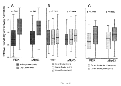
Figs. 1A-1C show that P13K and ONp63 are differentially
activated in smokers with lung cancer.
- 8 -
CA 02700200 2010-03-18
WO 2009/039457 PCT/US2008/077136
Using binary regression models trained on in vitro gene
expression signatures, pathway activation probabilities were
calculated in samples obtained from the cytologically normal
airway. Pathway levels are summarized using box plots, where
the bar represents the median value, the box denotes the
range of the data points from the 25th to 75th percentile,
and the whiskers specify the range of the remaining lst and
4th quartile. As shown in Fig. 1A, when grouping the
activation levels by lung cancer status (blue for no lung
cancer, red for lung cancer), two pathways were found to be
statistically different after random permutation tests: P13K
(p<0.001), and ONp63 (p<0.001). To account for variables
that could possibly confound the observed differences in
pathway activation seen in the airway of smokers with lung
cancer, pathway activation probabilities were also
calculated for healthy never (green), former (brown) and
current smokers (gray) (Fig. 1B), as well as current smokers
with (orange) or without (gray) chronic obstructive
pulmonary disease (COPD) (Fig. 1C). Neither of the potential
confounding variables showed a statistically significant
difference in pathway activation.
Fig. 2 shows oncogenic pathway activity in lung tumor
and adjacent normal tissue. Oncogenic pathway activity was
calculated for a dataset of lung adenocarcinoma and adjacent
normal tissue [32]. An increase in P13K (p<0.001) and ONp63
(p=0.002) was observed when comparing adjacent normal and
its paired tumor sample. Increases were also seen in Myc and
E2F3, and there was a decrease in Src. Error whiskers are
reported as SEM.
Figs. 3A-3B show the biochemical validation of P13K
activity in prospectively collected airway samples. Airway
brushings were collected prospectively from patients under
suspicion of having lung cancer in Boston and Utah. As shown
- 9 -
CA 02700200 2010-03-18
WO 2009/039457 PCT/US2008/077136
in Fig. 3A, kinase assays were used to measure in vivo
levels of P13K pathway activity. Patients with lung cancer
generally had higher levels of P13K activity than those
without lung cancer. A subset of the Boston cohort had extra
sample run on microarray so that computational predicted
P13K activity could be correlated to in vivo activity. The
probability of pathway activity is shown below the patients
that had extra samples. Pearson correlation of the
computationally predicted P13K activity and the
biochemically measured activity was 0.48. As shown in Fig.
3B, Western blots querying proteins both upstream and
downstream of P13K were quantified and then correlated with
P13K kinase levels measured in Fig. 3A. Correlations are
presented in a heatmap manner, where blue represents
negative correlation, and red represents positive
correlation. Correlation analysis is also broken down into
all samples, only samples with lung cancer, and only control
samples. Both p-IGF1R and p-PKC are positively correlated
with P13K activity in patients with lung cancer, suggesting
possible sub-pathways driving the increased P13K pathway
activity in the airway of smokers with lung cancer.
Fig. 4 demonstrates that smokers with dysplasia have an
increased activation of the P13K pathway. Genes that
increase when the P13K pathway is activated, as defined by
in vitro perturbation, are displayed in a heatmap. Blue
represents low expression of a gene, while red represents
higher expression of a gene. When comparing the expression
levels of these genes in the cytologically normal bronchial
airway of smokers with dysplasia against healthy smokers, an
increased activation of the P13K pathway in smokers with
dysplasia was observed. GSEA was used to quantify the
enrichment of this gene set (p<0.001, FDR Q < 0.001). Genes
were ranked for GSEA using a linear model that takes into
- 10 -
CA 02700200 2010-03-18
WO 2009/039457 PCT/US2008/077136
account dysplasia status, pack-years as well as a random
variable accounting for batch effects.
Fig. 5A-5C show that Myo-inositol inhibits the P13K
pathway in vitro. Insulin was used to activate the P13K
pathway in three different cell lines: (Fig. 5A) BEAS-2B
(bronchial airway cell line), (Fig. 5B) BT549 (breast cancer
cell line), and (Fig. 5C) HEK293 (human embryonic kidney
cell line). The cell lines were then treated with varying
doses of myo-inositol and LY-294002. PIP3 levels were
measured to quantify the activation levels of the P13K
pathway (y-axis). In each cell line tested, there was a drop
in PIP3 levels following treatment with either myo-inositol
or LY-294002 (a known P13K inhibitor), suggesting that myo-
inositol inhibits the P13K pathway in vitro. Replication of
these experiments produced similar results.
DETAILED DESCRIPTION OF THE INVENTION
Based on the concept that genomic changes in the
epithelial cells that line the entire respiratory tract
reflect host response to and damage from cigarette smoke, a
better understanding of the early events leading to
tumorigenesis may be gained by identifying which pathways
are deregulated in the airway of smokers with or at risk for
having lung cancer. One approach to assess pathway activity
uses gene expression data to link in vitro activation of an
isolated signaling pathway to predict status of that pathway
in patient samples. This approach has been successful at
predicting pathway status in cell lines as well as tumors
where the initiation event is known [13]. A strength of in
vitro defined pathway signatures is that they are capable of
identifying pathway activity at the gene expression level,
allowing the measurement of multiple pathways using a single
microarray experiment. Further, gene expression based
- 11 -
CA 02700200 2010-03-18
WO 2009/039457 PCT/US2008/077136
predictions of pathway activity have been found to correlate
significantly to drugs that target the specific pathway [13-
20]. Numerous studies have also found correlation of
predicted pathway status and therapeutic responsiveness in
clinical trials with targeted therapies [22-25].
Work described herein utilized expression signatures
developed through in vitro perturbation. Metagene models
were trained to compare oncogenic pathway activity in the
cytologically normal airway epithelium of smokers with and
without lung cancer. As described herein, using the gene-
expression based pathway approach described above, the data
show that the P13K pathway has an increased level of
activity in cytologically normal bronchial airway cells of
smokers with lung cancer, as well as higher levels in the
lung tumor tissue itself. Biochemical assays measuring in
vivo P13K activity in a prospectively collected cohort of
airway samples from patients with and without lung cancer
validated the computational predictions. An exploration of
the expression profiles from the cytologically normal airway
of high-risk smokers with dysplastic lesions in their airway
again revealed an increased activity of P13K. As dysplasia
is considered a pre-neoplastic event, this is suggestive
that P13K levels increased before the development of lung
cancer. Providing possible therapeutic relevance to this
result, high-risk subjects responsive to the
chemoprophylactic agent myo-inositol show a significant
reduction in P13K activity and regression of dysplastic
lesions. The relationship between myo-inositol and P13K was
further elucidated by showing that myo-inositol inhibits
P13K in vitro.
Together, these results demonstrate that the P13K
pathway is activated in the cytologically normal airway
epithelium prior to the development of lung cancer, and the
- 12 -
CA 02700200 2010-03-18
WO 2009/039457 PCT/US2008/077136
levels of this pathway associate with response to
chemoprophylaxis with myo-inositol. More broadly, these
findings suggest that airway gene-expression reflects
perturbation of specific oncogenic pathways within a smoker,
potentially allowing for personalized approaches to
chemoprophylaxis and therapy.
P13K and ONp63 pathways have an increased activation in
the normal airway of smokers with lung cancer (Figure 1A).
This is an intriguing finding, because a priori
cytologically normal cells are not expected to show signs of
oncogenic pathway deregulation. Additionally, it is
important to note that non-lung cancer controls used as
described herein have an extensive range of alternative
pathologies that could also impact the P13K pathway, and are
not just healthy volunteers. However, the increased
activity is not correlated to smoking status or COPD (Figure
1B, 1C).
Pre-neoplastic increases in the P13K pathway were also
seen in the cytologically normal airway of high-risk smokers
with dysplastic airway lesions when compared to healthy
smokers. This supports the hypothesis that P13K activity is
induced prior to the development of lung neoplasms. In
addition, there are higher levels of P13K activity in lung
tumors as compared to adjacent normal tissue (Figure 2),
suggesting a further increase in P13K activity as cells
transform.
Previous studies using mouse models of lung
adenocarcinoma have shown that P13K is required for
malignant progression in lung cancer, and that inhibition of
this pathway blocks tumorigenesis [40]. Increased activity
of the P13K pathway has also previously been observed in
many different cancers, including lung cancer [41]. Some of
the common causes of deregulation that confer constitutive
- 13 -
CA 02700200 2010-03-18
WO 2009/039457 PCT/US2008/077136
activation in tumors include a mutation in the tyrosine
kinase domain of EGFR; a mutation, deletion or suppression
of the tumor suppressor PTEN; increased P13K gene copy
number [42] or a mutation in p110a, the catalytic subunit of
P13K [41].
In the studies described herein, P13K activity in the
normal airway epithelium of lung cancer patients is
positively correlated with activation of IGF1R (upstream)
and PKC (downstream), and is not positively correlated with
HER2 (upstream) and AKT (downstream) (Figure 3). These
results suggest a specific signaling cascade leading to P13K
activation and subsequent downstream effects in these cells.
Increased levels of IGF signaling have been associated with
lung cancer in some studies. Further, current inhibitors of
the IGF pathway have been found to have significant
responses in lung cancer patients. PKC is a kinase
downstream of P13K, and has previously been found to have
increased levels in dysplastic lesions and lung cancer [33,
34]. Together, results described herein implicate the
IGFR1/PI3K/PKC pathway as central to lung cancer
development, even at a pre-malignant state.
Of clinical importance is whether a reduction in P13K
levels prior to the development of lung cancer would offer
any therapeutic potential. Current P13K pathway inhibitors
on the market, such as sirolimus, have harmful side effects
that would prohibit their use as a long-term prevention
option. To help address this crucial question, a study was
conducted on a high-risk cohort that had undergone treatment
with a lung cancer chemoprophylactic agent called myo-
inositol, which has previously been found to reduce
dysplastic lesions in the airway following oral treatment
for 2-3 months [35]. In contrast to sirolimus, myo-inositol
has the potential to be taken orally for long periods of
- 14 -
CA 02700200 2010-03-18
WO 2009/039457 PCT/US2008/077136
time because it causes very minor side effects. In patients
from this cohort that had responded to myo-inositol as
evidenced by a regression of dysplasia in their airway, gene
expression patterns were observed that reflect a reduction
in P13K activity. Given the relatively limited samples size
for this study (n=10), in vitro studies were conducted, and
it was found that myo-inositol directly inhibits P13K
activity, suggesting a possible mechanism of action for this
compound (Figure 5). If the chemoprophylactic properties of
myo-inositol are confirmed in larger clinical trials, use of
this compound in high-risk smokers with perturbed P13K
activity in the airway could decrease lung cancer
occurrence. Airway gene expression profiling on these
subjects post-treatment may also help identify a subset of
patients that would benefit from long-term therapy. More
broadly, these results suggest that a smoker's pattern of
airway gene-expression reflects perturbation of specific
oncogenic pathways, potentially allowing for personalized
chemoprophylaxis and therapy.
In principal, there are multiple hypotheses that could
explain the increased activity of oncogenic pathways in
cytologically normal airway epithelium. Importantly, these
concepts are not mutually exclusive and likely work in a
synergistic manner to promote disease. First, deregulation
could be caused by a genetic predisposition to lung cancer,
such as oncogenic germ-line mutations. Second, following the
field of injury hypothesis, cigarette smoke exposure damages
the entire respiratory tract, and the damage, such as
somatic mutations, could be the source of oncogenic activity
in the airway. The susceptibility to damage will partly
depend on host response to cigarette smoke, which will be
influenced by oncogenic germ-line mutations. Finally,
somatic mutations conferring growth advantages could cause
- 15 -
CA 02700200 2010-03-18
WO 2009/039457 PCT/US2008/077136
increased oncogenic activity in the airway due to clonal
expansion.
Cumulatively, these studies successfully use a
computational approach to identify the signaling pathways
driving lung cancer oncogenesis, and to identify rational
targeted therapeutic approaches which may be preventative of
cancer development in high-risk populations. Further, our
biochemical measurements correlate with computational
analysis in patient samples, and highlight the specificity
and sensitivity of this approach. Verification of pathway
activation is also seen in in vitro and in vivo studies
linking myo-inositol treatment to inhibition of the P13K
pathway. This suggests that the deregulation of the P13K
pathway is an early, measurable and reversible step in the
development of lung cancer and may serve to guide
chemopreventative approaches in high-risk smokers.
Accordingly the invention provides a general approach
to identifying pathway status (e.g., oncogenic pathway
status) in cytologically normal cells of the airway which
may be useful as an early predictor of lung disease and/or
which may provide targets for therapeutic intervention
(e.g., early intervention). According to this approach,
oncogenic or other pathways of interest are activated in a
cell (e.g., human epithelial cell, human primary epithelial
cell culture) in vitro to identify gene expression
signatures or patterns which are associated with pathway
activation. For example, cells can be perturbed using an
adenovirus expressing an activating or necessary component
of the pathway of interest (e.g., an adenovirus expressing
p110 or other suitable agent). Differentially classified
samples (e.g., a sample from a lung cancer patient v. sample
from an individual without lung cancer, a sample from a
treatment responsive patient v. a sample from a treatment
- 16 -
CA 02700200 2010-03-18
WO 2009/039457 PCT/US2008/077136
non-responsive patient, etc.) can then be assessed to
identify class associations with a gene expression profile
indicative of pathway activation. That is, pathway status
(e.g., activation) is correlated with phenotype (e.g.,
disease state, treatment response, etc.). Thereafter
histologically normal airway cells can be tested to identify
a gene expression pattern associated with activation of a
particular pathway, and based on the correlation between
pathway status and phenotype, the phenotype (e.g., disease
state such as cancerous, non-cancerous) of the individual
from whom the cell sample is obtained can be predicted. In
this manner associations between disease state and pathway
status (as indicated by gene expression) can be identified,
and these associations can be leveraged in therapeutic and
prognostic applications. Similarly, the impact of candidate
agents and treatment regimens on activation of one or more
pathways can be assessed by monitoring gene expression
associated with said pathway(s) to identify agents and/or
regimens having a desired effect.
The invention also relates to a method of identifying an
individual at increased risk of lung disease, comprising
determining the activation status of an oncogenic pathway,
e.g., the Np63 and/or P13K pathway, in a cytologically
normal airway epithelial cell from said individual.
Activation of, e.g., the Np63 and/or P13K pathway is
indicative that said individual is at increased risk of lung
disease as compared with an individual in whom the Np63
and/or P13K pathway is not activated. In particular
embodiments the individual is a smoker or a non-smoker. In
other embodiments the lung disease is lung cancer. I some
embodiments the activation status of multiple pathways
(e.g., oncogenic pathways) is assessed simultaneously.
- 17 -
CA 02700200 2010-03-18
WO 2009/039457 PCT/US2008/077136
In one embodiment the activation status of the P13K
pathway is determined using gene expression data for one or
more (i.e., 1, 2, 3, 4, 5 or more than 5) biomarkers of the
P13K pathway. For example, in some embodiments at least one
of said one or more biomarkers is a gene which is increased
upon P13K activation, and in other embodiments at least one
of said one or more biomarkers is a gene which is decreased
upon P13K activation. Combinations of biomarkers which are
increased and decreased upon P13K activation may also be
used. In particular embodiments at least one of said one or
more biomarkers is a gene which is upstream of P13K
activation, while in other embodiments at least one of said
one or more biomarkers is a gene which is downstream of P13K
activation.
In one embodiment of the present invention, the
isolated nucleic acid is obtained from a cytologically
normal airway epithelial cell and used to evaluate
expression of a gene or multiple genes using any method
known in the art for measuring gene expression, including
analysis of mRNA transcripts as well as analysis of DNA
methylation.
Methods for assessing mRNA levels are well known to
those skilled in the art. In one preferred embodiment, gene
expression can be determined by detection of RNA
transcripts, for example by Northern blotting, for example,
wherein a preparation of RNA is run on a denaturing agarose
gel, and transferred to a suitable support, such as
activated cellulose, nitrocellulose or glass or nylon
membranes. Labeled (e.g. radiolabeled) cDNA or RNA is then
hybridized to the preparation, washed and analyzed using
methods well known in the art, such as autoradiography.
- 18 -
CA 02700200 2010-03-18
WO 2009/039457 PCT/US2008/077136
Detection of RNA transcripts can further be
accomplished using known amplification methods. For example,
it is within the scope of the present invention to reverse
transcribe mRNA into cDNA followed by polymerase chain
reaction (RT-PCR); or, to use a single enzyme for both steps
as described in U.S. Pat. No. 5,322,770, or reverse
transcribe mRNA into cDNA followed by symmetric gap ligase
chain reaction (RT-AGLCR) as described by R. L. Marshall, et
al., PCR Methods and Applications 4: 80-84 (1994).
Other known amplification methods which can be utilized
herein include but are not limited to the so-called "NASBA"
or "3SR" technique described in PNAS USA 87: 1874-1878
(1990) and also described in Nature 350 (No. 6313): 91-92
(1991); Q-beta amplification as described in published
European Patent Application (EPA) No. 4544610; strand
displacement amplification (as described in G. T. Walker et
al., Clin. Chem. 42: 9-13 (1996) and European Patent
Application No. 684315; and target mediated amplification,
as described by PCT Publication WO 9322461.
In situ hybridization visualization may also be
employed, wherein a radioactively labeled antisense RNA
probe is hybridized with a thin section of a biopsy sample,
washed, cleaved with RNase and exposed to a sensitive
emulsion for autoradiography. The samples may be stained
with haematoxylin to demonstrate the histological
composition of the sample, and dark field imaging with a
suitable light filter shows the developed emulsion. Non-
radioactive labels such as digoxigenin may also be used.
Alternatively, RNA expression, including MRNA
expression, can be detected on a DNA array, chip or a
microarray. Oligonucleotides corresponding to a gene(s) of
interest are immobilized on a chip which is then hybridized
with labeled nucleic acids of a test sample obtained from a
- 19 -
CA 02700200 2010-03-18
WO 2009/039457 PCT/US2008/077136
patient. Positive hybridization signal is obtained with the
sample containing transcripts of the gene of interest.
Methods of preparing DNA arrays and their use are well known
in the art. (See, for example U.S. Pat. Nos: 6,618,6796;
6,379,897; 6,664,377; 6,451,536; 548,257; U.S. 20030157485
and Schena et al. 1995 Science 20:467-470; Gerhold et al.
1999 Trends in Biochem. Sci. 24, 168-173; and Lennon et al.
2000 Drug discovery Today 5: 59-65, which are herein
incorporated by reference in their entirety). Serial
Analysis of Gene Expression (SAGE) can also be performed
(See for example U.S. Patent Application 20030215858).
The methods of the present invention can employ solid
substrates, including arrays in some preferred embodiments.
Methods and techniques applicable to polymer array synthesis
have been described in U.S. Ser. No. 09/536,841, WO
00/58516, U.S. Pat. Nos. 5,143,854, 5,242,974, 5,252,743,
5,324,633, 5,384,261, 5,405,783, 5,424,186, 5,451,683,
5,482,867, 5,491,074, 5,527,681, 5,550,215, 5,571,639,
5,578,832, 5,593,839, 5,599,695, 5,624,711, 5,631,734,
5,795,716, 5,831,070, 5,837,832, 5,856,101, 5,858,659,
5, 936, 324, 5, 968, 740, 5, 974, 164, 5, 981, 185, 5, 981, 956,
6, 025, 601, 6, 033, 860, 6, 040, 193, 6, 090, 555, 6, 136, 269,
6,269,846 and 6,428,752, in PCT Applications Nos.
PCT/US99/00730 (International Publication Number WO
99/36760) and PCT/US01/04285, which are all incorporated
herein by reference in their entirety for all purposes.
Patents that describe synthesis techniques in specific
embodiments include U.S. Pat. Nos. 5,412,087, 6,147,205,
6, 262, 216, 6, 310, 189, 5, 889, 165, and 5,959,098.
Nucleic acid arrays that are useful in the present
invention include, but are not limited to those that are
commercially available from Affymetrix (Santa Clara, Calif.)
- 20 -
CA 02700200 2010-03-18
WO 2009/039457 PCT/US2008/077136
under the brand name GeneChip7. Example arrays are shown on
the website at affymetrix.com.
The present invention also contemplates many uses for
polymers attached to solid substrates. These uses include
gene expression monitoring, profiling, library screening,
genotyping and diagnostics. Examples of gene expression
monitoring, and profiling methods are shown in U.S. Pat.
Nos. 5,800,992, 6,013,449, 6,020,135, 6,033,860, 6,040,138,
6,177,248 and 6,309,822. Examples of genotyping and uses
therefore are shown in U.S. Ser. No. 60/319,253, 10/013,598,
and U.S. Pat. Nos. 5,856,092, 6,300,063, 5,858,659,
6,284,460, 6,361,947, 6,368,799 and 6,333,179. Other
examples of uses are embodied in U.S. Pat. Nos. 5,871,928,
5, 902, 723, 6, 045, 996, 5, 541, 061, and 6, 197, 506.
To monitor mRNA levels, for example, mRNA is extracted
from the biological sample to be tested, reverse
transcribed, and fluorescent-labeled cDNA probes are
generated. The microarrays capable of hybridizing to the
gene of interest are then probed with the labeled cDNA
probes, the slides scanned and fluorescence intensity
measured. This intensity correlates with the hybridization
intensity and expression levels.
In one preferred embodiment, gene expression is
measured using quantitative real time PCR. Quantitative
real-time PCR refers to a polymerase chain reaction which is
monitored, usually by fluorescence, over time during the
amplification process, to measure a parameter related to the
extent of amplification of a particular sequence. The amount
of fluorescence released during the amplification cycle is
proportional to the amount of product amplified in each PCR
cycle.
The present invention also contemplates sample
preparation methods in certain preferred embodiments. Prior
- 21 -
CA 02700200 2010-03-18
WO 2009/039457 PCT/US2008/077136
to or concurrent with expression analysis, the nucleic acid
sample may be amplified by a variety of mechanisms, some of
which may employ PCR. See, e.g., PCR Technology: Principles
and Applications for DNA Amplification (Ed. H. A. Erlich,
Freeman Press, NY, N.Y., 1992); PCR Protocols: A Guide to
Methods and Applications (Eds. Innis, et al., Academic
Press, San Diego, Calif., 1990); Mattila et al., Nucleic
Acids Res. 19, 4967 (1991); Eckert et al., PCR Methods and
Applications 1, 17 (1991); PCR (Eds. McPherson et al., IRL
Press, Oxford); and U.S. Pat. Nos. 4,683,202, 4,683,195,
4,800,159 4,965,188, and 5,333,675, and each of which is
incorporated herein by reference in their entireties for all
purposes. The sample may be amplified on the array. See, for
example, U.S. Pat. No 6,300,070 and U.S. patent application
Ser. No. 09/513,300, which are incorporated herein by
reference.
Other suitable amplification methods include the ligase
chain reaction (LCR) (e.g., Wu and Wallace, Genomics 4, 560
(1989), Landegren et al., Science 241, 1077 (1988) and
Barringer et al. Gene 89:117 (1990)), transcription
amplification (Kwoh et al., Proc. Natl. Acad. Sci. USA 86,
1173 (1989) and W088/10315), self-sustained sequence
replication (Guatelli et al., Proc. Nat. Acad. Sci. USA, 87,
1874 (1990) and W090/06995), selective amplification of
target polyhucleotide sequences (U.S. Pat. No. 6,410,276),
consensus sequence primed polymerase chain reaction (CP-PCR)
(U.S. Pat. No. 4,437,975), arbitrarily primed polymerase
chain reaction (AP-PCR) (U.S. Pat. Nos. 5,413,909,
5,861,245) and nucleic acid based sequence amplification
(NABSA). (See, U.S. Pat. Nos. 5,409,818, 5,554,517, and
6,063,603, each of which is incorporated herein by
reference). Other amplification methods that may be used are
described in, U.S. Pat. Nos. 5,242,794, 5,494,810, 4,988,617
- 22 -
CA 02700200 2010-03-18
WO 2009/039457 PCT/US2008/077136
and in U.S. Ser. No. 09/854,317, each of which is
incorporated herein by reference.
Additional methods of sample preparation and techniques
for reducing the complexity of a nucleic sample are
described, for example, in Dong et al., Genome Research 11,
1418 (2001), in U.S. Pat. Nos. 6,361,947, 6,391,592 and U.S.
Patent application Ser. Nos. 09/916,135, 09/920,491,
09/910,292, and 10/013,598.
Methods for conducting polynucleotide hybridization
assays have been well developed in the art. Hybridization
assay procedures and conditions will vary depending on the
application and are selected in accordance with the general
binding methods known including those referred to in:
Maniatis et al. Molecular Cloning: A Laboratory Manual
(2nd Ed. Cold Spring Harbor, N.Y., 1989); Berger and
Kimmel Methods in Enzymology, Vol. 152, Guide to Molecular
Cloning Techniques (Academic Press, Inc., San Diego, Calif.,
1987); Young and Davism, P.N.A.S, 80: 1194 (1983). Methods
and apparatus for carrying out repeated and controlled
hybridization reactions have been described, for example, in
U.S. Pat. Nos. 5,871,928, 5,874,219, 6,045,996 and
6,386,749, 6,391,623 each of which are incorporated herein
by reference.
The present invention also contemplates signal
detection of hybridization between ligands in certain
preferred embodiments. See, for example, U.S. Pat. Nos.
5,143,854, 5,578,832; 5,631,734; 5,834,758; 5,936,324;
5, 981, 956; 6, 025, 601; 6, 141, 096; 6, 185, 030; 6, 201, 639;
6,218,803; and 6,225,625, in provisional U.S. Patent
application 60/364,731 and in PCT Application PCT/US99/06097
published as W099/47964), each of which also is hereby
incorporated by reference in its entirety for all purposes.
- 23 -
CA 02700200 2010-03-18
WO 2009/039457 PCT/US2008/077136
Examples of methods and apparatus for signal detection
and processing of intensity data are disclosed in, for
example, U.S. Pat. Nos. 5,143,854, 5,547,839, 5,578,832,
5,631,734, 5,800,992, 5,834,758; 5,856,092, 5,902,723,
5, 936, 324, 5, 981, 956, 6, 025, 601, 6, 090, 555, 6, 141, 096,
6, 185, 030, 6, 201, 639; 6, 218, 803; and 6, 225, 625, in U.S.
Patent application 60/364,731 and in PCT Application
PCT/US99/06097 (published as W099/47964), each of which also
is hereby incorporated by reference in its entirety for all
purposes.
The practice of the present invention may also employ
conventional biology methods, software and systems. Computer
software products of the invention typically include
computer readable medium having computer-executable
instructions for performing the logic steps of the method of
the invention. Suitable computer readable medium include
floppy disk, CD-ROM/DVD/DVD-ROM, hard-disk drive, flash
memory, ROM/RAM, magnetic tapes and etc. The computer
executable instructions may be written in a suitable
computer language or combination of several languages. Basic
computational biology methods are described in, e.g. Setubal
and Meidanis et al., Introduction to Computational Biology
Methods (PWS Publishing Company, Boston, 1997); Salzberg,
Searles, Kasif, (Ed.), Computational Methods in Molecular
Biology, (Elsevier, Amsterdam, 1998); Rashidi and Buehler,
Bioinformatics Basics: Application in Biological Science and
Medicine (CRC Press, London, 2000) and Ouelette and Bzevansi
Bioinformatics: A Practical Guide for Analysis of Gene and
Proteins (Wiley & Sons, Inc., 2nd ed., 2001).
The present invention also makes use of various
computer program products and software for a variety of
purposes, such as probe design, management of data,
analysis, and instrument operation. See, for example, U.S.
- 24 -
CA 02700200 2010-03-18
WO 2009/039457 PCT/US2008/077136
Pat. Nos. 5,593,839, 5,795,716, 5,733,729, 5,974,164,
6,066,454, 6,090,555, 6,185,561, 6,188,783, 6,223,127,
6,229,911 and 6,308,170.
Additionally, the present invention may have preferred
embodiments that include methods for providing genetic
information over networks such as the Internet as shown in,
for example, U.S. patent applications Ser. No. 10/063,559,
60/349,546, 60/376,003, 60/394,574, 60/403,381.
Throughout this specification, various aspects of this
invention are presented in a range format. It should be
understood that the description in range format is merely
for convenience and brevity and should not be construed as
an inflexible limitation on the scope of the invention.
Accordingly, the description of a range should be considered
to have specifically disclosed all the possible subranges as
well as individual numerical values within that range. For
example, description of a range such as from 1 to 6 should
be considered to have specifically disclosed subranges such
as from 1 to 3, from 1 to 4, from 1 to 5, from 2 to 4, from
2 to 6, from 3 to 6 etc., as well as individual numbers
within that range, for example, 1, 2, 3, 4, 5, and 6. This
applies regardless of the breadth of the range. In addition,
the fractional ranges are also included in the exemplified
amounts that are described. Therefore, for example, a range
between 1-3 includes fractions such as 1.1, 1.2, 1.3, 1.4,
1.5, 1.6, etc.
In other embodiments the activation status of the P13K
pathway is determined using one or more gene expression
products of one or more biomarkers of the P13K pathway.
Said gene expression products may be nucleotide or amino
acid products and can be detected using methods known in the
art.
- 25 -
CA 02700200 2010-03-18
WO 2009/039457 PCT/US2008/077136
In some embodiments of the invention the activation
status of the P13K pathway is determined by assessing the
activation of IGF1R, wherein activation of IGF1R is
indicative of activation of the P13K pathway. In other
embodiments of the invention the activation status of the
P13K pathway is determined by assessing the activation of
PKC, wherein activation of PKC is indicative of activation
of the P13K pathway. In some embodiments of the invention
the activation status of the P13K pathway is determined by
assessing the expression of one or more biomarkers for the
P13K pathway disclosed in [29], the teachings of which are
incorporated herein by reference.
In one embodiment the activation status of the Np63
pathway is determined using gene expression data for one or
more biomarkers of the Np63 pathway. For example, in some
embodiments at least one of said one or more biomarkers is a
gene which is increased upon Np63 activation, and in other
embodiments at least one of said one or more biomarkers is a
gene which is decreased upon Np63 activation. Combinations
of biomarkers which are increased and decreased upon Np63
activation may also be used. In particular embodiments at
least one of said one or more biomarkers is a gene which is
upstream of Np63 activation, while in other embodiments at
least one of said one or more biomarkers is a gene which is
downstream of Np63 activation.
In particular embodiments, expression data for said one
or more biomarkers of the Np63 pathway is obtained using an
oligonucleotide microarray. In other embodiments the
activation status of the Np63 pathway is determined using
one or more gene expression products of one or more
biomarkers of the Np63 pathway. Said gene expression
products may be nucleotide or amino acid products and can be
detected using methods known in the art.
- 26 -
CA 02700200 2010-03-18
WO 2009/039457 PCT/US2008/077136
The invention also relates to a method of identifying
an individual at increased risk of lung disease, comprising
determining the activation status of PKC in a cytologically
normal airway epithelial cell from said individual, wherein
activation of PKC is indicative that said individual is at
increased risk of lung disease as compared with an
individual in whom PKC is not activated.
The invention further relates to a method of
identifying an individual at increased risk of lung disease,
comprising determining the activation status of IGF1R in a
cytologically normal airway epithelial cell from said
individual, wherein activation of IGF1R is indicative that
said individual is at increased risk of lung disease as
compared with an individual in whom IGF1R is not activated.
In other embodiments the invention provides an
oligonucleotide array having immobilized thereon one or more
probes for one or more biomarkers of the P13K pathway, and
wherein said array does not have immobilized thereon probes
for other biomarkers. In preferred embodiments said one or
more biomarkers of the P13K pathway are selected from the
group consisting of IGF1R, PKC, the biomarkers disclosed in
[29], and combinations thereof.
The invention also relates to a method of reducing the
risk of lung disease in an individual comprising
administering to an individual at risk of lung disease one
or more agents (e.g., one or more agents, regimens or
treatments or combinations thereof) which inhibit the P13K
pathway. In particular embodiments the P13K pathway is
activated in said individual prior to administration of said
one or more agents. In one embodiment the lung disease is
lung cancer. In another embodiment said one or more agents
are administered to said individual prophylactically before
the development of lung disease.
- 27 -
CA 02700200 2010-03-18
WO 2009/039457 PCT/US2008/077136
The practice of the present invention will employ,
unless otherwise indicated, conventional techniques of
molecular biology (including recombinant techniques),
microbiology, cell biology, biochemistry, nucleic acid
chemistry, and immunology, which are well known to those
skilled in the art. Such techniques are explained fully in
the literature, such as, Molecular Cloning: A Laboratory
Manual, second edition (Sambrook et al., 1989) and Molecular
Cloning: A Laboratory Manual, third edition (Sambrook and
Russel, 2001), (jointly referred to herein as "Sambrook");
Current Protocols in Molecular Biology (F.M. Ausubel et al.,
eds., 1987, including supplements through 2001); PCR: The
Polymerase Chain Reaction, (Mullis et al., eds., 1994);
Harlow and Lane (1988) Antibodies, A Laboratory Manual, Cold
Spring Harbor Publications, New York; Harlow and Lane (1999)
Using Antibodies: A Laboratory Manual Cold Spring Harbor
Laboratory Press, Cold Spring Harbor, NY (jointly referred
to herein as "Harlow and Lane"), and Beaucage et al. eds.,
Current Protocols in Nucleic Acid Chemistry John Wiley &
Sons, Inc., New York, 2000).
The teachings of all references and websites cited
herein are incorporated herein by reference in their
entirety. The invention will be further described by the
following non-limiting exemplary embodiment.
Exemplary Embodiment
Methods
Patient Population
Airway epithelial brushings were collected from current
and former smokers under suspicion of lung cancer who were
undergoing diagnostic flexible bronchoscopy from four
institutions- Boston University Medical Center, Boston
Veterans Administration, Lahey Clinic and St. James's
- 28 -
CA 02700200 2010-03-18
WO 2009/039457 PCT/US2008/077136
Hospital (previously described in [7], see demographics in
Table 1 for samples used in this study). Additional brushes
were collected from volunteer healthy current, former and
never smokers, as well as smokers with COPD, who were
undergoing bronchoscopy (some previously published in [31],
demographics in Table 1 and 2). Brushings from the
cytologically normal bronchial airway of current and former
smokers with airway dysplasia was collected at the
University of British Columbia from volunteers who were
between 40-74, had ?30 pack-years of cumulative smoke
history and had one or more sites of bronchial dysplasia on
autofluorescence bronchoscopy (see demographics in Table 2).
A subset of these volunteers were treated with myo-inositol
for a period of 2-3 months, and an additional brush was
collected from cytologically-normal airway epithelium at the
end of the treatment when autofluorescence bronchoscopy and
endobronchial biopsy was used to measure changes in
dysplasia (n=20 samples, 10 individuals) [35].
Prospective samples used for biochemical validation
were collected at both Boston University Medical Center and
University of Utah Hospital. Cytologically-normal bronchial
airway brushings were collected on subjects undergoing
bronchoscopy for clinical suspicion of lung cancer (see
Table 3). Subjects were followed post-bronchoscopy until a
final diagnosis or lung cancer or an alternative lung
pathology was made.
The study was approved by the Institutional Review
Board of all participating institutions and all subjects
provided written informed consent.
Sample Collection and Processing
Cytologically normal airway epithelial samples from
smokers with and without cancer (n=129, GSE4115), as well as
- 29 -
CA 02700200 2010-03-18
WO 2009/039457 PCT/US2008/077136
current, former and never smokers (n=104, GSE7895) and
smokers with (n=127) and without (n=20) COPD (subset of
GSE4115, GSE7895 and GSEYYYY) were collected and hybridized
onto Affymetrix HG-U133A microarrays as previously described
[7, 31]. For lung tumor and adjacent normal studies,
GSE10072 was used [32].
Cytologically normal airway epithelial samples from
patients with dysplastic airway lesions at the University of
British Columbia were collected before and after 2-3 months
of treatment with myo-inositol (n=20, 10 patients with two
samples each), as well as six additional samples collected
pre-treatment with myo-inositol as part of a dose response
study. In that study, bronchial brushing was performed in
three separate 6-8th generation bronchial airways using a
1.7 mm diameter bronchial cytology brush (Hobbs Medical,
Stafford Springs, CT). The brush was retrieved and
immediately immersed in RNALater and kept frozen at
-80 C until assayed. Epithelial cell content of
representative bronchial brushing samples has been
quantitated by cytocentrifugation (ThermoShandon Cytospin,
Pittsburgh, PA) of the cell pellet and staining with a
cytokeratin antibody (Signet, Dedham MA). The cells in the
bronchial brush contained >90% bronchial epithelial cells.
At least 1pg of each sample were later hybridized to
Affymetrix Human Exon ST microarrays according to the
manufacturer's protocol. Data from exon arrays were
normalized using RMA-sketch in the Affymetrix Expression
Console software.
Airway samples collected prospectively for biochemical
validation were snap frozen in liquid nitrogen. For a subset
of patients, additional brushes were collected and
hybridized to Affymetrix HG U133A 2.0 chips. Microarrays
were MAS5.0 normalized in Affymetrix Expression Console.
- 30 -
CA 02700200 2010-03-18
WO 2009/039457 PCT/US2008/077136
All new expression data for this study is available for
download at GEO under the accession GSEYYYY.
Oncogenic Pathway Activation Probability Calculation
Oncogenic pathways from a prior study were utilized,
and calculated as detailed previously [13]. Briefly, primary
mammary epithelial cells were cultured and allowed to grow
to quiescence. Cell cultures were then infected with
adenovirus constructs for key members of a specific pathway
(e.g., p110, the catalytic subunit of P13K) in order to
activate the pathway of interest. Samples from the perturbed
and normal cell culture were processed and hybridized onto
Affymetrix microarrays in replicate (approximately 10
samples for each pathway) 18 hours after infection. Before
statistical analysis, probesets were filtered based on low
expression or low variance (lowest 25% of each was removed).
A gene signature for each pathway was defined by selecting
200 probesets based on correlation with the class variable
(e.g., perturbed vs. GFP control). Training of the metagene
model was accomplished using the perturbed and GFP control
samples, by first summarizing the pathway signature in the
training data using the most dominant component from
singular value decomposition (SVD), and then using Bayesian
fitting of a probit regression model. This was done for each
of the pathways, and each model was applied to samples of
interest. Resulting pathway probabilities were scaled
between zero and one. To determine whether an oncogenic
pathway was differentially activated, first a rank-sum test
was performed, and for p-values less than 0.05, a random
permutation analysis was performed. During random
permutation, gene identifiers in the dataset of interest
(e.g., bronchial airway) were randomized, and a p-value from
a Wilcoxon rank sum test was calculated to measure
- 31 -
CA 02700200 2010-03-18
WO 2009/039457 PCT/US2008/077136
differential activation between class variables (e.g., lung
cancer vs. no lung cancer). This was repeated 1,000 times.
Microarray data was initially preprocessed by RMA
normalizing, and then corrected for batch effects using DWD
[43]. Specifically, to standardize expression data in the
development of metagene models, DWD was applied to correct
batch effects between the oncogenic pathway signature
microarray samples, and bronchial airway microarray samples.
Samples in the prospective series that were run on
microarrays in order to compare predicted P13K activity to
biochemical measurements were normalized by mas5 due to the
small samples size (n=4). Affymetrix U133A 2.0 chips were
used. Pathway activity was calculated using MAS5.0
normalized oncogenic pathway signatures, and no DWD
standardization was utilized.
Differences between the metagene model used in this
manuscript over the original framework are as follows.
Previously, to standardize the training dataset with the
samples of interest, singular value decomposition was
analyzed across all samples, and the two most dominant
components were used in the model. The first component
generally explained the variance caused by batch effects
between the signature dataset and the dataset of interest,
while the second component explained the pathway of
interest. To remove the standardization from the model,
which also removes influence of the test dataset when
training the model, batch correction was done in advance
(e.g., using DWD), and SVD was applied to the training and
test datasets separately (once for the training set to train
the model, and a second time for the dataset of interest to
apply the model).
GSEA
- 32 -
CA 02700200 2010-03-18
WO 2009/039457 PCT/US2008/077136
Gene Set Enrichment Analysis [30] was calculated using
GSEA v2. The genes making up the oncogenic pathway signature
for P13K were used to define a P13K gene set. Importantly,
two gene sets were created, one with genes that are
increased upon P13K activation, and one with genes that are
decreased upon P13K activation. Three different analyses
were conducted with GSEA, two of which used microarray data
from Affymetrix exon arrays. First, healthy current smokers
(n=11) were compared to smokers with dysplasia (n=14). Genes
were ranked using the following linear model (as calculated
in R) :
Y=Ro+R1+Rz+b+
Where Y is the expression of a gene, Ro is the intercept, R1
measures lung cancer risk (whether the sample has dysplasia
or not), R2 is the cumulative cigarette smoke exposure for
each person (pack-years), b is a random effect correcting
for batch differences, and c is the error term. The
coefficient of R1 was used to rank the genes for GSEA.
Second, to compare pre- and post-treatment with myo-
inositol, samples were ranked using a paired Wilcoxon rank
sum test. The GSEA analysis done in the U133A airway dataset
used the default signal to noise ranking. 100 gene set
permutations were used to calculate FDR.
Kinase Assay
80% confluent BEAS- 2B, BT549, or HEK293 cells were
starved in BEBM, RPMI, or DMEM medium, respectively
(Clonetics, GibcoBRL). This media contained either 0.1%
added supplements for the BEAS-2B cells or 0.1% fetal bovine
serum for BT549 and HEK293 cells for 24 hrs. Cells were then
pretreated with increased concentrations of myo-inositol
(Sigma), or LY294002 (Sigma) for 16 hrs. at 37 C. Prior
- 33 -
CA 02700200 2010-03-18
WO 2009/039457 PCT/US2008/077136
stimulation, the cells were treated with fresh drugs for
another 30 min, then 500 uM of insulin (SIGMA) were added
for 15 minutes at 37 C. The cells were lysed in RIPA buffer
(20 mM TRIS (pH 7.4), 150 mM NaCl, 1% NP-40, 0.5% Sodium
Deoxycholate, 1 mM EDTA, 0.1% SDS) containing 0.1 mM sodium
orthovanadate, 2 mM PMSF, 100uM protease inhibitors (Sigma).
Lysates were centrifuged at 14000 rpm for 20 minutes at 4 C
and incubated with monoclonal anti-p85 P13K (Santa Cruz)
antibody for 1 hr at 4 C. The bounded proteins were
precipitated with 50 ul of 50 % slurry protein G Sepharose
(Sigma) and washed three times with lysis buffer, three
times with buffer containing 0.1 mM Tris (pH 7.4), 5 mM
LiCl, 0.1 mM sodium orthovanadate, and two times with buffer
containing 10 mM Tris ( pH 7.4), 150 mM NaCl, 5 mM EDTA, 0.1
mM sodium orthovanadate. The beads were washed in kinase
buffer (50 mM Tris (pH 7.4), 10 mM MgC12) containing 20 uM
cold ATP (Sigma), and resuspended in 45 ul of kinase buffer
containing 5 ul of L-a- phosphatidylinositol-4,5-
bisphosphate (Avanti Polar Lipids) (1 mg/ml), and 20 uCi ATP
(32-P) for 20 minutes at RT. The reactions were stopped by
addition of 100 ul 1N HC1, and the lipids were extracted
with 160 ul of CHC13/MeOH (1:1). The phosphorylated products
were separated by TLC on Silica 60 plates pretreated with
potassium oxalate in a CHC13/MeOH/NH4 solution (45:35:1.5).
The production of PIP3 was evaluated by autoradiography and
quantified by densitometry analysis and scintillation
analysis. All experiments for each cell line were repeated
at least twice with similar results.
Western Blot Analysis
Patient tissue samples were collected by bronchoscopy
and snap frozen immediately in liquid nitrogen . Cell
extracts from bronchoscopy brushes were prepared by adding
- 34 -
CA 02700200 2010-03-18
WO 2009/039457 PCT/US2008/077136
200 ul of RIPA buffer. To facilitate the detachment of the
cells from the brushes, the tubes were vortexed three times
for 5 seconds. Both cell and bronchoscopy brushing extracts
were centrifuged at 14000 rpm for 20 minutes at 4 C and the
pellets discarded. The protein yield was quantified by
Bradford assay, and equivalent amount of protein was loaded
to 7% SDS-PAGE gels. The membrane were blocked for 1 h in
blocking buffer (Tris buffer saline containing 0.1% Tween 20
and 2.5% BSA, or Tris buffer saline containing 0.1% of Tween
20 and 5% low fat milk), and placed in primary antibody
(Tris buffer saline containing 0.1% Tween 20 and 2.5% BSA,
0.02% sodium azide ) overnight at 4 C. The primary
antibodies used in this study are the followed: rabbit
phospho-PKC (pan)(RII Ser660) ratio 1:100 (Cell Signaling
Techn.); rabbit phospho-IGF-I Receptor R(Tyr1131)/Insulin
Receptor R(Tyr 1146) ratio 1:100 (Cell Signaling Techn.);
rabbit phospho-PLCyl (Tyr783), ratio 1:500 (Cell Signaling
Techn.); rabbit phospho-AKT (Ser473) ratio 1:100 (Cell
Signaling Techn.); goat P13-Kinase p110a (C17) ratio 1:50
(Santa Cruz), rabbit phospho-ERK ratio 1:100 (Cell
Signaling) rabbit GAPDH , ratio 1:1000 (AbCam).
Nitrocellulose were washed three times in Tris buffer saline
containing 0.1% Tween 20 and/or 0.1% NP-40. Primary antibody
was detected using horseradish peroxidase-linked secondary
antibody and visualized with the ECL Plus Western Blot
Detection system (GE Healthcare).
Results
P13K pathway activation in cytologically normal bronchial
airway epithelial cells of smokers with lung cancer
Cytologically normal bronchial airway epithelial cell
brushings were obtained from current and former smokers
undergoing flexible bronchoscopy for suspicion of lung
- 35 -
CA 02700200 2010-03-18
WO 2009/039457 PCT/US2008/077136
cancer and were hybridized to DNA microarrays as previously
described [7] (n=129, see patient demographics in Table 1).
To help elucidate oncogenic pathway signaling changes in
these cells, we utilized a previously published gene
expression dataset and computational approach [13, 26-28].
Oncogenic pathway signatures [13] were experimentally
derived by activating a pathway via expression of a specific
oncogene in primary human epithelial cells. A gene
expression signature was then defined by identifying which
genes are altered following pathway activation, and used to
predict pathway activity in other in vivo samples. Using
this methodology, oncogenic pathway activation probabilities
for seven signaling pathways (Ras, Myc, E2F3, Src, ~-
catenin, ONp63 and phosphatidylinositol 3' kinase (P13K))
were calculated for the bronchial airway epithelial of
current and former smokers with suspicion for lung
cancer[7]. It is important to note that although
approximately half of these patients were ultimately
diagnosed with primary lung cancer (the remainder were found
to have alternate lung pathologies), the brushings collected
from the proximal mainstem bronchus (i.e. not adjacent to
the tumor or lung lesion), were cytologically normal and
were >90% epithelial. Thus, a priori one would not expect
differential oncogenic pathway activity in the normal airway
of smokers with lung cancer.
Of the seven pathways tested, only two were found to be
significantly and differentially activated in the airway of
smokers with lung cancer compared to controls with alternate
lung pathologies after a random permutation analysis: ONp63
and P13K (p<0.001, Figure 1A). Furthermore, the genes that
have been found to play roles in the phosphatidylinositol
signaling system pathway [29, the teachings of which are
incorporated by reference herein] were also found to be
- 36 -
CA 02700200 2010-03-18
WO 2009/039457 PCT/US2008/077136
significantly upregulated in lung cancer patients using Gene
Set Enrichment Analysis [30] (GSEA, p=0.034, FDR q=0.099).
Upregulation of P13K was not limited to a specific cancer
cell type, tumor location or tumor stage (data not shown).
P13K activation is not significantly correlated to
cumulative smoke exposure or COPD
We next sought to determine whether the increased
pathway activity of P13K was due to the presence of cancer
in the lung, or caused by other confounding factors such as
differences in cumulative smoke exposure (patients with
cancer had higher cumulative exposure, see Table 1) or other
pulmonary diseases. First, an ANCOVA using cumulative smoke
exposure as a covariate was used to test for differential
pathway activation between patients with and without lung
cancer. P13K (p = 2.08x10⁻⁸) remained significantly
differentially activated after addressing the possible
confounding influence of differences in tobacco exposure.
Second, using a whole-genome gene expression dataset of
bronchial airway epithelium collected from current (n=52),
former (n=31) and never (n=21) healthy smokers [31], we
calculated the pathway activation probabilities for all
seven pathways using the same methodologies previously
described. While none of the seven pathways were
differentially activated between healthy current, never and
former smokers, the ANp63 pathway trended towards being
significantly activated when comparing never and current
smokers (p=0.09, Figure 1B). Finally, using a bronchial
airway gene expression dataset obtained from smokers with
(n=17) and without (n=20) chronic obstructive pulmonary
disease (COPD), Ras was the only pathway that was
differentially activated (p<0.001, data not shown) (Figure
1C). This lends evidence that the significant differential
- 37 -
CA 02700200 2010-03-18
WO 2009/039457 PCT/US2008/077136
activation of the oncogenic pathway P13K in the normal
bronchial airway is specific to individuals with lung
cancer, though the mechanism of differential activation is
unknown. Due to the known deregulation of p53 related
pathways (such as ANp63) in response to environmental
stressors, as well as the lack of any known therapeutic
modulators of the p53/p63 pathway, we chose to focus on the
P13K pathway for all further studies.
P13K is activated in lung cancer tissue
The deregulation of P13K in cytologically normal airway
epithelial cells of patients with lung cancer led us to
examine P13K activity in lung cancer tissue. We would expect
a significant increase in oncogenic pathways essential for
tumor growth and survival during lung cancer development.
For this analysis, we used a published dataset comprised of
lung adenocarcinoma and matched adjacent non-tumor tissue
(n=107) [32]. Pathway status was predicted using the same
genomic approach detailed above. As shown in Figure 2,
malignant lung tumors had a highly significant (p<0.001)
increase in P13K activity as compared to the adjacent non-
tumor tissue. ONp63 pathway was also increased to a lesser
extent in tumor cells (p=0.002). This result highlights a
central role for the P13K pathway in the malignant
progression of cells, and supports our hypothesis that P13K
activation is important for lung cancer tumorigenesis. A
significant increase was also seen for the Myc, E2F3 and Src
pathways in lung tumors, confirming their roles in lung
tumor growth and development. As lung tumors are comprised
of dividing cells and have higher levels of proliferation
than normal tissue, we would expect pathway involved in cell
growth (Myc, E2F3, Src) to also be increased in this
analysis.
- 38 -
CA 02700200 2010-03-18
WO 2009/039457 PCT/US2008/077136
Biochemical analysis of P13K pathway activity in the
bronchial airway
To validate our gene expression findings, we measured
P13K enzymatic activity in a prospectively collected cohort
of cytologically-normal airway epithelial samples from
subjects undergoing bronchoscopy for clinical suspicion of
lung cancer. Samples were independently obtained from Boston
University Medical Center and University of Utah Hospital
between October 2007 and June 2008. Subjects were followed
post-bronchoscopy until a final diagnosis of lung cancer or
an alternate lung pathology was made. Importantly, subjects
without lung cancer had a range of other pathologies,
including metastatic cancer of non-lung origin, sarcoidosis,
septic emboli, and pneumonia. Following protein extraction
from the airway brushings, a P13K kinase assay was
performed. Based on our genomic predictions, we would expect
a majority of samples from patients with cancer to have high
P13K activity and only a minority of the samples from
patients with alternative pathologies to have high P13K
activity.
As seen in Figure 3A, P13K showed increased activation
in the majority of patients with lung cancer (70% of the
lung cancer samples in the genomic analysis were in the top
half of P13K activity), as compared to patients without lung
cancer (30%). Specifically, we see high correlation between
P13K activity and lung cancer status for both cohorts
(Boston: R = 0.499, Utah: R = 0.389). For a subset of these
samples (n=4), we were able to collect additional bronchial
epithelial cells to perform microarray analysis and predict
P13K activity using the same approach as was used on the
original dataset. Predicted P13K activity was correlated
with the P13K levels measured biochemically within the same
- 39 -
CA 02700200 2010-03-18
WO 2009/039457 PCT/US2008/077136
individual (R=0.48). Together, these results support the
conclusion that P13K activity is increased in the normal
airway of patients with lung cancer and validate our
computational predictions of P13K activity calculated from
gene expression data. These results are even more striking
when considering the variety of pathologies in the control
patients that might also affect P13K activity.
In an attempt to further define the causes and
consequences of elevated P13K activity, we performed Western
Blotting analysis of the phosphorylation state of key P13K
pathway components. Phosphorylation of IGF1R (Boston: R=
0.91, Utah: R= 0.34), but not HER2 (Boston R= -0.61, Utah R=
-0.41), positively correlated to P13K kinase activity
(Figure 3B), suggestive of this receptor being an upstream
effector for P13K activation in the normal airway epithelial
tissue of patients with lung cancer. Correlation between
biochemically measured P13K activity and IGF1R in patients
with lung cancer shows a high positive correlation, further
supporting a potential relationship between IGF1R activation
and P13K activity (Boston R= 0.98, Utah R= 0.90) (Figure
3B). Next, measured phosphorylation of Akt and PKC, the
major downstream effectors of P13K, was measured. PKC had
higher correlation with biochemically measured P13K activity
in patients with lung cancer, while no significant
correlation was seen for Akt (Figure 3B), again highlighting
activation of specific P13K pathways in patients with lung
cancer. Cumulatively, these results support our genomic
findings that P13K activity is enriched in the normal lung
cells of patients with lung cancer compared to patients with
other pathologies, and the activation of IGF1R is a
potential mechanism for this activity [33, 34].
- 40 -
CA 02700200 2010-03-18
WO 2009/039457 PCT/US2008/077136
P13K pathway activation in high-risk smokers with dysplasia
To evaluate the hypothesis that an increased activation
of P13K is an early event in the development of lung cancer,
we compared gene expression in cytologically normal airway
epithelia from a group of healthy smokers (n=11) to a group
of smokers with moderate-severe airway dysplasia (n=14, from
[35]) (See Table 2). As dysplasia is considered to be a pre-
neoplastic event, this cohort represents "high-risk" smokers
who have an increased likelihood of getting lung cancer [36,
37]. If high-risk smokers with dysplasia have higher levels
of P13K than healthy smokers, it would suggest that the
increased activity in the airway epithelium precede the
development of lung cancer. The high-risk cohort, as well as
a cohort of healthy current smokers, was hybridized to
Affymetrix Human Exon arrays. Due to platform differences
with the oncogenic pathway signatures, we were unable to use
the metagene model to calculate pathway activity. Instead,
GSEA was used to compare P13K activity between the two
groups, and the P13K gene signature defined by the in vitro
experiments was split into two gene sets defined by the
genes that go up or down with P13K activation (PI3K Up,
PI3K Down). Given that there were significant differences in
cumulative tobacco exposure between the two groups, a linear
model incorporating both batch effects and pack-years was
used to rank the genes for use in GSEA. A significant
increase in P13K activity in the cytologically normal airway
of subjects with dysplasia was found (p<0.001, FDR Q=0.022
and p<0.001, Q<0.001 for P13K_Up and PI3K Down gene sets
respectively) (Figure 4). The increased activity of P13K in
the airway of people with dysplasia indicates that the
deregulation of P13K is an early event in lung cancer
tumorigenesis.
- 41 -
CA 02700200 2010-03-18
WO 2009/039457 PCT/US2008/077136
Reversibility of P13K activity in high-risk smokers treated
with myo-inositol
Given elevated P13K activity in both high-risk smokers
as well as smokers with lung cancer, we next wanted to
determine if a decrease of P13K activity would correlate
with regression of dysplastic lesions. A recent phase 1
clinical study published by Lam et al. [35] studied myo-
inositol as a lung cancer chemoprophylactic agent in high-
risk smokers. In this study, volunteer smokers with ?30
pack-years of smoking history were screened for the presence
of dysplasia in their airway using autofluorescent
bronchoscopy. Ten current and former smokers with moderate-
severe dysplasia documented on endobronchial biopsy were
then given oral myo-inositol for 2-3 months, and the status
of their dysplastic lesions was again measured by repeat
endobronchial biopsy of the site. When compared to control
patients who were treated with placebo, myo-inositol was
found to significantly increase the rate of dysplastic
regression. Airway brushings of cytologically normal
bronchial epithelium were also collected both before and
after treatment, and gene expression profiling was performed
(see demographics in Table 2).
GSEA analysis was again used to measure changes in the
expression of genes in the P13K pathway. When comparing
before treatment with myo-inositol versus after treatment,
subjects who did respond to treatment (n=6, 12 samples)
showed increased expression of genes in the P13K Down gene
set (p=0.04, FDR Q=0.177), which is suggestive of a decrease
in P13K levels following treatment with myo-inositol. Those
that did not respond to treatment (n=3, 6 samples) had no
change in the levels of the P13K gene sets. The decrease in
airway P13K activity seen in patients who respond to myo-
- 42 -
CA 02700200 2010-03-18
WO 2009/039457 PCT/US2008/077136
inositol demonstrates that regression of dysplasia is
correlated to the activity level of this pathway.
Myo-inositol as a P13K inhibitor
A reduction in P13K activity in patients that responded
to myo-inositol with a regression of dysplasia in their
airway reinforces the association between elevated P13K
activity levels and the presence of pre-neoplastic airway
lesions. However, the mechanism of action of myo-inositol
has remained undefined, and the study was limited by a
relatively small sample size. To further explore the
relationship between myo-inositol and the P13K activity, we
tested the ability of myo-inositol to inhibit P13K in vitro.
Following activation of P13K by treating cells with insulin,
cells were treated with increasing doses of either myo-
inositol or LY-294002 (a known P13K inhibitor). P13K
activity, as measured by PIP3 levels, was then quantified
using a standard kinase assay protocol [38, 39]. For this
experiment, three different cell lines were analyzed in
replicate: BEAS-2B (airway), BT549 (breast cancer) and
HEK293 (embryonic kidney). In all three cell lines, myo-
inositol inhibited P13K activity levels in a dose-dependent
manner (Figure 5). Thus, myo-inositol is an inhibitor of
P13K, and has chemoprophylactic properties associated with
regression of dysplasia in airway epithelial cells.
- 43 -
CA 02700200 2010-03-18
WO 2009/039457 PCT/US2008/077136 r-i
co
co O ~ (-H O O ~
F~ F~ O
-P N ~-A m r-i ~11
-(I' (D ; ~5 I
-P ~ a ~
(D (D r-i
choU r > co
O
c~ N tO >i ( 1 ) +) -~I
~ OV ~
+) b) U b)
o~i t~ ~
co = ~- rl
+) -H N Ul +) 0
aa MrnU N +) -rl Ul +)
~U C) oM c~ c~ (1) O U
N ~ . Ln o +) = ,~
~M U ~-A +~ U (-H
M -~ (1) ~11 -~ O
u a) ~-i
N N 04 U ~-I (~ (~ -I
: c~~m o O O U a~ -rl
R LC) " 0 4H U U
~U ~M~ co O -rl II
0 r =
E
co -rl O a
1-7 LL c~i FC - rl O co O
,t ~ (' ) co +~ +~ U - rl U
p (' ) N- rl co ~ Ul
LL O) C~ O N rl u ,5 - rl = 0
V ~ M - ~ y N u~ N N N U 1
E x -P ~-i
0 -H a) a) Ou O
,~ ~ ~4-~~~ (D ~-A
(D +) (-H O ~-A Cn =A
>
0)zNM
O
z N
>1 I
M 4-- N 4- ~ r O-~
0 4--I ~-I O -P ~-I Ul ~-I
U o r~ 0 co O N~5
o N 44
cl~ a ~5 N
UoLo 1-- 04 Lo x -H II
+~ N rl +~ c~ N
~co~~~~ N 44 tn
oirno -o O
~p 0 t N M i6 O~~-P 4-I CO
r-~ -~ -~ O +~
`' -0 a 3 c~ ~
0 co N 4) -P O
. Ul
U U co
E >1
CD c2 m co N t l
LO co~ v N 3 0 -11~ -rl
m o 04 ~-I ~-I '(1, O O rl
Ii M ~-I N- rl co r~ U N
3 CDLO rl_~ 0 co ~ co -P
S (~ H
~ ~ ~ -S~-i (D
~ ~ m (D
-P O rl -P >1
O c~
O o N H U ~ c~ U
~ +) N rl -A 11 r ~
_ c
CU co co -1 CO -1 CO
a N cd t~ FC
y U c ' U r
3 ~
i6 C~i -~ H r r11 = ~
E = O D m m
~ Q Y w ? -I -0 O -P N
E 0 -P - H co co O
R rn5. E 0 LL co -P >1 rl -P -H ~-A
~ Qav)~ H co U U uD
un o un
1-1 1-1
CA 02700200 2010-03-18
WO 2009/039457 PCT/US2008/077136
II
co -P
w
-P
~-! >I
4-- O CO
0 -(1' U
(D N co N -rl
UO +) U
~5 N ~ -moa
co U co O
~ 4-)
~ N O N +) >1
u
E +~ > rl c~ ~ 4-~
fn co (D -rl O
tU N0
c~ U b)
M (D
2 O -P - rl -I
4-I co U co
-P O U ~5
CO +) +) - rl >1
N -H co 0
O r~ rl ~5 co U
>1 - rl +) U +) - rl Ul
co ~-I co co O
O r~ o ~-A Q~ N ~--I +~
~ ~-I ~-I O U) U
(0
O ~O co >1 -P 4 N ~-i
r
tll ~ O O -P -P O
c0 V - rl U ~-I
ca - O - i N - rl N~
~ a 3 ~ o ~
E) U O ~5 r~ O
w x co H~ u?. U-H
0 ~ H rl ~-
O N co ~
o a) a~ -P cn 4H N
~
co ~ r w ~'~ 0,~
a> u~ >1 +) N r N
~ 1= >, 4-I N- rl ~~D c~
~~ ~-H
a a = = 4- a
w
-0 cu O j:~ o rn ~~~ H-~ O o
M rn ca -P N O co -P
~ N c0 N (6 ~4 -P N m
LL: ~-I O (' ) - rl rl
N~ O+~ II co +~ co - rl
m~cO04 o ~- 4-- W O
O
O N -rl ~
O U '(I,
E 0 c: U co - rl I co -P
fn N -rl O Q, co -rl
c/) N N
~ N N ~- b) -' - rl co
N co >1
0 ,~ o 3
u (2) -H -0 ~-A
(D -P b) -P - H
~-I co c~ co rl c~
O co -rl ~
E -P tn >
~:i O rl - rl 4) c~
0 L- N U rl x -0 = -rl
rl N co ~-I '(I,
= ~n +) N N U
y c~ ~ a~~n ~ o 0
-c,
= H O ~-i m ~
V) S V
-P
C tll d = r
_ N>1 N O Ul N co
Q ~ Y rl -o u r~ r~ r~
E 0 ~Q ~5 N N ~-I ~-I
NaaN0 Q ~ +~ O O
H Ul U U-P 4--I
i-n
CA 02700200 2010-03-18
WO 2009/039457 PCT/US2008/077136
References:
1. United States. Public Health Service. Office of the
Surgeon General., National Center for Chronic Disease
Prevention and Health Promotion (U.S.). Office on Smoking
and Health., and Centers for Disease Control and Prevention
(U.S.), The health consequences of smoking : a report of the
Surgeon General : executive summary. 2004, [Atlanta, Ga.]:
U.S. Dept. of Health and Human Services Publc Health Service
Office of the Surgeon General.
2. Powell, C.A., et al., Loss of heterozygosity in
epithelial cells obtained by bronchial brushing: clinical
utility in lung cancer. Clin Cancer Res, 1999. 5(8): p.
2025-34.
3. Wistuba, II, et al., Molecular damage in the bronchial
epithelium of current and former smokers. J Natl Cancer
Inst, 1997. 89(18): p. 1366-73.
4. Guo, M., et al., Promoter hypermethylation of resected
bronchial margins: a field defect of changes? Clin Cancer
Res, 2004. 10(15): p. 5131-6.
5. Franklin, W.A., et al., Widely dispersed p53 mutation
in respiratory epithelium. A novel mechanism for field
carcinogenesis. J Clin Invest, 1997. 100(8): p. 2133-7.
6. Miyazu, Y.M., et al., Telomerase expression in
noncancerous bronchial epithelia is a possible marker of
early development of lung cancer. Cancer Res, 2005. 65(21):
p. 9623-7.
7. Spira, A., et al., Airway epithelial gene expression in
the diagnostic evaluation of smokers with suspect lung
cancer. Nat Med, 2007. 13(3): p. 361-6.
- 46 -
CA 02700200 2010-03-18
WO 2009/039457 PCT/US2008/077136
8. Beane, J., et al., A Prediction Model for Lung Cancer
Diagnosis that Integrates Genomic and Clinical Features
10.1158/1940-6207.CAPR-08-0011. Cancer Prev Res, 2008. 1(1):
p. 56-64.
9. Marinov, M., B. Fischer, and A. Arcaro, Targeting mTOR
signaling in lung cancer. Crit Rev Oncol Hematol, 2007.
63 (2) : p. 172-82.
10. Takahashi, T., et al., p53: a frequent target for
genetic abnormalities in lung cancer. Science, 1989.
246(4929): p. 491-4.
11. Downward, J., Targeting RAS signalling pathways in
cancer therapy. Nat Rev Cancer, 2003. 3(1): p. 11-22.
12. Grepmeier, U., et al., Deletions at chromosome 2q and
12p are early and frequent molecular alterations in
bronchial epithelium and NSCLC of long-term smokers. Int J
Oncol, 2005. 27(2): p. 481-8.
13. Bild, A.H., et al., Oncogenic pathway signatures in
human cancers as a guide to targeted therapies. Nature,
2006. 439(7074): p. 353-7.
14. Massague, J., Sorting out breast-cancer gene
signatures. N Engl J Med, 2007. 356(3): p. 294-7.
15. Huang, F., et al., Identification of candidate
molecular markers predicting sensitivity in solid tumors to
dasatinib: rationale for patient selection. Cancer Res,
2007. 67(5): p. 2226-38.
16. Sato, M., et al., A translational view of the molecular
pathogenesis of lung cancer. J Thorac Oncol, 2007. 2(4): p.
327-43.
17. Rhodes, D.R., et al., Molecular concepts analysis links
tumors, pathways, mechanisms, and drugs. Neoplasia, 2007.
9 (5) : p. 443-54.
18. Finn, R.S., et al., Dasatinib, an orally active small
mnlerule inhibitor of both the src and abl kinases,
- 47 -
CA 02700200 2010-03-18
WO 2009/039457 PCT/US2008/077136
selectively inhibits growth of basal-type/"triple-negative"
breast cancer cell lines growing in vitro. Breast Cancer Res
Treat, 2007. 105(3): p. 319-26.
19. Horvath, S., et al., Analysis of oncogenic signaling
networks in glioblastoma identifies ASPM as a molecular
target. Proc Natl Acad Sci U S A, 2006. 103(46): p. 17402-7.
20. Garraway, L.A., et al., Integrative genomic analyses
identify MITF as a lineage survival oncogene amplified in
malignant melanoma. Nature, 2005. 436(7047): p. 117-22.
21. Lamb, J., et al., The Connectivity Map: using gene-
expression signatures to connect small molecules, genes, and
disease. Science, 2006. 313(5795): p. 1929-35.
22. Baselga, J., Targeting tyrosine kinases in cancer: the
second wave. Science, 2006. 312(5777): p. 1175-8.
23. Paez, J.G., et al., EGFR mutations in lung cancer:
correlation with clinical response to gefitinib therapy.
Science, 2004. 304(5676): p. 1497-500.
24. van't Veer, L.J. and R. Bernards, Enabling personalized
cancer medicine through analysis of gene-expression
patterns. Nature, 2008. 452(7187): p. 564-70.
25. Hennessy, B.T., et al., Exploiting the PI3K/AKT pathway
for cancer drug discovery. Nat Rev Drug Discov, 2005. 4(12):
p. 988-1004.
26. Black, E.P., et al., Distinctions in the specificity of
E2F function revealed by gene expression signatures. Proc
Natl Acad Sci U S A, 2005. 102(44): p. 15948-53.
27. Black, E.P., et al., Distinct gene expression
phenotypes of cells lacking Rb and Rb family members. Cancer
Res, 2003. 63(13): p. 3716-23.
28. Huang, E., et al., Gene expression phenotypic models
that predict the activity of oncogenic pathways. Nat Genet,
2003. 34 (2) : p. 226-30.
- 48 -
CA 02700200 2010-03-18
WO 2009/039457 PCT/US2008/077136
29. Kanehisa, M., et al., KEGG for linking genomes to life
and the environment. Nucleic Acids Res, 2008. 36(Database
issue): p. D480-4.
30. Subramanian, A., et al., Gene set enrichment analysis:
a knowledge-based approach for interpreting genome-wide
expression profiles. Proc Natl Acad Sci U S A, 2005.
102 (43) : p. 15545-50.
31. Beane, J., et al., Reversible and permanent effects of
tobacco smoke exposure on airway epithelial gene expression.
Genome Biol, 2007. 8(9): p. R201.
32. Landi, M.T., et al., Gene expression signature of
cigarette smoking and its role in lung adenocarcinoma
development and survival. PLoS ONE, 2008. 3(2): p. e1651.
33. Mackay, H.J. and C.J. Twelves, Targeting the protein
kinase C family: are we there yet? Nat Rev Cancer, 2007.
7 (7) : p. 554-62.
34. Dempsey, E.C., C.D. Cool, and C.M. Littler, Lung
disease and PKCs. Pharmacol Res, 2007. 55(6): p. 545-59.
35. Lam, S., et al., A phase I study of myo-inositol for
lung cancer chemoprevention. Cancer Epidemiol Biomarkers
Prev, 2006. 15(8): p. 1526-31.
36. Greenberg, A.K., H. Yee, and W.N. Rom, Preneoplastic
lesions of the lung. Respir Res, 2002. 3: p. 20.
37. Salaun, M., et al., Molecular predictive factors for
progression of high-grade preinvasive bronchial lesions. Am
J Respir Crit Care Med, 2008. 177(8): p. 880-6.
38. Kamalati, T., et al., Expression of the BRK tyrosine
kinase in mammary epithelial cells enhances the coupling of
EGF signalling to PI 3-kinase and Akt, via erbB3
phosphorylation. Oncogene, 2000. 19(48): p. 5471-6.
39. Whitman, M., et al., Association of
phosphatidylinositol kinase activity with polyoma middle-T
- 49 -
CA 02700200 2010-03-18
WO 2009/039457 PCT/US2008/077136
competent for transformation. Nature, 1985. 315(6016): p.
239-42.
40. Yang, Y., et al., Phosphatidylinositol 3-kinase
mediates bronchioalveolar stem cell expansion in mouse
models of oncogenic K-ras-induced lung cancer. PLoS ONE,
2008. 3 (5) : p. e2220.
41. Bader, A.G., et al., Oncogenic P13K deregulates
transcription and translation. Nat Rev Cancer, 2005. 5(12):
p. 921-9.
42. Yamamoto, H., et al., PIK3CA mutations and copy number
gains in human lung cancers. Cancer Res, 2008. 68(17): p.
6913-21.
43. Benito, M., et al., Adjustment of systematic microarray
data biases. Bioinformatics, 2004. 20(1): p. 105-14.
- 50 -