Note: Descriptions are shown in the official language in which they were submitted.
CA 02700948 2010-03-25
Description
MAGNETIC RESONANCE IMAGING APPARATUS AND OPERATING METHOD
THEREFOR, DIAGNOSTIC IMAGING SYSTEM, AND DIAGNOSIS METHOD
Technical Field
[0001]
The present teaching relates to a magnetic resonance imaging device and an
operating
method therefor, a diagnostic imaging system, and a diagnosis method.
Background Art
[0002]
Early detection and early treatment of lesions is strongly required in the
field of health care
not only for the sake of patients but also from the viewpoint of health care
costs. For example,
diagnostic imaging by magnetic resonance imaging (MRI) is a useful technique
for discovering
tissue lesions and abnormalities. A technique that applies magnetization
transfer (MT) effect
has been proposed as an imaging technology that uses MRI (cf e.g., Patent
Documents 1 and 2).
[0003]
In addition, the inventors of the present teaching previously proposed a new
indicator in
the form of equivalent cross-relaxation rate (ECR) that constructs images by
emphasizing
portions affected by MT effects (cf, e.g., Non-Patent Document 1). This Non-
Patent Document
I discloses that, in the case of comparing the ECR of breast cancer metastatic
lymph node tissue
and normal tissue, the breast cancer metastatic lymph node tissue exhibits
lower values than
normal tissue.
[0004]
Patent Document l: Japanese Patent Application Laid-open No. H11-313810
Patent Document 2: Japanese Patent Application Laid-open No. 2006-116299
Non-Patent Document 1: Magnetic Resonance in Medicine, 2005, 54, pp. 1300-1304
Disclosure of the Invention
[0005]
In general, tissue lesions are only subjected to testing and detected after a
functional
abnormality of a tissue appears in the form of an abnormal value in a blood
test or biochemical
test and the like. However, there are cases in which the tissue lesion may
have already
progressed when biochemical test data or other data has indicated an abnormal
value. In
addition, since conventional MR diagnostic imaging technology determines a
disease state based
I
CA 02700948 2010-03-25
on morphological changes in tissue or changes in blood flow and the like,
lesions were unable to
be discovered until abnormalities in tissue function appeared. Moreover, with
respect to the
ECR previously proposed by the inventors of the present teaching, although
evaluations are
carried out with respect to the properties of body tissue in the manner of a
difference in ECR
between cancer tissue and normal tissue, the relationship between the degree
of progression of a
lesion and ECR was unclear.
[0006]
In this manner, diagnosis of a disease has conventionally consisted primarily
of a diagnosis
of the presence of abnormal test values or tissue morphological abnormalities,
and did not
involve detection of a presage (predictive information) of a tissue lesion in
the form of an
abnormality. Thus, under the present circumstances, it was not possible to
realize early
treatment by detecting even a slight tissue lesion as the presage of a disease
at the stage at which
functional abnormalities or changes in tissue morphology had not yet appeared.
[0007]
With this in mind, an object of the present teaching is to provide a magnetic
resonance
imaging apparatus capable of extremely early detection of tissue lesions, an
operating method
thereof, a diagnostic imaging system and a diagnosis method.
[0008]
The inventors of the present teaching made a comparison of patient blood
and/or
biochemical test values with a tissue diagnostic rating index, which is
represented as a function
of a signal intensity Mo and a signal intensity Ms of a second magnetic
resonance signal
received from a subject according to a second imaging condition, to examine
whether or not the
lesion diagnostic rating index is able to reflect subtle changes in the
molecular structure of a
tissue to be diagnosed. Whereupon, the lesion diagnostic rating index was
surprisingly found to
allow sensitive detection and diagnosis of presages or the presence of tissue
abnormalities at an
earlier stage than the appearance of abnormalities in blood and biochemical
test values.
Namely, it was found that the lesion diagnostic rating index had already
demonstrated values
capable of being distinguished from those of healthy subjects at the stage at
which MRF and
biochemical data had not yet reached an abnormal range. Moreover, it was also
found that
biochemical test values subsequently reach an abnormal range in patients
distinguished from
health subjects on the basis of the lesion diagnostic rating index. The
inventors of the present
teaching completed the present teaching on the basis of these findings.
Namely, the followings
are provided by the present teaching.
[0004]
According to the present teaching, a magnetic resonance imaging apparatus is
provided
2
CA 02700948 2010-03-25
that includes: a transmission/reception module configured to receive a
magnetic resonance signal
from a subject after transmitting a high-frequency signal; a control module
configured to control
the transmission/reception module such that scans are executed based on pulse
sequences
corresponding to a first imaging condition unaccompanied by application of an
MT pulse and to
a second imaging condition accompanied by application of the MT pulse; a
calculation module
configured to calculate a lesion diagnostic rating index represented as a
function of a signal
intensity Mo of a first magnetic resonance signal received from the subject
according to the first
imaging condition and a signal intensity Ms of a second magnetic resonance
signal received
from the subject according to the second imaging condition; and a detection
module configured
to detect a lesion in living tissue or a presage thereof in the imaged region
based on the lesion
diagnostic rating index. The lesion diagnostic rating index may be calculated
based on the
following equation. The lesion diagnostic rating index represented by the
following equation
(1) is the ECR previously proposed by the inventors of the present teaching.
[0010]
(Equation 1)
ECR(%) _ ((Mo-Ms)IMs) x 100 (1)
[0011]
According to the present teaching, the apparatus may also be provided in which
the
detection module is a module configured to detect the lesion in living tissue
or the presage
thereof based on time-based changes in the lesion diagnostic rating index.
Further, the
detection module may be a module configured to detect the lesion in living
tissue or the presage
thereof based on threshold information on the lesion diagnostic rating index
that has been
correlated with testing information relating to a patient for which a
definitive diagnosis has been
made according to a method differing from diagnosis according to the lesion
diagnostic rating
index.
(0012]
According to the present teaching, the detection module may be a module
configured to
perform detection based on the threshold information on the lesion diagnostic
rating index that
has been correlated with testing information relating to blood of the patient.
[0013]
The detection module may further be a module configured to detect the lesion
in living
tissue or the presage thereof based on the threshold information. In this
aspect, the detection
module may be a module configured to detect normalcy of a tissue when a value
of the tissue
diagnostic rating index exceeds a prescribed threshold information E 1, detect
a presage of a
tissue lesion when the value of the tissue diagnostic rating index has
exceeded a prescribed
3
CA 02700948 2010-03-25
threshold information E2 but is equal to or less than the prescribed threshold
information El, and
detect a presence of the tissue lesion when the value of the tissue diagnostic
rating index is equal
to or less than the prescribed threshold information E2.
[0014]
Moreover, according to the present teaching, the apparatus may also be
provided in which
the detection module is a module configured to perform detection based on a
use of a correlation
between testing information other than tissue diagnostic rating index obtained
from a healthy
subject group and a patient group, for which a definitive diagnosis has been
made according to a
method differing from diagnosis using the tissue diagnostic rating index and
the lesion diagnostic
rating index that has been correlated with this testing information, and also
based on a lesion
diagnostic rating index of a subject and the subject's testing information
other than his/her lesion
diagnostic rating index .
[0015]
According to the present teaching, the living tissue may be liver tissue, and
the testing
information may be hematological information on one type or two or more types
of components
selected from a group consisting of glutamic oxaloacetic transaminase (GOT),
glutamic pyruvic
transaminase (OPT), albumin, bilirubin, cholinesterase and indocyanine green
(ICG).
[0016]
In addition, the MT pulse in the present teaching may be that in which an
irradiation
frequency is near a resonance frequency of water.
[0017]
According to the present teaching, an operating method for a magnetic
resonance imaging
apparatus may be provided that comprises: a calculation step of calculating a
lesion diagnostic
rating index reprresented as a function of a first magnetic resonance signal
Mo received from a
subject according to a first imaging condition unaccompanied by application of
an MT pulse and
a second magnetic resonance signal Ms received from the subject according to a
second imaging
condition accompanied by application of the MT pulse, and a detection step of
detecting a
presage of a lesion in living tissue in an imaged region based on the lesion
diagnostic rating
index.
[0018]
According to the present teaching, a diagnostic imaging system may be provided
that is
provided with a transmission/reception module configured to receive a magnetic
resonance
signal from a subject after transmitting a high-frequency signal; a control
module configured to
control the transmission/reception module such that scans are executed based
on pulse sequences
corresponding to a first imaging condition unaccompanied by application of an
MT pulse and to
4
CA 02700948 2010-03-25
a second imaging condition accompanied by application of the MT pulse; a
calculation module
configured to calculate a lesion diagnostic rating index represented as a
function of a signal
intensity Mo of a first magnetic resonance signal received from the subject
according to the first
imaging condition and a signal intensity Ms of a second magnetic resonance
signal received
from the subject according to the second imaging condition; and a detection
module configured
to detect a presage of a lesion in living tissue in the imaged region based on
the lesion diagnostic
rating index.
[0019]
According to the present teaching, a method of detecting a lesion in living
tissue may be
provided that comprises: a detection step of detecting a lesion in living
tissue or a presage
thereof based on a lesion diagnostic rating index represented as a function of
a signal intensity
Mo of a first magnetic resonance signal received from a subject according to a
first imaging
condition unaccompanied by application of an MT pulse and a signal intensity
Ms of a second
magnetic resonance signal received from the subject according to a second
imaging condition
accompanied by application of the MT pulse.
[0020]
According to the present teaching, a tissue lesion detection apparatus may be
provided that
is provided with a module configured to acquire a lesion diagnostic rating
index represented as a
function of a signal intensity Mo of a first magnetic resonance signal
received from an imaged
region of a subject according to a first imaging condition unaccompanied by
application of an
MT pulse and a signal intensity Ms of a second magnetic resonance signal
received from the
imaged region of the subject according to a second imaging condition
accompanied by
application of the MT pulse, and a detection module configured to detect a
lesion in living
tissue or a presage thereof in the imaged region based on the lesion
diagnostic rating index.
[0021]
The detection apparatus of the present teaching may further be provided with
an imaging
module configured to image a degree of the lesion in the imaged region based
on the lesion
diagnostic rating index in the imaged region, and an output module configured
to output an
image of the degree of the lesion based on the lesion diagnostic rating index.
Brief Description of the Drawings
[0022]
FIG. I is an explanatory drawing showing an example of a pulse train used in
imaging for
calculating a lesion diagnostic rating index.
FIG 2 is an explanatory drawing showing an example of a graph for setting
threshold
CA 02700948 2010-03-25
information of a lesion diagnostic rating index.
FIG 3 is an explanatory drawing showing an example of threshold information of
a lesion
diagnostic rating index.
FIG. 4 is an explanatory drawing showing an example of using a lesion
diagnostic rating
index and testing information relating to blood to detect a lesion.
FIG. 5 is a block diagram showing the general configuration of a magnetic
resonance
imaging apparatus 10 of the present teaching.
FIG. 6 is a flow chart illustrating an operating method for a magnetic
resonance imaging
apparatus 10 of the present teaching.
FIG. 7 is a drawing illustrating correlations between lesion diagnostic rating
index data and
blood data of Example 1, wherein FIG 7(a) represents the correlation between
lesion diagnostic
rating index data and GOP, FIG. 7(b) between that and GPT, FIG. 7(c) between
that and albumin,
FIG. 7(d) between that and bilirubin, FIG 7(e) between that and
cholinesterase, and FIG: 7(t)
between that and ICG.
FIG. 8 is a drawing indicating time-based changes in blood data following
calculation of
lesion diagnostic rating index of Example 2, wherein FIG. 8(a) indicates the
changes in albumin,
and FIG. 8(b) indicates the changes in cholinesterase.
FIG 9 is a graph indicating cell densities and lesion diagnostic rating
indices for normal
tissue and abnormal tissue.
FIG. 10 is a drawing showing the results of classifying a cirrhosis group, a
hepatitis group
and a normal group based on combinations of blood test results.
FIG. I I is a drawing showing a graph of investigating a distribution of
lesion diagnostic
rating indices among patients included in each of the groups shown in FIG: 10.
FIG; 12 is a drawing showing a graph of the numbers of patients of each of the
groups
shown in FIG. 10 contained with the range of each lesion diagnostic rating
index.
FIG 13 is a drawing showing time-based changes (1 month) in lesion diagnostic
rating
indices and platelet test results in I 1 "subjects.
Best Mode for Carrying Out the Invention
[0023]
According to an MRI apparatus, diagnosis method and the like of the present
teaching,
minute structural changes in tissue molecules can be accurately determined by
using a lesion
diagnostic rating index as an indicator of a presage or presence of a tissue
lesion. The lesion
diagnostic rating index is represented as a function of a signal intensity Mo
and a signal intensity
Ms, where the signal intensity Mo is of a first magnetic resonance signal
received from an
6
CA 02700948 2010-03-25
imaged region of a subject according to a first imaging condition
unaccompanied by application
of an MT pulse, and the signal intensity Ms is of a second magnetic resonance
signal received
from the imaged region of the subject according to a second imaging condition
accompanied by
the application of the MT pulse. According to the inventors of the present
teaching, the lesion
diagnostic rating index is known to demonstrate values in tumor tissue or
other diseased tissue
that differ from those of normal tissue. However, as shown in FIG. 9, when
cell density
decreases accompanying tissue fibrosis due to tumor metastasis and the like
from normal tissue,
the lesion diagnostic rating index also decreases, and when cell density is
plotted versus lesion
diagnostic rating index for normal tissue and abnormal tissue (referring here
to tumor metastatic
tissue), both tissues were determined to be able to be clearly distinguished
on the basis of lesion
diagnostic rating indices. In addition, it was determined for the first time
here that the lesion
diagnostic rating index demonstrates abnormal values that can already be
distinguished from
healthy subjects even at a stage prior to detection of an abnormality on the
basis of diagnostic
imaging or biochemical testing data, namely at a stage at which abnormalities
would be
overlooked as being `normal' by the conventional techniques. Moreover, the
lesion diagnostic
rating index was also determined to decrease starting at the stage of presages
to the stage at
which tissue lesions are detected even by using other diagnostic techniques
and corresponding to
the degree of subsequent progression thereof.
[0024]
Although the new finding obtained here that the lesion diagnostic rating index
can serve as
an indicator for diagnosing the presence of a tissue lesion and indicator for
detecting a tissue
lesion at the stage of a presage (indicator for diagnosing a presage) is
thought to be based on the
lesion diagnostic rating index being able to detect a change in tissue
microstructure, there had
been no correlation made whatsoever between the detection performance thereof
and tissue
lesion presages. In the past, the lesion diagnostic rating index was only used
to reflect subtle
changes in the state of diseased tissue.
[0025]
On the basis of the above, the present teaching is able to provide a novel
diagnosis method
and apparatus capable of early detection of tissue lesions. Moreover, since
the present teaching
enables detection of tissue lesions from the stage of a presage to an advanced
stage both
non-invasively and quantitatively, it is extremely useful as a method for
performing regular
health examinations on healthy subjects as well as a first diagnostic method
of choice at the
stages of diagnosis and treatment.
[0026]
In addition, since the present teaching can be carried out simply by adding a
function
7
CA 02700948 2010-03-25
enabling detection of MT effects to an ordinary MRI apparatus used in the
routine clinical setting,
the present teaching does not require any special devices and has a high
degree of universality.
Moreover, since the present teaching only requires processing of image data
captured with an
MRI apparatus, lesions can be detected both quickly and easily. In this
manner, the lesion
diagnostic rating index is extremely useful as a novel indicator for early
discovery and diagnosis
of tissue lesions.
[0027]
In addition to a magnetic resonance imaging apparatus, the present teaching
also provides
an operating method thereof, a diagnostic imaging system, and a diagnosis
method. The
following provides a detailed explanation of embodiments of the present
teaching while suitably
using the drawings. Here, FIG. I is an explanatory drawing showing an example
of a pulse
train used during imaging for calculating a lesion diagnostic rating index,
FIG. 2 is an
explanatory drawing showing an example of a graph for setting threshold
information of a lesion
diagnostic rating index, FIG. 3 is an explanatory drawing showing an example
of threshold
information of a lesion diagnostic rating index, FIG 4 is an explanatory
drawing showing an
example of the use of a lesion diagnostic rating index and testing information
relating to blood to
detect a lesion, FIG 5 is a block diagram showing the general configuration of
a magnetic
resonance imaging apparatus 10 of the present teaching, and FIG. 6 is a flow
chart illustrating an
operating method for a magnetic resonance imaging apparatus 10. Furthermore,
although the
embodiments indicated below are prefer ed embodiments of the present teaching,
they are not
intended to limit the present teaching.
[0028]
(Diagnosis Method)
The diagnosis method of the present teaching is a method for diagnosing a
lesion in living
tissue by analyzing information relating to a lesion diagnostic rating index
obtained from a
subject to be diagnosed. There are no particular limitations on the subject of
the present
teaching, and the subject may be a human or non-human animal. Examples of non-
human
animals include livestock, poultry and pets. In addition, there are also no
particular limitations
on the type of body tissue, and tissue of an organ of the head region,
thoracic region, abdominal
region, lumbar region or any other region throughout the entire body can be
used as a subject
tissue. Preferably, the organ is an organ that allows the progression of a
lesion to be correlated
with factors that increase or decrease in the body and more preferably testing
information in
which the factors are related to the blood, and even more preferably the organ
is the liver.
[0029]
There are also no particular limitations on the type of lesion to be
diagnosed. For
8
CA 02700948 2010-03-25
example, in the case of the liver, the diagnosis method can be applied to
diagnosis of various
types of hepatitis such as acute hepatitis, chronic hepatitis or alcoholic
hepatitis, as well as
various liver function disorders such as cirrhosis or hepatocellular
carcinoma.
[0030]
(Detection Step)
The diagnosis method is provided with a detection s-tep_ The detection step is
a step of
detecting the lesion in living tissue or presage thereof based on the lesion
diagnostic rating index
represented as the function of the signal intensity Mo of the first magnetic
resonance signal
received from the subject according to the first imaging condition
unaccompanied by the
application of the MT pulse, and the signal intensity Ms of the second
magnetic resonance signal
received from the subject according to the second imaging condition
accompanied by the
application of the MT pulse.
[0031 ]
(MT Pulse)
The MT pulse is a radio wave used to induce cross-relaxation (magnetization
transfer
effect) between protons (bound water) bound to a high molecular weight
compound such as body
tissue and free water protons. The there are no particular limitations on the
form of the MT
pulse used in the present teaching provided it allows the obtaining of
effective magnetization
transfer effect in body tissue. For example, the MT pulse may have a sine
waveform or a
waveform having a Gaussian distribution.
[0032]
The MT pulse excites a frequency band separated by a prescribed frequency from
the
resonance frequency of protons contained in water. Although there are no
particular limitations
on the frequency used for excitation, the frequency is preferably near the
resonance frequency of
water. More specifically, the location from the resonance frequency of water
(offset frequency)
is preferably 20 ppm or less and more preferably 10 ppm or less.
[0033]
(First Imaging Condition and Second Imaging Condition)
The signal intensity Mo of the first magnetic resonance signal and the signal
intensity Ms
of the second magnetic resonance signal used to calculate the lesion
diagnostic rating index can
be acquired by imaging tissue with the magnetic resonance imaging apparatus
according to the
first imaging condition unaccompanied by application of the MT pulse and the
second imaging
condition accompanied by application of the MT pulse. For example, in the case
the first
imaging condition executes scans based on imaging pulse sequences, the second
imaging
condition preferably radiates the MT pulse before the imaging sequence pulses.
Furthermore,
9
CA 02700948 2010-03-25
pulses other than the imaging pulses and MT pulse may also be added to the
pulse sequences of
the first imaging condition and the second imaging condition either
simultaneous to or separate
there from-
[0034]
In the second imaging condition, there are no particular limitations on the
form in which
the MT pulse is radiated provided it is able to induce magnetization transfer
effect. For
example, the MT pulse may be composed as a single, independent RF pulse or
composed of a
plurality of cyclically repeated RF pulses. The MT pulse is preferably
radiated in the form of a
single RF pulse.
[0035]
There are no particular limitations on the application time, application
intensity, flip angle
or bend width and the like of the MT pulse at this time, and can be suitably
set corresponding to
the target tissue or irradiated region.
[0036]
Furthermore, there are no particular limitations on the form of the imaging
pulse
sequences, and although they may be two-dimensional scans or three-dimensional
scans,
three-dimensional scans are preferable. In addition, there are also no
particular limitations on
the form of the pulse train, and various types of pulse trains can be suitably
employed, such as
spin echo (SE), gradient echo (GRE), spoiled gradient recalled acquisition in
steady state
(SPGR), high-speed SPQR, field echo (FE) or high-speed SE. SPGR is used
preferably.
[0037]
In FIG. 1, an example of a pulse train used in imaging for calculating the
lesion diagnostic
rating index is shown. In FIG. 1, FIG 1(a) indicates a pulse sequence set with
the first imaging
condition, while FIG 1(b) indicates a pulse sequence set with the second
imaging condition.
As shown in FIG. 1, in contrast to the pulse train being composed of imaging
pulse sequences for
gathering imaging data under the first imaging condition, under the second
imaging condition, an
MT pulse sequence is set so as to be applied to a subject in the form of a
free pulse prior to
gathering of imaging data. Namely, as shown in FIG 1(b), under the second
imaging condition,
an MT pulse sequence is first executed followed by the execution of scans
based on imaging
pulse sequences.
[0038]
Furthermore, there are no particular limitations on the configuration and the
like of the
magnetic resonance imaging (MRI) apparatus used for imaging provided it is
configured to be
able to apply an MT pulse. For example, this type of apparatus can be
configured by adding a
program and the like for setting a function that applies an MT pulse to an
ordinary MRI
CA 02700948 2010-03-25
apparatus commonly used in the routine clinical setting.
[0039]
(Lesion Diagnostic Rating Index)
This diagnosis method detects a presage of a lesion in living tissue based on
the lesion
diagnostic rating index. The lesion diagnostic rating index is represented as
a function of a
signal intensity when not irradiating a region of interest with the MT pulse
(the signal intensity
Mo of the first magnetic resonance signal), and a signal intensity when the
region of interest is
irradiated with the MT pulse (the signal intensity Ms of the second magnetic
resonance signal).
Examples of functions (numerical formulas) that include the Mo and Ms that
represent the lesion
diagnostic rating index include any numerical formula selected from the group
consisting of
I Mo-Msl, Mo/Ms and Ms/Mo as well as numerical formulas including one type or
two or more
types of numerical formulas selected from this group. The function is
typically represented by
the following equation (1). Namely, the lesion diagnostic rating index can be
represented by
the rate of decrease (%) of signal intensity represented by dividing the
difference between Mo
and Ms by the signal intensity during irradiation (the signal intensity Ms of
the second magnetic
resonance signal).
(0040)
(Equation 2)
ECR(%) = ((Mo-Ms)/Ms) x 100 (1)
[0041]
The lesion diagnosis rating index is preferably represented by equation (1)
above. When
the lesion diagnostic rating index is represented by equation (I) above,
subtle changes in signal
intensity between when radiating and not radiating the MT pulse can be
amplified as compared
with a magnetization transfer ratio (MTR) that represents signal intensity in
terms of a rate of
decrease. In other words, according to the lesion diagnostic rating index
represented by
equation (1) above, the portion affected by magnetization transfer effect can
be emphasized.
Furthermore, MTR conventionally refers to the rate of decrease (%) of signal
intensity
represented by dividing the difference between signal intensity when a region
of interest is not
irradiated with an MT pulse and signal intensity when a region of interest is
irradiated with an
MT pulse by the signal intensity when not irradiated with an MT pulse.
[0042]
The lesion diagnostic rating index is able to quantitatively represent the
effect of
magnetization transfer effect. This magnetization transfer effect makes it
possible to provide
information that reflects intermolecular cross-relaxation between water and
high molecular
weight substances or proteins and the like, and is therefore considered to be
able be used to
11
CA 02700948 2010-03-25
evaluate minute structural changes in body tissue. Thus, the lesion diagnostic
rating index is
thought to be able to provide information for evaluating minute structural
changes in tissue.
More specifically, the lesion diagnostic rating index is thought to
demonstrate elevated values in
normal tissue in which fibrosis has not occurred due to a large magnetization
transfer effect, and
demonstrate low values in abnormal tissue exhibiting fibrosis due to a small
magnetization
transfer effect.
[0043]
The lesion diagnostic rating index also makes it possible to detect lesions or
abnormalities
in body tissue earlier than blood or biochemical tests. This is thought to be
because lesions or
abnormalities can be determined at the stage of a presage prior to the
appearance of tissue
functional abnormalities, since the lesion diagnostic rating index is able to
detect subtle changes
in the molecular structure of body tissue.
[0044]
Although there are no particular limitations on the subject region of body
tissue from
which the lesion diagnostic rating index is to be acquired, in the case the
objective is to detect a
presage, it is preferable to use normal tissue for the subject region in which
abnormalities have
not been detected in other diagnostic imaging or biopsy. The use of the lesion
diagnostic rating
index makes it possible to detect minute structural changes in the tissue as a
lesion or presage
thereof based on a decrease in the tissue diagnostic rating index even in
tissue which at first may
appear `normal' according other testing information.
[0045]
There are no particular limitations on the size of location and so forth of
the region
targeted for calculation of the lesion diagnostic rating index provided it is
a measurement site
that is identical or considered to be identical in terms of the first magnetic
resonance signal and
the second magnetic resonance signal. The size of the region, for example, may
use one pixel
or two or more pixels in images obtained according to the first imaging
condition and the second
imaging condition, or may be a prescribed area (for example, several to
several tens of square
millimeters) of an image or body tissue. In the case of calculating the lesion
diagnostic rating
index in a prescribed area of an image, for example, the lesion diagnostic
rating index for each
pixel contained in the prescribed area may be calculated followed by
determination of the
average value thereof.
[0046]
(Mode for Detecting a Lesion in living tissue or Presage Thereof)
There are no particular limitations on the mode for detecting a lesion in
living tissue or a
presage thereof based on the lesion diagnostic rating index calculated in this
manner, and various
12
CA 02700948 2010-03-25
modes can be employed. Here, "detection of a lesion in living tissue or a
presage thereof' at
least includes the detection of the presence or absence of a presage of a
lesion in body tissue. It
may further include detection of the presence of a lesion, its disease state
and its degree of
progression.
[0047]
One form for detecting a lesion in living tissue or presage based on the
lesion diagnostic
rating index consists of setting one type or two or more types of lesion
diagnostic rating indices
in the form of threshold information for detecting a presage from lesion
diagnostic rating indices
of a healthy subject group and a patient group ultimately definitely diagnosed
with a disease
related to the lesion in question, and then detecting the lesion or presage
thereof based on the
lesion diagnostic rating index. More specifically, a single lesion diagnostic
rating index is set in
the form of threshold information for which a lesion or a presage thereof is
suspected in the
target body tissue from the lesion diagnostic rating indices of a healthy
subject group and a
patient group. In addition to this, the presage is detected based on the
difference between the
lesion diagnostic rating index of a subject patient and the set threshold
information. for
example, the lesion diagnostic rating index in the form of threshold
information for which the
presage is suspected is set as an upper limit value. Namely, in the case a
measured lesion
diagnostic rating index is equal to or less than the fixed value, the presage
can be detected by
considering the measured lesion diagnostic rating index to be indicative of
the presage of the
lesion. In addition, a lesion diagnostic rating index in the form of threshold
information that is
considered to be normal and a lesion diagnostic rating index in the form of
threshold information
that is considered to be abnormal may both be set, and thereby a presage can
be detected by
considering the measured lesion diagnostic rating index to be indicative of
the presage of a lesion
when the lesion diagnostic rating index is within the range of `normal to
abnormal'. Threshold
information may also be set by further dividing the range of `normal to
abnormal'.
[0048]
Another form of detecting a lesion in living tissue or presage thereof based
on the lesion
diagnostic rating index consists of using changes in the lesion diagnostic
rating index as a
function of time from before diagnosis to after diagnosis (accompanying
progression of a
disease) obtained from a patient group for which a definitive diagnosis of the
disease has already
been made. In this form, diagnosis is made based on set threshold information
by using lesion
diagnostic rating index data of a subject disease tissue of the patient group,
and setting a lesion
diagnostic rating index in the form of threshold information for assessing the
presage, presence
and degree of lesion progression of a tissue lesion from that data.
[0049]
13
CA 02700948 2010-03-25
For example, a prescribed rate of decrease based on the amount of change
(versus time) of
the lesion diagnostic rating index of the patient group can be used as
threshold information for
detecting a presage of a tissue lesion and detecting the presence of a tissue
lesion. In particular,
the rate of decrease of the lesion diagnostic rating index prior to diagnosis
is preferable as the
threshold information for detecting the presage of the tissue lesion, the
rates of decrease of the
lesion diagnostic tissue rating before and after diagnosis are preferable as
the threshold
information for detecting the presence of the tissue lesion, and the rate of
decrease of the lesion
diagnostic rating index after diagnosis is preferable as the threshold
information for assessing the
degree of progression of the tissue lesion.
[0050]
furthermore, another form for detecting the tissue lesion or the presage
thereof includes a
form in which the lesion is detected based on rates of decrease (decreasing
tendency) over time
of the lesion diagnostic rating index without being particularly dependent on
an amount of
change in the lesion diagnostic rating index in the form of threshold
information. According to
this form, the progression of a disease and the like of the target region can
be assessed. In
addition, the accuracy of the diagnosis can be further enhanced in comparison
with the case of
assessing the presence or degree of progression of the lesion only on the
basis of the value of the
lesion diagnostic rating index at a given point in time. Namely, in the case
of observing the
lesion diagnostic rating index of the same patient over time, it is thought
that the larger the
degree of decrease in the lesion diagnostic rating index at the same region of
interest, the greater
the structural change in the molecules of that region. Thus, in a region where
the rate of
decrease of the lesion diagnostic rating index is large, the lesion can be
judged to have a high
possibility of progressing rapidly. Thus, monitoring the lesion diagnostic
rating index is useful
for early diagnosis and early treatment.
[00511
In addition, the lesion diagnostic rating index can be set in the form of
threshold
information for detecting the presage of the tissue lesion, in the form of
threshold information for
detecting the presence of the tissue lesion, and in the form of threshold
information for
respectively diagnosing the degree of progression of a disease, from lesion
diagnostic rating
index data of before diagnosis, during diagnosis and after diagnosis of a
patient group. In this
form, the lesion diagnostic rating index in the form of threshold information
for which a presage
or presence of a lesion is suspected is set in as the upper limit value. In
addition, the amount of
change in the lesion diagnostic rating index is set in the form of a lower
limit value. For
example, when the lesion diagnostic rating index is equal to or less than the
fixed value or below
the fixed value, the lesion diagnostic rating index can be detected as
indicating the presage of a
14
CA 02700948 2010-03-25
tissue lesion. Further, when an amount of change in the lesion diagnostic
rating index is equal
to or greater than. the fixed value or exceeds the fixed value, the lesion
diagnostic rating index
can be detected as indicating the presage of a tissue Iesion.
[0052]
In setting threshold information with respect to the lesion diagnostic rating
index in this
manner, there are no particular limitations on the procedure for making a
definitive diagnosis for
the patient group provided it is that other than the lesion diagnostic rating
index. For example,
a blood test, urinalysis, imaging test or pathology test and the like can be
used separately or in
the form of suitably combinations thereof. An example of a preferable
technique for making a
definitive diagnosis is testing involving various types of clinical laboratory
testing. By using
information obtained from testing in this manner (testing information),
statistically significant
threshold information can be set by using a correlation with the lesion
diagnostic rating index.
100531
For example, as shown in FIG. 2, a graph can be prepared of the correlation
between lesion
diagnostic rating index data and prescribed testing information for a healthy
subject group and a
patient group, and the lesion diagnostic rating, indices can be set in the
form of threshold
information based on the lesion diagnostic rating indices when equivalent to a
boundary value
(upper limit or lower limit of an abnormal range) of the testing information.
Namely, a lesion
diagnostic rating index for tissue being normal can be set as threshold
information El based on
the lesion diagnostic rating indices of the healthy subject group, and a
lesion diagnostic rating
index when equivalent to the boundary value of the testing information can be
set for the patient
group as threshold information E2. Furthermore, the threshold information E2
is effective for
detecting the presence of a tissue lesion. For example, when a calculated
lesion diagnostic
rating index exceeds the threshold information E2 but is equal to or less than
the threshold
information El, the lesion diagnostic rating index can be considered to
indicate the presence of
the presage of a tissue lesion. In addition, when the calculated lesion
diagnostic rating index is
less than or equal to the threshold information E2, the lesion diagnostic
rating index can be
considered to indicate the presence of a lesion.
[0054]
Particularly, the lesion diagnostic rating index for assessing a future
patient group for
which the presage of a tissue lesion is present can be set as threshold
information E3 from the
graph of lesion diagnostic rating indices of the healthy subject group and the
patient group. For
example, as for the threshold information E3, when the lesion diagnostic
rating index is higher
than the threshold information E3, the lesion diagnostic rating index can be
considered to
indicate the absence of the presage of a tissue lesion, and when it is equal
to or below the
CA 02700948 2010-03-25
threshold information E3, the lesion diagnostic rating index can be considered
to indicate the
presence of the presage of a tissue lesion.
[0055]
An example of threshold information of the lesion diagnostic rating index is
shown in FIG.
3. In FIG. 3, tissue is evaluated as normal when the lesion diagnostic rating
index exceeds the
threshold information E I, is evaluated as indicating the presence of the
presage of a tissue lesion
when it exceeds the threshold information E2 but is equal to or less than the
threshold
information El, and is evaluated as indicating the presence of the tissue
lesion when it is equal to
or less than the threshold information E2. Furthermore, although the state of
the lesion is
evaluated to one of three stages in FIG. 3, the present teaching is not
limited thereto, but rather it
may also be evaluated to one of two stages, or alternately to one of four
stages or more.
[0056]
For example, the threshold information E2 may be set with a value within the
range of
70% to 90%. The threshold information E2 is more preferably set to a value in
the vicinity of
80%, and even more preferably is set to a value in the vicinity of 80% in the
case of applying to
a liver disease.
[0057]
When setting a logical expression and the like based on a prescribed value
selected for use
as threshold information during detection of the tissue lesion and the like
using the lesion
diagnostic rating index, whether or not that prescribed value is to be
included can be set as is
suitable. Thus, in the present specification, when detecting the presence of a
tissue lesion or
normalcy in the tissue based on the threshold information, determination of
being equal to or
greater than the prescribed threshold information may be substituted with
determination of
whether the prescribed threshold information is exceeded. In addition,
determination of equal
to or less than the prescribed threshold information may similarly be
substituted with
determination of being less than the prescribed threshold information.
[0058]
Although there are no particular limitations on the testing information for
setting the
threshold information, it is preferable to use testing information related to
a biological factor that
increases or decreases with the progression of the lesion to be diagnosed. For
example, testing
information relating to blood, urine, stool, saliva, sputum or cell density
and the like can be used.
Testing information relating to blood is preferable-
[0059]
In the case of using testing information relating to blood, there are no
particular limitations
on the type thereof. For example, in the case the body tissue is liver tissue,
various
16
CA 02700948 2010-03-25
hematological data that serves as an indicator of liver function can be used,
such as glutamic
oxaloacetic transaminase (GOT), glutamic pyruvic transaminase (GPT), albumin,
bilirubin,
cholinesterase, indocyanine green (ICG), lactate dehydrogenase (LDH),
prothrombin time (PT),
y-GTP, alkaline phosphatase (ALP), a-fetoprotein (AFP), abnormal prothrombins,
carcinoembryonic antigen (CEA) or CA19-9. The hematological data is preferably
one type or
two or more types of data selected from the group consisting of GOP, GPT,
albumin, bilirubin,
cholinesterase and )CQ more preferably one type or two or more types of
hematological data
selected from the group consisting of albumin, bilirubin and cholinesterase,
and even more
preferably albumin and/or cholinesterase. In addition, platelet count may also
be used as
hematological data.
[0060]
The lesion diagnostic rating index data is preferably indicated in a state
able to be
visualized on an image. This enables the presence of a presage or a presence
of a tissue lesion
in a targeted region to be accurately determined. Visualization can be carried
out by, for
example, displaying each region of lesion diagnostic rating indices divided
according to
threshold information of the lesion diagnostic rating indices by
distinguishing their hue,
saturation or contrast and the like.
[0061]
Another form of detecting a lesion in living tissue or presage thereof based
on the lesion
diagnostic rating index consists of using a correlation between testing
information other than the
lesion diagnostic rating index obtained from a healthy subject group and a
patient group for
which a definitive diagnosis has been made by a method differing from
diagnosis using the
lesion diagnostic rating index and the lesion diagnostic rating index
correlated with the testing
information, as well as carrying out detection based on the testing
information of a subject other
than the lesion diagnostic rating index and the lesion diagnostic rating
index. Namely, the
correlation between testing information and lesion diagnostic rating index
data is used. The
testing information is related to a tissue lesion or disease other than the
lesion diagnostic rating
index that is obtained from the healthy subject group and the patient group in
which the
prescribed disease has ultimately been confirmed,. As a result, the certainty
of diagnosis of the
subject disease can be increased. In this form, as shown in FIG 4, for
example, a graph of the
distribution of a healthy subject group (correlation diagram thereof) and the
distribution of a
patient group (correlation diagram thereof) can be used by plotting testing
information on the
horizontal axis and plotting lesion diagnostic rating indices on the vertical
axis for the healthy
subject group and the patient group. Namely, when the lesion diagnostic rating
index and
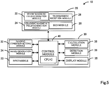
testing information have been acquired for a patient to be diagnosed, whether
or not a point
17
CA 02700948 2010-03-25
based on this lesion diagnostic rating index and testing information falls
within either
distribution (correlation diagram thereof) can be assessed based on
statistical techniques.
According to this technique, a lesion having a high probability of appearing
in the future, namely
a lesion or presage thereof, can be detected even if testing information does
not exhibit abnormal
values.
[0062]
There are no particular limitations no the testing information used in this
form. For
example, testing information relating to various biological factors such as
blood, urine, stool,
sputum or saliva, information relating to pathological tests, or information
relating to diagnostic
imaging such as CT or ultrasound tests can be used. Testing information used
when setting the
threshold information is used preferably. Testing information at least
relating to blood is used
more preferably.
[0063]
When the lesion diagnostic rating index is calculated according to the
equation (1), it is
preferable for being able to quantitatively evaluate a lesion, presage thereof
or progression of a
disease. In addition, since the evaluation is a quantitative evaluation,
minute lesions or
presages thereof of a degree that cannot be seen by ordinary diagnostic
imaging can be
determined, and this leads to a decrease in the rate of false negatives.
Moreover, differing from
testing information such as hematological data, the region where the lesion
has appeared can be
specified. Moreover, since evaluations can be made non-invasively, diagnoses
can be made at
affected regions where biopsy is inappropriate and there is less of a burden
on patients.
[0064]
In addition, according to the lesion diagnostic rating index, the tissue
lesion or the presage
thereof can be detected even in tissue that has been diagnosed as being normal
on the basis of
testing information and pathology information such as clinical laboratory data
or image
information other than the lesion diagnostic rating index. More specifically,
in addition to
being able to diagnose the tissue lesion or presage thereof in tissue that has
been tentatively
diagnosed as being normal tissue, in the case a portion of an organ has been
diagnosed as
abnormal, the presage can be detected in the case the lesion has progressed to
tissue tentatively
diagnosed as 'normal' that has yet to be diagnosed as abnormal. In addition,
by investigating
the lesion diagnostic rating index of an organ that has yet to be diagnosed
with cancer of a cancer
patient, a presage of metastasis to that organ can also be detected. In
addition, by following the
lesion diagnostic rating index of an organ that has been completely cured over
time, a presage of
recurrence can also be detected. Thus, by detecting the tissue lesion or
presage thereof based
on the lesion diagnostic rating index, preventive medicine or early treatment
can be performed in
18
CA 02700948 2010-03-25
anticipation of the appearance of the tissue lesion.
[0065]
Furthermore, although diagnosis using the lesion diagnostic rating index as
described
above can demonstrate adequate effectiveness even by examining the lesion
diagnostic rating
index at a fixed point in time, it is able to demonstrate even greater
effectiveness and improve the
accuracy of detecting presages preferably by periodic monitoring thereof Thus,
both
examination of the lesion diagnostic rating index and monitoring examination
thereof can greatly
contribute to early diagnosis and early treatment by using in examinations
conducted e.g., in
health examinations, examinations conducted prior to making definitive
diagnoses, examinations
during the course of monitoring the progress of a patient such as during
relapse, metastasis or
recurrence, examinations conducted during the course of treatment,
examinations conducted for
the purpose of determining prognoses, or examinations conducted following
relapse or
recurrence.
[0066]
The present diagnosis method may also be realized manually or may be realized
with one
or a plurality of computers. In the case of realizing by computer, the
computer may be
composed separately from the magnetic resonance imaging apparatus or may
compose a portion
of the magnetic resonance imaging apparatus.
[0067]
The following provides an explanation of the magnetic resonance imaging
apparatus of the
present teaching with reference to FiG 5. This apparatus is suitable for
executing the
previously described diagnosis method of the present teaching.
[0068]
(Magnetic Resonance Imaging Apparatus)
The magnetic resonance imaging apparatus (to also be abbreviated as MRI) 10 of
the
present teaching has a module capable of applying an MT pulse, a module for
calculating the
lesion diagnostic rating index, and a module for detecting a lesion or presage
thereof based on
the lesion diagnostic rating index, added to an MRI apparatus used in the
routine clinical setting.
Furthermore, the MT pulse is has been previously explained with regard to the
diagnosis method
of the present teaching. As shown in FIG 5, this apparatus is provided with a
scanner 20 that
executes scans based on pulse sequences, and a controller 30 that controls the
scanner 20 and
carries out processing of image data.
[0069]
(Scanner)
As shown in FIG 5, the scanner 20 is provided with a static magnetic field
generation
19
CA 02700948 2010-03-25
section 22 that generates a static magnetic field in an imaging space, a
gradient magnetic field
generation section 24 that generates a gradient magnetic field for adding
location information to
the static magnetic Field, a transmission/reception module 26 that transmits
an RF signal to a
subject and receives an MR signal from the subject, and a bed section 28 on
which the subject
lies down.
[0070
The static magnetic field generation section 22 forms the static magnetic
field in an
imaging space in which the subject is contained. There are no particular
limitations on the form
of the static magnetic field generation section 22, and for example, may be
composed of a static
magnetic field magnet and a static magnetic field power supply or composed of
a permanent
magnet. In addition, although there are limitations on the direction in which
the static magnetic
field is formed, it is preferably formed so that the direction of the static
magnetic field is in line
with a direction perpendicular to the axial direction of the subject's body.
[0071]
The gradient magnetic field generation section 24 adds location information to
the MR
signal from the subject by applying a gradient to the magnetic field strength
of the imaging space
formed by the static magnetic field. The gradient magnetic field generation
section 24 can use
an ordinary gradient magnetic field coil composed of three pairs of coils,
corresponding to the
three directions of the x direction, y direction and z direction, inside a
primary magnet.
[0072]
The transmission/reception module 26 is composed of an RF coil, for example,
and carries
out transmission and reception of electromagnetic signals. Namely, the
transmission/reception
module 26 transmits an RF pulse to the subject arranged within the imaging
space formed by the
static magnetic field. As a result, a high-frequency magnetic field is formed
and the spin of
protons in the imaged region of the subject is excited. Electromagnetic waves
emitted from the
excited protons are then received from the subject in the form of the magnetic
resonance signal
(MR signal).
[0073]
The bed section 28 is a bed that allows the subject to lie down thereon. The
bed section
28 is configured so as to be able to move between the inside and outside of
the imaging space.
[0074]
(Controller)
The controller 30 is provided with an imaging condition setting module32 that
allows
setting of the first imaging condition and the second imaging condition, an
image generation.
module 34 that generates images, a calculation module 36 that calculates the
lesion diagnostic
CA 02700948 2010-03-25
rating index, a detection module 38 that detects a presage or presence of a
lesion of body tissue
based on the calculated lesion diagnostic rating index, and a control module
40 that controls
operation of the entire controller 30 and each section of the scanner 20.
[0075]
The imaging condition setting module32 sets an imaging condition based on
input data
input to an input section 33 into which instructions of an operator have been
input (such as a
number of scans, flip angle, number of echoes, echo time and other scan
parameters and imaging
commands). Namely, pulse sequences to be executed by the scanner 20 are
generated based on
the input data. This imaging condition setting module32 at least allows the
setting of a first
imaging condition that is unaccompanied by application of an MT pulse and a
second imaging
condition that is accompanied by the application of the MT pulse-
[0076]
The image generation module 34 carries out image processing based on the MR
signal
input from the transmission/reception module 26 to the control module 40.
Namely, the image
generation module 34 arranges digital data from the transmission/reception
module 26 in a
Fourier space (k-space), and reconstructs the data to image data of real space
by applying
two-dimensional or three-dimensional Fourier transformation. There are no
particular
limitations on the form of image processing by the image regeneration section
34, examples of
which include addition processing among multiple frames of image data (such as
simple addition
processing, averaging processing or weighted addition processing), difference
operation
processing and maximum intensity projection (MIP) processing. Images
reconstructed in this
manner are displayed on a display section 35. In addition, information that
correlates threshold
information of the lesion diagnostic rating indices with colors is stored in
the image generation
module 34, and the image generation module 34 carries out imaging based on the
values of the
lesion diagnostic rating index for each pixel.
[0077]
The calculation module 36 calculates the lesion diagnostic rating index based
on a
preliminarily stored program. More specifically, image data imaged under the
first imaging
condition and image data imaged under the second imaging condition are input
from the control
module 40, the lesion diagnostic rating index is calculated, and the
calculated lesion diagnostic
rating index is output to the control module 40.
[0078]
The detection module 38 detects the presage or the presence of a lesion in
living tissue
based on the lesion diagnostic rating index calculated in the calculation
module 36. This
detection module 38 stores preset threshold information of the lesion
diagnostic rating index
21
CA 02700948 2010-03-25
(such as the threshold information shown in FIG. 3), and detects the presence
of the presage or
the presence of a tissue lesion, or assesses the disease state or degree of
progression thereof,
based on this threshold information. Furthermore, a setting method used in the
previously
described diagnosis method of the present teaching can be applied as is for
setting the threshold
information of the lesion diagnostic rating index.
[0079]
The control module 40 controls the scanner 20 so that scans are executed based
on pulse
sequences set with the imaging condition setting module32. The control module
40 is
configured, for example, in the form a microprocessor having a CPU 42 as the
core thereof, and
control operation of each section that composes the scanner 20 as well as the
entire controller 30.
Namely, the control module 40 is electrically connected to the static magnetic
field generation
section 22, the gradient magnetic field generation section 24, the
transmission/reception module
26 and the bed section 28, outputs control signals and the like to each of
these sections, and
inputs MR signals and the like from the transmission/reception module 26. In
addition, the
control module 40 is also electrically connected to the imaging condition
setting module32, the
input section 33, the image generation module 34, the display section 35, the
calculation module
36 and the detection module 38 that compose the controller 30, and carries out
exchange of
control signals, command signals and the like.
[0080]
(Operating Method)
Next, an explanation is provided of the functions of the magnetic resonance
imaging
apparatus 10 of the present teaching configured in this manner, and
particularly a module for
detecting the lesion in living tissue or the presage thereof using the lesion
diagnostic rating index
as an indicator. The CPU 42 of the control module 40 executes the lesion
diagnostic rating
index indicator diagnosis routine shown in FIG 6 when a scan start command is
input from the
input section 33.
[0081]
When this lesion diagnostic rating index indicator diagnosis routine is
started, the CPU 42
of the control module 40 first executes a scan based on the first imaging
condition set with the
imaging condition setting module32 (Step S100). More specifically, together
with forming a
magnetic field in the imaging space by driving the static magnetic field
generation section 22 and
the gradient magnetic field generation section 24 corresponding to a preset
pulse train, the
transmission/reception module 26 is driven so that a scan is executed based on
an ordinary
imaging sequence in which an MT pulse is not applied. As a result, a first
magnetic resonance
signal is obtained that is not affected by magnetization transfer effect.
22
CA 02700948 2010-03-25
[0082]
When all scans based on imaging sequences have been completed, the CPU 42
carries out
three-dimensional Fourier transformation on k-space digital data that has been
gathered and
arranged as a result of imaging under the first imaging condition, and
reconstructs that data into
real space image data (Step S 110)_
[0083]
Continuing, the CPU 42 executes a scan based on the second imaging condition
set with
the imaging condition setting module32 (Step S120). Under the second imaging
condition,
together with driving the static magnetic field generation section 22 and the
gradient magnetic
field generation section 24, the transmission/reception module 26 is driven to
that an MT pulse
and an imaging pulse are applied to a subject on the bed section 28. As a
result, an MR signal
that reflects a magnetization transfer effect (second magnetic resonance
signal) can be obtained
from the subject. When all scans based on imaging sequences having an MT pulse
added to the
leading end thereof have been completed, k-space digital data that has been
gathered and
arranged is reconstructed to real space image data in the same manner as the
first imaging
condition (Step S130).
[0084]
When imaging based on the first imaging condition and the second imaging
condition has
been completed, the CPU 42 detects the lesion diagnostic rating index using
the signal intensity
Mo of the first magnetic resonance signal and the signal intensity Ms of the
second magnetic
resonance signal (Step S 140). Here, calculation processing is carried out for
each
corresponding pixel.
[0085]
After having calculated the lesion diagnostic rating index data, the CPU 42
detects the
presage and the presence of a lesion in living tissue based on the calculated
lesion diagnostic
rating index data and displays the result on the display section 35 (Step
S150), thereby
completing this routine. In the detection step of Step 5150, together with
comparing the
calculated lesion diagnostic rating index data of each pixel with a
corresponding relationship
with threshold information and saturation of preliminarily stored lesion
diagnostic rating indices,
specifying a color corresponding to the lesion diagnostic rating index data
for each pixel, and
displaying an image composed of each specified color on the display section
35, information that
correlates tissue state with color is displayed on the display section 35. Asa
result, since the
lesion diagnostic rating index data is visualized in a state that is
correlated with the state of the
tissue, an operator who has viewed the display section 35 can easily and
accurately determine the
lesion or presage thereof and the disease state.
23
CA 02700948 2010-03-25
[0086]
Furthermore, although detection results are displayed by imaging the lesion
diagnostic
rating index in Step S150 in the present embodiment, if information relating
to a lesion in the
manner of the region where the lesion is present or the degree of progression
thereof is displayed,
the detection results may be displayed by a method other than imaging the
lesion diagnostic
rating index (such as by using characters or numerical values).
[0087]
In addition, in Step S140, the lesion diagnostic rating index may be
calculated from signal
intensity of a region of interest selected by the operator on an image imaged
according to the first
imaging condition or the second imaging condition. At this time, the presage
of a lesion may
be detected in that region of interest in Step S150.
[0088]
Moreover, although a tissue lesion or presage thereof was detected in Step
5150 based on
the lesion diagnostic rating index at a fixed point in time, instead of or in
addition thereto, the
amount of change (versus time) in lesion diagnostic rating indices may be
calculated based on a
preliminarily stored past lesion diagnostic rating index of the same patient
and the current
calculated lesion diagnostic rating index, and the degree of progression or
metastasis of a lesion
may also be analyzed based on the calculated amount of change.
[0089]
Furthermore, the functions, applications and the like of the previously
explained diagnosis
method of the present teaching can be directly applied to the magnetic
resonance imaging
apparatus and operating method thereof of the present teaching. Thus, the
present teaching
includes each of various forms for realizing each of the forms of the
diagnosis method of the
present teaching.
[0090]
(Diagnostic Imaging System)
The diagnostic imaging system of the present teaching is provided with a
transmission/reception module, a control module, a calculation module and a
detection module
in the previously described magnetic resonance imaging apparatus of the
present teaching. The
configuration and functions and so forth of the previously explained diagnosis
method of the
present teaching, magnetic resonance imaging apparatus and operating method
thereof can be
directly applied to this diagnostic imaging system. Thus, the present teaching
includes each of
various forms for realizing each of the above-mentioned forms of the diagnosis
method, the
magnetic resonance imaging apparatus and the operating method thereof of the
present teaching.
[0091]
24
CA 02700948 2010-03-25
(Lesion in Living Tissue Detection Apparatus)
The detection apparatus of the present teaching can be provided with a module
for
acquiring the lesion diagnostic rating index represented by the equation (1)
using the signal
intensity Mo of the first magnetic resonance signal received from an imaged
region of a subject
according to a first imaging condition unaccompanied by the application of the
MT pulse and the
signal intensity Ms of the second magnetic resonance signal received from the
imaged region of
the subject according to the second imaging condition accompanied by the
application of the MT
pulse, and a detection module for detecting the lesion in living tissue or the
presage thereof in the
imaged region based on the lesion diagnostic rating index.
[0092]
According to this detection apparatus, the lesion diagnostic rating index
represented by the
equation (1) can be acquired by inputting the signal intensities Mo and Ms
acquired by a nuclear
magnetic resonance imaging apparatus. Moreover, a lesion or presage thereof in
the imaged
region can be detected based on the acquired lesion diagnostic rating index.
According to this
detection apparatus, by acquiring the above-mentioned signal intensities from
a conventional
nuclear magnetic resonance imaging apparatus, and calculating the lesion
diagnostic rating index
or acquiring the lesion diagnostic rating index directly, the lesion in living
tissue or presage
thereof that was unable to be detected solely with a conventional nuclear
magnetic resonance
imaging apparatus can be easily detected.
[0093]
The module for acquiring the lesion diagnostic rating index may be a module
for inputting
the lesion diagnostic rating index calculated on the basis of the equation (1)
in a nuclear
magnetic resonance imaging apparatus and the like, or may be a module for
calculating the
lesion diagnostic rating index based on the equation (1) within the present
detection apparatus
based on the signal intensities Mo and Ms input into the detection apparatus
after having been
acquired by a nuclear magnetic resonance imaging apparatus and the like. A
module for
executing a step in which the lesion diagnostic rating index is acquired with
the previously
explained nuclear magnetic resonance imaging apparatus can be employed for
this module.
[0094]
The detection module for detecting the lesion or presage thereof provided in
this detection
apparatus can employ a module for executing a step in which the lesion in
living tissue or
presage thereof is detected in the nuclear magnetic resonance imaging
apparatus. In addition,
each of type of embodiment of this module can also be applied directly to the
present detection
apparatus.
[0095)
CA 02700948 2010-03-25
Moreover, the present detection apparatus can be provided with an imaging
module for
imaging the degree of the lesion in the imaged region based on the lesion
diagnostic rating index
of that imaged region. The degree of the lesion of the imaged region, namely
the degree of
progression from the lesion or the presage thereof, can be easily determined
by imaging the
degree of the lesion in the imaged region based on the lesion diagnostic
rating index. It is
necessary to preliminarily correlate the degree of the lesion with lesion
diagnostic rating indices
based on the detection module in order to output an image of the degree of the
lesion in the
imaged region based on the lesion diagnostic rating index. Here, time-based
changes in the
lesion diagnostic rating index, threshold information of the lesion diagnostic
rating index and the
correlation (graph) between testing information and the lesion diagnostic
rating index can be
used to correlate the degree of the lesion with the lesion diagnostic rating
index as previously
explained.
[0096]
The imaging module is only required to form image information obtained by
imaging the
degree of a lesion. Thus, the formed image information may be output directly
a suitable
external image display module such as a monitor or nuclear magnetic resonance
imaging device,
or in the case the present detection apparatus is provided with an image
display module, the
image may be displayed on the image display module.
[0097]
(Program)
According to the present teaching, a diagnostic imaging program is provided
for detecting
the lesion in living tissue or the presage thereof based on the lesion
diagnostic rating index.
The diagnostic imaging program of the present teaching is a program that
executes the detection
step of the above-mentioned operating method in one or a plurality of
computers. This program
may be recorded on a computer-readable recording medium (such as a hard disc,
ROM, FD, CD
or DVD), may be distributed to a different computer from a certain computer
through a transfer
medium (such as the Internet, LAN or other communication network), or may be
received by any
other mode. Effects similar to the above-mentioned operating method are
obtained by
executing this program in a single computer or by executing in a plurality of
computers by
assigning respective operations thereof to each computer.
[0098]
Although the following provides an explanation of the present teaching through
specific
examples thereof, the present teaching is not limited to the specific examples
described below.
[0099)
(Example 1)
26
CA 02700948 2010-03-25
(MRI)
Table I shows the results of imaging the livers of 17 patients (consisting of
3 patients with
normal livers, 3 patients with metastatic liver cancer and I 1 patients with
hepatocellular
carcinoma) using a magnetic resonance imaging apparatus. Definitive diagnosis
of the patients
shown in Table 1 was made by suitably combining techniques other than the
lesion diagnostic
rating index, such as MRI and CT imaging testing, blood testing and the like.
Furthermore,
among the disease names shown in Table 1, HCC indicates hepatocellular
carcinoma, TAE
indicates transcatheter arterial embolization, LC indicates liver cirrhosis
and FNH indicates focal
nodular hyperplasia.
[0100]
["fable 1]
Case Age Gender Height Weight BMI Disease name Child-Pugh
(m) (kg) classification
1 58 M 1.665 66.2 23.88 HCC, postTAE B
2 58 M 1.665 66.2 23.88 HCC, postTAE B
3 61 M -- -- -- Suspected FNH A
4 63 M 1.694 79.5 27.70 HCC A
71 F 1 514 57.6 25.13 Colorectal cancer A
w/liver metastasis
6 67 F 1.445 56.2 26.92 HCC,LC B
7 49 M 1.660 79.0 28.67 Bladder cancer A
8 60 M 1.695 65.6 22.83 Lung cancer w/liver A
metastasis
9 66 M 1.626 63.6 24.06 HCC, LC C
79 M 1.647 75.8 27.94 Colon cancer w/liver A
metastasis
11 46 F 1.490 44.0 19.82 Suspected pancreatic A
tumor
12 71 M 1.720 80.6 27.24 Suspected pancreatic A
tumor
13 75 F 1.520 50.0 21.64 HCC B
14 74 M 1.620 54.0 20.58 HCC recurrence B
62 F -- -- -- HCC --
16 67 M - -- -- HCC --
27
CA 02700948 2010-03-25
17 67 M -- -- - FICC, postOP --
[0101]
A 1.5 T superconducting MR apparatus (GE, SIGNA MR/i 1.5 T High Speed Ver.
9.0) was
used for the MR apparatus. Furthermore, this apparatus was installed with a
program enabling
radiation of MT-RF pulses- Imaging with this MR apparatus was carried out
using 3D-spoiled
gradient recalled acquisition in steady state (SPGR) without applying an MT-RF
pulse, and
MT-SPUR in which the MT-RF pulse was applied prior to SPQR.
[01021
The imaging conditions consisted of a repeat time TR of 39 msec, echo time TE
of 6.9
msee, flip angle of 30 , matrix size of 256 x 96, FOV of 40 cm, slice
thickness of 10 mm,
overlap location of 0 mm, and location per slab of 8. In addition, the MT-RF
pulse employed a
frequency 7 ppm away from the resonance frequency of water to the side of the
lower magnetic
field for the offset frequency, and the pulses were radiated for 18 msec in
the form of a sine
waveform (radiation intensity: 3.26 T).
[0103]
(Calculation of Lesion Diagnostic Rating Index)
Lesion diagnostic rating indices were calculated using image data obtained by
imaging by
SPGR and imaging by MT SPUR. More specifically, the same region of an image of
the same
cross-section was extracted, the lesion diagnostic rating index was calculated
for each pixel of
the extracted region (region of interest) using the equation (1), and the
average value of each
pixel in that region was determined. Furthermore, a region judged to be normal
tissue by MR
was selected when extracting the region of interest on the images. The results
are shown in
Table 2.
[01041
[Table 2]
Case ECR ICG GOP GPT CITE Albumin Bil:irubin
1 92.1 16.8 -- 68 37 183 3.6 0.5
2 82.8 12.4 3.2 48 66 211 4.4 0.5
3 95.8 6.7 -- 20 29 199 4.7 0.3
4 81.3 6.9 17.1 74 70 152 4.0 0.9
92.7 8.6 -- 20 11 122 3.9 1.1
6 68.3 12.5 28.2 65 75 91 3.2 1.6
7 115.4 20.6 - 30 53 178 4.6 0.4
28
CA 02700948 2010-03-25
8 92.5 6.5 4.4 18 16 182 4.1 0.7
9 68.0 13.3 -- 58 24 37 2.8 2.4
91.0 20.0 18.7 28 19 146 4.2 0.5
11 94.0 16.2 -- 17 13 116 3.8 0.4
12 98.0 13.5 -- 17 16 161 4.1 0.3
13 74.1- 6.3 -- 101 92 101 3.7 1.2
14 82.7 8.2 -- 35 30 131 3.9 0.8
84.4 8.5 14.5 32 34 193 4.3 0.6
16 75.0 6.6 21.6 57 65 73 3.6 1.0
17 77.1 4.5 21.7 90 84 123 4.0 1.2
[0105]
(Acquisition of Blood Data)
Blood tests were carried out on the patients shown in Table 1. The tests were
carried out
for the six parameters shown in Table 2 according to ordinary techniques used
in the routine
clinical setting. The results are shown in Table 2.
[0106]
(Evaluation of Correlation Coefficients and Setting of Threshold Information
for Lesion
Diagnostic Rating Indices)
In addition to evaluating correlation coefficients between lesion diagnostic
rating index
data and blood data acquired from the patients, threshold information was set
for the lesion
diagnostic rating indices. In evaluating the correlation coefficients, a graph
was prepared of
correlation coefficients between the lesion diagnostic rating index data and
each parameter of
blood data of the patients as shown in Table 2, and the results were evaluated
by calculating
Spearman's rank correlation coefficient (r). In addition, setting of threshold
information was
carried out by determining the lesion diagnostic rating index equivalent to a
boundary value for
which each parameter of blood data was considered to be abnormal using the
previously
prepared graph. The resulting graphs and Spearman's rank correlation
coefficients are shown.
Furthermore, in FIG. 7, FIG. 7(a) indicates GOP (also AST (aspartate
aminotransferase)) for the
blood data, FIG. 7(b) indicates GPT (also ALT (alanine aminotransferase)), FIG
7(c) indicates
albumin, FIG 7(d) indicates bilirubin, FIG 7(e) indicates cholinesterase, and
FIG 7(f) indicates
ICG.
[0107]
As shown in FIG 7, Spearman's rank correlation coefficients were -0.73 for
GOP, -0.69 for
29
CA 02700948 2010-03-25
GPT, 0.66 for albumin, -0.83 for bilirubin, 0.67 for cholinesterase and -0.71
for JCG. Strong
correlations were observed between both parameters for GOP, bilirubin and ICG
in particular.
In addition, the lesion diagnostic rating indices equivalent to the boundary
values of the blood
data were determined to yield values in the proximity of 80% for each of FIG.
7(a) through 7(f)
(to be referred to as the respective boundary values). Moreover, among FIG.
7(a) through 7(0,
the distribution of a normal liver group and abnormal liver group were clearly
distinguished for
albumin in FIG 7(c), bilirubin in l;iG 7(d) and cholinesterase in FIG 7(e). In
other words, in a
comparison of lesion diagnostic rating indices when blood data is in the
normal range, lesion
diagnostic rating indices in the abnormal liver group were distributed in a
lower range than those
in the normal liver group.
[0108]
On the basis of the above results, lesion diagnostic rating indices were
determined to
exhibit a strong correlation with blood data, and the values were observed to
decrease as blood
data became worse. In addition, liver function disorders were determined to be
able to be
detected when using values in the vicinity of 80% for the threshold values of
the lesion
diagnostic rating indices. Moreover, in the case albumin, bilirubin or
cholinesterase was used
for the blood data, since results were obtained for which the distributions
(graphs) of the normal
liver group and abnormal liver group were able to be distinguished, it was
determined that a
region of interest could be evaluated as normal or abnormal by evaluating to
which distribution
(graph) the lesion diagnostic rating index of that region belonged. Moreover,
since the lesion
diagnostic rating indices of the abnormal liver group were distributed in a
range lower than that
of the normal liver group despite blood data being in the normal range, lesion
diagnostic rating
indices in the abnormal liver group were suggested to exhibit values capable
of being
distinguished from the normal liver group at an earlier stage than blood data.
[0109]
On the basis of these findings, the lesion diagnostic rating index was
suggested to be able
to serve as a novel indicator for detecting tissue abnormalities. In addition,
the use of the lesion
diagnostic rating index was suggested to enable earlier detection of a lesion
than that based on
blood test results. Moreover, when considering that the tissue diagnostic
rating indices of the
abnormal liver group yielded values capable of being distinguished from those
of the normal
liver group even when blood data was within the normal group, and that the
lesion diagnostic
rating index is considered to allow detection of microstructural changes in
tissue, the lesion
diagnostic rating index was suggested to be an extremely useful indicator for
detecting and
diagnosing not only the presence of a tissue lesion, but also a presage
thereof.
[0110]
CA 02700948 2010-03-25
(Example 2)
In Example 2, lesion diagnostic rating indices were calculated for those
patients listed in
Table I in which blood data was generally within the normal range and lesion
diagnostic rating
indices were in the vicinity of boundary values, followed by evaluating
changes in blood data
over time. Evaluations were carried out by comparing blood data at the time of
calculating the
lesion diagnostic rating indices and blood data one month later. Albumin and
cholinesterase
levels were used for the blood data. The results are shown in FIG 8_
Furthermore, in FIG 8,
FIG. 8(a) indicates the results for albumin, while FIG. 8(b) indicates the
results for cholinesterase.
In addition, data from Cases 2, 13, 16 and 17 in Tables 1 and 2 was used to
carry out evaluations
for albumin, while data from Case 2 was used to carry out evaluations for
cholinesterase.
[0111]
As shown in FIGS. 8(a) and 8(b), values for both albumin and cholinesterase
decreased
one month after calculation of lesion diagnostic rating indices in all cases
tested. In the case of
albumin, values decreased considerably in Cases 2, 13 and 17. In addition, in
contrast to
albumin levels being in the normal range at the time of calculation of lesion
diagnostic rating
indices in Cases 2 and 13, albumin levels reached the abnormal range one month
after
calculation of lesion diagnostic rating indices. Moreover, in contrast to the
cholinesterase level
at the time of calculation of the lesion diagnostic rating index in Case 2
being within the normal
range at the time of calculation of the lesion diagnostic rating index, the
cholinesterase level
reached the abnormal range one month after calculation of the lesion
diagnostic rating index.
[0112]
On the basis of the above results, the lesion diagnostic rating index was
determined to
already exhibit values that were able to be distinguished from patients of the
normal liver group
(values in the vicinity of 80%) at even an earlier stage than detection of
abnormalities in blood
data. In addition, among patients exhibiting lesion diagnostic rating indices
in the vicinity of
80%, blood data was determined to subsequently reach the abnormal range
despite blood data
having been in the normal range at the time of calculation of the lesion
diagnostic rating indices.
[0113]
On the basis of these findings, the lesion diagnostic rating index was
determined to be an
indicator that allows sensitive detection and diagnosis of tissue lesions or
presages thereof at
even an earlier stage than the appearance of abnormal values in blood tests;
namely at an early
stage at which blood tests are diagnosed as being `normal'. In other words, it
was determined
that threshold values for assessing tissue lesions or presages thereof can be
set on the basis of the
lesion diagnostic rating index and other testing information, and that the
threshold values alone
can be used to detect tissue lesions or presages thereof. Thus, according to
the lesion diagnostic
31
CA 02700948 2010-03-25
rating index, a tissue lesion or presage thereof can be said to be able to be
detected at an
extremely early stage as compared with conventional tissue lesion diagnosis
methods. As a
result, treatment and diagnosis for tissue lesions can be performed at an
extremely early stage.
[0114]
(Example 3)
In the present example, when a total of 30 definitively diagnosed patients
(consisting of
liver cirrhosis patients, hepatitis patients and healthy (normal) subjects)
were classified into a
liver cirrhosis group, hepatitis group and normal group using data from blood
test results for
AST (aspartate aminotransferase, GOT), ALT (GPT), LDH (lactate dehydrogenase),
Che
(cholinesterase), Alb (albumin), Bil (bilirubin), y-GPT, ALP (alkaline
phosphatase) and Pit
(platelets), the groups were able to be classified according to factor 2 and
factor 3 indicated by
the following equations. The results are shown in FIG. 10.
(Equation 3)
Factor 2 = 0.663*AST+ 1.086*ALT - 0.015*LDH - 0.045*Che
- 0.218*Alb + 0.072*Bil + 0.601 *y-GTP + 0.582*ALP - 0.383*Plt
Factor 3 = 0.419*AST + 0.091 *ALT - 0.052*LDH - 0.858*Che
- 0.891 *Alb + 0.737*Bil + 0.111 *y-GTP - 0.135*ALP - 0.718*Plt
[0115]
Next, the lesion diagnostic rating indices were calculated based on the
equation (1) for
each of these groups, and after grouping the acquired lesion diagnostic rating
indices at 5%
intervals, the distributions of the lesion diagnostic rating indices of the
patients included in each
group were investigated. The results are shown in. FIG. 11. In addition, the
numbers of
patients in each group included in the range of each lesion diagnostic rating
index were
investigated. Those results are shown in FIG. 12.
[0116]
As shown in FIGS. 10 to 12, the patients were able to be classified into a
liver cirrhosis
group, a hepatitis group and a normal group on the basis of the lesion
diagnostic rating indices as
well. Namely, if a threshold value is set for the lesion diagnostic rating
indices in the vicinity
of 90%, when the lesion diagnostic rating index is equal to or greater than
the threshold value,
the possibility of hepatitis or liver cirrhosis based on the presence of a
tissue lesion thereof can,
be diagnosed as being low, or the patient can be diagnosed as normal, and when
the lesion
diagnostic rating index is equal to or less than the threshold value, the
possibility of hepatitis or
liver cirrhosis based on the presence of a tissue lesion thereof can be
diagnosed as being high.
In addition, if a value in the vicinity of 80% is used for the threshold
value, when the lesion
32
CA 02700948 2010-03-25
diagnostic rating index is equal to or less than the threshold value, the
possibility of liver
cirrhosis can be diagnosed as being high, and when the lesion diagnostic
rating index is equal to
or greater than the threshold value but equal to or less than 90%, then the
possibility of hepatitis,
but not liver cirrhosis, can be diagnosed as being high.
[0117]
(Example 4)
In the present example, in addition to acquiring lesion diagnostic rating
indices represented
by the equation (1) along with platelet test results at about the same time
from 11 subjects
suspected of liver disease, platelet test results were acquired from the same
subjects one month
later. These results are shown in FIG. 13. Furthermore, a value of 100,000 to
400,0001 l was
used for the platelet reference value (normal range).
[0118]
As shown in FIG 13, when the lesion diagnostic rating index was equal to or
less than
84.4%, there was no improvement in platelet test results observed at the time
of initial testing or
one month later. In addition, when the lesion diagnostic rating index was
within the range of
85.5% to 91.0%, platelet test results after one month were equal to or had
decreased as compared
initial testing results. Moreover, when the lesion diagnostic rating index was
equal to or greater
than 92.7%, an improving trend was observed in platelet test results. Namely,
it was
determined that even if platelet test results are in the normal range, in the
case the lesion
diagnostic rating index is equal or less than a fixed value, a tendency for
platelet count to
decrease in the future can be detected, namely at a preliminary stage of liver
disease or an
extremely early stage of liver disease.
[0119]
On the basis of the above, a high possibility of liver disease can be
diagnosed in the case,
for example, the lesion diagnostic rating index is equal to or less than about
85%, and a high
possibility of the presence of a tissue change of a preliminary stage of liver
disease or a tissue
lesion at an extremely early stage of liver disease can be diagnosed in the
case the tissue
diagnostic rating index is within the range of about 85% to about 90%. Namely,
the possibility
of a preliminary stage of liver disease or extremely early stage of liver
disease can be diagnosed
as being high. In addition, when the tissue diagnostic rating index is equal
to or greater than
about 90%, this type of tissue change or tissue lesion can be diagnosed as
being absent.
Namely, the possibility of the absence of liver disease can be diagnosed as
being high.
[0120]
Since the present teaching is as has been previously explained, it can clearly
be carried out
in the following forms.
33
CA 02700948 2010-03-25
[0121]
According to the present teaching, a magnetic resonance imaging device can be
provided
that is provided with a transmission/reception module that receives a magnetic
resonance signal
from a subject after having transmitted a high-frequency signal, a control
module that controls
the transmission/reception module such that scans are executed based on pulse
sequences
corresponding to both a first imaging condition unaccompanied by application
of an MT pulse
and a second imaging condition accompanied by the application of the MT pulse,
a calculation
module that calculates a lesion diagnostic rating index represented by the
above-mentioned
equation (1) using a signal intensity Mo of a first magnetic resonance signal
received from the
subject according to the first imaging condition and a signal intensity Ms of
a second magnetic
resonance signal received from the subject according to the second imaging
condition, and a
detection module that detects a lesion in living tissue or a presage thereof
in the imaged region
based on the lesion diagnostic rating index; wherein, when the detection
module is a module that
detects the lesion in living tissue or the presage thereof based on threshold
information of the
lesion diagnostic rating index correlated with testing information relating to
a patient for which a
definitive diagnosis has been made by a method differing from the diagnosis
using the lesion
diagnostic rating index, the threshold information may be set based on the
lesion diagnostic
rating index equivalent to a boundary value of the testing information
relating to the patient. In
this form, the threshold information can be a value equal to or greater than
the lesion diagnostic
rating index equivalent to the boundary value of the testing information
relating to the patient.
[0122]
In addition, the testing information relating to the patient may be one type
or two or more
types of data selected from the group consisting of testing information
relating to blood in the
.form albumin, bilirubin and cholinesterase. Alternatively, the testing
information relating to the
patient may be albumin and/or cholinesterase.
[0123]
In addition, the detection module can also be a module that detects the lesion
in living
tissue or the presage thereof at an earlier stage than detection of the lesion
or the presage thereof
by testing information relating to blood.
[0124]
In the above-mentioned form, a display module may also be provided so that it
may image
and display tissue diagnostic rating indices calculated with the calculation
module. In this form,
the display module may carry out imaging based on threshold information of the
lesion
diagnostic rating indices.
[0125]
34
CA 02700948 2010-03-25
The detection module may also be a module for detecting a presage of a liver
disease. In
this form, the liver disease may be one type or two or more types selected
from the group
consisting of hepatitis, liver cirrhosis and hepatocellular carcinoma.
[0126]
According to the present teaching, a diagnosis method for lesion in living
tissues is
provided that is comprised of a detection step of detecting a lesion in living
tissue of presage
thereof based on the lesion diagnostic rating index represented by the above-
mentioned equation
(1) using a signal intensity Mo of a first magnetic resonance signal received
from a subject
according to a first imaging condition unaccompanied by application of an MT
pulse and a signal
intensity Ms of a second magnetic resonance signal received from the subject
according to a
second imaging condition accompanied by the application of the MT pulse,
wherein the
detection step is a step is carried out earlier than detection of the lesion
in living tissue by testing
information relating to blood.
[0127]
According to the present teaching, it is clear that the present teaching can
be carried out in
the form of an operating method and diagnostic imaging system of a magnetic
resonance
imaging apparatus for carrying out the diagnosis method described above. In
addition, the
present teaching can also be carried out in the form of a detection method for
lesion in living
tissues that carries out a detection step of detecting a lesion in living
tissue or presage thereof in
the diagnosis method described above, a detection apparatus of a lesion in
living tissue that
executes this detection step, and a detection apparatus provided with an
imaging module for
imaging the degree of a detected lesion.