Note: Descriptions are shown in the official language in which they were submitted.
CA 02701393 2015-04-08
Method and Apparatus for Preparing Bone for a Prosthetic Device
Background of the Invention
I. Field of the Invention
[0002] This invention relates generally to hip surgeries and, more
particularly, relates
to surgical methods and tools for preparing a femur and trialing the femur for
an implant.
2. Related Art
[0003] Most modular systems require instrumentation comprising distal
stem trials,
proximal sleeve trials, proximal body segment trials, and trial necks to
replicate the implant.
An implant is then selected based on the trial instrumentation that provides
the best fit for
the patient. Using distal stem trials adds to the number of instruments in
system, therefore
increasing inventory overhead and cost of manufacture. More instruments may
also lead to
confusion in the OR, and a larger cleanup/sterilization operation. There is
also a need to
simplify instrumentation in such a way that surgical assistants are not
burdened by handling
large, heavy instrument sets.
[0004] With conventional systems, a surgeon must remove distal reamers from
the
femoral canal and replace them with a distal stem trial connected to a
proximal stem trial.
Each time a reamer is removed and a distal stem trial is inserted in its
place, there are risks
of fracture and/or excessive bone removal, because the distal stem trial adds
an additional
scratch-fit step. In cases of poor bone quality, these risks may be
substantially increased. In
addition to an increased risk of fracture for each reamer removal step, there
is also an
-1---
1
CA 02701393 2015-04-08
increased OR time for the surgeon. The time it takes to ream the femoral
canal, remove the
reamer from the femoral canal, and implant a distal stem trial can be costly.
[0005] The
present invention is more advantageous than conventional methods,
because the distal reamer serves two purposes instead of one, thereby reducing
the number
of steps and parts needed to perform the same surgical procedure. The distal
reamer is first
used to distally ream the femoral canal, and then it is left in place within
the canal. The
distal reamer is configured to allow a proximal trial and trial neck assembly
to be connected
to it in such a way that trial reduction may be performed immediately after
the distal reamer
has stopped cutting. Modular neck segments of the present invention allow
quick trial
reduction with the reamer still within the femoral canal. By trialing off of
the reamer, the
present invention avoids a set of separate stem trials.
[0006] The
standard practice for implanting a hip system is to prepare the bone using
a first set of instruments, and then perform a trial reduction using a second
set of
instruments that is different from the first set. Conventionally, reamers
clear a passage
1 5 within a
femoral canal, and then a trial stem is selected from a kit and inserted into
said
passage. Trial reduction takes place using the trial stein. After the joint is
reduced, the trial
stem is removed from the femoral canal and then replaced with the
correspondingly sized
implant stem. This standard practice generally takes more time to perform and
is less
efficient than the present invention, because more instruments and method
steps are needed
to carry out the same procedure. More time in the OR means exposing patients
to more risk,
and increasing hospital overhead. Another practice for implanting a hip system
is to prepare
the bone using a first set of instruments, and then perform a trial reduction
using the same
first set of instruments.
-2-
CA 02701393 2015-04-08
Summary of the Invention
[0007] In one aspect of the invention, a system for preparing a long
bone for a
prosthetic, the long bone having a long axis comprises a distal instrument and
a proximal
instrument. The distal instrument is configured to remove bone from a distal
portion of bone.
The distal instrument has a bone removing portion configured to remove bone
along the long
axis of the long bone and a shaft extending from the bone removing portion
along the long
axis of the long bone. The proximal instrument is configured to overlie the
shaft of the distal
element within the long bone. The distal shaft guides the proximal instrument
to prepare the
proximal portion of the long bone after the distal instrument has prepared the
distal portion of
the long bone.
[0008] In another embodiment, the distal instrument is a reamer.
[0009] In yet another embodiment, the proximal instrument is a reamer.
[0010] Still another embodiment further comprises a trial neck body
configured to
attach to at least one of the distal instrument and the proximal instrument.
[0011] Alternatively, another embodiment further comprises a trial spacer
configured
to space the trial neck body from distal instrument.
[0012] Another embodiment includes the trial spacer and the trial neck
body
comprising a mating feature configured to adjustably orient a radial position.
[0013] In another embodiment, the mating feature is incrementally
adjustable.
[0014] In yet another embodiment, the mating feature is lockable.
[0015] Alternatively, an embodiment further comprises a modular trial
neck segment.
The modular trial neck segment is configured to adjust at least one of an
offset, version, or
height of a trial component.
[0016] Another embodiment comprises a method for preparing a long bone
having a
long axis. The method includes removing bone from a distal portion of bone
thereby leaving a
-3-
CA 02701393 2015-04-08
cavity along the long axis of the long bone. The method also includes leaving
a distal
instrument within the long bone. Another step includes guiding a proximal
instrument over a
portion of the distal instrument to remove bone in a proximal portion of the
bone.
[0017] Alternatively, the guiding step may comprise guiding the
proximal instrument
over a shaft aligned along the long axis of the bone.
[0018] In another embodiment, the removing step may comprise reaming
the bone.
[0019] In yet another embodiment, a further step may include attaching
a trial neck
body to at least one of the distal instrument and the proximal instrument.
[0020] Another embodiment further comprises spacing the trial neck
body from distal
instrument.
[0021] Alternatively, another embodiment further comprises adjustably
mating the
trial neck body relative to the distal instrument to orient a radial position.
[0022] In another embodiment, the adjustably mating step is
incrementally adjustable.
[0023] In yet another embodiment, the adjustably mating step may
comprise locking
the trial neck body to the distal instrument.
[0024] Another embodiment includes adjusting at least one of an
offset, version, or
height of a trial component by interchangeably using a plurality of multiple
trial neck
segments.
[0025] Further features, aspects, and advantages of the present
invention, as well as
the structure and operation of various embodiments of the present invention,
are described in
detail below with reference to the accompanying drawings.
Brief Description of the Drawings
[0026] The accompanying drawings, which are incorporated in and form a
part of the
specification, illustrate embodiments of the present invention and together
with the
description, serve to explain the principles of the invention. In the
drawings:
-4-
CA 02701393 2015-04-08
[0027] Figure 1 is an exploded view and an assembled view of a reaming
system
according to an aspect of the invention;
[0028] Figure 2 is an exploded view and an assembled view of a
trialing system
according to an aspect of the invention;
[0029] Figure 3 is a view of a quick connect assembly for a reaming system;
[0030] Figure 4 is a view of a trial neck body;
[0031] Figure 5 is a view of a distal reamer with a cap;
[0032] Figure 6 is an exploded view and an assembled view of an insert
and a
proximal reamer;
[0033] Figure 7 is a view of steps for preparing a femur and trialing the
femur;
[0034] Figure 8 is a view of a reaming system including a spacer;
[0035] Figure 9 is an exploded view and an assembled view of a distal
reamer and an
extender; and
[0036] Figure 10 is an exploded view of a distal reamer and a starter
proximal reamer.
Detailed Description of the Embodiments
[0037] Referring to the accompanying drawings in which like reference
numbers
indicate like elements, Figure 1 is an exploded view and an assembled view of
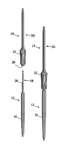
a reaming
system 10 according to an aspect of the invention. The system 10 for removing
bone material
includes a distal cutting instrument 12 (e.g., a reamer) and a proximal
cutting instrument 14.
The distal reamer 12 includes at least one cutting edge 16, a shaft portion
18, and a
mechanical stop feature 20. The proximal cutting instrument 14 includes at
least one cutting
edge 22, a shaft portion 24, a bore 26, and a mechanical stop feature within
the bore 26. The
bore 26 of the proximal cutting instrument 14 accepts the shaft 18 of the
distal cutting
instrument 12. The mechanical stops (either integral with the shaft portions
or individual
-5-
CA 02701393 2015-04-08
parts) of the two cutting instruments 12 and 14 abut to restrict axial
movement further along
the distal cutting instrument 12.
[0038] In one
embodiment, each shaft portion 18 and 24 are configured to receive a
drill. The drill may rotate the distal instrument 12 first to ream a distal
portion of bone, then
disconnect from the distal shaft portion 18, and reconnect to the proximal
shaft portion 24 to
ream a proximal bone portion. The cutting flutes 16 and 22 may be oriented to
cut in the
same direction or oriented to cut in opposite directions so that rotating the
proximal
instrument 14 would not further cut the distal portion of bone. The shafts 18
and 24 may
also be isolated from one another so that rotating the proximal instrument 14
does not rotate
the distal instrument 12.
[0039] The bore
26 of the proximal instrument 14 and the shaft 18 of the distal
instrument 12 are axially aligned so that the cutting instruments 12 and 14
are coaxially
aligned. The bore 26 may be formed to transmit torque to the distal instrument
12 or may be
formed to rotate freely about the shaft 18. In such an embodiment, the distal
shaft 18 serves
to direct the proximal cutting instrument 14 on top of the distal cutting
instrument 12.
[0040] While in
this embodiment, the system 10 includes reamers, other
embodiments may include impactors for impacting bone material. Such a system
could
include a distal impaction instrument and a proximal impaction instrument. The
distal
impaction instrument may include a smooth circular portion, a shaft portion,
and a
mechanical stop feature. The proximal impaction instrument may include a
smooth circular
portion, a shaft portion, a bore, and a mechanical stop feature. The bore of
the proximal
impaction instrument accepts the shaft of the distal impaction instrument, and
the
mechanical stop features of the two impaction instruments abut to restrict
axial movement.
[0041] Figure 2
is an exploded view and an assembled view of a trialing system 28
according to an aspect of the invention. The trialing device 28 of the
preferred embodiment
-6-
CA 02701393 2015-04-08
generally comprises three components: a proximal trial spacer 30, a trial neck
body
assembly 32, and the distal cutting instrument 12. The proximal trial spacer
30 is primarily
used to support the trial neck body assembly 32 on the distal reamer shaft 18
through a
threaded portion 34 of the distal shaft 18, although this spacer 30 may not be
necessary.
[0042] A trial modular
neck segment (not shown) interfaces with the trial neck body
assembly 38 through an interface surface 38. The neck segment is configured to
support a
head (e.g., femoral or humeral head), and may comprise a plurality of trial
modular neck
segments of different sizes and shapes to assess varying height, offset,
version, and range of
motion for the proposed implant. The trial modular neck segment may be
configured to
have a "stick fit" or press fit with the trial neck body assembly 32 (e.g.,
via use of a Morse
taper on the interface surface 38), and the trial modular neck segment (and
thus the interface
surface 38) may or may not be reversible and may or may not be keyed.
[0043] The
proximal trial spacer 30 and trial neck body assembly 32 may include a
mating feature 36 such as a set of radially-extending, axially-disposed ridges
and grooves
which allow the orientation of the trial neck body assembly 32 to be adjusted
incrementally
with respect to both the proximal trial spacer 30 and the distal reamer 12.
The trial neck
body assembly 32 also may include a cut-out feature that allows a surgeon to
mark neck
orientation of the system onto bone with a surgical marker or bovie, in order
to properly
orient the final implant.
[0044] Figure 3 is a view
of a quick connect assembly 40 for a reaming system. The
quick connect assembly 40 includes an inner cylinder 42, an outer cylinder 44,
a flange 46 and
a drill connector 50. The inner cylinder 42 is axially slidable within the
outer cylinder 44, and
is biased so that the drill connector 50 is pressed away from the flange 46.
The biasing means
may be fixed axially to the outer cylinder 42, for example, through a pin (not
shown) and
extends through the outer and inner cylinders 42 and 44. The pin extends
through the inner
-7-
CA 02701393 2015-04-08
cylinder 42 within a slot. The slot also houses a spring which biases the
drill connector 50 of
the inner cylinder 42 axially away from the flange 46.
[0045] When the
inner cylinder 42 is axially slid within the outer cylinder 44 (i.e., the
drill connection 50 is depressed toward the flange 46), a pair of bearings may
be slid out of the
bottom of the quick connect assembly 40. The bearings extend radially outward
from the
quick connect assembly 40 to a distance greater than the inner diameter of the
outer cylinder
44. With the bearings extending out from the inner cylinder 42, the quick
connect assembly
40 is positioned to connect to the reamers 12 and 14.
[0046] The quick
connect assembly 40 then may be used to quickly connect and
disconnect the drill (attached to the quick connect assembly 40) from the
distal reamer 12 and
the proximal reamer 14. Thus, time lost to connecting, disconnecting and
reconnecting the
drill to the reamers is minimized. This may reduce total surgical time,
particularly when
additional passes of the distal reamer 12 and proximal reamer 14 may be
necessary.
[0047] Figure 4
is a view of a trial neck body 32. The trial neck body assembly 32
may include the mating feature 36. The mating feature 36 may be a set of
radially-
extending, axially-disposed ridges and grooves which allow the orientation of
the trial neck
body assembly 32 to be adjusted incrementally with respect to both the
proximal trial spacer
30 and the distal reamer 12. The trial neck body assembly 32 may be
rotationally
incremented to allow for proper orientation of the trial neck during trailing.
The trial neck
body assembly 32 also may include a cut-out feature 60 that allows a surgeon
to mark neck
orientation of the system onto bone with a surgical marker or bovie, in order
to properly
orient the final implant in the position that the trial neck body assembly 32
was oriented
upon a final determined implant position. A neck bore 62 through the trial
neck body
assembly 32 allows the trial neck body assembly 32 to be fixed to the threaded
portion 34 of
2.5 the distal
reamer 12 by a locking nut. The connection between the trial neck body
assembly
-8-
CA 02701393 2015-04-08
32 and the distal reamer 12 compresses the mating surfaces 36 to lock the
trial in place.
While this embodiment includes a locking nut and radially extending, axially
disposed
ridges, other devices that provisionally lock the trial neck body assembly 32
in place would
achieve the same results as the current embodiment.
[0048] Figure 5 is a view
of the distal reamer 12 with a cap 64. The cap 64 serves as a
protector of the threaded portion 34 of the reamer. The cap 64 may protect the
threaded
portion from damage as the drill rotates the distal reamer 12.
[0049] Figure 6
is a view of an insert 70 and the proximal reamer 14. The insert 70
may slide into the bore 26 of the proximal reamer 14. The insert 70 may
protect the
proximal reamer 14 and the distal reamer 12 from friction, which could bind
the
reamers 12 and 14, or could increase chatter between the reamers 12 and 14.
Additionally, chatter could cause the distal reamer to continue to cut through
distal
bone. Such additional movement may cause poor distal fixation of the implant.
[0050] Figure 7
is a view of steps for preparing a femur 80 and trialing the femur 80.
A femoral neck osteotomy and acetabular preparation (not shown) are first
performed. The
femoral canal is prepared for reaming. A quick connect device may be attached
to the
appropriately sized distal reamer, or the reamer may be directly attached to a
drill. Reaming
begins with a distal reamer that is 4-6mm smaller than the templated size. At
all times, the
reaming should be done so that the reamer has little or no resistance, which
may minimize
heat in the bone. The reamers may use depth mark either on the reamer or on
the quick
connect instrument corresponding to the neutral head center of the prosthesis,
to gauge the
appropriate reaming depth. The depth marks may reference the greater
trochanter. The
femoral canal is sequentially expanded using the distal reamers. For example,
the reamers
may change size by 0.5mm increments until the last reamer matches the selected
implant
-9-
CA 02701393 2015-04-08
size. The final reamer size may need to be adjusted based on bone quality,
anatomy and
surgeon preference.
[0051] Distal
reamer depth may also change. The distal reamer 12 may be the
shortest reamer. A mid size distal reamer 96 may allow for more distal
fixation while a long
reamer 98 may allow for deep distal reaming. The diameter size of the reamers
may also
change according to bone quality, anatomy and surgeon preference. The choice
for length
will also depend on bone quality, anatomy and surgeon preference both distally
and
proximally.
[0052] After
distal reaming is complete, the final distal reamer is left in-situ. A
starter reamer over the top of the final distal reamer reams the proximal
femur. The starter
reamer may remove any trochanteric bone that may impede the proximal reaming
process.
The starter reamer has a mechanical axial stop that will abut the distal
reamer and prevent
excess bone removal.
[0053] Proximal
reamers 14, 100 and 102 prepare the femur for the modular sleeve
implant. These reamers are sized according to cone size and distal diameter.
First the
reamer with the smallest cone size that corresponds with the distal diameter
of the stem
reams over the top of the distal reamer (for example, for a size 13, select a
13S proximal
reamer, first.) Progressively, reamers of larger diameters and cone sizes are
used to ream
until the desired fit is achieved. The length of the distal reamer corresponds
to the length of
the sleeve and may effect the leg length (A) of the trial.
[0054] After the
proximal femur is reamed, then a trial spacer 108 is placed within
the proximal femur recess 110. The trial neck body assembly 32 is attached to
the distal
reamer. A neck segment 120 and head 122 are assembled to the trial to reduce
the hip and
assess leg length and joint tension/stability. By varying the trial neck
segment 120, neck
offset (D) and neck height (C) may be adjusted by selecting a different trial
modular neck
-10-
CA 02701393 2015-04-08
segment 120. Neck version (E) may be adjusted by either using a trial modular
neck
segment having a built-in specified version angle, or by unscrewing a trial
neck body
assembly screw and radially orienting the trial neck body assembly with the
optimum
version (B). Desired version may be marked on the bone with a bovie or skin
marker with
respect to the orientation marker on the trial neck body assembly. The trial
instruments are
removed from the bone.
[0055] The
distal stem and proximal sleeve/body are assembled. The stem and
sleeve/body implant are impacted into the prepared canal, and oriented
referencing any
marks made with the skin marker or bovie. Select modular neck segment and head
according to the trial components used in the trial reduction procedure
previously. The
modular neck segment is impacted onto the body and the head is assembled. The
hip is
reduced to ensure correct stability and joint tension.
[0056] While
this procedure has been described with respect to a primary hip
system, a revision hip system would proceed similarly after the failed primary
system has
been removed. This implant system may be more beneficial in a revision as the
modular
choices of the implant allows for accommodating different bone deficiencies.
It should be
appreciated that there could obviously be extra steps involved with
reconstruction of the
femur comprising the use of cables, struts, and augments (metal, active,
and/or
bioabsorbable) etc. especially if there is an ETO (extended trochanteric
osteotomy)
involved.
[0057]
Alternative embodiments of the present invention may include reamers
having some of the features above in combination with any or all of the
following features.
The top of the distal reamer shaft may act as the mechanical stop feature to
restrict axial
movement of the proximal reamer when reaming atop the distal reamer shaft. The
mechanical stop feature may broadly comprise any one of a shelf, ledge, step,
ring, flange,
-11-
CA 02701393 2015-04-08
plate, end portion, male/female connection, or any other feature which may
prevent further
axial movement.
[0058] The distal
and proximal reamers may connect directly to the hospital's
standard powered drill/reamer device with or without the need for the quick
connect adapter
device of the present invention. The distal and proximal reamers may connect
directly to a
non-powered T-handle with or without the need for the quick connect adapter
base of the
present invention. The top of the bore of the proximal reamer may act as the
mechanical
stop feature to restrict axial movement of the proximal reamer when reaming
atop the distal
reamer shaft. The distal end portion of the proximal reamer may act as the
mechanical stop
feature which restricts axial movement of the proximal reamer when reaming
atop the distal
reamer shaft. The proximal reamer may or may not have a depth mark or other
orientation
marks corresponding with references to bony or other anatomy. The proximal
trial spacer
may or may not have a depth mark or other orientation marks corresponding with
references
to bony or other anatomy. The trial neck body assembly may or may not have a
depth mark
or other orientation marks corresponding with references to bony or other
anatomy.
[0059] Figure 8
is a view of a reaming system including a spacer 130. The spacer 130
may space the proximal reamer 14 from the distal reamer 12. One or
more spacer
components 130 may fit between the distal and proximal reamers. The spacer
components
130 may vary in size, length, and geometry, although it is preferred to have a
single length
spacer component.
[0060] Figure 9
is an exploded view and an assembled view of a distal reamer 12 and
an extender 140. The removable distal reamer extension may fit proximally atop
(as shown)
or distally below (not shown) the distal reamer to allow the distal reamer to
ream further
into the femoral canal. The reamer and extension may be formed as a single
integral piece.
-12-
i
1
CA 02701393 2015-04-08
[00611 Figure 10
is an exploded view of a distal reamer 144 and a starter proximal
reamer 150. The additional starter reamer component may be utilized to open
the canal prior
to the proximal reamer step. This "starter" reamer may fit atop the distal
reamer component.
Alternatively, if there is significant proximal bone loss, proximal reaming
with either the
proximal reamer Or starter reamer may be omitted.
[0062] An
alternative embodiment of the present invention comprises making two of
the at least three components (distal stem, proximal trial spacer, and trial
neck body
assembly) of the preferred embodiment integral with each other. For example,
the proximal
trial spacer and trial neck body assembly may be formed as a single integral
piece and may
come available several available sizes and shapes. Alternatively, the trial
neck body
assembly and the trial modular neck segments (not shown) may be formed as a
single
integral piece. In another embodiment, the distal reamer extension disclosed
above may be
made integral with the distal reamer. Other embodiments may include the
protective sleeve
disclosed above and the proximal reamer may be made integral with each other.
That is, the
protective sleeve may be formed of a similar or dissimilar material and
pressed into the
proximal reamer, or the protective sleeve geometries may be "built into" the
proximal
reamer and formed as a single integral piece.
[0063] The trial
modular neck segments used with the present invention may be
designed such that an array of trial modular neck segments can address various
neck lengths,
heights, offsets, and versions, such to replicate the final implant.
[0064] The
proximal trial spacer may abut the reamed bone created by the proximal
reamer. There may be a plurality of sizes of proximal trial spacers such that
a single
proximal trial can be selected to fit the reamed area of several differently-
sized proximal
reamers. The proximal trial spacer may be made integral with the distal reamer
if proximal
reaming is not necessary.
-13-
CA 02701393 2015-04-08
[0065] The
proximal trial spacer, trial neck body assembly, and trial modular neck
segment may be attached prior to reaming for the proximal implant. In other
words,
proximal reaming may be done after a first trial reduction and leg length
assessment is
completed.
[0066] In yet another
embodiment, a system of the present invention comprises at
least two differently-sized distal reamers, at least two differently-sized
proximal reamers,
and a proximal trial spacer. The proximal trial spacer may be one size fits
all, or a plurality
of sizes and geometries of proximal trial spacer components may be provided.
In addition,
the system may further comprise one or more trial neck body assemblies, and/or
one or more
trial modular neck segments. The trial modular neck segments may be keyed such
that they
may be capable of universal or selective use between revision product lines
and/or primary
product lines, or use to within a specified range of stem sizes within a
particular product
line.
[0067] Moreover
in yet another embodiment, the present invention may be used in
knee arthroplasty to bore holes in the tibia or femur. For example, in a knee
revision, a
distal intramedullary tibial reamer may be used to bore a hole and fix itself
into the bone. A
proximal intramedullary reamer may then be placed over the distal
intramedullary tibial
reamer to clean up the proximal portion of the tibia or enlarge the hole more
proximally for
fins or other stem features requiring a larger diameter bore. A trial tibial
tray/insert
component may then be attached to said distal intramedullary reamer with or
without the use
of a supporting proximal trial spacer between the distal intramedullary reamer
and said trial
tibial tray/insert component. Trial reduction is performed, and then a best
fit implant size
and geometry is selected. The distal intramedullary reamer (and supporting
proximal trial
spacer if applicable) is then removed, and then the implant is installed.
Using the present
-14-
CA 02701393 2015-04-08
invention, there is no need to insert a trial tibial tray into the tibia,
since all trialing is done
off of the distal intramedullary reamer left in-situ.
[0068] In view of the foregoing, it will be seen that the several
advantages of the
invention are achieved and attained.
[0069] The embodiments were chosen and described in order to best explain
the
principles of the invention and its practical application to thereby enable
others skilled in the
art to best utilize the invention in various embodiments and with various
modifications as are
suited to the particular use contemplated.
[0070] As various modifications could be made in the constructions and
methods
herein described and illustrated without departing from the scope of the
invention, it is
intended that all matter contained in the foregoing description or shown in
the accompanying
drawings shall be interpreted as illustrative rather than limiting. Thus, the
breadth and scope
of the present invention should not be limited by any of the above-described
exemplary
embodiments, but should be defined only in accordance with the following
claims appended
hereto and their equivalents.
-15-