Note: Descriptions are shown in the official language in which they were submitted.
CA 02701694 2010-04-01
WO 2009/044284 PCT/IB2008/003310
ANTIGEN-BINDING PROTEINS HAVING SPECIFICITY FOR HUMAN HEPCIDIN
The present invention relates to antibodies or fragments thereof recognizing
the mature form of human hepcidin, and to their use for treating and
diagnosing diseases
associated with hepcidin.
Iron is an essential element required for growth and survival of almost every
organism. Therefore, disturbances of iron metabolism have been implicated in a
number of
significant mammalian diseases, including, but not limited to iron deficiency
anemia,
hemosiderosis or the iron overload disease hemochromatosis (PIETRANGELO, Am J
Physiol
Gastrointest Liver Physiol, 282, G403-14, 2002; ANDREWS, Annu Rev Genomics Hum
Genet, 1, 75-98, 2000; PHILPOTT, Hepatology, 35, 993-1001, 2002; ANDERSON &
POWELL, Int J Hematol, 76, 203-7, 2002 ; BEUTLER et al., Drug Metab Dispos,
29, 495-9,
2001).
Iron deficiency is the most common nutritional disorder in the world. As
many as 4-5 billion people (i.e., 65-80% of the world's population) may be
iron deficient, and
2 billion people (over 30% of the world's population, mostly children and
women of
childbearing age) are anemic, mainly due to iron deficiency. Iron deficiency
affects more
people than any other condition, constituting a public health condition of
epidemic
proportions.
In mammals, the iron balance is primarily regulated at the level of duodenal
absorption of dietary iron. Following absorption, ferric iron is loaded into
apo-transferrin in
the circulation and transported to the tissues, including erythroid
precursors, where it is taken
up by transferrin receptor-mediated endocytosis. Reticuloendothelial
macrophages play a
major role in the recycling of iron from the degradation of hemoglobin of
senescent
erythrocytes, while hepatocytes contain most of the iron stores of the
organism in ferritin
polymers. A feedback mechanism exists that enhances iron absorption in
individuals who are
iron deficient, whereas iron absorption is reduced in persons with iron
overload. In hereditary
hemochromatosis (HH), however, this regulatory mechanism seems to be impaired;
despite
iron overload, elevated amounts of iron are absorbed from the diet and lead to
accumulation
of excess iron in internal organs, resulting in organ dysfunction and failure.
The molecular
mechanism by which the intestine responds to alterations in body iron
requirements is poorly
understood. In this context, hepcidin, a recently identified mammalian peptide
(KRAUSE et
al., FEBS Lett, 480, 147-50, 2000; PARK et al., J Biol Chem, 276, 7806-10,
2001) was
shown to be a key signaling component regulating iron homeostasis (NICOLAS et
al., Proc
Natl Acad Sci U S A, 99, 4596-601, 2002).
Hepcidin regulates iron homeostasis by binding to the cellular iron exporter
ferroportin and causing its internalization and degradation (NEMETH et al.,
Science, 306,
2090-3, 2004). The consequence of the degradation of ferroportin is the
retention of iron in
the cells and thus a diminution of circulating iron. By this mechanism,
hepcidin decreases iron
CA 02701694 2010-04-01
WO 2009/044284 PCT/IB2008/003310
2
efflux from iron exporting tissues into plasma and thus reduces dietary iron
absorption,
release of recycled iron from macrophages, release of iron stored in
hepatocytes, ant transfer
of iron across placenta.
Hepcidin is a small cysteine-rich peptide predominantly produced in the
liver. This molecule regulates the absorption of iron in the intestine and
inhibits release of
iron from macrophages. Hepcidin was initially isolated from human plasma and
urine as a 25
amino acid (aa) peptide exhibiting antimicrobial activity (KRAUSE et al., FEBS
Lett, 480,
147-50, 2000; PARK et al., J Biol Chem, 276, 7806-10, 2001). Hepcidin cDNAs
encoding an
83 as precursor in mice and an 84 as precursor in rat and man, including a
putative 24 as
signal peptide, were subsequently identified searching for liver-specific
genes that were
regulated by iron (PIGEON et al., J Biol Chem, 276, 7811-9, 2001). Human
hepcidin is
expressed as an 84 amino acid prepropeptide that is amino terminally processed
to a 60 amino
acid residue precursor (prohepcidin) of about 10 kDa, which is further
processed into a 25
amino acid mature peptide (hepcidin-25) of about 3 kDa. In addition to the 25-
amino acid
form, 20- and 22-amino acid forms truncated at the N-terminus were also
detected in urine
(PARK et al., J Biol Chem, 276, 7806-10, 2001). However, these N-truncated
variants appear
to have no iron-regulatory function (RIVERA et al., Blood, 106, 2196-9, 2005;
NEMETH et
al., Blood, 107, 328-33, 2006). Accordingly, it is generally admitted that
hepcidin-25 is the
bioactive form which is mainly responsible of the hypoferremic effect of
hepcidin.
Hepcidin is a central regulator of iron homeostasis. Hepcidin deficiency
plays a central role in most iron overload disorders, and it has also be shown
that hepcidin
excess is involved in several forms of anemia. For example, Nicolas G. et al
(2002) showed
that overexpression of hepcidin resulted in severe anemia in transgenic mice
(NICOLAS et
al., Proc Natl Acad Sci U S A, 99, 4596-601, 2002). A recent study reported
that hepcidin is a
key mediator of anemia of inflammation (NEMETH et al., Blood, 101, 2461-3,
2003).
Moreover, abnormal high concentration of hepcidin was reported in anemia with
different
aetiologies, such as anemia associated with renal disease (TOMOSUGI et al.,
Blood, 108,
1381-7, 2006), anemia associated with severe sepsis (KEMNA et al., Blood, 106,
3268-70,
2005), anemia associated with Crohn's disease (SEMRIN et al., Inflamm Bowel
Dis, 12,
1101-6, 2006) and iron refractory anemia associated with hepatic adenomas
(WEINSTEIN et
al., Blood, 100, 3776-81, 2002).
Due to involvement of hepcidin in disorders of iron homeostasis, various
assays for its detection and quantification in plasma or urine in view of the
diagnosis and
monitoring of these disorders have been proposed.
However, the development of immunochemical reagents has been hampered
by the lack of availability of anti-hepcidin antibodies. An immunochemical
assay using
polyclonal anti-hepcidin rabbit antibodies has been described (NEMETH et al.,
Blood, 101,
2461-3, 2003), but it only allows to quantify hepcidin in urin, and not in
plasma. Rabbit
CA 02701694 2010-04-01
WO 2009/044284 PCT/IB2008/003310
3
antisera produced against as 28-47 (EG(l)-HepN and EG(2)-HepN) and as 70-84
(EG(l)-
HepC) of prohepcidin have also been described (PCT application WO 2004/058044
;
(KULAKSIZ et al., Gut, 53, 735-43, 2004). These antisera detected prohepcidin
in human
serum; however, none of them recognized the bioactive hepcidin-25.
Until now, no antibody able to recognize hepcidin-25 in serum has been
described, and the only available assays for determination of hepcidin-25 in
serum are based
on mass spectrometry (TOMOSUGI et al., Blood, 108, 1381-7, 2006) (MURPHY et
al.,
Blood, 110, 1048-54, 2007).
Furthermore, antibodies acting as hepcidin antagonists, i.e. able to inhibit
the binding of hepcidin to ferroportin and the subsequent internalization and
degradation of
ferroportin would be useful in the treatment of conditions resulting from an
excess of
hepcidin.
Thus, antibodies recognizing hepcidin-25 and allowing its determination in
serum, and further able to inhibit the binding of hepcidin to ferroportin
appear highly
desirable.
The inventors have now succeeded in obtaining such an antibody.
This monoclonal antibody, hereinafter designated "AN-LP 1" is produced by
the hybridoma deposited in accordance with the terms of Budapest Treaty, at
the CNCM
(Collection Nationale de Cultures de Microorganismes, Institut Pasteur, 25 rue
du Docteur
Roux, 75724 Paris Cedex 15, France), on August 14, 2007, under the deposit
number 1-3794.
The inventors have cloned and sequenced the variable domain (VL) of the
light chain, and the variable domain (VH) of the heavy chain of the monoclonal
antibody AN-
LP 1. The limits of the sequences encoding the complementarity determining
regions (CDRs)
of said antibody have been obtained, classically, by aligning these VH and VL
sequences
against the IMGT reference database (LEFRANC et al., Nucl. Acids Res., 33,
Database issue
D593-597, 2005), using the software program IMGT/V-QUEST (GIUDICELLI et al.
Nucl.
Acids Res., 32, Web Server issueW435-440, 2004). These sequences are described
below in
Table 1 (for the heavy chain) and Table 2 (for the light chain).
CA 02701694 2010-04-01
WO 2009/044284 PCT/IB2008/003310
4
TABLE 1
mAb AN-LP1 Domains Sequence
GAGGTACAGCTGGAGGAGTCTGGGGGAGGTTTAGTGCAGCCT
GGAGGGTCCCTGAAACTCTCCTGTGCAGCCTCTGGATTCACTT
TCAGTAGATATAGCATGTCTTGGGTTCGCCAGACTCCAGAGAA
GAGGCTGGAGTGGGTCGCATACATTAGTGATGGTGGTGGTAG
VH CACCTACTCTCCAGACACTGTAAAGGGCCGATTCAGCATTTCC
AGAGACAATGCCCAGAACACCCTTTACCTACAAATGAGCAGTC
TGAAGTCTGAGGACACGGCCATATTTTACTGTGTAAGACATGC
GCGATTAGAGGGATACTTCGATGTCTGGGGCGCAGGGACCTC
GGTCACCGTCTCCTCAGCCAAAACGACACCCCATCTGTCTAT
(SEQ ID NO:1
VH-CDR1 GGATTCACTTTCAGTAGATATAGC
(SEQ ID NO:2)
VH-CDR2 ATTAGTGATGGTGGTGGTAGCACC
(SEQ ID NO:3)
VH-CDR3 GTAAGACATGCGCGATTAGAGGGATACTTCGATGTC
(SEQ ID NO:4
TABLE 2
mAb AN-LPI Domains Sequence
GACGTGTTGACGCAGTCTCCAGCTTCTGTGGCTGTGTCTCTAG
GGCAGAGGGCCACCATATCCTGCAGAGCCAGTGAAAGTGTTG
ATAATTATGGCAATAGTTTTATGAACTGGTACCAGCAGAAACCA
GGACAGCCACCCAAACTCCTCATCTATCGTGCATCCAACCTAG
AATCTGGGATCCCTGCCAGGTTCAGTGGCAGTGGGTCTAGGA
CAGACTTCACCCTCACCATTAATCCTGTGGAGGCTGATGATGTT
GCAACCTATTACTGTCAGCAAAGTAATGAGGATCCGACGTTCG
GTGGAGGCACCAAGCTGGAAATCAAACGGGCTGATGCTGCAC
VL CAACTGTATCCATCTTCCCACCATCCAGTGAGCAGTTAACATCT
GGAGGTGCCTCAGTCGTGTGCTTCTTGAACAACTTCTACCCCA
AAGACATCAATGTCAAGTGGAAGATTGATGGCAGTGAACGACA
AAATGGCGTCCTGAACAGTTGGACTGATCAGGACAGCAAAGAC
AGCACCTACAGCATGAGCAGCACCCTCACGTTGACCCAGGAC
GAGTATGAACGACATAACAGCTATACCTGTGAGGCCACTCACA
AGACATCAACCTCACCCATTGTCAAGAGCCTCAACAGGGGAAA
GTGTTAG
SEQ ID NO:5
VL-CDR1 GAAAGTGTTGATAATTATGGCAATAGTTTT
(SEQ ID NO:6)
VL-CDR2 CGTGCATCC
(SEQ ID NO:7)
VL-CDR3 CAGCAAAGTAATGAGGATCCGACG
(SEQ ID NO:8)
An object of the present invention is an antigen-binding protein
characterized in that it is capable of binding human hepcidin-25, and in that
it comprises at
least the VH-CDR3 of the heavy chain and the VL-CDR3 of the light chain of the
antibody
AN-LP 1.
According to a preferred embodiment, said antigen-binding protein further
comprises the VH-CDR1 of the heavy chain and the VL-CDR1 of the light chain of
the
antibody AN-LP I.
According to another preferred embodiment, said antigen-binding protein
further comprises the VH-CDR2 of the heavy chain and the VL-CDR2 of the light
chain of
the antibody AN-LP I.
CA 02701694 2010-04-01
WO 2009/044284 PCT/IB2008/003310
The VH-CDR3 and the VL-CDR3 of AN-LP1 are respectively encoded by
SEQ ID NO: 4 and SEQ ID NO: 8. The VH-CDRI and the VL-CDR1 of AN-LPI are
respectively encoded by SEQ ID NO: 2 and SEQ ID NO: 6. The VH-CDR2 and the VL-
CDR2 of AN-LP1 are respectively encoded by SEQ ID NO: 3 and SEQ ID NO: 7.
5 Antigen binding proteins of the invention encompass in particular:
a) the monoclonal antibody AN-LP1 produced by the hybridoma CNCM 1-3794;
b) the antigen binding fragments of the antibody AN-LP 1;
c) the chimeric or humanized antibodies obtained from AN-LP1;
d) the antigen-binding fragments of the antibodies c) above.
Unless otherwise specified, the term "hepcidin-25" herein refers to the
human hepcidin polypeptide having the following sequence:
DTHFPICIFCCGCCHRSKCGMCCKT (SEQ ID NO: 9).
The CDRs (complementarity determining regions) of an antibody are the
portions of the variable domains which are involved in antigen recognition
specificity. Each
light and heavy chain of an immunoglobulin has three CDRs, designated VL-CDRI,
VL-
CDR2, VL-CDR3 and VH-CDRI, VH-CDR2, VH-CDR3, respectively.
Antigen-binding fragments of an antibody contain the variable domains
comprising the CDRs of said antibody. The basic antigen-binding fragments
include Fv, dsFv,
scFv, Fab, Fab', F(ab')2.
Fv fragments consist of the VL and VH domains of an antibody associated
together by hydrophobic interactions; in dsFv fragments, the VH::VL
heterodimer is
stabilised by a disulphide bond; in scFv fragments, the VL and VH domains are
connected to
one another via a flexible peptide linker thus forming a single-chain protein.
Fab fragments
are obtainable by papain digestion of an antibody; they comprise the entire L
chain, and a
about a half of the N-terminal side of H chain, bound together through a
disulfide bond. The
F(ab')2 fragment can be produced by pepsin digestion of an antibody; it
comprises two Fab
fragments, and additionally a portion of the hinge region of the
immunoglobulin molecule.
The Fab' fragments are obtainable from F(ab')2 fragments by cutting a
disulfide bond in the
hinge region. F(ab')2 fragments are divalent, i.e. they comprise two antigen-
binding sites, like
the native immunoglobulin molecule; on the other hand, Fv, dsFv, scFv, Fab,
and Fab'
fragments are monovalent, i.e. they comprise a single antigen-binding site.
These basic antigen-binding fragments can be combined together to obtain
multivalent antigen-binding fragments, such as diabodies, tribodies or
tetrabodies. These
multivalent antigen-binding fragments are also part of the present invention.
The terms "chimeric antibody" herein refers to an engineered antibody
having the variable domains of the monoclonal antibody from which it is
derived, and having
constant domains from another antibody, preferably a from a human antibody.
CA 02701694 2010-04-01
WO 2009/044284 PCT/IB2008/003310
6
The term "humanized antibody" herein refers to an antibody which has been
engineered in order to reduce its immunogenicity, while retaining its antigen-
binding
specificity by replacing as much as possible of the murine sequences by their
human
counterparts. Within the variable domains, these sequence replacements
generally target the
framework regions (FRs), i.e the amino acid sequences interposed between the
CDRs.
However, some methods of humanizing antibodies involve sequence replacements
within the
CDRs 1 and 2.
The chimeric and humanized antibodies of the invention can belong to any
class of immunoglobulins. Preferably, they will belong to a subclass of the
IgG class such as
IgGl, IgG2, IgG3 or IgG4.
According to a preferred embodiment of an antigen-binding protein of the
invention, it is able to inhibit the binding of hepcidin to ferroportin,
thereby inhibiting the
degradation of ferroportin.
The ability to inhibit the binding of hepcidin to ferroportin can easily be
tested using for instance, an in vitro assay using cells expressing
ferroportin at their surface.
In presence of hepcidin, ferroportin is internalized and degraded. In presence
of an inhibitor
of the binding of hepcidin to ferroportin, ferroportin internalization and
degradation are
reduced or suppressed. The evaluation of the ferroportin level allows to
determine the
inhibitory properties.
Antigen-binding proteins in accordance with the invention can be obtained
by conventional techniques. For instance, antigen-binding fragments like Fv,
Fab or F(ab')2,
may be obtained by enzyme digestion of the whole antibody.
These fragments as well as other monovalent and multivalent antigen-
binding fragments, and chimeric or humanized antibodies, can also be prepared
by classical
genetic engineering techniques, such as those described by SAMBROOK et al.
[MOLECULAR CLONING, A LABORATORY MANUAL, 2nd Ed., Cold Spring Harbor
Laboratory Press, Cold Spring Harbor, N.Y., (1989)].
Polynucleotides encoding the variable regions of the antibody AN-LP 1 or
the CDRs thereof, can, for example, be obtained by cloning said regions from a
cDNA library
of the hybridoma CNCM I-3794. They can also be prepared, completely or
partially, by
nucleic acid synthesis, based on the nucleotide sequences provided herein.
Methods for preparing recombinant antigen-binding fragments, or chimeric
antibodies by combining the variable regions of an antibody with appropriate
linkers, or with
the constant regions of another antibody, are well known in themselves.
Methods for humanizing antibodies are also well known in the art and are
described for instance by ROUTLEDGE et al. ["Reshaping antibodies for
therapy", in Protein
Engineering of Antibody Molecules for Prophylactic and Therapeutic
Applications in Man,
13-44, Academic Titles, Nottingham, England (1993)] or by ROGUSKA et al.
Protein
CA 02701694 2010-04-01
WO 2009/044284 PCT/IB2008/003310
7
Engineering, 9(10), 895-904, (1996)]. These methods can also apply to antigen-
binding
fragments, such as scFvs.
By way of example, the method known as "resurfacing" consists in
replacing the set of surface residues in the frameworks of the variable region
of a nonhuman
antibody with a human set of surface residues, while the method known as CDR
grafting
consists of transferring the CDRs from a non-human antibody into the framework
regions of a
human antibody. CDR grafting is generally completed by framework optimization,
consisting
in the replacement of some residues of the human framework, in order to
optimize the binding
affinity.
The step of framework optimization has been recently simplified by the use
of combinatorial libraries (ROSOK. et al. J. Biol. Chem. 271, 22611-22618,
1996; BACA et
al. J. Biol. Chem. 272, 10678-10684, 1997).
Another recent strategy for antibody humanization preserves only the
original nonhuman CDR3 sequences of light and heavy chain while the remaining
sequence is
selected from naive human V gene libraries (RADER et al., Proc. Natl. Acad.
Sci. U.S.A. 95,
8910-8915, 1998).
A subject of the present invention is also any polynucleotide encoding an
antigen-binding protein of the invention comprising the CDRs of the antibody
AN-LP1, and
also any recombinant vector, in particular any expression vector, comprising
said
polynucleotide.
A subject of the present invention is also any cell expressing an antigen-
binding protein in accordance with the invention comprising the CDRs of the
antibody AN-
LP1. This encompasses in particular the hybridoma CNCM 1-3794, and also any
host cell
genetically transformed with a polynucleotide of the invention.
Polynucleotides of the invention may advantageously comprise, besides a
sequence encoding an antigen-binding protein in accordance with the invention,
a sequence
encoding a signal peptide allowing secretion of said protein. They may also
comprise one or
more sequence(s) encoding one or more marker peptide(s) for detecting, and/or
facilitating
the purification of, said protein.
Expression vectors in accordance with the invention comprise at least one
nucleic acid sequence encoding an antigen-binding protein in accordance with
the invention,
associated with transcription- and translation-controlling elements which are
active in the host
cell chosen. There is a broad variety of host vectors, known in themselves,
which can be used
to construct expression vectors in accordance with the invention; the choice
of an appropriate
vector depends mainly on the host cell intended to be used.
Host cells which can be used in the context of the present invention can be
prokaryotic or eukaryotic cells. Examples of eukaryotic host cells include
bacteria such as E.
coli. Among the eukaryotic cells which can be used, mention will in particular
be made of
CA 02701694 2010-04-01
WO 2009/044284 PCT/IB2008/003310
8
plant cells (in the case of plantibodies), cells from yeast, such as
Saccharomyces,
Kluyveromyces, or Pichia pastoris, insect cells, such as Drosophila or
Spodoptera cells, and
mammalian cells such as HeLa, CHO, 3T3, C127, BHK, Heck 293, COS, etc., cells.
The construction of expression vectors in accordance with the invention and
the transformation of the host cells can be carried out by the conventional
techniques of
molecular biology.
A subject of the invention is also a method for producing an antigen-binding
protein in accordance with the invention, characterized in that it comprises
culturing at least
one cell in accordance with the invention, and recovering said protein from
said culture.
If the protein is secreted, it can be recovered directly from the culture
medium; if not, cell lysis will be carried out beforehand.
The protein can then be purified from the culture medium or from the cell
lysate, by conventional procedures, known in themselves to those skilled in
the art, for
example by fractionated precipitation, in particular precipitation with
ammonium sulfate,
electrophoresis, gel filtration, affinity chromatography, etc.
If desired, the antigen-binding proteins of the invention can be further
modified in order for instance, to facilitate their detection, to facilitate
their administration in
vivo, or to enhance their therapeutic properties. By way of non-limitative
examples, they may
be labelled with a detectable molecule or substance, such as a fluorescent
molecule, a
radioactive molecule, a spin label for nuclear magnetic resonance (NMR)
imaging, or any
others labels known in the art, they may also be coupled with molecules, such
as polyethylene
glycol, which prolong their plasma half-life.
An antibody of the invention may be labelled with a radioactive molecule
by any method known to the art. For example radioactive molecules include but
are not
limited radioactive atom for scintigraphic studies such as 1123, 1124, Inl11,
Re186, Re188.
Antibodies of the invention may be also labelled with (also known as magnetic
resonance
imaging, mri), such as iodine-123, iodine-131, indium-Ill, fluorine-19, carbon-
13, nitrogen-
15, oxygen- 17, gadolinium, manganese or iron.
The antigen-binding proteins of the invention can be used for diagnostic of
hepcidin-related diseases.
In particular, they can be used for detecting hepcidin, and/or evaluating its
amount in a biological sample, in particular blood, urine, amniotic fluid
samples, or organ
biopsies. Therefore they can be used for diagnosing all diseases associated
with abnormal
hepcidin levels, whether they are associated with hepcidin excess or with
hepcidin deficiency.
An object of the invention is a method for detecting hepcidin, and/or
evaluating its amount in a biological sample, in particular a serum or plasma
sample, from an
human subject, wherein said method comprises contacting said sample with an
antigen-
binding protein of the invention under conditions allowing the formation of an
immune
CA 02701694 2010-04-01
WO 2009/044284 PCT/IB2008/003310
9
complex between hepcidin and said antigen-binding protein, and detecting or
measuring the
immune complex formed.
The immune complex formed can be detected or measured by a variety of
methods using standard techniques, including, by way of non-limitative
examples, enzyme-
linked immunosorbent assay (ELISA) or other solid phase immunoassays,
radioimmunoassay,
electrophoresis, immunofluorescence, or Western blot.
A further object of the invention is a method for diagnosing a disease
associated with abnormal hepcidin levels, wherein said method comprising
evaluating the
amount of hepcidin, as indicated above, in a biological sample from a subject
to be tested, and
0 comparing the determined amount with a control value of hepcidin in a normal
subject.
The method of the invention can be used for diagnosing diseases associated
with excessive hepcidin levels, such as anemia of chronic disease, anemia of
cancer, and
anemia of renal insufficiency as well as for diagnosing diseases associated
with insufficient
hepcidin levels, such as congenital chronic anemias or iron deficient anemia
(chronic
.5 bleeding, ulcerative gastritis...) or with a relative or complete hepcidin
deficiency such as
hereditary hemochromatosis.
The invention also provides kits comprising an antigen-binding protein of
the invention, associated with one or more devices and/or reagents for
performing an
immunoassay. For instance, kits of the invention can contain an antigen-
binding protein of the
:0 invention coupled to a solid support, e.g., a tissue culture plate or beads
(e.g., sepharose
beads), and reagents for performing an immunoassay.
The antigen-binding proteins of the invention, able to inhibit the binding of
hepcidin to ferroportin can also be used as medicaments. They are useful for
the treatment of
diseases associated with an excess of hepcidin, in particular anemia of
cancer, anemia of renal
:5 insufficiency and anemia of chronic disease.' Anemia of chronic disease,
also known as
anemia of inflammation, is likewise the most frequent anemia in hospitalized
patients. This
mild to moderate normocytic to microcytic anemia is found with a frequency
between 8% and
95% in patients suffering from diseases that are associated with chronic
immune activation,
such as autoimmune disorders including rheumatoid arthritis and malignancies
and chronic
0 infections including HIV.
The invention thus provides a method of treating anemia in a patient in need
thereof, comprising administering to said patient a therapeutically effective
amount of an
antigen-binding protein of the invention, able to inhibit the binding of
hepcidin to ferroportin.
The conditions treatable by the antigen-binding proteins of the present
5 invention include for instance anemia of chronic disease, anemia associated
with a decline or
loss of kidney function (chronic renal failure), anemia associated with
myelosuppressive
therapy, such as chemotherapeutic or anti-viral drugs (such as AZT), anemia
associated with
the progression of non-myeloid cancers, anemia associated with viral
infections (such as
CA 02701694 2010-04-01
WO 2009/044284 PCT/1B2008/003310
HIV), anemia in patients with Crohn's disease, anemia with chronic immune
activation, late-
life anemia and anemia of thermal injury.
The invention also provides pharmaceutical compositions comprising an
antigen-binding protein of the invention. The antigen-binding proteins of the
invention can be
administered by themselves, or mixed with pharmaceutically acceptable carriers
or
excipient(s). They can be used systemically or locally. A preferred route of
administration is
the parenteral route, including for instance intramuscular, subcutaneous,
intravenous, or
intraperitoneal injections. The oral route can also be used, provided that the
medicament is in
a form suitable for oral administration, able to protect the active principle
from the gastric and
J intestinal enzymes.
The present invention will be further illustrated by the additional
description
which follows, which refers to examples describing the monoclonal antibody AN-
LPL It
should be understood however that these examples are given only by way of
illustration of the
invention and do not constitute in any way a limitation thereof.
5 EXAMPLE 1: PRODUCTION AND CHARACTERIZATION OF THE ANTI-
HEPCIDIN ANTIBODY AN-LP1:
The hepcidin binding properties of AN-LP 1 produced by hybridoma CNCM
1-3794 were tested by ELISA. Synthetic human hepcidin-25 was coated on 96-well
boxes,
with a concentration of 1-10 microgram/ml in 100mM carbonate buffer, pH 9.5.
One part of
;0 the wells is coated with an irrelevant peptide (PELAPVSSNLKYTLDC, SEQ ID
NO: 10) to
be able to determine the specific component of measured signal. After one
night of contact,
the wells are washed 3 times with a solution of PBS/0.05%tween 20, then
saturated with a
solution of 0.1M Tris 20%, sucrose pH 7.8. The mouse serums are added in
duplicates, with
10-fold serial dilutions. After a 6h incubation, the wells were washed three
times with a
25 solution of PBS 0.05%, tween 20 and an anti-mouse antibody coupled with
peroxidase
(Biosource) diluted to 1/5,000 in PBS, 0.1% BSA, 0.01% Tween 20 was added for
an
additional 1h30-2h. Binding of the antibody was revealed using ABTS [2,2'-
azino-bis(3-
ethylbenzthiazoline-6-sulfonic acid)] as a substrate, and the reading was
carried out at
405 nm.
30 The results are illustrated by Figure 1. They show that AN-LP1 specifically
binds human hepcidin-25 coated on the wells, and does not bind the control
peptide.
Cells of the hybridoma CNCM 1-3794, producing the antibody, were used
for ascites production of the AN-LP1 antibody. The mice were treated with an
I.P. injection of
0.5 ml pristane, 8 days before the injection of 107 hybridoma cells. Two weeks
later, the
35 ascites were withdrawn, and the antibody purified by the technique of
sequential precipitation
with caprylic acid. The caprylic acid precipitates the proteins of a molecular
weight lower
than 100-120 kDa. The precipitate was spin down, and immunoglobulins present
in the
supernatant were then precipitated using ammonium sulphate at a 45% final
concentration
CA 02701694 2010-04-01
WO 2009/044284 PCT/IB2008/003310
11
(w/v). These two successive precipitations make it possible to obtain,
starting from 2 mice, 30
milligrams of purified antibody.
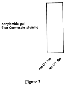
The secreted AN-LP1 immunoglobulin is an IgG1 Kappa. The results of
analysis by SDS-PAGE are presented on Figure 2. These results show that AN-LPI
presents
the conventional features of IgG (heavy chain 50 kDa, light chain 25 kDa).
Further experiments were performed in order to better characterize the
antibody.
1-Dot blot analyses:
Samples of synthetic human hepcidin (100 to 500 ng) or synthetic mouse
hepcidin (500ng) (Peptide International, Louisville, KY, USA), or of 10 or
40p,1 of sera from a
patient with an inflammatory disease, or from a healthy volunteer, were used.
In one
experiment, one sample of synthetic human hepcidin (500 ng) was treated with
Laemmli
buffer. The samples were directly spotted onto the nitrocellulose membrane and
allowed to
dry overnight. Non-specific sites were blocked by soaking the membrane in 5%
skim milk in
TBS-T (1 hr, at room temperature).
The primary antibodies (AN-LP1 antibody, or irrelevant antibody HPC4
directed against the epitope EDQVDPRLIDGK (SEQ ID NO: 11) of human protein C)
were
added at a dilution of 1:200 from a solution at 6mg/ml. Incubation was
performed for 2h at
room temperature or overnight at +4 C.
;0 After 3 washes with TBS-T, the membranes were incubated with the
secondary antibody conjugated with horseradish peroxidase (1:5000) for 1 h at
room
temperature.
The signals were visualized by chemiluminescence using an ECL reagent,
followed by autoradiography.
?5 The results are presented in Figure 3. These results show:
-that revelation of hepcidin with the antibody AN-LPI is specific (no signal
with the irrelevant antibody HPC4)
-that revelation of hepcidin with the antibody AN-LP1 requires the native
structure of the peptide (no signal when the sample is treated with Laemmli)
30 -that the antibody AN-LPl do not cross-react with murine hepcidin-25;
-that serum hepcidin from human samples is well recognized by the
antibody AN-LP 1.
2- Western blot analysis:
Samples of synthetic human or mouse hepcidin (Peptide International) were
35 separated in a 16 % Novex Tricine gel in non-reducing conditions and
blotting was
performed onto PVDF membrane for lh at room temperature. Non-specific sites
were
blocked by soaking the membrane in 5% skim milk in TBS-T (2 hr, room
temperature).
CA 02701694 2010-04-01
WO 2009/044284 PCT/IB2008/003310
12
Incubation with the primary antibody (AN-LP1 antibody, 6mg/ml, diluted at
1:100) was performed overnight at +4 C.
After 3 washes with TBS-T, the membrane was incubated with the
secondary antibody conjugated with horseradish peroxidase (1:5000) for 1 h at
room
temperature.
The signal was visualized by chemiluminescence using an ECL reagent,
followed by autoradiography.
The results are presented in Figure 4. These results show that the AN-LP I
antibody is efficient for detecting human hepcidin in Western Blot analysis
revealing a
product of the correct size (approximately 3kDa for the peptide of 25 AA) and
an additional
band most likely corresponding to dimers of hepcidin. In contrast, no signal
is observed for
mouse hepcidin, confirming the results of dot-blot analyses.
3-Immunohistochemistry:
Immunochemistry was performed on paraffine-embedded human liver. The
AN-LP1 antibody was used at dilution of 1:50 overnight at +4 C. After
incubation with the
secondary antibody conjugated with horseradish peroxidase, sections were
revealed with
diaminobenzidine.
The results are presented in Figure 5. These results show that the AN-LP 1
antibody is efficient for revealing hepcidin in human liver biopsies
0 EXAMPLE 2: FUNCTIONAL PROPERTIES OF THE ANTIBODY AN-LP1:
A screening test for hepcidin biological activity was developed. This test is
based on the capacity of the hepcidin to degrade the iron exporter,
ferroportin. It consists in
incubating macrophages (J774murine cell line), which express ferroportin, in
the presence of
hepcidin during a few hours. If the hepcidin is biologically active, it will
bind to ferroportin
5 and induce its degradation.
More specifically:
J774 cells were treated overnight with 200 M iron-NTA to induce
ferroportin production. Synthetic hepcidin-25 (100nM) alone, or linked to KLH
(200nM), or
an irrelevant peptide linked to KLH (100nM) were added to the cell culture. A
culture with no
30 peptide added was used as a control.
After incubation for 5h at 37 C, the cells were washed, then lysed.
Membrane extracts were prepared, and analyzed by Western blot with anti-
ferroportin
antibodies. The results are presented in Figure 6 A (Lane 1: control)
These results show that hepcidin alone or linked to KLH induces ferroportin
35 degradation, while the irrelevant peptide has no effect.
The same experiment was repeated with synthetic hepcidin-25 (100nM)
preincubated for 1 hour at 37 C with 3 or 30 g of AN-LP1 or of the irrelevant
antibody
HPC4, before addition to the cell culture.
CA 02701694 2010-04-01
WO 2009/044284 PCT/IB2008/003310
13
The results are illustrated by Figures 6B (hepcidin preincubated with the
irrelevant antibody HPC4) and 6C (hepcidin preincubated with the antibody AN-
LP1), and
6D (effect of increasing concentrations of AN-LP1).
These results show that while hepcidin normally induces the degradation of
ferroportin in the presence of the irrelevant antibody, the binding of the AN-
LP1 antibody to
hepcidin neutralizes its action, thus preventing the internalisation and the
degradation of
ferroportin.
EXAMPLE 3: AFFINITY OF THE HEPCIDIN/AN-LP1 INTERACTION
Immobilization of the ligands onto the sensor surfaces
To estimate AN-LP1 affinity constant and the antibody-antigen interactions,
surface plasmon resonance (SPR) measurements were performed with a BlAcore
2000
instrument using carboxymethylated dextran CM5 chips (BlAcore, Piscataway,
NJ).
AN-LP1 mAb, dilued at 2 g/ml in 5mM maleate buffer (pH 5.75), was immobilized
on the
CM5 chips by amine coupling.
Binding assays and data analysis
Hepcidin was diluted in running buffer HBS-EP (0,01M HEPES pH 7.4 ;
0.15M NaCl, 3mM EDTA and 0.005 % polysorbate 20). Analyte was injected in 3
min,
60 L/min injections and dissociation was monitored during 10 min. Surface Ab
were
generated by a 30-s to 1-min injection of glycine-HCL 10mM pH 2. Kinetic rate
constants
0 were determined with purified antibodies. Concentrated hepcidin (0.39 nM to
12.5 nM) were
injected over the chip surface at a rate of 60 L/min to collect binding data.
Data analyses
were carried out with the BlAevaluation 3.0 software.
The results are shown on Figure 7. The concentration of hepcidin is
indicated for each curve (Blk=blank).
'.5 The affinity constant (Kd) of AN-LP1 is of 9.9 x E-11 M.