Note: Descriptions are shown in the official language in which they were submitted.
CA 02701957 2010-04-08
WO 2008/044013
PCT/GB2007/003842
1
NEEDLE STRUCTURE
AND METHOD OF PERFORMING NEEDLE BIOPSIES
TECHNICAL FIELD
The present invention relates to biopsy needles,
i.e. needles adapted for the purpose of extracting fluid
or cells (e.g. tissue) from the body e.g. for the purpose
of idenifying cancerous growths.
BACKGROUND TO THE INVENTION
A fine needle biopsy normally uses a thin hollow
needle to remove a small tissue sample from an organ or a
tumour. A common type of fine needle biopsy is a fine
needle aspiration, where a fine needle and a syringe are
used to remove either fluid from a cyst or clusters of
cells from a solid mass. The procedure for fine needle
aspiration and fine needle biopsy is basically the same,
and the two procedures are sometimes performed together.
Fine needle biopsies can be obtained from organs or
tumours located within the human anatomy. Common sites
that may be considered for biopsy procedures to be
performed include: breasts, kidneys, the liver, the
pancreas, the prostate, the thyroid, lungs, ovaries and
lymph nodes. Fine needle biopsy is a diagnostic tool used
to evaluate organ or tumour tissue, and may also be used
to establish whether or not certain treatments are
working. It is normal for a local anaesthetic to be used
to numb the area where the needle will be inserted. The
thin hollow biopsy needle is inserted through the skin to
the biopsy site. In current procedures, the needle may be
CA 02701957 2010-04-08
WO 2008/044013
PCT/GB2007/003842
2
inserted more than once for correct positioning or to
obtain multiple samples.
When taking needle biopsies to identify potential
breast tumours, it is normal practice for the surgeon to
guide the biopsy needle into the area of concern by
palpitating or feeling the lump, whenever this is
physically possible, and then the needle may be located
into the tumour based on this information. There is a
high risk of false negatives occurring when taking
biopsies in this manner, and it may be necessary to
perform several needle biopsies in the region where the
lump has been felt in order for there to be a good chance
of locating the cancerous tissue.
If the lump is non-palpable, then the biopsy may be
performed under image guidance, e.g. using ultrasound.
However, even when ultrasound-guided needle biopsies are
performed, it is normal to make several attempts at
locating the cancerous site. Imaging using ionising
radiation is also used to locate the biopsy needle inside
the tumour. Fluoroscopy, where X-rays are directed onto a
fluorescent plate, which is linked to a television
camera, is used to see live images of the insertion of
the biopsy needle on a monitor, and to establish the most
appropriate position to take the biopsy. Computer
tomography (CT) or computer aided tomography (CAT), where
a scanner is used to rotate X-rays around the patient, is
also used to guide the biopsy needle. This form of image
guidance has the obvious drawback of exposing the patient
to potentially harmful doses of X-ray radiation. Other
drawbacks include: X-ray imaging procedures are expensive
and can be time consuming, they require specialist
CA 02701957 2015-01-08
WO 2008/044013
PCT/GB2007/003842
3
support to drive the equipment, and they are not always
successful in locating the cancerous site.
Needle biopsies are widely used and accepted as a
safe and reliable test for the determination of the
manifestation of cancer in the human body, but currently
used methods may lead to cancerous cells being spread
around the body when the biopsy needle is withdrawn. The
concern is that during the procedure of removing the
needle from the biopsy site, malignant cells may break
away from the tumour and be deposited along the needle
track that contains healthy tissue. This may lead to
seeding and the development of new tumours. It has been
reported' that a needle biopsy may increase the spread of
cancer by 50% compared to patients who undergo
lumpectomies.
Cases have been reported where the use of fine
needle biopsies to diagnose liver tumours has led to
metastases seeding along the biopsy needle track. In one
clinical review2 it is stated that the occurrence of
seeding is likely to'be the cause of the death of a
particular individual described in the case study.
SUMMARY OF THE INVENTION
At its most general, the present invention proposes
forming an antenna structure with the needle (hereinafter
a `needle antenna')., whereby the needle has the ability
not only to perform conventional tissue extraction but
also to couple microwave energy to and from the tissue to
in an article by Dr Joseph Mercola
2 Metcalfe M. S, Bridgewater F. H. G., Mullin E. J., and Maddern
G.J., Br. Med. Jou., 328, 28th February 2004, pp. 507 - 508
CA 02701957 2010-04-08
WO 2008/044013
PCT/GB2007/003842
4
perform measurements and/or ablation of tissue e.g. at
the needle tip.
The ability to measure dielectric properties of the
tissue (the measured information) may offer significant
advantage in terms of locating the cancerous tissue the
first time the needle antenna is inserted into the region
of tissue where it is suspected that a tumour is present,
i.e. there may be no need to take a number of tissue
samples. Also, the ability to measure tissue properties
in this manner may reduce the risk of false negatives
occurring.
The ability to measure information relating to the
tissue at the exact location, where the end of the biopsy
needle is located, may also offer significant advantage'
over location techniques that use the imaging (e.g.
scanning) arrangements described above in that the
scanning equipment may be unable to provide full or
reliable details regarding the region where the tumour or
cancerous tissue is located, due to certain biological
structures obscuring the image, or due to image
resolution or signal processing limitations. The present
invention may not suffer from these limitations.
The seeding of new tumours caused by biopsy needles
may be prevented by the ability to controllably ablate
the needle track e.g. during withdrawal to kill any
cancerous cells that would otherwise have been left
behind. The present invention may be arranged to
selectively perform both this ablation function and the
measurement function.
Thus, the needle antenna disclosed herein may have
the capability of directly measuring information relating
to the tissue in the form of tissue type and/or the state
CA 02701957 2010-04-08
WO 2008/044013
PCT/GB2007/003842
of the tissue. The needle antenna described in this
invention can also be used to perform controlled tissue
ablation.
According to one aspect of the invention, there may
5 be provided a biopsy needle insertable into tissue for
introducing or extracting a sample therefrom, the needle
having an elongate body terminating with an insertion
tip, a longitudinal channel formed within the body for
transporting the sample, and a coaxial antenna comprising
an inner conductor and an outer conductor coaxial with
the inner conductor and separated from it by a dielectric
material, wherein the coaxial antenna is arranged to
couple microwave energy to/from tissue at the insertion
tip, and the channel is formed within the inner conductor
or in an outer portion of the outer conductor. The inner
conductor may be a conductive layer along an inside wall
of the channel. Preferably, the inner conductor is a
conductive layer (tube) that defines the channel.
Preferably, the outer conductor comprises a conductive
layer formed on the outer surface of the elongate body.
The outer conductor may comprise a conductive layer
formed on the dielectric material and an annular or part
annular channel formed on that conductive layer. The
coupled microwave energy may be selectable either to
measure properties of tissue at the insertion tip or to
ablate tissue at the tip.
In another aspect of the invention, there may be
provided needle biopsy apparatus comprising a biopsy
needle as described above and a microwave power source
arranged to deliver microwave frequency energy to the
coaxial antenna in the needle in order to measure and/or
ablate tissue at the insertion tip of the needle. The
CA 02701957 2010-04-08
WO 2008/044013
PCT/GB2007/003842
6
apparatus may include a dynamic impedance tuner arranged
to adjust the impedance of the needle e.g. to match the
impedance of the tissue at the insertion tip in order to
ensure even (uniform) energy delivery into the tissue.
This aspect of the invention offers an advantage in that
it enables uniform ablation of the channel through which
the antenna is inserted to prevent the occurrence of
seeding. The ability to dynamically match into various
tissue structures prevents uneven ablation due to
variations in matching to various tissue types as the tip
of the antenna moves through the various structures.
In other words, the needle antenna described in this
specification can couple microwave frequency energy into
a co-axial structure for the purpose of making tissue
type/state measurements, and/or for performing controlled
tissue ablation, and has a hollow tube centre conductor
to enable tissue biopsies to be performed before, after,
or during the tissue ablation process. The structure
disclosed in the current invention may, therefore, be
considered as a tri-functional needle antenna. The
frequency of choice used in the current invention, and
the microwave aspects of the design of the tri-functional
antenna structure makes it possible to measure
information regarding the state of the biological tissue
at the same location (position) as where the tissue
biopsy is to be physically taken, i.e. at the distal tip.
In this specification, microwave frequency means a
frequency range of between 1 GHz to 100 GHz, preferably 5
GHz to 60 GHz. Higher frequencies, e.g. up to 200 GHz may
also be used. More preferably, the frequency source used
operates at a frequency of between 14 GHz and 15 GHz,
CA 02701957 2010-04-08
WO 2008/044013
PCT/GB2007/003842
7
and, even more preferably, operates at a spot frequency
of 14.5GHz.
This invention may overcome problems associated with
conventional needle biopsies and other similar tissue
= biopsy systems. The ability to perform tissue
measurements and to controllably ablate tissue offers
advantages in terms of preventing the seeding of
cancerous cells, that is often associated with
conventional needle biopsy procedures, by using
controlled microwave energy to seal the track or channel
made by the needle, allow fluid/tissue biopsies to be
taken with a high degree of confidence that false
negatives will not occur due to the ability of the system
to distinguish between healthy and cancerous tissue by
performing dielectric measurements at the tip of the
needle using a low power microwave transceiver; it may be
possible to eliminate the need to take multiple
fluid/tissue samples as is often the case in current
procedures (even when the needle is guided using
ultrasound or X-ray imaging), where hitting the target
only once out of several attempts is deemed to be
sufficient to qualify as constituting a successful
procedure. The current invention may also allow for
biopsy samples to be taken before, after, and during
ablation to help prevent loss of pathological information
as occurs in normal percutaneous tumour ablation
procedures using RF or microwave energy.
The tissue biopsy aspect of the current invention is
not limited to the extraction of cancerous tissue, or for
use in regions of the human body where cancerous tissue
may exist.
CA 02701957 2010-04-08
WO 2008/044013
PCT/GB2007/003842
8
It should be noted that the microwave design of the
needle antenna structure described in this specification
enables impedance mismatches at the distal tip of the
needle antenna structure to be reflected back along the
shaft of the antenna and the cable assembly attached
thereto to the generator, where measurements of the
reflected signal are used to calculate the requirements
to enable the distal tip of the antenna to be matched to
the generator, which may be a microwave power amplifier
with an output impedance of 50 0.
It may also be desirable to use a dynamically
adjustable tuning filter, for example, a waveguide cavity
containing three tuning stubs with a spacing of a quarter
of the guide wavelength at the frequency of interest, to
create a conjugate match between the distal tip of the
needle antenna and the load presented by the biological
tissue structure. It should be understood that the tuning
filter is positioned between the output from the power
amplifier and the distal tip of the needle antenna to
enable the output impedance of the amplifier to be
matched to the input impedance of the tuning filter, and
the output impedance of the tuning filter to be matched
to the impedance of the biological tissue. This feature
enables the needle antenna to be used to perform
controlled ablation of a volume of cancerous tissue or to
perform controlled ablation (or sealing) of the needle
track (or channel).
The ability of the needle antenna to convey
information back to the measurement system to allow
dynamic impedance matching to be performed between the
changing tissue impedance and the generator enables the
energy delivered into the various tissue structures that
CA 02701957 2010-04-08
WO 2008/044013
PCT/GB2007/003842
9
exist along the track between the site where the tissue
biopsy (or the tumour ablation) takes place and the
outside world to be automatically regulated to provide
uniform tissue ablation of healthy tissue structures en
route, i.e. it may be desirable to ablate a channel of 4
mm diameter of healthy tissue along the track (or
channel) to prevent the seeding of cancerous cells. The
ability of the needle antenna structure to allow for the
mode of operation described above to be performed may be
an additional feature of the current invention.
The invention may also be used in the future for
performing percutaneous musculoskeletal biopsies for
helping clinicians diagnose benign or malignant
musculoskeletal lesions due to the fact that the role of
fine-needle aspiration in the diagnosis and management of
musculoskeletal lesions is slowly gaining acceptance. It
is also expected that cytopathology of bone and soft
tissue tumours will serve to widen the usage of the fine
needle aspiration technique.
The invention may also be used to perform biopsies
of lung tissue. In this instance, depending on the exact
location, a biopsy will be obtained either by a
bronchoscopy or a needle biopsy. Needle biopsy is better
for cancers near the periphery of the lungs (i.e. closer
to the ribs than the centre of the chest), beyond the
reach of the bronchoscope. In this procedure, the biopsy
needle is inserted percutaneously through the chest wall
to take a tissue sample.
The invention may not be limited to introducing the
needle antenna percutaneously into the human body. The
needle antenna described here may be introduced into the
body by other means; examples include: through a trocar,
CA 02701957 2010-04-08
WO 2008/044013
PCT/GB2007/003842
through an endoscope, through a bronchoscope, through a
natural orifice, through a cystoscope, or during an open
surgical procedure.
The invention may not be limited to using a single
5 frequency source for performing controlled ablation and
making dielectric measurement. A plurality of frequency
sources may be used. For example, it may be advantageous
to use a lower microwave frequency, for example 1 GHz to
10 GHz, for performing controlled ablation, and a higher
10 microwave frequency, for example, 20 GHz to 100 GHz, for
performing dielectric measurements. The embodiments of
=
the invention described below use a single frequency
source operating at 14.5GHz, which has the advantage of
producing a high energy density for controlled ablation
of small tumours and effective track (or channel)
sealing, and a small enough radiation distance to allow
for dielectric measurements that are localised to the end
of the distal tip to be performed. The advantage of
using lower microwave frequencies for tumour ablation is
that the larger penetration depths associated with low
frequency microwave energy may be beneficial in terms of
producing effective ablation of large tumours, and the
advantage of using higher microwave frequencies for
dielectric measurement is that the small radiation
distances associated with high frequency microwave energy
may be beneficial in terms of effectively performing
local tissue measurements that are unaffected by
surrounding tissue structures.
From the above, it can be seen that this invention
may be particularly useful in helping to promote the
widespread use of needle biopsies. The ability to guide
the needle to the exact location of the suspected tumour
CA 02701957 2010-04-08
WO 2008/044013
PCT/GB2007/003842
11
and the capability of being able to controllably ablate
or seal the needle track to prevent seeding, may offer
great advantage over existing biopsy and location
techniques. The added ability of also being able to
controllably ablate the tumour whilst performing a tissue
biopsy may offer an extra advantage.
In another aspect, the present invention may provide
a method of performing a needle biopsy comprising any or
all of the following steps:
percutaneously inserting the biopsy needle antenna
through healthy tissue to the cancerous site under the
control of the tissue measurement system (i.e. performing
the dielectric measurement),
taking a first tissue (fluid or cell) sample,
commencing controlled tumour ablation under the
control of dynamic impedance matching,
taking further tissue samples during the controlled
ablation process (for example, the measurement interval
may be 30 seconds),
continuing ablation to achieve complete ablation of
tumour and controlled ablation of an extra portion of
healthy tissue to leave a safe margin,
taking a final sample of tissue,
sealing the needle track during needle withdrawal
using the microwave energy source configured to a lower
power setting, for example, between 2.5 W and 20 W, under
the control of dynamic impedance matching to ensure that
the various layers of tissue seen by the end of the
needle antenna are ablated by the same amount and that
this ablation process is well controlled.
The invention may be used to introduce material or
treatment into the body e.g. during bracytherapy.
CA 02701957 2010-04-08
WO 2008/044013
PCT/GB2007/003842
12
Materials introduced through the longitudinal
channel e.g. at the centre of the inner conductor and/or
in the outer portion (outer jacket) of the outer
conductor may be used to augment the plume of ablation
produced by the antenna or to adjust the shape of the
plume. For example, a lossy material may be introduced or
an additional electrode may be introduced that produces
energy at a lower frequency where cable loss is of little
significance.
A channel in the outer portion of the outer
conductor may be used to circulate a coolant, for example
saline or water.
In summary, the modes of operation of the current
invention may be as follows:
1. Tissue biopsy and controlled track ablation to
prevent seeding. Embodiments of the invention may
provide the following advantages:
- high microwave frequency and associated depth of
penetration of energy ensures minimal damage to healthy
tissue during channel ablation, and
- controlled solid state energy source ensures that
the power delivered can be adjusted in accordance with
tissue layer.
2. Tissue measurement (e.g. to recognise signature
of cancerous tissue type) and tissue biopsy. Embodiments
of the invention may provide the following advantages:
- information from the measurement system can be
used to ensure that tip of antenna is located at the
centre of the tumour to reduce the risk of a false
negative, and
- impedance measurement information can be used to
complement the biopsy information.
CA 02701957 2010-04-08
WO 2008/044013
PCT/GB2007/003842
13
3. Tissue measurement, tissue biopsy and
controlled track ablation to prevent seeding. Embodiments
of the invention may provide the following advantages:
- measurement information gathered as antenna is
withdrawn can be used in a feedback loop to control
tissue impedance matching system to ensure that a uniform
channel (around needle track) is ablated with minimal
damage to healthy tissue,
- information gathered during pre-clinical studies
regarding ablation plume shape and size can be used to
ensure that no nodal information will be lost,
- ablation may be automatically started when (or
prevented until) the tip of the antenna is located a pre-
determined distance from the node,
- impedance measurement information can be used to
complement the biopsy information.
4. Tissue measurement and introduction of material
into the body. Embodiments of the invention may provide
the following advantages:
- material can be introduced into the body using
measurement system to locate the centre of the target
tissue,
- introduced material can be used to augment the
ablation process, i.e. by using a material that reacts
with the microwave energy,
- a radioactive implant can be introduced accurately
and the entrance channel can be controllably ablated to
prevent seeding.
BRIEF DESCRIPTION OF THE DRAWINGS
CA 02701957 2010-04-08
WO 2008/044013
PCT/GB2007/003842
14
Examples of the invention are discussed below with
reference to the accompanying drawings, in which:
Fig. 1 is a block diagram of a needle biopsy
apparatus that is a first embodiment of the invention;
Fig. 2 is a more detailed diagram of the apparatus
shown in Fig. 1;
Fig. 3 is a diagram showing a needle biopsy
apparatus that is a second embodiment of the invention;
Fig. 4 illustrates the circuit layout for the
transceiver shown in Fig. 3;
Fig. 5 is a diagram showing a needle biopsy
apparatus that is a third embodiment of the invention;
Fig. 6 is a diagram illustrating the skin effect;
Fig. 7 is a graph representing the amount of power
transferred using an energy source operating at a
frequency of 14.5GHz as a function of metal layer
thickness for various metals;
Fig. 8 shows a needle antenna that is a fifth
embodiment of the invention;
Figs. 9a and 9b illustrate a needle antenna that is
a sixth embodiment of the invention;
Figs. 10a and 10b illustrate a needle antenna that
is a seventh embodiment of the invention;
Fig. 11 is a cross-section through a model of a
needle antenna according to the present invention for use
in a computer simulation;
Fig. 12 shows the result of a simulation of the
energy distribution from the needle antenna shown in Fig.
11;
Fig. 13 shown the result of a simulation similar to
Fig. 12 but where there is no biopsy channel through the
centre of the needle;
CA 02701957 2010-04-08
WO 2008/044013
PCT/GB2007/003842
Figs. 14 and 15 are diagrams illustrating the
impedance match for a needle antenna with and without a
biopsy channel;
Fig. 16 is a plot showing the impedance values of
5 various materials on a Smith chart;
Fig. 17 shows connection pipes for a central biopsy
channel in a needle antenna;
Fig. 18 shows the energy density distribution for a
needle antenna with the connection pipes shown in Fig.
10 17;
Fig. 19 is a close up view of the energy
distribution around the connection pipes shown in Fig.
17;
Fig. 20 is a diagram illustrating the impedance
15 match of four equally spaced connecting pipes;
Fig. 21 shows the energy density distribution for a
configuration of four connection pipes; and
Fig. 22 is a diagram illustrating the impedance
match of the four connecting pipes shown in Fig. 21.
DETAILED DESCRIPTION; FURTHER OPTIONS AND PREFERENCES
In this description the term ablation may refer to
the ablation of a region of cancerous tissue, for example
a tumour, or for sealing a track or channel made as the
needle antenna passes through layers of healthy tissue.
The latter will generally require lower levels of power
and the track ablation may be performed with dynamic
energy matching to the tissue impedance seen en route to
ensure that controlled amounts of energy is launched into
the various tissue types as the needle antenna traverses
through the tissue to the outside world. However this
CA 02701957 2010-04-08
WO 2008/044013
PCT/GB2007/003842
16
invention need not be limited to performing controlled
ablation with dynamic impedance matching being in place.
In general terms, one embodiment of the needle
antenna structure comprises a rigid stainless steel
structure with an outside diameter of around 2.2 mm and a
sharp ceramic pointed cone at the distal tip to enable
percutaneous insertion. However, the invention need not
be limited to this geometry or construction and may be
realised using any suitable antenna structure that
enables microwave energy to be transferred in the forward
and reverse direction to enable the measurement of
dielectric information, and to cause controlled tissue
ablation, whilst allowing tissue samples (fluid or cells)
to be extracted without upsetting the environment set-up
to allow microwave signals to propagate for the purpose
of making a dielectric measurement or for the purpose of
introducing a high enough level of microwave energy into
biological tissue to cause controlled tissue ablation.
The invention makes use of the fact that the centre
conductor within the antenna is around 0.5 mm in
diameter, but a wall thickness of about 0.01 mm only is
required to enable almost all of the microwave energy to
flow, or to be transported, along an appropriate
conductive material when the frequency of operation is
14.5 GHz. Thus, in theory the centre of the centre
conductor can be removed to leave a bore having a
diameter of over 0.4 mm available as a channel that can
be used to remove fluid from a cyst or cells within a
solid mass. It =is worthwhile noting that this channel
could also be used to transport other liquids and/or
solids in and out of the needle antenna. For example
CA 02701957 2010-04-08
WO 2008/044013
PCT/GB2007/003842
17
imaging or contrast media for specific tissue marking
and/or identification.
It was recently reported that a Japanese scientist
won a Good Design Award for the development of a 0.2 mm
diameter needle for infusion, therefore, it can be the
with confidence that the technology exists to enable the
manufacture of a hollow centre conductor that suits these
requirements.
For practical implementation in systems suitable for
taking biopsies of breast tissue, the preferred hollow
section for the needle centre conductor may be between
0.3 mm and 0.4 mm in diameter. This should provide enough
mechanical strength, and ensure that all of the
electromagnetic fields propagate along the outside of the
centre conductor, i.e. the wall thickness is much greater
than 0.01 mm, thus the electromagnetic fields set-up
inside the structure would be unaware that the structure
is hollow. To ensure that the overall needle antenna
structure is rigid, and that it is allowed to be
percutaneously inserted inside a patient, it is
preferable to use stainless steel as the outer jacket of
the needle antenna structure.
In this description, an antenna structure and
apparatus is described that has the potential to perform
the following functions:
- measure dielectric information to determine the
type, state and location of healthy and cancerous tissue,
- perform a needle biopsy with confidence that the
tip of the needle is located inside the centre of the
tumour, or other biological tissue that may require
treatment,
CA 02701957 2010-04-08
WO 2008/044013 PCT/GB2007/003842
18
- controllably ablate the tumour or other unhealthy
tissue structures and a small region of healthy tissue (a
safe margin),
- take further needle biopsies during and after the
treatment process, and
- controllably ablate the channel made by the needle
antenna during needle withdrawal to prevent seeding.
The new combined procedure involving tissue
measurement, tissue biopsy, and tissue ablation can allow
cancerous tissue (fluid or cells) to be located during a
first attempt, and the risk of dragging cancerous cells
back through the channel can be mitigated due to the fact
that the needle channel (or track) is subjected to
controlled ablation, thus causing the death of any
cancerous cells that may be present at or around the
distal tip of the needle antenna.
It should be noted that this device can be used to
perform any combination of the above listed functions,
for example, it could be used to locate the needle
antenna into the centre of the tumour and take the
biopsy, or it could be used to take the biopsy and seal
up, or controllably ablate, the channel to prevent the
risk of seeding; it could be used to take the biopsy,
ablate the cancerous tumour and then seal the channel, or
it may be possible to take a tissue biopsy before, during
and after tissue ablation has taken place to ensure that
the tumour has been successfully destroyed using thermal
ablation.
It may also be desirable to use the current
invention to deposit materials into the biological system
rather than removing tissue from the biological system.
In this mode of operation, the tissue measurement and
=
CA 02701957 2010-04-08
WO 2008/044013
PCT/GB2007/003842
19
characterisation feature may be used to identify the
region of the body where a material (solid or liquid) is
required to be located with a high degree of accuracy,
and the material may be deposited at the exact desired
location (features associated with the use of the low
power microwave frequency transceiver facilitates this).
This aspect of the current invention may be particularly
useful for depositing a particular drug or a radioactive
dye into the body for example. This idea may be used with
brachytherapy. The ability to target the exact location
where a drug is to be delivered may offer significant
advantage in terms of minimising the concentration and
amount of drug required.
It should also be noted that the centre tube may be
used to suck out or remove ablated tissue in order to
increase the zone of ablation. This may be of particular
use where the ablated tissue has become charred. Once the
tissue has been removed the ablation process may commence
again and the process repeated a number of times. Since
it is not only the centre of the centre conductor that is
transparent to the microwave field, but also the outer of
the outer conductor may also be hollow, it is possible to
use this as a second channel for taking the biopsy and
for removing tissue.
This invention is not limited to removing fluid or
cells associated with cancerous tumours; the needle
antenna may be used to remove other tissue from sensitive
regions of the body where it is required to accurately
locate the biopsy tissue inside target tissue. In these
applications, the invention may be operated in
measurement mode only.
CA 02701957 2010-04-08
WO 2008/044013
PCT/GB2007/003842
The tri-functional nature of embodiments discussed
herein may be particularly useful for performing biopsies
on underarm lymph nodes, in which the presence or absence
of breast cancer cells is a powerful predictor of whether
5 the cancer has spread. It is now possible to examine
such biopsies to help decide appropriate therapy for
metastatic breast cancer without the need to remove a
sentinel lymph node for examination under a microscope.
The biopsy method is much quicker and less invasive than
10 the surgical procedure. The present invention can make
the biopsy method more accurate and controlled.
The present invention permits accurate location of
the lymph node using the measurement mode, extraction of
tissue for examination through the biopsy channel, and
15 controlled ablation to seal the needle track during
withdrawal using the ablation mode. To avoid damaging
the lymph node (e.g. by ablation), the measurement mode
may be used to determine when the tip of the probe leaves
the node. By leaving the node intact, it can be used for
20 further measurement in future. The size of the ablation
plume (e.g. extent of high power microwave radiation
field) produced by the needle is repeatable and well
defined for a given power level and pulse profile.
Accordingly, in combination with the measurement mode it
is possible to accurately and repeatably insert a safety
zone e.g. of 1-5 mm between a measurement position (e.g.
lymph node) and the start of ablation (e.g. higher power
mode (e.g. 10 W) to seal the needle track and prevent
seeding of cancer cells). The ablation mode may be
automatically enabled based on information obtained in
the measurement mode.
CA 02701957 2010-04-08
WO 2008/044013
PCT/GB2007/003842
21
Moreover, if the embodiment includes a tuner for
implementing the dynamic impedance matching discussed
above, evenly ablation of the needle track during
withdrawal can be achieved even when that track passes
through different tissue types i.e. materials having
different conductivities and permittivities at the
frequency of interest.
A further feature of the current invention may be to
pump water or saline through the biopsy channel during
ablation to keep the needle antenna as cool as possible.
It may be advantageous to use this feature in
applications where it is desirable to treat large
lesions. In this instance, it may be required for the
level of microwave power to be increased from that used
when operating in the treatment mode under normal
conditions, for example, where spherical tumours of
diameter greater than 2 cm are to be treated, or where it
is required to deliver power over longer durations of
time. For example it may be required to generate up to
100 W of continuous wave (CW) power for ten minutes in
order to treat a spherical lesion of, for example, 10 cm
in diameter.
Alternatively, the biopsy may be used to introduce a
material (e.g. lossy biocompatible material) which can
augment the ablation effect, e.g. increase the ablation
volume that is achievable with the apparatus. The
presence of the material within the needle may not affect
the generated microwave field because the microwave
energy only flows in the outer section of the inner
conductor.
The method and device used to either collect tissue
(or other substances) from the human body, or to
CA 02701957 2010-04-08
WO 2008/044013
PCT/GB2007/003842
22
introduce substances into the human body, through a
channel contained within the needle antenna introduced
here, will be determined by the specific application of
the current invention. In most procedures, a syringe may
be used but this invention is not limited to using a
syringe.
In one embodiment, the biopsy channel may be used to
suck necrosed or charred tissue from the needle tip
during ablation. This may be particularly beneficial
where dynamic impedance matching is implemented because
it removes the charred tissue. that the needle would
otherwise have to be matched with. Typically charred
tissue presents a load that is very different from that
which the needle may be designed to match with in the
absence of a tuner.
In another embodiment, the device may be used in
liposuction. The delivered microwave radiation may be
used to heat fat which can then be sucked out via the
biopsy channel. When used with a dynamic impedance
matching apparatus, the needle impedance can be matched
to the impedance of fat to target the heating. This
apparatus may reduce the invasive nature of liposuction
and facilitate its use on fine tissue structures or in
cosmetic surgery, where it is desirable to minimise
scarring or other permanent damage. Moreover, the
configuration of the channel may act to concentrate the
microwave energy around the channel, which can be
beneficial in targeting heating at the location where it
= is required.
In the following full description of the current
= invention, certain aspects relating to apparatus, or
electronic instrumentation, for producing controlled
CA 02701957 2010-04-08
WO 2008/044013
PCT/GB2007/003842
23
tissue ablation, and apparatus for detecting changes in
tissue state is given as an overview only, since the
inventor's earlier applications WO 2004/047659 and WO
2005/115235 describe these aspects in detail. On the
other hand, it should be noted that the current
description does address particular aspects of a
sensitive measurement transceiver (for measurement mode)
and the operation of the matching filter (for controlled
ablation mode).
Skin effect & needle antenna dimensions
This invention makes use of the fact that as the
frequency of the energy increases, conduction begins to
move from an equal distribution over the entire cross
section of a conductor to only existing at the surface of
the conductor. Microwave frequencies in the super high
frequency band (SHF), e.g. where the frequency is greater
than 3 GHz, are preferably in the invention since these
lend themselves particularly well where it is desirable
for the thickness of the conductor to be less than 0.1
mm, or more preferably less than 0.01 mm. The preferred
frequency used in the current invention is 14.5 GHz and
the conductor preferably has a high conductivity, thus
enabling conductor thicknesses to be in the micrometer
(pm) region. The phenomenon associated with the reduction
in conductor thickness as the frequency of the
electromagnetic energy increases, is known as the skin
effect.
The use of high microwave frequency radiation, i.e.
radiation from a source operating at a frequency above 10
GHz, is advantageous in that the microwave energy
CA 02701957 2010-04-08
WO 2008/044013
PCT/GB2007/003842
24
produces a low propagation distance or depth of
penetration inside the tissue, hence the dielectric
measurement is localised to the end of the needle antenna
where the fluid or cells are extracted from the
biological system. It should be clear from the above that
the current invention will enable the point where the
dielectric (or tissue type/state) measurement is taken to
be the same as where the tissue biopsy is extracted. It
should also be understood from this statement that it is
advantageous to design the tip of the needle antenna to
be sensitive to changes in tissue impedance. For example,
the material used at the distal tip of the needle antenna
preferably exhibits a low dissipation factor at the
frequency of interest, and the relative permittivity of
this material may be chosen to provide a good impedance
match between a representative tissue impedance and the
impedance of the rest of the needle antenna structure. It
should be noted that the tissue impedance is a function
of the relative permittivity and the conductivity of the
tissue at the frequency of interest. These two parameters
can be used to describe the behaviour of dielectric
materials at microwave frequencies.
When current is flowing through a conductor, the
magnetic flux that results is in the form of concentric
circles. Some of the flux exists within the conductor and
links more strongly with the current in the centre. The
result is that the inductance of the central part of the
conductor is greater than the inductance of the conductor
near the surface. This is because of the greater number
of flux linkages existing in the central region. At high
frequencies the reactance of the extra inductance is
sufficiently large to enable the current to flow along
CA 02701957 2010-04-08
WO 2008/044013
PCT/GB2007/003842
the surface of the conductor where the impedance is low
rather than near the centre of the conductor where the
impedance is high.
Depending on the bulk resistivity of the conductor,
5 at sufficiently high frequency all of the microwave
current will flow within a very small thickness of
conductor. Also, the current tends to concentrate nearest
to the surface that abuts the highest relative
permittivity. The use of materials with low bulk
10 resistivity leads to shallower skin depths.
For a solid wire, the current concentrates on the
outer surface. For this reason, when skin depth is
shallow, the solid conductor can be replaced by a hollow
tube with no loss in performance. This phenomenon is
15 illustrated in Fig. 6. Skin depth can be calculated
using either equation 1 or equation 2:
2
..1
s¨
a Quo-
s¨
irpf
where o's is skin depth in metres (m), co is radian
frequency (2nf) in Hertz (Hz), c is conductivity in
siemens (S or Q/m), p is resistivity in ohms metres (Om),
f is frequency in Hertz (Hz), and p is permeability of
free space in Henry per metre (H/m) (= 4nx10-7 H/m).
Table 1 provides values of skin depth at spot
frequencies of 1 MHz, 10 MHz, 100 MHz, 1 GHz and 10 GHz
for commonly used conductive materials. This table
illustrates the need for using high microwave frequencies
CA 02701957 2010-04-08
WO 2008/044013
PCT/GB2007/003842
26
in small diameter structures when metallisation thickness
is to be kept to a minimum, for example, in a co-axial
arrangement where a hollow centre conductor with the
largest diameter biopsy channel is required.
-r-
Bulk
Resistivity Skin Depth (pm at frequency)
at 20 C
Material Symbol (Qx10-8m) 1MHz 10MHz 100MHz 1GHz lOGHz
Aluminium Al 2.65 81.9 25.9 8.19 2.59 0.819
Beryllium Be 3.3 91.4 28.9 9.14 2.89 0.914
rBrass Cu70/Zn30 7 133 42.1 13.3 4.21 1.33
IBronze r-Cu89/Snll I 15 195 61.6 19.5
6.16 1 1.95
I Copper Cu 1.69 65.4 20.7 6.54
2.07 0.654
1Gold Au 2.2 74.7 23.6 7.47 2.36 0.747
'Graphite t-C 783.7 1409 446 141
44.6 14.1
Nickel , Ni 6.9 132 41.8 13.2
4.18 1.32
Silver Ag 1.63 64.3 120.3 6.43 2.03 0.643
Table 1: Skin depth (in pm) for various commonly used
materials at frequencies of 1MHz, 10MHz, 100MHz, 1GHz and
lOGHz
The percentage of power transferred as a function of
material thickness can be described by equation 3
%P =(1- ell x100 , .
.3
where x is the thickness of the layer of metallisation in
metres (m), and %P is percentage of the power flowing in
given thickness of metallisation in watts (W). For
example, equation 3 predicts that for a thickness of
CA 02701957 2010-04-08
WO 2008/044013
PCT/GB2007/003842
27
metallisation of six skin depths, 99.75% of the power
will be transported.
In the embodiments described below, four commonly
used metallic materials were considered. These were:
copper, silver, nickel, and steel. Fig. 7 shows the
variation of power transported as a function of
metallisation thickness for these four materials based on
skin depth calculations using equation 1, and the
exponential relationship given in equation 3. For the
generation of Fig. 7, the frequency of operation used was
14.5 GHz, it is assumed that the materials are non-
magnetic, and the following conductivity values (a)
apply:
Silver: a = 5.80x10⁷ S/m
Copper: a = 6.14x10⁷ S/m
Nickel: a = 1.28x10⁶ S/m
Steel: a = 5.0x10⁶ S/m
It can be seen that the best material to use in
order to minimise the thickness of metallisation is
silver. This if followed very closely by copper. The
thickest layer of metallisation is required when using
steel. It should be noted that the line for steel does
not converge with the other three materials on the graph
shown in Fig. 7, where the maximum thickness shown is
8pm.
Table 2 provides figures for the required thickness
of metallisation for 90%, 99% and 99.9% of power flow for
silver, copper, nickel and steel:
CA 02701957 2010-04-08
WO 2008/044013
PCT/GB2007/003842
28
Silver Copper Nickel Steel
Power transferred (%) Thickness of metallization (pm)
90 1.23 1.26 2.69 4.30
99 2.46 2.53 5.38 8.61
99.9 3.68 3.79 8.07 12.91
Table 2: Thickness of metallization requirements for
commonly used conductors when the operating frequency is
14.5GHz
It should be noted that the ability to minimise the
thickness of the metallisation layer leads to the ability
to fabricate needle antenna structures with minimal
inside and outside conductor diameter thicknesses. This
has the advantage of minimising the outside diameter of
the needle antenna structure and/or maximising the
diameter of the biopsy channel (this analysis assumes
that it is required to keep the characteristic impedance
of the structure constant and that the relative
permittivity of the dielectric material between the inner
and outer conductors is constant. These features may be
advantageous where percutaneous systems are used to
perform tissue ablation and/or dielectric (tissue
state/type) measurement and for taking tissue biopsies.
The characteristic impedance of the co-axial needle
antenna structure is described by equation 4 given below:
138
Z0
.. 4
where Zo is the characteristic impedance of the co-axial
line in ohms (0), Er is the relative permittivity of the
dielectric material between the centre conductor and the
CA 02701957 2010-04-08
WO 2008/044013
PCT/GB2007/003842
29
outer conductor, c is the inner diameter of outer
conductor in metres (m) and e is the outer diameter of
inner conductor in metres (m). The location of diameters
c and e are illustrated on the tri-functional needle
antenna structure shown in Fig. 8.
Referring to Fig. 8, in a structure that uses steel
as the metallic material, has a dielectric filling
between the two conductors with a relative permittivity
of 3.045, operates at a frequency of 14.5 GHz, and has a
characteristic impedance of 50 0, the following physical
dimensions (calculated using equations' 1, 3 and 4) may be
used:
- thickness b of steel for 99.9% of the energy to be
transported = 12.91pm
- outer diameter of needle antenna a = 2.2 mm
- inner diameter of outer conductor c = 2.18 mm
- outer diameter of inner conductor e = 0.51 mm
- inner diameter of inner conductor d = 0.49 mm
This is an illustrative example that enables a
biopsy channel with a diameter of up to 0.49 mm (in
theory up to 0.49709 mm) to be used. This example
assumes that the dielectric material used is a hard rod
of material with a hole of 0.51 mm diameter bored through
the centre. It would be necessary for a first layer of
steel of thickness 12.91 pm to be deposited onto the
outside of the rod, and a second layer of steel of
thickness 12.91 pm to be deposited onto the inside wall
of the hole bored through the centre of the rod.
The idea of taking a hard dielectric and Coating the
outer surface and the inner wall of the centre hole may
be an independent aspect of the invention.
CA 02701957 2010-04-08
WO 2008/044013
PCT/GB2007/003842
The idea of limiting the coating thickness of the
metallisation attached to the wall on the inside bore
hole to a thickness whereby it is considered that all the
energy will flow, but no greater than this, i.e. in this
5 case 12.91 pm, enables the diameter of the biopsy channel
to be maximised and allows the maximum amount of tissue
to be transported along the biopsy channel.
In the example given here, steel has been used
because it exhibits the lowest conductivity out of the
10 four materials considered as possible candidates for this
work.
Biopsy apparatus
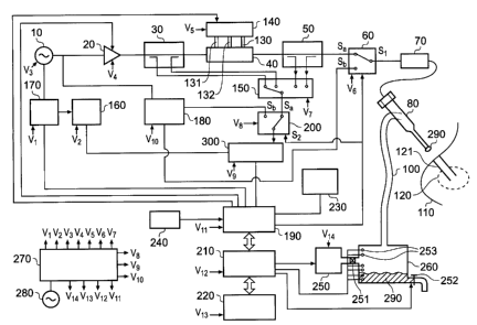
15 Fig. 1 shows a block diagram of the overall system.
This configuration enables all three modes of operation
to be performed using a single needle antenna structure
80. In the dielectric measurement (or tissue recognition
or location mode), stable frequency source 10 is used as
20 the low power transmitter signal and is fed into the low
power transmitter circuit 180, where it is channelled
into needle antenna 80 through mode select switch 60, and
cable assembly 70. The measurement signal, taken with
needle antenna 80 inserted into biological tissue 110 to
25 the area of concern 120, is then fed back into receiver
300 via cable assembly 70, mode select switch 60, low
power measurement transmitter 180 (containing high
isolation circulator with a carrier cancellation
circuit), channel select switch 200, and into receiver
30 300. Receiver 300 uses local oscillator 160 to produce a
first intermediate frequency (IF) signal that is used to
convert the measurement signal into a form that enables
CA 02701957 2010-04-08
WO 2008/044013
PCT/GB2007/003842
31
digital signal processor 190 to extract both magnitude
and phase information from the signal. Receiver 300 may
contain a second IF stage (this is not shown in Fig. 1).
The phase and magnitude information is then processed
using digital signal processor 190 and/or microprocessor
210 to determine the type of tissue 290 that the tip of
needle antenna 80 is in contact with. The tissue type 290
may be displayed using the user interface 220.
In the controlled ablation mode, stable frequency
source 10 is fed into power amplifier and control sub-
system 20, which is used to control the level and the
duration of the power being delivered (the energy
profile) into target tissue 120 to enable controlled
ablation to be performed, or into the needle channel 121
for controlled sealing of the track. The output from
power amplifier and control unit 20 is fed into first
forward and reflected power coupler unit 30, whose
function is to measure a portion of the forward power
coming out of power amplifier 20 and the power reflected
back due to a mismatch at the input of matching filter
40. The portions of forward and reflected powers are fed
into the inputs to monitor select switch 150. The output
from first forward and reflected power coupler unit 30 is
fed into the input of matching filter 40, whose function
=
is to impedance match power source 20 to load impedance
seen by the distal tip of needle antenna 80, which may be
either the treatment tissue 120, or the needle track 121.
The tuning of matching filter 40 is performed by moving
three tuning stubs 130, 131, 132 in and out of a
waveguide cavity that forms a part of the matching filter
40. The movement of stubs 130, 131, 132 is performed
using stub-actuator and a suitable control unit 140. It
CA 02701957 2010-04-08
WO 2008/044013
PCT/GB2007/003842
32
may be preferable to use linear actuators and a
proportional-integral-differential (PID) control system
(not shown here). The output from matching filter 40 is
fed into a second forward and reflected power coupler
unit 50, whose function is to measure a portion of the
forward power coming out of matching filter 40 and the
power reflected back due to a mismatch at the distal tip
of needle antenna 80. The portions of forward and
reflected powers are fed into the inputs to monitor
select switch 150.
The position of stubs 130, 131, 132 is determined by
the signals at the coupled ports of first and second
forward and reflected power couplers 30 and 50
respectively. These signals are measured by polling each
of the four switch positions of monitor select switch
150. The switch position is determined by a select signal
provided by digital signal processor 190. The single
output from monitor select switch 150 is fed into
receiver 300 via channel select switch 200, where the
switch contact connects the monitor select switch 150 to
the input to receiver 300. The receiver 300 has an
internal frequency mixer (not shown) that uses the
selected signal from first and second forward and
reflected power couplers 30 and 50 respectively and the
local oscillator signal 160 to produce a first IF
frequency. A second internal frequency mixer is used to
form a second IF stage (not shown) and the output signal
from the second IF stage is fed into digital signal
processor 190, where phase and magnitude extraction is
performed. The digital signal processor 190 uses the
phase and magnitude information to determine the required
signals to send to stub actuator and control unit 140 to
CA 02701957 2010-04-08
WO 2008/044013
PCT/GB2007/003842
33
enable tuning stubs 130,131,132 to be moved inside the
waveguide of matching filter 40 to positions whereby the
output from power amplifier 20 is matched to the
impedance seen at the distal tip of needle antenna 80.
The output from second forward and reflected power
coupler 50 is connected to mode channel switch 60, which
is configured to connect the output from the forward and
reflected power coupler 50 to the input to cable assemble
70. The output of cable assembly 70 is connected to the
input, or the proximal end, of needle antenna 80. A
control signal from digital signal processor 190 is used
to change the switch contacts within channel select
switch 200 and the mode select switch 60 to enable the
controlled ablation mode or the tissue measurement mode
to be selected.
It has been assumed here that the digital signal
processor 190 contains an analogue to digital converter
(ADC) to convert the analogue signals from receiver 300
into a digital format. In practice, it may be preferable
to use an external ADC unit. A footswitch 240 is used to
activate tissue ablation and measurement. The microwave
energy output from the generator 60 and the input line
from footswitch 240 contain DC isolation barriers (not
shown here); these are required to prevent the generator
from being connected to the user or patient circuit via a
DC path (not shown here). In the ablation mode, the user
interface 220 may indicate the energy dosage delivered
into the tissue, the treatment time, and any other useful
and/or relevant information. In biopsy mode, it may be
desirable for user interface 220 to show the level of
tissue contained in vessel 290 and when pump 250 has been
activated. In tissue measurement mode, it may be
CA 02701957 2010-04-08
WO 2008/044013 PCT/GB2007/003842
34
desirable for user interface 220 to show display tissue
type and/or tissue state. It may also be desirable to
sound an audible alarm or flash the display when the
distal tip of the antenna comes into contact with
cancerous tissue.
Stable frequency source 10 and local oscillator 160
use the same temperature compensated stable crystal
oscillator 170 as a reference source. The reference may
also be fed into digital signal processor 190 and may be
used as a timing reference.
Before the commencement of tissue measurement, or
dynamic controlled ablation, it is necessary to calibrate
needle antenna 80 using needle antenna calibration unit
230. Calibration is performed by inserting needle antenna
80 inside a cavity, or slot, contained within needle
antenna calibration unit 230 (the cavity or slot may be
co-axial or waveguide). A control signal from digital
signal processor 190 is used to actuate a linear motor
contained within calibration unit 230 to move a sliding
short along a waveguide cavity to enable a number of
calibration points to be measured. During calibration,
the proximal end of cable assembly 70 must be connected
to the RF output port of the microwave generator 60, and
distal end of cable assembly 70 must be connected to the
proximal end of needle antenna 80. It should be noted
that it may be preferable to integrate cable assembly 70
and needle antenna 80 into one assembly during the device -
manufacturing stage.
Mode select switch 60 and channel select switch 200
are configured in such a way that they change contact
position at the same time; these two switches enable
controlled ablation or measurement mode to be selected.
CA 02701957 2010-04-08
WO 2008/044013
PCT/GB2007/003842
The position control comes from a select signal provided
by digital signal processor 190. In the first switch
position (S,) the system is operated in ablation mode, and
in the second position (Sb) the system is operated in
5 measurement or tissue recognition mode.
A fluid feed pipe 100 is connected to needle antenna
80, preferably through a side wall of needle antenna 80,
and fluid feed pipe 100 connects to a collection tank or
vessel 260, which is used to collect biopsy tissue (fluid
10 or cells) 290. An internal pipe connects the outer jacket
of needle antenna 80 to the centre conductor of needle
antenna 80 (not shown here). A pump 250 is used to suak
the tissue sample 290 along a hollow channel contained
within needle antenna 80 (not shown here), and suck the
15 tissue through tissue feed pipe 100 into tank 260. It
must be ensured that there are no leaks in the system. A
valve 251 is used to ensure that tissue 290 cannot be
directed into pump 250. Microprocessor 210 is used to
control the operation of pump 250. It may be desirable to
20 attach fluid level monitors or sensors 253 inside tissue
vessel 260 to monitor the level of tissue inside the
vessel; microprocessor 210 may be used to process signals
from level monitors or sensors 253 and this information
may be displayed using user interface 220. Microprocessor
25 210 may also be used to control the operation of valve
252, which is used to empty vessel 260. The operation of
valve 252 may be based on information obtained from level
sensors 253. It should be noted that tank 253 and pump
250 may be replaced by a syringe.
30 Cable assembly 70 is preferably a low loss coaxial
cable with low random phase variation with flexure, but
other cable assemblies, such as flexible waveguide, may
CA 02701957 2010-04-08
WO 2008/044013
PCT/GB2007/003842
36
be used. It is preferable for the insertion loss of the
cable to be less than ldB per metre and for the random
phase variation with flexure to be less than 10 rms.
DC power supply 270 is used to supply the sub-
assemblies and units with DC power.
It may be preferable to apply a conformal coating of
Parylene C material to the needle antenna structure. A
coating thickness of around 10 pm will not affect the
microwave behaviour of the structure but will reduce the
coefficient of friction on the needle surface and help
reduce friction between the needle antenna and the tissue
as the needle antenna is pushed through various types of
tissue. Parylene C is easy to apply and is a
biocompatible material that has undergone extensive
material tests concerning its use inside the human body.
Should the tip of the tri-functional needle antenna be
made from a non-biocompatible material, the inclusion of
a layer (or coating) of Parylene C may enable the
structure to be used inside the human body.
It must be ensured that the fluid pumping system is
a sealed system and that no fluid or tissue is able to
leak in the region of the pipe that connects the centre
conductor and the outer conductor. In the instance where
a ceramic cone tip is used and the fluid is fed through
the ceramic tip, it must be ensured that the interface
between the hollow ceramic section and the centre
conductor is sealed. It may be desirable to extend the
centre conductor to the end of the ceramic cone tip. Full
electromagnetic field analysis is performed on the new
structure to take into account discontinuities and to
ensure that the microwave operation of the structure is
not in any way impaired.
1
CA 02701957 2010-04-08
WO 2008/044013
PCT/GB2007/003842
37
It should be pointed out that if leaks exist in the
system then it may be difficult to pump tissue or fluid
from the sample site 120 to the tank or vessel 260. If
there are leakage points then air will get into the
system, which may make it more difficult to transport
tissue from the cancerous site 120 to the tank or vessel
260. It may be desirable to remove any air bubbles that
may occur in the system before pumping fluid from the
body.
Fig. 2 shows a block diagram of the complete system,
where the construction of needle antenna 80 is shown in
detail. It should be noted that the functionality of Fig.
2 is identical to that of Fig. 1, which has already been
described in detail above. Physically, other than details
of needle antenna 80, Fig. 2 is identical to Fig. 1,
except for the following differences in component or sub-
assembly partitioning: In Fig. 2, power amplifier and
control unit 20 has been split up into two units, namely:
power/modulation control unit 21, and power amplifier 22,
stub actuator and control unit 140 has been split up
into: linear actuators 141 and actuator controller 142;
and microprocessor 210 and digital signal processor 190
has been combined into microprocessor and signal
processor 211.
Needle antenna 80 comprises an input microwave
connector 81, which may be any suitable microwave
connector that can be used at the microwave frequency
that is of interest for use in the current invention, for
example: SMA, MCX, or SMC types. The microwave connector
81 is used to connect the needle antenna 80 to the cable
assembly 70 and is also used to couple microwave energy
into and out of the needle antenna 80. The proximal end
CA 02701957 2010-04-08
WO 2008/044013
PCT/GB2007/003842
38
of the centre conductor 88 is connected to the centre
conductor of microwave connector 81. It may be preferable
for the first section of centre conductor 88 to be a
solid conductor up until a connection is made between the
centre conductor 88 and connection pipe 101, which
attaches to the tissue transport tube 100. Centre
conductor 88 is hollow from the interface between tissue
connection pipe 101 and centre conductor 88 to the distal
tip of the needle antenna structure 80, where tissue 290
is sucked into centre conductor 88. The hollow section 84
has a diameter such that the wall thickness 89 between
the solid section and the distal tip of centre conductor
88 is such that the transport of microwave energy is
unaffected by the removal of the centre section of the
centre conductor, and the wall of conductor 89 has enough
strength to support itself and to allow for the needle
antenna structure to be assembled with ease when the
instrument is manufactured. It is preferable for the
thickness of the wall of centre conductor 88 to be at
least six skin depths in thickness in order to ensure
that most of the microwave energy is transferred. The
skin depth is determined by the properties of the
material and the frequency of operation; full details of
skin depth characteristics and calculations of skin depth
for suitable materials have already been given in this
description. A connection pipe 101 connects the hollow
region 84 of centre conductor 88 to the tissue transport
tube 100, which is attached to collection vessel 260. The
pipe 101 may be made from a dielectric material or a
conductor. It is preferable for pipe 101 to be made from
a similar material to that of first dielectric material
87 in order to preserve the characteristic impedance of
CA 02701957 2010-04-08
WO 2008/044013
PCT/GB2007/003842
39
the co-axial structure and to minimise discontinuities
within the structure. The location, size and the material
used for pipe 101 may affect the transverse
electromagnetic (TEM) fields set up in the co-axial
structure, but any changes to the field distribution may
be compensated for by including a matching transformer
inside the structure near pipe 101; the matching
transformer may be a tuning stub, which may be a
conductive pin or a dielectric post. If a means of
matching out the effect of the connection pipe 101 is
required, then the matching structure may simply be a
change in relative permittivity of dielectric material 87
or an additional pin inserted through the wall of the
outer conductor 85 in the region of connection pipe 101.
The specific embodiment of the matching structure will be
dependent upon the specific geometry of the needle
antenna structure 80 and it may be necessary to perform
an electromagnetic field simulation of the complete
needle antenna to determine the best matching structure
to use. It should be noted that for small feed channels
84 and small connection pipes 101, the field
discontinuity produced by including the connection pipe
101 into the structure will be negligible and, therefore,
it may be ignored. This invention is not limited to the
use of a single feed pipe 101. It may be preferable to
use a plurality of feed pipes in order to minimise the
constriction of flow inside biopsy (or material) channel
84. For example, four feed pipes may be used rather than
the single feed pipe 101 shown in Fig. 2. It may be
preferable to arrange the four feed pipes such that the
total cross-section of the pipes equals the cross-section
of the biopsy channel 84 in order to minimise a possible
CA 02701957 2010-04-08
WO 2008/044013
PCT/GB2007/003842
constriction that may occur. In this instance, the biopsy
sample (or other material) would be gathered from four
outlets (or inlets if material is to be delivered into
the body) in the wall of outer conductor 85. The spacing
5 between the feed pipes may be adjusted to minimise the
mismatch caused by the introduction of the single feed
pipe 101 into the system, i.e. this may remove the need
for a separate impedance transformer (or matching stub)
to be introduced into the tri-functional needle antenna
10 design (already described above). More details on this
aspect of the tri-functional needle antenna design are
given at the end of this description, where results from
initial electromagnetic field simulations for a typical
needle antenna structure are given. The outer conductor
15 85 of the co-axial needle antenna structure 80 is the
second conductor in the co-axial arrangement. Outer
conductor 85 is connected to microwave connector 81 at
the proximal end, and to ceramic tip and matching
transformer 82 at the distal end. The outer conductor 85
20 is made from a suitable conductive material that provides
rigidity for the overall needle antenna structure 80, and
is preferably a biocompatible material to allow for
percutaneous insertion into the human body. In theory,
the thickness of outer conductor 85 only needs to be
25 around six skin depths, which may be as low as 12pm at
the preferred frequency of operation. In practice, this
will be increased by about a factor of ten in order to
provide the required rigidity for the overall needle
antenna structure 80 to enable it to be pushed through
30 tissue layers unaided. From equation 4 and the drawing of
the needle antenna 80 shown in Fig. 2, it can be seen
that the need for limited conductor thickness has the
CA 02701957 2010-04-08
WO 2008/044013
PCT/GB2007/003842
41
advantage of maximising the diameter of tissue channel 84
and minimising the outer diameter of the overall needle
antenna 80. A first dielectric 87 between inner conductor
88 and outer conductor 85 is used to determine the
characteristic impedance of the co-axial section of
needle antenna 80. First dielectric material 87 can also
be used to increase the potential breakdown voltage
between the two conductors and to ensure that the inner
conductor is centrally aligned. It is preferable for
first dielectric material 87 to exhibit a low dielectric
loss at the frequency of operation. Possible materials
for first dielectric material 87 include: low density
polytetrafluorethylene (PTFE), expanded PTFE, or tape
wrapped PTFE. In certain cases where the needle antenna
structure is short, for example less than 10cm, and where
breakdown voltage is not an issue, and dielectric loading
(where relative permittivity is greater than unity) is
not required to reduce the overall diameter of the
structure, it may be preferable to suspend the centre
conductor in air. A second dielectric material 82 is used
at the distal end of the needle antenna structure 80. It
is preferable for the second dielectric material 82 to be
a microwave ceramic material. The ceramic used is
preferably a hard material that allows the needle antenna
to be inserted into the body percutaneously, and exhibits
a low loss at the frequency of operation to prevent the
ceramic tip from reaching excessively high temperatures
that may cause unwanted tissue damage. The tissue channel
84 is extended into second dielectric 82 to enable the
extraction of tissue 290 to take place at the tip of the
needle antenna structure =80. A hole with a diameter
similar to that of the hole through centre conductor 88
CA 02701957 2010-04-08
WO 2008/044013
PCT/GB2007/003842
42
may be made in dielectric material 82 to implement this
feature. It may be desirable to perform an
electromagnetic (EM) field simulation in order to
optimise the effect of including the hole inside the
ceramic cone. This feature provides the advantage of
allowing the tissue sample, or biopsy, to take place at
the same location as where the dielectric measurement is
being performed to determine the tissue type or state. It
may be preferable for the hole to be located in the side
of second dielectric material 82 for the purpose of
preventing tissue clogging up the cone tip and also to
ensure that the cone tip is sharp enough to puncture
through skin to allow for percutaneous needle insertion.
It must be ensured that there is a good seal at the
interface between the hollow centre conductor 88 and the
hollow region of second dielectric 82 to ensure that
there are no leakages in the system. This feature is
important where the tissue transport channel 84 has a
small diameter, especially where the size of a leakage
point is comparable with the diameter of the transport
channel 84. An additional function of second dielectric
material 82 is that of performing an impedance match
between the co-axial section of needle antenna 80
(described by equation 4) and a typical representative
value for the complex impedance of treatment tissue 290.
The impedance transformer may be a quarter-wave
transformer, where the dielectric constant of the
material used for 82 is chosen to create a matched
condition between the dielectric constant of first
dielectric material 87 and a representative dielectric
constant for biological tissue 290. The interface between
first and second dielectric materials 87 and 82
CA 02701957 2010-04-08
WO 2008/044013
PCT/GB2007/003842
43
respectively should be well defined, i.e. if second
dielectric material 82 is a hard ceramic and first
dielectric material 87 is a low density PTFE, the hard
ceramic should not squash or deform the low density PTFE,
otherwise the characteristic impedance of the co-axial
section in this region may be altered or the interface
will be ill defined and this could lead to mismatches or
reflections at this interface between first and second
dielectric materials 87 and 82 respectively. A second
matching transformer 83 is shown in needle antenna
assembly 80. This may be a small metal stub or swage,
which is used to cancel out an undesirable reactance
(inductive or capacitive) seen at this point. It should
be noted that the combined effect of matching provided by
second dielectric material 82 and metal swage 83 is
effective for providing impedance matching in the
particular needle antenna structure 80 shown in Fig. 2,
which has been optimised to deliver energy into a tumour
using a particular tumour model. Each individual
structure may require a particular solution to suit the
particular geometry associated with the individual needle
antenna 80, the frequency of operation and the
representative tissue load 290. It may be desirable to
perform an electromagnetic (EM) field simulation in order
to optimise the particular antenna structure 80. An
example EM field simulation package that has been used to
optimise the antenna structures presented here is
Computer Simulation Technology (CST) Microwave Studio.
The distal tip of needle antenna 80 should be sharp
enough to allow for the antenna structure to be pushed
through the skin without having to make an incision using
a scalpel. Should it be necessary to make an incision,
CA 02701957 2010-04-08
WO 2008/044013
PCT/GB2007/003842
44
then this incision should be as small as possible, for
example, less than 2mm, and the incision should only be
used to pierce the outer layer of the skin tissue. Once a
small incision has been made, it should be possible for
the antenna structure to be pushed through healthy
tissue, towards the area of concern 120, with ease. It is
advantageous to coat the needle antenna structure with a
biocompatible material that provides a minimal amount of
friction, for example, Parylene C.
It should be noted that pump 250 and vessel 260 may
be replaced by a syringe (not shown). In this
arrangement, tube 100 is used to connect needle antenna
80 to the syringe. The syringe may be a standard medical
syringe, such as those used to remove blood samples from
the human body or those used to inject drugs into the
body. It may be preferable to use a syringe to extract
fluid or cells rather than to use the pump and vessel
arrangement described above.
Fig. 3 shows a diagram for a system that can be used
solely to perform a tissue type/state measurement and
take a needle biopsy. The functionality of the individual
components and blocks has already been given. The only
difference is that an analogue to digital converter (ADC)
191 is shown. The function of the ADC is to take the
analogue signal from the receiver section of transceiver
181 and convert the analogue signal into a digital signal
that is in an acceptable format for digital signal
processor 190 to accept. Since the arrangement shown in
Fig. 3 does not ablate tissue, the following units
necessary for tri-functional operation are no longer
required: power modulation and control unit 21, power
amplifier 22, first forward and reflected power monitors
CA 02701957 2010-04-08
WO 2008/044013
PCT/GB2007/003842
30, three-stub tuner 40, second forward and reflected
power monitors 50, measurement/ablation switch 60, tuning
stubs 130-132, linear actuators 141, and actuator
controller 142. There are a number of advantages
5 associated with using this system to percutaneously guide
the needle antenna along the needle channel 121 to the
cancerous tissue site 120 where tissue biopsy 290 is to
be taken. It may be possible to locate the cancerous
tissue with greater accuracy than that possible using
10 conventional ultrasound or X-ray techniques. It may be
desirable to use this system together with ultrasound or
X-ray imaging to provide additional information regarding
the accurate location of the cancerous tissue 120. It may
be preferable to use this system in regions of the body
15 where it is difficult to image tissue, i.e. where bones
obstruct the image, or where the area of concern 120 is
very small. This system may also be used to eliminate the
need to take multiple tissue samples as is currently
often the case. The region depicted by a dotted line 1000
20 shows the blocks needed for the operation of a sensitive
low power transmitter and receiver (transceiver) unit
181; these blocks are broken down into individual
microwave components in Fig. 4.
It should be possible for the given arrangement for
25 the tissue type/state measurement and needle biopsy
system (or unit) to be manufactured to produce a
relatively small and portable location/biopsy unit due to
the fact that this system does not require a high power
tissue ablation amplifier and associated high current
30 power supply, a forward/reflected power monitors and stub
tuner, a stub actuator system, and an actuator controller
unit.
CA 02701957 2010-04-08
WO 2008/044013
PCT/GB2007/003842
46
Fig. 4 shows a second diagram for the system that
can be used solely to perform a tissue type/state
measurement and take a needle biopsy. In this arrangement
a specific embodiment for the low power transceiver
circuit and signal oscillators 1000 is given. Except for
individual microwave components contained within 1000,
all components given in Fig. 4 are identical to those
previously discussed in this text above. The transceiver
given here uses a microwave circulator, 304 to separate
the transmitting and receiving signal paths. The
principle of operation of the transceiver is as follows:
a low amplitude stable 14.5GHz microwave signal,
generated using source oscillator 10, passes through
circulator 304 from port 1 to port 2 and is transmitted
along cable assembly 70 through needle antenna 80 into
the area of concern 120. A portion of the signal incident
at the tissue/needle tip is then reflected back along the
shaft of needle antenna 80, and cable assembly 70, back
to port 2 of circulator 304. The internal path for the
signal flowing back into circulator 304 is from port 2 to
port 3. The received signal, passing from port 2 to port
3 of circulator 304, is then frequency down converted to
provide an analogue signal at a frequency that is
suitable for ADC 191 which is preferably a standard ADC.
The transmitter circuit comprises source oscillator 10,
which produces a single frequency at 14.5GHz. The source
10 preferably comprises a dielectric resonator oscillator
(DRO) that is phase locked to a temperature compensated
crystal reference 170 to provide a single frequency with
a small variation around the desired centre frequency,
for example, a carrier frequency of 14.5GHz with a
variation of +/-1 KHz. The output from source oscillator
CA 02701957 2010-04-08
WO 2008/044013
PCT/GB2007/003842
47
is connected to the input port of first band-pass
filter 301, whose function is to pass the signal produced
by source oscillator signal 10, but reject all other
signals that are present at other frequencies. It is
5 necessary for first band-pass filter 301 to block any
signals present at the frequency of the first local
oscillator 160. It is preferable for any signals that may
be present at the frequency of local oscillator 160 to be
attenuated by greater than 40dB with respect to the
10 signal level produced by source oscillator 10 in order to
avoid the signal from first local oscillator 160
degrading the performance of the overall measurement
system. The output from first band-pass filter 301 is
connected to the input of first isolator 302, whose
function is to ensure that any reflected signal present
at port 1 of microwave circulator 304 cannot get back
into the output of source oscillator 10 and affect the
operation, for example, cause frequency variations due to
load pulling or output power level variation. It is
preferable that the signal isolation provided by isolator
302 is at least 20dB. The output from isolator 302 is
connected to the input of first directional coupler 303,
whose function is to tap off a portion of the signal from
source oscillator 10 in order to perform carrier
cancellation for the received signal (this aspect is
described later when we address the function of the
receiver circuit). The output from the through path (main
signal line) of first coupler 303 (the output port) is
passed into port one of microwave circulator 304.
Microwave circulator 304 acts as a roundabout for
microwave signals, i.e. it allows signals to flow in one
direction only; the signal paths through microwave
CA 02701957 2010-04-08
WO 2008/044013
PCT/GB2007/003842
48
circulator 304 are as follows: input on port 1 and output
on port 2, input on port 2 and output on port 3, and
input on port 3 and output on port 1. Ideally, it should
not be possible for any signal to travel from port 1 to
port 3, from port 3 to port 2, or from 2 to port 1. Also,
path loss or insertion loss from ports 1 to 2, 2 to 3 and
3 to I should ideally be zero. In practice, some signal
passes from port 1 to port 3, from port 3 to port 2, and
from 2 to port 1, and the level of signal is determined
by a property known as the isolation. For a good
circulator, the valueof isolation between the ports is
as high as possible, for example, an optimised circulator
may exhibit isolation of up to 35dB if narrow bandwidth
operation is required. Insertion loss between
transmission ports is normally around 0.1dB for a good
circulator that can be operated in the frequency band
that is of interest for this work. The output signal from
the transmitter stage comes out of circulator 304 at port
2. This signal is then passed down cable assembly 70,
through needle antenna 80 and into the area of concern
120. The level of signal emerging from the distal tip of
needle antenna 80 is such that the biological tissue
structure 290 will not be affected in any way, i.e. the
power level will be less than 10mW (10dBm) and most
likely will be around lmW (OdBm).
On the receiver side, the signal reflected back
along needle antenna 80, through cable assembly 70
arrives at port 2 of microwave circulator 304, where it
travels from port 2 to port 3. The received signal coming
out of port 3 goes into the input port of second
directional coupler 307. First and second directional
couplers 303 and 307 respectively form a part of a
CA 02701957 2010-04-08
WO 2008/044013
PCT/GB2007/003842
49
carrier cancellation circuit, which is used to increase
the level of signal isolation between the transmitted and
received signals. The carrier cancellation circuit
comprises first directional coupler 303, a variable phase
adjuster 305, a variable attenuator 306, and second
directional coupler 307. The operation of the carrier
cancellation circuit is as follolAis: a portion of the
forward going signal from source 10, in this case -10dB
(or 10%), from the coupled port of first directional
coupler 303 is fed into the input of phase adjuster 3o5,
and the output from phase adjuster 305 is fed into the
input of variable attenuator 306. The output from
variable attenuator 306 is connected to the coupled port
of second directional coupler 307. Second directional
coupler 307 is configured such that the received signal
from port 3 of microwave circulator 304 passes through
the coupler in the 'low loss' path. As already mentioned,
the purpose of the carrier cancellation circuit is to
increase the isolation between the transmitted and
received signals, i.e. reduce the effect of transmitted
power at port 1 of circulator 304 getting through to port
3 of circulator 304 via the isolated path from port 1 to
port 3. In addition, there will be signals that result
from unwanted reflections due to mismatches in the output
circuit between port 2 of circulator 304 and the needle
antenna. The carrier cancellation circuit will also
reduce the magnitude of these signals. In the
configuration shown, the portion of the forward power
from source oscillator 10 is adjusted in phase, using
phase adjuster 305, and adjusted in magnitude, using
attenuation adjuster 306, until the signal injected onto
the main line of second directional coupler 307, via the
CA 02701957 2010-04-08
WO 2008/044013
PCT/GB2007/003842
coupled port of second directional coupler 307, is in
anti-phase and equal in magnitude to the component of the
unwanted transmitted signal coupling into port 3 of
circulator 304 from port 1. If the signal that is coupled
5 into the main line of second directional coupler 307 is
in anti-phase and of the same magnitude as the unwanted
signals that are added to the wanted received signal, the
unwanted signals, which will be made up of both the
finite isolation across ports 1 and 3 of circulator 304
10 and the unwanted reflections in the output path, will be
removed and the signal seen at the output of second
directional coupler 307 will be the wanted received
signal. It is preferable for the coupling factors of
first and second directional couplers 303 and 307
15 respectively to be the same; in this case 10 dB. It
- should be noted that the use of a single frequency
transmitter signal is advantageous in terms of being able
to increase the breakthrough isolation between ports 1
and 3 of circulator 304 due to the need for one fixed
20 phase adjustment only; this feature also helps to enable
effective cancellation of any reflected signals coming
back along the reflected path due to mismatches that may
be present along the path. This feature may also be used
to increase the measurement sensitivity of the overall
=
25 system.
The output port of second directional coupler 307 is
connected to the input of second isolator 308, whose
function is to prevent any mismatch or reflection at the
input to low noise amplifier 309 from effecting the
30 operation of the carrier cancellation circuit. The output
from second isolator 308 is connected to the input port
of the low noise amplifier 309, whose function is to
CA 02701957 2010-04-08
WO 2008/044013
PCT/GB2007/003842
51
boost the level of the received signal to a level that is
acceptable at the RF input to first frequency mixer 310
to enable the frequency mixer 310 to operate. It is
preferable for a amplifier 309 to be a low noise
amplifier to ensure that the received signal at its input
is not corrupted with noise, for example, thermal or shot
noise produced by the amplifier itself, which will add to
the received signal and limit the sensitivity of the
measurement system. The local oscillator input signal to
first frequency mixer is a 14.45 GHz signal that is
produced by first local oscillator source 160. The first
local oscillator source 160 is preferably a dielectric
resonator oscillator (DRO), which is phase locked to a
temperature compensated crystal reference 170 to provide
a single frequency with a small variation around the
desired centre frequency, for example, a 14.45 GHz signal
with a variation of less than +/-1 kHz. It is preferable
for the source oscillator 10 (and measured RF signal) to
be synchronised to first local oscillator 160, and this
may be achieved by using the same crystal reference 170.
The output from first local oscillator 160 is connected
to the input of third signal isolator 311, whose purpose
is to prevent any mismatch or reflected signal seen at
the input to first driver amplifier 312 from varying the
frequency produced by first local oscillator 160 caused
by load pulling. The output of third isolator 311 is
connected to the input of the first driver amplifier 312,
whose function is to boost the level of the signal
produced by first local oscillator 160 to a level that is
acceptable by first frequency mixer 310 as a local
oscillator signal that will enable the first mixer 310 to
operate correctly. The output from driver amplifier 312
CA 02701957 2010-04-08
WO 2008/044013
PCT/GB2007/003842
52
is connected to the input of second band-pass filter 313,
whose function is to pass the signal produced by first
local oscillator signal 160, but reject all other signals
that are present at other frequencies. It is necessary
for second band-pass filter 313 to block any signals
present at the frequency of the source oscillator 10. It
is preferable for any signals that may be present at the
frequency of the source oscillator 10 to be attenuated by
greater than 40dB with respect to the signal level
produced by first local oscillator 160 in order to avoid
the signal from source oscillator 10 degrading the
performance of the overall measurement system. The output
from second band-pass filter 313 is fed into the local
oscillator input to first frequency mixer 310. First
frequency mixer 310 produces two output frequencies,
which are the sum and difference of the RF and local
oscillator (LO) frequencies, i.e. RF + LO and RF - LO. In
this particular embodiment, 14.5GHz + 14.45GHz =
28.95GHz, and 14.5GHz - 14.45GHz = 50MHz. These
frequencies are known as intermediate frequencies (IF).
The 50MHz IF is required in this work as this is a
practical frequency that can be used to extract magnitude
and phase from the measurement signal. The output IF from
first frequency mixer 310 is fed into the input of a
third band-pass filter 314, whose function is to filter
out the signal at the sum frequency (RF + LO) and any
other undesirable signals that may be present, for
example, the source oscillator 10 signal, the first local
oscillator 160 signal, the crystal reference signal 170,
and the second local oscillator signal. The band-pass
filter shown in the particular embodiment given in Fig. 4
allows the 50MHz IF signal to pass through the filter
CA 02701957 2010-04-08
WO 2008/044013
PCT/GB2007/003842
53
unadulterated. The output from third band-pass filter 314
is fed into the RF input to second frequency mixer 317,
whose function is to further frequency down-convert the
50 MHz IF signal. The local oscillator input to second
frequency mixer 317 comes from second local oscillator
source 319, which is preferably a crystal oscillator or a
voltage controlled oscillator (VCO) module. It is
preferable for second local oscillator source 319 to be
connected to temperature compensated crystal reference
170 to provide a single frequency with a small variation
around the desired centre frequency. It is required that
the main source oscillator 10, the first local oscillator
160, and the second local oscillator 319 be synchronised
together, and this may be achieved using the same crystal
reference 170. The output from second local oscillator
319 is connected to the input of a two way power splitter
315, whose function is to split the power level produced
by second local oscillator 319 into two equal parts
without causing an impedance mismatch. It may be
preferable to use a co-axial 3dB power splitter. The
first output from power splitter 315 is fed into second
driver amplifier 316, whose function is to boost the
level of the signal produced by second local oscillator
319 to a level that is acceptable by second frequency
mixer 317 as a local oscillator signal that will enable
the second frequency mixer 317 to operate correctly. The
output from second driver amplifier 316 is fed into the
local oscillator input of second frequency mixer 317.
Second frequency mixer 317 produces two output
frequencies, which are the sum and difference of the RF
and local oscillator (LO) frequencies, i.e. RF + LO and
RF - LO. In this particular embodiment, 50MHz + 40MHz =
CA 02701957 2010-04-08
WO 2008/044013
PCT/GB2007/003842
54
90MHz, and 50MHz - 40MHz = 10MHz. The 10MHz IF is
required in this work as this is a practical frequency
that can be used by a standard ADC 191 to extract
magnitude and phase from the measurement signal. The
advantage of using a lower frequency ADC is that greater
linearity and dynamic range is normally available. The
output IF from second frequency mixer 317 is fed into the
input of a fourth band-pass filter 318, whose function is
to filter out the signal at sum frequency (RF + LO), in
this case 90MHz, and any other undesirable signals that
may be present, for example, the source oscillator 10
signal, the first local oscillator 160 signal, the
crystal reference signal 170, and/or the second local
oscillator signal. The band-pass filter shown in the
particular embodiment given in Fig. 4 allows the 10MHz IF
signal to pass through the filter unadulterated. The
second output from power splitter 315 is fed into the
digital signal processor 190 and is used for timing
functions and synchronisation of the measurement signals.
All other blocks and components contained within Fig. 4
have already been described in detail above.
Fig. 5 shows a system for producing controlled
ablation of the channel or track made by the needle
antenna and/or to produce controlled ablation of the
tissue or tumour to be treated, and for taking a tissue
biopsy. The receiver shown in Fig. 5 is identical to that
shown in Fig. 4, and described above, from the input port
of signal isolator 309, i.e. the operation and
configuration of components: 308, 309, 310, 160, 170,
311, 312, 313, 314, 319, 316, 317, 318 and 191 are the
same as that given in Fig. 4, and described above. Due to
the fact that the function of the arrangement shown in
CA 02701957 2010-04-08
WO 2008/044013
PCT/GB2007/003842
Fig. 5 is to enable controlled ablation of the needle
channel, or track, and/or to enable controlled ablation
of the treatment tissue, the receiver uses signals taken
from forward and reflected power directional couplers
5 connected either side of tuning filter 40, to control the
position of tuning rods 130, 131, and 132, which enable
the impedance seen at the tip of needle antenna 80 to be
matched with the output impedance of power amplifier 26
to provide efficient power delivery into tissue. The
10 operation of the system to enable desired constant power
to be delivered into the changing tissue load impedance
based on a user controlled demand requires low insertion
loss between the tuning filter 40 and the distal tip of
needle antenna 80. The combination of tuning filter 40,
15 forward and reflected power monitor 50, cable assembly
70, and needle antenna 80 may be considered as a single
resonant filter: The filter should have as high a quality
factor (Q) as possible since the filter operates as a
resonant cavity, where multiple reflections between the
20 tuning filter 40 and the distal tip of needle antenna 80
are used to enable effective impedance matching between
the power amplifier 26 and tissue load 290. In the
arrangement shown in Fig. 5, the output from source
oscillator 10 (already described) is fed into the input
25 of first band-pass filter 11, whose function is to pass
the signal produced by source oscillator signal 10, but
reject all other signals that are present at other
frequencies. It is necessary for first band-pass filter
11 to block any signals present at the frequency of the
30 first local oscillator 160. It is preferable for any
signals that may be present at the frequency of local
oscillator 160 to be attenuated by greater than 40dB with
CA 02701957 2010-04-08
WO 2008/044013
PCT/GB2007/003842
56
respect to the signal level produced by source oscillator
in order to avoid the signal from first local
oscillator 160 degrading the performance of the impedance
matching system. The output from first band-pass filter
5 11 is fed into modulation switch 23, whose function is to
switch (or modulate) the signal produced by source
oscillator 10 by toggling signal control line DSP1, which
is controlled by digital signal processor 190. The output
from modulation switch 23 is fed into the input of power
10 controller 24, whose function is to attenuate the level
of power produced by source oscillator 10 to provide a
means of controlling the power level produced at the
output of power amplifier 26, and subsequently control
the level of power delivered into biological tissue 290.
The level of attenuation is determined by the signals
present on digital control lines DSP2, which are set by
digital signal processor 190. The output signal from
power controller 24 is fed into the input of pre-
amplifier 25, whose function is to amplify the incident
signal by a fixed amount of gain. It may be preferable to
use high gain MMIC devices in pre-amplifier 25. The
output from pre-amplifier 25 is fed into the input to
power amplifier 26, whose function is to boost the power
from the output of pre-amplifier 25 to a level that can
be used to cause efficient tissue ablation. It is normal
for power amplifier output stages, such as those
associated with power amplifier 26, to use low gain, high
- power microwave transistors, and it may be necessary to
combine the output from a number of such power
transistors in order to produce the desired output power
level from the system. The output from power amplifier 26
is protected against damage that may be caused by
=
CA 02701957 2010-04-08
WO 2008/044013
PCT/GB2007/003842
57
reflected signals returning to the output of amplifier 26
using a microwave circulator with a 500 power dump load
connected to port 3, i.e. the port where reflected power
will be incident after it has travelled back along cable
assembly 70. This arrangement also protects the amplifier
from load pulling caused by changes in the impedance seen
at the output point of the power devices. Without
protection, such load pulling may cause the amplifier to
act as a power oscillator, which will inevitably result
in damage occurring to the amplifier. The output from
power amplifier 26 is connected to the input of first
forward/reflected power monitor 30, whose function is to
provide a portion of the forward and reflected power that
can be fed into the microwave receiver for subsequent
processing for use in controlling the position of the
tuning stubs to create the necessary matched condition.
The output from first forward/reflected power monitor 30
is fed into the input to tuning filter 40, whose function
is to produce the matched condition and create a resonant
cavity between the distal tip of needle antenna 80 and
tuning filter 40. The three tuning stubs 130, 131, and
132 are controlled using suitable linear actuators 141.
The linear actuators 141 are connected to actuator
controller 142, whose control signals are provided by
digital control lines DSP4, which are connected to
digital signal processor 190. The output from tuning
filter 40 is connected to the input second
forward/reflected power monitor 50, whose function is to
provide a portion of the forward and reflected power that
can be fed into the microwave receiver for subsequent
processing for use in controlling the position of the
tuning stubs to create the necessary matched condition to
CA 02701957 2010-04-08
WO 2008/044013
PCT/GB2007/003842
58
enable precise control of energy being delivered into
tissue structures. This feature enables uniform ablation
of the track or channel subsequent to the needle biopsy.
The coupled ports from first forward/reflected power
monitor 30 and second forward/reflected power monitor 50
are fed into a single-pole-four-throw (SP4T) time domain
multiplexing switch 150, whose function is to transfer
measurement signals from first and second
forward/reflected power directional couplers 30 and 50
respectively into the measurement receiver (comprising:
308, 309, 310, 160, 170, 311, 312, 313, 314, 319, 316,
317, 318 and 191) and digital signal processor 190 to
enable phase and magnitude extraction and subsequent
processing to determine the required position of tuning
stubs 130, 131, 132 to set-up the resonant or matched
condition. Fixed attenuators 31, 32, 51, 52 are shown
connected between the coupled ports of forward/reflected
power monitors 30, 50 and the four input ports connected
to SP4T switch 150. The switch position is controlled
using control signal DSP3, which is connected to digital
signal processor 190. The signals from first and second
forward/reflected power monitors 30 and 50 respectively
are polled using SP4T switch 150 at a high enough speed
to enable phase and magnitude information from the
forward and reflected signals measured at the input and
output ports of tuning filter 40, to be compared with one
another to enable the necessary adjustment of the
position of the tuning stubs (rods) to be determined.
Needle structures
CA 02701957 2010-04-08
WO 2008/044013
PCT/GB2007/003842
59
Fig. 6 illustrates the effect of skin depth on a
solid conductor 500. It can be seen that the amount of
cross-sectional area required for the microwave energy
(or signal) to flow 520 is small compared with the total
cross-sectional area of the conductor. The region of the
conductor where no conductor is required 510 is
effectively transparent to the microwave energy that is
propagating (or flowing) backwards and forwards along
conductor 500. The region of conductor 510 can be made
hollow and may be filled with any material, for example,
biological fluid, biological cells, drugs, radioactive
dyes, radiological contrast media, saline, or water.
Fig. 7 shows a graph of the percentage power
transferred as a function of the thickness of the
metallisation layer, or, put in another way, the amount
of cross-sectional area required for four commonly used
conductive materials at an operating frequency of
14.5GHz. The materials chosen are: copper, silver, nickel
and steel. It can be seen that copper and silver are very
similar, with silver allowing for a slightly thinner
layer of metallisation to be deposited. It can be seen
that for both copper 'and silver, the required thickness
for all of the microwave energy to be transported is 8pm.
Nickel and steel require thicker layers of metallisation
to be deposited to enable all of the microwave energy to
flow along the conductor. Steel requires the thickest
layer of metallisation to be deposited to provide a
conduit for all of the microwave energy to flow.
Calculations show that with steel as the conductor, 99.9%
of the microwave energy is transferred when the thickness
is 12.91pm.
CA 02701957 2010-04-08
WO 2008/044013
PCT/GB2007/003842
Fig. 8 shows a detailed drawing of a representative
tri-functional needle antenna construction. The salient
features of the structure have already been described
above. In the construction shown in Fig. 8, the biopsy
5 channel 84 takes tissue 290 through the side of a cone
tip, made from second dielectric material 82. It is
preferable for the second dielectric material 82 to be a
hard material; a microwave ceramic maybe used as the
material of choice. This construction, where the inlet to
10 the biopsy channel 84 is located at the side of the
ceramic cone tip, has the advantage of allowing the tip
of the cone to be sharp to facilitate percutaneous
insertion through biological tissue. In this drawing, the
outer diameter of the overall structure of the needle
15 antenna is denoted by the letter a 800, the thickness of
the layer of metallisation of the outer conductor is
denoted by the letter b 801, the inner diameter of the
outer conductor is denoted by the letter c 802, the outer
diameter of the inner conductor is denoted by the letter
20 e 804, the inner diameter of the inner conductor is
denoted by the letter d 803, and the thickness of the
inner conductor is denoted by the letter f 805.
Dimensions c 802 and e 804 are used in calculating the
characteristic impedance of the co-axial structure;
25 equation 4 can be used for the calculation.
Fig. 9a gives a specific embodiment for a practical
tri-functional needle antenna, where the inlet to the
biopsy channel 84 is in the centre of the structure. The
specific embodiment shown in Fig. 9a includes the
30 following dimensions: the radius of the second dielectric
809, the outside diameter of the second dielectric 808,
the diameter of the second dielectric when inserted
CA 02701957 2010-04-08
WO 2008/044013
PCT/GB2007/003842
61
inside the outer conductor of the co-axial structure 815,
the diameter of the second dielectric 82 to enable the
second matching transformer swage 83 to be fitted 807,
the length of the taper at the proximal end of second
dielectric material 813, the length of the metal swage
814, the inside diameter of the second dielectric 806,
the length of the second dielectric between the end of
the co-axial structure and the distal tip of the needle
antenna 811, and the length of the second dielectric
material inside the co-axial structure 810. The geometry
of the second dielectric material 82 has been designed to
act as a first impedance transformer to perform impedance
matching between the complex impedance of a
representative tissue (or tumour) structure 290, the
second dielectric material 82, and the first dielectric
material 87. Metal swage 83 is a second matching
transformer and is used to perform an impedance match
between the co-axial structure and second dielectric
material 82. Second transformer 83 may be a single stub,
with a capacitive or inductive reactance that may be used
to cancel out reactive elements that may inherently exist
in the region between first and second dielectric
materials 87 and 82 respectively. Fig. 9b shows an
= expanded view of the tip of the needle antenna 80, where
the biopsy channel 84 passes through the centre of the
distal tip. In the instance where dynamic impedance
matching is used, it is not necessary to design the
structure to provide a good match into a specific
impedance, since the operation of the tuner should enable
the antenna to match into any impedance.
Fig. 10a gives a specific embodiment for a practical
tri-functional needle antenna, where the inlet to the
CA 02701957 2010-04-08
WO 2008/044013
PCT/GB2007/003842
62
biopsy channel 84 takes tissue 290 through the side of
the cone tip, made from second dielectric material 82. It
is preferable for the second dielectric material 82 to be
a hard material; a microwave ceramic maybe the material
of choice. Fig. 10b shows an expanded view of the tip of
the needle antenna 80, where the biopsy channel 84 passes
through the side of the ceramic cone made from second
dielectric material 82. All other details relating to
Figs. 10a and 10b have already been given in this
description.
Electromagnetic field simulations
Electromagnetic field simulations have been
performed to examine the effects of including the biopsy
channel 84 within the needle antenna structure 80.
The initial simulation results show that a 0.4 mm
diameter biopsy (or material) channel 84 can be
incorporated down the centre of inner conductor 88 of the
needle antenna 80. In the simulation model used here, the
channel 84 has been extended out through the ceramic tip
82, so that biopsies may be taken through the biopsy (or
material) channel 84. As far as microwave parameters are
concerned, it has been shown that the hole or channel 84
in the centre conductor 88 has no effect. However, the
hole in the ceramic tip 82 does affect the microwave
parameters, as might be expected, but this may be
compensated for using the dynamic impedance matching and
tuning mechanism described earlier in this description as
a part of the invention.
Simulation results
CA 02701957 2010-04-08
WO 2008/044013
PCT/GB2007/003842
63
The biopsy channel 84 was modelled running down the
axis of the centre-conductor 88 of the coaxial line, and
through the end of the ceramic tip 82. The channel 84
extended for 2 mm from the input port to the end of the
ceramic tip 82, 25 mm from the input port. The entire
biopsy channel 84 was modelled in every case as being
full of the same material 290 as that surrounding the
tri-functional needle antenna 80 i.e. it is assumed that
the tip of the needle is immersed inside the cancerous
lesion. The tissue materials 290 used were: tumour,
breast fat and air.
A cross-section of the tri-functional needle antenna
80 is shown in Fig. 11. The biopsy channel 84 is shown
with horizontal hatching. In each case the presence of
the biopsy sample 290 in the tip of the ceramic inlet 82
modified the match to the needle antenna 80. The
presence of the biopsy sample 290 inside the channel 84
of the centre-conductor 88 has no effect on the microwave
performance of the needle antenna 80, except for the
first millimetre at the tip. This is because the wall 86
of the centre-conductor 88 is more than several skin-
depths in thickness, so that the biopsy sample 290 is
shielded from fields outside the centre-conductor 88, and
the biopsy channel 84 is well below the cut-off frequency
for propagation of waves along the channel 84, even when
taking into account that the biopsy sample 290 may have a
very high dielectric constant (or permittivity). For a
dielectric constant of 50, which is the highest likely to
be found in the use envisaged, the tube or channel 84
would need to be over 3.5 mm in internal diameter for
propagation to take place, and even if this were to be
CA 02701957 2010-04-08
WO 2008/044013
PCT/GB2007/003842
64
the case, the high losses in the sample 290 would result
in very rapid attenuation of the signal in the first few
millimetres.
Fig. 12 shows the energy distribution 1200 for the
tri-functional needle antenna 80. Power density 1200
inside the biopsy channel 84, the ceramic tip 82, and the
surrounding tumour 290 is shown. It can be seen that
there is very little loss in the biopsy channel 84.
Fig. 13 shows the energy distribution for the needle
antenna 80 without the biopsy channel present. It can be
seen that the presence of the biopsy channel 84 results
in more power absorption near the tip of the needle
antenna. There is also a slight lowering of the peak
absorption. This is probably due to two effects: the
first is that when a biopsy channel 84 is present, the
power is spread over a larger volume, since there is more
power absorbed near the tip, and secondly, the biopsy
sample 290 worsens the match between the needle antenna
80 and the biological tissue 290, so slightly less power
is delivered in total. As already mentioned, the dynamic
tuning mechanism, using the three-stub tuner, will
recover most of the loss due to the second effect. This
mismatched condition could also be tuned out by a slight
re-design of the needle antenna, and this approach would
be preferable in the instance where the current invention
is used only to take a tissue biopsy for measuring the .
dielectric properties of the layers of biological tissue.
The change in the power absorption pattern or energy
distribution near the tip when the biopsy channel 84 is
introduced may be advantageous as the simulations
indicate that the addition of the channel 84 results in
more tissue heating near the tip of the structure 80.
CA 02701957 2010-04-08
WO 2008/044013
PCT/GB2007/003842
This may be particularly useful in track (or channel)
sealing where it is required to minimise the amount of
ablation of healthy tissue whilst preventing cancerous
cells from being left behind.
5 The change in impedance match introduced by the
biopsy channel 84 at the tip of the needle antenna 80 is
illustrated in Figs. 14 and 15. Fig. 14 shows the
impedance match for the needle antenna 80 with no biopsy
channel present, and Fig. 15 shows the impedance match of
10 the new tri-functional needle antenna 80, which includes
the biopsy channel 84. The impedance match is shown for a
range of frequencies of between 14 GHz and 15 GHz, with a
square marker at 14.5 GHz.
It can be seen that there is a significant change in
15 the impedance match between the two structures. The phase
has been rotated by about 80 degrees, and the return loss
has changed from about 17 dB to around 8
dB. Impedance
values referenced to the proximal end of the needle
antenna assembly 80, with the biopsy channel 84 included,
20 for various representative biological tissue structures;
i.e. structures that the distal tip of the needle antenna
80 may be subjected to in practice is shown in Fig. 16.
These simulation results show a region near the centre of
the polar plot, where the axes cross. The scale in Fig.
25 16 is approximately twice as large as in Figs. 14 and 15.
It can be,seen from the simulation results given in Fig.
16 that it is possible to differentiate between various
types of biological tissue even when the biopsy channel
84 is included in the overall needle antenna structure
30 80; this indicates that the current invention may be used
to take a tissue biopsy and also effectively measure the
various dielectric properties of the biological system.
CA 02701957 2010-04-08
WO 2008/044013
PCT/GB2007/003842
66
It may be observed that the cluster of points
towards the top of Fig. 16 is for blood-rich tissue, and
the points at the bottom are for fatty tissue.
In order to remove the biopsy sample 290 from the
distal tip of the tri-functional needle antenna 80, a
connection pipe 101 is required between the wall of the
inner conductor 88 and the wall of the outer conductor 85
to a feed pipe 100. At some point, the connection pipe
101 must pass through the wall of the coaxial feed. A
number of designs of connection pipes 101 have been
modelled in this work. In the end, four connection pipes
101, 102, 103, 104 were used with a total cross section
equal to the cross-section of the biopsy tube 84 through
the distal tip of the needle antenna. The total cross-
section is a compromise between minimising the
constriction of flow of the biopsy sample 290, and
leaving sufficient width of wall 86 on the inner
conductor 88, between connection pipes 101,102,104,104,
to provide good microwave conduction, and physical
strength.
A ring of connection pipes 101,102,103,104 near to
the proximal feed end of the needle antenna structure 80
was also modelled. The connection pipes 101,102,103,104
are positioned between the biopsy sample 290 inside the
inner conductor 888 and the outside of the outer
conductor 85. The connection tubes 101,102, 103, 104 run
through the wall 86 of the inner conductor 88, the first
dielectric insulator 87, and the wall of the outer
conductor 85. There is a very close proximity between
these parts, so it is expected that no extra wall will be
required to prevent leakage of the biopsy sample 290
between them, particularly as this is intended that the
CA 02701957 2010-04-08
WO 2008/044013
PCT/GB2007/003842
67
tri-functional needle antenna is a single use disposable
instrument.
The first simulations performed used a configuration
where four connection pipes of 0.2 mm diameter were
positioned at the same distance from the proximal end of
the needle antenna, forming a right-angled cross. The
total cross-section of the four connection pipes
101,102,103,104 was equal to the cross-section of the
channel 84 inside the inner conductor 88 of the co-axial
arrangement. A cross-section through the co-axial needle
antenna is shown in Fig. 17. Three of the connection
pipes 101,102,103 can be seen. The biopsy sample 290
would be gathered from the four holes in the outer
conductor 85 of the coaxial line by an exterior sleeve
(or feed pipe 100), which has not been modelled here, as
. it does not affect microwave performance.
The power loss density or energy density 1200 in the
whole needle antenna structure 80 is shown in Fig. 18,
and a magnified picture of the fields at the base, are
shown in Fig. 19.
It can be seen that the introduction of the four
connection pipes 101,102,103,104, with tumour 290 inside,
taken from the biopsy sample, has reduced the total power
delivered to the tumour. It is expected that this is due
to a combination of impedance mismatch at the connection
pipes 101,102,103,104 and the loss in the pipes. The
magnified picture shown in Fig. 19 confirms that there is
significant power loss density in and around connection
pipes 101,102,103,104.
The impedance match to the needle antenna structure
80 for the four connection tubes 101,102,103,104
connected at the same point is shown in Fig. 20.
CA 02701957 2010-04-08
WO 2008/044013
PCT/GB2007/003842
68
The arrangement of the holes between the inner and
outer conductors 88 and 85 respectively was then modified
in order to try to reduce the impedance mismatch and/or
loss caused by the holes. Instead of placing the four
holes the same distance from the distal tip, at 90 degree
spacing around the axis, they were arranged as two in-
line pairs, spaced 180 degrees around the axis.
Several arrangements were tried, starting with a
quarter wavelength separation, i.e. 3.5 mm, which is the
ideal separation to give cancellation of two simple
identical lossless mismatches. Quarter wavelength proved
to give no improvement, so the separation was then
reduced, first to 2 mm and then 1.5 mm. The power loss
density around the four holes and the match are shown in
Figs. 21 and 22. Although there is no apparent reduction
in the loss density around the holes in Fig. 21, it is
clear from Fig. 22 that the mismatch has been reduced.
This should result in an overall improvement in
performance compared to having all the holes at the same
distance from the probe tip.
The simulation results show that the needle antenna
structure 80 may be modified to introduce a biopsy
channel 84 with a 0.4 mm diameter, with either a ceramic
or a metal tube, without significantly degrading the
controlled ablation, and dielectric measurement features
which form an integral part of the current invention.
It is preferable to make the connection to the
biopsy channel 84 from the outside of the needle antenna
structure 80 using four 0.2 mm diameter holes passing
through the inner and outer conductor walls 86 and 85
respectively, and through the intervening first
dielectric 87.
CA 02701957 2010-04-08
WO 2008/044013
PCT/GB2007/003842
69
The presence of biopsy sample material 290 may
result in a small, but acceptable, reduction in the
overall performance of the tri-functional needle antenna
described in the current invention.