Note: Descriptions are shown in the official language in which they were submitted.
CA 02702000 2010-04-08
WO 2009/047300 PCT/EP2008/063550
1
Device for administering cells and cell-therapy methods using said device
The present application claims the benefit of U.S. Provisional Application No.
60/978,518 filed October 9, 2007.
Field of the invention
The present invention relates to a device suitable for administering cells at
sites of an individual body where they are needed. The invention also relates
to
different therapeutical use of such a device.
Background of the invention
Cell therapy intends to correct defects, for example skin or vascular defects,
in tissues of an individual by administering cells suitable to cure said
defect to the
individual.
Various methods for administering cells at a defect site exist. For instance,
in
the case of skin wounds or burns in an individual, autologous dermal
fibroblasts
can be cultivated on a biocompatible lattice which is then grafted onto the
individual e.g. as described by Coulomb et al. (1998) Plast. Reconstr. Surg.
101:1891-1903. The main drawback associated to this technique lies in the long
culture time necessary for the dermal fibroblast to colonize the lattice prior
to
implantation.
In the case of the treatment of vascular defects, the cells can be injected at
the site of defect for instance using an angioplasty balloon catheter provided
with
micro needles (e.g. Infiltrator ). However, the drawback of this technique is
that
blood circulation is interrupted in the vessel during administration of the
cells.
Besides, this technique is not adapted for protocol in large vessels and may
be
harmful by itself.
Accordingly, it is an object of the invention to provide devices and methods
capable of overcoming these drawbacks.
CA 02702000 2016-02-26
2
Summary of the invention
The present invention thus relates to a device for cell therapy, said device
being designed to be applied on a living tissue and having at least a tight
preferably
biocompatible first wall, designed to form a cavity between said wall and said
tissue,
and further comprising means to feed a healing substance in said cavity.
In another embodiment, the present invention relates to a device for cell
therapy, said device being of a generally tubular shape and designed to be
applied
on living tissues of biological conduits or hollow organs having
- at least a tight biocompatible internal wall,
- a biocompatible external wall with at least a porous part, a cavity being
provided between said internal and external walls,
- means to feed a healing substance in said cavity, the healing substance
being intended to be administered through said porous part,
- expansion means providing a radial expansion on the device, to bring an
outward radial pressure on the internal wall so as to urge said internal wall
toward said external wall, the internal wall exerting a pressure on the
healing
substance which is sufficient for the healing substance to peer through the
porous part of the external wall while allowing fluid or gas flow in the
biological conduit or the hollow organ during treatment,
- wherein said expansion means are a type of stent.
In another embodiment of the above-defined device said feeding means
comprise a valve to connect a feeding catheter to supply the substance, and,
after
feeding, to prevent the supplied substance to leak through the feeding means.
In another embodiment of the above-defined device there are at least two
valves, one at each of two substantially opposite ends of the device.
CA 02702000 2015-05-28
3
In another embodiment of the above-defined device, a catheter portion is
provided to branch, in a removable way, the feeding catheter.
In another embodiment, the above-defined device further comprises a second
wall with at least a porous part, the cavity being provided between said first
and
second walls, the healing agent being to be administered through said porous
part,
which is preferably made of a micro-perforated material, of a weave fabric,
and/or of
collagen, said wall second wall being biocompatible and preferably
bioresorbable.
In another embodiment of the above-defined device, at least one of the first
and second walls, preferably the first wall, comprises on a face turned toward
the
other of the two walls, a pattern in relief.
In another embodiment, the above-defined device is substantially cylindrically
shaped, the first wall being a tight internal wall, and the second wall being
an
external wall. Preferably, the external wall has two tight annular ends and,
between
said ends, said porous part. Besides, expansion means are also preferably
provided
to the above-defined device to bring an outward radial pressure on the
internal wall
so as to urge said internal wall toward said external wall, and tend to
provide a
radial expansion on the device. Preferably, the expansion means are resilient
means. Also preferably, the expansion means are a type of stent.
In another embodiment, the above-defined device is substantially flatly
shaped. In that case it is preferred that the porous portion of the second
wall is
surrounded by a peripheral tight portion, said peripheral portion preferably
having an
adhesive face to maintain said porous part against a skin wound.
In yet another embodiment of the above-defined device, the healing
agent filled up in the cavity is a suspension of cells, preferably gingival
fibroblasts. In
a further embodiment, the above-defined device is for use in the treatment of
defects of biological conduits, such as vascular defects, of hollow organs, or
of flat
wounds.
The present invention also relates to gingival fibroblasts for use in the
treatment of defects of biological conduits, such as vascular defects, of
hollow
CA 02702000 2015-05-28
3a
organs, or of flat wounds, wherein the gingival fibroblasts are administered
with a
device as defined above.
In another embodiment, the invention relates to a use of a device as defined
hereinabove, for the administration of gingival fibroblasts for the treatment
of defects
of biological conduits or of hollow organs.
The present invention also relates to the use of a device as defined above,
for the manufacture of an intraluminal implant intended for the treatment of
defects
biological conduits or of hollow organs or for the manufacture of a plaster
intended
for the treatment of flat wounds.
In an embodiment, the present invention notably relates to the use of a device
as defined above, for the manufacture of an intravascular implant intended for
the
treatment of vascular defects.
The present invention also relates to the use of cells, for instance of
gingival
fibroblasts, for the manufacture of a medicament intended for the treatment of
defects of biological conduits or of hollow organs, or for the treatment of
flat wounds,
wherein the cells are administered with a device as defined above.
In an embodiment, the present invention notably relates to the use of cells,
for
instance of gingival fibroblasts, for the manufacture of a medicament intended
for
the treatment of vascular defects, wherein the cells are administered with a
device
as defined above.
The present invention also relates to a method for treating a patient in need
of
cell therapy, wherein a therapeutically effective quantity of cells suitable
for said cell
therapy are administered with a device as defined above.
CA 02702000 2010-04-08
WO 2009/047300 PCT/EP2008/063550
4
The present invention further relates to a method for treating defects of
biological conduits or of hollow organs in a patient, in particular vascular
defects,
comprising:
- positioning a device as defined above at a site of defect of a biological
conduit or
of a hollow organ, in particular at a site of vascular defect;
- filling said device with a suspension of cells suitable to treat said
defect, in
particular gingival fibroblasts; and
- maintaining the device in place at least for a time sufficient for a
therapeutically
effective quantity of cells to have migrated in direction of the defect and/or
for the
cells to have exerted a therapeutically effective paracrine effect.
Similarly, the invention also relates to a method for treating flat wounds, in
particular skin wounds, in a patient, comprising
- positioning a device as defined above at a site of flat wound, in
particular at a site
of skin wound;
- filling said device with a suspension of cells suitable for treating said
wound, in
particular gingival fibroblasts; and
- maintaining the device in place at least for a time sufficient for a
therapeutically
effective quantity of cells to have migrated in direction of the wound and/or
for the
cells to have exerted a therapeutically effective paracrine effect.
In a preferred embodiment, the above-defined device is suitable for a single
use only, i.e. it cannot be re-used a second time when it has already been
used for
cell therapy in an individual.
Brief description of the drawings:
Figure 1 is a partially longitudinally cut of diagrammatic perspective view of
a first embodiment device according to the invention in position in a blood
vessel.
Figure 2 is a figure similar to figure 1, showing the use of a balloon to set
up
the device according to the invention;
Figure 3 is a diagramatic transversal cut of the device of figure 1 or 2;
Figure 4 is a diagramatic longitudinal cut of the same device;
CA 02702000 2010-04-08
WO 2009/047300 PCT/EP2008/063550
Figure 5 is a cut of diagrammatic perspective view of a second embodiment
device according to the invention in position on a flat surface, such as the
skin;
and,
Figure 6 is a plane view of the device of figure 5.
5
Detailed description of the invention
Definitions:
As intended herein "cell therapy", for example using gingival fibroblasts,
relates to the correction of defects, for example vascular defects or skin
wounds,
in tissues of an individual by administering cells suitable to cure said
defect to said
individual.
As intended herein "healing agent" relates to any agent who has the ability to
promote, accelerate, or improve wound healing. The agent can be a compound in
solution, such as a compound selected from the group constituted of growth
factors and cytokines. The agent can also be a cell suspension.
As intended herein a "cell suspension" for a cell therapy relates to a liquid
composition comprising cells in a medium suitable to sustain survival and
optionally growth of these cells. Such media are well known to the man skilled
in
the art.
As intended herein a "biological conduit" relates to any conduit which can be
found in a human or an animal body which function is to conduct fluids or
gases
within the body. Biological conduits notably encompass the vascular,
digestive,
respiratory or uro-genital conduits.
As intended herein a "defect of a biological conduit" relates to a lesion or a
disease of the internal wall of said conduit.
As intended herein a "hollow organ", or a cavitary organ, relates to any organ
which comprises a cavity. Hollow organs notably encompass the heart and
vesicular organs, such as the biliary vesicule or the bladder.
As intended herein a "defect of a hollow organ" relates to a lesion or a
disease of the internal wall of said conduit.
CA 02702000 2010-04-08
WO 2009/047300 PCT/EP2008/063550
6
In particular, as intended herein a "vascular defect" relates to a defect of
vascular walls, preferably of arterial walls, which occurs upon abnormal
cicatrisation of lesions of these walls. The lesions are of various origins,
such as
hypoxia, lipid overload, hemodynamic factors, atheroma, or hypertension.
Abnormal cicatrisation notably results from disequilibrium between degradation
and synthesis of the extracellular matrix, which disequilibrium induces
pathological
vascular remodelling. Manifestations of abnormal cicatrisation particularly
encompass vascular enlargement (e.g. aneurism), loss of elastin and vascular
constriction (e.g. stenosis, occurring in the course of atherogenesis, or
restenosis,
in particular post-angioplasty restenosis).
As intended herein a "flat wound" relates to any wound afflicting a flat
surface
of a tissue. Flat wounds notably encompass skin wounds.
As intended herein a "skin wound" relates to any rupture of the epidermis
and/or the dermis.
Skin wounds according to the invention can be particularly selected from the
group consisting of chronic wounds, pressure ulcers, venous ulcers, skin burns
and accidental or medically-related wounds, including irradiation.
Furthermore skin wounds according to the invention can also be surgical
wounds, i.e. wounds voluntarily made during a surgical procedure. Such
surgical
wounds notably encompass wounds occurring in the course of plastic and
reconstructive surgery or scar revision wounds (e.g. hypertrophic scars).
The plastic and reconstructive surgery procedures according to the invention
can be of any type, e.g. breast surgery, abdominal surgery, nose surgery, ear
surgery, or removal of skin wounds. As intended herein, skin wounds relate to
an
abnormal skin formation found in genetically predisposed individuals or to the
consequences of an abnormal skin development during embryogenesis, and
notably comprise giant naevi, cheiloschisis, and keloids.
As intended herein "treating a flat wound" or "treating a skin wound" relates
to
the promotion, the acceleration, or the improvement of healing at the wounded
site, in particular to the formation of a functional skin at the wounded site.
CA 02702000 2010-04-08
WO 2009/047300 PCT/EP2008/063550
7
As intended herein a "functional skin" relates to skin having in particular
recovered its mechanical properties and its barrier function, with respect to
non-
wounded skin areas.
As intended herein "gingival fibroblasts" relate to mesenchymal cells which
are capable of migrating, adhering and proliferating within the soft
connective
tissues of the gum, thereby maintaining the integrity of the gingival tissue
which is
exposed to numerous aggressions, such as mechanical stresses, bacterial
infections, or pH and temperature variations. Gingival fibroblasts are in
particular
described in Gogly etal., (1997) Clin. Oral Invest. 1:147-152; Gogly etal.
(1998)
Biochem. Pharmacol. 56:1447-1454; and Ejeil etal. (2003) J. Periodontol.
74:188-
195.
Depending on environmental conditions, gingival fibroblasts are capable to
modulate their phenotype, and to respond by proliferating, migrating,
synthesising
matrix components or matrix-related enzymes.
Gingival fibroblasts synthesise collagens (e.g. types I, Ill, V, VI, VII,
XII),
elastic fibers (oxytalan, elaunin and elastin), proteoglycans and
glycosaminoglycans (e.g. decorin, biglycan), glycoproteins (e.g. fibronectin,
tenascin). Simultaneously, gingival fibroblasts synthesise enzymes that are
able to
degrade the macromolecular compounds (matrix metelloproteinases; MMPs), but
also enzymes inhibiting active forms of MMPs (Inhibitors of
metalloproteinases;
TIMPs). Gingival fibroblasts are thus important actors of extracellular matrix
remodelling.
Procedures for taking, culturing and preserving gingival fibroblasts are well
known to the man skilled in the art and are particularly described in Naveau
et al.
(2006) J. Periodontol. 77:238-47.
Preferred embodiments:
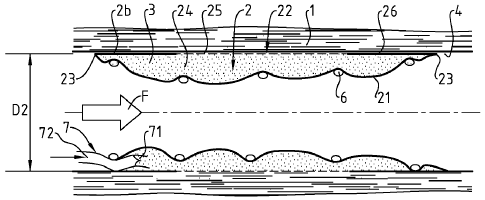
Figures 1 ¨ 4 illustrate a first embodiment for a device according the
invention, this embodiment being adapted to the treatment of vascular defects.
In figures 1, 2 and 4, a blood vessel 1 is illustrated longitudinally cut, and
on
figure 3, transversally cut. A healing device 2 is disposed in the internal
cavity of
said vessel, along a defect site thereof.
CA 02702000 2010-04-08
WO 2009/047300 PCT/EP2008/063550
8
The device 2 of figure 1-4 is of a generally tubular shape. It is expandable,
so
as it may be moved toward the defect site of the vessel, and then expanded so
that its external diameter D2 match with the internal diameter of the vessel.
Of
course, this external diameter may vary all along the device, as a vessel has
neither a constant diameter nor a perfect circular section.
The device comprises two walls 21,22 substantially tubular, one first wall 21,
and one wall 22, respectively internal and external. The two walls are joined
together at their common longitudinal ends, so that a cavity 24 is provided
between the two walls. The cavity is suitable to contain a substance 3 with a
healing agent, preferably under pressure.
The internal wall 21 is tight, so that the substance 3, shown as dots on the
figures, is prevented to leak through the internal wall 21. It can be
biodegradable.
On the contrary, the external wall, in a part 25 in contact with the wounded
portion of the vessel, is a porous portion. This porous portion 25 is designed
to let
the healing agent seep through, to dispense it to the wounded area.
Beyond each longitudinal end of this porous portion 25, at longitudinal ends
of the external wall 22, there are two tight portions 26. As these thigh
portions 26
are provided to be in contact with the wall 4 of the vessel, they prevent
leaking,
between the external wall 22 and the wall 4, of the agent seeping through
porous
portion 25.
As the device 2 is substantially tubular, when in place, it allows blood flow
F
during treatment. Then the intervention is less a trauma for the patient, and
needs
far less time and equipment.
As particularly shown on figure 1, the device 2 can comprise a stent 6 which
can be used to set the radial extension of the device, and along treatment,
maintain the extension of the device. In particular, the stent 6 is designed
to
maintain the external wall 22 substantially in contact with the wall 4 of the
vessel,
even when the cavity is emptying. Regarding the stent used to widen blood
vessels, such a stent 6 can be of a lighter design, as less strong is needed
to
maintain the device 2 than to widen a vessel.
These stents can be auto-expandable, for example, it can be maintained at a
reduced diameter a temperature of the operating room, for example at 20 or
25cC,
CA 02702000 2010-04-08
WO 2009/047300 PCT/EP2008/063550
9
then, using a memory of form, gain a larger diameter, the temperature of the
patient's body, around 37C.
As shown on figure 2, if not auto-expendable, the stent may be first
expanded by a balloon, the remaining resilience of the stent allowing
compensating the emptying of the cavity, by urging internal wall 21 toward
external
wall 22.
When the device is released in place, the cavity is generally empty. The
device, as shown in figure 4, is then provided with means 7 to fill up the
cavity.
Those means comprise a pipe portion 72 to connect a feeding catheter (not
shown) and a valve 71, to prevent leaking of substance, through the pipe
portion
72, after filling of the cavity 3. Optionally, the device can be provided with
a second
filling means which comprises a discharge pipe portion and a discharge valve;
when a sufficient pressure is reached in the cavity 24 the valve is designed
to
open, so that, in particular, an excess quantity of substance is released
through
the discharge pipe.
Figures Sand 6 illustrate a second embodiment, adapted to the healing of flat
wounds, particularly of skin wounds.
In this second embodiment device 2 is substantially flatly shaped. This device
2 also has one first wall 21 and one second wall 22 the two walls being joined
together at their common peripheral portion 26, so that the cavity 24 is
provided
between the two walls. The cavity is suitable to contain a substance with a
healing
agent, preferably under pressure. The first wall 21 is tight, so that the
substance is
prevented to leak through the fist wall 21.
The second wall 22 is intended to be in contact with the flat surface 10,
while
the first wall is also a protection for a wounded area 101 of the flat surface
10
against environmental aggressions. The second wall 22 comprises a porous
portion 25, in contact with the wounded area 101, and designed to let the
healing
agent seep through, to dispense it to the wounded area.
The porous portion 25 is surrounded by a peripheral tight portion 26, said
peripheral portion preferably having an adhesive face to maintain the porous
part
against the wounded area.
CA 02702000 2010-04-08
WO 2009/047300 PCT/EP2008/063550
In the flat arrangement of figure 5, the adhesive portion of the second wall
is
peripheral to the cavity, and its porous portion covers a whole side of the
cavity. In
the flat arrangement of figure 6, the adhesive portion of the second wall is
disposed between the cavity and the flat surface, so that the porous portion
covers
5 only partially this side of the cavity.
Advantageously, the first wall is resilient, so as, when the cavity contains
some substance, the first wall exert a pressure on the substance in the
cavity, the
pressure being sufficient for the substance to peer through the porous
portion.
The device, as shown in figure 5, is provided with means 7 to fill up the
10 cavity. Those means comprise first and second filling means 7A,7B. The
first filling
means 7A comprise a feeding pipe portion 72 to connect a feeding catheter (not
shown) and a valve 71, to prevent leaking of substance, through the pipe
portion
72, after filling of the cavity 24. The second filling means 7B comprise a
discharge
pipe portion 74 and a discharge valve 75. When a sufficient pressure is
reached in
the cavity 24, the valve is designed to open, so that, in particular, an
excess
quantity of substance is released through the discharge pipe 74.
The agent filled up in the cavity is preferably a suspension of cells. The
suspension of cells can also comprise healing compounds. It is particularly
preferred that the cells in suspension are gingival fibroblasts.
Advantageously, gingival fibroblasts have been shown to treat arterial-
remodelling pathologies (WO 2006/013261) and more recently to promote and to
accelerate skin wound healing. Advantageously also, gingival fibroblasts are
easily
sampled and cultured. Besides, gingival fibroblasts possess a high expansion
rate.
Accordingly, gingival fibroblasts provide for an almost limitless source of
autologous fibroblasts.
It is also preferred that the cells used for cell therapy are autologous, that
is
they are taken from the individual to whom they are intended to be
administered.
Preferably the individual is a mammal and more preferably a human. However,
the
cells can also be allogenic, that is taken from another individual of the same
species or heterologous, that is taken from another individual of another
species.
The number of cells in the device should preferably be of from 105 to 109/ml.
The volume of the cavity when it is filled is preferably such that the agent
filled up
CA 02702000 2010-04-08
WO 2009/047300 PCT/EP2008/063550
11
in the cavity is under pressure, so that it tends to migrate in direction of
the defect,
optionally through the porous wall. Preferably, the volume is of from 100 pl
to
20 ml.
The implantation procedure of the device of the invention will be apparent to
one of skill in the art.
For instance, for skin wounds, the porous wall of the device is apposed onto
the wound and maintained in close contact for instance through adhesive means.
As regards, the implantation of the device of the invention in vessels, one
skilled in
the art can for instance follow the general procedure adopted for stent
implantation. Briefly, a sheath is inserted in the femoral artery and then a
wire is
advanced through the aorta. Thereafter, the catheter carrying the device is
advanced over the wire until it reaches the desired site.
It shall be evident to a man skill in the art, that the many arrangements and
embodiments, not precisely set forth, could be practiced under the teachings
of the
present invention, as set forth in the following claims.
For example, the use of a stent is not obligatory required. Thus, the internal
wall may be sufficiently resilient to expend progressively, as the cavity is
emptying,
thus maintaining a sufficient pressure in the cavity to cause the substance to
seep
through the porous portion 25, and to maintain the internal wall 22 against
the wall
4 of the vessel.
Preferably, the stent is made of a resorbable matter, such as polylactic acid.
If not resorbable, the stent may be made of steel or Nitrinol.
Depending on the indications, the second wall can be omitted. Then the
cavity is formed between the tissue and the first tight wall.
In a preferred embodiment, the above-defined device is suitable for a single
use only, i.e. it cannot be re-used a second time when it has already been
used for
cell therapy in an individual.