Note: Descriptions are shown in the official language in which they were submitted.
CA 02702075 2010-04-08
WO 2009/049016
PCT/US2008/079295
ULTRASONIC DEVICE FOR CUTTING AND COAGULATING
Reference to Related Applications
[0001] The present application claims the priority benefit of U.S.
provisional patent
application serial no. 60/978,883, filed on October 10, 2007.
Field of the Invention
[0002] The present invention generally relates to ultrasonic surgical
systems and,
more particularly, to an ultrasonic device that is optimized to allow surgeons
to
perform cutting, coagulation, and fine dissection required in fine and
delicate
surgical procedures such as a thyroidectomy.
Background of the Invention
[0003] Ultrasonic surgical instruments are finding increasingly widespread
applications in surgical procedures by virtue of the unique performance
characteristics of such instruments. Depending upon specific instrument
configurations and operational parameters, ultrasonic surgical instruments
can provide substantially simultaneous cutting of tissue and homeostasis by
coagulation, desirably minimizing patient trauma. The cutting action is
typically effected by an end-effector at the distal end of the instrument,
which
transmits ultrasonic energy to tissue brought into contact with the end-
effector. Ultrasonic instruments of this nature can be configured for open
surgical use, laparoscopic or endoscopic surgical procedures including
robotic-assisted procedures.
[0004] Ultrasonic surgical instruments have been developed that include a
clamp
mechanism to press tissue against the blade of the end-effector in order to
couple ultrasonic energy to the tissue of a patient. Such an arrangement
(sometimes referred to as a clamp coagulator shears or an ultrasonic
transector) is disclosed in U.S. Pat. Nos. 5,322,055; 5,873,873 and 6,325,811.
-1-
CA 02702075 2010-04-08
WO 2009/049016
PCT/US2008/079295
The surgeon activates the clamp arm to press the clamp pad against the
blade by squeezing on the handgrip or handle.
[0005] Some current designs of clamp coagulator shears utilize a foot pedal
to
energize the surgical instrument. The surgeon operates the foot pedal while
simultaneously applying pressure to the handle to press tissue between the
jaw and blade to activate a generator that provides energy that is transmitted
to the cutting blade for cutting and coagulating tissue. Key drawbacks with
this type of instrument activation include the loss of focus on the surgical
field
while the surgeon searches for the foot pedal, the foot pedal getting in the
way of the surgeon's movement during a procedure and surgeon leg fatigue
during long cases.
[0006] Various methods have been disclosed for curved end effector
balancing,
which include repositioning the mass along the end effector. The drawbacks
of such methods are i) high stresses in the curved region, which makes the
end effector more prone to fracture if it comes in contact with metal during
surgery; ii) a shorter active length, which limits the vessel size that can be
operated on, (the active length is defined as the length from the distal end
of
the blade to where the displacement is one half of the displacement at its
distal end); and/or iii) the inability to separately balance orthogonal
displacements.
[0007] Some current designs of clamp coagulator shears utilize handles that
are
either of a pistol or scissors grips design. The scissor grip designs may have
one thumb or finger grip that is immovable and fixed to the housing and one
movable thumb or finger grip. This type of grip may not be entirely familiar
to
surgeons who use other open-type surgical instruments, such as hemostats,
where both thumb and finger grips move in opposition to one another.
Current designs have scissor arms that rotate around a fixed pivot or rotation
point that is perpendicular to the longitudinal axis of the working element.
This approach is limited since the relative motion between the two arms is
-2-
CA 02702075 2010-04-08
WO 2009/049016
PCT/US2008/079295
completely rotational. This feature limits the ability to control the pressure
profile between the two working ends when fully closed.
[0008] Some current designs of clamp coagulator shears are not specifically
designed for delicate procedures where precise dissection, cutting and
coagulation are required. An exemplary procedure is a thyroidectomy where
precise dissection, cutting and coagulation is required to avoid critical
blood
vessels and nerve bundles.
[0009] Some current designs of clamp coagulator shears have an uneven
pressure
profile across the blade with a higher pressure at the proximal end than the
distal end for a given clamp force. An uneven pressure profile can affect the
speed and completeness of tissue transaction, especially at the most distal
tip
of the blade.
[0010] Some current designs of clamp coagulator shears incorporate a spring
in the
handle mechanism to limit the amount of force applied to the blade by the
clamp arm. A disadvantage of the spring limiting force mechanism is an
increase in pad wear. As the device is used, the pad wears and a groove
begins to form in the surface of the pad. This is prevalent in the abuse case
where the device is activated when fully closed but with no tissue present
between the blade and clamp pad. When the groove becomes deeper, a
similar amount of force is placed on the blade due to the spring limit force
feature. Therefore, the slope of the force vs. displacement curve is
relatively
flat.
[0011] It would be desirable to provide an ultrasonic surgical instrument
that
overcomes some of the deficiencies of current instruments. The ultrasonic
surgical instrument described herein overcomes those deficiencies.
-3-
CA 02702075 2015-07-20
,
Brief Summary of the Invention
[0012] An ultrasonic clamp coagulator assembly embodying the principles of
the present
invention is configured to permit selective dissection, cutting, coagulation
and
clamping of tissue during surgical procedures.
[0012A] In one aspect, there is provided an ultrasonic surgical instrument
comprising:
a. a housing having a proximal end and a distal end;
b. a first shroud having a proximal end joined to the housing and a
distal end, the first shroud defining a longitudinal axis, wherein a
inner diameter of the first shroud is from about 0.175 inches to
about 0.22 inches.;
c. a second shroud having a proximal end joined to the distal end of
the first shroud, and aligned with the longitudinal axis, wherein a
inner diameter of the second shroud is from about 0.185 inches to
about 0.20 inches; and
an ultrasonic waveguide positioned within the first and second shroud and
having
a proximal end, a distal end and an ultrasonically actuated blade positioned
at
the distal end of the waveguide.
[0013] A first expression of a first disclosed embodiment is for an
ultrasonic waveguide
including an ultrasonically actuated blade attached to the distal end of the
waveguide; a tissue pad having a tissue engaging surface having a first width
and a second width less than the first width; and a clamp member defining a
distal portion and a proximal portion and moveable with respect to the blade
and
having an open position in which at least a portion of the clamp member is
spaced
from the blade and a closed position in which the clamp member is adjacent to
the
blade for clamping tissue between the tissue pad and the blade.
[0014] A second expression of a first embodiment includes a clamp member
defining a
first dimension and a second dimension in a first plane and the first
dimension is
greater than the second dimension and further defining a first dimension and a
-4-
CA 02702075 2015-07-20
second dimension in a second plane and the first dimension is greater than the
second dimension.
[0015] A first expression of a third embodiment includes a method of
assembling a
sterilized ultrasonic clamp coagulator apparatus including the steps of
providing
an ultrasonic waveguide having a proximal end and a distal end and an
ultrasonically actuated blade attached to the distal end of the waveguide; a
tissue
pad having a flange and a tissue engaging surface having a first width and a
second width less than the first width; a clamp member moveable with respect
to
said end-effector and having an open position in which at least a portion of
the
clamp member is spaced from the blade and a closed position in which the clamp
member is adjacent to the blade for clamping tissue between the tissue pad and
the blade, and where the clamp member includes a slot for
-4a-
CA 02702075 2010-04-08
WO 2009/049016
PCT/US2008/079295
slidably receiving the flange and slidably engaging the flange within the slot
and then sterilizing the clamp coagulator apparatus.
[0016] A first expression of a fourth embodiment of an ultrasonic surgical
instrument
is for a housing configured to accept a transducer and further defining a
longitudinal axis; a first switch positioned on the housing for actuation by
one
or more fingers of a user in a direction parallel to the longitudinal axis and
further electrically connected to a generator for providing an electrical
signal
to the generator for controlling a first level of ultrasonic energy delivered
by
the transducer.
[0017] A second expression of a fourth embodiment of an ultrasonic surgical
instrument is for a second switch positioned on the housing for actuation by
one or more fingers of a user in a direction parallel to the longitudinal axis
and
further electrically connected to a generator for providing an electrical
signal
to the generator for controlling a second level of ultrasonic energy delivered
by the transducer.
[0018] A first expression of a fifth embodiment of an ultrasonic surgical
instrument is
for an ultrasonic waveguide defining a longitudinal axis, having a proximal
end, a most distal node and a distal end and an ultrasonically actuated blade
positioned at the distal end of the waveguide and which defines a functional
asymmetry within a first plane, a first balance asymmetry distal to the most
distal node and proximal to the blade; and a second balance asymmetry
proximal to the most distal node.
[0019] A first expression of a sixth embodiment of an ultrasonic surgical
instrument is
for a housing, an outer shroud having a proximal end joined to the housing, an
ultrasonic waveguide positioned within the outer tube, an ultrasonically
actuated blade positioned at the distal end of the waveguide, and an actuating
lever for operating a clamp arm located at the distal end of the lever. The
actuating lever has camming members, which operatively engage the outer
-5-
CA 02702075 2010-04-08
WO 2009/049016
PCT/US2008/079295
tube such that movement of the actuating lever positions the clamp arm
between open and clamped positions relative to the blade.
[0020] A second expression of a sixth embodiment of an ultrasonic
instrument is for
stationary finger ring that defines an opening having a length L and the
housing and a transducer are sized to position a center of gravity of the
surgical instrument at the housing within the dimension of length L.
[0021] A first expression of a first embodiment of a torque wrench for use
with an
ultrasonic clamp coagulator apparatus is for a hand wrench body, a cantilever
arm movably attached to said wrench body, at least one tooth located at the
cantilever arm's distal end, and an adaptor rotatably attached to the hand
wrench and comprising a cam for operatively engaging the tooth.
Brief Description of the Figures
[0022] The novel features of the invention are set forth with particularity
in the
appended claims. The invention itself, however, both as to organization and
methods of operation, may best be understood by reference to the following
description, taken in conjunction with the accompanying drawings in which:
[0023] FIG. 1 is a perspective view illustrating an embodiment of an
ultrasonic
surgical instrument in accordance with the present invention;
[0024] FIG. 2 is a perspective assembly view of Fig. 1;
[0025] FIG. 3A is a perspective view of one embodiment of a waveguide and
blade in
accordance with the present invention;
[0026] FIG. 3B is an elevation view of the waveguide and blade of Fig. 3A;
[0027] FIG. 3C is an elevation view of an alternate embodiment of a
waveguide and
blade in accordance with the present invention;
[0028] FIG. 3D is an elevation view of an alternate embodiment of a
waveguide and
blade in accordance with the present invention;
-6-
CA 02702075 2010-04-08
WO 2009/049016
PCT/US2008/079295
[0029] FIG 3E is an elevation view of an alternate embodiment of a
waveguide and
blade in accordance with the present invention;
[0030] FIG 3F is an alternate view of the embodiment of the waveguide and
blade of
FIG. 3E;
[0031] FIG. 3G is an elevation view of an alternate embodiment of a
waveguide and
blade in accordance with the present invention;
[0032] FIG. 4 is a graph illustrating the displacement profile of the
present invention
and the prior art;
[0033] FIG. 5 is a graph illustrating an alternate displacement profile of
the present
invention and the prior art;
[0034] FIG. 6A is an elevation view of the waveguide and blade of FIGS. 3E-
F
illustrating one embodiment of the radius of curvature of the blade;
[0035] FIG. 6B is an exploded view of one embodiment of the blade of FIG.
6A and a
radius cut;
[0036] FIG. 6C is an alternate view of the embodiment of FIG. 6A;
[0037] FIG. 6D is a section view of the embodiment of FIG. 6B;
[0038] Fig. 7A is an elevation view of an end effector in accordance with
the present
invention;
[0039] FIG. 7B is a plan view of the end effector of FIG. 7A;
[0040] FIG. 8 is a perspective view from proximal to distal end of a clamp
member in
accordance with the present invention;
[0041] FIG. 9A is a plan view of a tissue pad in accordance with the
present
invention;
[0042] FIG. 9B is a plan view of the opposite face of the tissue pad of
FIG. 9A;
[0043] FIG. 9C is an elevation view the tissue pad of FIGS. 9A-B;
-7-
CA 02702075 2010-04-08
WO 2009/049016
PCT/US2008/079295
[0044] FIG. 10A is a perspective view of an alternate expression of the
clamp
member;
[0045] FIG. 10B is a perspective view of the clamp member of FIG. 10A and a
first
tissue pad;
[0046] FIG. 10C is a perspective view of the clamp member of FIG. 10A and a
first
and second tissue pad;
[0047] FIG. 11A-B are an alternate expressions for a first and second
tissue pad;
[0048] FIG. 11C is a perspective view of an alternate expression of a clamp
arm for
use with the tissue pads of FIGS. 11A-B;
[0049] FIG. 11D is an alternate view of the clamp are of FIG. 11C;
[0050] FIG. 11E is a cut-away view of an assembled clamp arm and tissue pad
assembly of FIGS. 11A-D
[0051] FIG. 12A is a perspective view of an alternate embodiment of a clamp
arm
having a distal connection point;
[0052] FIG. 12B is a perspective view of an alternate embodiment of a
tissue pad
having a distal connection member;
[0053] FIG. 12C is a perspective view of an assembled clamp arm and tissue
pad of
FIGS. 12A-B;
[0054] FIG. 13 is a partial view of the distal end of the ultrasonic
instrument in
accordance with the present invention;
[0055] FIG. 14 is an exploded elevation view of one part of the clamp arm
and clamp
member and cam members;
[0056] FIG. 15A is an exploded view of the outer shroud and cam slots;
[0057] FIG. 15B is a cut away view of one embodiment of the outer shroud;
[0058] FIG. 15C is a cut away view of one embodiment of the distal shroud;
-8-
CA 02702075 2010-04-08
WO 2009/049016
PCT/US2008/079295
[0059] FIG. 16A is an elevation view of an ultrasonic instrument and
pushbutton
assembly in accordance with the present invention;
[0060] FIG. 16B is an elevation view of the two piece assembly of a push
button in
accordance with the present invention;
[0061] FIG. 16C is a cut-away elevation view showing the interface among
the switch
housing, transducer, waveguide and housing;
[0062] FIG. 16D is a perspective elevation view of a switch housing in
accordance
with the present invention;
[0063] FIG. 16E is an alternate view of the switch housing of FIG. 16D;
[0064] FIG. 16F is a view of a flex circuit in accordance with the present
invention;
[0065] FIG. 16G is an electrical schematic of the hand switch circuit;
[0066] FIG. 16H is an alternate embodiment of the switch housing and
actuation;
[0067] FIG. 17A is an elevation view of an ultrasonic instrument in
accordance with
the present invention as may be grasped by a user;
[0068] FIG. 17B is an exploded view of the finger and thumb interface of a
ultrasonic
instrument in accordance with the present invention;
[0069] FIG. 18 is an elevation view of an ultrasonic instrument in
accordance with the
present invention as may be grasped by a user and defining a center of
gravity;
[0070] FIG. 19A is a perspective view of a two-piece torque wrench in
accordance
with the present invention;
[0071] FIG. 19B is a perspective view of a hand wrench in accordance with
the
present invention;
[0072] FIG. 19C is an elevation view of the hand wrench of FIG. 19B;
-9-
CA 02702075 2010-04-08
WO 2009/049016
PCT/US2008/079295
[0073] FIG. 19D is a cross sectional end view of the distal end of a hand
wrench
depicting cantilever arm and teeth geometry;
[0074] FIG. 19E is a cross sectional view of an adaptor depicting spline
gear
geometry;
[0075] FIG. 19F is a perspective view of an adaptor for use with a hand
wrench in
accordance with the present invention;
[0076] FIG. 19G is a partial perspective view of a hand wrench interfacing
with an
ultrasonic instrument in accordance with the present invention;
[0077] FIG. 20 is a graph illustrating the pressure profile across the
blade in
accordance with the present invention relative to the prior art;
[0078] Fig. 21 is a graphical illustration of the pivot radius in
accordance with the
present invention; and
[0079] FIG. 22 is a graph illustrating thumb ring displacement vs. thumb
ring force in
accordance with one embodiment of the present invention.
Detailed Description of the Invention
[0080] Before explaining the present invention in detail, it should be
noted that the
invention is not limited in its application or use to the details of
construction
and arrangement of parts illustrated in the accompanying drawings and
description. The illustrative embodiments of the invention may be
implemented or incorporated in other embodiments, variations and
modifications, and may be practiced or carried out in various ways. Further,
unless otherwise indicated, the terms and expressions employed herein have
been chosen for the purpose of describing the illustrative embodiments of the
present invention for the convenience of the reader and are not for the
purpose of limiting the invention.
-10-
CA 02702075 2010-04-08
WO 2009/049016
PCT/US2008/079295
[0081] Further, it is understood that any one or more of the following-
described
embodiments, expressions of embodiments, examples, etc. can be combined
with any one or more of the other following-described embodiments,
expressions of embodiments, examples, etc.
[0082] The present invention is particularly directed to an improved
ultrasonic
surgical clamp coagulator apparatus which is configured for effecting tissue
cutting, coagulation, and/or clamping during surgical procedures, including
delicate surgical procedures, such as a thyroidectomy. The present apparatus
is configured for use in open surgical procedures. Versatile use is
facilitated
by selective use of ultrasonic energy. When ultrasonic components of the
apparatus are inactive, tissue can be readily gripped and manipulated, as
desired, without tissue cutting or damage. When the ultrasonic components
are activated, the apparatus permits tissue to be gripped for coupling with
the
ultrasonic energy to effect tissue coagulation, with application of increased
pressure efficiently effecting tissue cutting and coagulation. If desired,
ultrasonic energy can be applied to tissue without use of the clamping
mechanism of the apparatus by appropriate manipulation of the ultrasonic
blade.
[0083] As will become apparent from the following description, the present
clamp
coagulator apparatus is particularly configured for disposable use by virtue
of
its straightforward construction. As such, it is contemplated that the
apparatus be used in association with an ultrasonic generator unit of a
surgical system, whereby ultrasonic energy from the generator unit provides
the desired ultrasonic actuation for the present clamp coagulator apparatus.
It
will be appreciated that a clamp coagulator apparatus embodying the
principles of the present invention can be configured for non-disposable or
multiple use, and non-detachably integrated with an associated ultrasonic
generator unit. However, detachable connection of the present clamp
coagulator apparatus with an associated ultrasonic generator unit is presently
preferred for single-patient use of the apparatus.
-11-
CA 02702075 2015-07-20
[0084] With specific reference now to Figs. 1 and 2, an embodiment of a
surgical
system 19, including an ultrasonic surgical instrument 100 in accordance with
the present invention is illustrated. The surgical system 19 includes an
ultrasonic generator 30 connected to an ultrasonic transducer 50 via cable 22,
and an ultrasonic surgical instrument 100. It will be noted that, in some
applications, the ultrasonic transducer 50 is referred to as a "hand piece
assembly" because the surgical instrument of the surgical system 19 is
configured such that a surgeon may grasp and manipulate the ultrasonic
transducer 50 during various procedures and operations. A suitable
generator is the GEN04 (also referred to as Generator 300) sold by Ethicon
Endo-Surgery, Inc. of Cincinnati, Ohio. A suitable transducer is disclosed in
co-pending U.S. patent application filed on October 10, 2006, serial no.
11/545,784, entitled MEDICAL ULTRASOUND SYSTEM AND HANDPIECE
AND METHODS FOR MAKING AND TUNING.
[0085] Ultrasonic transducer 50, and an ultrasonic waveguide 80 together
provide an
acoustic assembly of the present surgical system 19, with the acoustic
assembly providing ultrasonic energy for surgical procedures when powered
by generator 30. The acoustic assembly of surgical instrument 100 generally
includes a first acoustic portion and a second acoustic portion. In the
present
embodiment, the first acoustic portion comprises the ultrasonically active
portions of ultrasonic transducer 50, and the second acoustic portion
comprises the ultrasonically active portions of transmission assembly 71.
Further, in the present embodiment, the distal end of the first acoustic
portion
is operatively coupled to the proximal end of the second acoustic portion by,
for example, a threaded connection.
[0086] The ultrasonic surgical instrument 100 includes a multi-piece handle
assembly
68 adapted to isolate the operator from the vibrations of the acoustic
assembly contained within transducer 50. The handle assembly 68 can be
shaped to be held by a user in a conventional manner, but it is contemplated
-12-
CA 02702075 2010-04-08
WO 2009/049016
PCT/US2008/079295
that the present ultrasonic surgical instrument 100 principally be grasped and
manipulated in a scissor-like arrangement provided by a handle assembly of
the instrument, as will be described. While multi-piece handle assembly 68 is
illustrated, the handle assembly 68 may comprise a single or unitary
component. The proximal end of the ultrasonic surgical instrument 100
receives and is fitted to the distal end of the ultrasonic transducer 50 by
insertion of the transducer into the handle assembly 68. The ultrasonic
surgical instrument 100 may be attached to and removed from the ultrasonic
transducer 50 as a unit. The ultrasonic surgical instrument 100 may include a
handle assembly 68, comprising mating housing portions 69 and 70 and an
ultrasonic transmission assembly 71. The elongated transmission assembly
71 of the ultrasonic surgical instrument 100 extends orthogonally from the
instrument handle assembly 68.
[0087] The handle assembly 68 may be constructed from a durable plastic,
such as
polycarbonate or a liquid crystal polymer. It is also contemplated that the
handle assembly 68 may alternatively be made from a variety of materials
including other plastics, ceramics or metals. Traditional unfilled
thermoplastics, however, have a thermal conductivity of only about 0.20
W/m K (Watt/ meter- Kelvin). In order to improve heat dissipation from the
instrument, the handle assembly may be constructed from heat conducting
thermoplastics, such as high heat resistant resins liquid crystal polymer
(LCP), Polyphenylene Sulfide (PPS), Polyetheretherketone (PEEK) and
Polysulfone having thermal conductivity in the range of 20-100 W/m K.
PEEK resin is a thermoplastics filled with aluminum nitride or boron nitride,
which are not electrically conductive. The thermally conductive resin helps to
manage the heat within smaller instruments.
[0088] The transmission assembly 71 includes a waveguide 80 and a blade 79.
It
will be noted that, in some applications, the transmission assembly is
sometimes referred to as a "blade assembly". The waveguide 80, which is
adapted to transmit ultrasonic energy from transducer 50 to the tip of blade
79
-13-
CA 02702075 2010-04-08
WO 2009/049016
PCT/US2008/079295
may be flexible, semi-flexible or rigid. The waveguide 80 may also be
configured to amplify the mechanical vibrations transmitted through the
waveguide 80 to the blade 79 as is well known in the art. The waveguide 80
may further have features to control the gain of the longitudinal vibration
along
the waveguide 80 and features to tune the waveguide 80 to the resonant
frequency of the system. In particular, waveguide 80 may have any suitable
cross-sectional dimension. For example, the waveguide 80 may have a
substantially uniform cross-section or the waveguide 80 may be tapered at
various sections or may be tapered along its entire length.
[0089] Ultrasonic waveguide 80 may, for example, have a length
substantially equal
to an integral number of one-half system wavelengths (n2J2). The ultrasonic
waveguide 80 and blade 79 may be preferably fabricated from a solid core
shaft constructed out of material, which propagates ultrasonic energy
efficiently, such as titanium alloy (i.e., Ti-6A1-4V), aluminum alloys,
sapphire,
stainless steel or any other acoustically compatible material.
[0090] Ultrasonic waveguide 80 may further include at least one radial hole
or
aperture 66 extending therethrough, substantially perpendicular to the
longitudinal axis of the waveguide 80. The aperture 66, which may be
positioned at a node, is configured to receive a connector pin 27, discussed
below, which connects the waveguide 80, to the handle assembly 70.
[0091] Blade 79 may be integral with the waveguide 80 and formed as a
single unit.
In an alternate expression of the current embodiment, blade 79 may be
connected by a threaded connection, a welded joint, or other coupling
mechanisms. The distal end of the blade 79 is disposed near an anti-node 85
in order to tune the acoustic assembly to a preferred resonant frequency fo
when the acoustic assembly is not loaded by tissue. When ultrasonic
transducer 50 is energized, the distal end of blade 79 or blade tip 79a is
configured to move substantially longitudinally (along the x axis) in the
range
of, for example, approximately 10 to 500 microns peak-to-peak, and
preferably in the range of about 20 to about 200 microns at a predetermined
-14-
CA 02702075 2010-04-08
WO 2009/049016
PCT/US2008/079295
vibrational frequency fo of, for example, 55,500 Hz. Blade tip 79a also
preferably vibrates in the y axis at about 1 to about 10 percent of the motion
in
the x axis.
[0092] The blade tip 79a provides a functional asymmetry or curved portion
for
improved visibility at the blade tip so that a surgeon can verify that the
blade
79 extends across the structure being cut or coagulated. This is especially
important in verifying margins for large blood vessels. The geometry also
provides for improved tissue access by more closely replicating the curvature
of biological structures. Blade 79 provides a multitude of edges and surfaces,
designed to provide a multitude of tissue effects: clamped coagulation,
clamped cutting, grasping, back-cutting, dissection, spot coagulation, tip
penetration and tip scoring.
[0093] Blade tip 79a is commonly referred to as a functional asymmetry.
That is, the
blade (functionally, the blade provides a multitude of tissue effects) lies
outside the longitudinal axis of waveguide 80 (that is, asymmetrical with the
longitudinal axis), and accordingly creates an imbalance in the ultrasonic
waveguide. If the imbalance is not corrected, then undesirable heat, noise,
and compromised tissue effect occur.
[0094] It is possible to minimize unwanted tip excursion in the y and z
axes, and
therefore maximize efficiency with improved tissue effect, by providing one or
more balance asymmetries or balancing features proximal to the blade
functional asymmetry.
[0095] Referring now to Figs. 3A-G, transmission assembly 71 includes one
or more
balancing features placed at blade 79, at a position proximal and/or distal to
the distal most node 84. In addition, the balancing features at the waveguide
80 are shaped to balance the two orthogonal modes in the y and z axes,
separately. The size and shape and location of the balance features allow
flexibility to reduce stress at the blade 79, make the active length longer
and
separately balance the two orthogonal modes.
-15-
CA 02702075 2010-04-08
WO 2009/049016
PCT/US2008/079295
[0096] Figures 3A-B show a single balance cut 82 at the waveguide 80 distal
to node
84. In this embodiment balance cut 82 has side walls perpendicular to the
longitudinal axis of waveguide 80 and the bottom cut is parallel to the
longitudinal axis of waveguide 80. In this embodiment the high stresses
experienced during operation are localized at the balancing cut 82, which is
away from the more sensitive curved region at the blade 79.
[0097] Figure 3C shows two balancing features 82 and 82a, one distal and
one
proximal to the node 84. Adding second balance cut 82a, proximal to node 84
further eliminates the orthogonal bending modes thereby providing a more
pure longitudinal motion (x direction) and removing the overlapping bending
modes (y and z direction). Accordingly, the blade 79 is better balanced and
has a longer active length.
[0098] Figure 3D shows two balancing features 82c and 82a, distal and
proximal to
the node 84. An angled bottom cut at balance feature 82c allows individual
balancing of the bending mode in the z direction.
[0099] Figures 3E-F show two balancing features 82 and 82d, distal and
proximal to
the node 84. The side walls of balance feature 82d are angled with respect to
each other in the x-z plane and provide for individual balancing of the
bending
mode in the y direction. The angled side walls define an included angle 0 of
between 1 and about 90 , preferably between about 15 and about 25 , and
more preferably between about 190 and about 210. The weight removed at
each balance feature is a function of multiple parameters including the radius
of curvature at blade tip 79a and the desired level of removal of the
overlapping bending modes in the y and z direction. In an illustrative
example, the balance cut 82 represents a weight reduction of about 0.003 to
about 0.004 oz., and most preferably about 0.0034 oz. The balance cut 82d
represents a weight reduction of about 0.004 to about 0.005 oz., and most
preferably about 0.0043 oz.
-16-
CA 02702075 2010-04-08
WO 2009/049016
PCT/US2008/079295
[00100] Figure 3G shows one balance cut 82e in the curved blade region in
addition
to balance feature 82, distal to node 84. Balance cut 82e allows for balancing
as well as improved acoustic performance as a result of wide frequency
separation of transverse modes from the fundamental frequency, which is the
longitudinal mode frequency.
[00101] As would be apparent to one skilled in the art, any combination of
balance
cuts 82 through 82e are possible to provide balancing of a waveguide and
curved blade.
[00102] Figure 4 shows that the profile produced by the balancing cut features
of Fig.
3E produces a 1.3 mm longer active length along the longitudinal
displacement direction than is available from an LCS-05 ultrasonic clamp
coagulator, sold by Ethicon Endo-Surgery, Inc. (where the y axis is
representative of the ratio between the displacement anywhere along blade
tip 79a and the displacement at the most distal end of blade tip 79a). A
longer
active length is desirable for cutting and coagulating large vessels, for
example, 5-7mm vessels.
[00103] Figure 5 shows that the profile produced by the balancing features of
Fig. 3E
produces a 2.5 mm longer active length (along the vector sum of
displacements in the x, y and z directions) than is available from an LCS-05
ultrasonic clamp coagulator, which is desirable for cutting and coagulating
large vessels, for example, 5-7mm vessels.
[00104] Referring back to Figs. 1 and 2 an outer tubular member or outer
shroud 72
attaches to the most proximal end of handle assembly 70. Attached to the
distal end of the outer shroud 72 is a distal shroud 76. Both the outer shroud
72 and distal shroud 76 may attach via a snap fit, press fit, glue or other
mechanical means. Extending distally from the distal shroud 76 is the end-
effector 81, which comprises the blade 79 and clamp member 56, also
commonly referred to as a jaw, in combination with one or more tissue pads
58. A seal 83 may be provided at the distal-most node 84, nearest the end-
-17-
CA 02702075 2010-04-08
WO 2009/049016
PCT/US2008/079295
effector 81, to abate passage of tissue, blood, and other material in the
region
between the waveguide 80 and the distal shroud 76. Seal 83 may be of any
known construction, such as an o-ring or silicon overmolded at node 84.
[00105] Referring now to Figs. 6A-D and 7A-B, blade 79 is curved along with
the
associated clamp member 56. This is illustrative only, and blade 79 and a
corresponding clamp member 56 may be of any shape as is known to the
skilled artisan. One benefit of the invention, however, is the ability to
perform
finer, more delicate surgical procedures. It is also multifunctional and able
to
dissect tissue as well as coagulate and transect.
[00106] The ability to finely dissect is enabled primarily by the tapering of
the end
effector 81. The end effector is tapered in two planes, which mimics typical
hemostats. This allows the user to create windows in the tissue and then
spread the tissue apart more easily. The blade 79 and clamp member 56 are
tapered in both the x and z directions from the proximal end to the distal
end.
The pad 58 is only tapered in the Z direction. That is, the clamp pad 58 has a
constant thickness, but the width of the clamp pad 58 at the distal end is
less
than the width at the proximal end. Accordingly, the surface area of section A
is greater than the surface area of section B.
[00107] In addition to the taper, the radius at the distal end of the blade 79
and clamp
member 56 also promotes fine dissection. The radius at the tip of the clamp
member 56 is approximately 0.040 inches, and the blade radius is
approximately 0.045 inches.
[00108] With specific reference to Fig. 6A, blade 79 is defined by an inside
radius R1
and an outside radius R2 measured at a distance D1 from the longitundinal
axis. The dimensions R1, R2 and D1 are selected in combination with the
balance cuts previously discussed. In one embodiment R1 is from about 0.80
inches to about 1.00 inches and most preferably about 0.95 inches; R2 is from
about 0.90 inches to about 1.10 inches and most preferably about 1.04
-18-
CA 02702075 2010-04-08
WO 2009/049016
PCT/US2008/079295
inches; and D1 is from about 0.90 inches to about 1.10 inches and most
preferably about 0.99 inches.
[00109] Figs. 6B and 6D further illustrate a second expression of the blade
79.
Illustrated is a radius cut 90 in blade 79 to provide two back cutting edges
92
and 92a. As will be appreciated by the skilled artisian, radius cut 90 also
provides a balance asymmetry within the functional symmetry to help balance
the orthogonal modes. The back cutting edges 92 and 92a are positioned
opposite the clamp pad 58 (Fig. 7B) to allow the surgeon to perform tissue
cutting procedures without the assistance of the clamp pad 58. Preferably,
the radius cut is distal to the most distal tip of blade 79 to allow for a
blunt
radius tip for tissue dissection as discussed above. In one example of the
second expression of blade 79, a radius cut R3 is swept across an angle (I)
measured at a distance D2 from the longitudinal axis and starting a distance
D3 from the distal tip of blade 79. In one embodiment R3 is from about 0.030
inches to about 0.060 inches and most preferably about 0.050 inches; angle
(I) is from about 20 to about 35 and most preferably about 30'; D2 is about
0.90 inches to about 1.10 inches and most preferably about 0.99 inches; and
D3 is from about 0.085 inches to about 0.11 inches and most preferably about
0.09 inches.
[00110] In a third expression of blade 79, Fig. 6C illustrates a taper defined
by angle 0
relative to an axis parallel to the longitudinal axis of waveguide 80 from the
proximal end of blade 79 to the distal end of blade 79. In one embodiment the
taper may be on the blade surface that contacts tissue pad 58 (Fig. 7A).
Alternatively, the taper may be the defined by the opposite surface comprising
radius cut 90. Referring to Fig. 6C, angle 0 ranges from about 0.5 to about
, and preferably from about 1.5 to about 2
[00111] Referring now to Fig. 15B, due to the unique curvature of the blade
tip 79a, it
is preferable that the sheath covering the blade 79 is made in two pieces,
outer shroud 72 and distal shroud 76, in order to maintain preferred inner
dimensions of the distal shroud 76. The length of the outer shroud 72 and
-19-
CA 02702075 2010-04-08
WO 2009/049016
PCT/US2008/079295
distal shroud 76 are critical because they allow passage of the blade during
assembly. Preferably the inner diameter D1 of the outer shroud 72 is from
about 0.175 inches to about 0.22 inches and most preferably about 0.197
inches. The length of the outer shroud 76 is between about 1.5 inches to
about 2.4 inches. With a preferred inner diameter of about 0.197 inches, the
maximum length of outer shroud 72 is about 2.311 inches.
[00112] Referring now to Fig. 15C, the distal shroud 76 has a critical inner
diameter
D2 of about 0.185 inches to about 0.20 inches and most preferably about
0.191 inches. This diameter mates with the overmold 84 of the blade 79.
This interaction between the overmold 84 of the blade and the inner diameter
of the distal shroud 76 performs two critical functions. First, the tight
tolerance
isolates the vibrating blade from the distal shroud 76 to avoid metal to metal
contact. Second, the tight tolerance provides stiffness to the blade system.
Stiffness of the blade system is critical in maintaining the appropriate clamp
force of the instrument.
[00113] During assembly the outer shroud 72 is passed over the blade 79 and
then
the distal shroud 76 is passed over the curved portion of the blade 79a. The
length of the distal shroud 76 allows the blade 79 to pass through the distal
shroud 76. In order to accommodate a preferred embodiment of the blade 79
to pass through the inner diameter of the distal shroud 76, the length of
distal
shroud 76 is preferably from about 0.600 inches to about 0.650 inches, and
most preferably 0.616 inches. Once the distal shroud 76 is pressed fit onto
the outer shroud 72 (or other attachment means, such as glue or mechanical
fastering) the overmold of the blade 84 is sercured within the 0.191" inner
diameter.
[00114] Referring back to Fig. 2, waveguide 80 is positioned within cavity 59
of handle
assembly 68. In order to properly locate the waveguide 80 both axially and
radially, pin 27 extends through opening 66 of waveguide 80 (located at a
node) and engages channel 28 (formed by the mating of housing portions 69
and 70). Preferably pin 27 is made of any compatible metal, such as stainless
-20-
CA 02702075 2010-04-08
WO 2009/049016
PCT/US2008/079295
steel or titanium or a durable plastic, such as polycarbonate or a liquid
crystal
polymer. In a first expression of one embodiment, pin 27 is partially coated
with an elasto-meric material 30, such as silicon for that portion 29 of pin
27
that extends through waveguide 80 and uncoated for that portion of pin 27
that engages members 69 and 70. The silicone provides insulation from the
vibrating blade throughout the length of hole 66. This enables high efficiency
operation whereby minimal overheating is generated and maximum ultrasonic
output power is available at the blade tip for cutting and coagulation. The
lack
of insulation allows pin 27 to be held firmly within handle assembly 68 due to
the lack of insulation, which would provide deformation and movement if pin
27 were completely coated with an insulating material.
[00115] Referring now to Figs. 8 and 9A-C a first expression of clamp member
56 has
a shaped slot 57 for accepting one or more tissue pads. This configuration
prevents mis-loading of the tissue pads and assures that the appropriate pad
is loaded at the correct location within clamp member 56. For example clamp
member 56 may comprise a T-shaped slot 57 to accept a T-shaped flange 55
of clamp pad 58. Two mechanical stops 59 and 59a, when depressed,
engage the proximal end of clamp pad 58 to secure the clamp pad within
clamp member 56. As would be appreciated by those skilled in the art,
flanges and corresponding slots may have alternate shapes and sizes to
secure the clamp pads to the clamp arm. The illustrated flange configurations
shown are exemplary only and accommodate the particular clamp pad
material of one embodiment, but the particular size and shape of the flange
may vary, including, but not limited to, flanges of the same size and shape.
For unitary tissue pads, the flange may be of one configuration. Further,
other
tab stops are possible and may include any of the multiple methods of
mechanically attaching the clamp pads to the clamp arm, such as rivets, glue,
press fit or any other fastening means well know to the artisan.
[00116] Referring to Figs. 10A-C, in a first expression of an alternate
embodiment,
clamp pad 58 consists of a first tissue pad 58b and a second pad portion 58a,
-21-
CA 02702075 2010-04-08
WO 2009/049016
PCT/US2008/079295
which may be an insert within pad 58b. Tissue pad 58b may comprise a
tissue engaging surface having saw tooth-like teeth and proximal portion 58a
may have a smoother surface relative to pad 58b. The advantage of two
separate components 58a and 58b is that each pad may be constructed from
different materials. For example, having a two-piece tissue pad allows the
use of a very lubricious material at the distal end that is not particularly
resistant to high temperatures compared to a very high temperature material
at the proximal end that is not particularly lubricious because the proximal
end
is an area of lower amplitude. Such a configuration matches the tissue pad
materials to the amplitude of the blade 79.
[00117] In a second expression of an alternate embodiment of the present
invention,
clamp pad 58b is formed from TEFLON or any other suitable low-friction
material. Clamp pad 58a is formed from a base material and at least one filler
material, which is a different material from the base material. The surface of
proximal clamp pad 58a may be smoother than distal clamp pad 58b, or
proximal clamp pad 58a may also have a similar type saw-tooth configuration.
[00118] Several benefits and advantages are obtained from one or more of the
expressions of the invention. Having a tissue pad with a base material and at-
least-one filler material allows the base material and the at-least-one filler
material to be chosen with a different hardness, stiffness, lubricity, dynamic
coefficient of friction, heat transfer coefficient, abradability, heat
deflection
temperature, glass transition temperature and/or melt temperature to improve
the wearability of the tissue pad, which is important when high clamping
forces are employed because tissue pads wear faster at higher clamping
forces than at lower clamping forces. In experiments, a 15% graphite-filled
polytetrafluoroethylene tissue pad showed substantially the same wear with a
7 pound clamping force as a 100% polytetrafluoroethylene tissue pad showed
with a 1.5 pound clamping force. Having a flexible clamping arm and/or a
flexible tissue pad should also improve the wearability of the tissue pad due
to
the ability of the flexible member to more evenly distribute the load across
the
-22-
CA 02702075 2010-04-08
WO 2009/049016
PCT/US2008/079295
entire surface of the tissue pad. Further benefits and expressions of this
embodiment are disclosed in United States provisional patent application,
serial number 60/548,301, filed on February 27, 2004 and commonly
assigned to the assignee of the present application.
[00119] In a third expression of an alternate embodiment, a tissue pad with a
base
material and at least two filler materials allows the base material and the at-
least-two filler materials to be chosen with a different hardness, stiffness,
lubricity, dynamic coefficient of friction, heat transfer coefficient,
abradability,
heat deflection temperature, and/or melt temperature to improve the
wearability of the tissue pad, which is important when high clamping forces
are employed because tissue pads wear faster at higher clamping forces than
at lower clamping forces. In experiments, a 15% graphite-filled, 30% PTFE-
filled polyimide tissue pad showed substantially the same or better wear with
a 4.5 pound clamping force as a 100% polytetrafluoroethylene tissue pad
showed with a 1.5 pound clamping force. The advantage of a 15% graphite-
filled, 30% PTFE-filled polyimide tissue pad is increased heat resistance,
which improves the overall wear resistance of the tissue pad. This polyimide-
composite clamp pad has a useful heat resistance up about 800 F to about
1200 F, as compared to a useful heat resistance up to about 660 F of a PTFE
clamp pad. Alternatively, Other materials are also useful for a portion of the
tissue pad, such as ceramics, metals, glasses and graphite.
[00120] Figs. 10A-C disclose a first expression of an embodiment of attaching
a two
part clamp pad 58a-b to a clamp member 56. In fig. 10A, at least two slots
57a and 57b are shaped to accept two correspondingly shaped flanges 55a
and 55'. In this example, T-slot 57a accepts a corresponding T-flange 55a of
clamp pad 58a, and wedge-shaped slot 57' accepts a corresponding wedge-
shaped flange 55' of clamp pad 58b.
[00121] Figs. 11A-E illustrate a second expression of attaching a clamp pad
58c to a
clamp arm 56c. Clamp pad 58c comprises one or more protrusions 62 for
insertion into one or more corresponding apertures 63 in clamp arm 56c. If a
-23-
CA 02702075 2010-04-08
WO 2009/049016
PCT/US2008/079295
second or more clamp pad(s) 58d is also used in accordance with the
previous discussion, then clamp pad 58c further comprises corresponding
aperture 61 for accepting one or more clamp pad(s) 58d. Clamp arm 56c has
corresponding aperture(s) 63 for accepting protrusions 62, as well as a
corresponding cavity 64 for accepting the one or more clamp pad 58d. Fig.
11E illustrates the components assembled together prior to staking. Clamp
pad 58d fits inside the aperture 61 and cavity 64, and pad 58c is aligned with
clamp arm 56c so that protrusions 62 align with chamfered aperture 63.
Protrusions 62 have additional height beyond the top surface of clamp arm
56c to provide additional material to fill the chamfered volume during
staking.
Heat is applied to protrusions 62 above the clamp arm 56c; the protrusions
deform and take the shape of the chamfered volume.
[00122] Figs. 12A-C illustrate a third expression of attaching a clamp pad 58d
to a
clamp arm 56d. In addition to a T-shaped flange 55, clamp pad 58d further
comprises a hook-like protrusion or clip 65 for attaching to a corresponding
opening 66 at the distal tip of clamp arm 56d. In this expression, the distal
tip
of clamp arm 56d is open and the clamp pad 58d is inserted from the distal to
proximal direction until the hook clip engages opening 66. Hook clip 65 may
be biased closed so when clip 65 engages opening 66, clip 65 applies
compressive forces against opening 66.
[00123] A first expression for a method for inserting a clamp pad on a clamp
arm
includes a) inserting a first clamp pad having a first width dimension greater
than a second width dimension and having a first-shaped flange into a clamp
arm having a slot that accepts the first-shaped flange; and b) engaging a pad
stop to secure the clamp pad within the clamp arm. In a second expression of
the method, the clamp pad consists of a second clamp pad fabricated from a
base material and at least one filler material, which is a different material
from
the base material. The second clamp pad may have a second-shaped flange
for engaging a second-shaped slot on the clamp arm. The tissue surfaces of
-24-
CA 02702075 2010-04-08
WO 2009/049016
PCT/US2008/079295
the clamp pads may be smooth or have tissue gripping features, such as a
saw-tooth configuration.
[00124] A first expression for a method for replacing clamp pads would include
the
steps of: a) disengaging a pad stop; b) removing a first clamp pad from the
clamp arm; c) removing a second clamp pad from the clamp arm, wherein at
least one of the first or second clamp pads has a first width dimension
greater
than a second width dimension; d) inserting third and fourth clamp pads into
the clamp arm wherein at least one of the third or fourth clamp pads has a
first
width dimension greater than a second width dimension ; and e) engaging a
pad stop to secure the third and fourth clamp pads within the clamp arm. In a
second expression of this method one of the third and fourth clamp pads may
be fabricated from a polymeric material such as TEFLON, and the other
clamp pad may be fabricated from a base material and at least one filler
material, which is a different material from the base material. The tissue
surfaces of the clamp pads may be smooth or have tissue gripping features,
such as a saw-tooth configuration.
[00125] Referring to Figs. 13-15, a clamp arm 60 is configured for use with
the present
ultrasonic surgical instrument 100 and for cooperative action with blade 79
and clamp member 56. The clamp arm 60 is rotatably mounted to the distal
end of outer shroud 72, detailed below, and connectably attaches at the distal
end of thumb ring or actuation member 34. Clamp pad 58 mounts on the
clamp member 56 for cooperation with blade 79, with rotational movement of
the clamp arm 60 positioning the clamp pad in substantially parallel
relationship to, and in contact with, blade79, thereby defining a tissue
treatment region. By this construction, tissue is grasped between clamp pad
58 and blade 79. Pivotal movement of the clamp member 56 with respect to
blade 79 is affected by the provision of a pair of camming members on the
clamp arm 60 that interface with the outer shroud 72. The outer shroud 72 is
grounded to handle 68.
-25-
CA 02702075 2010-04-08
WO 2009/049016
PCT/US2008/079295
[00126] A first expression of clamp arm 60 comprises jaw-carrying member 60a
and
mating member 60b. Jaw-carrying member 60a includes two camming
members 94a and 94b for mating with two corresponding camming slots 95a
and 95b located outer shroud 72. Mating member 60b includes two camming
members 96a and 96b for mating with two corresponding camming slots 97a
and 97b located outer shroud 72. Corresponding camming members 94a/94b
and 96a/96b (and corresponding camming slots 95a/95b and 97a/97b) may
align along common axes perpendicular to the longitudinal axis of waveguide
80 or camming members may be offset to facilitate the assembly process.
Members 60a and 60b fixedly attach to each other as shown in Fig. 13 to form
clamp arm 60 via press fit or snap fit. Other attaching methods are available
as is known to those skilled in the art, such as welding, glue, screwing, etc.
Once assembled, clamp arm 60 defines an opening 93 for receiving outer
shroud 72 and the interlocking of the respective cam members and cam slots.
Alternatively, members 60a and 60b may be assembly around outer shroud
72 and all three elements mated together in one operation. One benefit of the
cam open and closure mechanism is that it can provide both a rotational
motion and linear motion of the clamp arm 60 and clamp member 56 thereby
providing better control of the pressure profile between clamp pad 58 and
blade 79.
[00127] In a second expression of clamp arm 60, the camming members may be
replaced with spherical elements that interface with cam slots. Alternatively
camming members may be replaced with spherical depressions for receiving
ball bearings that interface with the cam slots. Other camming mechanism
would be useful as is well known to the skilled artisian.
[00128] With solid camming members and corresponding slots, the force
delivered
between the clamp pad 58 and blade 79 is directly related to the force that
the
user applies at the thumb ring 35 and finger ring 36. In a third expression of
clamp arm 60, a force limiting element 98, such as an elastomer or coil or
leaf
spring, may be inserted within one or more cam slots and provide a force limit
-26-
CA 02702075 2010-04-08
WO 2009/049016
PCT/US2008/079295
to the coaptation force seen at the end effector 81. Preferably, the spring
constant of an elastomer or spring ranges from 10-500 lb./in.
[00129] Outer shroud 72, distal shroud 76 and clamp arm 60 may be constructed
from
any number of biocompatible materials, such as titanium, stainless steel or
plastics. Preferably, however, these elements are constructed of either 7075
or 6061 T6 aluminum. The aluminum provides a large benefit in terms of heat
dissipation. Devices of the prior art have sheaths and clamp arms made of
stainless steel. Typical values for thermal conductivity for aluminum are
around 250 W/m K. The values for stainless steel are around 16 W/m K.
Thus, aluminum has approximately 15 times greater capability to transmit
heat through the same amount of volume.
[00130] The inventors have found through testing of similar inputs (clamp
force and
blade displacement), the present invention operates approximately 150 F
lower in temperature than instruments of the prior art. The aluminum
components more effectively draw the heat away from the pad and the blade,
thus keeping the end effector cooler than other prior art instruments.
[00131] Referring now to Figs. 1, 2 and 16A-G housing 68 includes a proximal
end, a
distal end, and a cavity 59 extending longitudinally therein. Cavity 59 is
configured to accept a switch assembly 300 and the transducer assembly 50.
[00132] In one expression of the current embodiment, the distal end of
transducer 50
threadedly attaches to the proximal end of transmission rod 80. The distal
end of transducer 50 also interfaces with switch assembly 300 to provide the
surgeon with finger-activated controls on surgical instrument 19.
[00133] Transducer 50 includes a first conductive ring 400 and a second
conductive
ring 410 which are securely disposed within the transducer body 50 as is
described in co-pending application serial no. [ ] (Attorney docket no.
END5747USNP2).
-27-
CA 02702075 2010-04-08
WO 2009/049016
PCT/US2008/079295
[00134] Switch assembly 300 comprises a pushbutton assembly 310, a flex
circuit
assembly 330, a switch housing 350, a first pin conductor 360 and a second
pin conductor 370. Switch housing 350 is saddle-shaped and is supported
within handle assembly 68 by way of corresponding supporting mounts on
switch housing 350 and housing portions 69 and 70. Housing 350 defines a
first receiving area 353 for a dome switch, and a second receiving area 351
for a dome switch.
[00135] With particular reference now to FIG. 16D and E, pins 360 and 370 are
electrically connected to dome switch 332 and 334 via conductors 337 and
335, respectively, at one end and to the distal end of transducer 50 at a
second end. Pins 360 and 370 each have a spring-loaded tip 361 and 371
that interface with transducer 50 as shown in Fig. 16C. Each end 361 and
371 have a 0.050 inch working travel to allow for manufacturing tolerances
associated with the stackup of the assembled parts. Slidably attached to
housing 68 are two triggers 320 and 322, each comprising first and second
halves 320a, 320a and 322a, 322b, respectively. Shown in Fig. 16B is trigger
320, which comprises ridges 321a and b and contact surface 323 (made up of
mating surfaces 323a and 323b). When assembled, triggers 320 and 322
(comprising contact surface 325, not shown) slidably attach to housing 68 and
contact surfaces 323 and 325 mechanically engage dome switches 332 and
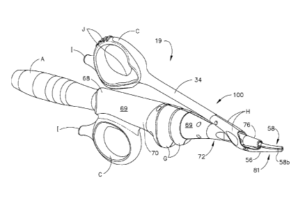
334, respectively. Ridges 321 and 326 provide interface between the user
and triggers 320 and 322. Ridges 321 and 326 are designed to provide as
much surface area for the user to depress in order to activate the instrument.
[00136] In a second expression of switch assembly 300 elastomeric connectors
having copper traces etched onto the elastomer press fit into switch housing
350 to provide the electrical interconnect between transducer 50 and flex
circuit 330. One end of the elastomer connectors electrically engage dome
swithches 332 and 334 via conductors 337 and 335. The other end of the
elastomer connectors slidably interface with conductors 400 and 410 of
transducer 50. Compression of the elastomer connectors allow a working
-28-
CA 02702075 2010-04-08
WO 2009/049016
PCT/US2008/079295
travel of up to 20% of the total height of the elastomer connectors to allow
for
manufacturing tolerances associated with the stackup of the assembled parts.
[00137] A flex circuit 330 provides for the electro-mechanical interface
between
pushbuttons 321 and 322 and the generator 30 via transducer 50. Flex circuit
comprises two dome switches 332 and 334 that are mechanically actuated by
depressing pushbuttons 321 or 322 axially in the x direction. Dome switches
332 and 334 are electrical contact switches, that when depressed provide an
electrical signal to generator 30 as shown by the electrical wiring schematic
of
Fig. 16G. Flex circuit 330 also comprises two diodes within a diode package
336 and conductors, 335 and 337 as is known to those in the art, that connect
to pins 360 and 370, respectively, which in turn provide electrical contact to
ring conductors 400 and 410, which in turn are connected to conductors in
cable 22 that connect to generator 30.
[00138] Flex circuit 330 generally sits within a channel 352 of switch
assembly 350 so
that dome switches 332 and 334 interface with the corresponding backing
surfaces 351 and 353. Backing surfaces provide a firm support for the dome
switches during operation, discussed below. Dome switches 332 and 334
may be fixedly attached to backing surfaces 351 and 353 by any convenient
method, such as, an adhesive.
[00139] As is readily apparent, by depressing pushbuttons 321 and 322 the
corresponding contact surfaces 323 and 324 depress against corresponding
dome switches 332 and 334 to activate the circuit illustrated in FIG. 16G.
When the surgeon depresses 321 pushbutton, the generator will respond with
a certain energy level, such as a maximum ("max") power setting; when the
surgeon depresses pushbutton 322, the generator will respond with a certain
energy level, such as a minimum ("min") power setting, which conforms to
accepted industry practice for pushbutton location and the corresponding
power setting.
-29-
CA 02702075 2010-04-08
WO 2009/049016
PCT/US2008/079295
[00140] In an alternate expression contact surfaces 323 and 325 contact a
living hinge
327 and 329, respectively. Each living hinge comprises an actuator 327a and
329a, which preferably extend across the width of the living hinge. The living
hinge 327 and 329 help eliminate assembly tolerance variations and any
significant amount of "play" in the triggers that rattle when the instrument
is
handled and apply a slight pre-load to the triggers that in turn can eliminate
any "play". The living hinge 327 and 329 further provide a more pronounced
tactile feel of the triggers since the actuators 327a 329a hit the respective
dome switch 332 and 334 of the flex circuit in an optimum location.
[00141] Referring now to Figs. 17A-B, the pushbutton axial actuation reduces
stress
on the surgeon's fingers and allows the fingers to actuate force in a more
ergonomic position preventing stresses at the hands and wrists. The switch
movement also allows comfortable button activation in less than optimal hand
positions, which surgeons often encounter throughout a typical procedure.
[00142] At the proximal end of each access ring 35 and 36 are protrusions 37
and 38,
respectively, that allow the surgeon to rest his or her pinky finger for added
control and comfort. This also allows the surgeon to use the pinky when
clamping on tissue, thereby reducing the force on the other fingers. Each
access ring 35 and 36 includes a soft-touch surface on the interior and
exterior surfaces whether by inserting fingers into the access rings or
palming
the access rings. This feature allows a greater number of hand sizes to
comfortably use the device.
[00143] Referring to Fig. 18, access rings 35 and 36 define a length L.
Perferably, the
center of gravity of the surgical instrument 100 in combination with the
transducer 50 is positioned within length L, more preferably within length L1,
and most preferably within length L2. This position of the center of gravity
allows the instrument to balance within the surgeon's hand to provide more
precise control of the instrument and eliminate hand fatigue during
procedures.
-30-
CA 02702075 2010-04-08
WO 2009/049016
PCT/US2008/079295
[00144] Referring now to Figs. 18 and 19A-E, a two-piece torque wrench 450 is
shown. The torque wrench includes a hand wrench 500 and an adaptor 550.
In one embodiment, hand wrench 500 is provided with cantilever arms 501
disposed in an annular fashion about the centerline of hand wrench 500.
Cantilever arms 501 include teeth 501a disposed, in one embodiment, in an
inward perpendicular fashion in relation to cantilever arms 501. Teeth 501a,
in one embodiment of the current invention, are disposed with a cam ramp
501b at a 25 angle with respect to the perpendicular angle between arm 501
and teeth 501a. Lumen 502 extends the entire length of hand wrench 500 for
accepting adaptor 550.
[00145] Adaptor 550 has a longitudinal shaft 552 with cantilevered tabs 554 at
its
distal end. At the proximal end of shaft 552 are spline gears 556 projecting
in
a perpendicular fashion along the outer circumference of shaft 552. Spline
gears 556 include cam ramps 556a disposed at an angle from about 23 to
about 28 with respect to the perpendicular angle between the outer
circumference of shaft 552 and spline gears 556. Shaft 552 further defines a
lateral opening (not shown) proximal to spline gears 556 for accepting curved
blade 79, discussed below. Adaptor further includes an interface 560 rigidly
connected to shaft 552 and defining an opening for rigidly engaging the distal
end of instrument 19. Optionally, a skirt 558 surrounds spline gears 556 to
prevent glove snags due to moving parts and forms a cavity 559.
[00146] In assembly, torque wrench opening 502 is aligned with shaft 552 and
guided
along substantially the entire length of shaft 552 until the tabs 554 flex
inward
and capture shoulder 505 (not shown) at the distal end of hand wrench 500.
Hand wrench lip 503 engages the distal end of optional skirt 558 allowing
cantilever teeth 501a to slidably engage spline gears 556. Cam ramp 501b
slidably engages retainer cam ramps 29b. The torque wrench assembly 450
slidably engages the distal end of instrument 19 and is held rigidly in place.
Flat surfaces 560b and 560a of interface 560 mate with flat surfaces 565b
(Fig. 18) and 565a (not shown) at the distal end of activation member 34
-31-
CA 02702075 2010-04-08
WO 2009/049016
PCT/US2008/079295
(clamp arm 60) and rail 562 slidably engaging slot 564 on clamp arm 60 and
distra shroud 76 and outer shroud 72 all provide structural support to
maintain
adapter 550 firmly engaged with instrument 19.
[00147] Clockwise annular motion or torque is imparted to hand wrench 500
through
paddles 504. The torque is transmitted through arms 501 and teeth 501a to
gears 556, which in turn transmit the torque to the waveguide 80 via clamp
arm assembly 60 via outer shroud 72 via insulated pin 27. When a user
imparts 5-12 lbs. of torque, the ramps 501b and 556 cause the arms 501 to
move or flex away from the centerline of wrench 500 ensuring that the user
does not over-tighten the waveguide 80 onto transducer 50. When a counter-
clockwise torque is applied to wrench 500 via paddles 504, the perpendicular
flat sides of teeth 501a and 556 abut allowing a user to impart a torque to
the
interface between the waveguide 80 and transducer 50 in proportion to the
force applied to the paddles facilitating removal of the instrument 100 from
the
transducer 50. The torque wrench 450 may be constructed from a durable
plastic, such as polycarbonate or a liquid crystal polymer. It is also
contemplated that the wrench 450 may alternatively be made from a variety of
materials including other plastics, ceramics or metals.
[00148] In another embodiment (not shown), the paddles and cantilever arm
assembly
may be separate components attached by mechanical means or chemical
means such as adhesives or glue.
[00149] Preferably, the ultrasonic clamp coagulator apparatus 19 described
above will
be processed before surgery. First, a new or used ultrasonic clamp
coagulator apparatus is obtained and if necessary cleaned. The ultrasonic
clamp coagulator apparatus can then be sterilized. In one sterilization
technique the ultrasonic clamp coagulator apparatus is placed in a closed and
sealed container, such as a plastic or TYVEK bag. Optionally, the ultrasonic
clamp coagulator apparatus can be bundled in the container as a kit with
other components, including a torque wrench 450. The container and
ultrasonic clamp coagulator apparatus, as well as any other components, are
-32-
CA 02702075 2010-04-08
WO 2009/049016
PCT/US2008/079295
then placed in a field of radiation that can penetrate the container, such as
gamma radiation, x-rays, or high-energy electrons. The radiation kills
bacteria
on the ultrasonic clamp coagulator apparatus and in the container. The
sterilized ultrasonic clamp coagulator apparatus can then be stored in the
sterile container. The sealed container keeps the ultrasonic clamp coagulator
apparatus sterile until it is opened in the medical facility.
[00150] Referring now to Figs. 20 and 21 the present invention enables a more
even
pressure profile across the blade from the proximal end to the distal end as
compared to the prior art. An even pressure profile along the blade provides
simultaneous tissue transaction along the blade as well as excellent cutting
at
the tip of the blade. The even pressure profile is accomplished by creating as
close a parallel closure of the clamp pad against the blade as possible.
[00151] The pivot radius of the prior art is less than 1.0 inches and in some
cases less
than 0.75 inches. In accordance with the present invention, the pivot radius
is
increased to be over two times the pivot radius of the prior art. In one
preferred embodiment, the pivot radius is equal to 1.5 inches. The pivot
radius is not limited to this dimension and exact dimensions are left to the
design artisian. What is important, however, is that as the clamp arm is
moved through its pivot radius the clamp arm exhibits a substantially parallel
closure with respect to the blade. Parallel closure means that as the clamp
pad closes against the blade, a substantially equal pressure is exerted across
the blade from the proximal end to the distal end. Parallel closure allows for
less variation in pressure profile across the blade from the proximal end to
the
distal end. In one example the pressure profile ranges from about 0.1 lbs.
measured at the distal tip to about 0.4 lbs. measured proximal of the distal
tip,
but most notably only ranging between 0.1 lbs. at the distal tip and less than
0.3 lbs. across substantially the entire length of the blade as shown in Fig.
20.
[00152] The present invention further comprises a displacement limited force
application, whereas prior art instruments comprise a force limiting element,
such as a spring, that limits the amount of force applied to the blade by the
-33-
CA 02702075 2010-04-08
WO 2009/049016
PCT/US2008/079295
clamp arm. In accordance with the present invention actuation member 34
deflects as increased load is applied. The force delivered to the tissue is
dependent upon the length of actuation member 34, the cross sectional area,
the modulus of elasticity and the amount of deflection allowed before it hits
a
hard stop on the shroud.
[00153] As actuation member 34 deflects and load is applied to blade 79, the
blade 79
deflects as well. Thus, the clamp force system is comprised of two members:
the inherent stiffness of actuation member 34 (mainly comprised of blade
deflection and distal seal compression); and the blade side stiffness (mainly
comprised of actuation member 34 and thumb ring stiffness). These two
stiffnesses can be calculated, measured and used to predict and manipulate
clamp force. In one preferred embodiment, the actuation member 34 has a
stiffness of approximately 3 lb/in, to about 7 lb/in., and the blade 79 has a
stiffness of between 1501b/in and about 250 lb/in.
[00154] Further, the thumb ring height gap G shown in Fig. 16A defines the
compression length of the actuation member 34 side of the spring system.
Height G preferably ranges from about 0.15 inches to about 0.33 inches.
[00155] One benefit of the displacement force limiting system is increased pad
life. As
the device is used, the pad wears and a groove begins to form. This is
prevalent in the abuse case where the device is activated when fully closed
with no tissue present between blade 79 and clamp pad 58. In prior art
ultrasonic instruments, when the groove became deeper, a very similar
amount of force was placed on the blade due to the force limiting spring. The
slope of the force vs. displacement curve is relatively flat.
[00156] In the present invention, however, as the pad wears, the thumb ring
only
rotates slightly downward due to the deflection of the lever system after
thumb
ring 35 bottoms out at handle 68 (distance G). Since there is less distance
for
the thumb ring to travel, the force on the blade decreases. The present
invention has a steeper force vs. displacement curve resulting in a larger
drop
-34-
CA 02702075 2015-07-20
,
in force due to pad wear. Thus, the pad groove does not increase as readily
as the prior art and the instrument still performs as needed with lower forces
dues to pad wear.
[00157] Referring to Fig. 22, the graph illustrates one embodiment of the
representative stiffness of the present invention. The y-axis is the force the
user exerts on the thumb ring and the y-axis is the thumb ring deflection.
The stiffness is approximately 3 lbs/in.
[00158] The present invention has been illustrated by description of
several
embodiments. Numerous variations, changes, and substitutions will occur to
those skilled in the art. Moreover, the structure of each element associated
with the present invention can be alternatively described as a means for
providing the function performed by the element. The scope of the claims
may be given the broadest interpretation consistent with the description as a
whole.
-35-