Note: Descriptions are shown in the official language in which they were submitted.
CA 02702242 2010-04-29
1
PORTABLE ELECTROCARDIOGRAM
FIELD OF THE INVENTION
[0004]The invention disclosed broadly relates to the field of equipment for
monitoring the electrical activity of the heart, and particularly to portable
electrocardiograph monitors.
BACKGROUND OF THE INVENTION
[0005]An electrocardiogram is a test that graphically records the electrical
activity
of the heart. The electrocardiogram or ECG (sometimes called EKG) is used
worldwide as a relatively simple way of diagnosing many heart conditions. It
records the small electric waves being generated during heart activity using
body
surface electrodes attached to a patient. The electrodes are placed in a
particular
pattern because electrical signals generated by a human heart appear in a
characteristic pattern throughout the body, and on its surface
[0006]A procedure developed by Willem Einthoven in 1901 inter-related three
electrodes specifically oriented on the body (right arm, left arm, and left
leg). These
electrodes are at the apices of a physiological triangle known as Einthoven's
triangle, as shown in FIG. la. The difference in electrical potential between
the left
and right arms is designated lead I; lead II is the difference in electrical
potential
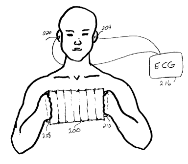
between the left leg and right arm; and lead III is the difference in
electrical
potential between the left leg and left arm. Thus, the Einthoven triangle
resembles
a triangle standing on its tip "."
[0007]These electrodes provide bipolar recordings of the voltage differential
between two electrodes. By convention, the positive electrode is placed on the
left
CA 02702242 2010-04-29
2
arm, with the negative electrode on the right arm. In the lead II
configuration, the
positive electrode is on the left leg and the negative electrode is on the
right arm.
Lead III has the positive electrode on the left leg and the negative electrode
on the
left arm. The limb leads can be attached to the end of the limb (wrists and
ankles)
or at the origin of the limb (shoulder or upper thigh). The difference in
electrical
potential between two of the electrodes constitutes the signal.
[0008]Referring to FIG. lb there is shown a simplified illustration of a
conventional
electrocardiograph 100 in place on a patient. The ECG 100 requires at least
three
leads (therefore three electrodes are needed). These three electrodes are
applied
one on each of the patient's arms 110 and 112. The third electrode 120 is
applied
on the patient's left leg.
[0009]A fourth electrode 140 is placed on the patient's right leg as an
electrical
ground. The ground can be at other locations on the body but at a reasonable
distance from the other electrodes to ensure a good signal. In addition, there
are
six precordial (chest) leads 160 designated V1-V6 (not shown here).
Their
conventional placement is illustrated in FIG. lc.
[0010]The electrodes are easy to apply and this conventional placement of
electrodes works well in a hospital setting and in doctor's office. The
problem
arises, however, when it is desirable and sometimes necessary for an ECG to be
used outside of a conventional medical setting. For example, a patient with
chronic
heart problems may want to have a portable ECG in the home or the office.
Airlines
may find it necessary to have a portable ECG in airplanes for in-flight
emergency
use. The signals produced by a portable unit can be transmitted to a doctor on
the
ground who can then interpret the signals and advise the airline staff as to
whether
to use an on-board defibrillator.
[0011]Electrodes must be positioned in an anatomically correct pattern so that
the
readings are valid. One problem with this conventional electrode placement is
that
CA 02702242 2010-04-29
3
leg electrodes are not conducive to portability.
SUMMARY OF THE INVENTION
[0012]Briefly, according to an embodiment of the invention, an apparatus for
measuring cardiac electrical activity of a patient includes: a malleable pad,
a first
electrocardiogram electrode, and a ground electrode. The pad includes: a right
handle for grasping with the patient's right hand, the right handle including
an
electrocardiogram electrode; a left handle for grasping with the patient's
left hand,
the left handle also including an electrocardiogram electrode; and an
electronic
circuit configured for receiving electrical signals from the electrodes and
also
configured to invert the signals from the electrodes for transmission to a
processor
to produce a conventional graphic recording of the differences in electrical
potential
between the electrodes. The pad also includes a multiplex cable for coupling
the
electronic circuit to leadwires attached to the electrocardiogram electrodes;
and a
port configured for coupling with the processor. The first electrode and the
handle
electrodes form an inverted Einthoven triangle with the first electrode at an
apex of
said triangle, and anatomically superior to the horizontal axis formed by the
second
and third electrodes.
[0013]According to another embodiment of the present invention, a method for
measuring cardiac electrical activity of a patient includes steps or acts of:
forming
an inverted Einthoven triangle of electrocardiograph electrodes on the
patient,
including steps of: attaching a first electrode-to an ear of the patient;
attaching an
electrical ground electrode to the patient; attaching second and third
electrocardiogram electrodes to locations on the patient to form a base of the
inverted Einthoven triangle; wherein the electrodes are coupled to a connector
by
leadwires and wherein the connector is operatively coupled to a processor; and
electronically inverting signals obtained from the electrodes to produce a
conventional electrocardiogram recording using the processor.
CA 02702242 2010-04-29
4
BRIEF DESCRIPTION OF THE DRAWINGS
[0014]To describe the foregoing and other exemplary purposes, aspects, and
advantages, we use the following detailed description of an exemplary
embodiment
of the invention with reference to the drawings, in which:
[0015]FIG. la shows an illustration of Einthoven's inverted triangle,
according to
the known art;
[0016]FIG. lb shows an illustration of an electrocardiograph system according
to
the known art;
[0017]FIG. lc shows the placement of leads V1-V6, according to the
known art;
[0018]FIG. 2 shows an illustration of the portable electrocardiograph,
according to
an embodiment of the present invention;
[0019]FIG. 3a shows the malleable form factor of the portable
electrocardiograph,
according to an embodiment of the present invention;
[0020]FIG. 3b shows, in sagittal view, the electrodes at the apices of each
hinged
articulation, according to an embodiment of the present invention;
[0021]FIG. 4a shows the contact side of the pad placed against the patient's
chest,
illustrating the exposed electrodes, according to an embodiment of the present
invention;
[0022]FIG. 4b shows a closer view of the connector, according to an embodiment
of
the present invention;
CA 02702242 2010-04-29
[0023]FIG. 5 shows the ECG pad with serpentine folds and V1-V6
electrodes, according to an embodiment of the present invention;
[0024]While the invention as claimed can be modified into alternative forms,
specific embodiments thereof are shown by way of example in the drawings and
will
herein be described in detail. It should be understood, however, that the
drawings
and detailed description thereto are not intended to limit the invention to
the
particular form disclosed, but on the contrary, the intention is to cover all
modifications, equivalents and alternatives falling within the scope of the
present
invention.
=
DETAILED DESCRIPTION
[0025]I describe a method and apparatus for recording the electrical activity
of the
heart using an inverted Einthoven triangle of electrodes. The apparatus is a
small,
lightweight, and portable pad. The pad is expanded across a patient's chest in
order
to record electrical activity from the chest. It is constructed of a malleable
material
such as mylar, soft vinyl or rubber so that it can be easily contracted and
expanded.
[0026]The pad forms the base of an inverted Einthoven triangle. The apex of
the
inverted Einthoven triangle is formed by a receiving electrode preferably
located in
or on the patient's ear. The other electrodes are preferably. located on the
pad
which is placed across the patient's Chest. This novel electrocardiograph
system is
small and portable so that it can be used for home use and travel. It embodies
a
small, compact form factor and can easily be carried in a small briefcase or
travel
bag. The portable system as will be described can be self-applied by a patient
and
is devoid of leg electrodes.
[0027]Referring now in specific detail to the drawings, and particularly FIG.
2, there
is illustrated a portable ECG device 200 according to an embodiment of the
present
CA 02702242 2010-04-29
6
invention. The device 200 is preferably a pad approximately the size of a book
that
expands to fit over a patient's chest. It contains electrodes for contact with
a
patient's chest, and hands.
[0028]Since patients have different size chests and their hearts may vary in
location within the chest cavity, the pad 200 is adjustable to fit the
different sizes
and contours of almost all patients. The pad 200 expands and contracts like an
accordion to adjust to the size and contour of a patient's chest. The pad 200
is
constructed from any suitable material that will be pliable enough to bend and
yet
not interfere with electrical signals. A preferred embodiment uses accordion-
like
ribbed pleats, but other constructs are acceptable provided they are able to
easily
expand and contract.
[0029]The pad 200 includes two handles 208 and 210 at either end. In a
preferred
embodiment, each handle contains at least one receiving electrode for each
respective hand. The patient grips both handles 208 and 210 to both position
the
pad 200 and provide electrical input from the arms. The patient holds the pad
200
lying substantially flat against the patient's chest.
[0030]The pad 200 preferably, but not necessarily, includes a number of
electrodes
embedded in the contact surface of the pad 200. As the pad 200 is held against
the
patient's chest, these electrodes make contact with the patient's skin and
pick up
intra-cutaneous electrical signals from the patient's heart.
[0031]One electrode 204 is placed in the patient's ear. Thus, electrical
potential
differences between the right arm, left arm, and ear can be measured. The ear
electrode 204 forms the apex of the inverted Einthoven triangle, with the
handles
208 and 210 forming the base of the triangle. Another electrode 220 used for
ground potential may optionally be placed in the patient's contralateral ear
as
shown.
CA 02702242 2010-04-29
7
[0032]With electrode placement on the right arm, left arm, left ear and right
ear
(as ground), the combination of right arm-left arm is equivalent to a standard
lead
I ECG configuration. The right arm-left ear combination acts as an inverted
lead II
and the left arm-left ear combination acts as an inverted lead III. Readings
from
these two latter leads are transformed back to the conventional Einthoven
configuration mathematically by taking into account: 1) the angular variation
of the
cardiac dipole measured by a left ear electrode as opposed to a left leg
electrode;
and 2) the magnitude variation of the cardiac dipole measured by a left ear
electrode as opposed to a left leg electrode.
[0033]Referring to FIG. 3a, a closer view of the device 200 is shown, from the
contact side that lies against a patient's chest. This embodiment carries six
additional electrodes 212. The placement of these six additional electrodes
212
substantially correlates to the V1-V6 voltages as shown in FIG. lc.
[0034]Referring to FIG. 3b, the pad 200 is shown from a side view as seen from
behind the patient, as it is expanded and ready to be placed over a patient's
chest
310. The patient is holding the handles 208 and 210 and has expanded the pad
200
to comfortably fit on the patient's chest 310. Electrodes 212 are located on
the
contact surface of the pad 200 so that the voltages V1-V6 can be
measured. The dashed lines indicate where the electrodes 212 will make contact
with the patient's chest 310. The patient grasps the handles 208 and 210, and
expands the pad 200 across his/her chest 310 while lying down flat. The
patient, or
an assistant, then positions the pad 200 by pressing down against the skin
surface
so that the electrodes 212 are in good contact with the surface of the chest
310.
The pad 200 not only houses the electrodes and leadwires, it also acts as an
electrical insulator between the electrodes and the patient, and between
adjacent
electrodes.
[0035]The embodiment of FIG. 3b uses an accordion principle to accommodate the
size and contour of an individual patient's chest 310. The pad 200 has been
CA 02702242 2010-04-29
8
expanded such that enclosed "pleats" 304 form oblique angles with electrodes
212
V1-V6 at the apex of each "pleat" 304. The pleats 304 provide
support
and are formed from a stiff, yet bending material. They produce the effect as
seen
in the ribs of an umbrella. The pleats 304 are encased in a compliant, non-
conductive material 302 such as rubber, mylar, silicone, or any other
appropriate
material. The pleats 304 may be hinged. The electrodes 212 are disposed on the
contact surface of the non-conductive material so that they can make contact
with
the patient's chest 310. Additional electrodes may also be placed in the pad
200.
For example, a twelve lead system may be configured by the addition of six
more
electrodes 212. The handles 208 and 210 also contain electrodes 212 to be used
in
place of the conventional arm electrodes, with contact effected by the
grasping
actions of the hands.
[0036]FIG. 4a shows the contact surface of the pad 200 which is placed against
the
patient's chest. The wires 450 extend from the electrodes 212 through the pad
200
to an electronic circuit 460 housed within a connector 420. The wires 450 may
be
combined into a multiplex cable (not shown). The electronic circuit 460, as
used in
conventional ECG devices, additionally inverts the electrical signals received
from
the electrodes to produce a conventional cardiac signal, which can be
displayed on
a monitor or transmitted to a remote location by landline or wireless means.
[0037]FIG. 4b shows a close-up view of an exemplary connector 420. One end of
the connector 420 may be exposed outside of the pad 200. A port 480 is located
at
this end for allowing the pad 20-0 to be coupled with the reader 416. The
reader
416 is used to read the electrical signals transmitted from the electrodes
200. The
reader 416 comprises a processor, an amplifier, and a filter. It is preferably
operatively coupled with a monitor or printer. The reader 416 may be a
conventional ECG monitor. The electronic circuit 460 may be placed as shown
within the connector 420, or the circuit 460 may be placed in any
configuration that
provides access to the wires 450.
CA 02702242 2010-04-29
9
[0038]FIG. 5 shows another embodiment using a pad 500 with serpentine folds to
achieve the accordion effect. In this embodiment the accordion effect may be
achieved using bendable ribs inserted into the pad 200.
[0039]To complete the inverted Einthoven triangle, a receiving electrode 204
is
attached to the patient's ear. This electrode 204 may be in an earphone form
factor, or it may be clipped to the earlobe. Another electrode can be placed
in the
patient's contralateral ear and act as an electrical ground 220. Six
electrodes 212
can be placed on the pad 200 to measure the V1-V6 voltages for added
reliability. Additional electrodes may be placed in the handles 208 and 210.
[0040]The electrodes 212 can be rectangular or some other shape. They may be
suction, button, or plate electrodes. Alternatively, the electrodes 212 may be
printed on the pad 200. Each electrode has a substantially flat surface for
secure
attachment to a patient's skin. Modern electrodes are self-adhesive; but to
aid in
electrical conduction, a conductive gel can be applied to the flat surface of
each
electrode 212 before attachment. If necessary, tape can be used to secure the
electrode 212. The non-contact surface of the electrode 212 is a conductor
attached
to an electrode leadwire 450 which in turn may be attached to a multiplex
cable
secured within the pad 200. The cable is coupled with the connector 420. The
connector 420 includes ports for coupling with the reader 416 and other
input/output devices.
[0041]To facilitate use by non-medical personnel, standardized color coding of
tlie
electrodes and/or leadwires can be used, and the color codes should be made
available to the patient. For example, the non-contact surface of the pad 200
may
have the color codes printed on it. The non-contact surface of the pad 200 may
optionally be marked with indicators showing optimal placement in order to
assure
that the maximum number of signals are received.
[0042]In one embodiment, the minimum number of electrodes that can be
CA 02702242 2012-08-31
=
1.0
advantageously used for cardiac monitoring is four: one receiving electrode in
the
patient's ear, two electrodes on the pad handles, and one electrode to be used
as =
ground. This ground electrode may be placed in the patient's contralateral
ear. In
another embodiment, additional electrodes can be embedded or affixed to the
pad
200, as has been previously shown.
[0043]Therefore, while there has been described what is presently considered
to be
the preferred embodiment, it will be understood by those skilled in the art
that
other modifications can be made within the invention.
=
.=
=