Note: Descriptions are shown in the official language in which they were submitted.
CA 02702420 2010-04-13
WO 2009/006008 PCT/US2008/067124
1
DEVICES AND METHODS FOR SELECTIVELY LYSING CELLS
Claim for Priority/ Reference to Co pending Applications.
This application claims priority to U.S. Utility Patent Application Serial
Number
11/515,634 filed September 5, 2006, U.S. Utility Patent Application Serial
Number
11/334,794 filed January 17, 2006, U.S. Utility Patent Application Serial
Number
11/334,805 filed January 17, 2006, and U.S. Utility Patent Application Serial
Number
11/292,950 filed December 2, 2005, the entirety of which are incorporated
herein by
reference.
Field of the Invention:
The present invention relates to a microbubble generation device and a system
for
selectively lysing cells by cavitating microbubbles.
Background of the Invention:
Gynoid lipodystrophy is a localized metabolic disorder of the subcutaneous
tissue
which leads to an alteration in the topography of the cutaneous surface
(skin), or a
dimpling effect caused by increased fluid retention and/or proliferation of
adipose tissue in
certain subdermal regions. This condition, commonly known as cellulite,
affects over 90%
of post-pubescent women, and some men. Cellulite commonly appears on the hips,
buttocks and legs, but is not necessarily caused by being overweight, as is a
common
perception. Cellulite is formed in the subcutaneous level of tissue below the
epidermis and
dermis layers. In this region, fat cells are arranged in chambers surrounded
by bands of
connective tissue called septae. As water is retained, fat cells held within
the perimeters
defined by these fibrous septae expand and stretch the septae and surrounding
connective
tissue. Furthermore, adipocyte expansion from weight gain may also stretch the
septae.
Eventually this connective tissue contracts and hardens (scleroses) holding
the skin at a
non-flexible length, while the chambers between the septae continue to expand
with
weight gain, or water gain. This results in areas of the skin being held down
while other
sections bulge outward, resulting in the lumpy, "orange peel" or "cottage-
cheese"
appearance on the skin surface.
Even though obesity is not considered to be a root cause of cellulite, it can
certainly
worsen the dimpled appearance of a cellulitic region due to the increased
number of fat
cells in the region. Traditional fat extraction techniques such as liposuction
that target deep
fat and larger regions of the anatomy, can sometimes worsen the appearance of
cellulite
since the subdermal fat pockets remain and are accentuated by the loss of
underlying bulk
CA 02702420 2010-04-13
WO 2009/006008 PCT/US2008/067124
2
(deep fat) in the region. Many times liposuction is performed and patients
still seek therapy
for remaining skin irregularities, such as cellulite.
A variety of approaches for treatment of skin irregularities such as cellulite
and
removal of unwanted adipose tissue have been proposed. For example, methods
and
devices that provide mechanical massage to the affected area, through either a
combination
of suction and massage or suction, massage and application of energy, in
addition to
application of various topical agents are currently available. Developed in
the 1950's,
mesotherapy is the injection of various treatment solutions through the skin
that has been
widely used in Europe for conditions ranging from sports injuries to chronic
pain, to
cosmetic procedures to treat wrinkles and cellulite. The treatment consists of
the injection
or transfer of various agents through the skin to provide increased
circulation and the
potential for fat oxidation, such as aminophylline, hyaluronic acid,
novocaine, plant
extracts and other vitamins. The treatment entitled Acthyderm (Turnwood
International,
Ontario, Canada) employs a roller system that electroporates the stratum
corneum to open
small channels in the dermis, followed by the application of various
mesotherapy agents,
such as vitamins, antifibrotics, lypolitics, anti-inflammatories and the like.
Massage techniques that improve lymphatic drainage were tried as early as the
1930's. Mechanical massage devices, or Pressotherapy, have also been developed
such as
the "Endermologie" device (LPG Systems, France), the "Synergie" device
(Dynatronics,
Salt Lake City, Utah) and the "Silklight" device (Lumenis, Tel Aviv, Israel),
all utilizing
subdermal massage via vacuum and mechanical rollers. Other approaches have
included a
variety of energy sources, such as Cynosure's "TriActive" device (Cynosure,
Westford,
Mass.) utilizing a pulsed semiconductor laser in addition to mechanical
massage, and the
"Cellulux" device (Palomar Medical, Burlington, Mass.) which emits infrared
light
through a cooled chiller to target subcutaneous adipose tissue. The
"VelaSmooth" system
(Syneron, Inc., Yokneam Illit, Israel) employs bipolar radiofrequency energy
in
conjunction with suction to increase metabolism in adipose tissue, and the
"Thermacool"
device (Thermage, Inc., Hayward, Calif.) utilizes radiofrequency energy to
shrink the
subdermal fibrous septae to treat wrinkles and other skin defects. Other
energy based
therapies such as electrolipophoresis, using several pairs of needles to apply
a low
frequency interstitial electromagnetic field to aid circulatory drainage have
also been
developed. Similarly, non-invasive ultrasound is used in the "Dermosonic"
device
(Symedex Medical, Minneapolis, Minn.) to promote reabsorption and drainage of
retained
fluids and toxins.
CA 02702420 2010-04-13
WO 2009/006008 PCT/US2008/067124
3
Another approach to the treatment of skin irregularities such as scarring and
dimpling is a technique called subcision. This technique involves the
insertion of a
relatively large gauge needle subdermally in the region of dimpling or
scarring, and then
mechanically manipulating the needle below the skin to break up the fibrous
septae in the
subdermal region. In at least one known method of subcision, a local
anesthetic is injected
into the targeted region, and an 18 gauge needle is inserted 10-20 mm below
the cutaneous
surface. The needle is then directed parallel to the epidermis to create a
dissection plane
beneath the skin to essentially tear through, or "free up" the tightened
septae causing the
dimpling or scarring. Pressure is then applied to control bleeding acutely,
and then by the
use of compressive clothing following the procedure. While clinically
effective in some
patients, pain, bruising, bleeding and scarring can result. The known art also
describes a
laterally deployed cutting mechanism for subcision, and a technique employing
an
ultrasonically assisted subcision technique.
Certain other techniques known as liposuction, tumescent liposuction,
lypolosis
and the like, target adipose tissue in the subdermal and deep fat regions of
the body. These
techniques may include also removing the fat cells once they are disrupted, or
leaving
them to be resorbed by the body's immune/lymphatic system. Traditional
liposuction
includes the use of a surgical cannula placed at the site of the fat to be
removed, and then
the use of an infusion of fluids and mechanical motion of the cannula to break
up the fatty
tissue, and suction to "vacuum" the disrupted fatty tissue directly out of the
patient.
The "Lysonix" system (Mentor Corporation, Santa Barbara, Calif.) utilizes an
ultrasonic transducer on the handpiece of the suction cannula to assist in
tissue disruption
(by cavitation of the tissue at the targeted site). Liposonix (Bothell, Wash.)
and Ultrashape
(TelAviv, Israel) employ the use of focused ultrasound to destroy adipose
tissue
noninvasively. In addition, cryogenic cooling has been proposed for destroying
adipose
tissue. A variation on the traditional liposuction technique known as
tumescent liposuction
was introduced in 1985 and is currently considered by some to be the standard
of care in
the United States. It involves the infusion of tumescent fluids to the
targeted region prior to
mechanical disruption and removal by the suction cannula. The fluids may help
to ease the
pain of the mechanical disruption, while also swelling the tissues making them
more
susceptible to mechanical removal. Various combinations of fluids may be
employed in
the tumescent solution including a local anesthetic such as lidocaine, a
vasoconstrictive
agent such as epinephrine, saline, potassium and the like. The benefits of
such an approach
are detailed in the articles, "Laboratory and Histopathologic Comparative
Study of Internal
CA 02702420 2010-04-13
WO 2009/006008 PCT/US2008/067124
4
Ultrasound-Assisted Lipoplasty and Tumescent Lipoplasty" Plastic and
Reconstructive
Surgery, Sep. 15, (2002) 110:4, 1158-1164, and "When One Liter Does Not Equal
1000
Milliliters: Implications for the Tumescent Technique" Dermatol. Surg. (2000)
26:1024-
1028, the contents of which are expressly incorporated herein by reference in
their entirety.
Various other approaches employing dermatologic creams, lotions, vitamins and
herbal supplements have also been proposed to treat cellulite. Private spas
and salons offer
cellulite massage treatments that include body scrubs, pressure point massage,
essential
oils, and herbal products using extracts from plant species such as seaweed,
horsetail and
clematis and ivy have also been proposed. Although a multitude of therapies
exist, most of
them do not provide a lasting effect on the skin irregularity, and for some,
one therapy may
cause the worsening of another (as in the case of liposuction causing scarring
or a more
pronounced appearance of cellulite). Yet other treatments for cellulite have
negative side
effects that limit their adoption. Most therapies require multiple treatments
on an ongoing
basis to maintain their effect at significant expense and with mixed results.
Medical ultrasound apparatus and methods are generally of two different types.
One type of medical ultrasound wave generating device known in the art is that
which
provides high intensity focused ultrasound or high acoustic pressure
ultrasound for tissue
treatment, for example for tumor destruction. High intensity or high acoustic
pressure
ultrasound is capable of providing direct tissue destruction. High intensity
or high acoustic
pressure ultrasound is most commonly focused at a point in order to
concentrate the energy
from the generated acoustic waves in a relatively small focus of tissue.
However, another
type of medical ultrasound is a lower intensity and less focused type of
ultrasound that is
used for diagnostic imaging and physical therapy applications. Low acoustic
pressure
ultrasound is commonly used, for example, for cardiac imaging and fetal
imaging. Low
acoustic pressure ultrasound may be used for tissue warning, without tissue
disruption, in
physical therapy applications. Low acoustic pressure ultrasound, using power
ranges for
diagnostic imaging, generally will not cause any significant tissue disruption
when used
for limited periods of time in the absence of certain enhancing agents.
Methods and apparatus of using high intensity focused ultrasound to disrupt
subcutaneous tissues directly has been described in the known art. Such
techniques may
utilize a high intensity ultrasound wave that is focused on a tissue within
the body, thereby
causing a localized destruction or injury to cells. The focusing of the high
intensity
ultrasound may be achieved utilizing, for example, a concave transducer or an
acoustic
lens. Use of high intensity focused ultrasound to disrupt fat, sometimes in
combination
CA 02702420 2010-04-13
WO 2009/006008 PCT/US2008/067124
with removal of the fat by liposuction, has been described in the known prior
art. Such use
of high intensity focused ultrasound should be distinguished from the low
acoustic
pressure ultrasound.
In light of the foregoing, it would be desirable to provide methods and
apparatus
5 for treating skin irregularities such as cellulite and to provide a
sustained aesthetic result to
a body region, such as the face, neck, arms, legs, thighs, buttocks, breasts,
stomach and
other targeted regions which are minimally or non-invasive. It would also be
desirable to
provide methods and apparatus for treating skin irregularities that enhance
prior techniques
and make them less invasive and subject to fewer side effects.
Therefore, there has been recognized by those skilled in the art a need for an
apparatus and method for the use of low intensity ultrasound to treat
subcutaneous tissues.
Use of low intensity ultrasound, in the power ranges of diagnostic ultrasound,
would be
safer to use, have fewer side effects, and could be used with less training.
The present
invention fulfills these needs and others.
SUMMARY OF THE INVENTION
Disclosed is a device for generating microbubbles in a gas and liquid mixture
and
injection device, which includes a housing defining a mixing chamber; means
for mixing
solution contained in the mixing chamber to generate microbubbles in the
solution; and a
needle array removably attached to the housing and in fluid connection with
the mixing
chamber, the needle array including at least one needle.
The mixing chamber may include a first mixing chamber in fluid communication
with a second mixing chamber. Moreover, the mixing means may include means for
expressing a solution of fluid and gas between the first and second mixing
chambers to
generate microbubbles in the solution.
The device may further include a fluid reservoir in fluid connection with at
least
one of the first and second mixing chambers; and a source of gas in fluid
connection with
at least one of the first and second mixing chambers. Optionally, the fluid
reservoir and/or
the mixing chamber(s) may be thermally insulated and/or include means for
maintaining
the fluid at a predetermined temperature. Still further, the source of gas may
be room air,
or may include air, oxygen, carbon dioxide, perfluoropropane or the like which
may be
maintained at greater than atmospheric pressure.
The solution expressing means may include first and second pistons mounted for
reciprocation within the first and second mixing chambers.
CA 02702420 2010-04-13
WO 2009/006008 PCT/US2008/067124
6
Still further, the device may include means for reciprocating the first and
second
pistons to express fluid and gas between the first and second cylinders to
create a
microbubble solution. The reciprocating means may be a source of compressed
air; and the
first and second cylinders may be pneumatic cylinders.
The device may include a needle deployment mechanism operably connected to the
needle array for deploying the at least one needle(s) between a retracted and
an extended
position. The needle array may include at least two needles and the needle
deployment
mechanism selectively deploys one or more of the at least two needles between
the
retracted and the extended position. Still further, the needle deployment
mechanism may
include at least one of a pneumatic piston, an electric motor, and a spring.
The device may include at least one pressure sensor for measuring tissue
apposition
pressure. The sensor may be provided on either or both of the housing and the
needle
array. Deployment of the at least one needle may be inhibited if a measured
apposition
pressure values falls beneath an initial threshold value or exceeds a
secondary threshold
value. The device may include two or more sensors wherein deployment of the at
least
one needle is inhibited if a difference in measured apposition pressure values
between any
two sensors exceeds a threshold value.
The aforementioned mixing means may include at least one of a blade, paddle,
whisk, and semi-permeable membrane positioned within the mixing chamber. The
mixing
means may further include one of a motor and a pneumatic source operably
coupled to the
at least one of a blade, paddle, whisk, and semi-permeable membrane.
The device of the present invention may include tissue apposition means for
pulling the needle array into apposition with tissue. The tissue apposition
means may
include at least one vacuum orifice defined in at least one of the housing and
the needle
array, whereby the vacuum orifice transmits suction from a source of partial
vacuum to
tissue bringing the needle array into apposition with the tissue. The vacuum
orifice may
be formed in the needle array, and the at least one needle may be positioned
within the
vacuum orifice. Still further, the vacuum orifice may define a receptacle,
whereby tissue is
pulled at least partially into the receptacle when the vacuum orifice
transmits suction from
the source of partial vacuum.
In some embodiments, the needle array includes a tissue apposition surface;
and the
tissue apposition means further includes at least one flange mounted on the
tissue
apposition surface and surrounding the vacuum orifice.
CA 02702420 2010-04-13
WO 2009/006008 PCT/US2008/067124
7
The device of the present invention may include means for adjusting a needle
insertion depth of the at least one needle. The needle array may include at
least two
needles and the insertion depth adjustment means may individually adjust the
insertion
depth of each needle. In one embodiment, the needle insertion depth adjustment
means
may include a plurality of discrete needle adjustment depths. Alternatively,
the needle
insertion depth adjustment means provides continuous adjustment of the needle
adjustment
depth. Still further, the needle insertion depth adjustment means may include
a readout
and/or a display indicative of the needle adjustment depth.
According to one embodiment, the needle array includes a tissue apposition
surface; and the at least one needle includes a distal end, the at least one
needle being
moveable between a retracted position in which the distal end of the needle is
maintained
beneath the tissue apposition surface and an extended position in which the
distal end of
the needle extends beyond the tissue apposition surface.
According to one embodiment an ultrasound transducer is operably connected to
one of the needle array, the housing and the at least one needle.
According to one aspect, the needle array may generally surround the
ultrasound
transducer. Alternatively, the ultrasound transducer may generally surround
the needle
array. Moreover, the ultrasound transducer may be integrally formed with the
needle
array.
The device may further include a fluid pressurization mechanism in fluid
communication with the at least one needle.
Still further, the device may include means for controlling a volume and
pressure
of fluid dispensed from the fluid reservoir into the mixing chamber. Moreover
the device
may include means for controlling the volume, pressure, and rate at which
fluid or solution
is injected into the tissue.
A machine readable identifier may be provided on the needle array. The
identifier
may be used to uniquely identify the ultrasound transducer, needle array
and/or
characteristics of the needle array.
According to one embodiment, the device includes a machine readable identifier
on
the needle array and means for reading the identifier operably connected to
the needle
deployment mechanism. Optionally, the needle deployment mechanism inhibits
deployment of the at least one needle unless the identifier reading means
authenticates the
identifier. Moreover, the needle deployment mechanism may optionally
accumulate the
number of times the needle array associated with a given identifier is
deployed and inhibit
CA 02702420 2010-04-13
WO 2009/006008 PCT/US2008/067124
8
deployment of the at least one needle if the accumulated number needle
deployments
associated with the identifier exceeds a predetermined value.
According to one embodiment, the device includes a machine readable identifier
on
the needle array and means for reading the identifier operably connected to
the fluid
pressurization mechanism, wherein the fluid pressurization mechanism adjusts
the fluid
injection pressure in response to information read from the identifier.
Also disclosed is a system comprising, a container containing a measured
amount
of a solution including at least one of a vasoconstrictor, a surfactant, and
an anesthetic, the
solution comprising a liquid and at least one of a gas and a fluid; a needle
array in fluid
connection with the container, the needle array including at least one needle.
The gas is at
least partially dissolved and may be fully dissolved in the fluid. Optionally,
the solution
container is enclosed, and the solution is maintained at greater than
atmospheric pressure.
The aforementioned system may include an ultrasound transducer apparatus
capable of operating in at least one of first, second, third, and fourth
energy settings,
wherein the first energy setting is selected to facilitate the absorption of
solution by the
tissue, the second energy setting is selected to facilitate stable cavitation,
the third energy
setting is selected to facilitate transient cavitation, and the fourth energy
setting is selected
to facilitate pushing bubbles within tissue. The transducer apparatus may
include first and
second transducers, wherein the first transducer facilitates popping of
bubbles and the
second transducer facilitates bringing dissolved gas out of solution.
According to one
embodiment, the transducer apparatus produces at least one of unfocussed and
defocused
ultrasound waves.
Also disclosed is a method for selectively lysing cells, comprising:
percutaneously
injecting a solution including at least one of a vasoconstrictor, a
surfactant, and an
anesthetic into subcutaneous tissue, insonating the tissue with ultrasound
setting to
distribute the solution by acoustic radiation force; and insonating the tissue
at a second
ultrasound setting to induce cell uptake of the solution and thereby lyse the
cells.
Also disclosed is a method for selectively lysing cells, comprising:
percutaneously
injecting a microbubble solution into subcutaneous tissue; insonating the
tissue at a first
ultrasound setting to distribute the solution and push the microbubble against
walls of the
cells by acoustic radiation force; and insonating the tissue at a second
ultrasound setting to
induce transient cavitation. The solution may include at least one of a
vasoconstrictor, a
surfactant, and an anesthetic.
CA 02702420 2010-04-13
WO 2009/006008 PCT/US2008/067124
9
Also disclosed is a method for selectively lysing cells, comprising:
percutaneously
injecting a solution into subcutaneous tissue, the solution containing at
least one of a
dissolved gas and a partially dissolved gas; insonating the tissue to induce
stable cavitation
and generate microbubbles; insonating the tissue with ultrasound to distribute
the solution
and push the microbubble against walls of the cells by acoustic radiation
force; insonating
the tissue with ultrasound to induce transient cavitation. The solution may
include at least
one of a vasoconstrictor, a surfactant, and an anesthetic.
Each of the aforementioned embodiments may include a needle or needles having
a
texture encouraging the creation of microbubbles.
BRIEF DESCRIPTION OF THE DRAWINGS
Further features of the invention, its nature and various advantages will be
more
apparent from the accompanying drawings and the following detailed
description, in
which:
FIGs. IA and lB are block diagrams of a bubble generator according to the
present
invention;
FIG. 1C is a block diagram of a first modification of the bubble generator of
FIG.
113;
FIG. 1D is a block diagram of a second modification of the bubble generator of
FIG. 113;
FIG. 2 is a block diagram of a tissue cavitation system according to the
present
invention;
FIGs. 3A-3C are views of a fluid injection device including a manifold and an
injection depth adjustment mechanism according to the present invention;
FIGs. 3D shows a modified mechanism for adjusting the injection depth of the
fluid injection device of FIG. 3A;
FIGs. 4A-4C show an alternate embodiment fluid injection device including a
mechanism for individually adjusting the fluid flow through each needle and a
mechanism
for individually adjusting the injection depth;
FIG. 5 shows a needle array including an optional sensor used in a fluid
injection
device according to the present invention;
FIGs. 6A and 6B show straight and side firing needles used in the needle array
of
FIG. 5;
FIG. 7 is a block diagram a fluid injection device including a mechanism for
rotating the needle in situ;
CA 02702420 2010-04-13
WO 2009/006008 PCT/US2008/067124
FIGs. 8A and 8B show the fluid injection device in a retracted and fully
extended
position;
FIGs. 9A-9C show a tissue apposition mechanism according to the present
invention;
5 FIGs. 10A and l0B show an alternate embodiment bubble generator and a system
for injecting and insonating bubbles using the same;
FIG. 11 shows a counterbalance arm for supporting a solution injection and
insonation system according to the present invention;
FIGs. 12A and 12B show a handpiece including a fluid injection mechanism used
10 as part of a solution injection and insonation system of the present
invention;
FIG. 13 is a block diagram of an alternate embodiment of the tissue cavitation
system which does not utilize a bubble generator; and
FIG. 14 is a section view of a transducer apparatus according to the present
invention.
DETAILED DESCRIPTION OF THE PREFERRED EMBODIMENTS
One aspect of the present invention relates to a device for generating a
microbubble
solution and for a system using the device to selectively lyse tissue.
According to a first embodiment of the invention the microbubble solution
includes
a fluid or mixture containing one or more of the following: active bubbles,
partially
dissolved bubbles, a saturated or supersaturated liquid containing fully
dissolved bubbles
or a material/chemical which generates bubbles in situ. The bubbles may be
encapsulated
within a lipid or the like, or may be unencapsulated (free) bubbles.
Active bubbles refer to gaseous or vapor bubbles which may include
encapsulated
gas or unencapsulated gas. These active bubbles may or may not be visible to
the naked
eye. Dissolved bubbles refer to gas which has dissolved into the liquid at a
given pressure
and temperature but which will come out of solution when the temperature
and/or pressure
of the solution changes or in response to ultrasound insonation. The
microbubbles may
come out of solution in situ, i.e., after the solution is injected into the
tissue. This may, for
example, occur when the solution reaches the temperature of the tissue or when
the tissue
is subjected to ultrasound insonation. Alternatively, the microbubble may come
out of
solution before the solution is injected into the tissue when reaching
atmospheric pressure.
Thus, the bubbles may come out of solution before or after the solution is
injected into the
tissue.
CA 02702420 2010-04-13
WO 2009/006008 PCT/US2008/067124
11
As noted, the solution includes a liquid (fluid) and a gas which may or may
not be
dissolved in the liquid. By manner of illustration, the liquid portion of
enhancing agent
may include an aqueous solution, isotonic saline, normal saline, hypotonic
saline,
hypotonic solution, or a hypertonic solution. The solution may optionally
include one or
more additives/agents to raise the pH (e.g., sodium bicarbonate) or a
buffering agent such
as known in the art. By manner of illustration the gaseous portion of the
solution may
include air drawn from the room ("room air" or "ambient air"), oxygen, carbon
dioxide,
perfluoropropane, argon, hydrogen, or a mixture of one or more of these gases.
However,
the invention is not limited to any particular gas. There are a number of
candidate gas and
liquid combinations, the primary limitation being that both the gas and the
liquid must be
biocompatible, and the gas must be compatible with the liquid.
According to a presently preferred embodiment the liquid portion of the
microbubble solution includes hypotonic buffered saline and the gaseous
portion includes
air.
It should be noted that the biocompatibility of overall solution depends on a
variety
of factors including the biocompatibility of the liquid and gas, the ratio of
gas to liquid,
and the size of the microbubbles. If the microbubbles are too large they may
not reach the
target tissue. Moreover, if the bubbles are too small they may go into
solution before they
can be used therapeutically. As will be explained in further detail below, the
microbubble
solution of the present invention may include a distribution of different
sized
microbubbles. Thus it is anticipated that the solution may contain at least
some
microbubbles which are too small to be therapeutically useful as well as some
which are
larger than the ideal size. It is anticipated that a filter, filtering
mechanism or the like may
be provided to ensure that bubbles larger than a threshold size are not
injected into the
tissue.
It should further be appreciated that "biocompatible" is a relative term in
that living
tissue may tolerate a small amount of a substance whereas a large amount of
the same
substance may be toxic with both dose and dosage as considerations. Thus, the
biocompatibility of the microbubble solution of the present invention should
be interpreted
in relation to the amount of solution being infused, the size of the
microbubbles, and the
ratio of gas to liquid. Moreover, since selective cell lysis is one of the
objects of the
present invention, the term biocompatible should be understood to include a
mixture or
solution which may result in localized cell lysis alone or in conjunction with
ultrasound
insonation.
CA 02702420 2010-04-13
WO 2009/006008 PCT/US2008/067124
12
The microbubble solution according to the present invention may include one or
more additives such as a surfactant to stabilize the microbubbles, a local
anesthetic, a
vasodilator, and a vasoconstrictor. By manner of illustration the local
anesthetic may be
lidocaine and the vasoconstrictor may be epinephrine. Table 1 is a non-
exclusive list of
other vasoconstrictors which may be included in the microbubble solution of
the present
invention. Table 2 is a non-exclusive list of other local anesthetics which
may be included
in the microbubble solution of the present invention. Table 3 is a non-
exclusive list of
gaseous anesthetics which may be included in the gaseous portion of the
solution of the
present invention. Table 4 is a non-exclusive list of surfactants which may be
included in
the solution of the present invention.
CA 02702420 2010-04-13
WO 2009/006008 PCT/US2008/067124
13
Table 1
Vasoconstrictors
Norepinephrine
Epinephrine
Angiotensin II
Vasopressin
Endothelin
Table 2
Anesthetics (Local)
Amino esters
Benzocaine
Chloroprocaine
Cocaine
Procaine
Tetracaine
Amino amides
Bupivacaine
Levobupivacaine
Lidocaine
Mepivacaine
Prilocaine
Ropivacaine
Articaine
Trimecaine
Table 3
Anesthetics aseous
Halothane
Desflurane
Sevoflurane
Isoflurane
Enflurane
CA 02702420 2010-04-13
WO 2009/006008 PCT/US2008/067124
14
Table 4
Surfactants
Anionic (based on sulfate, sulfonate or carboxylate anions)
Sodium dodecyl sulfate (SDS), ammonium lauryl sulfate, and other alkyl sulfate
salts
Sodium laureth sulfate, also known as sodium lauryl ether sulfate (SLES)
Alkyl benzene sulfonate
Soaps, or fatty acid salts
Cationic (based on quaternary ammonium cations)
Cetyl trimethylammonium bromide (CTAB) a.k.a. hexadecyl trimethyl
ammonium bromide, and other alkyltrimethylammonium salts
Cetylpyridinium chloride (CPC)
Polyethoxylated tallow amine (POEA)
Benzalkonium chloride (BAC)
Benzethonium chloride (BZT)
Zwitterionic (amphoteric)
Dodecyl betaine
Dodecyl dimethylamine oxide
Cocamidopropyl betaine
Coco ampho glycinate
Nonionic
Alkyl poly(ethylene oxide) called Poloxamers or Poloxamines)
Alkyl polyglucosides, including:
Octyl glucoside
Decyl maltoside
Fatty alcohols
Cetyl alcohol
Oleyl alcohol
Cocamide MEA, cocamide DEA, cocamide TEA
The enhancing solution may further include a buffering agent such as sodium
bicarbonate. Table 5 is a non-exclusive list of buffers which may be included
in the
solution of the present invention.
Table 5
Buffer
H3PO4 / NaH2PO4 (pKat) NaH2PO4 / Na2HPO4 (PKa2)
1,3-Diaza-2,4-cyclopentadiene and Glyoxaline N-Tris(hydroxymethyl)methyl-2-
(Imidazole) aminoethanesulfonic acid (TES)
CA 02702420 2010-04-13
WO 2009/006008 PCT/US2008/067124
ampholyte N-(2-hydroxyethyl) piperazine-N'-2- N-2-Hydroxyethylpiperazine-N'-2-
hydroxypropanesulfonic acid (HEPPSO) ethanesulfonic acid (HEPES)
Acetic acid Citric acid (pKai)
N-Tris(hydroxymethyl)methyl-3- Triethanolamine (2,2',2"-Nitrilotriethanol
aminopropanesulfonic acid (TAPS) Tris(2-hydroxyethyl)amine)
Bis(2- N-[Tris(hydroxymethyl)methyl]glycine,
hydroxyethyl)iminotris(hydroxymethyl)methane 3-[(3-
(Bis-Tris) Cho lamidopropyl)dimethylammonio]prop
anesulfonic acid (Tricine)
Cacodylic acid 2-Amino-2-(hydroxymethyl)-1,3-
propanediol (Tris)
H2CO3 / NaHCO3 (pKai) Glycine amide
Citric acid (pKa3) N,N-Bis(2-hydroxyethyl)glycine (Bicine)
2-(N-Morpholino)ethanesulfonic Acid (MES) Glycylglycine (pKa2)
N-(2-Acetamido)iminodiacetic Acid (ADA) Citric acid (pKa2)
Bis-Tris Propane (pKai) Bis-Tris Propane (pKa2)
Piperazine-1,4-bis(2-ethanesulfonic acid) N-(2-Acetamido)-2-
aminoethanesulfonic
(PIPES) acid (ACES)
Boric acid (H3B03 / Na2B4O7) N-Cyclohexyl-2-aminoethanesulfonic
acid (CHES
Glycine (pKai) Glycine (pKa2)
N,N-Bis(2-hydroxyethyl)-2- NaHCO3 / Na2CO3 (pKa2)
aminoethanesulfonic acid (BES)
3-Morpholinopropanesulfonic acid (MOPS) N-Cyclohexyl-3-aminopropanesulfonic
acid (CAPS)
Na2HPO4 / Na3PO4 (pKa3) Hexahydropyridine (Piperidine)
*The anhydrous molecular weight is reported in the table. Actual molecular
weight will
depend on the degree of hydration.
It should be noted that like reference numerals are intended to identify like
parts of
the invention, and that dashed lines are intended to represent optional
components.
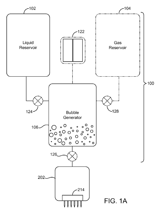
5 FIG. IA depicts a first embodiment of a device 100 for generating
microbubbles in
the enhancing solution. The device 100 consists of a liquid reservoir 102, a
gas vapor
CA 02702420 2010-04-13
WO 2009/006008 PCT/US2008/067124
16
reservoir 104 (shown in dashed lines) and a bubble generator 106. The bubble
generator
106 is a vessel or vessels in which the fluid and gas are mixed. Fluid from
the liquid
reservoir 102 and gas/vapor from the gas reservoir 104 flow into the bubble
generator 106
and are mixed to create microbubbles and/or supersaturate the fluid.
The device 100 may include a fluid metering device 124 (shown in dashed lines)
controlling the amount of fluid dispensed into the bubble generator 106 and/or
a fluid
metering device 126 (shown in dashed lines) controlling the amount of
microbubble
solution to be injected into the tissue. The device 100 may further include a
gas metering
device 128 (shown in dashed lines) used to control the amount of gas dispensed
into the
bubble generator 106. The device 100 depicted in FIG. IA includes both of the
fluid
metering devices 124 and 126 and the gas metering device 128; however, in
practice one
or more of these devices may be eliminated. As noted previously, two or more
components may be integrated together. For example, the fluid metering device
124 may
be integrated into the fluid injection device 202.
FIG. lB is a more detailed illustration of a first embodiment of the bubble
generator 106 and includes a housing 108, a pair of cylinders 116
interconnected by a
pathway 118. At least one of the cylinders 116 is in fluid communication with
the liquid
reservoir 102, and at least one of the cylinders 116 is in fluid communication
with the gas
reservoir 104 (which may be ambient environment). The fluid pathway 118
provides fluid
communication between the cylinders 116.
One or more of the cylinder(s) 116 may be provided with a reciprocating piston
120 driven by an external power source 122 such as a source of compressed air,
spring,
elastomeric member, motor, stepper motor or the like. According to one
embodiment, the
reciprocating piston 120 is a pneumatic piston manufactured by the Bimba
Corporation.
Liquid from the liquid reservoir 102 may be pushed into the bubble generator
106
under positive pressure from an external pressurization source 110 (shown in
dashed
lines); it can be drawn into the bubble generator 106 under partial pressure
which may for
example be generated by the reciprocating piston 120; or it can flow into the
generator 106
under gravity. Similarly, gas from the gas reservoir 104 may be pushed into
the bubble
generator 106 under positive pressure from an external pressurization source
112 (shown
in dashed lines) or it can be drawn into the bubble generator 106 under
partial pressure.
As will be described below, the piston 120 may also serve a dual purpose as a
fluid
pressurization mechanism for injecting the fluid into the tissue.
CA 02702420 2010-04-13
WO 2009/006008 PCT/US2008/067124
17
The bubble generator 106 may or may not be pressurized to enhance the
saturation
of the gas in the solution or prevent dissolved gas from coming out of
solution. An
optional fluid pressurization mechanism 110 (shown in dashed lines) may be
used to
maintain the fluid at a desired pressurization. As will be described in
further detail below,
the fluid may be chilled to further enhance solubility/saturation of the gas
in the solution.
FIG. IC is an alternate embodiment of the microbubble generator 106, which
utilizes a member 120' (rotor) such as a blade, paddle, whisk, semi-permeable
membrane
or the like driven by an external power source 122 to generate the
microbubbles within a
cylinder or mixing chamber (stator) 116. As will be appreciated by one of
ordinary skill in
the art the member 120' is rotationally driven by the external power source
122 within a
cylinder 116 or the like. An optional fluid pressurization mechanism 130 may
be used for
injecting the fluid into the tissue.
The fluid in the reservoir 102 may be at ambient temperature. Alternatively,
the
fluid may be chilled slightly to enhance gas solubility (super saturation).
The fluid
reservoir 102 may be thermally insulated to maintain the fluid at its present
temperature
and or/ the fluid reservoir 102 may include a heating/cooling mechanism (not
illustrated)
to maintain the fluid at a predetermined temperature.
If the gas used is air then the gas reservoir 104 may be eliminated in favor
of
simply drawing air from the environment, i.e., the room housing the device 100
("room
air"). If room air is used, the device 100 may include an air filter 114
(shown in dashed
lines) such as a HEPA filter or the like.
FIG. 1D is an alternate embodiment of the microbubble generator 106, which
utilizes an agitator 133 to agitate or shake a container or cartridge 132
containing
measured amounts of liquid and gas and generate the microbubbles within the
cartridge
132. The microbubble solution is dispensed from the cartridge 132 to fluid
injection
device 202 (FIG. 2). Additionally, this cartridge 132 may incorporate an
active
heating/cooling mechanism to control the temperature of the fluid at a
predetermined
setting. Furthermore, the cartridge 132 may be pressurized, such as by
compressed air or
mechanical mechanism to allow dispensation of the contents at a predetermined
rate and
pressure.
FIG. 2 is a block diagram of a liposculpture system 200 according to the
present
invention. The system 200 includes device 100, a fluid injection device 202,
an ultrasound
transducer apparatus 204, an ultrasound generator 206, an ultrasound control
unit 208, and
CA 02702420 2010-04-13
WO 2009/006008 PCT/US2008/067124
18
an injection control unit 210. Device 100 may include the bubble generator 106
depicted
in FIGs. IA - 1D or may be one of the alternative embodiments disclosed herein
below.
The fluid injection device 202 may include a needle array 214 which may
include
one or more needles 218. Alternatively, the fluid injection device 202 may,
for example,
include one or more hypodermic syringes.
The fluid injection device 202 further includes or is operably connected to a
fluid
pressurization mechanism 110 for pushing the solution into the tissue. As
noted above, the
piston 120 or the like used to express fluid between the cylinders 116 may
serve as the
fluid pressurization mechanism 210.
One or more of the components collectively termed system 200 may be combined.
For example the fluid injection device 202 may be integrated as a single
component with
the ultrasound transducer apparatus 204 and/or the fluid injection control
unit 210.
Likewise, the ultrasound control unit 208 can be integrated as a single
component with the
ultrasound generator 206. Such integration of components is contemplated and
falls within
the scope of the present invention.
The fluid injection control unit 210 may control the amount of fluid and gas
dispensed into the bubble generator 106 and/or the amount of solution injected
into the
tissue. Optionally, the control unit 210 may be interfaced directly or
indirectly with the
fluid metering device(s) 124, 126 and the gas metering device 128. The fluid
injection
control unit 210 may control the mixing or agitation (if any) of the solution
within the
bubble generator 106. The fluid injection control unit 210 may control the
injection of
solution into the tissue 220 by the injection device 202, including the
deployment of a
needle array 214, the depth to which the needle array 214 is deployed, and the
amount of
solution injected.
The fluid injection control unit 210 may control the individual deployment and
retraction of one more needles (or hypodermic syringes) of the needle array
214. Thus, the
control unit 210 may deploy or retract the needles 218 (or hypodermic
syringes) one at a
time, may deploy or retract two or more needles 218 at a time, or may deploy
or retract all
of the needles simultaneously.
Additionally, the fluid injection control unit 210 may individually control
the
amount of solution delivered to each needle 218. One of ordinary skill in the
art will
appreciate that there are many ways to control the amount of solution
delivered to each
needle 218. For example, it may be desirable to deliver more solution in the
center of the
treatment area and less to the peripheral portion of the treatment area or
vice-versa.
CA 02702420 2010-04-13
WO 2009/006008 PCT/US2008/067124
19
If the injection device 202 utilizes hypodermic syringes, then the fluid
injection
control unit 210 may control the amount of fluid distributed to each syringe.
As noted
above it may be desirable to provide differing amounts of solution to
different areas of the
treatment area, and this may be achieved by varying the amount of solution in
each
syringe.
As best seen in FIGs. 3A-3C, the fluid injection device 202 may include a
manifold
or fluid distribution pathway 212 (shown in dashed lines) in fluid connection
with device
100 and needle array 214, and a needle deployment mechanism 216 operably
connected to
the needle array 214. The manifold 212 is the fluid pathway used to transport
the
microbubble solution from the microbubble generator 106 to the needle array
214.
One or more flow control devices 222 may be provided in the fluid pathway 212
to
enable individualized control of the amount of fluid dispensed to each of the
needles or
syringes 218. The manifold 212 alone or in combination with the flow control
devices 222
controls the distribution of the microbubble solution among the needles 218.
The manifold
212 may be configured to deliver a uniform amount of solution to each of the
needles 218
(or hypodermic syringes), or it may be configured to deliver differing amounts
of solution
to different needles 218. The flow control devices 222 may be manually
adjustable and/or
maybe controlled by the injection control unit 210. An alternate embodiment
may include
infinitely variable volume control at each needle or hypodermic through active
means,
such as with an electronic flow meter and controller.
It may be desirable to deploy all of the needles 218 simultaneously into the
tissue
but deliver solution to one or more needles 218 individually. For example, it
may be
desirable to deliver solution sequentially to groups of one or more needles
218. If needles
218 are deployed individually or in groups of two or more it may be desirable
to deliver
solution only to the deployed needles 218.
As will be explained below, the injection depth may be manually determined by
selecting an appropriate needle length or setting a desired injection depth.
The needle deployment mechanism 216 (FIGs. 2 and 3A) deploys one or more
needles 218 (or hypodermic syringes) of the needle array 214 such that needles
218
penetrate a desired distance into the tissue. The needle deployment mechanism
216 may
be configured to deploy the needle(s) 218 to a fixed predetermined depth or
may include
means for adjusting the depth that the needle(s) 218 are deployed.
There are several broad approaches for adjusting the injection depth which may
be
utilized. One way to adjust the injection depth is to provide needle arrays
214 of varying
CA 02702420 2010-04-13
WO 2009/006008 PCT/US2008/067124
length needles. According to this embodiment, the user simply selects an array
214 having
shorter/longer needles 218 to achieve a desired injection depth. Moreover, the
different
length needles 218 may be used within a given array 214.
According to another approach, the needle array 214 is displaced vertically in
order
5 to adjust the injection depth.
FIG. 3A shows aspects of an adjusting means, which may include a flange 244A
and a groove 244B arrangement for vertically adjusting the needle array in
discrete
intervals.
FIG. 3D shows aspects of an adjusting means, which may include mating screw
10 threads 240 formed on the needle array 214 and the fluid injection device
202 or housing
108 which enable the user to vertically adjust the needle array 214 thereby
altering the
injection depth.
According to one embodiment, the injection depth may be continuously adjusted
within a given range of injection depths. For example, the user may be able to
continually
15 adjust the injection depth between 5 and 12 millimeters by rotating the
needle array 214.
According to an alternate embodiment, the injection depth may be adjusted in
discrete
intervals. For example, the user may be able to adjust the injection depth
between 3 and
15 millimeters in 1 millimeter increments. In yet another embodiment, the
needle depth
may be controlled electronically whereby the user enters a specified depth on
the control
20 unit 210.
The injection depth adjustment described above may specify the injection depth
for
the entire needle array 214. However, according to yet another approach it may
be
desirable to facilitate the individualized adjustment of one or more needles
218 of the
needle array 214. The needle deployment mechanism 216 may allow for the
independent
adjustment of the injection depth for one or more of the needles 218 or
syringes.
One or more of the needles 218 or syringes may be displaced vertically in
order to
adjust the injection depth of individual needles. The adjustment of the
injection depth
(vertical needle displacement) may be continuous or in discrete intervals, and
may be
manual or may be adjusted via the injection control unit 210.
As noted above, the injection depth may be adjusted by providing mating screw
threads 246 to dial in the desired injection depth (FIG. 4A), a standoff 248
to provide a
means for adjusting the injection depth in discrete intervals (FIG. 4B), or
the like on the
needle array 214 to adjust the vertical height of the needles 218 relative to
the tissue
apposition surface 226A.
CA 02702420 2010-04-13
WO 2009/006008 PCT/US2008/067124
21
Yet another approach to individualized injection depth control is to deploy
individual needles or syringes 218 as opposed to deploying the entire needle
array 214.
The injection control unit 210 or needle deployment mechanism 216 selects the
injection
depth of each individual needle or syringe 218 (FIG. 4C).
One of ordinary skill in the art will appreciate that there are many other
ways to
implement the adjustment of the injection depth. The invention is not limited
to the
embodiments depicted in the drawings.
The needle deployment mechanism 216 deploys the needles 218 in response to a
signal from the fluid injection control unit 210. The deployment mechanism 216
may
include a spring, pneumatic ram, or the like which deploys the needles 218
with sufficient
force to penetrate the tissue 220. The fluid injection control unit 210
synchronizes the
deployment mechanism 216 with the injection of the microbubble solution into
the tissue.
A predetermined amount of the solution may be injected at a single injection
depth.
Alternatively, the fluid injection control unit 210 in synchronism with the
deployment
mechanism 216 may inject solution at each of plural injection depths, or may
inject
continuously as the needle array 214 on either the forward (penetration) or
rearward
(withdrawal) strokes. It may be desirable to deploy the needles to a first
depth within the
tissue and then retract the needles to a slightly shallower injection depth
before injecting
the solution.
FIG. 5 is an enlarged view of the needle array 214 including at least one
hypodermic needle or micro-needle 218. The invention is not limited to any
particular
length or gauge needle, and needles 218 are selected in accordance with the
depth of the
tissue to be treated and to accommodate patient comfort. Moreover, it may be
desirable
for the needle array 214 to include needles of varying length and/or needles
of varying
gauge.
The embodiment depicted in FIG. 5 includes a plurality of uniformly spaced
needles 218. However, the scope of the invention is not limited to any
particular number
of needles 218; moreover, the invention is not limited to any particular
geometric
arrangement or configuration of needles 218. It may be desirable to have non-
uniform
needle spacing. For example, it may be desirable to have a smaller (denser)
needle spacing
in one portion of the treatment region and a greater (sparser) needle spacing
in another
portion. The use of additional needles 218 may facilitate uniform distribution
of the
microbubble solution in the tissue 220 and/or reduce the number of distinct
injection
cycles needed to treat a given area.
CA 02702420 2010-04-13
WO 2009/006008 PCT/US2008/067124
22
FIG. 6A depicts a needle 218 having a single injection orifice 242, which is
linearly aligned with the needle shaft 224. The hypodermic needle 218 is a
tubular
member having a lumen configured for injection of the solution through the
needle and
into the tissue. The lumen may include a textured surface for promoting the
generation of
microbubbles.
FIG. 6B depicts an alternative needle 218A having one or more side firing
orifice(s) 242A which are generally orthogonal to longitudinal axis of the
shaft 224A. The
side firing orifice(s) may be formed at different heights along the length of
the needle shaft
such that solution is injected at varying injection depths. These orifice(s)
may also be
arranged in a specific radial pattern to preferentially direct the flow
distribution.
Depending on the characteristics of the tissue undergoing treatment the user
may
find that needle 218 is preferable over needle 218A or vice versa. Reference
to the needles
218 should be understood to refer generally to both the needles 218 (FIG. 6A)
and the
needles 218A (FIG. 6B).
As shown in FIG. 7, some embodiments of the invention may include a mechanism
256 for selectively rotating one or more of the needles 218 in situ. This
feature may
facilitate the uniform distribution of solution in the tissue.
According to some embodiments of the invention it may be desirable for the
needle
deployment mechanism 216 to ultrasonically vibrate one or more of the needles
218. This
feature may facilitate tissue penetration and/or bringing dissolved gas out of
solution. For
example, an ultrasound transducer 258 may be operably coupled to the needles
218 and/or
the needle array 214. The ultrasound transducer 258 is shown for the sake of
convenience
in FIG. 7 however, the transducer 258 may be used in a device which does not
include the
needle rotation mechanism 256 and vice versa.
As best seen in FIG. 8A, the hypodermic needle 218 has a proximal end
connected
to the fluid distribution pathway 212 and a distal end configured for
penetrating into the
tissue 220 to be treated. In one embodiment, the needles 218 may include micro-
needles.
In one embodiment, the fluid injection device 202 includes needle deployment
mechanism 216 for moving the hypodermic needle 218 from a fully retracted
position
(FIG. 8A) in which the distal end of the needle 218 is housed inside the
solution injection
member 202 to a fully extended position (FIG. 8B).
As shown in FIGs. 9A-9C, the fluid injection device 202 may optionally be
provided with a tissue apposition mechanism which urges the device 202 into
firm
apposition with the tissue 220 undergoing treatment. According to one
embodiment the
CA 02702420 2010-04-13
WO 2009/006008 PCT/US2008/067124
23
tissue apposition mechanism includes at least one vacuum port 228 and a vacuum
source
230 in fluid communication with the vacuum port 228. The vacuum port 228 may
be
defined in the needle array 214 and/or the housing 108. In operation the
tissue apposition
surface 226A is pulled into apposition with the tissue 220 when vacuum from
the vacuum
source 230 is transmitted through the vacuum port 228 to the tissue 220.
In some embodiments it may be desirable to provide a one-to-one relationship
between needles 218 and vacuum ports 228. Moreover, the needle(s) 218 may be
positioned within the vacuum port(s) 228. The vacuum port 228 may define a
recess or
receptacle 229 such that the tissue 220 is at least partially pulled (sucked)
into the recess
229 by the vacuum force. Moreover, the needles 218 may be at least partially
housed
within and deployed through the recess 229.
An optional flange 232 (show in dashed lines) may surround (skirt) the
periphery
of the needles 218 (or 218A) to channel/contain the suction force.
Alternatively, a
separate flange 232A may surround (skirt) each of the needles 218 (or 218A) to
channel/contain the suction force.
It may be desirable to have one or more vacuum ports 228 spaced along a
periphery
of the apposition surface 226A. Moreover, it may be desirable to include a
central portion
apposition surface 226A, which does not include any vacuum ports 228 (no
suction zone).
Alternatively, it may be desirable to have vacuum ports confined to a central
portion of the
apposition surface 226A.
It should be appreciated that the liquid reservoir 102 and gas reservoir 104,
in each
of the aforementioned embodiments may be replaced with a cartridge 132 (FIG.
1D)
containing a pre-measured amount of liquid and gas. The gas may be fully or
partially
dissolved in the fluid. In its simplest form the cartridge 132 is simply a
sealed container
filled with a predetermined amount of gas and liquid, e.g., a soda can.
FIG. l0A shows an enhanced cartridge 106A ("Guinness can"), which may be used
to replace the liquid reservoir 102, gas reservoir 104, and bubble generator
106 in each of
the aforementioned embodiments. In this embodiment, the cartridge 106A
includes a
hollow pressurized pod 134 such as disclosed in U.S. 4,832,968, which is
hereby
incorporated by reference. Both the cartridge 106A and the pod 134 contain a
solution of
gas and liquid under greater than ambient pressure which may for example be
achieved by
providing or introducing a dose of liquid nitrogen into the solution before
sealing the
cartridge 106A.
CA 02702420 2010-04-13
WO 2009/006008 PCT/US2008/067124
24
The cartridge 106A includes a headspace 136, which is bounded between a top
inner surface 138 and a gas-liquid interface 140. The pod 134 includes a
similar
headspace 142, which is bounded between a top inner surface 144 and a gas-
liquid
interface 146.
The pod 134 includes a small opening or orifice 148, which enables the
pressure
within the headspace 136 of the cartridge 106A to reach equilibrium with the
pressure
within the headspace 142 of the pod 134. When a seal 150 of the cartridge 106A
is pierced
the pressure within the headspace 136 rapidly reaches equilibrium with the
ambient
pressure. In the moments after seal 150 is pierced the pressure within the pod
134 is
greater than the pressure in the headspace 136 of the cartridge 106A because
the orifice
148 restricts the rate of flow of solution out of the pod 134. A jet of
solution forcefully
streams out of the orifice 148 into the solution within the cartridge 106A,
which agitates
and/or shears the solution within the cartridge causing some of the dissolved
bubbles to
come out of solution thereby generating microbubbles in the solution.
The pod 134 is preferably situated at or near the bottom of the cartridge 106A
such
that the orifice 148 is maintained below the liquid gas interface 140.
FIG. l0B is a block diagram showing the system 200 including cartridge 106A in
place of bubble generator 106.
The microbubble generator 106 may be mounted on (integrated with) the fluid
injection device 202 thereby minimizing the distance that the solution travels
before being
injected into the tissue. The liquid reservoir 102 and gas reservoir 104 (if
provided) may
be removably connected to the microbubble generator 106 as needed to generate
microbubble solution. The injection device 202 may be manually supported by
the
operator. Alternatively, the injection device 202 may be supported on an arm
302 (FIG.
11) which may include a counterbalance to facilitate manipulation of the
injection device
202.
FIG. 12A depicts a handpiece 300 which includes fluid injection device 202 and
which is coupled to the microbubble generator 106 (not illustrated) by a
flexible conduit
236. This design minimizes the size and weight of handpiece 300 being handled
by the
operator since the handpiece 300 does not include the microbubble generator
106.
FIG. 12B depicts a handpiece 300 using the cartridge 106A mounted on the fluid
injection device 202. This embodiment minimizes the distance that the
microbubble
solution travels before being injected into the tissue.
CA 02702420 2010-04-13
WO 2009/006008 PCT/US2008/067124
According to one embodiment the system of the invention includes a container
which may be an enclosed or sealed cartridge 106A or it may be an open
container. If the
container is sealed it includes a measured amount of a solution. Obviously, if
the container
is not sealed then solution may be freely added as needed.
5 The system includes a needle array including at least one needle. The needle
array
214 being in fluid connection with the container.
The solution includes any of the solutions disclosed herein. The solution
includes a
liquid. The solution may further include a gas which may be partially or fully
dissolved
within the solution.
10 The container may be enclosed and the solution may be maintained at greater
than
atmospheric pressure.
The needle array 214 includes at least one needle 218 which may be any of the
needles disclosed herein.
The aforementioned gas may include one or more gases selected from the group
of
15 air, oxygen, carbon dioxide, carbon dioxide, perfluoropropane, argon,
hydrogen,
Halothane, Desflurane, Sevoflurane, Isoflurane, and Enflurane.
The solution may include one or more of a vasoconstrictor, a surfactant, and
an
anesthetic. Moreover, the vasoconstrictor may include one or more of
Norepinephrine,
Epinephrine, Angiotensin II, Vasopressin and Endothelin.
20 Optionally, the system may include refrigeration means for maintaining the
container at a predefined temperature range. Moreover, the container may be
thermally
insulated.
The system may further include an ultrasound transducer apparatus 204 for
transmitting ultrasound waves to the tissue. Preferably, the transducer
apparatus 204 is
25 operated in synchronism with the injection of solution into the tissue.
The transducer apparatus 204 may transmit ultrasound energy at a first setting
to
facilitate the distribution, absorption and/or uptake of solution by the
tissue, i.e.,
sonoporation.
Ultrasound parameters that enhance the distribution of the solution include
those
conditions which create microstreaming, such as large duty cycle pulsed
ultrasound (>
10% duty cycle) or continuous wave ultrasound at a range of frequencies from
500 kHz to
15 MHz, focused or unfocused, and a mechanical index less than 4. According to
one
embodiment the mechanical index (MI) falls within the range .5 < MI < 4.
According to
another embodiment the mechanical index falls within the range .5 < MI < 1.9.
CA 02702420 2010-04-13
WO 2009/006008 PCT/US2008/067124
26
Sonoporation leading to increased absorption and/or uptake of the solution in
the
tissue can be generated by pulsed wave or continuous wave ultrasound, at a
range of
frequencies from 500 kHz to 15 MHz, focused or unfocused and medium to high
mechanical index (MI > 1.0). The preferred embodiment is pulsed wave
ultrasound at a
frequency of 500 kHz, unfocused, with high mechanical index (MI > 1.9) in
order to
reproducibly create pores that are temporary or longer lasting pores.
The transducer apparatus 204 may transmit ultrasound energy at a second
setting to
facilitate the generation of bubbles by bringing dissolved gas out of
solution, i.e., stable
cavitation.
Ultrasound parameters for stable cavitation such as large duty cycle pulsed
ultrasound (> 10% duty cycle) or continuous wave ultrasound at a range of
frequencies
from 2MHz to 15 MHz, focused or unfocused, and a mechanical index (MI) .05 <
MI <2Ø
The transducer apparatus 204 may transmit ultrasound energy at a third setting
to
facilitate transient cavitation, i.e., popping bubbles.
Ultrasound parameters for transient cavitation at a range of frequencies from
500kHz to 2 MHz, focused or unfocused, and a mechanical index (MI) greater
than 1.9.
The duty cycle required for transient cavitation may be very low, and the
preferred
embodiment is a wideband pulse (1 to 20 cycles) transmitted at a duty cycle
less than 5%.
The transducer apparatus 204 may include any of the transducers disclosed
herein,
and may be operably connected to the needle array 214.
The transducer apparatus 204 may transmit ultrasound energy at a fourth
frequency
range to facilitate the pushing of bubbles within the tissue by acoustic
streaming and/or
acoustic radiation force.
Ultrasound Acoustic Streaming and Radiation Force
Sound propagating through a medium produces a force on particles suspended in
the medium, and also upon the medium itself. Ultrasound produces a radiation
force that
is exerted upon objects in a medium with an acoustic impedance different than
that of the
medium. An example is a nanoparticle in blood, although, as one of ordinary
skill will
recognize, ultrasound radiation forces also may be generated on non-liquid
core carrier
particles. When the medium is a liquid, the fluid translation resulting from
application of
ultrasound is called acoustic streaming.
The ability of radiation force to concentrate microbubbles in-vitro and in-
vivo has
been demonstrated, e.g., Dayton, et al., Ultrasound in Med. & Biol.,
25(8):1195-
1201(1999). An ultrasound transducer pulsing at 5 MHz center frequency, 10 kHz
pulse
CA 02702420 2010-04-13
WO 2009/006008 PCT/US2008/067124
27
repetition frequency ("PRF"), and 800 kPa peak pressure, has been shown to
concentrate
microbubbles against a vessel wall in-vivo, and reduce the velocity of these
flowing agents
an order of magnitude. In addition, the application of radiation to
concentrate drug
delivery carrier particles and the combined effects of radiation force-induced
concentration
and carrier fragmentation has been demonstrated. See U.S. patent application
Ser. No.
10/928,648, entitled "Ultrasonic Concentration of Drug Delivery Capsules,"
filed Aug. 26,
2004 by Paul Dayton et al., which is incorporated herein by reference.
Acoustic streaming and optionally radiation force may be used to "push" or
concentrate microbubbles injected into the tissue along a cell membrane.
Notably,
acoustic streaming has previously been used to push or concentrate carrier
particles within
a blood vessel. In contrast, the present invention utilizes acoustic streaming
to push
bubbles within subcutaneous tissue to concentrate the bubble against the walls
of cells to
be treated.
According to one aspect of the present invention, a solution containing
microbubbles is injected into subcutaneous tissue or a solution containing
dissolved gas is
injected into subcutaneous tissue and insonated to bring the gas out of
solution thereby
generating bubbles within the subcutaneous tissue. The bubbles are pushed
against the cell
walls using acoustic streaming, and then insonated to induce transient
cavitation to
enhance the transport of the solution through the cell membrane and/or
mechanically
disrupt the cell membrane to selectively lyse cells.
The ultrasound parameters useful for inducing acoustic streaming include
ultrasound waves having center frequencies about 0.1-20 MHz, at an acoustic
pressure
about 100 kPa-20 MPa, a long cycle length (e.g., about >10 cycles and
continuous-wave)
OR a short cycle length (e.g., about <10 cycle), and high pulse repetition
frequency (e.g.,
about >500 Hz). The specific parameters will depend on the choice of carrier
particle, as
detailed further below, and can be readily determined by one of ordinary skill
in the art.
According to one embodiment, the transducer apparatus 204 includes a single
transducer capable of operating a plurality of operating modes to facilitate
stable
cavitation, transient cavitation, acoustic streaming, and sonoporation.
According to
another embodiment, the transducer apparatus 204 includes first and second
transducers
with first transducer optimized for popping bubbles (transient cavitation) and
the second
transducer optimized for bringing dissolved gas out of solution (stable
cavitation) and/or
pushing the bubbles using acoustic radiation force.
CA 02702420 2010-04-13
WO 2009/006008 PCT/US2008/067124
28
The transducer apparatus may produce focused, unfocused, or defocused
ultrasound waves. Focused ultrasound refers to generally converging ultrasound
waves,
unfocused ultrasound refers to generally parallel ultrasound waves and
defocused
ultrasound wave refers to generally diverging ultrasound waves.
However, according to a preferred embodiment, the transducer apparatus 204
selectively produces unfocused and/or defocused ultrasound waves. For example,
it may
be desirable to utilize unfocused waves during transient cavitation, and
defocused waves
during stable cavitation. To this end the transducer apparatus may include a
flat
transducer, i.e., a transducer having a generally planar acoustic wear layer
(acoustic
window) for producing unfocused ultrasound waves (nonconverging waves) and/or
a
convex transducer, i.e., a transducer having a convex acoustic wear layer for
producing
defocused ultrasound waves (diverging waves).
As will be appreciated by one of ordinary skill in the art, there are many
different
configurations for the ultrasound apparatus. FIG. 14 depicts an embodiment in
which the
transducer apparatus 204 includes an inner transducer 204A and an outer
transducer 204B.
In the illustrated embodiment, the inner transducer 204A has a convex shaped
acoustic
wear layer for producing defocused waves 205A, and the outer transducer 204B
has a
planar shaped acoustic wear layer for producing unfocused waves 205B. However,
both of
the inner and outer transducers 204A and 204B may be planar or both may be
convex.
Still further, one or both of the inner and outer transducers may be concave,
i.e., may have
a concave acoustic wear layer for producing focused waves. Thus, the
ultrasound
apparatus 204 may include any combination of focused, unfocused, and defocused
transducers.
The inner and outer transducers depicted in FIG. 14 are both circular and the
outer
transducer surrounds (encircles) the inner transducer. However, other
configurations are
contemplated and fall within the scope of the invention. According to a
presently
preferred embodiment, the inner transducer is used to produce stable
cavitation and the
outer transducer is used to create transient cavitation. However, the relative
positions may
be swapped with the inner transducer producing transient cavitation and the
outer
transducer producing stable cavitation.
The ultrasound apparatus 204 illustrated in FIG. 14 includes a needle array
214 of
the type described elsewhere in this disclosure. The transducer apparatus 204
of FIG. 14
may be incorporated in any of the embodiments disclosed herein which include
an
CA 02702420 2010-04-13
WO 2009/006008 PCT/US2008/067124
29
ultrasound transducer. Notably, the transducer apparatus 204 may be
incorporated in
system 200.
It should be noted that the transducer apparatus 204 may include one or more
arrays of transducers. For example, the transducer apparatus may include an
array of
transducers for stable cavitation and/or an array of transducers for transient
cavitation.
According to another aspect of the present invention, a solution which may or
may
not include microbubbles is injected into subcutaneous tissue. The solution is
pushed
against the cell walls using acoustic streaming, and then the subcutaneous
tissue is
insonated to induce sonoporation and facilitate the uptake/absorption of
solution by the
tissue. Solution is injected an insonated using a system such as system 200
depicted in
FIG. 13 which does not include a bubble generator 100. Absorption of the
solution
preferably results in cell lysis.
As described in U.S. Utility Patent Application Serial Number 11/292,950 filed
December 2, 2005, the ultrasound energy from ultrasound generator 206 is
applied to the
tissue 220 via ultrasound transducer 204. Ultrasound control unit 208 controls
the various
ultrasound parameters and generally controls the supply of ultrasound by
generator 206.
Preferably, ultrasound control unit 208 communicates with the injection
control unit 210 to
synchronize the application or ultrasound energy with the injection of fluid.
It may for
example be desirable to quickly apply energy to the tissue before the
microbubbles
dissipate or are absorbed by the tissue.
The ultrasound transducer 204 is preferably configured to deliver unfocused
ultrasound at an intensity and pressure sufficient to noninvasively cavitate
the
microbubbles within tissue thereby causing cell lysis. The intensity and
pressure of the
ultrasound applied to the tissue is preferably selected to minimize the
heating of tissue and
in particular avoid burning the patient's skin. The transducer 204 may include
a
thermocouple 238 or the like to monitor the temperature of the transducer 204.
In at least one embodiment the liposculpture system 200 (FIG. 2) includes an
ID
reader 250 (shown in dashed lines), and the needle array 214 includes an
identifier 252
(shown in dashed lines), which uniquely identifies the needle array 214. The
ID reader
250 reads the identifier 252, and preferably authenticates or verifies the
needle array 214.
The identifier 252 may contain information identifying the characteristics of
the needle
array 214 such as length and gauge of needles. The identifier 252 may further
include
identifying information which may be used to track the number of injection
cycles (needle
deployments) or use time for a given array 214.
CA 02702420 2010-04-13
WO 2009/006008 PCT/US2008/067124
The reader 250 preferably communicates with the injection control unit 210.
The
injection control unit 210 may count the number of injection cycles that a
given needle
array 214 has been used, and may warn the operator if the number exceeds a
threshold
number. The injection control unit 250 may use information stored on the
identifier 252 to
5 adjust the injection depth or injection flow rate. The injection control
unit 210 may further
inhibit usage of a needle array if it cannot authenticate, verify or read the
identifier 252.
The identifier 252 may be a barcode label, a radio frequency tag, smart chip
or
other machine-readable medium such as known in the art.
The ultrasound transducer 204 may also include an identifier 252. The
identifier
10 252 may be used to store information identifying the characteristics of the
transducer 204,
which is used by the ultrasound control unit 208 in setting or verifying the
treatment
settings. The ultrasound control unit 208 may inhibit insonation if it cannot
authenticate,
verify or read the identifier 252.
As described above, the transducer 204 may be integrated with the needle array
15 214 in which case a single identifier 252 may store information describing
characteristics
of both the needle(s) 218 and the transducer 204. The ultrasound control unit
208 may use
information on the identifier 252 to track the amount of time the identified
ultrasound
transducer 204 has been operated and at what power levels, and may inhibit
insonation if
the accumulated insonation time exceeds a threshold value.
20 The constituent components of the device 100 may be formed of any
sterilizable,
biocompatible material. Moreover, some or all of the components may be
disposable, i.e.,
manufactured for single-patient use, to minimize potential cross-contamination
of patients.
The needle array 214 is preferably a disposable component, as the needles 218
will likely
dull with use.
25 One or more optical or pressure sensors 254 (FIG. 5) may be provided to
measure
pressure exerted on the handpiece 300 (FIG. 12A) when the handpiece is placed
in
abutment with the tissue. The pressure sensor(s) 254 may provide a safety
interlock
function to prevent inadvertent deployment of the needle array 214 and/or
actuation of the
transducer 204 unless pressure is detected as the handpiece 300 is placed in
abutment with
30 the tissue. If two or more pressure sensors 254 are provided the injection
of solution
and/or insonation may be inhibited unless each of the measured pressure values
fall within
a predefined window and/or so long as the difference between any given two
measured
pressure values is less than a threshold value. The pressure sensor(s) 254
may, for
example, be provided on the needle array 214 (FIG. 4) or on the fluid
injection device 202
CA 02702420 2010-04-13
WO 2009/006008 PCT/US2008/067124
31
(not illustrated). Alternatively, other sensing means, possibly optical or
capacitive, may be
used to detect proper positioning of the needle array against the tissue to be
treated.
It may be advantageous to couple the needles 218 with the ultrasound
transducer
204 such that ultrasound is transmitted through the needle(s) 218 to the
tissue. Applying
ultrasound in this manner may facilitate targeted cavitation and/or may
facilitate
penetration of the needle(s) 218 into the tissue.
FIG. 13 is a block diagram of a system 200 for a fat lysing system according
to the
present invention. The system 200 is identical to the system 200 of FIG. 2 but
excludes
the bubble generator 100. Moreover, the ultrasound transducer 204, ultrasound
generator
206, and ultrasound control unit 208 are shown in dashed lines to indicate
that these are
optional components. The system 500 may be used to inject a fat lysing
solution (as will
be described below in greater detail) with or without the use of ultrasound.
According to one embodiment, the fat lysing solution includes epinephrine as
its
active ingredient. The epinephrine may be combined with an aqueous solution,
isotonic
saline, normal saline, hypotonic saline, hypotonic solution, or a hypertonic
solution. The
solution may optionally include one or more additives/agents to raise the pH
(e.g., sodium
bicarbonate) or a buffering agent such those listed in Table 5 above or other
buffering
agents such as known in the art.
According to a presently preferred embodiment the fat lysing solution includes
epinephrine in hypotonic buffered saline.
The inclusion of ultrasound in system 200 may facilitate the absorption and/or
distribution of the fat lysing solution. The inclusion of ultrasound in system
200 may
facilitate the absorption and/or distribution of the fat lysing solution. More
particularly,
the ultrasound may be used to enhance the distribution, absorption, and/or
uptake of the
solution in the tissue by permanently or temporarily opening pores in the cell
membrane
(sonoporation), generating microstreaming in the solution, or locally heating
the solution
or the tissue. According to one aspect of the invention, the ultrasound
generator 206 may
be operated at a first setting to facilitate distribution of the solution and
then it may be
operated at a second setting to facilitate absorption. The sonoporation may be
reversible or
irreversible.
The system 200 may include an optional ultrasound transducer 258 for vibrating
the needles 218 to facilitate tissue penetration and/or a needle rotation
mechanism 256
which may be used in conjunction with side-firing needles 218 to facilitate
even
distribution of the solution. The same transducer apparatus 204 used to
facilitate
CA 02702420 2010-04-13
WO 2009/006008 PCT/US2008/067124
32
absorption and/or distribution of the solution may be used to facilitate
tissue penetration
thereby eliminating the need for a separate transducer 258.
The system 200 may include any or all of the features described in this
disclosure
including means for selectively adjusting the amount of solution injected by
each of the
needles 218 and/or the rate or pressure at which the solution is injected into
the tissue.
Still further the system 200 may include the selective adjustment of the
injection depth
and/or the tissue apposition mechanism as described above.
Mode of Operation/ Method of Use
According to a first mode of operation, solution is percutaneously injected
into
subcutaneous tissue, and the tissue is insonated at a first ultrasound setting
to distribute the
solution. Once the solution has been distributed the tissue is insonated at a
second setting
to induce sonoporation thereby inducing cell lysis. According to this mode of
operation
the solution need not contain microbubbles as they do not contribute to cell
lysis. To
increase the efficacy of this mode of operation it is recommended to repeat
the injection
and insonation of the tissue through 10 to 50 cycles.
According to a second mode of operation, a solution containing microbubbles is
percutaneously injected into subcutaneous tissue, and the tissue is insonated
at a first
ultrasound setting to distribute the solution and/or push the microbubbles
against the cell
walls. Thereafter the tissue is insonated at a second setting (for between 1
millisecond and
1 second) to induce transient cavitation inducing cell lysis. To increase the
efficacy of this
mode of operation it is recommended to repeat the injection and insonation of
the tissue
through 10 to 50 cycles.
It should be appreciated that it is important to synchronize the timing of the
insonation. Notably, the microbubbles will be absorbed by the tissue and/or go
into
solution within a relatively short period of time. Thus, it is important to
distribute the
microbubbles (using acoustic radiation force) and induce transient cavitation
within a
relatively short time after the solution has been injected into the
subcutaneous tissue.
According to a presently preferred embodiment, the tissue is insonated to
facilitate
distribution of the microbubble solution through acoustic radiation force
and/or
microstreaming occurs simultaneously as the solution is injected into the
tissue or within a
very short amount of time afterward. The injection of a small amount of the
microbubble
solution takes approximately 200 milliseconds and insonation to induce
distribution
through acoustic radiation force takes between 1 millisecond and 1 second.
Next, the
tissue is insonated to induce transient cavitation for approximately 400
milliseconds.
CA 02702420 2010-04-13
WO 2009/006008 PCT/US2008/067124
33
According to a third mode of operation, a solution containing dissolved gas,
i.e.,
dissolved gas bubbles is percutaneously injected into subcutaneous tissue, and
the tissue is
insonated at a first ultrasound setting to bring the bubbles out of solution
(for between 100
microseconds and 1 millisecond) followed immediately by insonation at a second
setting
(for between 1 millisecond and 1 second) to distribute the solution and/or
push the
microbubbles against the cell walls. Thereafter the tissue is insonated at a
third setting (for
between 100 microseconds and 1 second) to induce transient cavitation inducing
cell lysis.
To increase the efficacy of this mode of operation it is recommended to repeat
the injection
and insonation of the tissue through 10 to 50 cycles.
It should be appreciated that it is important to synchronize the timing of the
insonation. Notably, the microbubbles will be absorbed by the tissue and/or go
into
solution within a relatively short period of time. Thus, it is important to
distribute the
microbubbles (using acoustic radiation force) and induce transient cavitation
within a
relatively short time after the bubbles have been brought out of solution.
According to a presently preferred embodiment, the tissue is insonated to
induce
stable cavitation and bring the bubbles out of solution after the solution has
been injected
into the subcutaneous tissue. Satisfactory stable cavitation results have been
achieved by
insonating for approximately 100 microseconds. Thereafter the tissue is
insonated to
facilitate distribution of the microbubble solution through acoustic radiation
force and/or
microstreaming occurs. Insonating for between 1 millisecond and 1 second is
required to
distribute the microbubbles. Immediately thereafter the tissue is insonated to
induce
transient cavitation for approximately 400 milliseconds.
The invention may be combined with other methods or apparatus for treating
tissues. For example, the invention may also include use of skin tightening
procedures, for
example, ThermaCoolTM available from Thermage Corporation located in Hayward,
California, Cutera TitanTM available from Cutera, Inc. located in Brisbane,
California, or
AlumaTM available from Lumenis, Inc. located in Santa Clara, California.
The invention may be embodied in other forms without departure from the spirit
and essential characteristics thereof. The embodiments described therefore are
to be
considered in all respects as illustrative and not restrictive. Although the
present invention
has been described in terms of certain preferred embodiments, other
embodiments that are
apparent to those of ordinary skill in the art are also within the scope of
the invention.
Accordingly, the scope of the invention is intended to be defined only by
reference to the
appended claims.