Note: Descriptions are shown in the official language in which they were submitted.
CA 02702615 2016-06-14
SYSTEMS AND METHODS FOR CARDIAC REMODELING
CROSS-REFERENCE TO RELATED APPLICATIONS
[0001]
BACKGROUND OF THE INVENTION
[0002] Blood returning to the heart from the peripheral circulation and the
lungs
generally flows into the atrial chambers of the heart and then to the
ventricular chambers,
which pump the blood back out of the heart. During ventricular contraction,
the atrio-
ventricular valves between the atria and ventricles, i.e. the tricuspid and
mitral valves, close
to prevent backflow or regurgitation of blood from the ventricles back to the
atria. The
closure of these valves, along with the aortic and pulmonary valves, maintains
the uni-
directional flow of blood through the cardiovascular system. Disease of the
valvular
apparatus can result in valve dysfunction, where some fraction of the
ventricular blood
regurgitates back into the atrial chambers.
[0003] There are several possible structural causes for atrio-ventricular
valve
dysfunction, including: loss of pliability of the annulus leading to decreased
contractibility;
widening of the annulus; thickening, shortening or swelling of the leaflets;
dilation of the
ventricle; elongation or breaking of the chordae tendineae; and elongation of
the attachment
of the chordae tendineae with the papillary muscles or ventricular wall.
Structural
abnormalities at one or more of these anatomical sites may eventually lead to
loss of
coaptation of the leaflets, loss of competence of the valve and decreased
efficiency of the
heart as a one-way pumping mechanism. When the latter occurs, various signs
and
symptoms may be seen in patients, including breathlessness or lack of stamina
and heart
murmurs.
[0004] Traditional treatment of heart valve stenosis or regurgitation, such as
mitral or tricuspid regurgitation, involves an open-heart surgical procedure
to replace or
repair the valve. Currently accepted treatments of the mitral and tricuspid
valves include:
valvuloplasty, in which the affected leaflets are remodeled to perform
normally; repair of the
chordae tendineae and/or papillary muscle attachments; and surgical insertion
of an
"annuloplasty" ring. This requires suturing a flexible support ring over the
annulus to
1
CA 02702615 2010-04-14
WO 2009/052427 PCT/US2008/080368
constrict the radial dimension. Other surgical techniques to treat heart valve
dysfunction
involve fastening (or stapling) the valve leaflets to each other or to other
regions of the valve
annulus to improve valve function (see, e.g., U.S. Pat. No. 6,575,971).
BRIEF SUMMARY OF THE INVENTION
[0005] Described herein are devices and methods for improving the
hemodynamic function of a patient. In particular, a first device adapted to
reshape an atrio-
ventricular valve is used with a second device configured to further alter the
blood flow
through the valve. The first device may be an implant positioned in the
subvalvular space of
a ventricle. The second device may be an annuloplasty implant, a non-annulus
valve
apparatus implant, a ventriculoplasty implant, or a cardiac rhythm management
device.
[0006] In one embodiment, a method for reshaping a heart is provided. The
method comprises accessing a first cardiac tissue at a subvalvular space of a
ventricle,
positioning a first therapy device adjacent the first cardiac tissue using a
first delivery tool,
reconfiguring the first cardiac tissue using the first therapy device and
reconfiguring a second
cardiac tissue at a different location from the first cardiac tissue using a
second therapy
device. Thus, more than one therapy device may be used. In some embodiments, a
septolateral dimension of a heart chamber is reduced.
[0007] In one embodiment, a method for treating an atrio-ventricular valve is
provided. The method comprises accessing a first cardiac tissue at a
subvalvular space of an
atrio-ventricular valve, wherein the first cardiac tissue is non-leaflet
cardiac tissue.
Sometimes, the subannular groove region of the left ventricle may be
specifically accessed.
A first therapy device may be positioned adjacent to the first cardiac tissue
using a first
delivery tool and the first therapy device may be used to reconfigure the
first cardiac tissue.
A second therapy device adapted to alter flow through the valve may be also
implanted.
Occasionally, a third therapy device adapted to alter flow through the valve
is also implanted.
In some embodiments, the first therapy device comprises a first plurality of
tissue anchors
slidably coupled to a first tether. Reconfiguring the first cardiac tissue may
occur before
implanting the second therapy device.
[0008] In some further embodiments, implanting the second therapy device may
comprise accessing a second cardiac tissue inferior to a third order chordae
tendineae,
positioning the second therapy device adjacent the second cardiac tissue and
reconfiguring
2
CA 02702615 2010-04-14
WO 2009/052427 PCT/US2008/080368
the second cardiac tissue using the second therapy device. The second cardiac
tissue may be
inferior or superior to a papillary muscle, and sometimes may be oriented
generally
perpendicular to a longitudinal axis of a ventricle, or generally parallel to
the base of the
ventricle. The second therapy device may be selected from a group consisting
of: an
annuloplasty device, a myocardial tensioning device, a myocardial compression
device, a
valve leaflet clip, a chordae tendineae clip device, a left ventricular assist
device, a cardiac
rhythm management device, and the like.
[0009] Sometimes, the method of treatment comprises passing a guide catheter
in a retrograde direction through an aorta, passing a first delivery catheter
through the guide
catheter and toward the first cardiac tissue, withdrawing the first delivery
catheter from the
guide catheter after reconfiguring the first cardiac tissue using the first
device, passing a
second delivery catheter through the guide catheter and toward the second
cardiac tissue, and
manipulating a cinching member of the first therapy device. In some further
embodiments,
manipulating the cinching member of the first therapy device is performed in
the left
ventricle. Also, in some particular embodiments, the second therapy device
comprises a
means for reducing a left ventricle dimension.
[0010] In another embodiment, a method for reducing valve regurgitation is
provided. The method comprises accessing a ventricle in a patient with a pre-
existing
annuloplasty implant, positioning a therapy device adjacent a wall of the
ventricle, and
reconfiguring the wall of the ventricle using the therapy device. The therapy
device may
comprise a plurality of tissue anchors movably coupled to a tether. At least
one tissue anchor
may be self-attaching or self-securing. The method may be performed to reduce
a distance
between a first papillary muscle and a second papillary muscle in the
ventricle, or reduce a
distance between a valve leaflet and a papillary muscle. The papillary muscle
may be
attached to the valve leaflet by a chordae tendineae, or may be an
unassociated papillary
muscle.
[0011] In still another embodiment, a kit for altering atrio-ventricular valve
flow is provided. The kit comprises a guide catheter, a first delivery
catheter configured for
insertion into the guide catheter, a first plurality of tissue anchors
slidably coupled to a first
tether and configured for loading into the first delivery catheter, a second
delivery catheter
configured for insertion into the guide catheter, and a second plurality of
tissue anchors
slidably coupled to a second tether and configured for loading into the second
delivery
3
CA 02702615 2010-04-14
WO 2009/052427 PCT/US2008/080368
catheter. In some embodiments, one or both of the delivery catheters is pre-
loaded with a
plurality of tissue anchors.
BRIEF DESCRIPTION OF THE DRAWINGS
[0012] The structure and method of using the invention will be better
understood with the following detailed description of embodiments of the
invention, along
with the accompanying illustrations, in which:
[0013] FIG. lA is a cross-sectional view of a heart with a guide catheter
device
advanced through the aorta into the left ventricle;
[0014] FIG. 1B is a flowchart representation of a method for delivering at
least
two anchors into a region of a heart valve annulus;
[0015] FIGS. 1C to 1K provide a detailed depiction of a method for advancing
at least two delivery catheters to the subannular groove region of a heart
valve to deliver at
least two anchors into a region of annular tissue;
[0016] FIGS. 2A and 2B are cross-sectional views of a portion of a heart,
schematically illustrating the positioning of a flexible device for treatment
of a mitral valve
annulus;
[0017] FIGS. 2C and 2D are cross-sectional views of a portion of a heart,
showing the positioning and deployment of a flexible anchor delivery device
for treatment of
a mitral valve annulus;
[0018] FIG. 3 is a perspective view of a distal portion of an anchor delivery
device;
[0019] FIG. 4 is a perspective view of a segment of a distal portion of an
anchor
delivery device, with the anchors in an undeployed shape and position;
[0020] FIG. 5 is a different perspective view of the segment of the device
shown in FIG. 4;
[0021] FIG. 6 is a perspective view of a segment of a distal portion of an
anchor
delivery device, with anchors in a deployed shape and position;
4
CA 02702615 2010-04-14
WO 2009/052427 PCT/US2008/080368
[0022] FIGS. 7A through 7E are cross-sectional views of an anchor delivery
device, illustrating a method for delivering anchors to valve annular tissue;
[0023] FIGS. 8A and 8B are top-views of a plurality of anchors coupled to a
self-deforming coupling member, with the coupling member shown in an
undeployed shape
and a deployed shape, respectively;
[0024] FIGS. 9A through 9C are various perspective views of a distal portion
of
a flexible anchor delivery device;
[0025] FIGS. 10A through 1OF demonstrate a method for applying anchors to a
valve annulus and cinching the anchors to tighten the annulus, using an anchor
delivery
device;
[0026] FIGS. 11A through 11C are schematic cross-sectional views of one
embodiment of the invention comprising a self-forming anchor attaching to
tissue;
[0027] FIGS. 12A and 12B illustrate transseptal and transapical approaches to
the left ventricle, respectively;
[0028] FIG. 13 is a schematic cut-away view of another embodiment of the
invention comprising a mitral valve reshaping implant and a ventricular
remodeling implant;
[0029] FIGS. 14A through 14D depict various embodiments of support
members for stabilizing an anchor delivery device against a myocardial
surface;
[0030] FIG. 15 is a schematic representation of a heart with a mitral valve
reshaping implant, a ventricular reshaping implant, and leads from a cardiac
rhythm
management system;
[0031] FIG. 16 is a schematic representation of a heart with a coronary sinus
reshaping implant and a ventricular reshaping implant;
[0032] FIG. 17 is a schematic representation of a heart with a mitral valve
leaflet clip and a ventricular reshaping implant;
[0033] FIG. 18 is a lateral schematic view of a left ventricle with a mitral
valve
reshaping implant and a ventricular tension implant;
CA 02702615 2010-04-14
WO 2009/052427 PCT/US2008/080368
[0034] FIG. 19A is a schematic representation of a left ventricle with a
dyskinetic wall segment; FIG. 19B is a schematic representation of the left
ventricle of FIG.
19A following myocardial splinting with a ventricular remodeling implant;
[0035] FIG. 20 is a schematic view of an external surface of the heart with an
external cardiac support device;
[0036] FIGS. 21A and 21B are schematic views of an external surface of the
heart with a mitral valve reshaping implant placed on the epicardial surface;
[0037] FIGS. 22A through 22C are schematic representations of an
implantation of another embodiment of a ventricular reshaping implant;
[0038] FIGS. 23A and 23B illustrate another embodiment of a ventricular
reshaping implant; FIGS. 23C and 23D depict embodiments of delivery catheters;
[0039] FIG. 24A is a perspective view of a delivery catheter, FIG. 24B is a
front view of the delivery catheter of FIG. 24A, and FIGS. 24C and 24D are
side and bottom
views, respectively, of a portion of the delivery catheter of FIG. 24A;
[0040] FIG. 25 is a schematic view of the heart illustrating various
dimensions
of a heart chamber; and
[0041] FIG. 26 is a schematic view of the heart illustrating various
dimensions
of a heart chamber.
DETAILED DESCRIPTION OF THE INVENTION
[0042] While existing treatment options, such as the implantation of an
annuloplasty ring or edge-to-edge leaflet repair, have been developed to treat
structural
abnormalities of the disease process, these treatments may fail to return the
patient to a
normal hemodynamic profile. Furthermore, atrio-ventricular valve regurgitation
itself can
also cause secondary changes to the cardiac function. For example,
compensatory volume
overload of the left ventricle may occur over time to maintain the net forward
flow from the
ventricle. This in turn will cause ventricular dilation, and further worsen
mitral valve
regurgitation by reducing valve coaptation. Ventricular dilation may also
cause non-
6
CA 02702615 2010-04-14
WO 2009/052427 PCT/US2008/080368
structural changes to the heart that can cause arrhythmias or
electrophysiological conduction
delays.
[0043] Devices, systems and methods are generally described herein for
reshaping or remodeling atrio-ventricular valves. In some variations,
procedural efficiencies
may be gained by facilitating the delivery of two or more treatment devices to
one or more
treatment sites using some common delivery components. The implantation
procedures may
be transvascular, minimally invasive or other "less invasive" surgical
procedures, but the
procedures can also be performed with open or limited access as well.
[0044] When used for treatment of a cardiac valve dysfunction, the methods
may generally involve contacting an anchor delivery device, delivering a
plurality of slidably
coupled anchors from the anchor delivery device, and drawing the anchors
together to tighten
the annulus or annular tissue. Devices include an elongate catheter with a
housing at or near
the distal end for releasably housing a plurality of coupled anchors, as well
as delivery
devices for facilitating advancement and/or positioning of an anchor delivery
device. Self-
securing anchors having any of a number of different configurations may be
used in some
embodiments. Additional devices include delivery devices for facilitating
delivery and/or
placement of an anchor delivery device at a treatment site.
Valve Reshaping
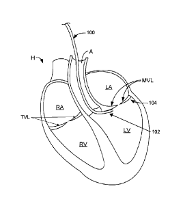
[0045] Referring now FIG. 1A, a cross-sectional depiction of a heart H is
shown with an anchor delivery device guide catheter 100 advanced in a
retrograde direction
through the aorta A and into the left ventricle LV. Retrograde, as used
herein, generally
refers to a direction opposite the expected flow of blood. In one embodiment,
this access
route is used to reach the subvalvular space 106. Guide catheter 100 is
generally a flexible
elongate catheter which may have one or more curves or bends toward its distal
end to
facilitate placement of the distal end 102 of the catheter 100 at the desired
location. The
distal end 102 of guide catheter 100 may be configured to be positioned at an
opening into
the subvalvular space 106 or within the subvalvular space 106, such that
subsequent delivery
devices may be passed through guide catheter 100 into the subvalvular space
106. Although
the retrograde aortic access route preferably starts from a percutaneous or
peripheral access
site, in some embodiments of the invention, aortic access may be achieved by
an incision in
7
CA 02702615 2010-04-14
WO 2009/052427 PCT/US2008/080368
the ascending aorta, descending aorta, aortic arch or iliac arteries,
following surgical,
thorascopic or laparoscopic access to a body cavity.
[0046] Access to the other chambers of the heart may be performed through
percutaneous or venous cut-down access, including but not limited to
transjugular, subclavian
and femoral vein access routes. When venous access is established, access to
the right atrium
RA, the right ventricle RV, the tricuspid valve TV and other right-sided
cardiac structures can
occur. Furthermore, access to left-sided heart structures, such as the left
atrium LA, left
ventricle LV, mitral valve and the aortic valve, may be subsequently achieved
by performing
a transseptal puncture procedure, which is discussed in greater detail below.
[0047] Access to the heart H may also be transthoracic, with a delivery device
being introduced into the heart via an incision or port in the heart wall.
Open heart surgical
procedures may also be used to provide access for the methods and devices
described herein.
In some embodiments, hybrid access involving a combination of access methods
described
herein may be used. In one specific example, dual access to a valve may be
achieved with a
combination of venous and arterial access sites. User manipulation of both
ends of a
guidewire placed across a valve may improve positioning and control of the
catheter and the
implants. In other examples of hybrid access, both minimally invasive and
surgical access is
used to implant one or more cardiac devices.
[0048] Other embodiments of the invention also include treatment of the
tricuspid valve annulus, tissue adjacent the tricuspid valve leaflets TVL, or
any other cardiac
or vascular valve. Thus, although the description herein discloses specific
examples of
devices and methods of the invention for mitral valve repair, the devices and
methods may be
used in any suitable procedure, both cardiac and non-cardiac. For example, in
other
embodiments, the mitral valve reshaping devices and procedures may be used
with the
tricuspid valves also, and certain embodiments may also be adapted for use
with the
pulmonary and aortic valves. Likewise, the other examples provided below are
directed to
the left ventricle, but the devices and methods may also be adapted by one of
ordinary skill in
the art for use in the right ventricle or either atrium. The devices and
methods may also be
used with the great vessels of the cardiovascular system, for example, to
treat aortic root
dilatation.
8
CA 02702615 2010-04-14
WO 2009/052427 PCT/US2008/080368
[0049] FIG. 1B is a flowchart of a method 120 for deploying at least two
anchors in the region of a heart valve annulus. As shown there, this
illustrative method
comprises advancing a guide catheter to the subannular groove region 122,
advancing a
guidewire through a lumen of the guide catheter 124, advancing a tunnel
catheter over the
guidewire 126, and proximally withdrawing the guidewire from the tunnel
catheter 128.
After the guidewire has been proximally withdrawn, a first delivery catheter
may be
advanced through the lumen of the tunnel catheter 130 and a first anchor may
be deployed
into a first region of the heart valve annular tissue 132. The first anchor
may then be fixedly
attached or otherwise secured to a guide element, such as a tether. In this
way, after the
anchor is deployed, the guide element may remain attached to the anchor and
the guide
element may be used as a track or monorail for the advancement of additional
delivery
catheters thereover.
[0050] The guide element may be made from any suitable or desirable
biocompatible material. The guide element may be braided or not braided, woven
or not
woven, reinforced or impregnated with additional materials, or may be made of
a single
material or a combination of materials. For example, the guide element may be
made from a
suture material (e.g., absorbable suture materials such as polyglycolic acid
and
polydioxanone, natural fibers such as silk, and artificial fibers such as
polypropylene,
polyester, polyester impregnated with polytetrafluoroethylene, nylon, etc.),
may be made
from a metal (absorbable or non-absorbable), may be made from a metal alloy
(e.g., stainless
steel), may be made from a shape memory material, such as a shape memory alloy
(e.g., a
nickel titanium alloy), may be made from combinations thereof, or may be made
from any
other biocompatible material. In some variations, when pulled proximally, the
guide element
will cinch or reduce the circumference of the atrio-ventricular valve annulus
or the annular
tissue. In certain variations, the guide element may be in the form of a wire.
The guide
element may include multiple layers, and/or may include one or more coatings.
For example,
the guide element may be in the form of a polymer-coated wire. In certain
variations, the
guide element may be formed of a combination of one or more sutures and one or
more
wires. As an example, the guide element may be formed of a suture that is
braided with a
wire. In some variations, the guide element may be formed of one or more
electrode
materials. In certain variations, the guide element may be formed of one or
more materials
that provide for the telemetry of information (e.g., regarding the condition
of the target site).
9
CA 02702615 2010-04-14
WO 2009/052427 PCT/US2008/080368
[0051] In some variations, the guide element may include one or more
therapeutic agents (e.g., drugs, such as time-release drugs). As an example,
the guide
element may be partially or entirely coated with one or more therapeutic
agents. In certain
variations, the guide element may be used to deliver one or more growth
factors and/or
genetic regenerative factors. In some variations, the guide element may be
coated with a
material (e.g., a polymer) that encapsulates one or more therapeutic agents,
or in which one
or more therapeutic agents are embedded. The therapeutic agents may be used,
for example,
to treat the target site to which the guide element is fixedly attached or
otherwise secured. In
certain variations, the guide element may include one or more lumens through
which a
therapeutic agent can be delivered.
[0052] After the first anchor has been deployed in the region of the heart
valve
annular tissue, the first delivery catheter may be withdrawn proximally and
the tunnel
catheter may then be positioned at a different location about the subannular
groove region
134. A second delivery catheter may then be advanced over the guide element
through the
lumen of the tunnel catheter 136. During advancement of the second delivery
catheter over
the guide element, the guide element may enter the second delivery catheter
through an
opening at its distal end, and exit the second delivery catheter through an
opening in its side
wall that is proximal to its distal end. Alternatively, the guide element may
enter the second
delivery catheter through an opening at its distal end, and exit the second
delivery catheter
through an opening at its proximal end. After the second delivery catheter has
been advanced
over the guide element through the lumen of the tunnel catheter, a second
anchor is deployed
into a second region of the heart valve annular tissue 138.
[0053] As illustrated in FIG. 2A, a distal portion 102 of the delivery device
100
is positioned in a desired location under a valve leaflet L and adjacent a
ventricular wall VW.
The valve annulus VA generally comprises an area of heart wall tissue at the
junction of the
ventricular wall VW and the atrial wall AW that is relatively fibrous and,
thus, significantly
stronger than leaflet tissue and other heart wall tissue. It is noted,
however, that considerable
structural variations of the annulus exist within patient populations and that
attempted
delivery of an implant to the valve annulus VA may instead contact or attach
to the tissue
adjacent to the valve annulus. The term "annular tissue" as used herein shall
include the
valve annulus and the tissue adjacent or surrounding the valve annulus.
CA 02702615 2010-04-14
WO 2009/052427 PCT/US2008/080368
[0054] The distal portion 102 of the delivery device 100 may be advanced into
position generally under the valve annulus VA by any suitable technique, some
of which are
described below. The distal portion 102 of the delivery device 100 may be used
to deliver
anchors to the valve annular tissue, to stabilize and/or expose the annulus,
or both. In one
embodiment, using a delivery device 100 having a flexible elongate body as
shown in FIG. 1,
a flexible distal portion 102 may be positioned in the left ventricle LV at
the level of the
mitral valve leaflets MVL using any of a variety of access routes described
herein. The distal
portion 102 may be advanced to a region 104 under the posterior valve leaflet.
Referring to
FIG. 2A, in some variations the region 104 may be generally bordered by the
inner surface of
the ventricular wall VW, the inferior surface of valve leaflets L, and the
third order chordae
tendineae CT connected directly to the ventricular wall VW and the leaflet L.
It has been
found that when a flexible anchor delivery device 100 is passed, for example,
under the
mitral valve via an intravascular approach, the delivery device 100 may be
inserted into the
space 104 and advanced along the subannular groove region 104 either partially
or
completely around the circumference of the valve. Other examples of deployment
locations
are described elsewhere herein. Once in the region 104, the distal portion 102
of the delivery
device 100 may be positioned proximate to the intersection of the valve
leaflet(s) and the
ventricular wall VW, which is near to the valve annulus VA. These are but
examples of
possible access routes of an anchor delivery device to a valve annulus, and
any other access
routes may be used.
[0055] In some embodiments, the guide catheter 100 may comprise a curvable
portion with a radius in an expanded/curved state that is greater than a
radius of the valve
annulus or the subannular groove region. The relative size of this portion of
the guide
catheter 100, when positioned within the smaller sized ventricle, may exert a
radially outward
force that can improve the surface contact between guide catheter 100 and the
left ventricle
LV. For example, in one embodiment guide catheter 100 in the expanded state
has a radius
about 25%-50% larger that the valve annulus or ventricle chamber.
[0056] In some variations, the distal portion 102 of the delivery device 100
may
include a shape-changing portion which enables distal portion 102 to conform
to the shape of
the valve annulus VA, the region 104, or other portion of the heart chamber.
The delivery
device 100 may be introduced through the vasculature with the shape-changing
distal portion
in a generally straight, flexible configuration. Once the delivery device 100
is generally
11
CA 02702615 2016-06-14
positioned beneath the leaflet in proximity to the intersection between the
leaflet and the
interior ventricular wall, the shape of the distal portion 102 may be changed
to conform to the
annulus and the shape may be "locked" to provide sufficient stiffness or
rigidity to permit the
application of force from the distal portion 102 to the annulus or annular
tissue.
[0057] In some embodiments, a shape-changing portion may be sectioned,
notched, slotted or segmented and one of more tensioning members such as
tensioning cords,
wires or other tensioning devices coupled with the shape-changing portion may
be used to
shape and rigidify distal portion 102. A segmented distal portion, for
example, may include
multiple segments coupled with two tensioning members, each providing a
different direction
of articulation to the distal portion. A first bend may be created by
tensioning a first member
to give the distal portion a C-shape or similar shape to conform to the
annular tissue, while a
second bend may be created by tensioning a second member to articulate the C-
shaped
member upwards against the annular tissue. In another embodiment, a shaped
expandable
member, such as a balloon, may be coupled with the distal portion 102 to
provide for shape
changing/deforming.
[0058] For example, in transthoracic delivery methods and other embodiments,
the distal portion 102 may be shaped, and the method may involve introducing
distal portion
102 under the valve leaflets. The shaped distal portion 102 may be rigid or
formed from any
suitable material such as spring stainless steel, a super-elastic or shape
memory material such
as nickel-titanium alloy (e.g., Nitinol), or the like. In embodiments
configured for open
surgical access, the delivery devices may be made with stiffer materials when
the
maneuverability through a transvascular route is not required, but in other
embodiments,
flexible, catheter-like delivery devices may still be used with open surgical
procedures.
[0059] In addition to delivering anchors to the annular tissue, the delivery
device 100 (and specifically distal portion 102) may be used to stabilize
and/or expose the
valve annulus or annular tissue. Such stabilization and exposure are described
fully in U.S.
Pat. Appl. Ser. No. 10/656,797. For
example, once the distal portion 102 is positioned generally under the annular
tissue, force
may be applied to the distal portion 102 to stabilize the valve annulus VA or
annular tissue,
as shown in FIG. 2B. Such force may be directed in any suitable direction to
expose, position
and/or stabilize the annulus or annular tissue. In another example, an upward
and lateral
force is shown in FIG. 2B by the solid-headed arrow drawn from the center of
the distal
12
CA 02702615 2016-06-14
portion 102. In other examples, only upward, only lateral, or any other
suitable force(s) may
be applied. With application of force to the distal portion 102, the annular
tissue may rise or
project outwardly, thus exposing the annular tissue for easier viewing or
access. The applied
force may also stabilize the valve annulus VA or valve annular tissue, also
facilitating
surgical procedures and visualization.
[0060] Some embodiments of the invention may include a stabilization
component as well as an anchor delivery component. For example, some
embodiments may
include two flexible members, one for contacting the atrial side of a valve
annulus and the
other for contacting the ventricular side. In some embodiments, such flexible
members may
be used to "clamp" the annulus between them. One of such members may be an
anchor
delivery member and the other may be a stabilization member, for example. Any
combination and configuration of stabilization and/or anchor delivery members
is
contemplated.
[0061] Referring now to FIGS. 2C and 2D, an anchor delivery device 108 is
schematically shown delivering an anchor 110 to a valve annulus VA. Anchor 110
is shown
first housed within delivery device 108 in FIG. 2C and then delivered to the
annulus VA, as
depicted in FIG. 2D. Of course, although the delivery and position of the
anchor 110 is
described with respect to the valve annulus VA, one or more anchors 110 may be
secured to
the valve annulus VA or other structures accessible from the region 104. As is
shown, in
some embodiments, anchors 110 may have a relatively straight configuration
when housed in
delivery device 108, with two sharpened tips and a loop in between the tips.
Upon
deployment from delivery device 108, the tips of anchor 110 may curve in
opposite directions
to form two semi-circles, circles, ovals, overlapping helices or the like.
Additional anchor
embodiments are described below, and may also be found in U.S. Pat. Appl. Ser.
No.
11/202,474. Multiple coupled
anchors 110 may be delivered, and the anchors 110 may be drawn together to
tighten the
valve annulus.
[0062] Although delivery device 108 is shown having a circular cross-sectional
shape in FIGS. 2C and 2D, it may alternatively have any other suitable shape.
In one
embodiment, for example, it may be advantageous to provide a delivery device
having an
ovoid or elliptical cross-sectional shape. Such a shape may help ensure that
the device is
aligned, when positioned between a corner formed by a ventricular wall and a
valve leaflet,
13
CA 02702615 2010-04-14
WO 2009/052427 PCT/US2008/080368
such that one or more openings in the delivery device is oriented to deliver
the anchors into
valve annulus tissue. To further enhance contacting of the annular tissue
and/or orientation of
the delivery device, some embodiments may further include an expandable
member, coupled
with the delivery device, which expands to urge or press or wedge the delivery
device into
the corner formed by the ventricle wall and the leaflet to contact the valve
annulus. Such
enhancements are described further below.
[0063] FIGS. 1C to 1K provide a more detailed depiction of the method shown
in flowchart form in FIG. 1B. In FIGS. 1C to 1K, the mitral valve MV of FIG.
lA is
depicted schematically from an inferior perspective looking up, but in other
embodiments the
tricuspid valve may be accessed. Referring to FIG. 1C, a guide catheter 140 is
advanced to
subannular groove region 142 using any of the access routes (or any other
suitable access
routes) previously described. After guide catheter 140 has been positioned at
the desired
location in subannular groove region 142, a guidewire 142 is advanced through
the lumen of
guide catheter 140. The guidewire 144 may then be advanced beyond the distal
end 146 of
guide catheter 140, so that guidewire 144 extends further along subannular
groove region 142
than guide catheter 140, as shown in FIG. 1D.
[0064] After the guidewire 144 has been positioned in the subannular groove
region 142, a tunnel catheter 148 may be advanced through guide catheter 140,
over
guidewire 144, which is shown in FIG. 1E. Tunnel catheter 148 may be any
suitable catheter,
and in some instances, it is desirable that the tunnel catheter be pre-shaped
or pre-formed at
its distal end, such as the tunnel catheter illustrated in FIG. 1E. The tunnel
catheter may have
a pre-shaped distal portion comprising a curve. In this way, the tunnel
catheter may more
easily conform to the geometry of the atrio-ventricular valve. It should also
be understood
that any of the catheters or guidewires described here may be pre-shaped or
pre-formed to
include any number of suitable curves. Of course, the guidewires and/or
catheters described
here may also be steerable.
[0065] After tunnel catheter 148 has been positioned in the subannular groove
region 142, guidewire 144 may be withdrawn proximally as shown in FIG. 1F.
After
guidewire 144 has been withdrawn, a delivery catheter 150 may then be advanced
through
the lumen of the tunnel catheter 148. As shown in FIG. 1G, a distal portion
152 of delivery
catheter 150 is advanced toward an opening 154 in distal portion 156 of tunnel
catheter 148.
In some embodiments, the delivery catheter 150 may be extended through the
opening 154 of
14
CA 02702615 2016-06-14
the tunnel catheter 148. As shown in FIG. 1H, an anchor 158, which is attached
to a guide
element (shown in FIG. 11 as a tether 158), may then be deployed from delivery
catheter 150.
The anchor 158 may be deployed from the delivery catheter 150 in any suitable
fashion,
including but not limited to a push-pull wire, using a plunger, or other
suitable actuation
technique. Similarly, anchor 158 may be attached to tether 158 by any suitable
attachment
method. For example, one or more knots, welded regions, and/or adhesives may
be used.
Alternate embodiments for anchor deployment and anchor attachments are
described in U.S.
Pat. Appl. Ser. Nos. 11/583,627, and 61/083,109.
[0066] The anchors for use with the methods and devices described here may be
any suitable anchor. The anchors may be made of any suitable material, may be
any suitable
size, and may be of any suitable shape. The anchors may be made of one
material or more
than one material. Examples of anchor materials include super-elastic or shape
memory
materials, such as nickel-titanium alloys and spring stainless steel. Examples
of anchor
shapes include T-tags, rivets, staples, hooks (e.g., C-shaped or semicircular
hooks, curved
hooks of other shapes, straight hooks, barbed hooks), multiple looped anchors,
and clips. The
anchors may be configured to self-expand and self-secure into tissue, but need
not be
configured in such a fashion. Additionally, while the delivery and deployment
of multiple
anchors of the same shape over a single guide element have been described, in
some
variations, a single guide element can be used to deliver and deploy multiple
anchors having
different shapes. Similarly, in certain variations, a single guide element can
be used in the
delivery and deployment of multiple anchors having different sizes.
Illustrative examples of
suitable anchors are described in more detail, for example, in U.S. Pat. Appl.
Ser. No.
11/202,474.
[0067] The anchor 158, shown in FIG. 1H, may be configured to self-expand as
it exits delivery catheter 150 and to self-secure into a region of the mitral
valve annulus, but
may also be used to in other regions of the heart. It should be understood
that the one or
more anchors may be deployed into the annulus directly, while other anchors
may be secured
to other tissue in the vicinity of the subannular groove region. For example,
one or more
anchors may be secured to the tissue below the annulus. After anchor 158 has
been deployed,
delivery catheter 150 may be proximally withdrawn. FIG. 11 shows anchor 158,
attached to
tether 160 and secured to the mitral valve annulus AN. As shown in FIG. 1J,
tunnel catheter
CA 02702615 2016-06-14
148 may then be moved to a different location or position in the subannular
groove region or
the heart, and a second delivery catheter 162 is advanced through the lumen of
tunnel catheter
148, over tether 160, as shown in FIG. 1K.
[0068] Before delivery catheter 162 is advanced through tunnel catheter 148,
the tether 160 may be threaded into delivery catheter 162, and slidably
engaged with a second
anchor 164. Any of a number of different methods can be used to thread a guide
element,
such as a tether, into a delivery catheter, and to engage the guide element
with an anchor.
Other methods are disclosed in U.S. Pat. Appl. Ser. No. 11/202,474, and
threading devices are described, for example, in U.S. Pat. Appl. Ser. No.
11/232,190. With reference now to FIG. 1K, after delivery catheter 162 has
been
advanced through tunnel catheter 148, and is used to deploy anchor 164 before
being withdrawn from the tunnel catheter 148.
[0069] Tunnel catheter 148 may be formed of any of a number of different
materials. Examples of suitable materials include polymers, such as polyether-
block co-
polyamide polymers, copolyester elastomers, thermoset polymers, polyolefins
(e.g.,
polypropylene or polyethylene, including high-density polyethylene and low-
density
polyethylene), polytetrafluoroethylene, ethylene vinyl acetate, polyamides,
polyimides,
polyurethanes, polyvinyl chloride (PVC, fluoropolymers (e.g., fluorinated
ethylene
propylene, perfluoroalkoxy (PFA) polymer, polyvinylidenefluoride, etc.),
polyetheretherketones (PEEKs), and silicones. Examples of polyamides that may
be included
in tunnel catheter (410) include Nylon 6 (e.g., Zytel HTN high performance
polyamides
from DuPontTm), Nylon 11 (e.g., Rilsan B polyamides from Arkema Inc.), and
Nylon 12
(e.g., Grilamid polyamides from EMS-Grivory, Rilsan A polyamides from Arkema
Inc.,
and Vestamid polyamides from Degussa Corp.). In some variations, tunnel
catheter 148
may be formed of multiple polymers. For example, tunnel catheter 148 may be
formed of a
blend of different polymers, such as a blend of high-density polyethylene and
low-density
polyethylene. While the wall of tunnel catheter 148 is formed of a single
layer, some
variations of tunnel catheters may include walls having multiple layers (e.g.,
two layers, three
layers). Furthermore, some variations of tunnel catheters may include at least
two sections
that are formed of different materials and/or that include different numbers
of layers.
16
CA 02702615 2016-06-14
Additionally, certain variations of tunnel catheters may include multiple
(e.g., two, three)
lumens. The lumens may, for example, be lined and/or reinforced (e.g., with
braiding).
[0070] FIGS. 24A to 24D show various detailed views of one embodiment of a
delivery catheter 1200 that can be used to deliver one or more anchors to a
target site. As
shown in FIG. 24A, the delivery catheter 1200 has a distal region 1204
including a tip 1202,
an anchor-holding region 1206 including a primary lumen 1208, an intermediate
region 1210,
a secondary lumen 1212, and a proximal region 1214 including primary lumen
1208. An
anchor 1216 is disposed within primary lumen 1208, in the anchor-holding
region 1206.
While only one anchor is shown in the anchor-holding region, some variations
of delivery
catheters may include an anchor-holding region that is adapted to hold
multiple anchors.
Similarly, while the variation shown in FIGS. 24A to 24D depict anchors
adapted to be
deployed from the distal end of the delivery catheter, it should be understood
that the anchors
may be deployed from any suitable region of the delivery catheter, as
desirable. For example,
if desirable, the anchor may be delivered out of a side port or hole on the
delivery catheter.
[0071] As shown in FIGS. 24A to 24D, a tether 1218 is threaded into a slot
1219 of tip 1202 (shown in FIGS. 24C and 24D), and through an eyelet 1226 of
anchor 1216.
After extending through the eyelet, the tether may exit the primary lumen
1208, and extend
along an exterior surface 1221 of delivery catheter 1200 for the remainder of
the length of the
anchor-holding region, as shown in FIG. 24C. The tether then enters secondary
lumen 1212,
and extends through the length of the secondary lumen, exiting the secondary
lumen at an end
of distal region 1214. An actuator 1220 may be slidably disposed within
primary lumen
1208, and can be used to deploy anchor 1216. The actuator is in the form of a
pushable
generally tubular member, although other forms of actuators may be used. For
example, in
some variations, a solid rod may be used as an actuator. Other embodiments of
the delivery
catheter are described in U.S. Pat. App!. Ser, No. 11/202,474.
[0072] It should also be understood that while some embodiments of the
invention utilize multiple anchors being delivered via multiple delivery
catheters, other
methods of delivering the anchors may be used. For example, in some instances,
it may be
desirable to deliver multiple anchors from a single delivery catheter, as
described in more
detail below and in U.S. Pat. Appl. Ser. No. 11/201,949.
Similarly, it may be desirable to combine multiple anchor delivery
17
CA 02702615 2010-04-14
WO 2009/052427 PCT/US2008/080368
and deployment via a single delivery catheter with single anchor delivery and
deployment via
a single delivery catheter.
[0073] With reference now to FIG. 3, one embodiment comprises an anchor
delivery device 200, which suitably includes an elongate shaft 204 having a
distal portion 202
configured to deliver a plurality of anchors 210, coupled with a tether 212,
and configured for
attachment to annular tissue. The tethered anchors 210 are housed within a
housing 206 of
the distal portion 202, along with one or more anchor retaining mandrels 214
and an
expandable member 208. Many variations may be made to include one or more of
these
features, and various parts may be added or eliminated. Some of these
variations are
described further below, but no specific variation(s) should be construed as
limiting.
[0074] Housing 206 may be flexible or rigid in some variations. In some
embodiments, for example, flexible housing 206 may comprise multiple segments
configured
such that housing 206 is deformable by tensioning a tensioning member coupled
to the
segments. In some embodiments, housing 206 is formed from an elastic material
having a
geometry selected to engage and optionally shape or constrict the annular
tissue. For
example, the rings may be formed from spring stainless steel, super-elastic
shape memory
alloys such as nickel-titanium alloys (e.g., Nitinol), or the like. In other
embodiments, the
housing 206 could be formed from an inflatable or other structure that can be
selectively
rigidified in situ, such as a gooseneck or lockable element shaft, any of the
rigidifying
structures described above, or any other rigidifying structure.
[0075] "Anchors," for the purposes of this application, are defined to include
any of a variety of fasteners. Thus, anchors 210 may comprise C-shaped or
semicircular
hooks, curved hooks of other shapes, straight hooks, barbed hooks, clips of
any kind, T-tags,
or any other suitable fastener(s). In one embodiment, as described above,
anchors may
comprise two tips that curve in opposite directions upon deployment, forming
two
intersecting semi-circles, circles, ovals, helices or the like. In some
embodiments, anchors
210 are self-deforming. By "self-deforming" it is meant that anchors 210 are
biased to
change from a first undeployed shape to a second deployed shape upon release
of anchors
210 from restraint in housing 206. Such self-deforming anchors 210 may change
shape as
they are released from housing 206 and enter annular tissue, and secure
themselves to the
tissue. Self-deforming anchors 210 may be made of any suitable material such
as spring
18
CA 02702615 2016-06-14
stainless steel, or a super-elastic or shape-memory material like nickel-
titanium alloy (e.g.,
Nitinol).
[0076] In other embodiments, the anchors 210 may be made of a non-shape-
memory material and may be loaded into housing 206 in such a way that they
change shape
upon release. For example, anchors 210 that are not self-deforming may be
secured to tissue
via crimping, firing or other application of mechanical force to facilitate
tissue penetration
and/or securement. Even self-securing anchors may be crimped in some
embodiments of the
invention, to provide enhanced attachment to tissue. In some embodiments,
anchors 210 may
comprise one or more bioactive agents. In another embodiment, anchors 210 may
comprise
electrode components. Such electrodes, for example, may sense various
parameters including
but not limited to impedance, temperature and electrical signals. In other
embodiments, such
electrodes may be used to supply energy to tissue at ablation or sub-ablation
amounts. In still
other embodiments, the anchors may be incorporated with an implantable pacing
lead or an
implanted sensor of a congestive heart failure monitor. Examples of a
congestive heart
failure monitor include the HeartPOD(TM) Implantable Heart Failure Therapy
System by
Savacor, Inc. (Los Angeles, CA) and the OptiVolO feature of the InSync
Sentry(TM) cardiac
resynchronization therapy-defibrillator by Medtronic, Inc. (Minneapolis, MN).
These
systems are described in greater detail in U.S. Patent Nos. 6,970,742 and
6,931,272.
Delivery of the anchors may be accomplished by any suitable device and
technique, such as
by simply releasing the anchors by hydraulic balloon delivery as discussed
further below.
Any number, size and shape of the anchors 210 may be included in housing 206.
[0077] In another embodiment, the anchors 210 may generally C-shaped or
semicircular in their undeployed form, with the ends of the "C" being
sufficiently sharpened
to penetrate tissue. Between the ends of the C-shaped anchor 210, an eyelet
may be formed
for allowing slidable passage of the tether 212. To maintain the anchors 210
in their C-
shaped, undeployed state, anchors 210 may be retained within housing 206 by
two mandrels
214, one mandrel 214 retaining each of the two arms of the C-shape of each
anchor 210.
Mandrels 214 may be retractable within elongate catheter body 204 to release
anchors 210
and allow them to change from their undeployed C-shape to a deployed shape.
The deployed
shape, for example, may approximate a partial or complete circle, or a circle
with overlapping
ends, the latter appearing similar to a key ring. Such anchors are described
further below, but
19
CA 02702615 2010-04-14
WO 2009/052427 PCT/US2008/080368
generally may be advantageous in their ability to secure themselves to annular
tissue by
changing from their undeployed to their deployed shape. In some variations,
anchors 210
may also be configured to lie flush with a tissue surface after being
deployed. By "flush" it is
meant that no significant amount of an anchor protrudes from the surface,
although some
small portion may protrude.
[0078] The retaining mandrels 214 may have any suitable cross-sectional shape,
cross-sectional area, length and be made of any suitable material, such as
stainless steel,
titanium, nickel-titanium alloys (e.g., Nitinol), or the like. Some
embodiments may not
include a mandrel, or may have one mandrel, two mandrels, or more than two
mandrels.
[0079] In some embodiments, the anchors 210 may be released from mandrels
214 to contact and secure themselves to annular tissue without any further
force applied by
the delivery device 200. Some embodiments, however, may also include one or
more
expandable members 208, which may be expanded to help drive anchors 210 into
tissue.
Expandable member(s) 208 may have any suitable size and configuration and may
be made
of any suitable material(s). Any of a variety of mechanical and hydraulic
expandable
members known in the art may be included in housing 206.
[0080] Referring now to FIGS. 4 and 5, a segment of a distal portion 302 of an
anchor delivery device suitably includes a housing 306, multiple tensioning
members 320 for
applying tension to housing 306 to change its shape, two anchor retaining
mandrels 314
slidably disposed in housing 306, multiple anchors 310 slidably coupled with a
tether 312,
and an expandable member 308 disposed between anchors 310 and housing 306. As
can be
seen in FIGS. 4 and 5, housing 306 may include multiple segments to allow the
overall shape
of housing 306 to be changed by applying tension to tensioning members 320. As
also is
evident from the drawings, "C-shaped" anchors 310 may actually have an almost
straight
configuration when retained by mandrels 314 in housing 306. Thus, for the
purposes of this
application, "C-shaped" or "semicircular" refers to a very broad range of
shapes including a
portion of a circle, a slightly curved line, a slightly curved line with an
eyelet at one point
along the line, and the like.
[0081] With reference now to FIG. 6, the same segment of distal portion 302 is
shown, but mandrels 314 have been withdrawn from two mandrel apertures 322, to
release
anchors 310 from housing 306. Additionally, expandable member 308 has been
expanded to
CA 02702615 2010-04-14
WO 2009/052427 PCT/US2008/080368
drive anchors out of housing 306. Anchors 310, having been released from
mandrels 314,
have begun to change from their undeployed, retained shape to their deployed,
released
shape.
[0082] Referring now to FIGS. 7A to 7E, a cross-section of a distal portion
402
of an anchor delivery device is shown in various stages of delivering an
anchor to annular
tissue. In FIG. 7A, distal portion 402 is positioned against the annular
tissue, an anchor 410
is retained by two mandrels 414, a tether 412 is slidably disposed through an
eyelet on anchor
410, and an expandable member 408 is coupled with housing 406 in a position to
drive
anchor 410 out of housing 406. When retained by mandrels 414, anchor 410 may
be in its
undeployed shape. As discussed above, mandrels 414 may be slidably retracted,
as
designated by the solid-tipped arrows in FIG. 7A, to release anchor 410. In
some
embodiments, anchors 410 may be released one at a time, such as by retracting
mandrels 414
slowly, may be released in groups, or may all be released simultaneously, such
as by rapid
retraction of mandrels 414.
[0083] In the example depicted in FIG. 7B, anchor 410 has begun to change
from its undeployed shape to its deployed shape (as demonstrated by the hollow-
tipped
arrows) and has also begun to penetrate the annular tissue. Empty mandrel
apertures 422
demonstrate that mandrels 414 have been retracted at least far enough to
release anchor 410.
In FIG. 7B, expandable member 408 has been expanded to drive anchor 410
partially out of
housing 406 and further into the annular tissue VA. Anchor 410 also continues
to move from
its undeployed towards its deployed shape, as shown by the hollow-tipped
arrows. In FIG.
7D, anchor 410 has reached its deployed shape, which is roughly a completed
circle with
overlapping ends or a "key ring" shape. In FIG. 7E, delivery device 402 has
been removed,
leaving a tethered anchor in place in the valve annulus. Of course, there will
typically be a
plurality of tethered anchors secured to the annular tissue. Tether 412 may
then be cinched to
apply force to anchors 410 and cinch and tighten the valve annulus.
[0084] With reference now to FIGS. 8A and 8B, a diagrammatic representation
of another embodiment comprising coupled anchors is shown. Here, anchors 510
are coupled
to a self-deforming or deformable coupling member or backbone 505. In some
examples,
this backbone 505 may be another embodiment of a tether. The backbone 505 may
be
fabricated, for example, from nickel-titanium alloys (e.g., Nitinol), spring
stainless steel, or
the like, and may have any suitable size or configuration. In one embodiment,
as in FIG. 8A,
21
CA 02702615 2010-04-14
WO 2009/052427 PCT/US2008/080368
backbone 505 is shaped as a generally straight line when held in an undeployed
state, such as
when restrained within a housing of an anchor deliver device. When released
from the
delivery device, backbone 505 may change to a deployed shape having multiple
bends, as
shown in FIG. 8B. By bending, backbone 505 shortens the longitudinal distance
between
anchors, as demonstrated by the solid-tipped arrows in FIG. 8B. This
shortening process may
act to reshape the annular tissue into which anchors 510 have been secured.
Thus, anchors
510 coupled to backbone 505 may be used to reshape annular tissue without
using a separate
tether or applying tethering force. In other embodiments, an elastic tether
may be used as the
backbone 505. In still other embodiments, backbone may also be coupled with a
termination
member to further cinch the annular tissue. In such an embodiment, the
backbone 505 is
adapted to be at least partially conformable or cinchable, such that when
force is applied to
anchors 510 and backbone 505 via a tether, backbone 505 bends further to allow
further
cinching of the annular tissue.
[0085] In another embodiment, shown in FIGS. 9A to 9C, a flexible distal
portion of an anchor delivery device 520 includes a housing 522 coupled with
an expandable
member 524. Housing 522 may be configured to house multiple coupled anchors
526 and an
anchor contacting member 530 coupled with a pull cord 532. Housing 522 may
also include
multiple apertures 528 for allowing egress of anchors 526. For clarity,
delivery device 520 is
shown without a tether in FIGS. 9A and 9C, but FIG. 9B shows that a tether 534
may extend
through an eyelet, loop or other portion of each anchor 526, and may exit each
aperture 528
to allow for release of the plurality of anchors 526. The various features of
this variation are
described further below.
[0086] In the specific embodiment in FIGS. 9A to 9C, anchors 526 are
relatively straight and lie relatively in parallel with the long axis of
delivery device 522.
Anchor contacting member 530, which may comprise a device such as a ball,
plate, hook,
knot, plunger, piston, or the like, may generally have an outer diameter that
is nearly equal to
or slightly less than the inner diameter of housing 522. Contacting member 530
is disposed
within the housing, distal to a distal-most anchor 526, and may be retracted
relative to
housing 522 by pulling pull cord 532. When retracted, anchor contacting member
530
contacts and applies force to a distal-most anchor 526 to cause release of
that anchor 526
from housing 522 via one of the apertures 528. Contacting member 530 is then
pulled farther
22
CA 02702615 2010-04-14
WO 2009/052427 PCT/US2008/080368
proximally to contact and apply force to the next anchor 526 to deploy that
anchor 526, and
so on.
[0087] Retracting contacting member 530 to push anchors 526 out of apertures
528 may help cause anchors 526 to secure themselves to the tissue adjacent the
apertures 528.
Using anchors 526 that are relatively straighter/flatter in configuration when
undeployed may
allow anchors 526 with relatively large deployed sizes to be disposed in (and
delivered from)
a relatively small housing 522. In one embodiment, for example, anchors 526
that deploy
into a shape approximating two intersecting semi-circles, circles, ovals,
helices, or the like,
and that have a radius of one of the semi-circles of about 3 mm may be
disposed within a
housing 522 having a diameter of about 5 French (1.67 mm) and more preferably
about 4
French (1.35 mm) or even smaller. Such anchors 526 may measure about 6 mm or
more in
their widest dimension. In some embodiments, housing 522 may have a
diametrical
dimension ("d") and anchor 526 may have a diametrical dimension ("D") in the
deployed
state, and the ratio of D to d may be at least about 3.5. In other
embodiments, the ratio of D
to d may be at least about 4.4, and more preferably at least about 7, and even
more preferably
at least about 8.8. These are only examples, however, and other larger or
smaller anchors 526
may be disposed within a larger or smaller housing 522. The dimensions of an
anchor may
vary depending on the particular usage. For example, anchors used for
ventriculoplasty may
permit the use of larger anchors than those used for annuloplasty due to fewer
space
constraints in the main compartment of the ventricles than in the subvalvular
spaces.
Furthermore, any convenient number of anchors 526 may be disposed within
housing 522. In
one variation, for example, housing 522 may hold about 1 to about 20 anchors
526, and more
preferably about 3 to about 10 anchors 526. Other variations may hold more
anchors 526.
[0088] Anchor contacting member 530 and pull cord 532 may have any suitable
configuration and may be manufactured from any material or combination of
materials. In
alternative embodiments of the invention, contacting member 530 may be pushed
by a pusher
member to contact and deploy anchors 526. Alternatively, any of the anchor
deployment
devices and methods previously described may be used.
[0089] Tether 534, as shown in FIG. 9B, may comprise any of the tethers or
tether-like devices described above, or any other suitable device.
Furthermore, in some
variations, multiple tethers may be provided. In such variation and each
tether may or may
not be coupled to every anchor, and some or all of the anchors may be coupled
to more than
23
CA 02702615 2010-04-14
WO 2009/052427 PCT/US2008/080368
one tether. Tether 534 may be generally attached to a distal-most anchor 526
at an
attachment point 536. The attachment itself may be achieved via a knot, weld,
adhesive, or
by any other suitable attachment mechanism. Tether 234 then extends through an
eyelet,
loop or other similar configuration on each of the anchors 526 so as to be
slidably coupled
with the anchors 526. In the particular embodiment shown, tether 534 exits
each aperture
528, then enters the next-most-proximal aperture, passes slidably through a
loop on an anchor
526, and exits the same aperture 528. By entering and exiting each aperture
528, tether 534
allows the plurality of anchors 526 to be deployed into tissue and cinched.
Alternate
embodiments of housing 522, anchors 526 and tether 534 may also be used. For
example,
housing 522 may include a longitudinal slit through which tether 534 may pass,
thus allowing
tether 534 to reside wholly within housing before deployment.
[0090] Expandable member 524 is an optional feature of anchor delivery device
520, and thus may be included in some embodiments and not in others. In some
embodiments, expandable member 524 will be coupled with a surface of housing
522, will
have a larger radius than housing 522, and will be configured such that when
it is expanded
as housing 522 nears or contacts the valve annulus, expandable member 524 will
push or
press housing 522 into enhanced contact with the annulus. For example,
expandable member
524 may be configured to expand within a space near the corner formed by a
left ventricular
wall and a mitral valve leaflet.
[0091] With reference now to FIGS. 10A to 10F, one embodiment of the
invention comprises a method for applying a plurality of tethered anchors 526
to the annular
tissue of a heart. As shown in FIG. 10A, an anchor delivery device 520 is
first contacted with
the valve annulus VA or annular tissue such that openings 528 are oriented to
deploy anchors
526 into the tissue. Such orientation may be achieved by any suitable
technique. In one
embodiment, for example, a housing 522 having an elliptical cross-sectional
shape may be
used to orient openings 528. Contact between housing 522 and the annular
tissue may be
enhanced by expanding expandable member 524 to wedge housing 522 within the
deepest
portion of the subannular groove region.
[0092] Generally, delivery device 520 may be advanced into any suitable
location for treating any valve by any suitable advancing or device placement
method. Many
catheter-based, minimally invasive devices and methods for performing
intravascular
procedures, for example, are well known, and any such devices and methods, as
well as any
24
CA 02702615 2010-04-14
WO 2009/052427 PCT/US2008/080368
other devices or method later developed, may be used to advance or position
delivery device
520 in a desired location. For example, in one embodiment a steerable guide
catheter is first
advanced in a retrograde fashion through an aorta, typically via access from a
femoral artery.
The steerable catheter is passed into the left ventricle of the heart and thus
into the space
formed by the mitral valve leaflets, the left ventricular wall and chordae
tendineae of the left
ventricle. Once in this space, the steerable catheter is advanced along a
portion (or all) of the
circumference of the mitral valve. A sheath is advanced over the steerable
catheter within the
space below the valve leaflets, and the steerable catheter is removed through
the sheath.
Anchor delivery device 520 may then be advanced through the sheath to a
desired position
within the space, and the sheath may be removed. In some cases, an expandable
member
coupled to delivery device 520 may be expanded to wedge or otherwise move
delivery device
520 into the corner formed by the left ventricular wall and the valve leaflets
to enhance its
contact with the valve annulus. This is but one exemplary method for advancing
delivery
device 520 to a position for treating a valve, and other suitable methods,
combinations of
devices, etc. may be used.
[0093] As shown in FIG. 10B, when delivery device 520 is positioned in a
desired location for deploying anchors 526, anchor contacting member 530 is
retracted to
contact and apply force to a most-distal anchor 526 to begin deploying anchor
526 through
aperture 528 and into the valve annulus VA (or annular tissue). FIG. 10C shows
anchor 526
further deployed out of aperture 528 and into valve annulus VA. FIG. 10D shows
the valve
annulus VA transparently so that further deployment of anchors 526 can be
seen. As shown,
in one embodiment, anchors 526 include two sharpened tips that move in
opposite directions
upon release from housing 522 and upon contacting the valve annulus VA.
Between the two
sharpened tips, an anchor 526 may be looped or have any other suitable eyelet
or other device
for allowing slidable coupling with a tether 534.
[0094] Referring now to FIG. 10E, anchors 526 are seen in their fully deployed
or nearly fully deployed shape, with each pointed tip (or "arm") of each
anchor 526 having
curved to form a circle or semi-circle. In some variations anchors 526 may
have any other
suitable deployed and undeployed shapes, as described more fully above. FIG.
1OF shows
anchors 526 deployed into the valve annulus VA and coupled to tether 534, with
the distal-
most anchor 526 fixedly coupled to tether 524 at attachment point 536. At this
stage, tether
534 may be cinched to tighten the annular tissue, thus reducing valve
regurgitation. In some
CA 02702615 2016-06-14
embodiments, valve function may be monitored by means such as echocardiogram
and/or
fluoroscopy, and tether 534 may be cinched, loosened, and adjusted to achieve
a desired
amount of tightening as evident via the employed visualization technique(s).
When a desired
amount of tightening is achieved, the implant may be fixed using any of a
variety of
termination devices and methods.
[0095] For example, in one embodiment, cinching tether 534, attaching tether
534 to most-proximal anchor 526, and cutting tether 534 are achieved using a
termination
device (not shown). The termination device may comprise, for example, a
catheter
advanceable over tether 534 that includes a cutting member and a nickel-
titanium alloy (e.g.,
Nitinol) knot or other attachment member for attaching tether 534 to most-
proximal anchor.
The termination catheter may be advanced over tether 534 to a location at or
near the
proximal end of the tethered anchors 526. It may then be used to apply
opposing force to the
most-proximal anchor 526 while tether 534 is cinched. Attachment and cutting
members
may then be used to attach tether 534 to most-proximal anchor 526 and cut
tether 534 just
proximal to most-proximal anchor 526. Such a termination device is only one
possible way
of accomplishing the cinching, attachment and cutting steps, and any other
suitable device(s)
or technique(s) may be used. Additional devices and methods for terminating
(e.g., cinching
and fastening) may be found, for example, in U.S. Pat. Appin. Ser. Nos.
11/232,190 and
11/270,034. In some
embodiments, the termination device is located in the same heart chamber as
the remaining
portions of the implant, which permits the implant to be wholly implanted in a
single heart
chamber. In other embodiments, however, a portion of the implant passes
transmurally
through a septal wall or an outer wall of a heart chamber. In these
embodiments, the
termination member and optionally one or more anchors may be located in a
different heart
chamber.
[0096] In some embodiments, it may be advantageous to deploy a first number
of anchors 526 along a first portion of annular tissue, cinch the first
anchors to tighten that
portion of the annular tissue, move the delivery device 520 to another portion
of the annular
tissue, and deploy and cinch a second number of anchors 526 along a second
portion of the
annular tissue. Such a method may be more convenient, in some cases, than
extending
delivery device 520 around all or most of the circumference of the annular
tissue, and may
allow a shorter, more maneuverable housing 522 to be used.
26
CA 02702615 2016-06-14
[0097] In other embodiments, similar to that shown in FIGS. 10A to 10F, the
anchors 526 may be driven out of delivery device 520 through a biocompatible
material
attached to delivery device 520, thereby attaching the biocompatible material
to the annular
tissue. Several devices and methods for attaching biocompatible material using
anchors are
described in U.S. Pat. Appl. Ser. No. 11/201,949.
For example, in one embodiment, a Dacron strip may be attached to delivery
device 520, extending along device 520 and covering apertures 528. Anchors 526
are then
driven out of delivery device 520, through the Dacron strip, into the annular
tissue, thus
detaching the Dacron strip from device 520 and attaching it to the annular
tissue. Such a
biocompatible material may facilitate tissue ingrowth of anchors 526 and may
enhance
attachment generally to the annular tissue. In an alternative embodiment,
multiple pieces of
biocompatible material, such as separate pieces of material disposed over each
of apertures
528, may be used. For example, in one embodiment multiple discs of Dacron
material are
disposed over multiple apertures 528.
[0098] In another embodiment, a distal portion of delivery device 520 may be
detachable from a proximal portion of delivery device 520. Such a variation
may be
configured such that when anchors 526 are deployed from device 520, the distal
portion of
device 520 detaches from the proximal portion and is attached, via anchors
526, to the
annular tissue. In one variation, for example, anchors 526 may pierce through
the distal
portion of device 520, rather than exiting device 520 through apertures 528.
The distal
portion may be detachable via any suitable means, such as perforations or the
Re.
[0099] In several embodiments of the invention, self-forming anchors 900 are
stored in the delivery device in a straightened configuration, coupled with a
tether 902, as
shown in FIG. 11A. Anchors 900 are held or restrained in that straightened
state, while their
deployed configuration is non-linear or curved. Thus, when the straightened
anchor 900 is
released from the delivery device into tissue T, the anchor 900 actually pulls
itself into the
tissue T, as shown in FIG. 11B, due to the storage of potential energy in the
straightened state
and the tendency of each of the arms 901 of anchors 900 to drive the tip of
the arm into the
tissue as illustrated. Arms 901 are joined together at a junction 903. Each
arm 901 is braced
against the other arm so that forces exerted by tissue T on each arm 901 are
opposed by the
other arm 901 wherein the arms are joined to one another. This eliminates the
need for an
anchor driving device, such as required with staples, thus substantially
simplifying the
27
CA 02702615 2016-06-14
assembly and method. In addition, bracing arms 901 against one another also
helps to reduce
or eliminate problems associated with tissue deflection. As shown by the
hollow-tipped
arrows in FIG. 11B, the anchor 900 pulls itself into tissue T as it assumes
its natural, curved
shape, and exerts forces in vertical, horizontal and curved directions.
Finally, after pulling
itself into tissue and assuming its natural shape, as in FIG. 11C, anchor 900
is substantially
embedded in the tissue T. Various anchor designs and deployment methods are
disclosed, for
example, in U.S. Pat. Appin. Nos. 10/741,130, 10/792,681, 10/900,980,
11/255,400, and
10/901,555.
[0100] As explained previously, although one access route to the region 104 or
space 106 is a retrograde route through the aorta A to the heart H, other
access routes may
also be used. Referring to FIG. 12A, with a heart H is shown in cross section,
an elongate
anchor delivery device 150 may be introduced within the heart H by a
transseptal puncture
procedure. Transseptal punctures may be performed using a Mullins introducer
sheath with a
Brockenbrough curved needle through the interatrial septum to access the left
atrium LA, but
any of a variety of other transseptal puncture devices may be used. From the
left atrium LA,
supravalvular access to the mitral valve may be achieved, as well as antegrade
access to the
left ventricle LV through the mitral valve. Similarly, access from the right
ventricle RV to
the left ventricle LV may be obtained by transseptal puncture of the
ventricular septum. In
still other embodiments, a catheter device may access the coronary sinus and a
valve
procedure may be performed directly from the sinus.
[0101] Surgical approaches that may be used have been described above but
also include but are not limited to transcatheter procedures made through
surgical incisions in
the aorta or myocardium. In one particular embodiment, depicted in FIG. 12B, a
transapical
approach with a surgical delivery device 114 is utilized. In some instances, a
transapical
approach may provide a more linear route to the subvalvular space 106. The
transapical
approach may also reduce potential effects of a myocardial incision on cardiac
output, as the
apical wall 112 may contribute less mechanical effect on left ventricular
ejection fraction
compared to other sections of the myocardial wall.
Synergistic Implants
[0102] In one embodiment, illustrated in FIG. 13, reshaping of the annular
tissue of the mitral valve with a cinching implant 706 may be combined with
the
28
CA 02702615 2010-04-14
WO 2009/052427 PCT/US2008/080368
reconfiguration of the subvalvular apparatus using one or more additional
cinching implants
710. The reshaping of the annular tissue may be performed with the embodiments
described
above, or with other implants. However, unlike some implants, the valve
reshaping implants
described herein may also be adaptable for implantation in a more inferior
position in
ventricle. A plurality of tethered anchors may be secured to the myocardium
adjacent the
papillary muscle and then cinched to tension the myocardium and cause
repositioning of one
or more papillary muscles. In some embodiments, one or more of the anchors may
be
attached to or looped around the papillary muscle itself.
[0103] In one embodiment, depicted schematically in FIG. 13, the anchors may
be oriented circumferentially with respect to the long axis of the ventricle
LV between the
base 702 and the apex 704 of the ventricle LV. When cinched, the implant 710
reduces the
relative distance between the papillary muscles 708. In some instances the
papillary muscle
708 may be displaced in the presence of dilated cardiomyopathy, or as a result
of ventricular
remodeling secondary to mitral valve regurgitation. By reducing the distance
between the
papillary muscles 708, the valve leaflet coaptation may be improved by
alleviating the pull of
the mitral valve leaflets MVL by the taut chordae tendineae (not shown)
attached to displaced
papillary muscles 708. One or more imaging modalities, including but not
limited to
magnetic resonance imaging, spiral CT, fluoroscopy or ultrasound, may be used
to visualize
the valvular apparatus and to determine the preferred orientation of the
cinching implant to
achieve the desired effect. For example, if ultrasound imaging identifies
redundant chordae
tendineae as one source of valve regurgitation, one or more cinching implants
may placed
with a longitudinal orientation between the associated papillary muscle 708
and the apex 704
of the ventricle LV to increase tension in the chordae and reduce leaflet
prolapse.
[0104] Even where a valve reshaping implant adequately treats the valve
regurgitation, the placement of cinching implant in an inferior location in
the ventricle may
still be beneficial for treating or limiting ventricular dilation. Under the
LaPlace principle, by
reducing the radius of the heart chamber, myocardial strain from volume
overload can be
reduced and may lead to some recovery of myocardial function over time.
Therefore, in
addition to repositioning of the papillary muscles 708 to improve valvular
function, the
ventricular implant 710 may also improve the contractile function of the left
ventricle LV.
Various imaging modalities mentioned previously can be used to identify
locations to reduce
29
CA 02702615 2010-04-14
WO 2009/052427 PCT/US2008/080368
ventricular dimensions, and in some embodiments, multiple cinching implants
may be used in
the ventricle to achieve the desired result.
[0105] The reshaping of a ventricle may be performed or assessed along any of
a variety of dimensions or vectors. For example, referring to FIG. 25, in some
embodiments
of the invention, the reshaping of a ventricle or a valve may occur with
respect to the
diameter B or the circumference C about a valve orifice. In one embodiment,
the diameter B
and the circumference C with respect to the region 104 of a ventricle is
reshaped. In addition
to the reshaping of to valvular structures, reshaping can also be performed
with respect to the
non-valvular structures of a heart chamber. For example, one or more of the
diameters or
circumferences of the ventricle may be reshaped. As shown in FIG. 25, the
diameter B' and
the circumference C' of the ventricle located generally at or above the
papillary muscles may
be reshaped. The diameter B" and circumference C" of the ventricle at or below
the
papillary muscles may also be reshaped. The orientation of the diameter and
circumference
that is reshaped or assessed can vary, but in some embodiments, the diameter
or
circumference may be in a generally perpendicular orientation with respect to
a longitudinal
axis of a ventricle. One of skill in the art will understand that the
longitudinal axis may be
characterized in a number of ways, including but not limited to a longitudinal
axis from a
valve orifice to an apex of a heart chamber, or from the apex of a heart
chamber to a point
that generally splits the ventricular volume in half. Similarly, some of the
implantation
dimensions or vectors may also be oriented with respect to the anterior-
posterior axis or the
septolateral axis of the heart chamber.
[0106] Referring to FIG. 26, in some embodiments, the distances A, D between
a papillary muscle and a valve leaflet may be reshaped. This distance A or D
may be
between a papillary muscle and its associated valve leaflet, or between a
papillary muscle and
an unassociated valve leaflet, respectively. Although the distances A, D
depicted in FIG. 26
are shown from the tip of the papillary muscle, these distances may also be
measured from
the base of the papillary muscle. Similarly, distances involving a valve
leaflet may be
measured from the distalmost section, the middle or the base of the valve
leaflet. In other
embodiments, the reshaping of the heart may occur between the apex of a heart
chamber and
one or more valves. For example, reshaping may occur along the distance E
between the
outlet valve and the apex of a heart chamber, and/or along the distance F
between the inlet
valve and the apex.
CA 02702615 2016-06-14
[0107] Thus, one or more shortening implants, including the cinching implants
described herein, may be generally placed or oriented between or along one or
more of the
dimensions or vectors, as exemplified above. In some embodiments, multiple
implants may
be placed in a generally parallel arrangement or in a fan-like pattern along
one or more of the
dimensions or vectors. The placement of a shortening implant is not limited to
the vectors or
locations described herein, and may occur with any angle, length or skewing as
needed.
Although the dimensions depicted in FIGS. 25 and 26 are wholly contained
within a single
heart chamber, in other embodiments, the dimensions may include cardiac sites
outside of a
single heart chamber.
[0108] Referring back to FIG. 13, although the two cinching implants depicted
have similar size anchors and tether lengths, in other embodiments these
features may be
optimized for the intended implant location. For example, larger anchors may
be used when
performing ventriculoplasty. Likewise, the length of the tether and the number
of coupled
anchors may be increased with myocardial wall applications due to the larger
circumferential
dimensions of the ventricle compared to the annular tissue regions.
Furthermore, the desired
tissue-related characteristics of the cinching implants may differ, depending
on the implant
location. For example, tissue fibrosis around a valve reshaping implant may be
desirable to
improve implant biocompatibility and to resist further annulus dilation.
Further details
regarding tissue fibrosis around a valve reshaping implant may be found in
U.S. Pat. Appl.
Ser. No. 11/255,400. Tissue fibrosis around
a ventricular implant, however, may reduce the contractility and compliance of
the
myocardial wall and result in reduced ejection fractions. For this reason, it
may be desirable
to configure valve and ventricular implants for different tissue responses.
For example,
ventricular implants may benefit from an anti-proliferative drug coating to
limit tissue
fibrosis. The anti-proliferative drug may be any of a variety of anti-
proliferative agents
known in the art, including but not limited to paclitaxel, sirolimus,
everolimus, a
corticosteroid and the like.
[0109] Although a number of surgically implanted ventricular devices and
procedures are known in the art, the percutaneous or transvascular
implantation of a
ventricular device may pose a significant challenge, due to the instability
from the wall
motion of a beating heart. To assure adequate contact between the delivery
device and the
myocardium and reliable positioning of a ventricular cinching implant, the
delivery device
31
CA 02702615 2010-04-14
WO 2009/052427 PCT/US2008/080368
may be stabilized against a less mobile portion of the cardiac structure
during the
implantation procedure. In some embodiments, the delivery device for a
ventricular implant
may be stabilized in the subannular groove, the subvalvular space, or the apex
of the left
ventricle.
[0110] FIG. 14A depicts an embodiment of the ventricular implant delivery
device, comprising a support member configured to seat in the apical region of
the left
ventricle during implantation. The support member depicted in FIG. 14A is a
helical support
member 652 coupled to a distal end of anchor delivery device 658, but other
shapes and
configurations may also be used. In other embodiments, helical support member
652 may
alternately extend out of a guide catheter 650 to contact the heart wall 651
and support the
anchor delivery device 658. Preferably the support member 652 has a delivery
configuration
with a reduced profile to facilitate passage of the support member 652 to the
target site, and
an expanded configuration with an enlarged profile for seating against the
apical region 704
of the left ventricle or other stable region of an anatomical structure.
Helical member 652
may be made of any suitable material, including but not limited to nickel-
titanium alloys
(e.g., Nitinol), stainless steel or the like. Any suitable mechanism may be
used for extending
helical member 652 into the left ventricle or other chamber. For example,
helical member
652 may be pushed out of guide catheter 650, but may alternatively be extended
out the guide
catheter with extension of anchor delivery device 658.
[0111] In another embodiment illustrated in FIG. 14B, the delivery device may
be stabilized against the superior surfaces of the papillary muscles 708. In
some examples,
stabilization against the papillary muscles 708 may provide mid-chamber
support during
implantation of a ventricular cinching implant 710. The anchor delivery device
668 may
optionally comprise a deployable J- or U-shaped support member 662 that is
movably
coupled with a distal portion of an anchor delivery device 668, both of which
are advanceable
through a guide catheter 660. Upon being advanced out of the distal end of
guide catheter
660, U-shaped member 662 may automatically spring out, or alternatively may be
manually
extended, to contact the inner surface of the heart wall and/or to contact a
papillary muscle
708. Manual extension of the U-shaped member 662 may permit the user to
titrate the
positioning of the delivery device to the desired location in the heart
chamber. Such a U-
shaped member 662 may automatically deform from a straight configuration for
delivery
through guide catheter 660 into a U-shaped configuration, such as if member
662 is made of
32
CA 02702615 2010-04-14
WO 2009/052427 PCT/US2008/080368
spring stainless steel or nickel-titanium alloys (e.g., Nitinol). In another
embodiment, the U-
shaped member 662 may be connected to anchor delivery device 668 at or near
the distal end
of the device 668 and may be pushed distally to force the U-shaped member 662
to expand
into its U-shape. In still another embodiment, the U-shaped member 662 may be
attached
proximally and may be pulled into its expanded configuration. Any suitable
method for
changing the shape of U-shaped member 662 from straight to U-shaped may be
used in some
variations.
[0112] In another embodiment depicted in FIG. 14C, the U-shaped member 662
may optionally include an expandable member 667, such as an inflatable
balloon.
Expandable member 667 may be expanded to provide further force against and
support of
anchor delivery device 668, to enhance its contact with ventricular wall 651.
In FIG. 14C,
the expandable member 667 is circumferentially mounted on the U-shaped member
662,
similar to a balloon angioplasty-type catheter but with a greater expansion
diameter. In some
embodiments, the balloon may have an expanded diameter of at least about 1 cm,
at least
about 2 cm, of at least about 3 cm. In other embodiments of the invention, the
expandable
member 667 may be mounted and inflated directly from the delivery device,
without a U-
shaped member 662.
[0113] In another embodiment of the invention, shown in FIG. 14D, multiple
spring members 672 may be coupled with a distal end of an anchor delivery
device 678 to
provide force against an inner surface of a heart wall (solid tipped arrows)
to support the
anchor delivery device 678 against the heart wall of the heart chamber (hollow
tipped
arrows). Thus, an anchor delivery device may include any of a number of
suitable support
members to support an anchor delivery device against the myocardium, thus
possibly
enhancing the ability of the delivery device to delivery tissue anchors to the
target tissue in
the left ventricle.
[0114] In some of the embodiments, the support members of an anchor delivery
device may have a fixed length or configuration such that the anchor delivery
device is
configured to position an implant at a single level or position relative to an
anatomical
structure or site in the heart, e.g. the apex of the left ventricle. Further
manipulation by the
physician may permit the anchor delivery device to be positioned at other
levels with a fixed
configuration device. In other embodiments, the length of the support
member(s) may be
manipulated with respect to the guide catheter or the anchor delivery device
to permit
33
CA 02702615 2010-04-14
WO 2009/052427 PCT/US2008/080368
variable positioning of the anchor delivery device at different levels or
sites of the heart
chamber. The different sites include but are not limited to the apex, the
region between the
apex and the lower boundary of the papillary muscles, the papillary muscles,
the subvalvular
space, and the subannular groove region. The implantation sites can also be
characterized by
a percentage or percentage range with respect to an axis of the particular
heart chamber.
These percentages include but are not limited to about 0%, about 10%, about
20%, about
30%, about 40%, about 50%, about 60%, about 70%, about 80%, about 90% and
about
100%. Along a longitudinal axis of the left ventricle, for example, the apex
may be
characterized as about 0% of the longitudinal axis while the subannular groove
region may be
characterized as about 100% of the longitudinal axis.
[0115] Although in some embodiments, the cinching implants may be oriented
at an angle in the heart chamber so that they are orthogonal to the
longitudinal axis of the
heart chamber, in other embodiments the implants may be oriented at any angle
or range of
angles, from about zero degrees to about 180 degrees with respect to the
longitudinal axis,
including but not limited to about 15 degrees, 30 degrees, about 45 degrees,
about 60 degrees,
about 75 degrees, about 90 degrees, about 105 degrees, about 120 degrees,
about 135
degrees, about 150 degrees, about 165 degrees. With non-orthogonal angles, the
implant may
be located across two or more levels of the heart chamber as described
previously. A
particular implantation angle may be facilitated by the fixed or variable
angle between the
support member and the anchor delivery device, or from manual positioning by
the physician.
[0116] Another challenge involving a papillary reconfiguration or
ventriculoplasty implant is the potential arrhythmogenic risk to a patient.
Patients who could
benefit from such implants may be at-risk for conduction abnormalities from
ventricular
dilatation. However, annular tissue may be relatively electro-physiologically
inert compared
to the myocardial tissue. Patients with tissue anchors attached to the
myocardium may
benefit from an implantation of a cardiac rhythm management device with a
defibrillator
component. FIG. 15 depicts one such embodiment, comprising multiple implants.
In some
examples, multiple implants may be used for synergistic treatment of mitral
regurgitation and
related sequelae. Here, the patient has a mitral valve reshaping implant 706
for treatment of
valve regurgitation, a ventriculoplasty implant 710 for treatment of ventricle
dilatation, and a
set of electrodes 712 for monitoring and treatment of arrhythmias and
conduction delays that
may reduce ventricular contractile efficiency. In addition to treating common
risks
34
CA 02702615 2016-06-14
associated with mitral regurgitation, the pacemaker-defibrillator leads 712
and the cinching
implants 706, 710 may be synergistically configured for implantation using a
common guide
catheter, which may reduce implantation procedure time and costs.
[0117] Although some of the preceding examples utilize two minimally
invasive tissue anchor implants for reshaping cardiac structures, not all of
the implants need
to have a design comprising tissue anchors. In FIG. 16, for example, a
coronary sinus
annuloplasty implant 714, such as the CCURETM device by Mitralife, Inc. (Santa
Rosa, CA),
may be used in conjunction with the tissue anchor implant. Different tissue
anchor-based
implants may be used, including those described in U.S. Pat. Pub. 2007/0112424
assigned to
Mitralign, Inc. Various designs of the coronary sinus annuloplasty implants
are disclosed in U.S. Pat. No. 6,402,781 to Langberg et al.
The embodiment depicted in FIG. 16 also illustrates the use of dual valve
reshaping implants
to achieve a further degree of annulus diameter reduction. The use of both
peripheral and
central reshaping forces from two difference types of mitral valve implants
706, 714 may
achieve better annulus reshaping than any annuloplasty implant alone.
[0118] Also, while both types of implants 706, 714 may be placed during the
same procedure, the second implant may be placed at a later date. With
reference again to
FIG. 16, a patient with an existing mitral valve reshaping implant 714 may
receive an
additional implant 706 to reduce any residual regurgitation from the original
surgery, or any
regurgitation that develops later as a result of disease progression. In other
embodiments, a
patient with a pre-existing surgically implanted annuloplasty ring may receive
a second mitral
valve annuloplasty implant that is translumenally implanted by a catheter. The
second
implant may also be placed several weeks, months or years after the original
implant.
[0119] The use of a tissue-anchor implant may allow further annular tissue
reshaping without requiring removal of an existing coronary sinus implant or
surgically
implanted annuloplasty ring. The self-deploying design of tissue anchor design
may also
generate less concern that the second implant is interfering with existing
implant because the
self-deploying design permits securement of the implant to a wider range of
structures or
surfaces.
CA 02702615 2016-06-14
[0120] FIGS. 17, 18 and 20 depict the use of an anchor-based ventricular
implant 710 along other complementary cardiac devices for the multimodal
treatment of
mitral valve regurgitation and related sequelae. In FIG. 17, a clip device
716, such as the one
produced by Evalve, Inc. (Redwood City, CA) may be used to restrain the free
edges of a
mitral valve for reducing regurgitation, while a cinching implant is used
synergistically to
reduce ventricle size and alleviate volume overload. Leaflet clips and other
suitable valvular
apparatus lasso devices are described in U.S. Patent No. 6,629,534,
Conversely in FIG. 18, a myocardial tension implant 718, such as the Coapsys
device by Myocor Inc, (Maple Grove, MN), may be used with a cinching valve
reshaping
implant 706. Various designs for transmural and transchamber myocardial
tension implants
718 and related implantation tools are described in U.S. Patent Nos. 5,961,440
and 6,260,552.
[0121] In addition to the transmural myocardial tension device shown in FIG.
18, other implants requiring access to the epicardial surface may also be used
with annular
tissue and ventricular cinching implants 706, 710. Another example of an
external cardiac
support device that limits cardiac dilatation is the CorCap(TM) cardiac
support device by
Acorn Cardiovascular, Inc. (St. Paul, MN), which depicted in FIG. 20 and
described in U.S.
Patent No. 7,278,964.
[0122] One or more cinching implants may also be applied to the epicardial
surface of the heart. Referring to FIG. 21A, an epicardial cinching implant
722 may be
placed on the heart H using a thorascopic procedure or an open surgical
procedure through an
incision in the pericardial sac. In one embodiment, the cinching implant 722
may be secured
at a circumferential epicardial location inferior to the left circumflex
artery LCX and then
cinched to reduce the diameter of the mitral valve annulus (not shown). During
some
procedures, when positioning the implant 722, to the cinching implant 722 may
be positioned
to limit or avoid impingement of the coronary arterial and venous system. This
can be done
with direct visualization of the epicardial surface 732 using a minimally
invasive fiber optic
scope or by direct visualization with the creation of a pericardial flap or
window.
36
CA 02702615 2010-04-14
WO 2009/052427 PCT/US2008/080368
Identification of the coronary surface vasculature can also be performed
indirectly with dye
injection into the vasculature during spiral CT scan or fluoroscopy.
[0123] The cinching implants applied to the epicardial surface may have a
similar size tissue anchor and tether as the various transvascular embodiments
described
herein, but in other embodiments, one or more implants may have a longer
tether and a
greater number of anchors to compensate for the greater diameter of the
epicardial surface.
In some embodiments, the implants 724 may have anchors 726 with wider eyelets
728 that
are configured for slidable coupling to a band-like tether 730, as depicted in
FIG. 21B, which
may permit the use of fewer tissue anchors 726 and allows the band-type tether
730 to contact
and restrain portions of the epicardium 732.
[0124] In addition to the use of the cinching implants to restrain ventricular
dilation and improve a patient's hemodynamic profile, the cinching implants
may also be
used to splint dyskinetic wall segments to the intact myocardium. In some
instances,
splinting of dyskinetic wall segments may reduce paradoxical wall motion
during systole.
The splinting of dyskinetic wall segments may also improve forward flow
through the
ventricle and increase the ejection fraction of the left ventricle, and/or
valve function when
one or more papillary muscles are adjacent to a dyskinetic wall segment.
Referring to FIG.
19A, for example, the papillary muscle 708 of the postero-lateral mitral valve
leaflet MVL
may be proximate to a dyskinetic lateral wall segment 736 that causes leaflet
insufficiency
during ventricular systole. By positioning a cinching implant 734 across
portions of the
dyskinetic wall segment 736 and the surrounding intact myocardium 738, the
splinted
dyskinetic wall segment 736 may resist outward bulging forces during
ventricular systole and
increase net forward blood flow. The cinching implants 734 used for splinting
wall segments
may rely on the tension of the tether for splinting effect, but in some
embodiments of the
invention, a rigid or semi-rigid tether or backbone may be used. Also, in the
particular
embodiment depicted in FIGS. 19A and 19B, the cinching implant is secured to
the
myocardium in a longitudinal orientation, but one of skill in the art can
image the heart
chamber and wall segments to determine the desired implant orientation.
[0125] In the embodiments of the cinching implant described above, the
implants are configured for generally planar implantation along an arcuate
target tissue such
as the ventricular wall or subannular groove region. In other embodiments of
the invention,
the cinching implants may have more complex configurations. FIGS. 22A to 22C,
for
37
CA 02702615 2010-04-14
WO 2009/052427 PCT/US2008/080368
example, depict the implantation of a helical ventriculoplasty implant 740.
The longitudinal
length of the helical implant 740 may permit redistribution of the restraining
force across a
greater number of tissue anchors 742. In some embodiments, the helical anchor
740 may
have a length of about 5 cm or more, preferably about 7 cm or more, and most
preferably
about 9 cm or more. The helical implant 740 may also be designed with a right-
handed or
left-handed twist configuration, which may complement the theoretical twisting
orientation of
the myocardial fibers comprising the left ventricle LV.
[0126] To implant a ventricular device in a beating heart contracting walls,
in
some embodiments one end of the implant may first attached to a less mobile
portion of the
ventricle chamber. In FIG. 22A, the distal end 744 of the implant 740 is first
secured to the
apical region 704 of the left ventricle LV. Once the distal end 744 of the
implant 740 is
stabilized, the delivery catheter 746 can be stabilized using the secured
distal end 744 and
provides sufficient stability to the delivery catheter 746 to assume the
desired geometric
configuration and orientation. This can occur with a delivery catheter 746
that is made from
a shape memory material with an helical geometry that can be reversibly
straightened with a
movable stiffening wire or element (not shown) within the delivery catheter
746. When the
stiffening element is removed and the delivery catheter 746 assumes the
helical configuration
as shown in FIG. 22B, surface contact between the delivery catheter 746 and
the heart wall
651 can be maintained with distally directed force on the delivery catheter
746. Manipulation
of the distally directed force can also be used to control the longitudinal
length of the heart
chamber over which the helical implant 740 is positioned. FIG. 22C depicts the
implant 740
after withdrawal of the delivery catheter 746.
[0127] In some alternate embodiments, the delivery catheter may be pre-
positioned along one or more portions of the subannular groove region or the
subvalvular
space before the distal tissue anchor is secured to the apex. In still other
alternative
embodiments, a detachable tissue anchor or engaging structure may be provided
about the
distal end of the guide wire, guide catheter or delivery catheter to
temporarily stabilize
delivery catheter for implantation of the cinching implant. After the implant
is secured to the
myocardium, the detachable tissue anchor or engaging structure may be
disengaged from the
myocardium and withdrawn from the patient with the other components of the
delivery
system.
38
CA 02702615 2010-04-14
WO 2009/052427 PCT/US2008/080368
[0128] Referring now to FIGS. 23A and 23B, although the embodiments
described herein may utilize a cinching implant with a linear or serial
configuration, other
embodiments may utilize a branched cinching implant 748 having one or more
branch
sections 750 where two or more arms 752, 754 of the implant 748 are joined.
The branched
implant 748 may comprise a single tether or multiple tethers 756, 758.
Multiple tethers 756,
758 may permit the individual arms 752, 754 of the implant 748 to be cinched
to different
degrees. One example of a branched implant 748 implanted in a ventricle is
shown in FIG.
23B. This particular implantation location may permit the reconfiguration of
each papillary
muscle to occur with different amount of tension. Alternatively, of course,
two or more
serially-configured cinching implants may also be used.
[0129] With respect to the delivery of a branched cinching implant, the
delivery
catheter 764 may be configured with separate openings for each tissue anchor
of the implant,
as shown in FIG. 23C, wherein the openings 766 for anchors on different arms
of the implant
are circumferentially separated on the delivery catheter. In some embodiments,
the
circumferentially separated openings may reduce the risk that a branch tether
may get tangled
during delivery. In other embodiments, however, all the tissue anchors are
delivered along a
series of longitudinally spaced openings 770 on the delivery catheter 768, as
in FIG. 23D, or
through a single opening on the delivery catheter. Referring to FIG. 23A, the
implant 748,
when loaded into the delivery catheter, may have one or more tether sections
768 without any
anchors and may require a substantial amount of cinching to take of the
additional slack on
the tether.
[0130] While this invention has been particularly shown and described with
references to embodiments thereof, it will be understood by those skilled in
the art that
various changes in form and details may be made therein without departing from
the scope of
the invention. For all of the embodiments described above, the steps of the
methods need not
be performed sequentially.
39