Note: Descriptions are shown in the official language in which they were submitted.
CA 02703103 2015-07-28
Use of a Regenerative Biofunctional Collagen Biomatrix for Treating
Visceral or Parietal Defects
BACKGROUND OF THE INVENTION
[0002] Embodiments of the present invention encompass the use of a
biofunctional,
regenerative, reconstituted collagen biomatrix in conjunction with or without
fibrin sealant,
polyethylene glycol, or other materials, for treating defects in a visceral or
parietal
membrane, such as for preventing post-surgical tissue leaks and air leaks.
[0003] Prolonged postoperative tissue leaks and air leaks are a major cause of
morbidity
after pulmonary resection and other types of visceral or parietal membrane
surgery and lead
to prolonged drainage time which is associated with pain and immobilization.
These
complications put the patients at an increased risk for development of
infections bleeding,
adhesions, pneumothorax and bronchopleural fistulae and consequently, a
prolonged hospital
stay, which increases healthcare costs. Surgical techniques to address this
issue include the
use of sutures or stapling devices with or without the concomitant use of
surgical sealants,
which have proven insufficient and have failed to eliminate tissue leaks or
air leakage during
pulmonary surgery.
[0004] A variety of complementary natural and synthetic materials have been
tried with
mixed results to overcome tissue leaks or air leaks during pulmonary
resection. These
materials include fibrin sealants and synthetic glues. In some cases, sealants
have been used
to enforce sutures or staple lines. However, they have had limited success and
cannot replace
an exact and precise surgical technique. Moreover, internal scarring,
fibrosis, and adhesions
after visceral or parietal membrane surgery are well known and undesired side
effects of such
surgery.
[0005] Consequently, a strong need exists for improved systems and techniques
for
directed and controlled tissue regeneration to treat or prevent post-surgical
or post-traumatic
1
CA 02703103 2010-04-20
WO 2009/056298
PCT/EP2008/009139
tissue leaks, fluid leaks (e.g. blood, serous fluids, bile), or air leaks in
lung tissue, and to
promote tissue healing and regeneration process following surgical and
traumatic injuries.
There is also a need for matrices which do not absorb blood, which support the
remodelling,
regeneration, and the wound healing process, which direct the growth and the
in-growth of
cells. Further, there is a need for techniques that involve the replacement
and regeneration of
severed visceralis, such as pleura that covers the lung.
[0006] Embodiments of the present invention provide solutions for such needs.
Aspects of
the present invention encompass the use of a bio functional collagen
biomatrix, optionally
with a fibrin sealant, for surgically treating visceral or parietal membranes
and tissue defects
after resection and for treating pulmonary tissue defects or defects of a
visceral membrane,
such as the pleura visceralis after lung resection surgery. The effectiveness
of such
techniques can be demonstrated by the results of an animal trial using a
collagen biomatrix
for the repair and regeneration of visceral defects. This collagen biomatrix
provides a matrix
with a special layer structure and includes pure naturally cross-linked
collagen of equine
origin. The biomatrix can act as a substitute for the severed visceralis or
visceral membrane,
and later, during the healing process as a regenerative biomatrix for the
ingrowth of cells and
formation of for example a visceral neo-pleura. The biomatrix may also act as
an effective
seal against fluid leaks, which is particularly advantageous as lung or organ
function is
greatly improved in the absence of fluid leaks in the visceral membrane.
Relatedly,
embodiments encompass the use of a collagen biomatrix for preventing post-
surgical fluid
leaks in pulmonary resection or other lung surgery or for treating defects of
a visceral
membrane such as a pleural membrane.
BRIEF SUMMARY OF THE INVENTION
[0007] Embodiments of the present invention include a novel biofunctional
collagen
biomatrix optionally in conjunction with fibrin sealant and its use for
visceral or parietal
membrane reparation, such as for pleural reparation and tissue regeneration in
patients
undergoing lung surgery, while avoiding or inhibiting persistent tissue leaks,
air leaks, fluid
leaks, and the like. The use of surgical sealants alone or as a support for
staples or suture
lines has not been generally effective in reducing the incidence of AALs
(Alveolar Air Leaks)
and PAALs (Persistent Alveolar Air Leaks). In contrast, embodiments of the
present
invention encompass the use of collagen foils applied optionally together with
fibrin sealant
on tissue defects, for example on an insufflated injured lung with the purpose
of realizing
2
CA 02703103 2010-04-20
WO 2009/056298
PCT/EP2008/009139
contemporary immediate and extended aerostasis and optionally hemostasis. The
foil
formulation of collagen biomatrix used for this purpose can improve lung
function during
respiration. The collagens fibrils of the collagen biomatrix can provide a
support matrix for
substitution and regeneration and facilitate the migration of fibroblasts and
repair cells. In
some cases, a collagen biomatrix is provided for directed cell ingrowth and de-
novo
formation of extracellular matrix for the regeneration of visceral and
parietal membranes, for
example in treating defects of the visceral membrane of the lung following
lung
decortication.
[0008] Embodiments encompass methods of using a substantially nonporous
collagen foil
to repair and regenerate visceral or parietal tissue, such as pleural tissue,
of mammals when
the tissue is damaged as a result of injury, tumors, surgery, and the like.
The nonporous
collagen foil include collagen fibrils which provide a replacement membrane
composition
that is elastic, liquid-tight and air-tight, and which has a high tensile
strength. The nonporous
collagen foil is furthermore resorbable and provides a biomatrix, wherein a
neo-visceral or
neo-parietal membrane, such as a neo-pleura, is rapidly formed which becomes
indistinguishable from the autologous membrane, such as an autologous pleura,
in a matter of
weeks. The process for making the collagen foil can reduce the likelihood of
disease
transmission.
[0009] Embodiments include methods for treating or preventing post-surgical or
post-traumatic cellular adhesion on the surface of a tissue such as the
pleura, or between a
wound surface and the adjacent anatomy, such as between the lung surface and
chest wall.
Methods may include covering the tissue with a multilayered bioactive and
biofunctional
collagen biomatrix foil, and directing cell growth and tissue repair. Methods
may also
include treating a disorder in a mammal by covering the tissue with a
multilayered collagen
foil biomatrix. Methods are useful for inhibiting or preventing adhesion and
scar tissue
formation by providing a biofunctional matrix for directed in-growth of cells
and controlled
tissue regeneration. Embodiments further encompass methods for treating or
inhibiting
AALs (Alveolar Air Leaks) or PAALs (Persistent Alveolar Air Leaks).
[0010] In one aspect, embodiments of the present invention encompass methods
for
treating a disorder in a patient characterized by a defect of a visceral or
parietal membrane.
Methods may include administering to the defect a biofunctional nonporous
multilayered
collagen foil biomatrix which directs cell growth within interstices of the
multilayered
3
CA 02703103 2010-04-20
WO 2009/056298
PCT/EP2008/009139
collagen foil biomatrix. In some cases, the multilayered collagen foil
biomatrix forms a
substantially liquid tight and air tight layer between the visceral or
parietal defect and an
adjacent tissue. In some cases, the administering step includes attaching the
multilayered
collagen foil biomatrix to the visceral or parietal defect with fibrin
sealant, attaching the
multilayered collagen foil biomatrix to the visceral or parietal defect with
surgical sealant,
attaching the multilayered collagen foil biomatrix to the visceral or parietal
defect with
surgical sutures, utilizing pressure fitting techniques, or utilizing natural
adhesion between
the multilayered collagen foil biomatrix and the visceral or parietal defect.
Optionally, the
multilayered collagen foil biomatrix is attached to the visceral or parietal
defect of the patient
using a fibrin sealant. In some instances, the multilayered collagen foil
biomatrix is coupled
or coated with a material comprising polyethylene glycol. In some instances,
the biomatrix
does not promote adhesions with an adjacent tissue after cell growth within
interstices of the
multilayered collagen foil biomatrix. The multilayered collagen foil biomatrix
can direct cell
growth on the outer surface of the multilayered collagen foil biomatrix. The
multilayered
collagen foil biomatrix may include an excipient such as an antibiotic, a
preservative, a
growth factor, or an additive that aids in the flexibility and elasticity of
the multilayered
collagen foil biomatrix. In some cases, the multilayered collagen foil
biomatrix includes
collagen derived from a such as a bovine source, a porcine source, an equine
source, an ovine
source, a primate source, a rodentia source, or a human source. The
multilayered collagen
foil biomatrix may include collagen derived from tendon tissue.
[0011] In another aspect, embodiments of the present invention encompass
methods for
regenerating a visceral or parietal membrane in a mammal. Methods may include
contacting
a defect in the visceral or parietal membrane with a collagen foil. The foil
may include a
non-naturally occurring biomatrix of multiple layers of collagen fibrils that
are not cross-
linked by chemicals or radiation. The biomatrix may be substantially
nonporous. In some
cases, the multilayered collagen foil biomatrix forms a substantially liquid
tight and air tight
layer between the visceral or parietal membrane and an adjacent tissue. The
multilayered
collagen foil biomatrix may be attached to the visceral or parietal defect of
the patient using a
fibrin sealant. The multilayered collagen foil biomatrix may be coupled or
coated with a
material that includes polyethylene glycol. In some cases, the biomatrix does
not promote
adhesions with an adjacent tissue after cell growth within interstices of the
multilayered
collagen foil biomatrix.
4
CA 02703103 2010-04-20
WO 2009/056298
PCT/EP2008/009139
[0012] In still another aspect, embodiments of the present invention encompass
methods
for directed cell in-growth and controlled tissue regeneration of a visceral
or parietal
membrane to prevent post-surgical or post-traumatic adhesion and fibrosis
formation on the
surface of a tissue in a mammal. Methods may include contacting the tissue
with a
nonporous microscopically multilayered collagen foil biomatrix. The
multilayered collagen
foil biomatrix may form a substantially liquid tight and air tight layer
between a visceral or
parietal membrane defect and an adjacent tissue. The multilayered collagen
foil biomatrix
may be attached with or to the visceral or parietal membrane defect of the
patient using a
fibrin sealant. In some cases, the multilayered collagen foil biomatrix is
coupled with a
material such as polyethylene glycol. In some cases, the biomatrix does not
promote
adhesions with an adjacent tissue after cell growth within interstices of the
multilayered
collagen foil biomatrix.
[0013] In yet another aspect, embodiments of the present invention encompass
the use of a
composition in the manufacture of a medicament for the repair of a visceral or
parietal defect
in a mammal. The composition may include a microscopically multilayered
collagen foil
biomatrix which directs the growth of cells in interstices between collagen
layers of the
biomatrix. The multilayered collagen foil biomatrix may form a substantially
liquid tight and
air tight layer between an organ surface and an adjacent cavity or tissue. The
multilayered
collagen foil biomatrix may be attached to a visceral or parietal membrane of
the patient
using a fibrin sealant. Optionally, the multilayered collagen foil biomatrix
may be coupled
with a material comprising polyethylene glycol. In some cases, the biomatrix
does not
promote adhesions with an adjacent tissue after cell growth within interstices
of the
multilayered collagen foil biomatrix.. The multilayered collagen foil
biomatrix may be
smooth and substantially nonporous. Optionally, the multilayered collagen foil
biomatrix
may be smooth and nonporous. In some cases, the multilayered collagen foil
biomatrix is
reabsorbed and remodeled into natural tissue. The composition may be provided
or available
in kit form.
[0014] In another aspect, embodiments of the present invention encompass a
collagen
biomatrix for use in inhibiting post-operative leaks in a visceral or parietal
tissue. The
collagen biomatrix can be applied post-operatively after resection of the
visceral or parietal
tissue to prevent or inhibit a tissue leak or an air leak. The collagen
biomatrix can recruit
fibroblasts and other tissue regenerating cells. In some cases, the collagen
biomatrix includes
a collagen biomatrix with interstices between collagen layers to permit cell
growth in-
5
CA 02703103 2010-04-20
WO 2009/056298
PCT/EP2008/009139
between the layers. The collagen biomatrix may be applied in conjunction with
fibrin
sealant. The collagen biomatrix in conjunction with fibrin sealant may prevent
or inhibit air
leakages up to 28 days after a lung surgery. The fibrin sealant may be applied
over the defect
with collagen biomatrix applied over or in conjunction with fibrin sealant. In
some cases, the
areas of the lung tissue covered with a collagen biomatrix regenerate in a
more rapid manner
than areas of the lung tissue covered with a fibrin sealant.
[0015] In one aspect, embodiments of the present invention encompass methods
for
treating a disorder in a patient characterized by a defect of a visceral
pleura. Methods may
include the step of administering to the defect a biofunctional nonporous
multilayered
collagen foil biomatrix which directs cell growth within interstices of the
multilayered
collagen foil biomatrix. The multilayered collagen foil biomatrix may form a
substantially
liquid tight and air tight layer between an outer lung surface and a pleural
cavity. The
administering step may include attaching the multilayered collagen foil
biomatrix to the
visceral pleura with fibrin sealant, attaching the multilayered collagen foil
biomatrix to the
visceral pleura with surgical sealant, attaching the multilayered collagen
foil biomatrix to the
visceral pleura with surgical sutures, utilizing pressure fitting techniques,
or utilizing natural
adhesion between the multilayered collagen foil biomatrix and the visceral
pleura. In some
cases, the multilayered collagen foil biomatrix is attached to the visceral
pleura of the patient
using a fibrin sealant. In some cases, the multilayered collagen foil
biomatrix is coupled with
a material that includes polyethylene glycol. In some cases, the biomatrix
does not promote
adhesions with parietal pleura after cell growth within interstices of the
multilayered collagen
foil biomatrix. The multilayered collagen foil biomatrix may direct cell
growth on the outer
surface of the multilayered collagen foil biomatrix. The multilayered collagen
foil biomatrix
may include an excipient such as a preservative, a growth factor, or an
additive that aids in
the flexibility and elasticity of the multilayered collagen foil biomatrix.
The multilayered
collagen foil biomatrix may include collagen derived from a source such as a
bovine source, a
porcine source, an equine source, an ovine source, a primate source, a
rodentia source, or a
human source. In some cases, the multilayered collagen foil biomatrix includes
collagen
derived from tendon tissue.
[0016] In another aspect, embodiments of the present invention encompass
methods for
regenerating visceral pleura in a mammal. Methods may include contacting the
visceral
pleura with a collagen foil having a non-naturally occurring biomatrix of
multiple layers of
collagen fibrils that are not cross-linked by chemicals or radiation. The
biomatrix may be
6
CA 02703103 2010-04-20
WO 2009/056298
PCT/EP2008/009139
substantially nonporous. The multilayered collagen foil biomatrix may form a
substantially
liquid tight and air tight layer between an outer lung surface and a pleural
cavity. The
multilayered collagen foil biomatrix may be attached to the visceral pleura of
the patient
using a fibrin sealant. The multilayered collagen foil biomatrix may be
coupled with an anti-
adhesive material such as polyethylene glycol. In some cases, the biomatrix
does not
promote adhesions with parietal pleura after cell growth within interstices of
the multilayered
collagen foil biomatrix.
[0017] In yet another aspect, embodiments of the present invention encompass
methods for
directed cell in-growth and controlled tissue regeneration to prevent or
inhibit post-surgical or
post-traumatic adhesion and fibrosis formation on the surface of a lung tissue
in a mammal.
Methods may include contacting the lung tissue with a nonporous
microscopically
multilayered collagen foil biomatrix. The multilayered collagen foil biomatrix
may form a
substantially liquid tight and air tight layer between an outer lung surface
and a pleural
cavity. The multilayered collagen foil biomatrix may be attached to the
visceral pleura of the
patient using a fibrin sealant. In some cases, the multilayered collagen foil
biomatrix is
coupled or coated with a material that includes polyethylene glycol. In some
cases, the
biomatrix does not promote adhesions with parietal pleura after cell growth
within interstices
of the multilayered collagen foil biomatrix.
[0018] In still another aspect, embodiments of the present invention encompass
the use of a
composition in the manufacture of a medicament for the repair of a visceral
pleura defect in a
mammal. The composition may include a microscopically multilayered collagen
foil
biomatrix. The multilayered collagen foil biomatrix can direct the growth of
cells in
interstices between collagen layers of the biomatrix. In some cases, the
multilayered collagen
foil biomatrix forms a substantially liquid tight and air tight layer between
an outer lung
surface and a pleural cavity. The multilayered collagen foil biomatrix may be
attached to the
visceral pleura of the patient using a fibrin sealant. In some cases, the
multilayered collagen
foil biomatrix is coupled with a material that includes polyethylene glycol.
Optionally, the
biomatrix may not promote adhesions with parietal pleura after cell growth
within interstices
of the multilayered collagen foil biomatrix. In some cases, the multilayered
collagen foil
biomatrix is smooth and substantially nonporous. In some cases, the
multilayered collagen
foil biomatrix is smooth and nonporous. The multilayered collagen foil
biomatrix can be
reabsorbed and remodeled into natural tissue. In some cases, the composition
is available in
kit form.
7
CA 02703103 2015-07-28
[0019] In some aspects, embodiments of the present invention encompass a
collagen
biomatrix for use in inhibiting post-operative air leaks in lungs. The
collagen biomatrix can be
applied post-operatively after pulmonary resection or other lung surgery to
prevent air leaks.
In some cases, the collagen biomatrix recruits fibroblasts and other tissue
regenerating cells.
In some cases, the collagen biomatrix includes a collagen biomatrix with
interstices between
collagen layers to permit cell growth in-between the layers. Optionally, the
collagen biomatrix
can be applied in conjunction with fibrin sealant. In some cases, the collagen
biomatrix in
conjunction with fibrin sealant prevents air leakages up to 28 days after
surgery. In some
cases, the fibrin sealant is applied over the defect with collagen biomatrix
applied over or in
conjunction with fibrin sealant. In some cases, the areas of the lung tissue
covered with the
collagen biomatrix regenerate in a more rapid manner than areas of the lung
tissue covered
with fibrin sealant.
[0019a] In accordance with another aspect of the present invention, there is
provided a
biofunctional nonporous microscopically multilayered collagen foil biomatrix,
which directs
cell growth therein, within interstices of said biomatrix, said collagen foil
biomatrix is a non-
naturally occurring biomatrix comprising multiple layers of collagen fibrils
that are not cross-
linked by chemicals or radiation, wherein said collagen foil biomatrix is for
use for treating a
disorder in a patient characterized by a defect of a visceral or parietal
membrane and to
prevent post-surgical or post-traumatic adhesion and fibrosis formation on the
surface of a
tissue in said patient.
[0020] For a fuller understanding of the nature and advantages of the present
invention,
reference should be had to the ensuing detailed description taken in
conjunction with the
accompanying drawings.
BRIEF DESCRIPTION OF THE DRAWINGS
[0021] Figure 1 illustrates selected aspects of patient's thoracic anatomy.
[0022] Figure 2 illustrates selected aspects of a patient's thoracic anatomy
and visceral
membrane and tissue defects.
[0023] Figure 2A illustrates selected aspects of a patient's thoracic anatomy
and visceral
membrane and tissue defects.
[0024] Figure 3 illustrates aspects of a treatment technique for pleural
visceral membrane
and lung tissue defects, according to embodiments of the present invention.
8
CA 02703103 2015-07-28
[0025] Figure 4 illustrates aspects of a treatment technique for pleural
visceral membrane
and lung tissue defects, according to embodiments of the present invention.
[0026] Figure 5 illustrates aspects of a treatment technique for pleural
visceral membrane
and lung tissue defects, according to embodiments of the present invention.
[0027] Figure 6 illustrates aspects of a treatment technique for pleural
visceral membrane
and lung tissue defects, according to embodiments of the present invention.
8a
CA 02703103 2010-04-20
WO 2009/056298
PCT/EP2008/009139
[0028] Figure 6A illustrates aspects of a treatment technique for pleural
defects, according
to embodiments of the present invention.
[0029] Figure 6B illustrates aspects of a collagen biomatrix for treating a
visceral
membrane defect, according to embodiments of the present invention.
[0030] Figure 7 is a SEM (scanning electron microscope) photograph
illustrating the
primarily poreless or nonporous fluid- and air-tight surface of a
biofunctional collagen foil
biomatrix according to embodiments of the present invention.
[0031] Figures 8A and 8B are photographs taken under ESEM (environmental
scanning
electron microscopy) conditions, which means near natural conditions in a
slightly humid
atmosphere, illustrating the upper surface, seen from the side of a
biofunctional collagen foil
biomatrix according to embodiments of the present invention.
[0032] Figures 9A and 9B are photographs taken under ESEM conditions
illustrating the
lower surface of a biofunctional collagen foil biomatrix according to
embodiments of the
present invention.
[0033] Figure 10 is a SEM photograph illustrating the surface of a hydrated
biofunctional
collagen foil biomatrix according to embodiments of the present invention.
[0034] Figures 11A, 11B, and 11C are photographs taken under ESEM conditions
(humid
atmosphere) illustrating the cross section of a biofunctional collagen foil
biomatrix according
to embodiments of the present invention.
[0035] Figures 12A and 12B are SEM photographs illustrating the cross section
of a dry
biofunctional collagen foil biomatrix according to embodiments of the present
invention.
[0036] Figure 13 shows lung tissue with tissue defects or leaks and air leaks
after the
resection of the pleural visceral membrane.
[0037] Figure 14 shows application of the collagen foil on the wound surface,
according to
embodiments of the present invention.
[0038] Figure 15 shows post application tissue leak or air leak evaluation
under water
(hydro pneumatic test) of the lung, according to embodiments of the present
invention.
9
CA 02703103 2010-04-20
WO 2009/056298
PCT/EP2008/009139
[0039] Figure 16 shows a collagen foil covering the lung tissue and providing
a liquid-tight
and air-tight closure while protecting the cellularity of the tissue,
according to embodiments
of the present invention.
[0040] Figure 17 shows a histological slide of lung tissue defects sealed with
a collagen foil
biomatrix fixed with fibrin sealant, according to embodiments of the present
invention.
[0041] Figure 18 shows a histological slide of lung tissue sealed with fibrin
sealant which
shows high affinity of cells, according to embodiments of the present
invention.
[0042] Figure 19 shows a histological slide of a collagen biomatrix (lower
part of slide)
sealing tissue defects according to embodiments of the present invention.
[0043] Figure 20 shows fibroblasts recruited and growing within the
interstices of the
collagen biomatrix according to embodiments of the present invention.
[0044] Figure 21 depicts a normal histological aspect of the pleural visceral
membrane on
the surface of the lung tissue.
[0045] Figure 22 shows remodeled collagen biomatrix and regenerated visceral
membrane
four weeks after implantation, according to embodiments of the present
invention.
DETAILED DESCRIPTION OF THE INVENTION
[0046] Serous membranes associated with various organs of the body typically
include a
visceral layer and a parietal layer. Serous cavities include the pericardial
cavity which
surrounds the heart, the pleural cavity which surrounds the lungs, and the
peritoneal cavity
which surrounds many abdominal organs. Embodiments of the present invention
encompass
the use of a collagen biomatrix for the treatment of tissue and visceral or
parietal membrane
defects or leaks, such as those which may be found in organs such as the lung.
The lung is
surrounded by a pleural visceral membrane, which is thin delicate serous
tissue. Damage to
the pleural visceral membrane and lung tissue, for example in conjunction with
resections to
different degrees (lung resection surgery), can present life-threatening
complications for the
patient. Postoperative tissue leaks and air leaks are a frequent complication
after pulmonary
resection for lung cancer or other pathologies in the lung tissue, such as
fibrosis and
emphysema. Air leaks may cause serious complications, such as empyema, or
prolong the
need for chest tube and hospitalization. Leakage of air (e.g. from the sutured
or stapled
CA 02703103 2010-04-20
WO 2009/056298
PCT/EP2008/009139
surface of lung resections) is known to negatively influence morbidity and
mortality after
lung surgery.
[0047] Exemplary visceral membranes include the visceral peritoneum, the
visceral pleura,
and the visceral pericardium or epicardium. As suggested above, visceral
membranes may
surround organs such as the heart, lungs, liver, spleen, gall bladder, and the
like. Exemplary
parietal membranes include the parietal peritoneum, the parietal pleura, and
the parietal
pericardium. Defects of such visceral or parietal membranes can lead to
unwanted fluid
leakage. Visceral or parietal membranes of the lung, liver, kidney, spleen and
the thoracic
and abdominal cavity have the same or similar wound healing reaction schemes
to injuries,
resections, damage, and the like, including hemostasis, fibrin formation,
fibrin and collagen
of the injured tissue as guide rail for wound healing and repair cells,
invasion of fibroblasts
and repair cells, rebuilding of the extracellular matrix/collagen structure,
and vascularisation.
The reaction of fibroblasts and repair cells to biofunctional collagen
biomatrices disclosed
herein may be based on the same or similar principles for many visceral or
parietal membrane
defects, for example by using properties of the biomatrix in a certain way,
such as by directed
ingrowth and steering of fibroblasts and repair cells. Embodiments of the
present invention
encompass techniques for treating visceral or parietal membrane or tissue
defects, including
techniques for treating parietal and visceral membrane defects or leaks of the
lung.
Typically, visceral membranes as part of a tissue or organ have
epithelial/mesothelial cell
layers and other layers and are significantly different from other types of
tissues found in the
body. According to some embodiments of the present invention, a multilayered,
bioactive
collagenous biomatrix can be used for the steering of the cell ingrowths and
de novo
formation of extracellular matrix in visceral membrane regeneration and
restoration, such as
for pleural visceral membrane regeneration.
[0048] Application of Collagen Biomatrix to Patient
[0049] Turning now to the drawings, Figure 1 illustrates relevant aspects of
patient's
thoracic anatomy. The lung 110 of the patient 100 is adjacent to and covered
by a visceral
pleura membrane 120. This visceral pleura membrane is attached directly to the
lung, and is
surrounded by an outer parietal pleural membrane 140 which is adjacent to the
chest wall 150
and lines the inside of the thoracic cavity. As shown here, the chest wall 150
includes the
ribs 152 and the intercostal muscles 154.
11
CA 02703103 2010-04-20
WO 2009/056298
PCT/EP2008/009139
[0050] Together, the visceral membrane 120 and parietal membrane 140 make up
mesothelium. A pleural cavity 130, sometimes referred to as the intrapleural
or interparietal
space, is the cavity or space disposed between the visceral pleural membrane
120 and the
parietal pleural membrane 140. The parietal layer 140 secretes pleural fluid
into the pleural
cavity 130, and the pleural fluid is resorbed by the visceral layer 120.
[0051] The visceral pleural membrane 120 and the parietal pleural membrane 140
continually tend to pull away from each other because of the stretched elastic
condition of the
lungs, and maintenance of the intrapleural pressure within the pleural cavity
130 is important
for pulmonary ventilation. For example, during inspiration there is a negative
pressure within
the pleural cavity 130, and during expiration there is a positive pressure
within the pleural
cavity 130. If the pleura is compromised, air can be sucked into the pleural
cavity 130, which
may separate the two pleural layers and lead to lung collapse. Accordingly,
the visceral
pleural membrane 120 and the parietal pleural membrane 140 play an important
role in
respiration, and air leaks or defects in the membrane and lung tissue can pose
a significant
risk to the patient.
[0052] Relatedly, maintenance of the pleural fluid within the pleural cavity
130 is also
important for respiratory functioning of the patient. The fluid lubricates the
plane pleural
membrane surfaces and helps the lungs move easily relative to the chest wall,
for example by
reducing friction between the lung and inner surface of the chest wall as the
lung expands and
contracts during normal breathing. If the visceral pleural membrane 120 or the
parietal
pleural membrane 140 are damaged and the fluid interface is disrupted,
pneumothorax may
occur.
[0053] Certain pulmonary surgery techniques or injuries may lead to tissue-,
air-, or fluid-
fluid leakage in a patient's lung. For example, the visceral pleural membrane
.120 may
become compromised. As noted above, the integrity of pleura plays an important
factor in
the mechanics of breathing. Collagen biomatrix embodiments described herein,
which
encourage or steer cell ingrowth into its multilayered plane structure, are
well suited for
preventing or treating such leaks or defects of the lung and for maintaining
or restoring the
plane fluid and gas-tight surface of the lung. Due to the elasticity of the
collagen biomatrix,
it can readily accommodate the movement of lung tissue as the patient
breathes.
[0054] Figure 2 provides another view of the thoracic cavity of a patient. As
shown here,
the lung tissue 210 is surrounded by the visceral pleura 220, which in turn is
surrounded by
12
CA 02703103 2010-04-20
WO 2009/056298
PCT/EP2008/009139
the parietal pleura 240. The visceral pleural membrane contains several
histologic layers.
The first layer includes a single layer of mesothelial cells, the second layer
includes a
submesothelial layer of loose connective tissue, the third layer is an elastic
layer of external
elastic lamina, the fourth layer is an interstitial or loose connective tissue
layer containing
lymphatics, large capillaries, and collagen, and the fifth layer includes
elastic fibers of
internal elastic lamina and fibrous tissue that contacts the lung. The
visceral membrane
covers the lung parenchyma or tissue and the interlobular fissures. The chest
wall 250
includes ribs 252 and muscle 254. Figure 2 also depicts a lung resection area
or defect 201,
whereby alveoli or lung tissue 210 is exposed, thus providing pulmonary air
leaks 202. Such
defects can be created during lung surgery, for example by a surgeon's
scalpel, or as a result
of injury. As the visceral pleural membrane 220 is removed or compromised,
fluid
communication between lung tissue 210 and the pleural cavity 230 is
established. Removal
of the visceral pleural membrane can lead to leakage as the alveoli are
ruptured or exposed to
the pleural cavity. In some cases, damaged bronchioli may be exposed to the
pleural cavity
as well. Embodiments of the present invention encompass techniques for sealing
or
diminishing such fluid communication. For example, fluid leaks 202 can be
closed or
covered using a collagen biomatrix.
[0055] Figure 2A provides still another view of the thoracic cavity of a
patient. As shown
here, the lung tissue 210a is surrounded by the visceral pleura 220a, which in
turn is
surrounded by the parietal pleura 240a. The visceral pleura contains several
histologic layers.
The first layer includes a single layer of mesothelial cells, the second layer
includes a
submesothelial layer of loose connective tissue, the third layer is an elastic
layer of external
elastic lamina, the fourth layer is an interstitial or loose connective tissue
layer containing
lymphatics, large capillaries, and collagen, and the fifth layer includes
elastic fibers of
internal elastic lamina and fibrous tissue that contacts the lung. The
visceral pleura covers
the lung parenchyma or tissue and the interlobular fissures. The chest wall
250a includes ribs
252a and muscle 254a. Figure 2A also depicts a lung resection area or defect
201a, whereby
alveoli or lung tissue 210a is exposed, thus providing pulmonary air leaks
202a. Such defects
can be created during lung surgery, for example by a surgeon's scalpel, or as
a result of
injury. As the visceral pleura 220a is removed or compromised, fluid
communication
between lung tissue 210a and the pleural cavity 230a is established. Removal
of the visceral
pleura can lead to leakage as the alveoli are ruptured or exposed to the
pleural cavity. In
some cases, damaged bronchioli may be exposed to the pleural cavity as well.
Embodiments
13
CA 02703103 2010-04-20
WO 2009/056298
PCT/EP2008/009139
of the present invention encompass techniques for sealing or diminishing such
fluid
communication. For example, fluid leaks 202a can be closed or covered with a
collagen
biomatrix.
[0056] Figure 3 illustrates a defect 305 in the visceral membrane 320. Lung
tissue 310 is
exposed to the pleural cavity 330 and the parietal pleural membrane 340. As
shown here,
chest wall 350 includes ribs 352 and muscle 354. Fluid leaks 302 are present
between lung
tissue 310 and the pleural cavity 330. For example, there may be fluid
communication
between exposed or damaged alveoli or bronchiole and the pleural cavity.
Embodiments of
the present invention encompass techniques for sealing or diminishing such
fluid
communication. For example, fluid or air leaks 302 can be closed or covered
with a collagen
biomatrix.
[0057] As depicted in Figure 4, a collagen foil biomatrix 460 may be attached
to the
patient's visceral pleural membrane 420, surface lung tissue 410, or both. The
collagen
biomatrix slightly overlaps the opening in the patient's visceral membrane to
which it is
attached. The biomatrix 460 provides a barrier between the lung surface 410
and the pleural
cavity 430 or parietal membrane 440, which is adjacent the chest wall 450. In
some cases,
the natural attraction between the collagen foil biomatrix and visceral
membrane or lung
surface tissue can be used to attach the collagen foil biomatrix to the
visceral membrane or
lung surface tissue without the use of any sealant, glue, sutures, or pressure
fitting techniques.
In some cases, the biomatrix is pre-hydrated, such that once hydrated, the
collagen foil can be
cut slightly larger than the surgical opening in the patient's visceral
membrane. The collagen
foil thereby can slightly overlap the opening in the patient's visceral
membrane to which it is
attached. In one embodiment, the hydrated collagen foil is sized to have an
approximately
0.5 cm to about 1 cm overlap with the visceral membrane. The amount of overlap
can vary
depending on the preferences and skill of the surgeon.
[0058] As depicted in Figure 5, a collagen foil or biomatrix 560 may be
attached to the
patient's visceral membrane 520, surface lung tissue 510, or both, using a
fibrin sealant 570.
Examples of fibrin sealant approved for surgical use include TISSUCOLTm and
TISSEELTm
fibrin sealants (Baxter AG, Vienna, Austria). Alternatively, a surgical
sealant that is
approved for surgical use may also be utilized. The fibrin sealant or surgical
sealant may be
applied in a continuous line around the portion of the collagen foil that
overlaps the visceral
membrane in order to form a liquid-tight and air-tight seal. The collagen foil
biomatrix may
14
CA 02703103 2010-04-20
WO 2009/056298
PCT/EP2008/009139
slightly overlap the opening in the patient's visceral membrane to which it is
attached. The
biomatrix 560, optionally in conjunction with the fibrin sealant 570, provides
a barrier
between the lung surface 510 and the pleural cavity 530 or parietal membrane
540, which is
adjacent the chest wall 550. As depicted here, the biomatrix is positioned at
or near the
surface of the lung tissue, and the sealant is disposed more deeply within the
lung tissue.
Sealant may help to close defects, for example by contributing to a barrier
between alveoli or
bronchiole and the pleural cavity.
100591 In some instances, the collagen foil biomatrix may be utilized in
conjunction with
other products. For instance, after applying the collagen foil biomatrix to
the tissue and
securing by any of the means described herein, an anti-adhesion product may be
applied to
the upper or lower surface of the collagen foil biomatrix, or to adjacent
tissues. Figure 6
illustrates a repair technique for treating a defect 605 in the visceral
membrane 620 with a
collagen foil or biomatrix 660. Optionally, the collagen biomatrix 660 can be
treated or
combined with an anti-adhesion material 680, such as polyethylene glycol
(PEG). For
example, PEG can be applied to, incorporated into, coupled with, or coated on
a surface of
the biomatrix, and can operate as a separating layer between the biomatrix 660
and the
surrounding tissue. When the biomatrix is applied to the patient, the surface
with PEG can be
placed facing toward the chest wall 650. Fibroblasts coming from the lung
surface can
migrate into the biomatrix, and the PEG can prevent or inhibit adhesion
formation between
the lung surface and parietal pleura 640 or chest wall. Hence, a PEG based
product may be
applied to the upper or lower surfaces, or both, of the collagen foil
biomatrix, or to adjacent
tissues. In some cases, a PEG-precoated collagen biomatrix can be used. As the
collagen foil
biomatrix prevents adhesion by directing tissue regeneration, rather than by
creating a
"slippery" surface, its action may be complemented by utilizing products that
temporarily
create a "slippery" surface to which cells will not adhere. In another
embodiment a ready-to-
use collagen foil biomatrix, which is already coated with a PEG-based product
on one or both
surfaces may be used. Optionally, the biomatrix 660 may be attached to the
patient's visceral
pleura 620, surface lung tissue 610, or both, using a fibrin sealant 670. The
biomatrix 660,
optionally in conjunction with the fibrin sealant 670, provides a barrier
between the lung
surface 610 and the pleural cavity 630 or parietal pleural membrane 640, which
is adjacent
the chest wall 650. A PEG layer may provide a separation layer, facing toward
the parietal
membrane, and may also provide a slippery surface allowing movement of pleural
membranes. A PEG layer may also be dissolved and resorbed quickly. The
collagen
CA 02703103 2010-04-20
WO 2009/056298
PCT/EP2008/009139
biomatrix can provide a multilayered bioactive regenerative material that
directs cell
ingrowth and enhances regeneration of a laminar visceral membrane. The
sealant, optionally
including fibrinogen and thrombin, can allow fixation, fill small gaps, and
support wound
healing and cell attachment and ingrowth on the order of days or weeks. As
shown here, the
sealant faces toward the injured tissue.
[00601 In some instances, an anti-adhesion product may be applied to both the
upper and
lower surfaces of the collagen foil biomatrix. Figure 6A illustrates a repair
technique for
treating a defect 605a in the visceral pleura 620a with such a collagen foil
or biomatrix 660a.
Chest wall 650a includes ribs 652a and muscle 654a. As depicted here, the
collagen
biomatrix 660a is treated or combined with an anti-adhesion material 680a,
such as
polyethylene glycol (PEG), on one side of the biomatrix, and is also treated
or combined with
an anti-adhesion material 680b, such as polyethylene glycol (PEG), on the
other side of the
biomatrix. The anti-adhesion material 680a, 680b can provide a separating
layer between the
biomatrix 660a and the surrounding tissue. When the biomatrix is applied to
the patient, one
surface with PEG can be placed facing toward the parietal membrane 640a or
chest wall
650a, and the other surface with PEG can be placed facing toward the lung
tissue 610a. In
some embodiments, the slippery outer PEG coating 680a can enhance undisturbed
movement
of the pleura visceralis and the parietalis during breathing, and can also
help to provide a
temporary closure of injuries and a viscose separation layer. The collagen
biomatrix 660a
can provide for directed cell ingrowth and enhanced regeneration and
reconstruction of a
sound, normal tissue, e.g. restitution ad integrum. The inner PEG coating
680a' can be placed
facing toward lung tissue having air leaks. Inner PEG coating 680a' can fill
small gaps in the
lung tissue, 610a, and can be quickly hydrolyzed and resorbed, for example
within hours, and
allow undisturbed physiological wound healing within, for example, days and
weeks. Such a
double sided PEG coated biomatrix can offer simple handling, because the
biomatrix may be
applied right side up, or upside down. Figure 6B illustrates an exemplary
multilayered
collagen biomatrix 660b having a surgical mesh 661b, which may provide for
enhanced
stability and suturability of the biomatrix. Surgical mesh 661b can include
any of a variety of
surgical fabrics or felts, and may include materials such as polypropylene,
polyester,
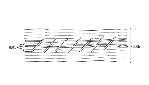
polytetrafluoroethylene, and the like.
[0061] Prior to use, the dry collagen foil may be hydrated, e.g., in
physiological saline. In
one embodiment, the physiological saline includes a 0.9% sodium chloride
solution. In
another embodiment, the collagen foil is hydrated in excipients or drug-
containing solutions.
16
CA 02703103 2015-07-28
The length of time necessary to hydrate the collagen foil may be related to
the thickness of
the foil. The collagen foil can be hydrated until it is consistent in
thickness across its entire
area. The collagen foil biomatrix may be resistant to disintegration in water
or
physiological saline solutions. In one embodiment the collagen foil is
hydrated between
about 1 second and about 1 hour in physiological saline. In another
embodiment, the
collagen foil is hydrated between about 1 second and about 30 minutes in
physiological
saline. In another embodiment, the collagen foil is hydrated between about 1
second and
about 20 minutes in physiological saline. In another embodiment, the collagen
foil is
hydrated between about 1 second and about 10 minutes in physiological saline.
In still
another embodiment, the collagen foil is hydrated between about 1 minute and
about 6
minutes in physiological saline. In another embodiment, the collagen foil is
dipped into the
physiological saline and immediately removed. Such semi-hydration can lead to
enhanced
adherence to tissues. In another embodiment, the collagen foil is hydrated
about 5 minutes
in physiological saline. In another embodiment, the collagen foil is not
hydrated prior to
implantation, which can provide immediate adherence at application to tissues
and
hydration in situ.
100621 Embodiments of the present invention encompass the use, manufacture, or
application of multilayered collagen foil biomatrices, such as those described
in WO
2004/108179 or WO 2007/137839. As noted above, a biomatrix may be used in
conjunction with fibrin sealant. In some cases, fibrin sealant may be applied
to a treatment
site or location prior to application of the biomatrix. In some cases, fibrin
sealant and the
biomatrix can be applied together. Fibrin sealants typically include two main
active
components: fibrinogen and thrombin. The thrombin helps to convert the
fibrinogen into
fibrin monomers, which can cross-link to form a fibrin matrix, which can link
to the
injured tissue, such as pulmonary tissue, and to the native collagen fibrils
of the collagen
biomatrix. A fibrin glue may include an autologous fibrin sealant, derived
from the
patient's own serum. In some cases, collagen biomatrix is applied in
combination with
fibrinogen, followed by application of thrombin. In some cases, fibrinogen is
applied,
followed by application of the collagen biomatrix in combination with
thrombin. In some
cases, collagen biomatrix is applied in combination with fibrinogen and
thrombin. Fibrin
sealant can be applied to a surface of the biomatrix, for example on a side of
the biomatrix
which faces afterwards toward the lung surface upon application of the
biomatrix to the
patient. The fibrin sealant can help fix or adhere the biomatrix to the
patient's lung tissue.
In some cases, fibrin sealant can at least partially fill gaps between a
17
CA 02703103 2010-04-20
WO 2009/056298
PCT/EP2008/009139
rough surface of the lung tissue and the biomatrix. Hence, the sealant can
facilitate close
contact between the biomatrix and the lung surface and support cell attachment
and cell
growth. Fibrin sealant may be applied to a collagen biomatrix in a thin layer.
Fibrin sealant
may also provide barrier properties, for example against fluids and gases.
[0063] Optionally, a collagen biomatrix, or a component associated with the
biomatrix such
as a PEG layer or fibrin sealant layer, can be treated with or include a
coloring agent, to allow
an individual to discern one side of the biomatrix from the other. For
example, one side of a
biomatrix that includes a fibrin sealant for fixation can have a coloring
agent or dye such as
methylene blue. Such visual markers can assist a surgeon in determining how to
apply the
biomatrix to a patient. In the example noted above, the surgeon could apply
the biomatrix so
that the blue side faces toward the lung surface.
[0064] As part of a surgical procedure, a surgeon or other healthcare
professional can apply
a rinse to the biomatrix. For example, a surgeon may hydrate the biomatrix
with a saline
solution prior to application of the biomatrix to the patient tissue. Upon
application to the ,
patient, the natural fluid between lung and chest wall can also provide
hydration to the
biomatrix. In some cases, a surgeon can apply the biomatrix to the patient,
and can peel off
and reposition the biomatrix as needed. Such techniques may be facilitated
with an
adequately hydrated biomatrix. Hydration can also be performed with solutions,
when the
biomatrix is already in situ.
[0065] In another embodiment, the collagen foil produces a liquid-tight and
air-tight seal
when attached to the autologous visceral pleura or lung surface with or
without a continuous
line of fibrin sealant or surgical sealant. In another embodiment, the
collagen foil that
overlaps the visceral pleura or lung surface can be dotted with fibrin sealant
or surgical
sealant to attach it to the visceral pleura or lung surface. A liquid-tight
seal fixation may be
advantageous as it avoids complications associated with contact of the
adjacent tissues with
hemorrhages, e.g., induction of adhesion formation by uncontrolled bleeding
and fibrin
exudation. In another example the collagen foil biomatrix produces a liquid-
tight and air-
tight seal when attached to the tissue with a continuous line of fibrin
sealant or surgical
sealant. In a further example the collagen foil biomatrix that overlaps the
tissue can be dotted
with fibrin sealant or surgical sealant to attach it to the tissue. In still
another example the
collagen foil biomatrix is attached by surgically suturing it to the tissue
once it has been
positioned to the desired contact site. If the collagen foil biomatrix is to
be sutured,
18
CA 02703103 2010-04-20
WO 2009/056298
PCT/EP2008/009139
tensionless suturing techniques can be used to prevent tearing the foil. It
may be desirable to
seal suture lines, for example, with a fibrin sealant. In another example, the
collagen foil
biomatrix is positioned and implanted according to known pressure fitting
techniques. In
some techniques, the collagen foil biomatrix is positioned in the desired
implantation site and
held in place by the surrounding tissues. Thus, the graft remains in place
without the use of
surgical sutures, fibrin sealant, or surgical sealant glue. In another
example, the collagen foil
biomatrix is positioned and implanted without the use of any sealant, glue,
sutures, or
pressure fitting techniques. In this technique, the collagen foil biomatrix is
positioned in the
desired implantation site and held in place by the natural attraction or
adhesion that occurs
between the collagen foil biomatrix and the mammalian tissue. In another
example, semi-
hydration of the biomatrix can enhance the adherence to injured wettish
tissue. In another
example, the collagen foil biomatrix may be applied to a tissue and affixed by
any of the
methods described herein, and then another collagen foil biomatrix may be
applied to an
adjacent tissue, and applied by any of the methods described herein, thus
resulting in adjacent
sheets of the collagen foil biomatrix.
[0066] In another embodiment, the collagen foil is attached by surgically
suturing it to the
injured tissue, e.g. the visceral pleura, once it has been positioned to the
desired implantation
site. This embodiment may be utilized to attach the collagen foil to the
autologous visceral
pleural membranes of the patient. If the collagen foil is to be sutured,
tensionless suturing
techniques may be used to prevent tearing the foil. Suture lines may be
sealed, for example,
with a fibrin sealant.
[0067] In another embodiment, the collagen foil is positioned and implanted
according to
pressure fitting techniques. In this technique, the collagen foil is
positioned in the desired
implantation site and held in place by the natural internal pressure present
in the respective
anatomy. Thus, the graft can remain in place without the use of surgical
sutures, fibrin
sealant, or tissue glue.
[0068] In another embodiment, the collagen foil is positioned and implanted
without the
use of any sealant, glue, sutures, or pressure fitting techniques. In this
technique, the collagen
foil is positioned in the desired implantation site and held in place by the
natural attraction or
adhesion that occurs between the collagen foil and the injured tissue, e.g.
parenchyma.
[0069] The collagen foil can be utilized as a temporary replacement visceral
membrane
graft to repair human visceral tissue, e.g. visceral pleural tissue, due to a
congenital condition,
19
CA 02703103 2010-04-20
WO 2009/056298
PCT/EP2008/009139
birth defect, disease, injury, tumor removal or other surgical procedure that
disrupts or
penetrates the visceral membrane of a patient, or any other condition which
may benefit from
the repair of visceral membrane, e.g. visceral pleura. The collagen foil may
also be utilized
to repair visceral membrane tissue of any of a variety of mammals, including,
but not limited
to sheep, monkeys, horses, rats, mice, humans, laboratory animals, or other
mammals.
Embodiments of the present invention are further directed to kits having
collagen foil and
instructions for its preparation and use as a replacement visceral membrane.
[0070] Methods of covering the tissue with a multilayered biofunctional
collagen foil
biomatrix may be carried out during the treatment of any injuries or defects
of visceral
membranes. In some cases, the step of applying the matrix to the patient can
be performed
during or as part of a lung surgery. In some cases, the multilayered collagen
foil biomatrix
can attract cells such as repair cells and regeneration cells and direct their
in-growth along the
multiple bioactive layers and through and on the foil biomatrix. The
multilayered collagen
foil biomatrix can be reabsorbed and remodeled to natural tissue by the in-
growth of cells.
The layerstructured collagen foil biomatrix can act as a bioactive and
biofunctional scaffold
for cellular in-growth in vivo and is replaced by mammalian tissue with
layerstructured
autologous collagen during regeneration and restoration. The collagen foil
biomatrix can be
resorbable by the mammal in which it is implanted. This property may be
enhanced by the
biofunctionality of the native cross-linked collagen fibers and the
multilayered structure of
the collagen foil biomatrix, as shown in Figures 11A-C and 12A-B for example.
[0071] According to some embodiments, the phrase "covering the tissue with a
multilayered collagen foil biomatrix" means, in general, bringing the tissue
into physical
contact with a multilayered collagen foil biomatrix. In some embodiments, the
contacting of
the tissue with a multilayered collagen foil biomatrix results in an
implantation of the foil.
Examples of the positioning of the multilayered collagen foil biomatrix are
illustrated in
Figures 3-6 and 14. According to some embodiments, a collagen biomatrix is
applied over a
defect (overlay). According to some embodiments, a collagen biomatrix is
applied under a
defect (underlay).
[0072] Collagen biomatrix products provide many useful properties. The
chemotactic
interaction in which they engage facilitates rapid infiltration of endothelial
cells and
fibroblasts, which in turn produce and deposit new collagen autologous fibres
in layers; a
concomitant limited lymphocytic inflammatory response in surrounding
structures promotes
CA 02703103 2010-04-20
WO 2009/056298
PCT/EP2008/009139
absorption of the collagen biomatrix. Collagen also possesses haemostatic
properties which
are put to therapeutic use. Platelets deposit themselves on the collagen
structure, disintegrate
and in doing so release clotting factors which facilitate fibrin formation in
conjunction with
plasma factors.
[0073] Collagen Biomatrix
[0074] According to some embodiments, the phrase "multilayered collagen foil
biomatrix"
or "collagen biomatrix" or "collagen foil" means a biomatrix (e.g. a matrix of
biocompatible
and biofunctional material) of native collagen fibrils treated to remove non-
collagenous
components and to form a sheet of collagen fibrils with a multilayered laminar
structure on a
microscopic level. A multilayered collagen foil may be from any source, such
as bovine,
ovine, porcine, equine, or human origin treated to remove non-collagenous
components and
to form a sheet of collagen fibrils, with the same physical characteristics. A
collagen foil
biomatrix according to some embodiments is substantially nonporous, as
determinable by
scanning electron microscopy.
[0075] According to some embodiments, the term "biofunctional" as used herein
in the
context of a biofunctional multilayered foil biomatrix means that the
biomatrix consists of
native collagen fibrils that are recognized and utilized by the cells of an
animal in a manner
similar to the native collagen fibrils in the animal. For example, without
limitation, such
functions may include migration of repair and regeneration cells along the
biofunctional
collagen fibrils and the multi-layered structure, and the deposition of new
extracellular matrix
by the cells including, or replacing, the biofunctional collagen fibrils.
[0076] According to some embodiments, the phrase "non-naturally occurring
biomatrix" as
used herein means a manufactured matrix or framework having native collagen
fibrils formed
from (i) a material existing in nature (i.e. natural material) that has been
treated or processed
in a manner in which the collagen fibrils contained in the natural material
have been moved
or repositioned from their naturally-occurring arrangement within the collagen
structure of
the natural material; or (ii) a material not existing in nature (i.e. a non-
natural, artificial
material, such as a recombinant material) treated or processed to manipulate
the arrangement
of the collagen fibrils. For example, a non-naturally occurring biomatrix may
be formed
from starting material which includes collagen that has been mechanically or
chemically
processed (e.g. grounded, chopped, etc.). A collagen biomatrix that is formed
from the
treatment or processing of starting material in a manner that preserves the
structure of the
21
CA 02703103 2010-04-20
WO 2009/056298
PCT/EP2008/009139
naturally occurring collagen framework is not a non-naturally occurring
biomatrix (e.g.
epidermal tissue treated to remove cellular components while preserving the
naturally
occurring collagen structure).
[0077] In some embodiments, the collagen foil biomatrix includes connective
tissue
proteins having collagen fibrils. For example, the collagen foil biomatrix may
include
connective tissue proteins with Type I collagen fibrils. In addition to having
collagen fibrils,
a collagen foil biomatrix can also include an excipient, a preservative, a
growth factor, or an
additive that aids in the flexibility and elasticity of the final product.
Each layer of collagen
fibrils can be substantially nonporous. According to some embodiments, the
phrase
"substantially nonporous" means that any pores that are present in a collagen
foil biomatrix as
a result of precipitation of collagen fibrils to form a collagen sheet are
primarily isolated from
one another, and the pores are not interconnected in a manner that traverses
the thickness of
the collagen foil. Mechanical perforations that create holes in the collagen
foil biomatrix are
not pores. In some cases, the material appears to be substantially free of
pores that would be
visible using a scanning electron microscope at 1500x magnification. Scanning
electron
microscope pictures illustrate the nonporous nature of the collagen foil
biomatrix as in
Figures 7, 8A-B, 9A-B, and 10.
[0078] According to some embodiments, the collagen foil is resorbable by the
mammal in
which it is implanted. Without being bound by any particular theory, it is
thought that this
property may be enhanced by the structure of the collagen foil. The process
utilized to
produce the equine collagen foil forms stacked layers of collagen fibrils.
Between each layer
are interstices into which cells and vasculature of the patient can migrate
and form visceral or
parietal membrane tissue, such as neo-pleura tissue. Each layer of collagen
fibrils can be
substantially nonporous. The few pores which may be present are typically
isolated from one
another and do not interconnect through multiple layers of collagen fibrils.
The multiple
layer structure of the present invention enhances the liquid-tight and air-
tight characteristics
of the collagen foil.
[0079] Collagen foil biomatrix embodiments can encompass a non-naturally
occurring
multi-layered collagen membrane having layers of numerous multi-directional
intertwined
collagen fibrils. Thus, the collagen fibrils can be arranged in a multi-
directional fashion in a
plane, and these planes form sheets, which create a multi-layered structure.
An illustration of
a dry collagen foil biomatrix may be seen in the photomicrograph (SEM) of
Figure 7, which
22
CA 02703103 2010-04-20
WO 2009/056298
PCT/EP2008/009139
illustrates the surface of the collagen foil biomatrix in which collagen
fibrils are embedded.
The collagen fibrils are visible on the surface on photographs of the upper
surface of the
collagen foil biomatrix under ESEM (Environmental Scanning Electron
Microscopy)
conditions, in which a slightly humid atmosphere provides near natural
conditions. As shown
in Figures 8A-B, the surface appears smooth and substantially nonporous.
Photographs
(ESEM) of the lower surface of collagen foil biomatrix illustrate the
substantial non-porosity
of the collagen foil biomatrix, as depicted in Figures 9A-B. Collagen fibrils
are evident.
[0080] The unique orientation of the native collagen fibrils in two-
dimensional directions in
the multiple layers is primarily responsible for a liquid-tightness and air-
tightness, even for
example under high hydrostatic pressure, and provides great strength with high
elasticity.
Due to the numerous parallel-oriented thin collagen fibril layers of the
collagen foil
biomatrix, this material is suitable for temporarily replacing the body's own
visceral or
parietal membranes in closing the defect after covering and provides a
biofunctional
biomatrix scaffold for cell in-growth for forming a new tissue and collagen
structures. The
multiple layer structure can enhance the liquid-tight and air-tight
characteristic of the
collagen foil biomatrix.
[0081] Prior to using the collagen foil to repair visceral membrane tissue of
a mammal, the
dry collagen foil material may be hydrated. Figure 10 is a SEM photograph
illustrating the
surface of a hydrated collagen foil wherein collagen fibrils are clearly
shown. Substantial
non-porosity of the surface is evident from the picture.
[0082] According to some embodiments, the collagen foil biomatrix is
substantially
nonporous, and interstices exist between the layers of collagen fibrils. The
collagen foil
biomatrix can be analogous to a stack of pages wherein each page is
substantially smooth and
nonporous, with a space between each page. When in its dry form the
interstices can be more
pronounced. The interstices become reduced when the collagen foil biomatrix is
observed
under near natural conditions in a slightly humid atmosphere. The reduction of
the interstices
of the collagen foil biomatrix and the layered characteristics are illustrated
in pictures of cross
sections of collagen foil biomatrix in a humid atmosphere in Figures 11A-C.
Humid ESEM
conditions may approach natural conditions. The material reveals a structure
like a stack of
sheets packed very tightly together. Interstices between the collagen layers
are evident. In
comparison, Figures 12A-B are ESEM photographs showing a dry collagen foil.
Multiple
layers of collagen and interstices between the collagen layers are evident. In
addition to
23
CA 02703103 2010-04-20
WO 2009/056298
PCT/EP2008/009139
promoting liquid-tight and air-tight properties, the numerous parallel-
oriented thin collagen
fibril layers of the collagen foil biomatrix simultaneously serve as a
bioequivalent
biofunctional scaffold for cell in-growth for de novo construction of the
body's own tissue.
[0083] In some cases, the change in volume of the collagen foil biomatrix is
small or
negligible when hydrated. The collagen foil biomatrix substantially retains
its size and shape
upon being hydrated, having excellent shape stability even after hydration,
and causing no
problems of swelling or shrinking following the contact with the tissue. Once
hydrated and
implanted, collagen foil biomatrix embodiments may. not significantly expand
or contract in
area or thickness to the extent that it would tear surgical sutures or break
apart fibrin or other
biocompatible glue seals that hold the collagen foil biomatrix to the
patient's tissue.
[0084] In some cases, the shrinking or swelling of the area of the dry
collagen foil
biomatrix may vary from about -5% to about 20% when completely hydrated. In
some case,
the area of the dry collagen foil biomatrix may vary between about -5% to
about 10% when
completely hydrated. Optionally, the area of the dry collagen foil biomatrix
can vary
between about -5% to about 5% when completely hydrated. For example, the area
of the dry
collagen foil biomatrix may increase no more than about 4 percent when
completely
hydrated.
[0085] In some cases, the collagen foil biomatrix increases up to about 6
times its dry
thickness when it is completely hydrated. In some cases, the collagen foil
biomatrix
increases up to about 3 times its dry thickness when it is completely
hydrated. In some cases,
the collagen foil biomatrix increases to about twice its dry thickness when it
is completely
hydrated.
[0086] The thickness of the collagen foil biomatrix may vary depending on the
particular
application. Varying the amount of starting material utilized to produce a
particular size of
collagen foil biomatrix can control the thickness of the collagen foil
biomatrix. In some
cases, the collagen foil biomatrix, when in its dry form, has a thickness
between about 0.01
mm to about 3.0 mm. In another example, the collagen foil biomatrix has a
thickness
between about 0.02 mm to about 2.0 mm. In a further example, the collagen foil
biomatrix
has a thickness between about 0.03 mm to about 1.5 mm. In another example, the
collagen
foil biomatrix has a thickness between about 0.05 min to about 1 mm. In still
another
example, the collagen foil biomatrix has a thickness of about 1.0 mm or less.
24
CA 02703103 2010-04-20
WO 2009/056298
PCT/EP2008/009139
[0087] The dry weight of the collagen foil biomatrix may be dependent on its
desired
thickness. In one example, the dry weight of the collagen foil biomatrix is
between about 1
mg/cm2 to about 50 mg/cm2. In another example, the dry weight of the collagen
foil
biomatrix is between about 1.5 mg/cm2 to about 30 mg/cm2. In still another
example, the dry
weight of the collagen foil biomatrix is between about 2 mg/cm2 to about 20
mg/cm2. In a
further example, the dry weight of the collagen foil biomatrix is between
about 2.5 mg/cm2 to
about 15 mg/cm2. For example, the dry weight of the collagen foil biomatrix
can be between
about 3 mg/cm2 to about 10 mg/cm2.
=
[0088] In some cases, the weight of the collagen foil biomatrix increases up
to about 15
times its dry weight upon hydration. In another example, the weight of the
collagen foil
biomatrix increases up to about 10 times its dry weight upon hydration. In
another example,
the weight of the collagen foil biomatrix increases up to about 7 times its
dry weight upon
hydration. In still another example, the weight of the collagen foil biomatrix
increases up to
about 5 times upon hydration from its dry state.
[0089] According to some embodiments, the collagen foil biomatrix beneficially
has high
tensile strength, which improves and supports the handling of the collagen
foil biomatrix, for
example during its surgical application, and provides an increased mechanical
stability, for
example after its implantation. Additionally, increasing the thickness of the
collagen foil
biomatrix can significantly increase the tensile strength.
[0090] The propensity of collagen foil biomatrix material to tear under
exerted pressure
may be measured as its "ultimate tensile load" or "ultimate tensile force,"
hereinafter referred
to as "ultimate tensile force." The ultimate tensile force of a collagen foil
biomatrix may be
determined by subjecting pressure to a strip of collagen foil biomatrix having
a specified
width and determining the amount of pressure applied that results in failure
(e.g., tearing or
rupturing) of the collagen foil biomatrix. Ultimate tensile force may be
quantified using the
following equation: "Ultimate Tensile Force" = force applied/width of collagen
foil biomatrix
strip = Newtons/cm-strip.
[0091] In some cases, the collagen foil biomatrix has an ultimate tensile
force between
about 1 and about 30 Newtons/cm-strip. In some cases, the collagen foil
biomatrix has an
ultimate tensile force between about 1.5 and about 15 Newtons/cm-strip. In
some cases, the
collagen foil biomatrix has an ultimate tensile force between about 2 and
about 10
Newtons/cm-strip. In some cases, the collagen foil biomatrix has an ultimate
tensile force
CA 02703103 2010-04-20
WO 2009/056298
PCT/EP2008/009139
between about 3 and about 6 Newtons/cm-strip. In some cases, a collagen foil
biomatrix with
an integrated surgical mesh, for example of the type illustrated in Figure 6B,
can have an
ultimate tensile force of more than 30 Newtons/cm-strip.
[0092] Collagen foil biomatrix embodiments can have a high tensile strength,
yet remains
elastic and flexible when hydrated. This feature permits a collagen foil
biomatrix to
optimally adapt to the anatomic conditions (e.g. curves) present at the
contact site.
[0093] When in its hydrated state, the collagen foil biomatrix can be easily
moved around.
For example, the biomatrix can be moved around in the surgical site and
optimally modeled
and adapted to the shape and position of the visceral or parietal membrane
defect, e.g. where
it is being implanted. Once implanted, the collagen foil biomatrix graft
remains smooth and
may be repositioned if necessary or desired. Over time, cells and vasculature
migrate
directed throughout the multiple layers of the multilayered collagen foil
biomatrix, eventually
replacing the multilayered collagen foil biomatrix with a new tissue and
autologous collagen
structures. As cells migrate and vasculature forms within the layers of the
collagen foil
biomatrix, the tissue takes on the form of the collagen foil biomatrix in a
directed way. After
cellular organization of the collagen foil biomatrix with the newly formed
connective tissue,
adhesion formation to adjacent tissues, e.g. to the parietal pleura or chest
wall, can be
minimized.
[0094] Collagen for use in manufacturing the collagen foil biomatrix may be
obtained from
any suitable source. For example, without limitation, collagen may be of
bovine, ovine,
porcine, equine, or human origin. The collagen may be harvested from a
naturally occurring
tissue, such as tendon, corium, or other collagen rich tissue or may be
produced by
recombinant genetic means. As described below, one exemplary embodiment of the
invention utilizes equine collagen derived from Achilles tendon.
[0095] According to some embodiments, a collagen biomatrix includes native
equine
collagen fibrils (mainly type I collagen) precipitated from purified minced
equine Achilles
tendon. A flexible formstable and elastic biomatrix can have a nonporous fluid-
tight and air-
tight multilayer structure. In a dry state, the biomatrix thickness may be
about 0.1 mm, and
in a wet state the membrane thickness can be about 0.3 mm.
[0096] A multilayered collagen foil biomatrix according to embodiments of the
present
invention can include a collagenous native cross-linked microscopically
multilayered
biomatrix having multiple layers of a substantially nonporous foil that
includes collagen
26
CA 02703103 2015-07-28
fibrils. Embodiments of such non-naturally occurring biomatrices are described
in the
international patent application WO 04/108179 and WO 07/137839. Collagen foils
which
may be used according to embodiments of the present invention are typically
biofiinctional, bioactive, mechanically stable, elastic, nonporous, air-tight
and fluid-tight,
especially blood and cell tight, and can provide a temporary barrier against
uncontrolled
distribution of blood, fibrinogen, necrotic material and damaged tissues. A
defined
bioactive separation layer between the visceral tissue and the adjacent
anatomical
structures thus initially shields the treated tissue. A multilayered collagen
foil biomatrix
can act as a hemostatic agent and inhibit uncontrolled fibrin band formation
and
distribution as well as hematomas, which are one of the main causes for
fibrosis and
adhesion formation, in anatomical areas which are located beside or close to
visceral
membranes, e.g. the lung surface.
100971 A mechanically stable, elastic, nonporous, and primary fluid-tight and
gas-tight
collagenous biomatrix can be applied to a visceral membrane defect. The
collagen
biomatrix can acts as an artificial epithelium/mesothelium and cover and
protect the
visceral or parietal surface. This results in the primary closure of the seal
of fluid and air
leaks at the organ surface. Advantageously, application of the collagen
biomatrix can
provide an immediate mechanical sealant, acting as a barrier for fluid or air.
Due to the
layered structure of the biomatrix, fibroblasts or wound healing cells such as
lymphocytes
or macrophages can be directed or allowed to migrate throughout the biomatrix,
leading to
the production of endogenous collagen. Accordingly, the patient's own visceral
or parietal
membrane can be regenerated or enhanced, for example as the wound healing
cells are
eventually replaced with normal visceral membrane. In this way, the biomatrix
can be
remodeled into the body's own visceral membrane. This can be accomplished with
little or
no adhesions or binding, particularly between the visceral tissue and adjacent
areas, e.g.
the lung surface and the chest wall.
100981 According to some embodiments, visceral membrane regeneration with a
collagen
biomatrix can replace or augment certain standard surgical techniques. For
example,
pleura regeneration can be carried out instead of lung sealing, coagulation
techniques,
suture techniques, and implantation of other "biomaterials". Biofunctional
collagenous
biomatrices can be used for the steering of cell ingrowths and enhanced de
novo formation
of extracellular matrix for the regenerative processes of visceral membranes.
A collagen
biomatrix can adheres to the wound surface, initially through adhesive forces.
The native
27
CA 02703103 2010-04-20
WO 2009/056298
PCT/EP2008/009139
collagen fibrils of the biomatrix can activate coagulation and the local
formation of fibrin,
which additionally supports the closure of leaks, and the adherence of the
collagen
membrane. Bioactive collagen can attract repair cells, especially fibroblasts.
This may
additionally be supported by the local presence of fibrin (e.g. physiologic
fibrin sealant).
[0099] Parallel layers in a structured multilayer biomatrix can direct the
ingrowth of
fibroblasts and repair cells, along the layers. The ingrowths along the
parallel layers of the
collagen matrix are typically faster than through the layers. The de novo
formation of the
extracellular matrix with endogenous collagen is also guided through the cell
(fibroblast) line
up. This process steers the remodeling of the biomatrix to living tissue, and
closes the
visceral defect, for example after traumatic rupture, surgical incisions,
resection or
decortication. In a decortication process, the surface layer membrane of the
lung is removed.
[0100] In this way, embodiments of the present invention encompass techniques
for
forming a multilayered extracellular matrix resulting from collagen structure
of the collagen
foil. A steering collagen biomatrix provides for directed cell ingrowth for
the regeneration of
visceral and parietal membranes. Such steered regeneration of tissue is well
suited for
closing or reducing leaks in pleural tissue, such as prolonged post-surgical
pulmonary air
leaks. In fact, animal studies have demonstrated the efficiency of a collagen
biomatrix in the
prevention of prolonged postoperative air leaks in pulmonary surgery, and in
the steered
regeneration of visceral membranes through directed cell ingrowth.
[0101] According to some embodiments, the collagen biomatrix of the present
invention is
an impermeable material for the purpose of sealing and the contact between
collagen and
blood cells determine platelet aggregation. This interaction with the collagen
biomatrix
induces the release of coagulation factors and formation of fibrin. The
collagen biomatrix
can be cut according to necessity and adapted to visceral defect. Collagen
biomatrix
embodiments of the present invention have proved successful in macroscopic
aspects of graft
incorporation, preventing early and late air leak in post operative subjects,
without harmful
reactions to adjacent tissue structures (inflammation, adhesion, fibrosis,
necrosis) and in
histological assessment of the incorporation process and connective tissue
organization.
Multilayered collagen foil biomatrix embodiments can attract cells such as
repair cells and
regeneration cells. Biomatrix materials can direct the cell growth on to the
surface of the
biomatrix, and provide for the in-growth of repair and regeneration cells.
Biomatrix
materials can be remodeled to natural tissue after the in-growth and can be
resorbed.
28
CA 02703103 2010-04-20
WO 2009/056298
PCT/EP2008/009139
[0102] Standard collagen based compositions are usually perceived as foreign
by the host
and often encapsulated. Therefore, recellularization and remodeling to the
respective
anatomical tissue does not occur or is impossible, there is no directed cell
in-growth and no
control of the regeneration process, and the collagen is merely tolerated as a
"biocompatible"
-- implant. In contrast, the multilayered collagen foil biomatrix according to
embodiments of
the present invention acts as a membrane (e.g., visceral pleura membrane)
functioning as a
bioactive temporary layer directing cell growth within the multilayered
collagen foil
biomatrix and on the surface of the collagen foil biomatrix. Rather than
acting solely as a
barrier against cell growth, as most anti-adhesion compositions do, the
multilayered collagen
-- foil biomatrix can be extremely bioactive and support the remodeling of the
tissues. For
example, within weeks after implantation, a multilayered collagen foil
biomatrix can be well
integrated into the restored anatomical structure of visceral membrane
tissues. Further,
during and after surgery, the nonporous, fluid-tight (e.g., blood, liquid,
air, gas, bile, etc.)
multilayered structure of the collagen membrane is capable of preventing
uncontrolled
-- distribution of blood (e.g., fibrinogen/fibrin) and necrotic material from
the pleural wound
areas, which may be responsible for supporting conditions of adhesion
formation in the initial
time period after surgery (in contrast to porous compositions). The collagen
biomatrix can
also prevent direct contact between the visceral surface and the adjacent
tissues such as the
lung surface and parietal pleura or chest wall, a primary area of the scar
formation and
-- fibrosis. This contributes also to the controlled remodeling of anatomical
structures with
prevention and minimization of uncontrolled adhesion and scar formation and
pleural
fibrosis. Such techniques can be used in any mammal, including without
limitation humans,
dogs, cats, mice, rats, and the like.
[0103] In addition to promoting liquid-tight and air-tight properties, the
numerous parallel-
-- oriented thin collagen fibril layers of the collagen foil simultaneously
serve as a biomatrix
scaffold for cell ingrowth for de novo construction of the body's own visceral
membrane. It
has been surprisingly discovered that the nonporous, layered structure of the
collagen foil
promotes the ingrowth of cells, vasculature, and the formation of new collagen
structures
across the collagen foil and in the interstices that exist between its
multiple layers, forming a
-- neo-pleura with a typical layer structure of a natural pleura within weeks
of implantation. As
described elsewhere herein, the ingrowth of cells, vasculature, and new
collagen structure is
so extensive that within weeks post-operation, the neo-pleura becomes
difficult to distinguish
29
CA 02703103 2010-04-20
WO 2009/056298
PCT/EP2008/009139
from a patient's previously existing visceral pleura tissue. Hence, eventually
the graft
ingrowth may become fully organized.
[0104] According to some embodiments of the present invention, a significant
benefit of
using the collagen foil is the substantially low risk of transmitting a
disease to a patient in
which it is implanted. Manufacturing process in which the collagen fibrils are
treated with
acids (e.g., hydrochloric acid, acetic acid, and the like) and bases, such as
sodium hydroxide,
to produce the collagen foil beneficially acts to inactivate or reduce the
infectious levels of
bacteria, viruses, and prions that may be present. Treatment of biomaterial
with hydrochloric =
acid, sodium hydroxide, ethylene oxide (ET0), and the like have been
recognized by
governmental agencies as approved methods within drug and biomaterial
regulations to
inactivate prions and viruses. Such treatment may, under some regulations,
reduce the
regulatory requirements for testing the collagen foil on a batch-by-batch
basis. Thus, the
treatment of the collagen fibrils during the manufacturing process enhances
the product safety
and reduces the risk of disease transmission to a patient.
[0105] Collagen material that has been subjected to the manufacturing process
described
herein is not known to transmit any pathogens to patients. Thus, in addition
to the
manufacturing process, utilization of equine collagen further avoids the risks
of transmitting
spongiform encephalitis that have been previously associated with human
cadaveric
substitutes. Use of collagen derived from an equine origin, such as collagen
derived from
equine Achilles tendons avoids the risks of transmitting transmissible
spongiform
encephalopathy (TSE), which is also known as bovine spongiform encephalopathy
(B SE) or
scrapie. Transmission of this disease has been associated with the use of
biological material
obtained from ruminant sources (e.g., biological material from cattle, goats,
sheep, and the
like).
[0106] The change in volume of the collagen foil is small or negligible when
hydrated. In
contrast to porous replacement products, the collagen foil substantially
retains its size and
shape upon being hydrated, having excellent shape stability, remaining bio
stable even after
hydration, and causing no problems of swelling or shrinking in the body
following
implantation. Once hydrated and implanted, collagen foil does not
significantly expand or
contract in area or thickness to the extent that it would tear surgical
sutures or break apart
fibrin glue seals that hold the collagen foil to the patient's tissue.
CA 02703103 2010-04-20
WO 2009/056298
PCT/EP2008/009139
[01071 The thickness of the collagen foil can be controlled by varying the
amount of
starting material utilized to produce a particular size of collagen foil. The
collagen foil can
be gas-sterilized with ethylene oxide (ETO) or similar sterilization gas or by
irradiation.
[0108] Manufacture Of Collagen Biomatrix
[01091 According to some embodiments, the collagen foil is a biomatrix of
collagen fibrils
treated to remove cellular components and to form a sheet of collagen fibrils.
During the
manufacturing process, for example as described in WO 04/108179 or WO
07/137839, the
collagen fibrils become naturally cross-linked as the fibrils precipitate out
of solution to form
a collagen foil. Unlike cross-linking the collagen fibrils with chemicals or
radiation (e.g.
ionizing or ultraviolet radiation), allowing natural cross-linking of the
collagen fibrils can
ensure their biofunctionality, promote accelerated regeneration, and reduce
resorption times
once the collagen foil biomatrix is brought into contact with the tissue.
Cross-linking
collagen fibrils with chemicals or radiation can result in increased
resorption times, or even
non-resorption, encapsulation, and scar formation. The natural cross-linking
of the fibrils in
the collagen foil biomatrix utilized in some embodiments occurs by natural,
physiological-
like means. Primarily this natural cross-linking is through non-covalent
interactions (e.g. van
der Waals or dipole-dipole interactions) or by the formation of readily
dissociable Schiff-base
bonds between the amino acid side chains of the collagen molecule.
Intermolecular cross-
linking of collagen is responsible for physical and chemical stability. A key
step in the
formation of collagen cross-links depends on the enzymatic conversion of
lysine or
hydroxylysine residues and gives rise to aldehydes, allysine and
hydroxyallysine. These
aldehyde groups spontaneously react with reactive amino groups resulting in
the formation of
Schiff-base components containing labile aldolcondensation products with
labile aldimine
links (like for example -CH=N-). Thus, the fibrils of the product may be
dissociated by
treatment with, for example, a weak acid. Cross-linking arising from the use
of chemical
cross-linking agents can be detected from the presence of stable covalently
cross-linked
cross-linking moieties. Commonly, this is accomplished by using a Schiff-base
reagent (e.g.
glutaraldehyde) to form Schiff-base reaction products, and then stabilizing
the bonds through
either an Amadori-rearrangement or reducing conditions. In addition collagen
can be cross-
linked by various bifunctional carbodiimide reagents. Cross-linking arising
from the use of
radiation can be detected by the presence of stable covalent bonds between the
collagen
fibrils, caused by the reaction of free radical moieties generated during
irradiation. The
fibrils in the biomatrix product, on the other hand, are substantially uncross-
linked with any
31
CA 02703103 2010-04-20
WO 2009/056298
PCT/EP2008/009139
stable covalent bonds, and have not been treated in a chemical or irradiative
manner. Thus,
any associations between the fibrils in the biomatrix product are
substantially non-covalent or
readily reversible, and are not stably cross-linked. Chemicals such as
cyanamide,
glutaraldehyde, formaldehyde, acrylamide, carbodiimidediones, diimidates,
bisacrylamides,
and the like have been utilized in the past to chemically cross-link collagen
fibrils. Use of
such chemicals, however, may result in toxicity risks associated with
inadvertently contacting
visceral tissue with residual chemicals in the collagen foil biomatrix. The
precipitation
process thereby avoids the toxicity risks of cross-linking chemicals and
longer resorption
times associated with cross-linking the collagen fibrils with chemicals or
radiation.
[0110] In some cases, the resulting dried, precipitated, collagen composition
forms a
collagen foil biomatrix having a high-molecular weight multi-layered collagen
membrane
that includes numerous layers of two-dimensionally multi-directional naturally
intertwined
collagen fibrils. The collagen foil biomatrix may primarily contain
interstitial Type I
collagen. The collagen foil biomatrix may have substantially no pores and can
be primarily
liquid-tight and air-tight. Immune diffusion tests may be conducted on the
product to
guarantee the absence of foreign protein. The collagen foil biomatrix may be
gas-sterilized
with ethylene oxide (ETO) or similar sterilization gas or by irradiation.
[0111] A significant benefit of using an collagen foil biomatrix is the
substantially low risk
of transmitting a disease to a patient being contacted with said foil. The
manufacturing
process in which the collagen fibrils are treated with acids (e.g.
hydrochloric acid, acetic acid,
and the like) and bases, such as sodium hydroxide, to produce the collagen
foil beneficially
acts to inactivate or reduce the infectious levels of bacteria, viruses, and
prions that may be
present. Treatment of biomaterial with hydrochloric acid, sodium hydroxide,
ethylene oxide
(ETO), and the like have been recognized as approved methods within drug and
biomaterial
regulations to inactivate prions and viruses. Such treatment may, under some
regulations,
reduce the regulatory requirements for testing the collagen foil on a batch-by-
batch basis.
Thus, the treatment of the collagen fibrils during the manufacturing process
enhances the
product safety and reduces the risk of disease transmission to a patient.
[0112] Additionally, embodiments encompass the use of a multilayered collagen
foil
biomatrix in the manufacture of a medicament, i.e. a medically applicable
material, for
treating a disorder such as e.g. injuries, surgeries, or pathogen-based
diseases, in a mammal
characterized by a defect in the visceral membrane or surrounding tissue.
32
CA 02703103 2010-04-20
WO 2009/056298
PCT/EP2008/009139
[0113] According to some embodiments, a collagen foil can be produced from
suspensions
of high molecular weight collagen fibrils through a controlled drying process.
A graded
precipitation of the collagen fibril suspension results from the evaporation
of water and
simultaneous pH elevation. The controlled drying process results in a multi-
layered
construction of a collagen foil that can be implanted by surgeons. The multi-
layered collagen
foil construction provides a number of properties that are beneficial in a
pleural substitute and
as a biomatrix for the regeneration of living pleural tissue.
[0114] In one embodiment, the process to produce the collagen foil removes all
cellular
components producing a collagen foil of collagen fibrils that primarily
includes acellular
components.
[0115] Using established procedures in collagen chemistry, collagen-containing
tissue can
be used as a starting material for the preparation of the collagen foil. In
one embodiment,
tendons, such as Achilles tendons, are used as a starting material. In a
further embodiment,
equine Achilles tendons are used as a starting material. According to some
embodiments,
collagen can be obtained from any of a variety of animals, including without
limitation sheep,
monkeys, cows, horses, rats, mice, humans, or laboratory animals. Hence,
collagen can be
derived from a bovine source, a porcine source, an equine source, an ovine
source, a primate
source, a rodentia source, or a human source, for example.
[0116] In one embodiment, the starting material, for example equine Achilles
tendons, is
first ground and treated for at least one hour with 1 N sodium hydroxide and
neutralized with
hydrochloric acid. The collagen starting material is treated in acid
conditions at pH 2. The
acid utilized may be hydrochloric acid, acetic acid, or the like.
Subsequently, the non-
collagenous proteins and intermolecular cross-linking bonds present in the
starting material
are degraded enzymatically with pepsin to form a suspension of collagen. The
suspension is
then neutralized. In one embodiment, the suspension is neutralized to between
about pH 6.5
to about pH 8Ø In another embodiment, the suspension is neutralized to
between about pH
6.9 to about pH 7.5. In another embodiment, the suspension is neutralized to
about pH 7.
The collagen suspension is centrifuged, the supernatant removed, and the
precipitate
resuspended in acetic acid at about pH 2-4.5. Non-collagenous proteins are
thereby
successfully removed from the suspension of collagen. Repetition of the above-
described
steps may be conducted as necessary to remove residual non-collagenous
proteins present in
the precipitate.
33
CA 02703103 2010-04-20
WO 2009/056298
PCT/EP2008/009139
[0117] A surprising result of the production process of the equine collagen
foil is that a
controlled pH elevation of the collagen suspension in acetic acid is achieved
due to the
specified removal of water by evaporation over a long period of time, e.g., 24
hours. The
specified elevation of pH causes the precipitation of the multi-directional
intertwined
collagen fibrils in two-dimensional direction layers forming a multi-layered
construction of
the equine collagen foil. In one embodiment, the process is performed in a
drying oven at a
temperature of about 20 C to about 55 C, with equipment to remove steam and
the
simultaneous steam neutralization of acetic acid. In another embodiment, the
process is
performed in a drying oven at a temperature of about 30 C to about 45 C.
[0118] The equine collagen foil that results from the production process can
be considered
to be in its dry form when further loss of water is not detected or is
negligible. The water
content of the "dry form" of equine collagen foil is typically between about
2% to about 18%
by weight. The relatively high residual water content present in the "dry
form" of the equine
collagen foil prevents or restrains the denaturation of collagen molecules
that comprise the
equine collagen foil.
[0119] The above-described process is responsible for the precipitation of the
collagen
fibrils from the suspension since components with low solubility fall out at
the beginning of
the process at a low pH elevation. This technique results in a precipitation
of collagen fibrils
during water evaporation and simultaneous pH elevation.
[0120] Processes utilized to produce a collagen foil biomatrix can form
stacked layers of
collagen fibrils. Between each layer of collagen fibrils are interstices into
which cells and
vasculature of the patient can migrate and form new collagen structures and
native-
conformation tissue. It is a beneficial property of some embodiments that the
biofunctional
native collagen fibers and the nonporous, layered structure of the collagen
foil biomatrix
promotes the in-growth of cells, vasculature, and the formation of new
collagen structures
across the collagen foil biomatrix and in the interstices that exist between
its multiple layers.
As compared to random, unguided, non-controlled cellular in-growth at the
wound or defect,
the directed in-growth and regeneration according to method embodiments can
inhibit or
prevent the formation of adhesions and fibrosis. Thus, pain and complications
associated
with adhesions and fibrosis can be avoided.
[0121] According to some embodiments, the purification process for producing
the equine
collagen foil begins with at least one hour of sodium hydroxide solution
treatment of the
34
CA 02703103 2010-04-20
WO 2009/056298
PCT/EP2008/009139
tendon starting material, followed by neutralization in hydrochloric acid.
Pepsin is then used
to break down the tendons. The colloidal collagen thus produced is
precipitated as fibrils.
Drying and gas sterilization then yields equine collagen foil with 5.6 mg of
native collagen
fibrils per square centimeter. Nothing else is added and no artificial methods
for cross
linkage (i.e. involving chemicals or radiation) are performed. Immunodiffusion
tests ensure
that no foreign proteins are present. An equine collagen foil can be made of
native equine
collagen fibrils (mainly interstitial type I collagen). One square centimeter
of the material
can contain 5.6 milligrams of collagen fibrils with no cellular components.
Fibrin glue, such
as TissucolTm Duo S (Baxter), can be used to attach the grafts to the visceral
membrane or
outer wound surface. This biological dual-component glue kit includes of a pre-
filled syringe
containing human plasma proteins, fibrinogen, clotting factor XIII, plasma
fibronectin and
aprotinin, and another pre-filled syringe containing thrombin and calcium
chloride.
[0122] Experimental Animals
[0123] The following example presents the results of experiments in pigs to
evaluate a
collagen foil for its suitability as a visceral pleura substitute used to
repair visceral pleura
defects and as a biomatrix for visceral pleura regeneration. For the
experiments, 5 Landrace
pigs, around 35 kg of weight, pigs were used. The animal studies were
performed after
formal approval by the authorities of the Ethic Committee of the Catholic
University of Italy.
The choice of this species and this weight is due to the similarity of the
swine pulmonary
parenchyma with the human one. The dimensions of the animals allow us to use
the surgical
instrumentation normally used in humans and act as an ideal animal model for
human
subjects. Before surgery, the animals were prepared at the "Laboratory animal
facility of
Holy Heart University ( Rome )" for a period of 5 days. The day of the
intervention the
animal was set in general anaesthesia through the following procedure:
[0124] Anaesthesia:
[0125] Preanaesthesia through an intramuscular injection of: Atropin 0,02
mg/kg +
Ketamine 15 mg/kg + Diazepan 0,1mg/kg ; induction with mask for anaesthesia
with 02 and
vapours of Isoflurane 2%; endotracheal intubation and maintenance with 02 and
vapours of
Isofluorane 1,5%; muscular relaxing with pancuronium bromide (0,1 mg/kg e.v.).
[0126] Surgical procedure:
CA 02703103 2015-07-28
[01271 After the induction of anaesthesia, the animals were maintained in
lateral decubitus
and a lateral thoracotomy on the fourth intercostal space was performed.
Visceral pleura was
damaged to obtain bleeding and air leak, as indicated in Figure 13. A piece of
the collagen
biomatrix (measuring 3.5 x 2.5 cm) was then cut to size and immersed for 5
minutes in sterile
0.9% saline, and applied to the damaged visceral pleura, as depicted in Figure
14. To close
the defect, the graft was tucked all round under the pleural whole margins and
sealed with
fibrin sealant to keep it in place, and to assure a water-tight or fluid-tight
closure. The
collagen biomatrix includes native equine collagen fibrils (5.6 mg/cm2)
purified from minced
equine Achilles tendon, and the fibrin sealant includes TissucolTm Duo S
(Baxter).
101281 To detect any post application air leak, the aerostatic power of the
collagen
biomatrix was evaluated through a hydro pneumatic test, as shown in Figure 15.
The lung
was covered with saline solution and the lung insufflated by the
anaesthesiologist. In absence
of an air leak, a water-drainage was removed before the animals were revived.
In case of air
leak, water-drainage was connected with a system of measurement of the air
leak. As
depicted in Figure 16, the collagen foil covers the lung tissue and provides a
water-tight or
air-tight closure while protecting the cellularity of the tissue.
[0129] The thoracotomy was sutured in layers with absorbable thread and the
skin sutured
in silk. The animals were revived and observed at the laboratory animal
facility for 7, 15, 21
and 28 days during which the entity of the possible air leak was appraised.
[0130] Post-operative analgesic (Ketoprophene, Findol 10%, 0,3 m1/10kg/die,
intramuscularly) and antibiotic (enrofloxacin 2,5 mg/kg/die i. m.) therapy was
lengthened for
7 days. The animals were given conventional mixed feed because the drainage
did not
interfere with physiological functions. Every day the health of the animal was
monitored. At
the VIII, XVI, XXII and )0(IX day, the animals have been again submitted to
general
anaesthesia through bronchial intubation, and rethoracotomy again performed to
the fifth
intercostal space through cutaneous incision along the intercostal space.
After incising the
intercostal muscle, a costal retractor (Finocchietto) and an autostatic
abdominal retractor were
placed in site. After the opening of the pleural space, the lobe, or the whole
lung, previously
TM
treated with collagen biomatrix, were removed. The thoracotomy was sutured
with Vicryl 2
and the skin with silk I. At the end of the procedures, the animal, still in
general aesthesia,
were euthanized with Tanax E.V. (3m1/10kg).
101311 Results:
36
CA 02703103 2010-04-20
WO 2009/056298
PCT/EP2008/009139
[0132] Anaesthesia, surgery and the postoperative follow-up periods were
uneventful in all
but two animals. None of the animals displayed signs of inflammation, or
impaired wound
healing. No persistent air leakage was observed either short term or long
after surgery as is
demonstrated in the data. The biomatrix was remodeled into a pleura-like
visceral
membrane, smooth and plane without signs of clinically relevant adhesions or
fibrosis.
[0133] Histological Slides: Week 2
[0134] Figure 17 shows a histological slide of lung tissue sealed with
collagen foil
biomatrix in conjunction with fibrin sealant, according to embodiments of the
present
invention. Figure 18 shows a histological slide of lung tissue sealed with
fibrin sealant
which shows high affinity of cells, according to embodiments of the present
invention.
Figure 19 shows a histological slide of a collagen biomatrix (lower part of
slide) sealing lung
tissue and within the collagen biomatrix, cells are colonizing promoting
tissue growth.
Figure 20 provides a close up of Figure 19, which shows fibroblasts recruited
and growing
within the interstices of the collagen biomatrix.
[0135] Histological Slide: Week 4
[0136] Figure 21 shows remodeled collagen biomatrix four weeks after
implantation. The
original lamellar structure of the collagen biomatrix is remodeled into
lamellar autologous
tissue. Endogenous collagen synthesis generates a neo-pleura, which exhibits
the same
parallel, lamellar structure as normal visceral pleura. As a result of the
parallel structure of
the collagen biomatrix, the endogenous synthesized collagen layers show also
the parallel,
lamellar structure. No inflammatory reaction at the borderline between the
remodeled
biomatrix/neo-pleura and the lung tissue is evident. Portion A illustrates
alveolar structure,
and Portion B illustrates remodeled biomatrix/neo-pleura with a lamellar
collagen structure
including repair cells (mainly fibroblasts) and blood vessels.
[0137] Figure 22 depicts a normal histological aspect of the visceral pleura
on the surface
of the lung. Portion A illustrates alveolar structure, and Portion B
illustrates pleura visceralis
with a lamellar collagen structure including cells fibroblasts and blood
vessels.
[0138] As shown by these experiments, a biofunctional collagen foil biomatrix
can be
placed over the edges of a surgically produced defect in the visceral pleura,
and on top of the
outer lung surface, in order to direct cell in-growth and control tissue
regeneration, thus
preventing adhesion of the regenerating wound tissue to the parietal pleura
and chest wall.
37
CA 02703103 2015-07-28
The edges of the biofunctional collagen foil biomatrix may be secured to
portions of the
visceral pleura near the defect. The structure of the biomatrix surface can be
nonporous and
form a mechanically stable temporary fluid- and air-tight barrier between the
outer lung
surface and the pleural cavity. Pleural fibroblasts can invade the biomatrix
and spread in a
directed longitudinal in-growth along the parallel multi-layered structures,
growing into the
collagen foil biomatrix, as directed by the multi-layer structure. The
multilayered collagen
foil biomatrix can be fully integrated. Pleural tissue repair cells can
infiltrate the multilayer
structure of the collagen biomatrix.
[0139]
[01401 While exemplary embodiments have been described in some detail, by way
of
example and for clarity of understanding, the skilled artisan will recognize
that a variety of
modification, adaptations, and changes may be employed. Hence, the scope of
the present
invention should be limited solely by the claims.
38