Note: Descriptions are shown in the official language in which they were submitted.
-ft CA 02703158 2010-04-20
DESCRIPTION
COMPOSITION FOR TREATMENT OF JOINT DISEASE
TECHNICAL FIELD
The present invention relates to a composition for treating a joint
disease, including veterinary applications.
BACKGROUND OF THE INVENTION
For example, articular cartilage is hyaline cartridge that is composed of
a small number of cells, collagenous extracellular matrix, abundant
proteoglycans and water. In the case of bone, since vascular and neural
networks are present and bone has the ability to self-repair, even if a
fracture
has occurred, the fracture is frequently completely repaired. However,
articular cartilage lacks vascular and neural networks. Consequently, it has
virtually no potential for self-repair, and in the case of the formation of
large
cartilage defects in particular, the cartilage defect is not adequately
repaired.
Even at those portions that are repaired, fibrous cartilage is formed that has
different mechanical properties than hyaline cartilage. Consequently, when
a cartilage defect is formed, joint pain and loss of function are brought
about
that frequently progress to osteoarthritis. In addition, a cartilage defect
can
reach over a broad range as a result of symptoms progressing from the initial
stages of osteoarthritis that began with wear of the surface of articular
cartilage due to aging or excessive joint usage.
Although osteoarthritis (OA) is a degenerative disease in which
articular cartilage is worn down due to aging and excessive joint use, in
addition to the mechanical cause of wear, local inflammatory responses, such
as the production of inflammatory cytokines by synovial cells and
chondrocytes and the induction of algesic substances and proteases by
inflammatory cytokines, are also said to be involved in joint destruction.
Namely, accompanying wear of articular cartilage (mechanical damage), an
inflammatory response is induced within joint tissue, self-destructive
cartilage damage progresses due to this inflammatory response and
mechanical damage further progresses due to decreased joint function,
thereby resulting in a vicious cycle that further exacerbates the disease.
Treatment of osteoarthritis focuses primarily on the removal of pain and
1
CA 02703158 2010-04-20
inflammation at the affected area, and is commonly treated overseas with
administration of non-steroid anti-inflammatory drugs.
However, since
renal function may be depressed in elderly patients, continuous oral
administration of non-steroid anti-inflammatory drugs may be difficult from
the viewpoint of safety. Products incorporating hyaluronic acid, which is a
component of cartilage synovial fluid, improve the lubricating function of
joints by being administered into a joint, and since these products also
having
analgesic action, they are widely used as joint function improving agents for
osteoarthritis. However, in severe cases of osteoarthritis associated with
advanced degeneration of cartilage and surrounding tissue, there is ultimately
no other choice but to replace the joint with an artificial joint, thus making
this one of the diseases for which there is a need to develop a novel
therapeutic drug that inhibits and ultimately improves the advance of tissue
degeneration (Reference 1).
Although the mechanism of occurrence of rheumatoid arthritis (RA) is
not fully understood, it has been reported to involve inflammation and
abnormal growth of the synovial membrane and an excessive immune
response mediated by activated T-cells, resulting in progressive destruction
of joint tissue. Although RA demonstrates symptoms resembling OA with
respect to being associated with degeneration of joint tissue, RA is a type of
autoimmune disease, and has a different pathology from that of OA.
Recently, biological preparations have come to be used as RA therapeutic
drugs targeted at an inflammatory cytokine in the form of TNF-a. These
preparations have as active ingredients thereof anti- TNF-a antibodies or
TNF receptors, and are thought to contribute to prevention of joint
destruction by inhibiting the function of TNF-a. On the other hand, since
these preparations inhibit the function of TNF-a systemically, serious
adverse effects, including infectious diseases such as pneumonia and
tuberculosis, present problems clinically. Thus, there is a need for a novel
therapeutic drug that is highly safe and capable of inhibiting the progression
of joint tissue degeneration.
Alginic acid is high molecular weight polysaccharide present in large
amounts in brown algae that is a polymer obtained by linearly polymerizing
two types of uronic acids in the form of D-mannuronic acid (M) and
L-gluronic acid (G). In addition to exhibiting viscosity when in solution,
2
CA 02703158 2010-04-20
since alginic acid also has the property of gelling in the presence of a
cation
having a valence of 2 or more, it is widely used as a thickener or gelling
agent in foods, cosmetics and base materials of pharmaceutical preparations.
A technology has come to be used that utilizes this gelling property of
alginic
acid by which beads embedded with cells are produced by dropping an
alginic acid solution with cells suspended therein into a calcium ion
solution.
Attempts have been made to embed chondrocytes and the like in such beads
followed by transplanting to a cartilage injury lesions. Reference 2 provides
a discussion indicating that alginic acid can be used as a carrier without
having any disadvantageous affects whatsoever on the cartilage injury lesions,
and that alginic acid per se does not have any therapeutic effects. In
addition, Reference 3 discloses that, although normal cartilage tissue was
formed in a graft following the suspension of chondrocytes in a sodium
alginate solution, injecting into a rabbit cartilage defect and curing the
surface with CaC12 solution, fibrous cartilage is formed in the case of
applying only alginic acid to the cartilage defect without containing cells
therein.
Reference 4 discloses a curable, self-gelling alginic acid
composition, comprising a mixture of a soluble alginic acid salt and an
insoluble alginic acid salt/gel, which contains chondrocytes and is injected
into a cartilage defect.
In this manner, alginic acid is known to be a biopolymer capable of
being used as a carrier of chondrocytes and the like, and has been attempted
to be used as a transplant carrier that is injected into a cartilage defect
together with cells and then cured by taking advantage of its gelling ability.
However, the therapeutic effects of an alginic acid composition not
containing cells are unknown, and the application of a non-curing alginic
acid composition to joint disease has yet to be attempted.
[REFERENCES]
1.
Harumoto Yamada et al., "Drug therapy for osteoarthritis", Clin.
Rheumatol., Vol. 18, 2006: pp. 298-306
2.
Cay M. Mierisch et al., "Transforming Growth Factor-f3 in Calcium
Alginate Beads for the Treatment of Articular Cartilage Defects in the
Rabbit", The Journal of Arthroscopic and Related Surgery, Vol. 18, No. 8
(October), 2002: pp. 892-900
3
CA 02703158 2010-04-20
3. E. Fragonas et al., "Articular Cartilage Repair in Rabbits by Using
Suspensions of Allogenic Chondrocytes in Alginate", Biomaterials, Vol. 21,
2000: pp. 795-801
4. International Publication WO 2006/044342
DISCLOSURE OF THE INVENTION
Therapeutic drugs for osteoarthritis are required to provide
comprehensive effects, including effects that protect cartilage from wear,
effects that inhibit degenerative changes in cartilage caused by wear and
inflammation, effects that repair cartilage injury lesion, and effects that
suppress inflammation and pain. If
a drug capable of inhibiting
inflammation and suppressing pain in joints was able to be obtained, it could
be applied to the treatment of frozen shoulder and suppression of joint pain
in chronic rheumatoid arthritis.
Therapeutic drugs for rheumatoid arthritis are required to have
therapeutic effects such as inhibiting abnormal proliferation of synovial
cells
and inhibiting destruction of osteochondral tissue accompanying an excessive
immune response while also being highly safe with few adverse effects.
Since RA is an autoimmune disease, it is difficult to avoid
immunosuppressive adverse effects when attempting to obtain therapeutic
effects. Although hyaluronic acid preparations are known to have a
comparatively high degree of safety among pharmaceuticals able to be used
for RA, intra-articular injection of hyaluronic acid is used for the purpose
of
suppressing pain in RA, and is not considered to be an RA therapeutic agent.
Namely, the issue is to provide a novel drug capable of realizing both
therapeutic effects for RA and a high degree of safety.
The inventors of the present invention conducted extensive studies to
solve the above-mentioned problems. It was found that by intra-articularly
injecting a composition containing a monovalent metal salt of alginic acid in
which the endotoxin level had been lowered to a degree so as to substantially
not cause inflammation or fever, cartilage degenerative changes are inhibited
and effects are obtained that protect the cartilage in an experimental
osteoarthritis model. In addition, it was found that this composition has the
effect of suppressing pain in an experimental arthritis pain model.
Moreover, it was also found that this composition has the effect of inhibiting
4
CA 02703158 2010-04-20
destruction and degeneration of osteochondral tissue and inhibiting
degeneration of synovial tissue in an experimental rheumatoid arthritis model,
thereby leading to completion of the present invention.
This is the first instance in which a substance other than hyaluronic acid,
which is a major component of synovial fluid, has been demonstrated to have
compound effects on cartilage tissue in this manner in osteoarthritis. It was
surprising to find that alginic acid, which is a polymer originating in algae
and is not inherently present in animals, has effects such as these.
Rheumatoid arthritis is a type of autoimmune disease and has a different
pathology than osteoarthritis. In rheumatoid arthritis, drugs having the
function of a "disease modifying drug" that inhibits degeneration and
destruction of joint tissue consist primarily of immunomodulators that act
systemically in the manner of anti-TNFa antibodies and methotrexate.
Although hyaluronic acid, which is a polysaccharide similar to alginic acid,
is a therapeutic agent for osteoarthritis, it is only used in symptomatic
therapy for joint pain in rheumatoid arthritis. Thus, even though therapeutic
effects have been obtained for osteoarthritis with alginic acid, it was
difficult
to predict whether or not it has therapeutic effects in rheumatoid arthritis.
It
was therefore surprising to find that a highly safe and naturally-occurring
polysaccharide polymer like alginic acid, commonly used in foods and
pharmaceutical bases, functions as a disease modifying drug of rheumatoid
arthritis by intra-articular injection.
Namely, the present invention provides a composition allowing the
obtaining of therapeutic effects by injecting into a joint of a patient having
a
joint disease.
(1-1) A
composition, which is used for treatment of a joint disease
and which is injected into a joint, containing as an active ingredient thereof
a
low endotoxin monovalent metal salt of alginic acid.
(1-2) A
composition, which is used for inhibition of cartilage
degenerative changes and which is injected into a joint, containing as an
active ingredient thereof a low endotoxin monovalent metal salt of alginic
acid.
(1-3) A
composition, which is used for cartilage protection and
which is injected into a joint, containing as an active ingredient thereof a
low
5
CA 02703158 2010-04-20
endotoxin monovalent metal salt of alginic acid.
(1-4) A
composition, which is used for cartilage repair and which is
injected into a joint, containing as an active ingredient thereof a low
endotoxin monovalent metal salt of alginic acid.
(1-5) A
composition, which is used for suppression of joint pain and
which is injected into a joint, containing as an active ingredient thereof a
low
endotoxin monovalent metal salt of alginic acid.
(1-6) A
composition, which is used for inhibition of joint
inflammation and which is injected into a joint, containing as an active
ingredient thereof a low endotoxin monovalent metal salt of alginic acid.
(1-7) A
composition, which is used for improvement of joint
function and which is injected into a joint, containing as an active
ingredient
thereof a low endotoxin monovalent metal salt of alginic acid.
(1-8) A
composition, which is used for treatment of osteoarthritis
and which is injected into a joint, containing as an active ingredient thereof
a
low endotoxin monovalent metal salt of alginic acid.
(1-9) A
composition, which is used for treatment of frozen shoulder
and which is injected into a joint, containing as an active ingredient thereof
a
low endotoxin monovalent metal salt of alginic acid.
(1-10) A
composition, which is used for suppression of joint pain
associated with rheumatoid arthritis and which is injected into a joint,
containing as an active ingredient thereof a low endotoxin monovalent metal
salt of alginic acid.
(1-11) A
composition for treating rheumatoid arthritis, which is
injected into a joint, containing as an active ingredient thereof a low
endotoxin monovalent metal salt of alginic acid.
(1-12) A
composition for inhibiting degeneration of synovial tissue
in rheumatoid arthritis, which is injected into a joint, containing as an
active
ingredient thereof a low endotoxin monovalent metal salt of alginic acid.
(1-13) A
composition for inhibiting joint destruction in rheumatoid
arthritis, which is injected into a joint, containing as an active ingredient
thereof a low endotoxin monovalent metal salt of alginic acid.
(1-14) A
composition for intra-articular injection having the effect of
alleviating, improving or curing symptoms associated with joint disease,
which contains as an active ingredient thereof a low endotoxin monovalent
6
CA 02703158 2010-04-20
metal salt of alginic acid.
(1-15)
The composition described in (1-14) above, wherein the effect
of alleviating, improving or curing symptoms associated with joint disease is
at least one effect selected from the group consisting of inhibition of
cartilage degenerative changes, cartilage protection, cartilage repair,
suppression of joint pain, inhibition of joint inflammation, improvement of
joint function, inhibition of synovial tissue degeneration, inhibition of
osteochondral destruction and inhibition of joint destruction.
(1-16)
The composition described in any of (1-1) to (1-15) above,
wherein the monovalent metal salt of alginic acid is sodium alginate.
(1-17) The composition described in (1-16) above, wherein the
sodium alginate is sodium alginate having a weight average molecular weight
of 500,000 or more as determined by gel filtration chromatography.
(1-18)
The composition described in any of (1-1) to (1-17) above, not
containing cells (for example, cells for cartilage tissue regeneration).
(1-19)
The composition described in any of (1-1) to (1-18) above, not
containing a curing agent of a monovalent metal salt of alginic acid.
(1-20) A
composition for treating rheumatoid arthritis, which is
injected into a joint, containing as an active ingredient thereof a low
endotoxin sodium alginate having a weight average molecular weight of
500,000 or more as determined by gel filtration chromatography, wherein the
composition does not contain cells and is non-curable.
Moreover, the present invention provides a treatment method for joint
disease and symptoms associated therewith.
(2-1) A method of
treating a joint disease comprising: injecting into
a joint a composition containing as an active ingredient thereof a low
endotoxin monovalent metal salt of alginic acid.
(2-2) A
method of inhibiting cartilage degenerative changes
comprising: injecting into a joint a composition containing as an active
ingredient thereof a low endotoxin monovalent metal salt of alginic acid.
(2-3) A
method of protecting cartilage comprising: injecting into a
joint a composition containing as an active ingredient thereof a low
endotoxin monovalent metal salt of alginic acid.
(2-4) A
method of repairing cartilage comprising: injecting into a
joint a composition containing as an active ingredient thereof a low
7
CA 02703158 2010-04-20
endotoxin monovalent metal salt of alginic acid.
(2-5) A
method of suppressing joint pain comprising: injecting into
a joint a composition containing as an active ingredient thereof a low
endotoxin monovalent metal salt of alginic acid.
(2-6) A method of
inhibiting joint inflammation comprising:
injecting into a joint a composition containing as an active ingredient
thereof
a low endotoxin monovalent metal salt of alginic acid.
(2-7) A
method of improving joint function comprising: injecting
into a joint a composition containing as an active ingredient thereof a low
endotoxin monovalent metal salt of alginic acid.
(2-8) A
method of treating osteoarthritis comprising: injecting into
a joint a composition containing as an active ingredient thereof a low
endotoxin monovalent metal salt of alginic acid.
(2-9) A
method of treating frozen shoulder comprising: injecting
into a joint a composition containing as an active ingredient thereof a low
endotoxin monovalent metal salt of alginic acid.
(2-10) A method of suppressing joint pain associated with
rheumatoid arthritis comprising: injecting into a joint a composition
containing as an active ingredient thereof a low endotoxin monovalent metal
salt of alginic acid.
(2-11) A method of treating rheumatoid arthritis comprising:
injecting into a joint a composition containing as an active ingredient
thereof
a low endotoxin monovalent metal salt of alginic acid.
(2-12) A
method of inhibiting degeneration of synovial tissue in
rheumatoid arthritis comprising: injecting into a joint a composition
containing as an active ingredient thereof a low endotoxin monovalent metal
salt of alginic acid.
(2-13) A method of inhibiting joint destruction in rheumatoid
arthritis comprising: injecting into a joint a composition containing as an
active ingredient thereof a low endotoxin monovalent metal salt of alginic
acid.
(2-14) A
method of alleviating, improving or curing symptoms
associated with joint disease comprising: injecting into a joint a composition
containing as an active ingredient thereof a low endotoxin monovalent metal
salt of alginic acid.
8
CA 02703158 2010-04-20
(2-15)
The method described in (2-14) above, wherein the effect of
alleviating, improving or curing symptoms associated with joint disease is at
least one effect selected from the group consisting of inhibition of cartilage
degenerative changes, cartilage protection, cartilage repair, suppression of
joint pain, inhibition of joint inflammation, improvement of joint function,
inhibition of synovial tissue degeneration, inhibition of osteochondral
destruction and inhibition of joint destruction.
(2-16)
The method described in any of (2-1) to (2-15) above, wherein
the monovalent metal salt of alginic acid is sodium alginate.
(2-17) The method
described in (2-16) above, wherein the sodium
alginate is sodium alginate having a weight average molecular weight of
500,000 or more as determined by gel filtration chromatography.
(2-18)
The method described in any of (2-1) to (2-17) above, wherein
the composition does not contain cells (such as cells for regenerating
cartilage tissue).
(2-19)
The method described in any of (2-1) to (2-18) above, wherein
the composition does not contain a curing agent of a monovalent metal salt of
alginic acid.
(2-20) A method of treating rheumatoid arthritis comprising:
injecting into a joint a composition containing as an active ingredient
thereof
a low endotoxin sodium alginate having a weight average molecular weight
of 500,000 or more as determined by gel filtration chromatography, wherein
the composition does not contain cells and is non-curable.
The composition for treating a joint disease of the present invention is
able to inhibit the progression of joint disease and symptoms associated with
joint disease, and alleviate or cure symptoms thereof by injecting into a
joint
in a liquid state. One aspect of the composition of the present invention
demonstrates reparative, protective and degeneration inhibitory effects on
mechanical damage of cartilage while also inhibiting inflammatory reactions
and pain in joint tissue.
Moreover, the composition demonstrates
therapeutic effects for joint destruction by inhibiting degeneration of
synovial tissue accompanying an autoimmune response and inhibiting
osteochondral destruction. The composition contributes to improving joint
function in joint disease through these combined effects. In particular, the
9
CA 02703158 2015-03-16
75455-
composition is useful in the treatment of osteoarthritis, treatment of frozen
shoulder,
alleviation of joint pain in rheumatoid arthritis, and treatment of rheumatoid
arthritis.
The present invention as claimed relates to:
- a composition for treating rheumatoid arthritis, which is for injection into
a
joint, containing a low endotoxin monovalent metal salt of alginic acid and a
solvent for use
in vivo, wherein the endotoxin content of the low endotoxin monovalent metal
salt of alginic
acid is 100 EU/g or less;
= - a composition for inhibiting degeneration of synovial tissue in
rheumatoid
arthritis, which is for injection into a joint, containing a low endotoxin
monovalent metal salt
of alginic acid and a solvent for use in vivo, wherein the endotoxin content
of the low
endotoxin monovalent metal salt of alginic acid is 100 EU/g or less;
- a composition for inhibiting joint destruction in rheumatoid arthritis,
which is
for injection into a joint, containing a low endotoxin monovalent metal salt
of alginic acid and
a solvent for use in vivo, wherein the endotoxin content of the low endotoxin
monovalent
metal salt of alginic acid is 100 EU/g or less;
- use of a low endotoxin monovalent metal salt of alginic acid for the
treatment
of rheumatoid arthritis, wherein the endotoxin content of the low endotoxin
monovalent metal
salt of alginic acid is 100 EU/g or less;
- use of a low endotoxin monovalent metal salt of alginic acid for the
inhibition
of (i) synovial tissue degeneration or (ii) joint destruction, in rheumatoid
arthritis, wherein the
endotoxin content of the low endotoxin monovalent metal salt of alginic acid
is 100 EU/g or
less;
- use of a low endotoxin monovalent metal salt of alginic acid for the
mahufacture of a composition for the treatment of rheumatoid arthritis,
wherein the endotoxin
content of the low endotoxin monovalent metal salt of alginic acid is 100 EU/g
or less; and
=
CA 02703158 2015-03-16
75455-6
- use of a low endotoxin monovalent metal salt of alginic acid for the
manufacture of a composition for the inhibition of (i) synovial tissue
degeneration or (ii) joint
destruction, in rheumatoid arthritis, wherein the endotoxin content of the low
endotoxin
monovalent metal salt of alginic acid is 100 EU/g or less.
10a
CA 02703158 2015-03-16
7545'5-6
=
BRIEF DESCRIPTION OF THE DRAWINGS
FIG. 1 is a chart showing the criteria for scoring overall observations in
a rabbit cartilage repair model of Example 1.
=
FIG. 2 is a chart showing the criteria for scoring the results of staining
in a rabbit cartilage repair model of Example 1.
FIG. 3 shows photographs of tissue staining of a control group A)
(empty) in a rabbit cartilage repair model of Example 1. FIG. 3A shows the
= results after 4 weeks while FIG. 3B shows the results after 12 weeks. The
results are shown for, moving from left to right, H-E staining, Safranin-O
staining and type I collagen and type II collagen immunostaining.
FIG. 4 shows photographs of tissue staining of a food grade alginate +
cells group C) in a rabbit cartilage repair model of Example 1. 'FIG. 4A
shows the results after 4 weeks while FIG. 4B shows the results after 12
weeks. The staining methods are the same as those of FIG. 3. =
FIG. 5 shows photographs of tissue staining of a purified alginate (no
= cells) group D) in a rabbit cartilage repair model of Example 1. FIG. 5A
shows the results after 4 weeks while FIG. 5B shows the results after 12
weeks. = The staining methods are the same as those of FIG. 3. =
FIG. 6 shows photographs of tissue 'staining of a purified alginate +
cells group E) in a rabbit cartilage repair model of Example 1. FIG. 6A
=
shows the results after 4 weeks while FIG. 6B shows the results after 12
. weeks. The staining methods are the same as those of FIG. 3.
=
= FIG. 7 shows the results of scoring overall observations and staining in
=
a rabbit cartilage repair model of Example 1.
FIG. 8 is a graph showing the results of measuring mechanical strength .
for a purified alginate groups D) and E) in a rabbit cartilage repair model of
=Example 1. =
FIG. 9 shows photographs of the appearance of knee joints in a rabbit
osteoarthritis model of Example 3. =
FIG. 10 shows photographs of tissue staining of knee joint tissue in a
rabbit osteoarthritis model of Example 3.
10b =
=
=
CA 02703158 2010-04-20
FIG. 11 shows photographs of the appearance of knee joints in a rabbit
osteoarthritis model of Example 4 after staining with India ink. In the
photographs, the encircled areas indicate boundaries between cartilage injury
lesions stained with India ink and normal cartilage. A) Control group; B)
1% sodium hyaluronate dose group; C) 2% sodium alginate dose group
(molecular weight: 400,000); D) 2% sodium alginate dose group (molecular
weight: 1,000,000); E) 2% sodium alginate dose group (molecular weight:
1,700,000).
Furthermore, the photographs show examples of multiple
specimens.
FIG. 12 shows the results of scoring macroscopic findings of knee joints
stained with India ink in a rabbit osteoarthritis model of Example 4. NS,
HA, AL40, AL100 and AL170 respectively correspond to A) to E) (same as
FIG. 11). Grade 1 indicates an uninjured surface not stained with India ink
(no uptake of India ink, indicating intact surface). Grade 2 indicates focal
staining with India ink and mild injury to the surface (minimal focal uptake
of India ink, mild surface irregularity).
Grade 3 indicates large,
well-defined staining with India ink and obvious fibrillation (evident large
focal dark patches of India ink, overt fibrillation).
Grade 4a indicates
cartilage erosion of less than 2 mm (erosion of cartilage <2 mm). Grade 4b
indicates cartilage erosion of 2 to 5 mm (erosion of cartilage 2-5 mm).
Grade 4c indicates cartilage erosion of greater than 5 mm (erosion of
cartilage >5 mm).
FIG. 13 shows photographs of staining of knee joint tissue with
Safranin-O in a rabbit osteoarthritis model of Example 4. A) to E) are the
same as in FIG. 11. Furthermore, the photographs show examples of
multiple specimens.
FIG. 14 shows the results of scoring general histopathological
evaluations in a rabbit osteoarthritis model of Example 4. NS, HA, AL40,
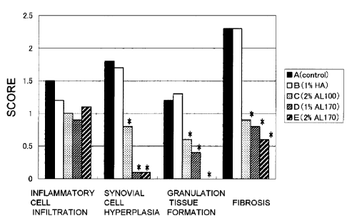
AL100 and AL170 respectively correspond to A) to E) (same as FIG. 11).
FIG. 15 shows time-based changes in gait scores in a rat experimental
arthritis pain model of Example 6. A) Control group (NS); B) 1% sodium
hyaluronate dose group (1% HA); C) 2% sodium alginate dose group
(molecular weight: 1,000,000) (2% AL100); D) 1% sodium alginate dose
group (molecular weight: 1,700,000) (1% AL170); E) 2% sodium alginate
dose group (molecular weight: 1,700,000) (2% AL170). *: p<0.05 vs. NS.
11
= CA 02703158 2010-04-20
FIG. 16 shows a photograph of humeral heads in a rabbit rotator cuff
rupture model of Example 7. The black arrow indicates a cartilage injury
lesions.
"Control" indicates a physiological saline dose group, while
"Alginate" indicates an alginic acid dose group.
FIG. 17 is a graph showing friction coefficients of knee joint specimens
of a rabbit osteoarthritis model of Example 8. Friction coefficients are
plotted on the vertical axis.
"Control (normal)" indicates a normal joint,
"HA(0A)" indicates an OA joint administered hyaluronic acid, and
"AL100(0A)" indicates an OA joint administered alginic acid.
FIG. 18 is a graph showing time-based changes in the degree of arthritis
following antigen sensitization in a rat collagen-induced arthritis model of
Example 9.
Arthritis scores are plotted on the vertical axis, while the
number of days after antigen sensitization is plotted on the horizontal axis.
A) indicates a control group, B) a 1% sodium hyaluronate dose group, C) a
2% sodium alginate dose group (molecular weight: 1,000,000), D) a 1%
sodium alginate dose group (molecular weight: 1,700,000) and E) a 2%
sodium alginate dose group (molecular weight: 1,700,000).
FIG. 19 shows the results of scoring histological evaluations of knee
joint synovial tissue in a rat collagen-induced arthritis model of Example 9.
A through E represent the same groups as in FIG. 18. *: p<0.05 vs. control
FIG. 20 shows the results of scoring histological evaluations of knee
joint patella in a rat collagen-induced arthritis model of FIG. 9. A through E
represent the same groups as in FIG. 18. *: p<0.05 vs. control
FIG. 21 shows the results of scoring histological evaluations of the
lateral femoral condoyle of the knee joint in a rat collagen-induced arthritis
model of Example 9. A through E represent the same groups as in FIG. 18.
*: p<0.05 vs. control
BEST MODE FOR CARRYING OUT THE INVENTION
Although the following provides a detailed explanation of the present
invention, the following embodiments are intended to be exemplary for
explaining the present invention, and the present invention can be carried out
in various forms without deviating from the purport thereof.
1. Introduction
12
= CA 02703158 2010-04-20
"Cartilage" is found in joints; thoracic wall; intervertebral discs;
meniscus; tubular structure such as throat, respiratory tract and ears and so
on, and is classified into three types consisting of hyaline cartilage,
elastic
cartilage and fibrous cartilage. For example, articular cartilage is
classified
as hyaline cartilage, is composed of chondrocytes, collagenous extracellular
matrix, proteoglycan and water, and is free of vascular intervention.
Hyaline cartilage is rich in type II collagen, and is stained by type II
collagen
antibodies. It is also characterized by being stained red by safranin-O stain
used to stain proteoglycan. A "cartilage injury" refers to a state in which
the cartilage has been damaged due to aging, trauma or other factors, and
includes a state in which cartilage function has decreased, such as a decrease
in the characteristic viscoelasticity of cartilage (which enables cartilage to
slowly compress when subjected to a load and then slowly return to its
original state when the load is removed) thereby bringing about impairment
of the ability of the cartilage to support a load while maintaining mobility.
The cartilage injury can also be observed in the disease such as
osteoarthritis
and rheumatoid arthritis.
In the present invention, "joint disease" refers to a disease that occurs
due to cartilage, cartilage tissue and/or joint tissue (such as the synovial
membrane, articular capsule or subchondral bone) having been injured by
mechanical irritation or inflammatory response. "Joint disease treatment"
refers to alleviating, improving and/or curing various symptoms of tissue that
has been injured by mechanical irritation or inflammatory response. For
example, in cases of osteoarthritis, there is compound occurrence of
symptoms such as articular cartilage wear, degeneration of cartilage tissue,
inflammation of the synovial membrane or pain associated with inflammation.
On the other hand, in cases of frozen shoulder, symptoms primarily consist of
inflammation of the synovial membrane and articular capsule as well as pain
associated therewith, while cartilage wear and degeneration may not be
observed. Although the mechanism of occurrence of rheumatoid arthritis is
not fully understood, synovial tissue and cartilage tissue are thought to be
destroyed by inflammatory cytokines resulting from an autoimmune response.
In this manner, joint disease is a disease that presents with compound
symptoms, and drugs for the treatment thereof are required to have compound
effects, including protection of cartilage from wear, inhibition of
13
CA 02703158 2010-04-20
degenerative changes in cartilage due to wear or inflammation, repair of
cartilage injury lesions, inhibition of inflammation and pain, inhibition of
synovial tissue degeneration, and inhibition of osteochondral destruction.
The "composition containing a low endotoxin monovalent metal salt of
alginic acid" of the present invention has the effects of protecting cartilage
from mechanical irritation, inhibiting degenerative changes in cartilage
caused by wear or inflammation, repairing cartilage injury lesions, inhibiting
inflammation and pain of joint tissue, inhibiting synovial tissue
degeneration,
and inhibiting osteochondral destruction. As a result, the composition is
able to inhibit the progress of joint disease, and alleviate, improve and/or
cure symptoms.
In particular, the composition is useful for treating
osteoarthritis, treating frozen shoulder, alleviating joint pain associated
with
rheumatoid arthritis and treating rheumatoid arthritis.
In addition, "injecting into a joint" refers to injection of a liquid
composition having fluidity into, for example, an articular cavity, synovial
bursa or peritenon.
In the case of using to treat osteoarthritis and
rheumatoid arthritis, the composition is preferably injected into an articular
cavity. Furthermore, although osteoarthritis and rheumatoid arthritis can
occur in various joints of the body, including those of the knees, shoulders,
hips, lower back, ankles, wrists and fingers, the composition of the present
invention can be applied to any of these joints.
2. Monovalent Metal Salt of Alginic Acid
The "monovalent metal salt of alginic acid" contained in the
composition for treating a joint disease of the present invention is a
water-soluble salt formed by ion exchange between a hydrogen atom of
carboxylic acid at position 6 of alginic acid and a monovalent metal ion such
as Na + or K. Although specific examples of monovalent metal salts of
alginic acid include sodium alginate and potassium alginate, sodium alginate
acquirable as a commercially available product is particularly preferable.
The "alginic acid" used in the present invention is a biodegradable, high
molecular weight polysaccharide that is a polymer obtained by linearly
polymerizing two types of uronic acids in the form of D-mannuronic acid (M)
and L-gluronic acid (G).
More specifically, the alginic acid is a block
copolymer in which a homopolymer fraction of D-mannuronic acid (MM
14
= CA 02703158 2010-04-20
fraction), homopolymer fraction of L-gluronic acid (GG fraction) and
fraction in which D-mannuronic acid and L-gluronic acid are randomly
arranged (MG fraction) are linked arbitrarily. The composite ratio of the
D-mannuronic acid to the L-gluronic acid of the alginic acid (M/G ratio)
mainly varies according to the type of algae or other organism serving as the
origin thereof, is affected by the habitat and season of that organism, and
extends over a wide range from a high G type having an M/G ratio of about
0.4 to a high M type having an M/G ratio of about 5.
A monovalent metal salt of alginic acid is a polysaccharide, and
although it is difficult to accurately determine molecular weight, it
generally
has a weight average molecular weight of 10,000 to 10,000,000 and
preferably 50,000 to 3,000,000. Sodium alginate having a weight average
molecular weight of about 1,000,000 and 1,700,000 as determined by gel
filtration chromatography demonstrated superior cartilage degenerative
change inhibitory effects, cartilage protective effects, cartilage repair
effects
and joint pain inhibitory effects as compared with sodium alginate having a
molecular weight of about 400,000. In the case of calculating the molecular
weight of a polysaccharide by gel filtration chromatography, there is
normally the potential for measurement error of 10 to 20%. For example, a
molecular weight of 400,000 can fluctuate within the range of 320,000 to
480,000, a molecular weight of 500,000 can fluctuate within the range of
400,000 to 600,000, and a molecular weight of 1,000,000 can fluctuate within
the range of 800,000 to 1,200,000
Thus, the preferable weight average
molecular weight range of a monovalent metal salt of alginic acid for which
effects on joint disease are particularly superior is at least 500,000 or
more,
more preferably 650,000 or more, and even more preferably 800,000 or more.
In addition to production being difficult, since problems occur such as
viscosity when preparing an aqueous solution being excessively high or
solubility decreasing if the molecular weight is excessively high, the weight
average molecular weight is preferably 5,000,000 or less and more preferably
3,000,000 or less.
Since high molecular weight substances derived from a natural origin
typically do not have a single molecular weight, but rather consist of an
aggregate of molecules having various molecular weights, molecular weight
is measured in the form of a molecular weight distribution having a certain
CA 02703158 2010-04-20
range. A typical measurement technique is gel filtration chromatography.
Typical examples of information obtained from molecular weight distribution
as determined by gel filtration chromatography include weight average
molecular weight (Mw), number average molecular weight (Mn) and variance
ratio (Mw/Mn).
Weight average molecular weight emphasizes the contribution of
average molecular weight of polymers having a large molecular weight, and
is represented with the following formula:
Mw = E(WiMi)/W = E(HiMi)/E(Hi)
Number average molecular weight is calculated by dividing the total
weight of polymers by the total number of polymers.
Mn = W/ENi = E(MiNi)/ENi = E(Hi)/E(Hi/Mi)
Here, W represents the total weight of all polymers, Wi represents the
weight of the ith polymer, Mi represents molecular weight at an ith elution
time, Ni represents the number of molecular weights Mi, and Hi represents
the height at the ith elution time.
Since cartilage regeneration effects (and particularly hyaline cartilage
regeneration effects) at cartilage injury lesions, cartilage repair effects,
effects inhibiting cartilage degenerative changes and/or cartilage protective
effects in the treatment of a joint disease are considered to be largely
contributed to by molecular species having large molecular weights, weight
average molecular weight may be used as an indicator of molecular weight.
Differences in values according to the measurement method are known
to occur in the measurement of molecular weights of high molecular weight
substances derived from a natural origin (example of hyaluronic acid:
Chikako Yomota et al., Bull. Natl. Health Sci., Vol. 117, pp. 135-139 (1999),
Chikako Yomota et al., Bull. Natl. Health Sci., Vol. 121, pp. 30-33 (2003)).
Methods for measuring the molecular weight of alginate described in the
literature include a method in which molecular weight is calculated from
intrinsic viscosity, and a method in which molecular weight is calculated by
Size Exclusion Chromatography with Multiple Angle Laser Light Scattering
Detection (SEC-MALLS) (ASTM F2064-00 (2006), published by ASTM
International). Furthermore, it is also described in the literature that in
the
measurement of molecular weight by size exclusion chromatography (gel
filtration chromatography), calculation from a calibration curve using
16
CA 02703158 2010-04-20
pullulan for the standard substance is insufficient, and it is recommended
that
measurement of molecular weight be used in combination with multiple angle
laser light scattering detector (MALLS) (namely, measurement by
SEC-MALLS). In addition, there are also examples of the use of molecular
weights determined by SEC-MALLS being used as catalog specifications of
alginates (FMC Biopolymer Inc., PRONOVATM Sodium Alginates Catalog).
The inventors of the present invention found there to be differences in
the therapeutic effects of sodium alginate having different molecular weights
in an OA model, and measured the molecular weights of these alginates by
gel filtration chromatography and SEC-MALLS. As a result, molecular
weights determined by gel filtration chromatography were determined to
demonstrate a higher correlation with viscosity and therapeutic effects of the
alginates. Namely, it was newly found that rather than the generally
recommended SEC-MALLS method, molecular weight determined by gel
filtration chromatography was found to be suitable as a parameter for
specifying the preferable molecular weight range of alginates used in a
composition for treatment of a joint disease. Thus, in the case of specifying
the molecular weight of an alginate in the present specification, that
molecular weight is the weight average molecular weight as calculated by gel
filtration chromatography unless specifically stated otherwise.
The preferable conditions for gel filtration chromatography as indicated
in the examples. A typical condition consists of the use of a calibration
curve using pullulan for the standard substance.
Pullulan having a
molecular weight of at least 1,600,000, 788,000, 404,000, 212,000 and
112,000 is preferably used for the pullulan used for the standard substance.
In addition, the eluate (200 mM sodium nitrate solution), column conditions
and the like can also be specified. Column conditions preferably consist of
using polymethacrylate resin-based filler and using at least one column
having a molecular weight cutoff of 10,000,000 or more. A typical example
of a column is the TSKgel GMPWx1 (diameter: 7.8 mm x 300 mm) (Tosoh
Corp.).
Although a monovalent metal salt of alginic acid has a large molecular
weight and high viscosity when initially isolated from brown algae,
molecular weight decreases and viscosity lowers during the course of
undergoing heat-drying, freeze-drying, purification and the like. Thus,
17
CA 02703158 2010-04-20
monovalent metal salts of alginic acid having different molecular weights can
be produced by suitably controlling the temperature in each step of
production. Monovalent metal salts of alginic acid having a high molecular
weight are obtained by controlling the temperature in each of step of
production to be somewhat low, while monovalent metal salts of alginic acid
having a low molecular weight are obtained by controlling the temperature in
each step of production to be somewhat high. In addition, monovalent metal
salts of alginic acid having different molecular weights can also be produced
by a technique such as suitably selecting the brown algae used for the raw
material, or fractionating according to molecular weight in the production
process. Moreover, a monovalent metal salt of alginic acid having a target
molecular weight can also be obtained by mixing a monovalent metal salt of
alginic acid produced according to various production processes with a
different lot of monovalent metal salt of alginic acid having a different
molecular weight or viscosity after having measured the molecular weight or
viscosity thereof.
Although the alginic acid used in the present invention may be of a
natural origin or synthetic, it is preferably derived from a natural origin.
Examples of naturally-occurring alginic acids include those extracted from
brown algae. Although brown algae containing alginic acid are prominently
found along seacoasts throughout the world, algae that can actually be used
as raw materials of alginic acid are limited, with typical examples thereof
including Lessonia species found in South America, Macrocystis species
found in North America, Laminaria and Ascophyllum species found in Europe,
and Durvillea species found in Australia. Examples of brown algae serving
as raw materials of alginic acid include Lessonia species, Macrocystis
species, Laminaria species, Ascophyllum species, Durvillea species, Eisenia
species and EckIonia species.
3.Endotoxin Reduction Treatment
The monovalent metal salt of alginic acid contained in the composition
for treatment of a joint disease of the present invention is a low endotoxin
monovalent metal salt of alginic acid. Low endotoxin refers to that in which
the endotoxin level thereof has been substantially lowered to an extent that
does not induce inflammation or fever. Namely, the monovalent metal salt
18
CA 02703158 2010-04-20
of alginic acid has been subjected to endotoxin reduction treatment. It was
surprisingly found that by subjecting to this endotoxin reduction treatment,
in
addition to being able to enhance the cartilage regenerative action of the
composition when applied to a cartilage injury lesion, the regeneration of
subchondral bone can be promoted and mechanical strength of the affected
area can be enhanced. Namely, by using low endotoxin alginic acid in the
composition of the present invention, a composition can be obtained having
high bioaffinity , and not inducing degeneration and inflammatory responses
in surrounding cartilage.
Endotoxin reduction treatment can be carried out by a known method or
a method complying therewith. For example, this treatment can be carried
out by the method of Suga et al. involving purification of sodium hyaluronate
(see, for example, Japanese Patent Application Laid-open No. H9-324001),
the method of Yoshida et al. involving purification of f31,3-g1ucan (see, for
example, Japanese Patent Application Laid-open No. H8-269102), the method
of William et al. involving purification of a biopolymer such as alginate or
gellan gum (see, for example, Published Japanese Translation No.
2002-530440 of PCT International Publication), the method of James et al.
involving purification of polysaccharide (see, for example, International
Publication No. 93/13136 pamphlet), the method of Lewis et al. (see, for
example, US Patent No. 5589591), the method of Hermanfranck et al.
involving purification of alginate (see, for example, Appl. Microbiol.
Biotechnol. (1994), 40:638-643) or a method complying therewith. The
endotoxin reduction treatment of the present invention is not limited thereto,
but rather can be carried out by a known method such as cleaning,
purification using filtration with filter (endotoxin removing filter or
electrification filter), ultrafiltration or a column (such as an endotoxin
adsorption affinity column, gel filtration column or ion exchange column),
adsorption to a hydrophobic substance, resin or activated carbon and the like,
organic solvent treatment (such as extraction with an organic solvent or
precipitation or deposition by addition of organic solvent), surfactant
treatment (see, for example, Japanese Patent Application Laid-open No.
2005-036036) or a suitable combination thereof. A known method such as
centrifugal separation may be suitably combined with these treatment steps.
Endotoxin reduction treatment is preferably suitably selected according to the
19
CA 02703158 2010-04-20
type of alginic acid.
Endotoxin level can be confirmed by a known method, and can be
measured using a known method such as a method using a limulus reagent
(LAL) or method using an Endospecy (registered trademark) ES-24S set
(Seikagaku Corp.). Although there are no particular limitations on the
endotoxin treatment method of the alginic acid contained in the composition
of the present invention, the endotoxin content of the monovalent metal salt
of alginic acid in the case of measuring endotoxin using a limulus reagent
(LAL) is preferably 500 endotoxin units (EU)/g or less, more preferably 100
EU/g or less, even more preferably 50 EU/g or less and particularly
preferably 30 EU/g or less as a result thereof. Sodium alginate that has
undergone endotoxin reduction treatment can be acquired as commercially
available products such as Sea Matrix (sterilized) (Kimica Corp., Mochida
International Ltd.) and PronovaTM UP LVG (FMC).
4. Preparation of Solution of Monovalent Metal Salt of Alginic Acid
The composition for treating a joint disease of the present invention
may be prepared by using a solution of a monovalent metal salt of alginic
acid.
The solution of a monovalent metal salt of alginic acid can be
prepared by a known method or method complying therewith. Namely, the
monovalent metal salt of alginic acid used in the present invention can be
produced by a known method such as an acid method or calcium method
using the previously described brown algae.
More specifically, after
extracting from these brown algae using an alkaline aqueous solution such as
aqueous sodium carbonate solution, for example, alginic acid be obtained by
adding an acid (such as hydrochloric acid or sulfuric acid), and a salt of
alginic acid can be obtained by ion exchange of the alginic acid. Endotoxin
reduction treatment is then carried out as previously described. There are
no particular limitations on the solvent of the alginic acid salt provided it
is a
solvent that can be applied in vivo, and examples of such solvents include
purified water, distilled water, ion exchange water, Milli-Q water,
physiological saline and phosphate-buffered saline (PBS). These are
preferably sterilized and preferably subjected to endotoxin reduction
treatment.
For example, Milli-Q water can be used after sterilizing by
filtration. In addition, the procedure for obtaining the composition of the
CA 02703158 2010-04-20
present invention is preferably carried out in an environment having low
levels of endotoxins and bacteria. For example, the procedure is preferably
carried out on a clean bench using sterilized apparatuses, and the apparatuses
used may be treated with a commercially available endotoxin removal agent.
In the case of producing a composition as described above using a
monovalent metal salt of alginic acid that has been purified to a preferable
endotoxin level, the endotoxin content of the composition is normally 500
EU/g or less, more preferably 300 EU/g or less and particularly preferably
150 EU/g or less.
5. Viscosity of Composition for Treating Joint Disease
Although there are no particular limitations on the viscosity of the
composition for treating a joint disease of the present invention in the case
of
injecting it into a joint provided therapeutic effects for joint disease are
obtained, it is preferably 100 to 20000 mPa=s. For example, the composition
for treating a joint disease of the present invention can be adjusted to a
suitable viscosity using the above-mentioned solvent. If viscosity is within
this range, the composition for treating a joint disease of the present
invention can be injected with a syringe and the like. The viscosity is
preferably 150 to 15000 mPa=s, more preferably 200 to 10000 mPa=s, and
particularly preferably 250 to 6000 mPa=s. The use of a suitable viscosity
makes it possible to demonstrate the effect of compensating for cushioning
function of synovial fluid, thereby making it possible to demonstrate the
effect of treating a joint disease in a state of being dispersed in synovial
fluid.
The viscosity of the composition for treating a joint disease can be
adjusted by, for example, controlling the concentration of alginic acid in the
solution of a monovalent metal salt of alginic acid or controlling the
molecular weight of the alginic acid.
The viscosity of the solution of the monovalent metal salt of alginic
acid increases when the concentration of alginic acid in the solution is high
and decreases when the concentration of alginic acid in the solution is low.
Although unable to be stated unequivocally as a result of being affected by
molecular weight, the preferable concentration of alginic acid in the solution
of the monovalent metal ion of alginic acid is roughly 0.2 to 5% w/v, more
21
CA 02703158 2010-04-20
preferably 0.5 to 3% w/v and particularly preferably 1 to 2.5% w/v.
A monovalent metal salt of alginic acid having a high molecular weight
can be selected to obtain a composition having high viscosity from a solution
of a monovalent metal salt of alginic acid having a low concentration. Since
the viscosity of the solution of a monovalent metal salt of alginic acid is
affected by the M/G ratio, an alginic acid can be suitably selected that has
an
M/G ratio more preferable for viscosity of the solution and the like. The
M/G ratio of alginic acid used in the present invention is about 0.4 to 4.0,
preferably about 0.8 to 3.0 and more preferably about 1.0 to 1.6.
As previously described, since the M/G ratio is determined primarily by
the type of algae, the type of brown algae used for the raw material has an
effect on the viscosity of the solution of the monovalent metal salt of
alginic
acid. The alginic acid used in the present invention is preferably derived
from brown algae of the genii Lessonia, Macrocystis, Laminaria,
Ascophyllum and Durvillea, more preferably derived from brown algae of the
genii Lessonia, and particularly preferably brown algae of Lessonia
nigrescens.
6. Formulation and Application of a Composition for Treating Joint
Disease Containing a Monovalent Metal Salt of Alginic Acid
The composition for treating a joint disease of the present invention is
used to treat a joint disease by injecting into a joint of a human or non-
human
mammal such as a cow, monkey, bird, cat, mouse, rat, guinea pig, hamster,
pig, dog, rabbit, sheep or horse.
The form of the composition for treating a joint disease of the present
invention is a fluid liquid, namely a solution. In the present invention, the
phrase "having fluidity" refers to the having of a property that causes the
form thereof to change to an amorphous form.
For example, the
composition preferably has fluidity such that it is able to be injected into
an
affected area. The composition of the present invention in the form of a
solution can be easily applied to a joint with a syringe, gel pipette or
special-purpose syringe.
The composition for treating a joint disease of the present invention
demonstrates therapeutic effects on joint disease in joint diseases such as
osteoarthritis, frozen shoulder and rheumatoid arthritis by having cartilage
22
CA 02703158 2010-04-20
repair effects, inhibitory effects on cartilage degenerative changes,
cartilage
protective effects, inhibitory effects on inflammation of joint tissue,
inhibitory effects on pain attributable to inflammation of joint tissue,
inhibitory effects on synovial tissue degeneration and/or inhibitory effects
on
osteochondral destruction. The composition for treating a joint disease of
the present invention inhibits joint destruction and improves joint function
through these combined effects.
One aspect of the composition for treating a joint disease of the present
invention is a composition for treating osteoarthritis. In the case a
cartilage
injury extends over a wide area of articular cartilage in the manner of
osteoarthritis, or when desiring to treat a type of cartilage injury
frequently
observed in a comparatively early stage of osteoarthritis such that
smoothness of the cartilage surface is disturbed and degenerative changes
have begun even though well-defined cartilage defects have not yet occurred,
the composition of the present invention is preferably applied by injecting
into an articular cavity and allowing to disperse throughout the synovial
fluid.
Contact of a monovalent metal salt of alginic acid with a cartilage injury
lesion promotes repair of the joint at the cartilage injury lesion, inhibits
degenerative changes caused by inflammation and wear, and protects the
cartilage. In addition, as a result of the active ingredient in the form of a
monovalent metal salt of alginic acid being dispersed throughout the synovial
fluid, inflammatory responses of surrounding tissue, including synovial
tissue,
are inhibited and effects that suppress pain are demonstrated. At the same
time, the presence of a monovalent metal salt of alginic acid within synovial
fluid fulfills the role of compensating for the function of synovial fluid by
serving as a cushion and lubricant.
Another aspect of the composition for treating a joint disease of the
present invention is a composition for treating frozen shoulder (periarthritis
humeroscapularis). Frozen shoulder presents primarily with inflammation
of the synovial membrane and articular capsule coupled with pain associated
therewith, and cartilage wear and degeneration may not be observed. Since
a monovalent metal salt of alginic acid demonstrates the effects of inhibiting
inflammatory responses of surrounding tissue, including synovial tissue and
suppressing pain, frozen should can be treated by administering the
composition of the present invention into the shoulder articular cavity,
23
CA 02703158 2010-04-20
subacromial bursa or biceps muscle tendon sheath.
Another aspect of the composition for treating a joint disease of the
present invention is a composition for suppressing joint pain. Joint pain is
frequently a problem in rheumatoid arthritis in addition to osteoarthritis,
frozen shoulder and the like as previously described. A preferable aspect of
the present invention is a composition for treating joint pain associated with
rheumatoid arthritis, and is particularly preferably a composition for
suppressing knee joint pain associated with chronic rheumatoid arthritis.
Although the mechanism of occurrence of rheumatoid arthritis is not yet fully
understood, synovial tissue and cartilage tissue are thought to be destroyed
by inflammatory cytokines resulting from an autoimmune response. Since a
monovalent metal salt of alginic acid demonstrates effects that inhibit
inflammatory responses of surrounding tissue, including synovial tissue and
suppress pain, the composition of the present invention is able to inhibit
inflammatory responses and suppress pain associated therewith by
administering into a joint suffering from rheumatoid arthritis.
One aspect of the composition for treating a joint disease of the present
invention is a composition for treating rheumatoid arthritis.
The
composition of the present invention inhibits osteochondral destruction and
degeneration of synovial tissue accompanying an autoimmune response. In
addition, when degeneration of joint tissue occurs due to an autoimmune
response, the joint is no longer able to demonstrate its inherent smooth
movement, thereby resulting in mechanical injury to cartilage in the same
manner as osteoarthritis. The composition of the present invention promotes
joint repair of cartilage injuries and protects cartilage and inhibits
degenerative changes in cartilage caused by inflammation and wear. The
composition of the present invention demonstrates therapeutic effects by
inhibiting joint destruction in rheumatoid arthritis through these combined
effect s.
Another aspect of the composition for treating a joint disease of the
present invention is a composition for alleviating, improving and/or curing
various symptoms associated with a joint disease.
In a joint disease,
cartilage, cartilage tissue and/or joint tissue (such as synovial membrane,
articular capsule or subchondral bone) are injured by mechanical irritation or
inflammatory response, and compound symptoms occur such as wear of
24
CA 02703158 2010-04-20
articular cartilage, degenerative changes in cartilage tissue due to
mechanical
irritation along with inflammatory responses, inflammation of the synovial
membrane and other joint tissue, joint pain attributable to inflammation,
synovial tissue degeneration, and osteochondral destruction.
Since the
composition of the present invention contains as an active ingredient thereof
a low endotoxin monovalent metal salt of alginic acid, it has the multiple
effects of protecting cartilage from mechanical irritation, inhibiting
degenerative changes in cartilage caused by wear and inflammation, repairing
cartilage injury lesions, inhibiting inflammation of joint tissue and pain,
inhibiting synovial tissue degeneration, and inhibiting osteochondral
destruction. As a result, the composition of the present invention is able to
inhibit the progress of a joint disease, and alleviate, improve and/or cure
symptoms. In addition, the composition for treating a joint disease of the
present invention has the effect of improving joint function through
alleviation, improvement and/or curing symptoms thereof. Improvement of
joint function refers to improving joint range of movement, improving
movement carried out during the course of daily life and the like.
In the case of applying the composition for treating a joint disease of
the present invention by injecting into a joint, the dose is suitably
determined
according to amount of synovial fluid of the joint into which the composition
is to be injected, and although there are no particular limitations thereon,
in
the case of administering to a human knee joint or shoulder joint, the dose is
normally 1 to 5 mL and more preferably 2 to 3 mL. In addition, the
administration method may consist of, for example, administering in five
consecutive administrations at one week intervals, followed by continuous
administrations every 2 to 4 weeks. Although there are no particular
limitations on the dose, the dose can be suitably adjusted according to the
symptoms and effects. For example, an administration method may be
adopted in which administration is suitably continued once every two weeks,
once every month, once every two months, once every three months or once
every six months. Since alginic acid is inherently not present in the body,
animals do not have an enzyme capable of specifically breaking down alginic
acid. Although alginic acid is normally gradually decomposed by hydrolysis
in an animal body, since its decomposition in the body is slow in comparison
with polymers such as hyaluronic acid, it can be expected to sustain
CA 02703158 2010-04-20
long-term effects in the case of being administered into a joint.
The composition for treating a joint disease of the present invention
contains as an active ingredient thereof a low endotoxin monovalent metal
salt of alginic acid. The inventors of the present invention found for the
first time that alginic acid itself demonstrates therapeutic effects on
cartilage
tissue and joint tissue in the case of administering alginic acid into a joint
of
the body. A monovalent metal salt of alginic acid preferably refers to
sodium alginate, and more preferably to sodium alginate having a weight
average molecular weight of 500,000 or more as determined by gel filtration
chromatography. The containing of alginic acid as an active ingredient
means that alginic acid is contained in an amount that enables it to
demonstrate therapeutic effects on cartilage tissue and joint tissue when
applied to an affected area, and that amount is preferably at least 0.1% w/v
or
more of the entire composition, more preferably 0.5% w/v or more, and
particularly preferably 1 to 3% w/v.
The composition for regenerating cartilage or treating a cartilage
disease of the present invention can also contain other pharmaceutically
active ingredients and components ordinarily used in pharmaceuticals, such
as commonly used stabilizers, emulsifiers, osmotic pressure adjusters,
buffers,
isotonic agents, preservatives, pain relievers or colorants as necessary.
Furthermore, in one aspect of the present invention, the composition of
the present invention does not contain a component demonstrating
pharmacological action on cartilage or joint tissue other than a low endotoxin
monovalent metal salt of alginic acid. A composition containing as an
active ingredient thereof only a low endotoxin monovalent metal salt of
alginic acid is also able to demonstrate adequate effects for treating a joint
disease.
For example, it is preferable to use a composition not containing cells
to facilitate the surgical procedure as well as reduce the risk of infection
by
viruses and the like attributable to the body or the culturing process without
placing an excessive burden on the body through such procedures as
harvesting chondrocytes, periosteum or bone marrow.
Cells specifically
refer to cells for regenerating cartilage tissue, examples of which include
bone marrow mesenchymal stem cells, bone marrow mesenchymal stromal
cells, cartilage precursor cells, chondrocytes, synoviocytes, erythropoietic
26
= CA 02703158 2010-04-20
stem cells and ES cells. The composition for treating a joint disease of the
present invention is a composition having for an active ingredient thereof a
low endotoxin monovalent metal salt of alginic acid, and is based on the
finding that alginic acid itself has therapeutic effects on joint disease. A
preferable example of a therapeutic composition is a cell-free composition
for treating a joint disease injected into a joint containing as an active
ingredient thereof low endotoxin sodium alginate having a weight average
molecular weight of 500,000 or more as determined by gel filtration
chromatography, that is able to demonstrate therapeutic effects superior to
those of hyaluronic acid preparations used in the prior art.
The composition for treating a joint disease of the present invention
preferable does not contain a curing agent for the monovalent metal salt of
alginic acid. A curing agent for a monovalent metal salt of alginic acid
refers to a component that causes curing or gelling of alginic acid in the
presence of a monovalent metal salt of alginic acid in solution, examples of
which include divalent or more metal ion compounds such as Ca2', Mg2+,
Ba2+ or Sr2+, and crosslinking reagents having 2 to 4 amino groups in a
molecule thereof.
Specific examples include CaC12, MgC12, CaSO4 and
BaC12, calcium gluconate and calcium alginate. When these components are
contained to a degree that causes curing and/or gelling of alginic acid,
injection with a syringe and the like becomes difficult due to gelling of
alginic acid.
As a result, problems occur such as obstruction of joint
function due to solidification of a large amount of alginic acid within a
joint.
A curable composition is suitable for using by filling into a hole and so
forth
of a joint defect. On the other hand, in order to demonstrate combined
therapeutic effects throughout all joint tissue of osteoarthritis or
rheumatoid
arthritis extending throughout the entire joint as in the manner of the
composition of the present invention, the composition itself is preferably
non-curable.
Although typical drug solvents contain trace amounts of
divalent metal ions, curing agents as referred to here are not applicable as
long as they are added with the intention of curing and/or gelling a
monovalent metal salt of alginic acid.
A preferable aspect of the
composition of the present invention is a composition not containing a curing
agent of a monovalent metal salt of alginic acid to a degree that causes
curing
and/or gelling of alginic acid. In other words, a preferable aspect of the
27
,
CA 02703158 2010-04-20
composition of the present invention is a non-curable composition.
Moreover, the present invention provides a method of treating joint
disease using the composition for treating a joint disease of the present
invention as described above. The method of treating joint disease of the
present invention inhibits the progress of joint disease and alleviates,
improves and/or cures symptoms by administering the composition for
treating a joint disease of the present invention into a joint. Administration
of the composition for treating a joint disease of the present invention into
a
joint inhibits the progress of joint disease and alleviates, improves and/or
cures symptoms by demonstrating at least one of the effects selected from the
group consisting of inhibition of cartilage degenerative changes, cartilage
protection, cartilage repair, suppression of joint pain, inhibition of joint
inflammation, inhibition of synovial tissue degeneration and inhibition of
osteochondral destruction. Joint function is improved and joint destruction
is inhibited through these combined effects.
There are no particular limitations on the method for applying the
composition for treating a joint disease of the present invention to a joint,
and for example, the composition may be injected directly into a joint with a
syringe, gel pipette or special-purpose syringe. In the case of applying by
injecting into a joint, an 18 to 27G needle is used preferably. "Injecting
into
a joint" refers to injecting a liquid composition having fluidity into an
articular cavity, bursa or tendon sheath and the like. In the case of using to
treat osteoarthritis or rheumatoid arthritis, the composition is preferably
applied by injecting into an articular cavity. Furthermore, although
osteoarthritis and rheumatoid arthritis may occur in various joints such as
joints of the knee, shoulder, hip, lower back, ankle, wrist or fingers, the
composition of the present invention can be applied to any of these joints.
In addition, concomitant drugs including antibiotics such as
streptomycin, penicillin, tobramycin, amikacin, gentamicin, neomycin or
amphotericin B or anti-inflammatory drugs such as aspirin, non-steroid
anti-inflammatory drugs (NSAIDs) or acetaminophen, or steroid drugs may
also be administered before, simultaneous to or after administration of the
composition of the present invention. These drugs may also be used by
mixing into the composition of the present invention.
28
CA 02703158 2015-03-16
754Þ5-6
7. Kit for Treating a Joint Disease
Moreover, the present invention provides a kit for treating a joint
disease. This kit may include the composition for treating a joint disease of
the present invention as described above, syringe, gel pipette,
special-purpose filler, instructions and the like. A preferable specific
example of a kit is that in which a monovalent metal salt of alginic acid is
sealed in one compartment of a syringe composed of two integrally formed
compartments divided by a partition, and a solution containing physiological
saline as a dissolving solution is sealed in the other compartment, and is
composed such that the partition between the compartments can be penetrated
easily at the time of use to enable the contents of both compartments to be
used by mixing and dissolving at the time of use. Another example of a kit
=is that a monovalent metal salt solution of alginic acid is sealed in a
pre-filled syringe allowing it to be administered directly at the time of use
=
without requiring a preparation procedure. = Moreover, the kit can also
contain concomitant drugs including antibiotics such as streptomycin,
penicillin, tobramycin, amikacin, gentamicin, neomycin or amphotericin B or
anti-inflammatory drugs such as aspirin, non-steroid antipyretic analgesic =
.drugs (NSAIDs) or acetaminophen, or steroid drugs.
The use of this kit enables joint disease therapy to be carried out
= smoothly.
=Although the following provides a detailed explanation of the present
invention through= examples thereof, the present invention is not limited to
these examples.
=
Example 1
= =
=
29
=
CA 02703158 2010-04-20
Rabbit Cartilage Repair Model
(1) Production of Transplant Cells
Bone marrow mesenchymal stromal cells (BMSC) were isolated and
cultured to obtain transplant cells. BMSC include erythropoietic cells and
the like in addition to bone marrow mesenchymal stem cells. 10 mL of bone
marrow were harvested from the tibia of four-month-old Japanese white
rabbits followed by washing twice with Ca-Mg-free PBS (Gibco BRL Lab.)
and suspending in DMEM-High Glucose (DMED-HG, Sigma Chemical, St.
Louis, MO). Blood clots were removed with a cell strainer having a pore
diameter of 70 pm (Falcon Co., Ltd.). The cells were then incubated while
humidifying at 37 C and 5% CO2 in a 100 mm culture dish containing a
culture medium consisting of DMEM-HG, 10% fetal bovine serum (FBS,
Gibco, Life Technology, Grand Island, NY) and 1% antibiotics
(Penicillin-Streptomycin-Fungizone 100X concentrated,
Cambrex
Biosciences, Walkersville, MD). The culture medium was replaced every
three days and non-adherent cells were removed. After monolayer culturing
the adherent cells for 10 to 14 days, the cells were removed with
trypsin-EDTA (10 mM, Sigma, UK) and counted followed by subculturing
every three days.
(2) Method
(Procedure)
Forty female Japanese white rabbits (body weights: 2.6 to 2.9 kg) were
anesthetized with isoflurane in 02 gas and intravenous injection of
pentobarbital (0.05 mg/kg) followed by intramuscular injection of antibiotic
(Penicillin G, Meiji-Seika, Japan) and shaving of the legs. A
2 cm
anteromedial incision was made in the skin and the trochlear groove was
accessed using a medial parapatellar approach.
Osteochondral defects
(diameter: 5 mm, depth: 2 mm) were created in the femoral trochlea using a
power drill (Rexon, Japan). The knees were then irrigated with
physiological saline, the absence of bleeding into the defects was confirmed
and the defects were allowed to dry.
In the present example, the experiment was conducted by dividing the
animals into five groups.
30
CA 02703158 2010-04-20
A) Control group (empty)
B) Food grade alginate group (no cells)
C) Food grade alginate + cells (2.5 x 10⁷/mL) group
D) Purified alginate group (no cells)
E) Purified alginate + cells (2.5 x 10⁷/mL) group
The defects were left untreated in the control group A). In addition,
2% w/v food grade sodium alginate solution was applied to the defects in the
food grade alginate group B) (no cells). 2% w/v purified sodium alginate
solution was applied to the defects in the purified alginate group D) (no
cells). Sodium Alginate 500 (serial no. 199-09961) manufactured by Wako
Pure Chemical Industries, Ltd. was used for the food grade alginate, while
Sea Matrix (Sterilized) (serial no. B5Y01) manufactured by Kimica Corp.,
Mochida International Ltd. was used for the purified alginate. Moreover,
the cells obtained in (1) were suspended in 2% w/v food grade sodium
alginate solution or 2% w/v purified sodium alginate solution and applied to
the articular cartilage defects in the food grade alginate + cells group C)
and
the purified alginate + cells group E), respectively. When endotoxin levels
were measured using a commercially available LAL assay kit (Limulus Color
KY Test Wako, Wako, Japan), the endotoxin level of the purified sodium
alginate was 5.76 EU (endotoxin units)/g and that of the food grade sodium
alginate was 75950 EU/g, thus indicating that the endotoxin level of the
purified sodium alginate was far lower than that of the food grade sodium
alginate. Namely, the purified sodium alginate had been subjected to
endotoxin reduction treatment. In addition, the heavy metal content of the
purified sodium alginate was 20 ppm or less, the lead sulfate content was
0.98% or less, and the arsenic content was 2 ppm or less.
The reason for making the concentration of the sodium alginate
solutions 2% w/v was that this allows the viscosity to be adjusted to a level
of 5000 to 6000 mPa.s suitable for the procedure. The rabbits were
immobilized with the defects facing upward, and each composition was
applied to the defects using a gel pipette.
Since the viscosity of the sodium alginate solution was suitable in
groups B) through E), the sodium alginate solutions did not flow out of the
defects despite conditions facilitating flow due to synovial fluid.
31
CA 02703158 2010-04-20
Subsequently, approximately 0.5 ml of 100 mM CaC12 solution was slowly
and continuously applied over the course of 10 seconds to the surface of the
graft using a 27G syringe. The surface layer of the graft gelled immediately
and the cells did not leave the affected area.
The CaC12 solution was
washed with physiological saline. Further immobilization was not required
and the affected area was sutured following the procedure. The rabbits were
able to move freely.
The subject rabbits were sacrificed by intravenous injection of an
excessive dose of pentobarbital at 4 weeks or 12 weeks after the procedure.
The distal ends of the femurs were excised with a power saw.
(Overall Observations)
The overall appearance was observed macroscopically and scored.
Overall appearance was scored according to the criteria of FIG. 1 with
reference to the method of Gabriele, G. et al. (Biomaterial, 21 (2000),
2561-2574).
(Staining)
Subsequently, the specimens were fixed with paraformaldehyde,
decalcified and embedded in paraffin. Sections located 5 pm from the
center of the defect were stained with Safranin-O, H-E stain and
immunostained with anti-type I collagen and anti-type II collagen. The
scoring system described in FIG. 2 was used to evaluate the newly formed
cartilaginous tissue and the tissue was evaluated microscopically.
Independent blinded observers performed the scoring.
(Measurement of Mechanical Strength)
The mechanical strength of the affected area was measured using an
indentation test.
The specimens were firmly clamped with the
femuropatellar joint facing upward, and the test was carried out at room
temperature. The indentator was automatically moved toward the center of
the regenerated cartilage and the displacement (mm) was recorded relative to
the load (N). The thickness of the regenerated tissue was measured from
histological sections. Young's modulus was then obtained from the linear
region of the load-displacement curves.
32
CA 02703158 2010-04-20
(3) Results
The results of staining are shown in FIG. 3 to FIG. 6.
As a result of H-E staining, Safranin-O staining and anti-type II
collagen immunostaining, the most prominent formation of hyaline cartilage
and type II collagen in comparison with the other groups was confirmed in
the purified alginate + cells group E) (FIG. 6) at an early stage 4 weeks
after
the procedure. Roughly 80% of the cartilage was observed to be regenerated
at 12 weeks after the procedure. The formation of subchondral bone was
extremely favorable based on the results of H-E staining. Safranin-O
staining revealed the formation of proteoglycan, and the formation of an
extracellular matrix was also able to be confirmed. On the other hand, there
was hardly any formation of fibrous cartilage observed based on the results
of H-E staining and anti-type I collagen immunostaining.
The purified alginate (no cells) group D) (FIG. 5) demonstrated
favorable formation of hyaline cartilage, type II collagen and subchondral
bone as compared with the food grade alginate + cells group C) (FIG. 4). In
group D), in which cells were not embedded, cartilage regeneration was
surprisingly found to have been obtained by hyaline chondrocytes. In
addition, it also unexpectedly found that group D) in which cells were not
embedded demonstrated a superior ability to regenerate cartilage injury as
compared with group C) in which cells were embedded.
On the other hand, there was hardly any neogenesis of cartilage and
type II collagen observed in control group A) (FIG. 3) in which the defects
were left untreated.
The evaluation results obtained by macroscopically scoring the overall
appearance (Macro) and the evaluation results obtained by scoring
observations based on the staining described above (Histological) are shown
in FIG. 7.
The total scores obtained by combining the Macro and Histological
scores in week 12 consisted of 22.71 for the purified alginate + cells group
E), 19.57 for the purified alginate (no cells) group D), 14.75 for the food
grade alginate + cells group C), 10.25 for the food grade alginate (no cells)
group B), and 8.43 for the control group A) (empty). Thus, the purified
alginate + cells group E) demonstrated the highest score followed by the
33
CA 02703158 2010-04-20
purified alginate (no cells) group D) and the food grade alginate + cells
group C) in that order. It was completely unexpected that group D) in which
cells were not embedded yielded a higher total score, and thereby
demonstrating superior ability to regenerate cartilage in cartilage injuries,
as
compared with group C) in which cells were embedded.
The scoring results for both macroscopic evaluation of overall
appearance (Macro total) and evaluation by staining (Histological total) were
the highest in the purified alginate + cells group E) in the same manner as
described above, and the next highest score was observed in the purified
alginate (no cells) group D).
In looking at the Macro evaluation parameters, groups D) and E), in
which purified alginate was used, were superior for all the parameters of
edge integration (new tissue relative to native cartilage), smoothness of
cartilage surface, cartilage surface, degree of filling, and color of
cartilage,
opacity or translucency of the neocartilage as compared with groups B) and
C) in which food grade alginate was used.
In looking at the Histological evaluation parameters, groups D) and E),
in which purified alginate was used, demonstrated higher scores than groups
B) and C), in which food grade alginate was used, for the parameters of
nature of predominant tissue, surface regularity, structural integrity and
homogeneity, thickness, bonding to adjacent cartilage, degenerative changes
in adjacent cartilage and inflammatory response.
On the basis of these findings, groups D) and E) demonstrated
extremely favorable formation of chondrocytes and cartilage tissue in a
cartilage injury, including the formation of hyaline cartilage, type II
collagen
and subchondral bone. There was hardly any formation of fibrous cartilage
observed.
Bonding of the regenerated tissue to host tissue using purified alginate
was also favorable, there was little degeneration or inflammation in adjacent
cartilage, and bioaffinity was determined to be high.
The results of measuring mechanical strength for the purified alginate
groups D) and E) are shown in FIG. 8.
As a result of measuring mechanical strength for the purified alginate
groups, the mechanical strength in the purified alginate + cells group E) was
a Young's modulus of 8 versus a Young's modulus of 10 in normal cartilage
34
CA 02703158 2010-04-20
tissue, thus indicating that strength had recovered to nearly a normal,
injury-free state. This finding also supported the claim that the composition
of the present invention embedded with cells has superior mechanical
strength, and is favorable with respect to regeneration of strong hyaline
cartilage and the formation of subchondral bone.
Example 2
Measurement of Molecular Weight Distribution of Purified Sodium
Alginate
(1) Method
The molecular weight distribution of purified sodium alginate was
measured by gel filtration chromatography under the conditions indicated
below.
Column: TSKgel GMPWx 1, 2 columns + TSKgel G2500PWx1, 1
column (Tosoh Corp.) (diameter 7.8 mm x 300 mm x 3 columns)
Column temperature: 40 C
Eluate: 200 mM aqueous sodium nitrate solution
Sample concentration: 0.05%
Flow rate: 1.0 mL/min
Injection volume: 200 tL
Detector: RI (differential refractometer)
Standards: Pullulan, glucose (molecular weights: 1,600,000, 788,000,
404,000, 212,000, 112,000, 47,300, 22,800, 11,800, 5900, 180)
(2) Results
(Table 1)
Measurement sample Number Weight Variance
(Reference)
average average ratio Viscosity
of
molecular molecular Mw/Mn 1% aqueous
weight Mn weight Mw solution
Purified sodium alginate 430,000 1,700,000 4.0 400 to 500
(Kimica Corp., Mochida mPa.s
International Ltd., Sea
MatrixTM (sterilized), Serial
No. B5Y01)
Purified sodium alginate 66,000 440,000 6.6 20 to 100
(FMC Biopolymer AS, mPa.s
PronovaTM SLG20)
CA 02703158 2010-04-20
(3) Discussion
The weight average molecular weight of the purified sodium alginate
used in the rabbit cartilage repair model of Example 1 was 1,700,000 as
measured using the method described above. As indicated in Example 1, the
sodium alginate demonstrated hyaline cartilage regenerative effects in the
rabbit cartilage repair model both with and without cells. On the other hand,
although a similar experiment was conducted using low endotoxin alginic
acid (PronovaTM LVG, currently PronovaTM UP LVG, FMC Biopolymer Inc.) as
described in Reference 3, it is disclosed that fibrous cartilage is formed in
the
case of applying only alginic acid not containing cells to a cartilage defect.
Furthermore, the sterilized version of PronovaTM LVG is designated as
PronovaTM SLG20, the weight average molecular weight thereof as
determined by the method described above was 440,000. Although Sea
MatrixTM and PronovaTM have a common characteristic of being low endotoxin
alginic acids, their alginic acids differ in terms of molecular weight, and
this
difference is thought to lead to differences in cartilage regenerative
effects.
Although viscosity can be adjusted by the concentration of alginic acid, in an
experiment in which different concentrations of alginic acid gels (0.5 to 4%)
were embedded with chondrocytes, transplanted beneath the skin of mice and
confirmed for the generation of cartilage, the concentration of alginic acid
was reported to not have an effect on cartilage generation effects (Keith T.
Paige et al., "De Novo Cartilage Generation Using Calcium
Alginate-Chondrocyte Constructs", Plastic and Reconstructive Surgery, Vol.
97: 1996, p.168-178). Thus, the difference in cartilage regenerative effects
between Sea MatrixTM and PronovaTM is thought to be attributable to
molecular weight. Namely, although the use of low endotoxin alginic acid
allows the obtaining of a composition having high bioaffinity with low levels
of degeneration and inflammatory responses in surrounding cartilage, by also
using alginic acid having a high molecular weight, it was found that a
composition for regenerating cartilage or therapeutic composition can be
obtained that has extremely superior cartilage regenerative effects allowing
regeneration of cartilage even without embedding cells therein.
Low
endotoxin alginic acid having a weight average molecular weight of at least
500,000 or more, and preferably 650,000 or more, is useful for cartilage
36
CA 02703158 2010-04-20
regeneration, and that having a weight average molecular weight of 1,000,000
to 2,000,000 was found to be more preferable, and that having a weight
average molecular weight of about 1,500,000 to 2,000,000 was found to be
particularly preferable.
Example 3
Rabbit Osteoarthritis Model (Anterior Cruciate Ligament (ACL)
Resection Model)
(1) Method
An OA model was created in both knee joints of female Japanese white
rabbits (body weights: 2.6 to 2.9 kg) in accordance with the method of
Vignon, E. et al. (Vignon, E., Bejui, J., Mathieu, P., Hartmann, JD, Ville,
G.,
Evreux, JC, et al., Histological cartilage changes in a rabbit model of
osteoarthritis, J. Rheumatol., 1987:14 (Spec No): 104-6). Three animals
each (6 knees) were assigned to the following four groups.
A) Control group (physiological saline)
B) 1% sodium hyaluronate solution dose group (molecular
weight: approx. 900,000, viscosity: approx. 2300 mPa=s)
C) 1% purified
sodium alginate solution dose group (molecular
weight: approx. 1,700,000, viscosity: approx. 500 mPa=s)
D) 2%
purified sodium alginate solution dose group (molecular
weight: approx. 1,700,000, viscosity: approx. 5000 mPa=s)
The solutions of B) to D) were prepared using physiological saline.
The purified sodium alginate of C) and D) were the same as the purified
sodium alginate used in Example 1 (Kimica Corp., Mochida International
Ltd., Sea Matrix (sterilized), Serial No. B5Y01).
Following resection of the anterior cruciate ligament, each of the
solutions A) to D) above were administered into the articular cavity in weeks
4, 5, 6, 7 and 8 (total of 5 administrations given once per week). The
solutions were administered using a 27G needle by penetrating the patellar
tendon and injecting 0.3 mL/knee per administration.
The rabbits were
sacrificed in week 9 to acquire knee joint tissue specimens. Inflammation
from infections, foreign body reactions and the like were not observed in any
37
CA 02703158 2010-04-20
of the knees.
(2) Results
(General Observations)
The appearance of the entire knee joint (knee articular cartilage of the
femur and tibia) was observed macroscopically. Those results are shown in
FIG. 9. In group A (physiological saline dose group), numerous findings of
osteoarthritis, including cartilage defects and osteophytes, were observed
macroscopically. The degree of cartilage injury (size, depth) was milder in
the other groups than in group A. Scoring of the macroscopic findings
yielded similar results.
(Staining)
The knee joint tissue specimens were fixed with paraformaldehyde,
decalcified and embedded in paraffin. The
specimens were evaluated
histologically by safranin-O staining. Those results are shown in FIG. 10.
The upper portions of each figure indicate femoral cartilage, while the lower
portions indicate tibial cartilage, and cartilage degenerative changes were
assessed in cartilage at both locations.
Decreased staining of cartilage
matrix and increased coarseness of cartilage surface were observed in group
A (physiological saline dose group). In group B (1% sodium hyaluronate
solution dose group), although cartilage surface was smoother than in group
A, decreased staining was observed. In group C (1% purified sodium
alginate solution dose group) and group D (2% purified sodium alginate
solution dose group), cartilage surface was smooth and decreases in staining
were mild as compared with groups A and B. In addition, residual alginic
acid was present on the cartilage surface.
On the basis of the above findings, intra-articular injection of sodium
alginate demonstrated action that inhibited cartilage degeneration and
protected cartilage in an ACL resection OA model, and effects were observed
that were equal to or better than administration of 1% sodium hyaluronate
solution used as a therapeutic drug for osteoarthritis. In addition, since
sodium alginate was adhered to the cartilage surface, sodium alginate was
confirmed to demonstrate affinity with articular cartilage as well as cover
and
38
CA 02703158 2010-04-20
protect cartilage surfaces.
Example 4
Evaluation of Therapeutic Effects of Alginic Acid of Different
Molecular Weights in a Rabbit Osteoarthritis Model (Anterior Cruciate
Ligament (ACL) Resection Model)
(1) Method
An OA model was created in both knee joints of female Japanese white
rabbits (body weights: 2.6 to 2.9 kg) in accordance with the method of
Vignon, E. et al. (Vignon, E., Bejui, J., Mathieu, P., Hartmann, JD, Ville,
G.,
Evreux, JC, et al., Histological cartilage changes in a rabbit model of
osteoarthritis, J. Rheumatol., 1987:14 (Spec No): 104-6). Five animals each
(10 knees) were assigned to the following five groups.
A) Control group (physiological saline)
B) 1% sodium hyaluronate solution dose group (ARTZ (registered
trademark), Kaken Pharmaceutical Co., Ltd., molecular weight: approx.
900,000, viscosity: approx. 2300 mPa=s)
C) 2% purified sodium alginate solution dose group (PronovaTM
SLM20, FMC Biopolymer Inc., molecular weight: approx. 400,000)
D) 2% purified sodium alginate solution dose group (Kimica
Corp., sterilized, molecular weight: approx. 1,000,000)
E) 2% purified sodium alginate solution dose group (Sea Matrix
(sterilized), Kimica Corp., molecular weight: approx. 1,700,000)
The solutions of C) to E) were prepared using physiological saline.
Following resection of the anterior cruciate ligament, each of the
solutions A) to E) above were administered into the articular cavity in weeks
4, 5, 6, 7 and 8 (total of 5 administrations given once per week). The
solutions were administered using a 27G needle by penetrating the patellar
tendon and injecting 0.3 mL/knee per administration. The rabbits were
sacrificed in week 9 to acquire knee joint tissue specimens. Inflammation
from infections, foreign body reactions and the like were not observed in
any of the knees.
39
CA 02703158 2010-04-20
(2) Results
(General Observations)
The appearance of the entire knee joint (knee articular cartilage of the
femur and tibia) was observed macroscopically. In order to evaluate the
degree of injury to the cartilage surface, the specimens were stained in India
ink in accordance with the method of Choji Shimizu et al. and then scored (J.
Rheumatol., Vol. 25, pp. 1813-1819, 1998). Macroscopic findings are
shown in FIG. 11. When staining with India ink, boundaries between
cartilage injury lesions and normal cartilage are colored. In group A
(physiological saline dose group), numerous findings of osteoarthritis,
including deep and wide-ranging cartilage defects and osteophytes, were
observed macroscopically. The degree of cartilage injury (size, depth) was
milder in the other groups than in group A. The results of scoring the
macroscopic findings are shown in FIG. 12. The knee joints were observed
at four locations consisting of the Medial Femoral Condyle (MFC), Medial
Tibial Plateau (MTP), Lateral Femoral Condyle (LFC) and Lateral Tibial
Plateau (LTP). The degree of cartilage injury was milder in groups B to E
than in group A at all of these sites. In addition, the degree of cartilage
injury tended to be milder in groups D and E than in groups B and C.
Differences in cartilage degenerative change inhibitory effects, cartilage
protective effects and cartilage repair effects were thought to be present due
to differences in molecular weight of alginic acid.
(Proteoglycan Staining)
The knee joint tissue specimens were fixed in paraformaldehyde,
decalcified and embedded in paraffin.
The specimens were evaluated
histologically by safranin-O staining. Those results are shown in FIG. 13.
The upper portions of each figure indicate femoral cartilage, while the lower
portions indicate tibial cartilage, and cartilage degenerative changes were
assessed in cartilage at both locations. Decreased staining of cartilage
matrix and increased coarseness of cartilage surface were observed in group
A (physiological saline dose group). In group B (1% sodium hyaluronate
solution dose group), although cartilage surface was smoother than in group
A, decreased staining was observed. In the sodium alginate solution dose
groups (groups C to E), cartilage surface was smooth and decreases in
CA 02703158 2010-04-20
staining were mild as compared with groups A and B. In addition, residual
alginic acid was present on the cartilage surface.
(Overall Histopathological Evaluation)
Macroscopic observations and observations by staining were
comprehensively evaluated by scoring in accordance with the method of
Toshiyuki Kikuchi et al. to evaluate effects of the administered drugs
(Osteoarthritis and Cartilage, Vol. 4, pp. 99-110, 1996). Medial femoral
condyle were evaluated to one of four levels for the 8 parameters indicated
below, and the total score was used as an osteoarthritis lesion score.
(1) Loss of cartilage surface, (2) cartilage erosion, (3) fibrosis and
cracking, (4) loss of stainable proteoglycan, (5) disturbances in chondrocyte
arrangement, (6) loss of chondrocytes, (7) loss of subchondral bone, and (8)
formation of chondrocyte clusters.
ANOVA was used to test for the presence of a significant difference
between groups, and subsequent comparisons between each group were made
at a level of significance of p<0.05 using a post hoc test.
The results are shown in FIG. 14. Osteoarthritis lesion scores were
significantly lower in groups B to E versus group A. In addition, although
superior effects were observed in the high molecular weight alginic acid dose
groups (groups D and E) as compared with the hyaluronic acid dose group
(group B), effects of the low molecular weight alginic acid dose group (group
C) were about the same as those of the hyaluronic acid dose group.
On the basis of the above findings, intra-articular injection of sodium
alginate demonstrated action that inhibited cartilage degenerative changes
and protected cartilage in an ACL resection OA model, and effects were
observed that were equal to or better than administration of 1% sodium
hyaluronate solution used as a therapeutic drug for osteoarthritis. In
particular, high molecular weight alginic acid demonstrated superior
therapeutic effects to hyaluronic acid. Furthermore, although the three
types of alginic acid differed in terms of viscosity, since alginic acid
having
viscosity lower than that of hyaluronic acid is observed to demonstrate
effects equal to or greater than those of hyaluronic acid, differences in
therapeutic effects are thought to be attributable to differences in the
41
CA 02703158 2010-04-20
substance used and molecular weight rather than differences in viscosity.
In the ACL resection OA model used in this experiment, the drugs were
administered starting 4 weeks after ACL resection.
Thus, decreases in
osteoarthritis lesion scores observed in the drug dose groups are thought to
be the combined result of effects inhibiting the progression of lesions due to
inhibition of cartilage degenerative changes and protection of cartilage, as
well as cartilage repair action on cartilage injuries that had already
occurred.
According to the paper by the above-mentioned Toshiyuki Kikuchi cited as a
reference in this experiment, OA scores are reported to reach 20 to 25 in
physiological saline dose groups. Since drug administration was started in
week 4 after ACL resection in this experiment, there is the possibility that
OA scores decreased as a result of improvement of cartilage status due to the
effects of the drugs as a result of starting administration from a state in
which OA scores were about 20 to 25. In addition, since the score for
normal joints is 8 in the evaluation system used in this experiment, the mean
OA score (11.3) in group E (alginic acid having a molecular weight of
1,700,000) can be said to approach the score for normal joints and be an
extremely good score.
Example 5
Study of Method of Measuring Molecular Weight of Alginic Acid
Different values are known to be obtained when measuring the
molecular weight of high molecular weight substances derived from a natural
origin depending on the measurement method.
According to ASTM
F2064-00 (ASTM International Publication (2006); the American Society for
Testing and Materials is an organization engaged in the international
standardization and establishment of specifications of industrial material
standards and testing method standards), the use of SEC-MALLS (Size
Exclusion Chromatography with Multiple Angle Laser Light Scattering
Detection) is recommended for measurement of molecular weight.
Therefore, a comparison was made between measurement of the molecular
weight of the sodium alginate used in Example 4 by SEC-MALLS and by gel
filtration chromatography as described in Example 2.
Furthermore,
SEC-MALLS combines the use of a multiple angle laser light scattering
detector (MALLS) with gel filtration chromatography.
42
CA 02703158 2010-04-20
(1) Method
Measurement by gel filtration chromatography was carried out in the
same manner as Example 2. Measurement by SEC-MALLS was carried out
under the conditions indicated below.
Multiple angle laser light scattering detector: DAWN HELEOS, Wyatt
Technology
Column: Shodex SB-806M, 2 columns (Showa Denko K.K.)
Eluate: 200 mM Aqueous sodium nitrate solution
Flow rate: 1.0 mL/min
(2) Results
(Table 2)
AL170 AL100 AL40
Weight average molecular weight
as determined by gel filtration
chromatography 1,700,000 1,000,000 410,000
Weight average molecular weight
as determined by SEC-MALLS
185,000 149,000 128,000
(Reference) Pharmacological
Very good Very good Good
effects in Example 4
The same purified (low endotoxin) sodium alginate used in Example 4
is used for AL170, AL100 and AL40.
AL170: Kimica Corp., Mochida International Ltd., Sea Matrix
(sterilized), 1% viscosity: approx. 500 mPa=s
AL100: Kimica Corp., sterilized, 1% viscosity: approx. 100 mPa=s
AL40: FMC Biopolymer Inc., PronovaTM SLM20, 1% viscosity: approx.
mPa=s
(3) Discussion
As shown in Table 2, differences in the molecular weights of three types
25 of alginates as determined by SEC-MALLS were only observed within a
43
CA 02703158 2010-04-20
range that did not definitively indicate a difference between them, and those
values differed considerably from measurement results obtained by gel
filtration chromatography. As
shown in Example 4, since there were
well-defined differences in pharmacological effects between the samples used,
molecular weights determined by gel filtration chromatography were found to
demonstrate a higher correlation with therapeutic effects of alginates than
molecular weights as determined by SEC-MALLS, and molecular weights
determined by gel filtration chromatography were found to be suitable as
parameters for specifying a preferable molecular weight range of alginates
used in the composition for treating a joint disease.
Example 6
Effects of Alginic Acid on Experimental Arthritis Pain in Rats
(1) Method
Rats with arthritis induced by intra-articular injection of needle-shaped
monosodium urate (MSU) crystals present with an abnormal gait due to pain.
A experimental arthritis pain model in rats administered MSU was prepared
in accordance with the method of Shizuhiko Ihara, et al. (Folia Pharmacol.
Japon, Vol. 100, pp. 359-365 (1992)) to assess the effects of intra-articular
administration of sodium alginate.
Male Crl:CD rats were purchased at age 5 weeks and used in the
experiment following a one week acclimation period. 0.05 mL of a 5.0%
physiological saline suspension of MSU were injected into the right knee
joint of the rats under anesthesia followed by observation of gait at 2, 4, 6
and 24 hours after injection. Gait was evaluated by scoring to one of five
grades consisting of normal gait (0 points), mild limping (1 point), moderate
limping (2 points), walking on toes (3 points) and walking on three legs (4
points). Ten animals were assigned to each of the five groups indicated
below.
A) Control group (physiological saline dose group)
B) 1% sodium hyaluronate solution dose group (ARTZ (registered
trademark), Kaken Pharmaceutical Co., Ltd., molecular weight: approx.
900,000)
C) 2% purified
sodium alginate solution dose group (Kimica
44
CA 02703158 2010-04-20
Corp., sterilized, molecular weight: approx. 1,000,000)
D) 1% purified sodium alginate solution dose group (Sea Matrix
(sterilized), Kimica Corp., molecular weight: approx. 1,700,000)
E) 2% purified sodium alginate solution dose group (Sea Matrix
(sterilized), Kimica Corp., molecular weight: approx. 1,700,000)
50 1.tL of each solution were administered to the same site of the joint
one hour prior to injection of MSU.
f2) Results and Discussion
Time-based changes in gait scores are shown in FIG. 15. The gait
scores of the 1% sodium hyaluronate solution dose group (group B) and 2%
sodium alginate solution dose groups (groups C and E) were significant lower
than the control group (group A), and pain suppressive effects were observed.
Dose-dependent pain suppressive effects were observed in a comparison of
the 1% and 2% solutions containing sodium alginate having a molecular
weight of about 1,700,00 (groups D and E). In addition, the 2% sodium
alginate solutions having molecular weights of 1,000,000 and 1,700,000
demonstrated equal pain suppressive effects despite having different
viscosities of about 300 mPa=s and about 5000 mPa=s, respectively.
In joints, MSU acts directly or indirectly on synovial cells and
neutrophils, and is thought to cause arthritis through the production of
cytokines and the like (above-mentioned publication by Shizuhiko Ihara, et
al.). Namely, MSU induces pain as a result of inflammatory reaction being
induced thereby. Sodium alginate solution demonstrated pain suppressive
effects in this model, and effects observed were equal to those of sodium
hyaluronate, which is used as a therapeutic drug for osteoarthritis and as a
joint pain suppressive drug for chronic rheumatoid arthritis. A monovalent
metal salt of alginic acid was confirmed to have effects that inhibit
inflammation and pain, and is believed to be useful as a therapeutic drug for
osteroarthritis, frozen shoulder and the like, while also being able to be
applied to joint pain associated with rheumatoid arthritis.
In addition, sodium alginate having a molecular weight of 430,000
(Kimica Corp., sterilized) was observed to tend to have weaker pain
suppressing effects than sodium alginate having a molecular weight of
CA 02703158 2010-04-20
1,000,000. This difference in pain suppressing effects was thought to be
attributable to the difference in molecular weight of the alginic acid.
Example 7
Effects of Intra-articular Administration of Alginic Acid in Rabbit
Rotator Cuff Rupture Model
(1) Production of Rotator Cuff Rupture Model
A rotator cuff rupture model was produced using Japanese white rabbits.
After shaving both shoulder joints of the animals under general anesthesia
using ketamine hydrochloride, the shoulder joints were exposed with a
posterior approach using a sterile procedure. The omovertebral muscle was
then separated to produce defects measuring 10 x 7 mm in the infraspinatus
muscle tendon and the insertion thereof on the side of the humeral head. 0.3
mL of a 2% purified sodium alginate solution (Sea Matrix (sterilized),
Kimica Corp., molecular weight: approx. 1,700,000) were injected into the
right shoulder joint. 0.3 mL of physiological saline (Otsuka Pharmaceutical
Co., Ltd.) were injected into the left shoulder joint using the same procedure
for use as a control. Following the procedure, the animals were allowed to
move freely in their cages without immobilizing the upper limbs. Following
the procedure, after continuously administering alginate solution into the
right shoulder and physiological saline into the left shoulder once a week for
five weeks (total of 5 administrations), the animals were sacrificed by
large-dose intravenous administration of pentobarbital followed by
acquisition of shoulder joint tissue specimens.
(2) Results
Although severe cartilage defects were observed at sites along the
rotator cuff tear in the control group following macroscopic observation of
the humeral head, there were no well-defined cartilage injuries observed in
the alginate dose group (FIG. 16). Sodium alginate demonstrated cartilage
protective effects and inhibited the occurrence and progress of cartilage
injury.
Example 8
Measurement of Knee Joint Friction Coefficients in a Rabbit
46
CA 02703158 2010-04-20
Osteoarthritis Model (Anterior Cruciate Ligament (ACL) Detachment Model)
(1) Method
A rabbit OA model was created in the same manner as Example 4, OA
knee joint specimens (n=4), administered with 1% sodium hyaluronate
solution (ARTZ (registered trademark), Kaken Pharmaceutical Co., Ltd.,
molecular weight: approx. 900,000, viscosity: approx. 2300 mPa=s) and OA
knee joint specimens (n=4) administered with 2% purified sodium alginate
solution (Kimica Corp., sterilized, molecular weight: approx. 1,000,000)
were acquired, and knee joint friction coefficients were measured in
accordance with the method of Tanaka, E. et al. (J. Dent. Res., 2004 May;
83(5): 404-7). Administration of each drug was carried out in the same
manner as Example 4, and the rabbits were sacrificed in week 9 following
anterior cruciate ligament excision to acquire knee joint specimens.
Measurements were carried out by bending the knee at an angle of 30 degrees
while applying a load of 1.8 kg for a measuring time of 120 seconds, and
measurements were carried out five times on each specimen. A normal knee
joint (n=1) was used as a control.
(2) Results
The OA knee joints administered alginic acid demonstrated significantly
lower friction coefficients than the OA knee joint specimens administered
hyaluronic acid (FIG. 17).
Although normal knee joints inherently
demonstrate low values for friction coefficient, as the pathology of OA
progresses, the friction coefficient increases and this increase further
promotes destruction of tissue. The reason for the low friction coefficients
in the OA knee joint specimens administered alginic acid is thought to be due
to OA symptoms being mild, and reflected the tissue being maintained in a
favorable state. Namely, the tissue status observed in the macroscopic
findings and histological evaluations of Example 4 are thought to have been
reflected in knee joint friction coefficients.
Example 9
Effects of Intra-Articular Administration of Alginic Acid for
Collagen-Induced Arthritis in Rats
A collagen-induced arthritis (CIA) model is frequently used as a model
47
CA 02703158 2010-04-20
of rheumatoid arthritis since the pathology resembles that of human
rheumatoid arthritis (RA). A collagen-induced arthritis model was created
in rats in order to examine the effects of intra-articular administration of
sodium alginate.
(1) Creation of Animal Model
Ten-week-old DA/Slc (SPF) male rats were purchased and used in the
experiment after a one-week acclimation period. An emulsion was prepared
by dissolving bovine type II collagen (Collagen Technology Research
Association) in 0.01 mol/L aqueous acetic acid solution to a concentration of
1.5 mg/mL and using an equal volume of Freund's incomplete adjuvant
(Difco). A total of 0.4 mL (collagen content: 300 1..tg) of this emulsion was
administered (sensitized) into the skin on the back of the rats (at 4 to 6
locations) to induce arthritis.
(2) Administration of Test Substances
Ten animals each were assigned to the five groups indicated below.
A) Control group (physiological saline dose group)
B) 1% aqueous sodium hyaluronate dose group (ARTZ (registered
trademark), Kaken Pharmaceutical Co., Ltd., molecular weight: approx.
900,000)
C) 2% aqueous purified sodium alginate dose group (Kimica Corp.,
sterilized, molecular weight: approx. 1,000,000)
D) 1% aqueous purified sodium alginate dose group (Sea Matrix
(sterilized), Kimica Corp., molecular weight: approx. 1,700,000)
E) 2% aqueous purified sodium alginate dose group (Sea Matrix
(sterilized), Kimica Corp., molecular weight: approx. 1,700,000)
The dose volume of each administered substance was 0.05 mL/rat, and
the substances were administered into the articular cavity of the left hind
knee joint of the animals using a 1 mL syringe and 26G injection needle.
The animals were dosed once a day on five days consisting of days 0 (day of
collagen administration), 5, 10, 15 and 20 following sensitization.
48
CA 02703158 2010-04-20
(3) Macroscopic Observation of Joint Inflammation
The left hind legs of the animals were macroscopically observed daily
after sensitization and the presence and degree of arthritis were evaluated by
scoring according to the criteria indicated below.
Score 0: Normal
Score 1: Redness observed
Score 2: Redness and slight edema observed in toes
Score 3: Edema extending from toes to full length of paw
Score 4: Severe edema observed
Score 5: Joint deformation observed
The results are shown in FIG. 18.
Rapid onset of arthritis was
observed starting on day 14 after sensitization in the control group, and
degree of arthritis increased through day 25. Although similar degrees of
onset were observed in all groups by day 25 after sensitization, onset tended
to be delayed in the hyaluronic acid dose group (group B) and aqueous
sodium alginate dose groups (groups C, D and E) as compared with the
control group. The degree of the delay was observed more strongly in the
aqueous sodium alginate dose groups (groups C, D and E) than in the
hyaluronic acid dose group (group B). The test substance was therefore
considered to have the potential to inhibit inflammation in joints.
(4) Histopathological Evaluations
The animals were sacrificed on day 25 after sensitization followed by
excision of knee joint tissue from the left hind limb, fixing in formalin,
decalcifying with aqueous 10% EDTA solution and fixing in paraffin. The
specimens were stained with hematoxylin and eosin and with safranin
followed by histological evaluation. The specimens were evaluated by
scoring to one of five levels consisting of no change (score: 0), slight
change
(score: 1), mild change (score: 2), intermediate change (score:3) and severe
change (score: 4) in accordance with evaluation parameters and evaluation
criteria of collagen-induced arthritis. The synovial membrane was observed
for infiltration of inflammatory cells, synovial cell hyperplasia, formation
of
granulation tissue and fibrosis, and the results of scoring those findings are
49
4 y
CA 02703158 2010-04-20
shown in FIG. 19. The patella was observed for the formation of pannus on
the surface of articular cartilage (including synovial hyperplasia),
destruction
of articular cartilage (including degeneration and fibrosis), bone destruction
(resorption) and decreased safranin 0 staining (reduced proteoglycans), and
the results of scoring those findings are shown in FIG. 20. Observation of
osteophyte formation (reactive osteoid formation and periosteal new bone
formation) was targeted at the lateral condoyle of the femur where it is
easiest to form osteophytes. The results are shown in FIG. 21.
The
presence of a significant difference was tested with the Mann-Whitney U test.
In the synovial membrane, the aqueous sodium alginate dose groups
(groups C, D and E) demonstrated significant synovial cell hyperplasia
inhibitory effects, granulation tissue formation inhibitory effects and
fibrosis
inhibitory effects with respect to the control group.
In addition, high
molecular weight alginic acid demonstrated more potent effects.
In the patella, the aqueous sodium alginate dose groups (groups C, D
and E) tended to inhibit pannus formation, articular cartilage destruction and
bone destruction.
In the lateral condoyle of the femur, the aqueous sodium alginate dose
groups (groups C, D and E) tended to inhibit osteophyte formation. In
addition, high molecular weight alginic acid demonstrated more potent
effect s.
Inflammation and abnormal growth of the synovial membrane and an
excessive immune response mediated by activated T-cells is said to be
involved in the onset of rheumatoid arthritis, and destruction of joint tissue
is
said to progress as a result thereof. Aqueous sodium alginate solution
strongly inhibited degeneration of synovial tissue by intra-articular
administration to collagen-induced arthritis model animals. In addition, the
aqueous alginate solution also demonstrated an inhibitory trend against
destruction and degeneration of bone and cartilage. The aqueous sodium
alginate solution was also observed to demonstrate tissue degeneration
inhibitory effects superior to those of sodium hyaluronate solutions used for
the treatment of joint pain in rheumatoid arthritis. Intra-articular injection
of a solution of a salt of alginic acid is thought to allow the obtaining of
therapeutic effects for rheumatoid arthritis in the form of inhibiting the
progress of and improving tissue lesions.
* I
CA 02703158 2010-04-20
INDUSTRIAL APPLICABILITY
The composition for treating a joint disease of the present invention has
cartilage repair effects, effects that suppress cartilage degenerative
changes,
cartilage protective effects, effects that inhibit inflammation of joint
tissue,
effects that suppress pain caused by inflammation of joint tissue, effects
that
inhibit synovial tissue degeneration, and/or effects that inhibit
osteochondral
destruction by being injected into a joint in a liquid state, thereby enabling
it
to demonstrate therapeutic effects on a joint disease. The composition is
particularly useful for the treatment of osteoarthritis, the treatment of
frozen
shoulder, alleviation of joint pain associated with rheumatoid arthritis, and
treatment of rheumatoid arthritis.
51