Note: Descriptions are shown in the official language in which they were submitted.
CA 02703177 2010-08-12
=
77292-44
1
BIOASSAY SYSTEM [NCLUDDNG OPTICAL DETECTION APPARATUSES, AND
METHOD FOR DETECTING BIOMOLECULES
.. Technical Field
[0002] The present invention relates to a bioassay system including a
plurality of optical
= detection apparatuses, and uses of the bioassay system for detecting and
analyzing biomolecules,
such as nucleic acids. More particularly, the present invention relates to a
bioassay system
including at least ten thousand optical detection apparatuses for monitoring,
in some
.. embodiments, a large number of fluorophore molecules in parallel for
detecting and analyzing
the biomolecules.
Background
[0003] The Human Genome Project (HGP) spurred a great increase in sequencing
.. throughput and resulted in a corresponding drop in sequencing costs. In
contrast to the 13 years
and cost of nearly three billion US dollars, per genome sequencing costs have
been reduced
significantly¨indeed two individual genomes have recently been completed
(McGuire et al.,
Science 317:1687 (2007)). Personal genomes represent a paradigm shift in
medical treatment
for both patients and health care providers. By managing genetic risk factors
for disease, health
care providers can more readily practice preventative medicine and provide
customized
treatment. With large banks of completed genomes, drug design and
administration can be
more efficient, pushing forward the nascent field of pharmacogenomics.
[0004] To popularize customized medical treatment for individuals, the US
National
Institutes of Health (NEH) National Human Genome Research Institute (NHGRI)
set a
.. benchmark of reducing per-genome sequencing costs from ten million to
approximately one
=
CA 02703177 2010-04-20
WO 2009/056065
PCT/CN2008/072824
2
thousand U.S. dollars. Conventional high-throughput capillary electrophoresis
and automated
genome sequencing technology, however, cannot satisfy the increased demand for
individual
genome sequencing. In addition, existing sequencing methods require
complicated and
error-prone image acquisition and analysis steps. For example, many existing
technologies
[0005] Accordingly, a need exists for devices to reduce the cost of nucleic
acid
sequencing. To approach the "$1000 genome" paradigm, devices should be capable
of
sequencing multiple molecules in parallel, have simplified design and
manufacture processes,
20 methods.
SUMMARY
[0006] The present invention provides a bioassay system including a plurality
of optical
detection apparatuses, and methods of using the bioassay system for nucleic
acid detection, e.g.,
25 sequencing. The bioassay system provided by the invention is capable of
large-scale parallel
sequencing reactions, i.e., simultaneously sequencing a large number of
different nucleic acid
templates. Each sequencing reaction uses a single molecule as the template
(i.e., single
molecule sequencing). The devices provided also have simplified
designs¨obviating the need
of current devices for complicated, expensive, and error-prone scanning and
detection steps.
30 The simplified design and function of the system provided by the
invention is based, in part, on
CA 02703177 2010-04-20
WO 2009/056065
PCT/CN2008/072824
3
the direct correspondence of linker sites to which the nucleic acids being
detected are attached
(either directly, or, e.g., by a polymerase molecule) and one or more
detecting units (e.g., light
detectors), and in part, on the short distance between the linker sites and
the detecting units.
This short distance between the nucleic acid and detecting unit is manifested,
in some
embodiments, by a large solid angle of detection.
[0007] In one aspect, there is provided a bioassay system for identifying a
single
biomolecule at a detecting unit. The bioassay system may include a plurality
of optical
detection apparatuses, each of which comprises a substrate having a light
detector, and a linker
site formed over the light detector, the linker site being treated to affix
the biomolecule to the
linker site, wherein the linker site is proximate to the light detector. In
some embodiments, the
linker site is spaced apart from the light detector by a distance of less than
or equal to 100
micrometers, and the light detector collects light emitted from the
biomolecule within a solid
angle of greater than or equal to 0.8 ST steridian. The optical detection
apparatus may further
include an excitation light source formed over the substrate so as to provide
a light source for
exciting a fluorophore attached to the biomolecule.
[0008] In another aspect, the invention provides a method of detecting a
nucleic acid by
linking at least one nucleic acid to a linker site of an optical detection
apparatus provided by the
invention (either directly or by binding a nucleic acid polymerase bound to
the linker site) and
detecting the nucleic acid on a corresponding light detector. In some
embodiments, the nucleic
acid is detected by hybridization, e.g., to a labeled probe. In some
embodiments the nucleic
acid is detected by performing nucleic acid sequencing on the optical
detection apparatus. In
some embodiments, the nucleic acid sequencing method is chosen from base-
extension
sequencing, terminally-labeled phosphate sequencing, and wobble sequencing. In
particular
embodiments, the sequencing reaction is a base-extension sequencing reaction.
In still more
particular embodiments, the base-extension sequencing reaction further
comprises the step of
adding blocked and labeled nucleotides to the optical detection apparatus. In
yet more
particular embodiments, the nucleotides are fluorescently labeled.
[0009] The invention also provides, in another aspect, methods of detecting a
sample
molecule. In some embodiments, these methods include the steps of affixing a
labeled sample
molecule to a linker site on an optical detection apparatus provided by the
invention and
CA 02703177 2013-12-12
,
51177-7PPH
4
detecting the sample molecule on a corresponding light detector. In some
embodiments, the
sample molecule is affixed to a linker site by a linking molecule. In some
embodiments the
linking molecule comprises 1) a capture molecule suitable for binding the
sample molecule
and 2) a nucleic acid tag. In particular embodiments, the sample molecule is
applied to an
optical detection apparatus provided by the invention, to which linking
molecules have
already been affixed to linker sites. In other embodiments, a sample molecule
is allowed to
bind a linking molecule and the bound complex is then applied to the optical
detection
apparatus and allowed to affix to a linker site. In particular embodiments,
the sample
molecule is a biomolecule, e.g., a polypeptide, nucleic acid, lipid,
polysaccharide, or
metabolite.
[009A] Specific aspects of the invention include:
- an apparatus for determining the presence of and identifying a single
biomolecule, comprising: a substrate having a light detector; a linker site
formed over the
light detector, the linker site being treated to have reactive functional
groups to affix one
single biomolecule to the linker site, wherein the linker site is proximate to
the light detector
and is spaced apart from the light detector by a distance of less than or
equal to
100 micrometers; a pinhole having a diameter, wherein the pinhole is included
in a blind sheet
formed over the substrate, and wherein the linker site is formed proximate to
the pinhole; and
a filter layer formed between the substrate and the blind sheet.
- an apparatus for determining the presence of and identifying a single
biomolecule, comprising: a substrate having a light detector; a linker site
formed over the
light detector, the linker site being treated to have reactive functional
groups to affix one
single biomolecule to the linker site, wherein the linker site is proximate to
the light detector
and is spaced apart from the light detector by a distance of less than or
equal to
100 micrometers; an excitation light source formed over the substrate; a
pinhole having a
diameter, wherein the pinhole is included in a blind sheet formed over the
substrate, and
wherein the linker site is formed proximate to the pinhole; and a filter layer
formed between
the substrate and the blind sheet.
CA 02703177 2013-12-12
51177-7PPH
4a
- an apparatus for determining the presence of and identifying a single
biomolecule, comprising: a substrate having a light detector, wherein the
light detector
collects light emitted from the biomolecule within a solid angle of greater
than or equal to 0.8
SI steridian; a linker site formed over the light detector, the linker site
being treated to have
reactive functional groups to affix one single biomolecule to the linker site;
a pinhole having a
diameter, wherein the pinhole is included in a blind sheet formed over the
substrate, and
wherein the linker site is formed proximate to the pinhole; and a filter layer
formed between
the substrate and the blind sheet.
- an apparatus for determining the presence of and identifying a single
biomolecule, comprising: a substrate having a light detector, wherein the
light detector
collects light emitted from the biomolecule within a solid angle of greater
than or equal to 0.8
SI steridian; a linker site formed over the light detector, the linker site
being treated to have
reactive functional groups to affix one single biomolecule to it; an
excitation light source
formed over the substrate; a pinhole having a diameter, wherein the pinhole is
included in a
blind sheet formed over the substrate, and wherein the linker site is formed
proximate to the
pinhole; and a filter layer formed between the substrate and the blind sheet.
- an optical detection system, comprising at least 10,000 apparatuses as
described herein;
- an optical detection system, comprising at least 250,000 apparatuses as
described herein;
- an optical detection system, comprising at least 2,000,000 apparatuses as
described herein;
- an optical detection system, comprising at least 10,000,000 apparatuses as
described herein;
- a method of sequencing a nucleic acid, comprising the steps of: affixing one
nucleic acid molecule to the linker site of the apparatus as described herein;
and performing
nucleic acid sequencing of the nucleic acid molecule on the apparatus;
CA 02703177 2013-12-12
,
51177-7PPH
4b
- a method of sequencing a plurality of nucleic acid molecules, the method
comprising the steps of: affixing a plurality of nucleic acid molecules to the
linker sites of the
optical detection system as described herein; and performing nucleic acid
sequencing of the
nucleic acid molecules in parallel on the optical detection system;
- a method of detecting a biomolecule, comprising the steps of: affixing one
or
more biomolecule to the linker site of the apparatus as described herein; and
detecting the
biomolecule on the apparatus;
- a method of detecting a plurality of biomolecules, the method comprising
the
steps of: affixing a plurality of biomolecules to the linker sites of the
optical detection system
as described herein; and detecting the biomolecules on the optical detection
system in parallel;
- a method for manufacturing an apparatus for determining the presence of
and
identifying a single biomolecule, comprising: forming a light detector and a
control circuit on
a substrate; forming a blind sheet having a pinhole over the substrate, the
pinhole having a
diameter; forming a linker site over the light detector and proximate to the
pinhole, the linker
site being treated to have reactive functional groups to affix one single
biomolecule to the
linker site, wherein the linker site is proximate to the light detector and is
spaced apart from
the light detector by a distance of less than or equal to 100 micrometers, and
forming a filter
layer between the substrate and the blind sheet;
- a method of providing biomolecule analysis service, comprising the steps
of:
providing a sample comprising a biomolecule from a service requester to a
service provider;
the service requester receiving analytical results from the service provider,
wherein the results
are produced using the apparatus as described herein;
- an apparatus for identifying a single biomolecule, comprising: a
substrate
having a light detector, the substrate being configured to detect light
emitted from the single
biomolecule; a blind sheet over the substrate, the blind sheet including a
pinhole having a
diameter; a filter layer provided between the blind sheet and the substrate
and provided under
the pinhole, the filter layer configured to filter the light emitted from the
single biomolecule;
CA 02703177 2013-12-12
51177-7PPH
4c
and a linker site provided proximate to the pinhole, the linker site being
treated to have
reactive functional groups to position the single biomolecule proximate to the
pinhole;
- an apparatus for identifying a single biomolecule, comprising: a substrate
having a light detector, the substrate being configured to detect light
emitted from the single
biomolecule; a light emitting layer formed over a filter layer, wherein the
light emitting layer
includes a cavity and the filter layer is provided between the cavity and the
substrate and
under a pinhole, the filter layer configured to filter the light emitted from
the single
biomolecule; and a linker site provided proximate to the cavity and the
pinhole, the linker site
being treated to have reactive functional groups to position the single
biomolecule proximate
to the pinhole;
- an apparatus for identifying a single biomolecule, comprising: a substrate
having a light detector; a blind sheet proximate to a filter layer, the blind
sheet including an
array of pinholes and the filter layer being formed under the array of
pinholes, wherein a
filter-layer structure is provided under each of at least some of the
pinholes; and a linker site
formed proximate to each pinhole, the linker site being treated to have
reactive functional
groups to affix the single biomolecule to the linker site; and
- a method for identifying a single biomolecule, comprising: attaching the
single biomolecule to a linker site, the linker site being treated to have
reactive functional
groups to attach the single biomolecule, and the linker site being provided
proximate to a
pinhole formed on a blind sheet, the pinhole having a diameter; filtering a
light emitted from
the single biomolecule by a filter layer, the filter layer (i) being provided
between the blind
sheet and a substrate having a light detector, and (ii) being provided under
the pinhole; and
using a light detector to detect the light passing through the filter layer.
[0010] It is to be understood that both the foregoing general
description and the
following detailed description are exemplary and explanatory only, and are not
restrictive of
the claimed invention.
CA 02703177 2013-12-12
51177-7PPH
4d
BRIEF DESCRIPTION OF THE DRAWINGS
[0011] Figure 1 is a plane view illustrating a bioassay system
including an array of
optical detection apparatuses consistent with the present invention.
[0012] Figure 2 is a sectional view, along line A-A of Figure 1,
illustrating an optical
detection apparatus in accordance with an embodiment consistent with the
present invention.
[0013] Figure 3 is a sectional view illustrating dimension details of
the optical
detection apparatus consistent with the present invention.
[0014] Figure 4 is a sectional view, along line A-A of Figure 1,
illustrating an optical
detection apparatus in accordance with another embodiment consistent with the
present
invention.
[0015] Figure 5 is a table illustrating the construction of a filter
layer in accordance
with an embodiment consistent with the present invention.
[0016] Figure 6 illustrates a nucleic acid linked on a linker site of
a device consistent
with the present invention.
[0017] Figure 7 illustrates a nucleic acid linked on a linker site of the
device after one
round of base extension with blocked and labeled nucleotides.
CA 02703177 2010-04-20
WO 2009/056065
PCT/CN2008/072824
[0018] Figure 8 illustrates an alternative embodiment of the base extension
sequencing
reaction shown in Figure 7.
[0019] Figure 9 illustrates one round of sequencing several nucleic acids in
parallel by
base-extension sequencing.
5
DETAILED DESCRIPTION
[0020] Reference will now be made in detail to embodiments consistent with the
present
invention, examples of which are illustrated in the accompanying drawings.
Wherever possible,
same reference numerals will be used throughout the drawings to refer to same
or like parts.
1. Bioassay System
[0021] The bioassay system consistent with the present invention can be used
to monitor
a large number (e.g., in some embodiments, more than 10,000) of single
biomolecules in parallel.
The bioassay system may include a plurality of optical detection apparatuses.
Each optical
detection apparatus may sense the existence of a fluorophore on the single
molecule by detecting
photons emitted from the fluorophore. By operating the optical detection
apparatuses in
parallel, the bioassay system consistent with the present invention may
determine, for example,
the sequence of a genome or the profile of expressed genes in a tissue sample
with high
throughput.
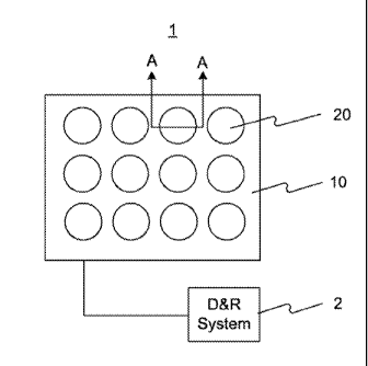
[0022] Referring to FIG. 1, a bioassay system 1 consistent with the present
invention is
illustrated. Bioassay system 1 may include a bioassay substrate 10 and a
plurality of optical
detection apparatuses 20 formed on substrate 10. Each optical detection
apparatus 20 may be
operated independently to detect and identify a single biomolecule affixed
thereto. For example,
the sequence of a single stranded DNA may be determined by sequentially
performing a base
extension and detecting light emitted from a fluorophore coupled with the
extended base using
optical detection apparatus 20. By integrating a huge number of optical
detection apparatuses
20 on substrate 10, a huge number of single biomolecules can be detected and
identified in
parallel. Depending on design choices, bioassay system 1 may include at least,
for example,
ten-thousand (10,000), two-hundred-fifty-thousand (250,000), two-million
(2,000,000), or even
ten-million (10,000,000), or more, optical detection apparatuses 20 formed on
substrate 10.
CA 02703177 2010-08-12
=
77292-44
6
[0023] Bioassay. system I may further include a detection and recordation
system 2
coupled with substrate 10 for controlling the operation of optical detection
apparatuses 20 and
for recording data acquired from optical detection apparatuses 20. In
addition, bioassay system
1 may further include an excitation light source (not shown). The excitation
light source may
produce excitation light, so as to induce the fluorophore to emit fluorescent
light. In one
embodiment, the excitation light source may be stand alone from optical
detection apparatuses
20 or bioassay substrate 10. In alternative embodiments, the excitation light
source may be
integrated with optical detection apparatuses 20 or bioassay substrate 10.
[0024] In this particular embodiment, as shown in FIG. 1, optical detection
apparatus 20
may have a circular shape when viewed from above. It is to be understood that
optical
detection apparatus 20 may have other geometrical shapes, such as a square
shape, a polygon
shape, an oval shape, and the like. In addition, FIG. 1 shows that the
plurality of optical
detection apparatuses 20 is arrayed in a square lattice pattern. It is to be
understood that optical
detection apparatuses 20 may be arrayed in other patterns, such as a
triangular lattice pattern, a
honeycomb lattice pattern, and the like.
[0025] Because the plurality of optical detection apparatuses 20 of bioassay
system 1 are
independently operable, only one optical detection apparatus 20 will be
described below in
accordance with various embodiments consistent with the present invention.
Although only
one optical detection apparatus 20 will be described, it is appreciated that
different optical
detection apparatuses 20 in bioassay system 1 are not necessarily the same.
Depending on
design choices, different types of optical detection apparatuses 20 may be
constructed according
to different embodiments consistent with the present invention.
[0026] Referring to FIG. 2, there is illustrated a section view, along line A-
A of FIG. 1,
of an optical detection apparatus 20 in accordance with one embodiment
consistent with the
present invention. As shown in FIG. 2, optical detection apparatus 20 includes
a light detector
210 formed on substrate 10, and a linker site 220 formed over light detector
210. In addition,
optical detection apparatus 20 may further include a control circuit 215
formed on substrate 10
for controlling the operation of light detector 210. Control circuit 215 may
be coupled with
detection and recordation system 2 so as to receive control instructions from
detection and
recordation system 2 and to transmit detected signals to detection and
recordation system 2. In
CA 02703177 2010-04-20
WO 2009/056065
PCT/CN2008/072824
7
some embodiments, substrate 10 may be a glass substrate, a semiconductor
substrate (e.g.,
silicon), or a plastics substrate. In some embodiments, one or more control
circuits 215 may
correspond to each light detector 210.
[0027] In some embodiments, light detector 210 may comprise a single
photoconductive
photon detector or a group of photoconductive photon detectors. In alternative
embodiments,
light detector 210 may comprise a single photovoltaic photon detector or a
group of photovoltaic
photon detectors. In alternative embodiments, light detector 210 may comprise
a single
photodiode or a group of photodiodes. In alternative embodiments, light
detector 210 may
comprise a single avalanche photodiode or a group of avalanche photodiodes. In
alternative
embodiments, light detector 210 may comprise a single phototransistor or a
group of
phototransistors.
[0028] In one embodiment, optical detection apparatus 20 may further include a
blind
sheet 230 over light detector 210. Blind sheet 230 may include a pinhole 235.
In one
embodiment, pinhole 235 may have a circular shape and may have a diameter D1
of less than or
equal to 1,000, 500, 300, 200, 150, or 100 nanometers. It is appreciated that
pinhole 235 may
have other shapes, such as an oval shape, a square shape, and the like. In one
embodiment,
blind sheet 230 may comprise an opaque material, so as to block away undesired
light from
reaching light detector 210. Therefore, desired light may reach light detector
210 via pinhole
235.
[0029] Linker site 220 may be formed proximate to pinhole 235. In the
embodiment
illustrated in this figure, linker site 220 is formed inside of pinhole 235.
In one embodiment,
linker site 220 formed proximate to pinhole 235 may be spaced apart from light
detector 210 by
a distance H1 of less than or equal to 100 micrometers. In alternative
embodiments, distance
H1 may be less than or equal to 75, 50, 25, 15, 10, 5, or 3 micrometers.
[0030] Optical detection apparatus 20 may further include a filter layer 240
(optional)
and a microlens 250 (optional) between light detector 210 and blind sheet 230.
Although FIG.
2 shows filter layer 240 is formed over microlens 250, it is appreciated that
filter layer 240 may
be formed under microlens 250. In some embodiments, filter layer 240 may
include a single
transparent layer, or a plurality of transparent sublayers having different
refractive indices.
When filter layer 240 includes a plurality of sublayers, filter layer 240 may
be formed by
CA 02703177 2010-08-12
õ
. ,
77292-44
8
sequentially depositing the sublayers over substrate 10. In some embodiments,
a sublayer
having a higher refractive index may be sandwiched by two sublayers having
lower refractive
indices. Alternatively, a sublayer having a lower refractive index may be
sandwiched by two
sublayers having higher refractive indices. In some embodiments, filter layer
240 may include
a layer with single region, or a layer with a plurality of sub-regions having
different transparency
to different wavelength ranges.
[0031] Referring still to FIG. 2, linker site 220 may be treated to affix a
single
biomolecule 30 thereto. In one embodiment, biomolecule 30 may include a single
stranded
DNA molecule 32 and an end link primer 34 coupled with DNA molecule 32.
Biomolecule 30 may
be affixed to linker site 220 via end link primer 34. Further, DNA molecule 32
may be labeled
with a fluorophore 36. When excited by excitation light of a first wavelength
1, fluorophore
36 may emit fluorescent light of a second wavelength 2. In some embodiments,
first
wavelength 1 is shorter than second wavelength 2. In some embodiments, first
wavelength
1 is longer than second wavelength 2, e.g., in multi-photon excitation. Light
detector 210
then detects the fluorescent light emitted from fluorophore 36, so as to
identify the type of base
that fluorophore 36 is attached to, thereby sequentially determining the
sequence of DNA
molecule 32.
[0032] Referring to FIG. 3, there is illustrated a sectional view of optical
detection
apparatus 20 in accordance with one embodiment consistent with the present
invention. As
shown in FIG. 3, blind sheet 230 is formed over light detector 210 and
vertically spaced apart
from light detector 210 by distance HI. Blind sheet 230, which has a thickness
T, includes
pinhole 235 having a radius R1 (i.e., one-half of diameter D1). In this
embodiment, linker site
220 may be formed in pinhole 235 to bind with a biomolecule (not shown).
[0033] When fluorophore 36 is found at a first location 36A above linker site
220 and
separate from linker site 220 by a distance 112, light detector 210 having a
radius R2 may collect
fluorescent light emitted from fluorophore 36 within a first solid angle I.
When fluorophore
36 is found at a second location 36B and almost contacts linker site 220
(i.e., distance H2
approaches zero or less than 1 micrometer), light detector 210 may then
collect fluorescent light
emitted from fluorophore 36 within a second solid angle 2. Second solid angle
2 is greater
than first solid angle 1, and provides a substantially stronger signai
CA 02703177 2010-04-20
WO 2009/056065
PCT/CN2008/072824
9
[0034] In order for light detector 210 to be exposed to the fluorescent light
emitted from
fluorophore 36 through pinhole 235, radius R2 of light detector 210 must be
greater than or equal
to the radius that corresponds to second solid angle 2 projected on an upper
surface of light
detector 210. By bringing blind sheet 230 (or linker site 220) closer to light
detector 210 (i.e.,
by decreasing distance H1), light detector 210 may then collect more
concentrated light (i.e., a
stronger light signal) from within a solid angle. In one embodiment, blind
sheet 230 (or linker
site 220) and light detector 210 are separated by a small distance H1, such
that second solid
angle 2 is at least 0.8 SI steridian.
[0035] Referring to FIG. 4, there is illustrated a section view, along line A-
A of FIG. 1,
of optical detection apparatus 20 in accordance with another embodiment
consistent with the
present invention. In this embodiment, an excitation light source 40 is
integrated with optical
detection apparatus 20. As shown in FIG. 4, excitation light source 40 is
formed on blind sheet
230 of optical detection apparatus 20. In one embodiment, excitation light
source 40 may
comprise a p-type and a n-type semiconductor layers (410 and 430), and a light
emitting layer
420 between the junction region of layer 410 and layer 430. Layer 410 and
layer 430 may be
connected to a power source. Depending on the materials and/or the materials'
physical and
atomic structure used for layers 410, 420, and 430, excitation light source 40
may be a light
emitting diode (LED), a light emitting laser diode (LD), an organic light
emitting diode (OLED),
or a polymer light emitting diode (PLED). Inorganic materials, such as gallium
arsenide,
indium phosphide, gallium antimonide, and gallium nitride, or organic
materials, such as
conjugated polymers with a poly(para-phenelyene-vinylene) backbone are all
examples of
semiconductor materials that can be used to create junction diodes that emit
light.
[0036] In other embodiments, excitation light source 40 may form blind sheet
230 or may
be formed within blind sheet 230. In some embodiments, excitation light source
40 integrated
with optical detection apparatus 20 may emit light of one wavelength band or a
plurality of
wavelength bands. Excitation light source 40 may emit light intermittently or
continuously.
Excitation light source 40 may emit light of one wavelength band at a time or
several wavelength
bands simultaneously.
[0037] Referring again to FIG. 4, excitation light source 40 may include a
cavity 450 at a
central portion thereof, so as to expose pinhole 235. In this embodiment,
linker site 220 may
CA 02703177 2010-04-20
WO 2009/056065
PCT/CN2008/072824
not be formed in pinhole 235. Rather, linker site 220 may be formed in cavity
450 and
proximate to pinhole 235. In the embodiments where excitation light source 40
forms blind
sheet 230 or is formed within blind sheet 230, cavity 450 forms pinhole 235 or
is formed within
pinhole 235. In some embodiments, pinhole 235 may be formed at a central
portion of both
5 layer 410 and blind sheet 230 by, for example, etching layer 410 and
blind sheet 230 using an
appropriate process.
[0038] In addition, excitation light source 40 may be coupled with a power
source 440
through a metal contact 415 formed on lower layer 410 and a metal contact 435
formed on upper
layer 430. Power source 440 may be stand alone and be controlled by detection
and recordation
10 system 2, or may be integrated with detection and recordation system 2.
[0039] Light emitting layer 420 of excitation light source 40 may emit
excitation light
into cavity 450 along a horizontal direction as indicated by arrows drawn on
light emitting layer
420 in FIG. 4. In this embodiment, excitation light is emitted along a
direction substantially
parallel to an upper surface of blind sheet 230. Accordingly, excitation light
may not interfere
with the fluorescent light that reaches light detector 210. Optical detection
apparatus 20
consistent with the present invention may thus more accurately identify
biomolecules than
conventional devices.
2. Nucleic Acid Detection
[0040] The bioassay system consistent with the present invention (including,
e.g., either a
single optical detection apparatus, or a plurality of such apparatuses) can be
used as part of a
system for or in methods or processes of molecule detection, e.g., nucleic
acid sequencing.
This bioassay system, and methods or processes utilizing it, is useful for,
e.g., analytical and
diagnostic applications. These applications may be private, public,
commercial, or industrial.
[0041] In some embodiments, the bioassay system is suitable for large-scale
parallel
sequencing of nucleic acids. Due, in part, to the direct correspondence of
linker sites and light
detectors of the bioassay system, and/or the close proximity of linker sites
and light detectors
(manifested, in some embodiments, as a large solid angle), the bioassay system
provided in the
present invention can be used to sequence nucleic acids without the need for
expensive,
complicated, and error-prone scanning and analysis systems, e.g., a moving
scanning lens or a
moving device stage and subsequent image analysis, thus reducing errors and
costs. The
CA 02703177 2010-04-20
WO 2009/056065
PCT/CN2008/072824
11
bioassay system can detect light signals with substantially improved signal
strength, which
makes single molecule analysis possible.
[0042] The bioassay system consistent with the present invention may be used
with a
wide variety of sequencing modalities and are suitable for sequencing single
molecules.
Additionally, the optical detection devices consistent with the present
invention have simplified
design, assembly, and production relative to existing biochip devices. For
example, the nucleic
acids to be sequenced can be affixed to random linker sites on the array,
avoiding the use of time
consuming and expensive robotics to deposit or synthesize nucleic acids at
predetermined
locations.
[0043] The bioassay system consistent with the present invention can be used
as part of a
system in methods and processes for biomolecule detection, including nucleic
acid hybridization
or sequencing for, e.g., whole genome sequencing, transcriptional profiling,
comparative
transcriptional profiling, or gene identification. Biomolecule detection can
also include
detection and/or measurement of binding interactions, e.g., protein/protein,
antibody/antigen,
receptor/ligand, and nucleic acid/protein. These applications are useful for
analytical or
diagnostic processes and methods.
[0044] Nucleic acids suitable for detection on the system provided by the
invention may,
in some embodiments, be part of a linking molecule, which affixes a molecule
suitable for
assaying binding interactions, e.g., proteins, other nucleic acids,
carbohydrate moieties, or small
molecules to a linker site on a device provided by the invention. The linking
molecule may, in
some embodiments, further comprise a capture molecule, which binds to the
molecule being
assayed for binding interactions. The nucleic acid in a linking molecule
serves as an identifying
tag for the capture molecule of the linking molecule by, e.g., direct
sequencing or hybridization.
[0045] The methods provided by the invention typically comprise a step of
affixing a
molecule to be detected to an address array of a device provided by the
invention. In some
embodiments, the address array may include a blind sheet 230 having a
plurality of pinholes 235,
and linker sites 220 may be formed in or around pinholes 235. See, for
example, FIGS. 1 and 2.
Thus, the bioassay system consistent with the present invention can
simultaneously read millions
of nucleic acid segments. If each segment is, for example, 1000 bases long, a
single device
CA 02703177 2010-04-20
WO 2009/056065
PCT/CN2008/072824
12
could obtain billions of bits of sequence information, making, e.g., whole
genome sequencing
and resequencing possible.
2.1 Molecules to be Detected
[0046] Nucleic acids suitable for detection by the methods provided by the
invention can
include any nucleic acid, including, for example, DNA, RNA, or PNA (peptide
nucleic acid), and
can contain any sequence¨both known and unknown, including naturally occurring
or artificial
sequences. The nucleic acid may be naturally derived, recombinantly produced,
or chemically
synthesized. The nucleic acid may comprise naturally-occurring nucleotides,
nucleotide
analogs not existing in nature, or modified nucleotides. The length of the
nucleic acid to be
detected will vary based on the actual application. In some embodiments, the
nucleic acid
includes at least 10, 20, 50, 100, 200, 500, 1000, 2000, 5000, 10000, 20000
bases, or more. In
some embodiments, the nucleic acid may be from 10 to 20, from 10 to 50, from
10 to 100, from
50 to 100, from 50 to 500, from 50 to 1000, from 50 to 5000, from 500 to 2000,
from 500 to
5000, or from 1000 to 5000 bases.
[0047] A nucleic acid can be single-stranded for detection. Single stranded
nucleic acid
templates can be derived from a double stranded molecule by means known in the
art including,
for example, heating or alkali or other chemical treatment. Single stranded
nucleic acid
templates can also be produced by, e.g., chemical or in vitro synthesis.
[0048] In some embodiments, the nucleic acid to be detected is attached to a
linker site at
its 5' or 3' end. In some embodiments, the nucleic acid may further comprise
one or more end
link primers coupled to the 5' end, the 3' end, or both the 5' end and the 3'
end of the nucleic
acid. In particular embodiments, an end link primer is affixed to the 3' end
of the nucleic acid.
End link primers can be used both to affix the nucleic acid to be detected to
linker sites on the
device and provide a complementary sequence for one or more detecting primers,
e.g., a
sequencing primer.
2.1.1 End Link Primer
[0049] End link primers are short nucleic acid molecules usually composed of
less than
100 nucleotides. In some embodiments, the end link primer is at least 5, 10,
15, 20, 25, 30, 50,
75, 90 nucleotides, or more, in length. In certain embodiments, end link
primers are from 8 to
CA 02703177 2010-04-20
WO 2009/056065
PCT/CN2008/072824
13
25, from 10 to 20, from 10 to 30, or from 10 to 50 nucleotides in length. In
some embodiments,
the end link primers are unbranched, however, in other embodiments, they may
be branched.
[0050] The end link primer can be used to attach the nucleic acid to be
detected to a
linker site on the address array. In some embodiments, the end link primer may
link the nucleic
acid to the array surface directly, e.g., by covalent linkage (e.g., ester or
thiol linkage) or
non-covalent linkage, e.g., antigen/antibody or biotin/avidin binding. See,
e.g., Fig. 5, Fig. 6, and
Fig. 7. In some embodiments, the end link primer may link the nucleic acid to
the array surface
indirectly, e.g., by binding an intermediate molecule, e.g., a polymerase.
See, e.g., Fig. 8.
Accordingly, the end link primer can contain modified nucleotides or be
otherwise modified to
facilitate attachment to a linker site by means known in the art, e.g.,
disulfide, thioester, amide,
phosphodiester, or ester linkages; or by, e.g., antibody/antigen or
biotin/avidin binding, e.g., the
end link primer contains a nucleotide comprising an antigen moiety or a
biotinylated nucleotide.
In particular embodiments, a modified nucleotide is on the 3' end of an end
link primer. In
some embodiments, the 5' end of an end link primer contains a modified
nucleotide.
[0051] The end link primer can also serve as a complement to one or more
primers used
to detect the nucleic acid, e.g., a sequencing primer. In some embodiments,
the primer is used
to detect the nucleic acid by hybridization, e.g., the primer contains a
detectable label, e.g., a
fluorescent or radioisotopic label. In some embodiments, the 5' end of the end
link primer
comprises a sequence complementary to a sequencing primer. In some
embodiments, the end
link primer sequence that is complementary to the sequencing primer is
oriented so that the 3'
end of the sequencing primer is immediately adjacent to the first nucleotide
in the nucleic acid to
be sequenced.
[0052] For example, Figure 6 is a graphical representation of one embodiment
of a
nucleic acid to be sequenced affixed to a optical detection apparatus 20
consistent with the
present invention. A single-stranded nucleic acid 32, end link primer 34, and
annealed
sequencing primer 346 are affixed at a linker site 220 treated to have
reactive functional groups,
which bind a modified nucleotide 344 on end link primer 34. In some
embodiments, nucleic
acid 32 may be attached to linker site 220 via its 5' end, and end link primer
34 may be attached
to the 3' end of nucleic acid 32 to serve as a complement to sequencing primer
346.
CA 02703177 2010-04-20
WO 2009/056065
PCT/CN2008/072824
14
[0053] In some embodiments, end link primers are added to ends of the nucleic
acid to be
detected by a ligase, for example, a DNA ligase. In some embodiments, the end
link primer
and nucleic acid to be detected are both single stranded before the ligation.
In other
embodiments, both are double stranded. In still other embodiments, one is
single stranded and
the other is double stranded. Ligation is well known in the art. For example,
in the polony
sequencing method, Shendure et al. (Science, 309:1728-1732 (2005)) ligated a
T30 end link
primer (32 bp) to a sample DNA segment with the New England Biolabs' (NEB)
Quick Ligation
kit. There, the ligation reaction solution included 0.26 pMole of DNA, 0.8
pMole of T30 end
link primer, 4.0 p,1 T4 DNA Ligase, in lx Quick Ligation Buffer. After mixing,
the reaction
solution was incubated for about 10 minutes at room temperature, and then
placed on ice. The
ligation reaction was stopped by heating the samples to 65 C for 10 minutes.
[0054] In other embodiments, the end link primer may be synthesized on the
nucleic acid
to be detected. For example, the end link primer may be a homopolymer added
by, e.g.,
terminal transferase. For example, Harris et al., (Science 320:106-109 (2008))
added a poly A
tail to DNA templates, which served as the complement to a poly T sequencing
primer in the
single molecule sequencing of a viral genome.
2.1.2 Sequencing Primer
[0055] A sequencing primer is a single-stranded oligonucleotide complementary
to a
segment of the nucleic acid to be detected or its associated end link primer.
In some
embodiments, the sequencing primer is at least 8, 10, 15, 20, 25, 30, 35, 40,
45, 50 nucleotides,
or more in length. In particular embodiments, the sequencing primer may be
from 8 to 25, from
10 to 20, from 10 to 30, or from 10 to 50 nucleotides in length. The
sequencing primer can be
made up of any type of nucleotide, including naturally-occurring nucleotides,
nucleotide analogs
not existing in nature, or modified nucleotides. In certain embodiments, the
5'-end of a
sequencing primer may be modified to facilitate binding to a linker site on
the address array after
the sequencing primer hybridizes with a nucleic acid to be sequenced,
including one or more end
link molecules.
[0056] In some embodiments, a sequencing primer contains modified nucleotides,
e.g.,
locked nucleic acids (LNAs; modified ribonucleotides, which provide enhanced
base stacking
interactions in a polynucleic acid). As an illustration of the utility of
LNAs, Levin et al.
CA 02703177 2010-08-12
.=
77292-44
(Nucleic Acid Research 34(20):142 (2006)) showed that a LNA-containing primer
had improved
specificity and exhibited stronger binding relative to the corresponding
unlocked primer. Three
variants of the MCP1 primer (5'-cttaaattttcttgaat-3') containing 3 LNA
nucleotides (in caps) at
different positions in the primer were made: MCP1-
LNA-3'(5'-cttaaattitCtTgaAt-3');
5 MCP1 -LNA-
5 '(5 '-CtTaAattttettgaat-3 ' ); and MCP1 -LNA-even (5 '-ctTaaatTttctTgaat-3
'). All
LNA-substituted primers had enhanced Tm, while the MCP1-LNA-5' primer
exhibited
particularly enhanced sequencing accuracy (Phred Q30 counts). Accordingly, in
particular
embodiments, the sequencing primer may contain at least one locked nucleotide
in its 5' region,
i.e., the 5' half, third, or quarter of the sequencing primer.
10 [0057]
Sequencing primers and single stranded sample nucleic acids (i.e., a nucleic
acid
to be detected including at least one end link primer) may be hybridized
before being applied to
an optical detection device consistent with the present invention. The
sequencing primer and
sample nucleic acid may be hybridized by mixing the sample nucleic acid with a
molar excess of
sequencing primer in a salt-containing solution, such as 5X SSC (or 5X SSPE),
0.1% Tween 20*
15 (or 0.1%
SDS), and 0.1% BSA buffer. The mixture may be heated to 65 C for at least 5
minutes and slowly cooled to room temperature, to allow primer/template
annealing. Residual
primers can be eliminated by appropriate means including, e.g., a molecular
sieve.
[0058] Primers, including both end link and sequencing primers, can be
designed by
appropriate means, including visual inspection of the sequence or computer-
assisted primer
design. Numerous software packages are available to assist in the primer
design, including
DNAStarTM (DNAStar, Inc., Madison, WI), OLIGO 4.0 (National Biosciences,
Inc.), Vector
NTe (Invitrogen), Primer Premier 5 (Premierbiosoft), and Primer3 (Whitehead
Institute for
Biomedical Research, Cambridge, MA). Primers are designed taking into account,
for example,
the molecule to be sequenced, specificity, length, desired melting
temperature, secondary
structure, primer dimers, GC content, pH and ionic strength of the buffer
solution, and the
enzyme used (i.e., polymerase or ligase). See, e.g., Joseph Sambrook and David
Russell,
Molecular Cloning: A Laboratory Manual Cold Spring Harbor Laboratory Press;
3rd edition
(2001)
*Trade mark
CA 02703177 2012-08-14
51177 ¨7 PPH
16
2.1.3 Bonding to the Array Surface
[0059] After the sequencing primer and nucleic acid to be sequenced, including
one or
more end link primers, are annealed, this complex is prepared in a suitable
buffer, applied to the
surface of an address array, and allowed to bind. In some embodiments the
sample nucleic acid
(nucleic acid to be detected and one or more end link primers) are affixed to
linker sites and
sequencing or detecting primers are later applied.. In other embodiments, the
complex is
hybridized before being applied to a device. Linker sites where only one
sample nucleic acid is
bound are known as effective addresses. In certain embodiments, the complex is
applied to the
optical detection device and the sample nucleic acids affix to random linker
sites on the address
array. In other embodiments, sample nucleic acids can be applied to
predetermined linker sites
on the address array by appropriate means, including; e.g., by robotics or
liquid handling
systems.
[0060] Appropriate means for affixing' a nucleic acid to a solid support are
well known in
the art. In some embodiments, the sample nucleic acid may be affixed directly
to a linker site
by covalent linkage, e.g., disulfide, thioester, amide, phosphodiester, or
ester linkages; or by
non-covalent linkage, e.g, antibody/antigen or biotin/avidin binding. In some
embodiments, the
sample nucleic acid may be affixed to a linker site by an intervening
molecule. In some
embodiments, the intervening molecule may be a polymerase, e.g., a DNA
polymerase.
[0061] As an illustrative example of direct, covalent attachment of a nucleic
acid, Adeesi
et al. (Nucleic Acid Research, 28:87 (2000)) modified the 5' end of a primer
to include a SH
functional group. According to the method of Adeesi et al., a nucleic acid may
be prepared in
50 p.M phosphate buffered saline ("PBS") (NaPi: 0.1 MNaH2PO4 pH 6.5, 0.1 M
NaC1). About
1-5 pi of primer solution may then be applied to a surface of a silanised
glass slide and incubated
in a humidity control box at room temperature for about 5 hours to bond the
primer to the chip
surface. After the binding reaction is completed, the PBS solution is
vibration washed twice at
room temperature for 5 minutes each to remove un-bonded DNA. After cleaning,
10 mM.
P-mercaptoethanol is added to a PBS solution and used to rinse the address
array surface under
room temperature, to deactivate the thiol group of un-bonded DNA. Next, the
array surface is
washed, e.g., once with 5X SSC 0.1% Tween* and once with 5X SSC buffer
solution.
Accordingly, in some embodiments, the method used by Adeesi et al. can be used
in the methods
*Trade mark
CA 02703177 2010-04-20
WO 2009/056065
PCT/CN2008/072824
17
provided by the invention to affix the sample nucleic acid complex to a linker
site, e.g., via the 5'
end of a sequencing primer or the sample nucleic acid.
[0062] In an alternative embodiment, the sample nucleic acid may comprise,
e.g., a
biotinylated nucleotide, and binds to avidin on the linker site surface. In
another embodiment,
the sample nucleic acid may comprise an antigenic moiety, e.g., BrdU or
digoxigenin, that is
bound by an antibody (or fragment thereof) on the linker site. By "antibody"
it is to be
understood that this term includes fragments of immunoglobin molecules,
including, for example,
one or more CDR domains; or variable heavy or variable light fragments.
Antibodies may be
naturally occurring, recombinant, or synthetic. Antibodies may also include,
e.g., polyclonal
and monoclonal variants. In some embodiments the antibodies bind their
antigen(s) with
association constants of at least 106, 107, 108, 109 M, or higher. The
structure, function, and
production of antibodies are well known in the art. See, for example, Gary
Howard and
Matthew Kasser, Making and Using Antibodies: A Practical Handbook CRC Press;
1' edition
(2006).
[0063] In yet another embodiment, the sample nucleic acid may be affixed to
the linker
site by a polymerase, e.g., DNA polymerase. The skilled artisan will
appreciate, that to retain
enzyme function available information, such as the primary, secondary, and
tertiary structures of
the enzyme, should be taken into consideration. For example, the structures of
Taq and Phi29
polymerases are known in the art, see: Kim et al., Nature, 376:612-616 (1995)
and Kamtekar et
al., MoL Cell, 16:609-618 (2004), respectively. Means for fixing a polymerase
to a surface,
while retaining activity are known in the art and are described in, e.g., U.S.
Patent Application
Publication No. 2008/0199932, published August 21, 2008 and Korlach et al.
PNAS
105:1176-1181 (2008). Figure 8 is a graphical representation of one embodiment
of the
invention where the sample nucleic acid (i.e., nucleic acid to be sequenced
32, end link primer 34,
and sequencing primer 346) is bound to a linker site 220 via a polymerase 38
already bound at
linker site 220 by means 384, e.g., direct non-covalent adsorption, an
antibody, biotin, or
chemical linkage, e.g., amide bond.
[0064] In some embodiments, an aldehyde-modified surface of a linker site is
treated
with aldehyde-containing silane reagent. The aldehydes readily react with
primary amines on
the proteins to form a Schiff 's base linkage. Because many proteins display
lysines on their
CA 02703177 2010-04-20
WO 2009/056065
PCT/CN2008/072824
18
surfaces in addition to the generally more reactive a-amine at the NH2-
terminus, they can attach
to the slide in a variety of orientations, permitting different sides of the
protein to interact with
other proteins or small molecules in solution. In another embodiment, a
photoNHS (a
N-hydroxy succimido carboxylate molecule linked to a azidonitrobenzene
molecule with a
carbon chain linker) attaches to an amine-modified surface on the device by UV
photoactivation.
In these embodiments, UV light excites the azidonitrobenzene moiety to produce
highly reactive
nitrene, by eliminating nitrogen. Nitrene is readily reacts with NH2 on the
surface of the device
and form a hydrazine bond. The other end of the linker is NHS carboxylate,
which react with
lysines on the surface of polymerase to produce an amide covalent bond. In
another
embodiment, an NHS carboxylate moiety is reacted with primary amine on the
surface of the
device under buffered conditions. UV light is used to activate an
azidonitrobenzene moiety and
form a highly reactive nitrene as an electron deficient group and readily
react with primary
amine of lysine residues on the polymerase. These methods are described in
further detail in
Example 4, below.
2.2 Sequencing Modalities
[0065] The bioassay system provided by the present invention can be used to
detect and
sequence nucleic acids by means known in the art, as reviewed in, e.g., U.S.
Patent No.
6,946,249 and Shendure et al., Nat. Rev. Genet. 5:335-44 (2004). In some
embodiments, the
sequencing methods rely on the specificity of either a DNA polymerase or DNA
ligase and
include, e.g., base extension sequencing (single base stepwise extensions),
multi-base sequencing
by synthesis (including, e.g., sequencing with terminally-labeled nucleotides)
and wobble
sequencing, which is ligation-based. All of the methods typically require a
single stranded
sample nucleic acid, including at least one end link primer to be affixed at a
linker site (either
directly or indirectly). Sequencing is then initiated at a sequencing primer
(ligase-based
sequencing commonly refers to anchor primers, which serve the analogous
purpose to
sequencing primers).
[0066] For all sequencing modalities, the present invention offers the
advantage of being
able to resequence single molecules. For example, after completion of a
sequencing read, the
sequencing primer and extended nucleotides can be stripped from the sample
nucleic acid, the
device is washed, and the sequencing is repeated. In various embodiments, the
resequencing
CA 02703177 2010-04-20
WO 2009/056065
PCT/CN2008/072824
19
may be done by the same or different methods. By resequencing the same
molecule,
sequencing errors are expected to fall as the power of the number of
sequencing reads. For
example, if per base error rates for a single read are 10-3, then after two
reads, this falls to (10-3)2,
i.e., 10-6. This is particularly advantageous for single molecule sequencing
since the modified
nucleotides used for sequencing can lose their labels or blocking groups
resulting in, e.g.,
spurious deletions.
2.2.1 Base Extension Sequencing: Stepwise Extension
[0067] In some embodiments, the light detection apparatuses provided by the
invention
can be used to perform base extension sequencing as disclosed in, e.g., U.S.
Patent No.
5,302,509. In some embodiments, base extension sequencing begins by attaching
a partial
duplex sample nucleic acid comprising a single stranded nucleic acid to be
sequenced 32, an end
link primer 34 associated with the 3' end of nucleic acid to be sequenced 32,
and a sequencing
primer 346 annealed thereto, to a linker site 220, as depicted in Figure 6. In
some embodiments,
polymerase 38 and modified nucleotides are then applied to the light detection
apparatus in a
suitable buffer. In some embodiments, the sample nucleic acid complex is
affixed to the linker
site by a polymerase at a linker site. In some embodiments, the nucleotides
include a
covalently-linked detectable label, e.g., a fluorescent label, and a blocking
group to prevent any
secondary extension. Accordingly, the sequencing pauses after the addition of
a single
nucleotide to the 3' end of sequencing primer 346.
[0068] Figure 7 is a graphical representation of the first step of one
embodiment of a base
extension sequencing reaction. A nucleotide 362 with a fluorescent blocking
group 364 is
added by a DNA polymerase 38 to the 3' end of sequencing primer 346. In some
embodiments,
the fluorescent label acts as the blocking group. In other embodiments, they
are separate
moieties. A single nucleotide is incorporated at the 3' end of sequencing
primer 346 and is
identified by its label by the corresponding light detector 210. The
fluorescent label and
blocking group are then removed, e.g., by chemical or enzymatic lysis, to
permit additional
cycles of base extension. In certain embodiments, the label and blocking
groups can be
removed simultaneously or sequentially and in any order. By compiling the
order of the bases
added, the sequence of the sample nucleic acid is deduced in the 3' to 5'
direction, one base at a
CA 02703177 2010-04-20
WO 2009/056065
PCT/CN2008/072824
time. Figure 9 illustrates one cycle of extension, detection, and
deblocking/delabeling of
several sample nucleic acids in parallel.
[0069] Generally, there are two ways to recognize the nucleotide added during
stepwise
extension. In the first case, the four nucleotides all have the same
detectable label, but are
5 added one at a time, in a predetermined order. The identity of the
extended nucleotide is
determined by the order that the nucleotide is added in the extension
reaction. In the second
mode for recognizing the base integrated during extension, four different
nucleotides are added
at the same time and each is coupled with a distinct detectable label. In
different embodiments,
the excitation or emission spectra and/or intensity of the labels may differ.
The identity of the
10 nucleotide added in the extension is determined by the intensity and/or
wavelength (i.e.,
excitation or emission spectra) of the detected label. Examples of these two
methodologies are
presented in Example 5.
2.2.2 Sequencing By Synthesis: Multi-step Extension
[0070] In some embodiments, sequencing by synthesis may proceed with multiple
15 uninterrupted extensions, e.g., without the use of blocking groups. In
these embodiments, the
polymerization reaction in monitored by detecting the release of the
pyrophosphate after
nucleoside triphosphate hydrolysis, i.e., the release of the 13 and y
phosphate complex. This
complex can be detected directly, for example, by a fluorescent moiety on the
complex, or
indirectly, by coupling the pyrophosphate to a chemi-or bio-luminescent
detection system.
20 [0071] In some embodiments, the sample nucleic acid is sequenced
essentially
continuously by using terminal-phosphate-labeled nucleotides. Exemplary
embodiments of
terminal-phosphate-labeled nucleotides and methods of their use are described
in, e.g., U.S.
Patent No. 7,361,466 and U.S. Patent Publication No. 2007/0141598, published
June 21, 2007.
Briefly, the nucleotides are applied to the apparatuses provided by the
invention and, when
hydrolyzed during the polymerization, the labeled pyrophosphate is detected by
a corresponding
light detector. In some embodiments, all four nucleotides comprise distinct
labels and can be
added simultaneously. In some embodiments, the nucleotides comprise
indistinguishable, e.g.,
identical, labels and are added sequentially in a predetermined order.
Sequential, cyclical
addition of nucleotides with indistinguishable labels still permits multiple,
uninterrupted
polymerization steps, e.g., in homopolymer sequences.
CA 02703177 2010-04-20
WO 2009/056065
PCT/CN2008/072824
21
2.2.3 Ligase-Based Sequencing
[0072] In other embodiments, a sample nucleic acid is sequenced on the optical
detection
apparatuses provided by the invention by ligase-based sequencing. Ligase-based
sequencing
methods are disclosed in, for example, U.S. Patent No. 5,750,341, PCT
publication WO
06/073504, and Shendure et al. Science, 309:1728-1732 (2005). In the method of
Shendure et
al., for example, an unknown single-stranded DNA sample may be flanked by two
end link
primers and immobilized on a solid support. A particular position in the
unknown sequence
(e.g., the rith base proximal to a particular end link primer) can be
interrogated by annealing a
so-called anchor primer (which is analogous to a sequencing primer) to one of
the end link
primers and then applying a pool of 4 degenerate nonamers to the mixture. The
four nonamers
all have distinct fluorescent labels and are degenerate at all positions
except for the query
position, where each nonamer interrogates with a distinct base¨A, C, G, or T.
The sample is
washed, fluorescently scanned, and the query base is identified. The anchor
primer and ligated
nonamer are then stripped from the sample nucleic acid, the device is washed,
and the process is
repeated, querying a different position. Advantageously, this method is non-
progressive, i.e.,
bases need not be queried in order. Thus, errors are not cumulative.
Additionally, this method
can query nucleotides from either the 5' or 3' direction, i.e., does not
require canonical 5'43'
DNA synthesis. A total of about 13 bases of a sample nucleic acid can
typically be sequenced
by this method.
2.3 Applications
[0073] The bioassay system consistent with the present invention can
simultaneously
detect millions of nucleic acid segments. If each segment is, for example,
1000 bases long, a
single device could obtain upwards of billions of base sequences at once.
Discussed below are
additional applications of the devices and methods provided herein.
2.3.1 Whole Genome Sequencing
[0074] The bioassay system consistent with the present invention can be used
to perform
whole or partial genome sequencing of, e.g., a virus, bacterium, fungi,
eukaryote, or vertebrate,
e.g., a mammal, e.g., a human.
[0075] Genomic DNA can be sheared into fragments of at least 20, 50, 100, 200,
300,
500, 800, 1200, 1500 nucleotides, or longer, for sequencing. In some
embodiments, the sheared
CA 02703177 2010-04-20
WO 2009/056065
PCT/CN2008/072824
22
genomic DNA may be from 20 to 50, from 20 to 100, from 20 to 500, from 20 to
1000, from 500
to 1200, or from 500 to 1500 nucleotides long. In some embodiments, the
nucleic acids to be
sequenced, along with associated end link primers, are made single stranded,
annealed to a
sequencing primer, and applied to a device provided by the invention for
sequencing as
2.3.2 Gene Expression Profiling
[0076] In other embodiments, the bioassay system consistent with the present
invention
can be used to sequence cDNA for gene expression profiling. For example, mRNA
levels can
be quantified by measuring the relative frequency that a particular sequence
is detected on a
15 [0077] CDNA synthesis is well known in the art and typically includes
total RNA
extraction with optional enrichment of mRNA. CDNA is produced from mRNA by
steps
including, for example: reverse transcription, for first strand synthesis;
RNAse treatment, to
remove residual RNA; random hexamer priming of the first strand, and second
strand synthesis
by DNA polymerase. The resultant cDNA is suitable for sequencing on the
devices provided
[0078] In some embodiments, cDNA can be sequenced by methods as disclosed in
U.S.
Patent Nos. 6,812,005 and 7,361,488. Briefly, cDNA is ligated with adapter
poly nucleic acids,
CA 02703177 2010-04-20
WO 2009/056065
PCT/CN2008/072824
23
2.3.3 Detecting and/or Measuring Binding Interactions
[0080] In other embodiments, the bioassay system can be used to detect various
binding
interactions including, e.g., DNA/DNA, RNA/RNA, or DNA/RNA base pairings,
nucleic
acid/protein interactions, antigen/antibody, receptor/ligand binding, and
enzyme/substrate
binding. In general, a sample molecule is affixed to a linking molecule that
comprises an
identifying nucleic acid tag (ID). In some embodiments, the linking molecule
further comprises
a capture molecule that binds the sample molecule. The linking molecule also
comprises a
means for binding to a linker site; e.g., a moiety to facilitate covalent
chemical linkage, such as
disulfide, thioester, amide, phosphodiester, or ester linkages; or by non-
covalent linkage, e.g.,
antibody/antigen or biotin/avidin binding. In some embodiments, a linking
molecule is affixed
to the array by the ID tag.
[0081] A sample molecule is applied to a device consistent with the present
invention
and affixed to a random linker site by its linker molecule, e.g., by binding a
capture molecule
located on the linking molecule. In some embodiments, the sample molecule and
linker
molecules are mixed, allowed to bind, and then applied to a device provided by
the invention.
In some embodiments, the linker molecule is first applied to the device,
allowed to affix to a
linker site, and then the sample molecule is applied. Next, the ID is detected
(e.g., by
hybridization or sequencing) by the methods consistent with the invention to
identify the
associated sample molecule. A plurality of sample molecule species can be
affixed to the same
array and are distinguished by their label while their binding interactions
can be characterized
using the unique IDs of the capture molecule it binds to. Thus, in some
embodiments, a method
of detecting a labeled sample molecule comprises the steps of linking a sample
molecule to a
linker site of a device consistent with the present invention by a linker
molecule comprising a
nucleic acid tag (ID), performing nucleic acid sequencing of the ID, and
detecting the labeled
sample molecule. In particular embodiments, the nucleic acid sequencing is
base extension
sequencing. In some embodiments the nucleic acid sequencing is chosen from
ligase-based
sequencing, or terminal-phosphate-labeled nucleotide sequencing.
[0082] By using nucleotide "bits," up to 411 distinct capture molecules can be
affixed and
identified on the bioassay system consistent with the present invention, where
n is natural
number representing the length of the ID sequenced. For example, 5 nucleotides
could provide
CA 02703177 2010-04-20
WO 2009/056065
PCT/CN2008/072824
24
over a thousand unique IDs, while 12 nucleotides provide over 16 million
combinations. For
example, linker molecules are affixed to a device consistent with the present
invention and their
locations are determined by detecting their corresponding ID tag. The linker
molecules then
serve as probes to, e.g., investigate binding interactions with one or more
labeled sample
molecules. That is, a device with one or more linker molecules affixed to it
can serve as a
probe array.
[0083] In certain embodiments, the labeled sample molecules are fluorescently
labeled.
When bound to the capture molecule of a linker molecule, a labeled sample
molecule is detected
by the light detector(s) corresponding to the linker site where the linker
molecule is affixed.
Accordingly, in some embodiments, methods consistent with the present
invention may further
comprise the steps of applying a labeled sample molecule to a device
consistent with the present
invention and detecting the labeled sample molecule. In particular
embodiments, the device has
linker molecules comprising a nucleic acid tag (ID) affixed to its linker
sites. Multiple labeled
sample molecules can be applied to a probe array simultaneously and be
differentiated by their
labels, e.g., by the intensity and/or wavelength of their fluorescent labels.
Dissociation
constants for binding interactions between sample molecules and labeled query
molecules can be
inferred based on both kinetics (e.g., rates of docking/undocking) and
statistics (e.g., the portions
of sample molecules in the bound or unbound state at any given time) at a
given concentration of
a labeled query molecule.
[0084] In some embodiments, the ID of a linking molecule is at least 5, 10,
15, 20, 25, 30,
40, 50, 75, 90, 100, 150, 200, or more, nucleotides long. In some embodiments,
the ID is from
5 to 10, 20, 40, 80, or 160; or from 10 to 20 or 50; or from 20 to 35
nucleotides long. The ID
contains a unique nucleic acid sequence, i.e., a nucleic acid to be detected.
In particular
embodiments, the unique nucleic acid sequence can be at least 1, 2, 4, 6, 8,
10, 12, 14, 16, 20, 24,
30, or more nucleotides long. In some embodiments, the unique nucleic acid
sequence is from
4 to 10, 12, 15, or 20; or from 10 to 20 nucleotides long. The ID comprises at
least one end link
primer, i.e., the ID contains a sequence complementary to a sequencing primer,
which, in some
embodiments, is modified to attach to a linker site, e.g., by containing a
biotinylated nucleotide.
In some embodiments, the end link primer portion of the ID is 3' to the unique
nucleic acid
sequence. In some embodiments, it is 5' to the unique nucleic acid sequence.
In still other
CA 02703177 2010-04-20
WO 2009/056065
PCT/CN2008/072824
embodiments, end link primers are present at both the 3' and 5' ends of the
unique nucleic acid
sequence.
[0085] In certain embodiments, sample molecules and capture molecules comprise
moieties chosen from a carbohydrate, lipid, protein, peptide, antigen, nucleic
acid, hormone,
5 small organic molecule (e.g., a pharmaceutical), or vitamin moiety; or a
combination thereof.
These moieties may be naturally-occurring (e.g., biochemically purified) or
synthetic (e.g.,
chemically synthesized or recombinantly produced). Additionally, these
substrates may contain
no, some, or all non-native components (e.g. non-natural amino acids, blocking
or protecting
groups, etc.). In particular embodiments, a sample molecule or capture
molecules are proteins,
10 e.g., a growth factor, peptide antigen, antibody, or receptor.
[0086] Various means for conjugating nucleic acids to linker molecules or
linker sites are
known in the art, as reviewed in, e.g., U.S. Patent Publication No.
2004/0038331. The '331
publication discloses methods of forming protein oligonucleotide conjugates on
a solid-phase
support. U.S. Patent No. 4,748,111 provides one example of conjugating a
protein to the 3' end
15 of a nucleic acid. There, terminal transferase is first used to add a
ribose residue to the 3'
portion of the molecule. A periodate oxidation reaction then generates a 3'
aldehyde group on
the nucleic acid, which then forms a covalent bond with an amide group of a
protein. When a
protein is conjugated to the 3' end of the ID, attachment to a linker site is
via the 5' end of the
ID.
20 [0087] In some embodiments, a capture molecule, e.g., a protein, is
linked to the 5' end
of an ID. In these embodiments, the 3' end of the ID or 5' end of a sequencing
primer is used
to affix capture molecule to a linker site. U.S. Patent No. 6,013,434, for
example, discloses
oligonucleotide-polyamide conjugates, where the connection is via the 5' end
of the
oligonucleotide. U.S. Patent No. 6,197,513 discloses both PNA and DNA
conjugates to
25 molecules with carboxylic acid moieties, e.g., proteins, via the 5' end
of the nucleic acid. The
PNA and DNA molecules contain arylamine or aminooxyacetyl moieties. U.S.
Patent No.
6,153,737 discloses oligonucleotides containing at least one 2' functionalized
nucleoside,
suitable for conjugating a variety of molecules to it.
CA 02703177 2010-04-20
WO 2009/056065
PCT/CN2008/072824
26
2.3.4 Additional Detection Methods
2.3.4.1 FRET
[0088] In some embodiments, a molecule is detected on a light detection
apparatus
provided by the invention by Forster resonance energy transfer (FRET),
sometimes known as
fluorescence resonance energy transfer. As is known in the art, FRET occurs
when an excited
donor molecule non-radiatively transfers energy to an acceptor molecule, which
emits the energy,
typically as light. FRET can help reduce background signals by, e.g.,
providing greater spectral
separation between effective excitation and emission wavelengths for a
molecule being detected.
FRET is often used to detect close molecular interactions since its efficiency
decays as the sixth
power of the distance between donor and acceptor molecules. For example, Zhang
et al.
(Nature Materials 4:826-31 (2005)) detected nucleic acid hybridization by
FRET. There, a
biotinylated nucleic acid target was conjugated to an avidin-coated quantum
dot donor, which
then excited a Cy5-conjugated DNA probe. In some embodiments of the invention,
a labeled
capture molecule and labeled sample molecule may form a donor/acceptor (or
vice versa) pair
for detection by FRET.
[0089] In some embodiments of nucleic acid sequencing provided by the
invention,
fluorescently labeled nucleotides act as acceptor chromophores for a donor
chromophore
attached to a polymerase or ligase. Accordingly, in these embodiments, the
donor chromophore
located on the polymerase or ligase excites an acceptor chromophore on a
nucleotide being
polymerized on, or ligated to, the sample nucleic acid. Nucleotides not
proximate to the
polymerase are not excited due to the rapid falloff in FRET efficiency. In
some embodiments
the donor molecule is, e.g., another fluorophore, e.g., a quantum dot. Quantum
dots, e.g.,
semiconductor quantum dots are known in the art and are described in, e.g.,
International
Publication No. WO 03/003015. Means of coupling quantum dots to, e.g.,
biomolecules are
known in the art, as reviewed in, e.g., Mednitz et al., Nature Materials 4:235-
46 (2005) and U.S.
Patent Publication Nos. 2006/0068506 and 2008/0087843, published March 30,
2006 and April
17, 2008, respectively. In some embodiments, quantum dots are conjugated to a
DNA
polymerase molecule, which is described further in Example 3, below. As
already discussed
above for conjugating enzymes to linker sites, the skilled artisan will
undoubtedly appreciate that
when conjugating flurophores to, e.g., a DNA polymerase or ligase, care must
be taken to retain
CA 02703177 2010-08-12
77292-44
27
enzyme function by mitigating any effect of conjugating the fluorophore on the
primary,
secondary, and tertiary structures of the enzyme.
2.3.4.2 Multi Photon Excitation
[0090] In some embodiments, a chromophore is excited by two or more photons.
For
example, in some embodiments, excitation of either a donor or acceptor
chromophore in FRET is
via two or more photons. Two photon and multi-photon excitation are described
further in, e.g.,
U.S. Patent Nos. 6,344,653 and 5,034,613.
23.4.3 Time Resolved Detection
[0091] In some embodiments, the light source and light detectors of a device
provided by
the invention can be modulated to have a characteristic phase shift. Using
methods known in
the art, for example, as disclosed in U.S. Patent Publication No.
2008/0037008, published
February 14, 2008, light emitted from a molecule being detected on a device
provided by the
invention can be measured by a corresponding light detector without
interference from an
excitation light source.
3. Biomolecule Analysis Service Using the Bioassay System
[0092] The present invention also provides a method of providing biomolecule
analysis
service using the bioassay system in accordance with embodiments consistent
with the present
invention. In some embodiments, the method includes the steps of providing a
sample
including a biomolecule to be analyzed from a service requester to a service
provider and the
service requester receiving analytical results from the service provider,
wherein the results are
produced using a device provided by the invention. In some embodiments, the
method is
performed for remunerative consideration, e.g., fee-for-service or contract
service agreements.
In addition, the sample may be shipped directly between the service requester
and the service
provider, or mediated by a vendor. In some embodiments, the service provider
or vendor may
be geographically located in a territory outside of the United States of
America, e.g. in another
country.
[0093] Where any conflict exits between a document described herein and the
present
application, this application will dominate.
=
CA 02703177 2012-01-04
51177-7PPH
28
[0094] The specification is most thoroughly understood in light of
the teachings of the
references cited within the specification. The embodiments within the
description and
drawings provide an illustration of embodiments of the invention and should
not be construed
to limit the scope of the invention. The skilled artisan readily recognizes
that many other
embodiments are encompassed by the invention. To the extent the material
described
herein contradicts or is inconsistent with the description or drawings, the
description or
drawings will supercede any such material. The citation of any references
herein is not an
admission that such references are prior art to the present invention.
[0095] Unless otherwise indicated, all numbers expressing quantities
of ingredients,
reaction conditions, and so forth used in the specification, including claims,
are to be
understood as being modified in all instances by the term "about".
Accordingly, unless
otherwise indicated to the contrary, the numerical parameters are
approximations and may
vary depending upon the desired properties sought to be obtained by the
present invention.
At the very least, and not as an attempt to limit the application of the
doctrine of equivalents
to the scope of the claims, each numerical parameter should be construed in
light of the
number of significant digits and ordinary rounding approaches.
[0096] Unless otherwise indicated, the term "at least" preceding a
series of elements
is to be understood to refer to every element in the series. Those skilled in
the art will
recognize, or be able to ascertain using no more than routine experimentation,
many
equivalents to the specific embodiments of the invention described herein.
Such equivalents
are intended to be encompassed by the following claims.
Examples
Example 1: Construction of a High Throughput Biossay System
[0097] Hereinafter, a method for manufacturing a bioassay system 1
will be described
in detail by referring to FIGs. 1-4. First, a silicon substrate 10 is provided
with a plurality of
light detectors 210 formed on an upper surface of substrate 10 by commercially
available
0.25 micro-meter semiconductor manufacturing process for general logic and
optical devices.
Light detectors 210 are photodiode photon detectors, each having a diameter of
24 m and an
CA 02703177 2010-04-20
WO 2009/056065
PCT/CN2008/072824
29
exposure area of 452 m2. Each light detector is arranged adjacent to each
other, such that an
array of 512 columns and 512 rows of light detectors 210 is formed on
substrate 10.
[0098] A plurality of control circuits 215 is formed on the upper surface of
substrate 10
where light detectors 210 are not formed. In this embodiment, one control
circuit 215
corresponds to one light detector 210, so as to control the operation of its
corresponding light
detectors 210 and to control the communication between light detectors 210 and
detection and
recordation system 2.
[0099] In this embodiment, a filter layer 240 is formed over the upper surface
of light
detectors 210 and control circuits 215. Global planarization process is
applied to the upper
surface of light detectors 210 and control circuits 215 before forming filter
layer 240. Filter
layer 240 includes a plurality of sublayers. In this embodiment, filter layer
240 is formed by
first depositing a sublayer having a higher refractive index over the
planarized upper surface of
light detectors 210 and control circuits 215. Then, a sublayer having a lower
refractive index is
deposited on the sublayer having a higher refractive index already formed. By
continuously
depositing sublayers of higher refractive indices and lower refractive
indices, filter layer 240 is
formed until a large number of sublayers has been so deposited. In this
embodiment, filter layer
240 includes a hundred and one sublayers.
[00100] Referring to FIG. 5, there is illustrated a table for summarizing an
example
construction of filter layer 240. In FIG. 5, a lower numbered sublayer denotes
a sublayer closer
to the bottom surface of filter layer 240 and a higher number sublayer denotes
a sublayer closer
to the upper surface of filter layer 240. As shown in FIG. 5, in this
particular embodiment, odd
numbered sublayers of filter layer 240 are made of, for example, Niobium Oxide
(Nb205), which
has a higher refractive index. Even numbered sublayers are made of, for
example, Silicon
Oxide (5i02), which has lower refractive index. The sublayers may be formed by
using a
sputtering system, an example of which includes Model No. RAS 1100 of Radical
Assisted
Sputtering Series, manufactured by Shincron Co., Ltd. (Shinagawa-ku, Tokyo,
JAPAN). The
thickness of each sublayer in this example is also provided in the table of
FIG. 5. The resultant
filter layer 240 is highly transparent with respect to the fluorescent light
of fluorophore Cy5 and
has a low transparency with respect to the light emitted from Helium-Neon
laser at a wavelength
of about 633 nm, which is used as an external light source to excite
fluorophore Cy5.
CA 02703177 2010-04-20
WO 2009/056065
PCT/CN2008/072824
[00101] Referring back to FIGS. 2 and 4, blind sheet 230 having pinhole 235 is
formed
on filter layer 240. Hereinafter, a process for fabricating blind sheet 230
having pinhole 235
over filter layer 240 or substrate 10 will be described in detail.
[00102] First, a photoresist layer is formed on filter layer 240 (if filter
layer 240 is
5 optionally formed on the upper surface of light detectors 210 and control
circuits 215) or on the
planarized upper surface of light detectors 210 and control circuits 215 (if
filter layer 240 is not
formed) by, for example, spin coating a resist material on filter layer 240.
Thereafter, the
photoresist layer is developed to form a photoresist pattern at pinhole
regions. The photoresist
pattern is formed by covering the pinhole regions using a photomask, and
exposing the
10 photoresist layer such that only the regions covered by the photomask
remain on filter layer 240
or the planarized upper surface of light detectors 210 and control circuits
215.
[00103] Then, a metal layer is deposited over filter layer 240 where the
photoresist
pattern has been formed. In this embodiment, the metal layer comprises
chromium (Cr), which
is deposited over filter layer 240 or the planarized upper surface of light
detectors 210 and
15 control circuits 215 by performing a magnetron sputtering process.
[00104] Subsequently, a portion of the metal layer on the pinhole regions and
the
photoresist pattern in the pinhole regions are removed, thereby forming blind
sheet 230 with
pinholes 235.
[00105] Alternatively, blind sheet 230 may be formed by first depositing a
metal layer
20 (e.g., Cr) on filter layer 240, and then forming a mask on the metal
layer, thereby exposing
portions of an upper surface of the metal layer. The exposed portions of the
metal layer are
then etched until filter layer 240 is exposed, thereby forming pinholes on the
metal layer. Then,
the mask is removed from the metal layer and blind sheet 230 having pinholes
235 is formed on
filter layer 240.
25 [00106] Referring again to FIGS. 2 and 4, in this embodiment, linker
site 220 is formed
by filling a supporting material into pinhole 235 or cavity 450. The
supporting material may be
polymers or inorganic materials transparent to the fluorescent light emitted
from fluorophore 36.
[00107] Referring again to FIG. 1, although only twelve optical detection
apparatuses 20
are illustrated. It is appreciated that at least ten thousand optical
detection apparatuses 20 can
30 be formed on substrate 10. For example, in this embodiment, each optical
detection apparatus
CA 02703177 2010-04-20
WO 2009/056065
PCT/CN2008/072824
31
20 has a circular shape with a radius of about 5 m or less, which may occupy
an area of about
100 m2. For a substrate 10 having an area of 1 inch2, (i.e., 2.54 cm by
2.54 cm), it is possible
to construct more than six million optical detection apparatuses 20 on one
substrate 10. By
simultaneously operating those six million optical detection apparatuses 20,
the biomolecules can
be detected with high throughput.
Example 2: Attachment and Detection of Biomolecules with High Throughput
Bioassay
System.
[00108] A fluorescent dye Cy5 labeled nucleic acid is used to test the
detection system.
Cy5 and biotin are affixed to the 3' and 5' ends, respectively, of a 60-mer
oligonucleotide. The
labeled and biotinylated DNA is dissolved in TrisMg (10 mM Tris, 10 mM NaC1,
100 mM
MgC12, pH 8.0) buffer, deposited on the address array, and incubated in humid
chamber. After
about 30 minutes, unbound DNA is washed off with Tris buffer.
[00109] Excitation light is provided via a 635 nm light emitting diode, which
may be
formed on a blind sheet. To read the signal from each pixel, excitation light
is on for about 1-5
seconds, the signal is recorded from each pixel, and the cycle is repeated for
100 runs.
Representative average signals and corresponding standard deviations of each
pixel are then
calculated accordingly. Signals before and after DNA sample deposition are
compared and
pixels with an average signal difference greater than 3 times of sum of the
standard deviations
are considered positive pixels, i.e.
(AvgAfter - AvgBefore) > 3X __ (STD
After + STDBefore)=
Example 3: Linking Quantum Dot to Polymerase
[00110] Below are two methods that conjugate functionalized quantum dots to
primary
amines on a polymerase molecule. The first uses amine-activated dots, the
second uses
carboxyl-activated dots.
3.1. Conjugation of Amine EVITAGTm to DNA Polymerase
[00111] Amine EVITAGTm(ex. Evident Technologies, cat# E2-C11-AM2-0620; the
EVITAGTm suite of QD products are also sold under the eFluorTm mark by
eBioscience, Inc., San
Diego, CA) functionalized quantum dots (QDs) are activated by B53 (Bis-
(sulfosuccinimidyl)
suberate), a homobifunctional, water-soluble crosslinker, which contains an
amine-reactive
N-hydroxysulfosuccinimide (NHS) ester at each end of an 8-carbon spacer arm.
NHS esters
CA 02703177 2010-04-20
WO 2009/056065
PCT/CN2008/072824
32
react with primary amines on the suface of QDs at pH 7-9 to form stable amide
bonds, and
release the N-hydroxysulfosuccinimide leaving group. Taq DNA Polymerase or
Phi29 DNA
Polymerase have several primary amines (e.g., lysine (K) residues and the N-
terminus of each
polypeptide) that are available as targets for NETS -ester crosslinking.
3.1.1. Surface Activation of Quantum Dot
[00112] Activate 2.0 nmol of EVITAGTmwith 25 [IL 10mM BS3
(Bis(sulfosuccinimidyl)
suberate, Pierce, part #21580) and 25 [IL 10X PBS (Phosphate buffer saline, pH
7.4) in a final
volume to 250 [IL with dH20. After incubating for 30 minutes, the solution is
desalted using a
PD-10 column (Amersham Biosciences, product code 17-0851-01), and eluted with
1X PBS.
The colored portion contains activated QDs.
3.1.2. DNA Polymerase Coupling
[00113] Polymerase is coupled by adding 100 ug of DNA polymerase in 0.1 M
Sodium
carbonate buffer, pH 9.2 to the mixture. After mixing well, the sample is
incubated at 4 C with
tilt rotation for 2 hours.
3.1.3. Purification of QD-Conjugated Polymerase
[00114] The conjugate is concentrated to a total volume of ¨200 ul by
centrifugation
with 30K Microspin filter (Pall Corporation, part # 0D100C33) at 6,000 rpm for
5-10 minutes.
The conjugate is washed over the 30K MicroSpin filter twice with dH20.
[00115] Next, the conjugate is purified by size exclusion over Superdex 30/75
Resin (GE
Healthcare, part # 17-0905-10 or 17-1044-10 for small proteins and peptides).
The column is
pre-equilibrated with dH20 before loading the concentrated coupling mix onto
the column and
allowing it to enter the column bed. The samples are eluted under blacklight
excitation with
dH20 and the fluorescent fractions are collected. The fluorescent fractions
are pooled and
concentrated to a total volume of ¨100 ul by centrifugation with a 100K
Microspin filter at 6,000
rpm for 5-10 minutes. The purified and concentrated conjugate may be stored at
4 C.
3.2. Conjugation of Carboxyl EVITAGTmto DNA Polymerase
[00116] Carboxyl EVITAGTm(ex. Evident Technologies, cat# E2-C11-CB2-0620)
functionalized QDs are activated via EDC-mediated Sulfo-NHS ester coupling
reactions. The
amine-reactive Sulfo-NHS ester react with primary amines in the side chain of
lysine (K)
residues on, e.g., Taq DNA Polymerase or Phi29 DNA Polymerase.
CA 02703177 2010-04-20
WO 2009/056065
PCT/CN2008/072824
33
3.2.1. Surface Activation of Quantum Dot
[00117] 2.0 nmol of EVITAGTmare diluted in 25 mM IVIES pH 5.0 buffer.
Immediately
before use, EDC (1-Ethy1-343-dimethylaminopropyl]carbodiimide Hydrochloride,
Pierce, part
#22980) is dissolved in cold 25 mM IVIES pH 5.0 to a concentration of 50
mg/ml. In parallel,
50 mg/ml solution of Sulfo-NHS (Pierce, part # 24525) in 25 mM IVIES, pH 5.0
are prepared
similarly.
[00118] Next, 50 p,1 of EDC solution and 50u1 of Sulfo-NHS solution are added
to the
EVITAGTmsolution. The mixture is mixed well and incubated with slow tilt
rotation at room
temperature for 30 minutes. The mixture is then desalted using a PD-10 column
(Amersham
Biosciences, product code 17-0851-01), eluting with 0.1 M Sodium carbonate
buffer, pH 9.2.
The colored portion containing the activated QDs is collected.
3.2.2. DNA Polymerase Coupling
[00119] Add 100 pg of DNA Polymerase in 0.1M Sodium carbonate buffer, pH 9.2
is
added to the mixture. After mixing well, the sample is incubated at 4 C with
tilt rotation for 2
hours.
3.2.3. Purification of QD-Conjugated Polymerase
[00120] The conjugate is concentrated to a total volume of ¨200 ul by
centrifugation
with 30K Microspin filter (Pall Corporation, part # 0D100C33) at 6,000 rpm for
5-10 minutes.
The conjugated is washed over the 30K MicroSpin filter twice with dH20. Next,
the conjugate
is purified by size exclusion with Superdex 30/75 Resin.
[00121] Briefly, the column is pre-equilibrated with dH20. The concentrated
coupling
mix is then loaded onto the column and allowed to enter the column bed. The
column is eluted
under blacklight excitation with dH20 and the fluorescent fractions are
collected. Fluorescent
fractions are pooled and concentrated to a total volume of ¨100 ul by
centrifugation with 100K
Microspin filter at 6,000 rpm for 5-10 minutes. The purified and concentrated
conjugate may
be stored at 4 C.
Example 4: Linking Polymerase to the Device
[00122] Two methods of using a photoNHS (N-hydroxy succinimido carboxylate
molecule linked to a azidonitrobenzene molecule with a carbon chain linker) to
attach an enzyme,
e.g., polymerase, to a device are described.
CA 02703177 2010-04-20
WO 2009/056065
PCT/CN2008/072824
34
4.1. UV Surface Activation
[00123] A photoNHS is used to attach a polymerase to an amine-modified surface
on the
apparatus by photoactivation with the UV light. UV light excites the
azidonitrobenzene moiety
to produce a highly reactive nitrene group by eliminating nitrogen. Nitrene
reacts with NH2 on
the surface of the device and forms a hydrazine bond. The other end of the
linker is NHS
carboxylate, which reacts with lysine residues on the polymerase to produce an
amide covalent
bond.
4.1.1. Surface Preparation
[00124] A solution of 1 mM photoNHS (Sigma, Art No. A3407, molecular weight
390.35) is prepared in 95% ethanol. The amine-modified surface is washed with
carbonate
buffer (pH 9.3) and then 95% ethanol. Next, the photoNHS solution is applied
to the
amine-modified surface. 254-365 nm UV light is shone on the surface for 3
minutes, followed
by three 95% ethanol rinses.
4.1.2. End-cap of the Amine
[00125] A solution of 10mM N-acetoxysuccinimide is prepared in carbonate
buffer (pH
9.3) and applied to the surface to end-cap any un-reacted amines. The
apparatus is incubated at
room temperature for two hours with gentle shaking. Next the surface is washed
three times
each with carbonate buffer and distilled, deionized water.
4.1.3. DNA Polymerase Coupling
[00126] Next, a solution of 1 mM DNA Polymerase in carbonate buffer (pH 9.3)
is
applied to the surface and incubated at room temperature for two hours under
continuous gentle
shaking. The surface is then washed three times each with carbonate buffer and
pH 7.4 PBS
(phosphate buffered saline). The polymerase-bonded surface may be stored at 4
C.
4.2. Buffered Surface Activation
[00127] In another embodiment, the photoNHS can be activated and conjugated to
the
surface under buffered conditions. UV light is then used to activate
aazidonitrobenzene moiety
in the presence of polymerase. Again, highly reactive nitrene is formed under
UV light as an
electron deficient group and readily reacts with primary amines on the surface
of a polymerase,
forming a covalent bond to the surface.
4.2.1. Surface Preparation
CA 02703177 2010-04-20
WO 2009/056065
PCT/CN2008/072824
[00128] A solution of 1 mM photoNHS (Sigma, Art No. A3407, molecular weight
390.35) is prepared in carbonate buffer (pH 9.3). An amine-modified surface is
rinsed with
carbonate buffer (pH 9.3). The photoNHS solution is applied to the amine-
modified surface
and incubated for two hours under continuous, gentle shaking. The surface is
then rinsed three
5 times with carbonate buffer.
4.2.2. End-cap of the Amine
[00129] A 10 mM N-acetoxysuccinimide solution prepared in carbonate buffer (pH
9.3)
is applied to the surface to end-cap the un-reacted amine group. The solution
is incubated at
room temperature for 2 hours under continuous, gentle shaking and then rinsed
three times with
10 PBS buffer (pH 7.4).
4.2.3. DNA Polymerase Coupling
[00130] A solution of 1 mM DNA Polymerase is prepared in PBS buffer (pH 7.4)
and
applied to the end-capped surface. UV light (254-365 nm) is shone on the
surface for 3
minutes. Next the surface is rinsed with PBS (pH 7.4) three times. The
polymerase-bonded
15 surface can then be stored at 4 C.
Example 5: Base Extension Sequencing Modalities
[00131] As discussed above, there are two general ways to recognize the
nucleotide
added during stepwise extension: sequentially adding four identically-labeled
nucleotides or
simultaneously adding four differentially-labeled nucleotides. Example of each
of these
20 modalities are provided below.
5.1 Four nucleotides with identical labels are added sequentially
[00132] 5.1.1 Adenine (A) molecule extension: Add blocked and labeled adenine
and suitable polymerase into a sequencing reaction buffer solution (e.g., 40
mM Tris-HC1 pH 9,
1 mM MgC12). Adenine will be added to the 3'-end of the sequencing primer only
when
25 thymine (T) is the nucleotide in the nucleic acid being sequenced is
adjacent to the 5'-end of the
end link primer. If the nucleotide of the nucleic acid closest to the 3'-end
of the primer is
guanine (G), cytosine (C), or adenine (A), then no extension will occur.
[00133] 5.1.2 Extension reaction cleaning step: After the extension reaction
is
completed, the array chip is washed once using 5X SSC and 0.1% SDS, and once
using 5X SSC,
30 to remove the adenine and unreacted solution
CA 02703177 2010-04-20
WO 2009/056065
PCT/CN2008/072824
36
[00134] 5.1.3 Fluorescent detection and recordation: Read the fluorescent
signal at
the linker site to determine whether an adenine was extended, which indicates
a corresponding
thymine in the nucleic acid being sequenced.
[00135] 5.1.4 Remove protection and fluorescent groups: After detection, the
protection and fluorescent groups are removed by chemical or enzymatic
cleavage.
[00136] 5.1.5 Cleaning step: The array chip is washed once with 5X SSC and
0.1%
SDS, and once with 5X SSC, to remove the cleaved protection and labeling
groups.
[00137] 5.1.6 Proofreading and recordation: Confirm the successful removal of
the
protection and fluorescent groups from the previous extension step. If there
are residual
protection and fluorescent groups, the detection and analysis software on, for
example, detection
and recordation system 2 will record the location. The recordation of the
sequencing reaction
can proceed only if removal of the protection and fluorescent groups is
confirmed in the next
cleaning step.
[00138] 5.1.7 Repeat 5.1.1-5.1.6, this time, using guanine for the extension
reaction.
[00139] 5.1.8 Repeat 5.1.1-5.1.6, this time, using cytosine for the extension
reaction.
[00140] 5.1.9 Repeat 5.1.1-5.1.6, this time, using thymine for the extension
reaction.
[00141] 5.1.10 Every four extension reactions using A, G, C, T is a cycle. By
repeating the cycle, the sequence of a nucleic acid is determined in a
stepwise fashion.
5.2. Four nucleotides with distinct labels are added simultaneously
[00142] 5.2.1 Base extension reaction: The four blocked and distinctly labeled
DNA
nucleotides (A, G, C, T) and nucleic acid polymerase are added to a sequencing
buffer on the
array. The extension reaction can only occur at the 3'-end of the sequencing
primer. The
nucleotide complementary to the nucleotide of the nucleic acid being sequenced
closest to the
5'-end of the linking primer will be added to the 3'-end of sequencing the
primer, see, e.g.,
Figure 6.
[00143] 5.2.2 Extension reaction cleaning step: After the extension reaction
is
completed, the chip is washed once with 5X SSC and 0.1% SDS, and once with 5X
SSC, to
remove residual materials in the reaction solution.
[00144] 5.2.3 Fluorescent detection and recordation: Detect each of the
distinct
fluorescent signals at each linker site to determine the nucleotide added.
CA 02703177 2010-04-20
WO 2009/056065
PCT/CN2008/072824
37
[00145] 5.2.4 Remove the protection and fluorescent groups: After detection,
the
protection and fluorescent groups are removed by chemical or enzymatic
cleavage.
[00146] 5.2.5 Cleaning step: The chip is washed once with 5X SSC and 0.1% SDS,
and once with 5X SSC, to remove the cleaved protection and fluorescent groups.
[00147] 5.2.6 Proof reading and recordation: Confirm the successful removal of
the
protection and fluorescent groups from the previous step. If there are
residual protection group
and fluorescent groups, the detection and analysis software on, for example,
detection and
recordation system 2 will record the location. The recordation of the
sequencing reaction can
proceed only if the removal of the protection group and fluorescent groups at
the location is
confirmed in the next cleaning step.
[00148] 5.2.7 Repeat 5.2.1-5.2.6.
Repeat the reaction cycle, to determine the
sequence of the nucleic acid.
Example 6: Sequencing of a Known Nucleic Acid
[00149] A chemically synthesized 60mer oligonucleotide, with known sequence:
biotin-5'-tcag tcag tcag tcag tcag tcag tcag tcag tcag tcag tcag tc
ACACGGAGGTTCTA-3',
serves as a sequencing template. The sequencing template is combined with a
14mer
oligonucleotide sequencing primer (5 '-TAGAACCTCCGTGT-3'). The 5'-end of the
template
is modified to include a biotin molecule. The template molecule is affixed to
a reactor surface
containing streptavidin. The sequencing reaction uses a DNA polymerase to
perform a
base-extension reaction in a 1X Sequencing buffer with 15 mM DTT. Each
extension reaction
step adds only one type of nucleotide which has a secondary extension
protection (blocking)
group and a fluorescent label (e.g., Cy5). If the nucleotide of the DNA
template adjacent to the
3'-end of the sequencing primer is complementary to the added nucleotide, the
fluorescently
labeled base is then added. After washing off unreacted base materials, the
fluorescent signal is
detected. If the added base is not complementary, no fluorescent signals will
be detected.
After detection, the protection fluorescent groups are removed chemically, and
the array is
further washed using a salt containing solution (e.g., 5X SSC; 0.1% SDS), and
detected once
again to confirm removal of fluorescent label. For locations where no
fluorescent signals are
detected after the removal and washing steps, it is regarded that the
fluorescent label has been
removed. The fluorescent signal obtained in the next reaction cycle is then
regarded as the
CA 02703177 2010-04-20
WO 2009/056065
PCT/CN2008/072824
38
signal of the next extended sequenced. For the locations where fluorescent
signals still remain
after removal and washing, the location is recorded as having an incomplete
removal reaction
using software. The signal from the location can continue to be recorded only
if the removal
step in the next cycle is confirmed. Based on this method, one can
sequentially add the four
types of reaction base materials and perform the reactions cyclically.
Accordingly, the
sequence of the template is determined.
[00150] Other embodiments consistent with the present invention will be
apparent to
those skilled in the art from consideration of the specification and practice
of the invention
disclosed herein. It is intended that the specification and examples be
considered as exemplary
only, with a true scope and spirit of the invention being indicated by the
following claims.