Note: Descriptions are shown in the official language in which they were submitted.
CA 02703562 2016-05-24
69666-253
SKELETAL MANIPULATION SYSTEM
Related Application
[0001] This Application claims priority to U.S. Provisional Patent
Application No.
60/983,917 filed on October 30, 2007.
Field of the Invention
[0002] The field of the invention generally relates to medical devices
for treating disorders
of the skeletal system.
Background of the Invention
[0003] Scoliosis is a general term for the sideways (lateral) curving of
the spine, usually in
the thoracic or thoraeolumbar region. Scoliosis is commonly broken up into
different
treatment groups, Adolescent Idiopathic Scoliosis, Early Onset Scoliosis and
Adult Scoliosis.
[0004] Adolescent Idiopathic Scoliosis (AIS) typically affects children
between ages 10
and 16, and becomes most severe during growth spurts that occur as the body is
developing.
One to two percent of children between ages 10 and 16 have some amount of
scoliosis. Of
every 1000 children, two to five develop curves that are serious enough to
require treatment.
The degree of scoliosis is typically described by the Cobb angle, which is
determined, usually
from x-ray images, by taking the most tilted vertebrae above and below the
apex of the
curved portion and measuring the angle between intersecting lines drawn
perpendicular to the
top of the top vertebrae and the bottom of the bottom. The term idiopathic
refers to the fact
that the exact cause of this curvature is unknown. Some have speculated that
scoliosis occurs
when, during rapid growth phases, the ligamentum flavum of the spine is too
tight and
hinders symmetric growth of the spine. For example, as the anterior portion of
the spine
elongates faster than the posterior portion, the thoracic spine begins to
straighten, until it
curves laterally, often with an accompanying rotation. In more severe cases,
this rotation
actually creates a noticeable deformity, wherein one shoulder is lower than
the other.
Currently, many school districts perform external visual assessment of spines,
for example in
all fifth grade students. For those students in whom an "S" shape or "C" shape
is identified,
instead of an "I" shape, a recommendation is given to have the spine examined
by a
physician, and commonly followed-up with periodic spinal x-rays.
[0005] Typically, patients with a Cobb angle of 20 or less are not
treated, but are
continually followed up, often with subsequent x-rays. Patients with a Cobb
angle of 40 or
1
CA 02703562 2010-04-22
WO 2009/058546
PCT/US2008/079743
greater are usually recommended for fusion surgery. It should be noted that
many patients do
not receive this spinal assessment, for numerous reasons. Many school
districts do not
perform this assessment, and many children do not regularly visit a physician,
so often, the
curve progresses rapidly and severely. There is a large population of grown
adults with
untreated scoliosis, in extreme cases with a Cobb angle as high as or greater
than 900. Many
of these adults, though, do not have pain associated with this deformity, and
live relatively
normal lives, though oftentimes with restricted mobility and motion. In AIS,
the ratio of
females to males for curves under 100 is about one to one, however, at angles
above 30 ,
females outnumber males by as much as eight to one. Fusion surgery can be
performed on
the AIS patients or on adult scoliosis patients. In a typical posterior fusion
surgery, an
incision is made down the length of the back and Titanium or stainless steel
straightening
rods are placed along the curved portion. These rods are typically secured to
the vertebral
bodies, for example with bone screws, or more specifically pedicle screws, in
a manner that
allows the spine to be straightened. Usually, at the section desired for
fusion, the
intervertebral disks are removed and bone graft material is placed to create
the fusion. If this
is autologous material, the bone is harvested from a hip via a separate
incision.
100061 Alternatively, the fusion surgery may be performed anteriorly. A
lateral and
anterior incision is made for access. Usually, one of the lungs is deflated in
order to allow
access to the spine from this anterior approach. In a less-invasive version of
the anterior
procedure, instead of the single long incision, approximately five incisions,
each about three
to four cm long are made in several of the intercostal spaces (between the
ribs) on one side of
the patient. In one version of this minimally invasive surgery, tethers and
bone screws are
placed and are secured to the vertebra on the anterior convex portion of the
curve. Currently,
clinical trials are being performed which use staples in place of the
tether/screw combination.
One advantage of this surgery in comparison with the posterior approach is
that the scars
from the incisions are not as dramatic, though they are still located in a
visible area, when a
bathing suit, for example, is worn. The staples have had some difficulty in
the clinical trials.
The staples tend to pull out of the bone when a critical stress level is
reached.
[0007] Commonly, after surgery, the patient will wear a brace for a few
months as the
fusing process occurs. Once the patient reaches spinal maturity, it is
difficult to remove the
rods and associated hardware in a subsequent surgery, because the fusion of
the vertebra
usually incorporates the rods themselves. Standard practice is to leave this
implant in for life.
With either of these two surgical methods, after fusion, the patient's spine
is now straight, but
depending on how many vertebra were fused, there are often limitations in the
degree of
2
CA 02703562 2010-04-22
WO 2009/058546
PCT/US2008/079743
flexibility, both in bending and twisting. As these fused patients mature, the
fused section
can impart large stresses on the adjacent non-fused vertebra, and often, other
problems
including pain can occur in these areas, sometimes necessitating further
surgery. Many
physicians are now interested in fusionless surgery for scoliosis, which may
be able to
eliminate some of the drawbacks of fusion.
[0008] One group of patients in which the spine is especially dynamic is
the subset known
as Early Onset Scoliosis (EOS), which typically occurs in children before the
age of five, and
more often in boys than in girls. This is a more rare condition, occurring in
only about one or
two out of 10,000 children, but can be severe, sometimes affecting the normal
development
of organs. Because of the fact that the spines of these children will still
grow a large amount
after treatment, non-fusion distraction devices known as growing rods and a
device known as
the VEPTR ¨ Vertical Expandable Prosthetic Titanium Rib ("Titanium Rib") have
been
developed. These devices are typically adjusted approximately every six
months, to match
the child's growth, until the child is at least eight years old, sometimes
until they are 15 years
old. Each adjustment requires a surgical incision to access the adjustable
portion of the
device. Because the patients may receive the device at an age as early as six
months old, this
treatment requires a large number of surgeries. Because of the multiple
surgeries, these
patients have a rather high preponderance of infection.
[0009] Returning to the AIS patients, the treatment methodology for those
with a Cobb
angle between 20 and 40 is quite controversial. Many physicians proscribe a
brace (for
example, the Boston Brace), that the patient must wear on their body and under
their clothes
18 to 23 hours a day until they become skeletally mature, for example to age
16. Because
these patients are all passing through their socially demanding adolescent
years, it is quite a
serious prospect to be forced with the choice of either wearing a somewhat
bulky brace that
covers most of the upper body, having fusion surgery that may leave large
scars and also
limit motion, or doing nothing and running the risk of becoming disfigured and
possibly
disabled. It is commonly known that many patients have at times hidden their
braces, for
example, in a bush outside of school, in order to escape any related
embarrassment. The
patient compliance with brace wearing has been so problematic, that there have
been special
braces constructed which sense the body of the patient, and keep track of the
amount of time
per day that the brace is worn. Patients have even been known to place objects
into unworn
braces of this type in order to fool the sensor. Coupled with the inconsistent
patient
compliance with brace usage, is a feeling by many physicians that braces, even
if used
properly, are not at all effective at curing scoliosis. These physicians may
agree that bracing
3
CA 02703562 2010-04-22
WO 2009/058546
PCT/US2008/079743
can possibly slow down or even temporarily stop curve (Cobb angle)
progression, but they
have noted that as soon as the treatment period ends and the brace is no
longer worn, often
the scoliosis rapidly progresses, to a Cobb angle even more severe than it was
at the
beginning of treatment. Some say the reason for the supposed ineffectiveness
of the brace is
that it works only on a portion of the torso, and not on the entire spine.
Currently a
prospective, randomized 500 patient clinical trial known as BrAIST (Bracing in
Adolescent
Idiopathic Scoliosis Trial) is enrolling patients, 50% of whom will be treated
with the brace
and 50% of who will simply be watched. The Cobb angle data will be measured
continually
up until skeletal maturity, or until a Cobb angle of 50 is reached, at which
time the patient
will likely undergo surgery.
[0010] Many physicians feel that the BrAIST trial will show that braces
are completely
ineffective. If this is the case, the quandary about what to do with AIS
patients who have a
Cobb angle of between 20 and 40 will only become more pronounced. It should
be noted
that the "20 to 40 " patient population is as much as ten times larger than
the "40 and
greater" patient population.
[0011] Currently, genetic scientists are at work to find one or more
genes that may
predispose scoliosis. Once identified, some are still skeptical as to whether
gene therapy
would be possible to prevent scoliosis, however the existence of a scoliosis
gene would no
doubt allow for easier and earlier identification of probable surgical
patients.
Summary of the Invention
[0012] In one embodiment, a system for manipulating a portion of the
skeletal system in
the body of a mammal includes an implant having a first portion and a second
portion, the
first portion configured for coupling to a first location of the skeletal
system and the second
portion configured for coupling to a second location of the skeletal system.
The system
further includes an adjustment device configured to change at least one of the
distance or
force between the first location and the second location, the adjustment
device having a
magnetic element configured for rotation about an axis of rotation, the
magnetic element
being operatively coupled to a drive element configured to alter at least one
of the distance or
the force between the first location and the second location. The system also
includes an
external adjustment device configured to magnetically couple to the adjustment
device from a
location external to the mammal, the external adjustment device include a
first permanent
magnet configured for rotation about a first axis and a second permanent
magnet configured
for rotation about a second axis, wherein cooperative rotation of the first
permanent magnet
4
CA 02703562 2016-05-24
69666-253
=
about the first axis and rotation of the second permanent magnet about the
second axis result
in rotation of the magnetic element about its axis of rotation.
[00131 In another embodiment, a system for manipulating a portion
of the skeletal system
in the body of a subject includes an implant having a first portion and a
second portion, the
first portion configured for placement on the skeletal system of the subject
at a first location
and the second portion configured for placement on the skeletal system of the
subject at a
second location. The system includes an adjustment device disposed on the
implant and
having a magnetic element with a mass of three grams or less configured for
rotation about an
axis of rotation, the magnetic element being operatively coupled a drive
element configured
to alter the relative position of the first portion and the second portion of
the implant. The
system also has an external adjustment device configured to magnetically
couple to the
adjustment device from a location external to the subject, the external
adjustment device
includes at least one magnet configured for rotation about an axis, wherein
rotation of the at
least one magnet about the axis results in rotation of the magnetic element of
the adjustment
device about its axis of rotation. The magnetic element has a mass of three
grams or less.
[0014] In still another embodiment, a system for manipulating a
portion of the skeletal
system in the body of a subject includes a first distraction device having
first and second
ends, the first distraction device being configured to attach to the skeletal
system at first and
second attachment points, the first distraction device including an adjustable
portion having a
permanent magnet rotationally mounted in the adjustable portion and coupled to
a drive
element, the adjustable portion configured to change the distance between the
first and
second ends of the first distraction device upon rotation of the permanent
magnet. The
system includes a second distraction device having first and second ends, the
second
distraction device being configured to attach to the skeletal system at first
and second
attachment points, the second distraction device including an adjustable
portion having a
permanent magnet rotationally mounted in the adjustable portion and coupled to
a drive
element, the adjustable portion configured to change the distance between the
first and
second ends of the second distraction device upon rotation of the permanent
magnet. The
system includes an external adjustment device configured to magnetically
couple to the
permanent magnets of the first and second distraction devices from a location
external to the
subject, the external adjustment device including at least one permanent
magnet configured
for rotation about an axis.
[0015] In yet another embodiment, a system for manipulating a
portion of the skeletal
system in the body of a mammal includes an implant having a first portion and
a second
5
CA 02703562 2010-04-22
WO 2009/058546
PCT/US2008/079743
portion, the first portion configured for coupling to a first location of the
skeletal system and
the second portion configured for coupling to a second location of the
skeletal system. The
system further includes an adjustment device disposed on the implant and
configured to
change at least one of the distance or force between the first location and
the second location,
the adjustment device including a magnetic element configured for cyclic
movement, the
magnetic element being operatively coupled to a drive element configured to
alter at least one
of the distance or the force between the first location and the second
location. The system
further includes an external adjustment device configured to magnetically
couple to the
adjustment device from a location external to the mammal, the external
adjustment device
including at least one permanent magnet configured for cyclic movement.
[0016] In another aspect of the invention, a system for manipulating a
portion of the
skeletal system in the body of a mammal includes an implant having a first
portion and a
second portion, the first portion configured for mounting at a first location
of the skeletal
system and the second portion configured for mounting at a second location of
the skeletal
system. The system further includes an adjustment device disposed on the
implant and
configured to apply a biasing force to the skeletal system, the adjustment
device including a
magnetic element configured for cyclic movement, the magnetic element being
operatively
coupled to a drive element configured to alter at least one of the distance or
the force between
the first location and the second location. The system further includes an
implantable
feedback device operatively coupled to the implant, the feedback device being
configured to
produce a response that is indicative of a condition of the implant which can
be identified
non-invasively.
[0017] In another embodiment, a system for manipulating a portion of the
skeletal system
in the body of a mammal includes an implant having a first portion and a
second portion, the
first portion configured for mounting to a first location of the skeletal
system and the second
portion configured for mounting to a second location of the skeletal system.
The system
further includes an adjustment device configured to change at least one of the
distance or the
force between the first location and the second location. The system includes
a clamp
disposed on the first portion, the clamp including a magnetic element
configured to allow for
non-invasive activation of the clamp to fixedly mount the first portion to the
first location.
[0018] In still another embodiment, a system for manipulating a portion
of the skeletal
system in the body of a mammal includes an implant having a first portion and
a second
portion, the first portion configured for coupling to a first location of the
skeletal system and
the second portion configured for coupling to a second location of the
skeletal system. The
6
CA 02703562 2010-04-22
WO 2009/058546
PCT/US2008/079743
system further includes an adjustment device operatively coupled to the
implant and
configured to change at least one of the distance or force between the first
location and the
second location, the adjustment device including a drive element configured to
alter at least
one of the distance or the force between the first location and the second
location. The
system also includes an external adjustment device configured to non-
invasively couple to the
adjustment device from a location external to the mammal, the external
adjustment device
configured to impart a driving torque to the drive element. The system further
has a slip
clutch operatively coupled to the drive element and configured to selectively
engage with the
adjustment device, the slip clutch configured to disengage from the adjustment
device when a
threshold torque level is reached or exceeded.
[0019] In yet another embodiment, a system for manipulating a portion of
the skeletal
system in the body of a mammal includes an implant having a first portion and
a second
portion, the first portion configured for coupling to a first location of the
skeletal system and
the second portion configured for coupling to a second location of the
skeletal system. The
system further includes an adjustment device disposed on the implant and
configured to
change at least one of the distance or force between the first location and
the second location,
the adjustment device including a magnetic element configured for rotational
movement, the
magnetic element being operatively coupled to a drive element configured to
alter at least one
of the distance or the force between the first location and the second
location. The system
has an external adjustment device configured to magnetically couple to the
adjustment device
from a location external to the mammal, the external adjustment device
including a first
permanent magnet configured for rotation about an axis and a second permanent
magnet
configured for rotation about a second, separate axis. Additionally, the
system includes a
feedback device operatively coupled to either the external adjustment device
or the magnetic
element, wherein the feedback device is configured to produce a response
indicative of the
extent of magnetic coupling between at least one of the first and second
permanent magnets
of the external adjustment device and the magnetic element of the adjustment
device.
[0020] In still another embodiment, system for manipulating a portion of
the skeletal
system in the body of a mammal includes an implant having a first portion and
a second
portion, the first portion configured for coupling to first location of the
skeletal system and
the second portion configured for coupling to a second location of the
skeletal system. The
system has an adjustment device configured to alter a distraction force
between the first
location and the second location, the adjustment device including a magnetic
element
configured for rotation about an axis of rotation, the magnetic element being
operatively
7
CA 02703562 2016-05-24
= 69666-253
coupled to a drive element configured to alter the distraction force between
the first location
and the second location. The system further includes an external adjustment
device
configured to magnetically couple to the adjustment device from a location
external to the
mammal, the external adjustment device having at least one permanent magnet
configured for
rotation about an axis. According to the above-noted system, at least one of
the first and second
portions of the implant configured to couple the skeletal system includes a
connecting rod
having a substantially 1800 curve.
Brief Description of the Drawings
[0021] FIG. 1 illustrates the spine of a person with scoliosis.
[0022] FIG. 2 illustrates the Cobb angle of a scoliotic spine.
[0023] FIG. 3 illustrates the large incision made during prior art
scoliosis fusion surgery.
[0024] FIG. 4 illustrates a two rod embodiment of the present
invention.
[0025] FIG. 5 illustrates a posterior view of the two rod
embodiment of the present
invention.
[0026] FIG. 6A illustrates a sectional view of a single rod in
accordance with an
embodiment of the present invention taken through line 6A-6A of FIG. 5.
[0027] FIG. 6B illustrates a detailed view of portion A of FIG. 6A
in accordance with an
embodiment of the present invention.
[0028] FIG. 6C illustrates a detailed view of portion B of FIG. 6A in
accordance with an
embodiment of the present invention.
[0029] FIG. 6D illustrates a detailed view of portion C of FIG. 6C
in accordance with an
embodiment of the present invention.
[0030] FIG. 6E illustrates an end view of a cylindrical magnetic
member for actuating a
clamp in accordance with an embodiment of the present invention.
[0031] FIG. 6F illustrates an end view of a cylindrical magnetic
member for adjusting a
distraction device in accordance with an embodiment of the present invention.
[0032] FIG. 60 illustrates the internal planetary gearing of
portion of FIG.7C in
accordance with an embodiment of the present invention.
[0033] FIG. 7 illustrates the two smaller incisions which are possible
using the system of
the invention.
[0034] FIG. 8 illustrates a single small incision which is possible
using another
embodiment of the system of the invention.
8
CA 02703562 2010-04-22
WO 2009/058546
PCT/US2008/079743
[0035] FIG. 9 illustrates a patient with an implanted distraction device
during a non-
invasive adjustment procedure.
[0036] FIG. 10 illustrates a perspective view of an external adjustment
device according to
one embodiment. The outer housing or cover is removed to illustrate the
various aspects of
the external adjustment device.
[0037] FIG. 11 illustrates a side or end view of the external adjustment
device of FIG. 10.
[0038] FIG. 12 illustrates a perspective view of an external adjustment
device of FIG. 10
with the outer housing or cover in place.
[0039] FIG. 13A illustrates a cross-sectional representation of the
external adjustment
device being positioned on a patient's skin. FIG. 13A illustrates the
permanent magnet of the
implantable interface in the 00 position.
[0040] FIG. 13B illustrates a cross-sectional representation of the
external adjustment
device being positioned on a patient's skin. FIG. 13B illustrates the
permanent magnet of the
implantable interface in the 90 position.
[0041] FIG. 13C illustrates a cross-sectional representation of the
external adjustment
device being positioned on a patient's skin. FIG. 13C illustrates the
permanent magnet of the
implantable interface in the 180 position.
[0042] FIG. 13D illustrates a cross-sectional representation of the
external adjustment
device being positioned on a patient's skin. FIG. 13D illustrates the
permanent magnet of the
implantable interface in the 270 position.
[0043] FIG. 14 schematically illustrates a system for driving the
external adjustment
device according to one embodiment.
[0044] FIGS. 15-22 illustrate cross-sectional views of the driven magnet
along with the
acoustic or sonic indicator housing illustrating the rotational orientation of
the magnet and the
magnetic ball. Various states are illustrated as the magnet rotates in the
clockwise direction.
[0045] FIGS. 23-30 illustrate cross-sectional views of the driven magnet
along with the
acoustic or sonic indicator housing illustrating the rotational orientation of
the magnet and the
magnetic ball. Various states are illustrated as the magnet rotates in the
counter-clockwise
direction.
[0046] FIG. 31 illustrates the acoustic signal as a function of time of an
embodiment of
the invention having an acoustic or sonic housing that contains a magnetic
ball. Peaks are
seen every 1/2 rotation of the driven magnet in the counter-clockwise
direction.
9
CA 02703562 2010-04-22
WO 2009/058546
PCT/US2008/079743
[0047] FIG. 32 illustrates the acoustic signal as a function of time of
an embodiment of
the invention having an acoustic or sonic housing that contains a magnetic
ball. Peaks are
seen every V2 rotation of the driven magnet in the clockwise direction.
[0048] FIG. 33 illustrates the frequency response of the acoustic or
sonic housing of the
type illustrated in FIGS. 15-30 during counter-clockwise rotation of the
driven magnet.
[0049] FIG. 34 illustrates the frequency response of the acoustic or
sonic housing of the
type illustrated in FIGS. 15-30 during clockwise rotation of the driven
magnet.
[0050] FIG. 35 illustrates a system for driving an internally located
driven magnet via an
external device using a feedback mechanism.
[0051] FIG. 36 illustrates a distraction device affixed to a spine of a
patient according to
one embodiment.
[0052] FIG. 37 illustrates a distraction device according to another
embodiment. Anchors
in the form of hooks are illustrated at opposing ends of the distraction rod.
[0053] FIG. 38 illustrates a side view of a pedicle screw system used in
accordance with
the embodiment illustrated in FIG. 36.
[0054] FIG. 39 illustrates the connection between an adjustable portion
of the distraction
device and a connecting rod that allows for, among other movements, free
rotation.
[0055] FIG. 40 is a perspective view of an adjustable portion of a
distraction device
according to another embodiment.
[0056] FIG. 41 is a perspective view of a remotely located magnetic
adjustment device
that is used in connection with the adjustable portion illustrated in FIG. 40.
[0057] FIG. 42 illustrates a perspective view of a cylindrical magnet
that is magnetized in
the radial direction according to one embodiment.
[0058] FIG. 43 illustrates a perspective view of a distraction device
according to another
embodiment.
[0059] FIG. 44 illustrates the adjustable portion of FIG. 43 without the
cover.
[0060] FIG. 45 illustrates a clamp used to affix the distraction device
to a patient's
anatomical structure according to one embodiment.
[0061] FIG. 46 illustrates a clamp used to affix the distraction device
to a patient's
anatomical structure according to another embodiment.
[0062] FIG. 47 illustrates an adjustable portion of a distraction device
according to one
embodiment.
[0063] FIG. 48 illustrates a cross-sectional view of the adjustable
portion of FIG. 47 taken
along the line 48-48 of FIG. 47.
CA 02703562 2010-04-22
WO 2009/058546
PCT/US2008/079743
[0064] FIG. 49 illustrates an adjustable portion of a distraction device
according to one
embodiment.
[0065] FIG. 50 illustrates a cross-sectional view of the adjustable
portion of FIG. 49 taken
along the line 50-50 of FIG. 49.
[0066] FIG. 51 illustrates an embodiment of a distraction device that
includes two (2)
adjustable rods, which each rod being independently adjustable.
[0067] FIG. 52 illustrates a technique of performing an emergency
adjustment of a
magnetically-actuated distraction device.
[0068] FIG. 53 illustrates an embodiment of a distraction device disposed
on a bone.
[0069] FIG. 54 illustrates an embodiment of a distraction device disposed
within the
intramedullary canal of a bone.
[0070] FIG. 55 illustrates an embodiment of a distraction device for
intervertebral
placement.
[0071] FIG. 56 illustrates a fractured vertebral body.
[0072] FIG. 57 illustrates a distraction device being placed into the
vertebral body of FIG.
56.
[0073] FIG. 58 illustrates a distraction device within a vertebral body.
[0074] FIG. 59 illustrates a distraction device manipulated to add height
to a vertebral
body.
[0075] FIG. 60 illustrates an alternative configuration of a distraction
device for use in a
vertebral body.
[0076] FIG. 61 illustrates a non-invasively adjustable dynamic
stabilization device.
Detailed Description of the Illustrated Embodiments
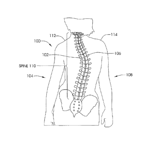
[0077] FIG. 1 illustrates a patient 100 with scoliosis. The concave portion
102 of the
spinal curve can be seen on the left side 104 of the patient 100, and the
convex portion 106
can be seen on the right side 108 of the patient 100. Of course, in other
patients, the concave
portion 102 may appear on the right side 108 of the patient 100 while the
convex portion 106
may be found on the left side 104 of the patient. In addition, as seen in FIG.
1, some rotation
of the spine 110 is present, and unevenness between the left shoulder 112 and
right shoulder
114 is seen.
[0078] FIG. 2 illustrates the Cobb angle 116 of a spine 110 of a patient
with scoliosis. To
determine the Cobb angle, lines 118 and 120 are drawn from vertebra 122 and
124,
respectively. Intersecting perpendicular lines 126 and 128 are drawn by
creating 90 angles
11
CA 02703562 2010-04-22
WO 2009/058546
PCT/US2008/079743
130 and 132 from lines 118 and 120. The angle 116 created from the crossing of
the
perpendicular lines 126 and 128 is defined as the Cobb angle. In a perfectly
straight spine,
this angle is 0 .
[0079] In many Adolescent Idiopathic Scoliosis (AIS) patients with a Cobb
angle of 40
or greater, spinal fusion surgery is typically the first option. FIG. 3
illustrates a long incision
134 formed in the patient 100 which is typically made during posterior
scoliosis fusion
surgery. This type of fusion surgery is known in the prior art. The long
incision 134 extends
between an upper end 136 and a lower end 138. The length of this incision 134
is longer than
the length of the section of the vertebra to be fused. The actual length
between the upper end
136 and the lower end 138 varies, depending on the size of the patient, and
the extent of the
scoliosis, but in AIS patients this length is significantly longer than 15 cm.
More typically, it
is longer than 25 cm.
[0080] FIGS. 4 and 5 illustrate a distraction device 140 for treating
scoliosis according to
one embodiment of the invention. The distraction device 140, which is an
implantable
device, includes a first adjustable rod 142 and a second adjustable rod 144.
For patient
distraction, a first adjustable rod 142 is positioned on one side of the spine
110 while the
second adjustable rod 144 is positioned on the opposing side of the spine 110.
The spine 110
is omitted from view in FIGS. 4 and 5 for sake of clarity. While the
distraction device 140
illustrated in FIGS. 4 and 5 comprises first and second adjustable rods 142,
144, it should be
understood that in alternative embodiments, the distraction device 140 may
include just a
single adjustable rod 142 (the second adjustable rod 144 being omitted
entirely) that is
implanted within the patient.
[0081] Referring back to FIGS. 4 and 5, each adjustable rod 142, 144
includes a first
elongate member 146, 148 and a second elongate member 150, 152, that are
coupled together
by an adjustable portion 158, 159. The adjustable portions 158, 159 include a
variable
overlapping region between the first elongate members 146, 148 and the second
elongate
members 150, 152 which allows for the non-invasive adjustment of the length of
each
adjustable rod 142, 144. In this particular embodiment, the first elongate
elements 146, 148
are telescopically contained within hollow receiving portions of the second
elongate elements
150, 152, and the adjustable portions 158, 159 are substantially straight. As
illustrated, the
adjustable rods 142, 144 have an upper curve 154 and a lower curve 156, which
allow them
to better conform to the natural front-to-back curve of the spine. For
example, the upper
curve 154 conforms to the normal kyphosis of the upper thoracic region and the
lower curve
156 conforms to the normal lordosis of the lumbar region. In one aspect of the
invention, the
12
CA 02703562 2010-04-22
WO 2009/058546
PCT/US2008/079743
curved portions 154, 156 are bendable in order to better conform with a
patient's specific
spinal configuration. For the example, the curved portions 154, 156 may be
made of a
malleable or elastic-type material such that the surgeon can manually alter
the particular
shape of each adjustable rod 142, 144 to the specific needs of the patient. In
a large number
of scoliosis patients, especially adolescent idiopathic scoliosis patients,
the scoliotic curve
does not include the lower lumbar levels of the spine and so the lower curve
156 is not
necessary. As explained above, the embodiment illustrated in FIGS. 4 and 5
represents a dual
rod configuration. With this configuration, both rods 142, 144 are inserted
through the same
incision, and can be placed along the spine 110 on two opposite sides of the
center line of the
spine 110. Alternatively, each may be placed through its own, smaller
incision.
[0082] Alternatively, a single adjustable rod version 142 can be used,
preferably
positioned on the concave side of the scoliosis curve. Yet another variation
includes a single
adjustable rod 142 that does not have either or both of the curves (i.e.,
curves 154 and 156
omitted). A straight adjustable rod 142 of this nature may be placed further
lateral (to the
side of the spine 110), and not necessarily have to hug the front-to-back
contours of the spine
110 or the muscle covering the spine 110. In still another embodiment, the
first elongate
member (e.g., 146, 148) and the second elongate member (e.g., 150, 152) do not
telescope in
relation to one another, but rather are in parallel, at least along the
adjustable portion 158,
159. The distraction device 140 is implanted in the patient 100 in order to
straighten the
scoliotic spine 110. For this reason, each end of the adjustable rods 142, 144
advantageously
contains an anchor 161 that allows for securement to a location in the
skeletal system. For
example, the anchor 161 at either end may include a clamp for clamping to a
skeletal
structure. Alternatively, either end may comprise a bracket for securing to a
section of bone
with the use of a bone screw or pedicle screw. The embodiment in FIG. 4
illustrates a clamp
160, 162 at the upper end of the first elongate members 146, 148 and brackets
164 at the end
of the second elongate members 150, 152. The brackets 164 can be secured to
the second
elongate members 150, 152 by a variety of methods, including set screws,
welding, soldering,
swaging, crimping or mechanical joints. Screws 166 secure the brackets 164 to
bony
structures, such as the vertebral bodies or the sacrum. The clamp 160, 162 can
be used to
clamp the distraction device 140 to a rib or the articulation of the rib with
the vertebra at the
facet. FIGS. 37 and 38, which are described in more detail below, illustrate
alternative
anchors 161 that may be used to secure the first elongate members 146, 148 or
second
elongate members 150, 152 to the skeletal structure.
13
CA 02703562 2010-04-22
WO 2009/058546
PCT/US2008/079743
[0083] The distraction device 140 is configured such that the adjustable
portion(s) 158,
159 change at least one of the distance or force between the anchor or
affixation points (e.g.,
at the spine or other anatomical structure) of the first elongate member(s)
146, 148 and the
second elongate member(s) 150, 152. For example, the adjustable portion(s)
158, 159 may
increase the length between the anchor or affixation points. Similarly, the
adjustable
portion(s) 158, 159 may increase the force (e.g., distraction force) between
the anchor or
affixation points. The adjustable portion(s) 158, 159 may alter both the
distance and force at
the same time.
[0084] FIG. 6A illustrates a sectional view of the first adjustable rod
142 indicating the
location of the adjustable portion 158 and the clamp 160. The tip 168 of clamp
160 is
shaped to allow for blunt dissection of tissue, so that the adjustable rod 142
may be placed
under the skin and pushed for much of the length of the spine 110, so that a
large portion of
the long incision 134 of FIG. 3 is not necessary. This allows for, for
instance, alternative
incision geometry, such as that illustrated in FIG. 7. As seen in FIG. 7, a
lower incision 170
is made having an upper end 176 and a lower end 178 (for example, by a
scalpel) and the first
adjustable rod 142 is placed through the lower incision 170 and under the
skin. Using a
dissection technique, the first adjustable rod 142 is inserted under the skin
along an
intermediate area 174. The dissection technique may include the use of a scope
(laparoscope,
arthroscope, endoscope, or the like) and an additional dissecting tool, but
usually can be done
without these tools. The additional dissecting tool may include, for example,
a tapered
sheath, which is advanced over the first adjustable rod 142, dissecting the
tissue along the
way, while being visualized by scope, for example on a monitor. Alternatively,
the additional
dissecting tool may be a blunt dissecting tool, consisting of two fingers
which can be spread
apart and brought together, once again, while being visualized by the scope.
[0085] Once the clamp 160 of the first adjustable rod 142 (as seen in FIG.
6A) is advanced
to the location near the anatomy to be clamped, an upper incision 172 is made
having an
upper end 180 and a lower end 182 and the location near the anatomy to be
clamped is
exposed by dissection. The clamp 160 is then actuated to clamp this anatomical
structure,
and additionally, the opposite end of the first adjustable rod 142 is secured,
for example by a
bone screw (e.g., pedicle screw) and bracket combination. The adjustment
device of the
adjustable rod 142 (to be described later) may be adjusted prior to the
securement of either
end of the first adjustable rod 142, so that the desired length is achieved.
After securement of
both ends, first adjustable rod 142 may then be adjusted in order to adjust
the distraction
distance or distraction force between the two locations in the anatomy to a
desired amount.
14
CA 02703562 2010-04-22
WO 2009/058546
PCT/US2008/079743
In one aspect of the invention, the length of the first adjustable rod 142 may
first be adjusted
manually by the physician without using the remotely-operated adjustment
device as
described herein. For example, the initial length of the adjustable rod 142
may be manually
set by the physician by pushing or pulling the first and second elongate
members 146, 150
relative to one another. Alternatively, the length of the adjustable rod 142
may be adjusted
by trimming or removing a portion of the length of the adjustable rod 142.
[0086] By having the physician adjust the length of the adjustable rod
142 during initial
placement, a distraction force may be applied to the spine 110 without having
to use any
displacement distance or force that is provided by the remotely-operated
adjustment device.
For example, there typically is a limited degree of movement that is provided
by the
remotely-operated adjustment device. When the physician applies a first or
initial distraction
force upon implantation, the budget of available displacement for the remotely-
operated
adjustment device is saved for later adjustments.
[0087] Still referring to FIG. 7, the two incisions are then closed using
standard
techniques. As described, the single long incision is now replaced by two,
shorter incisions
170, 172, whose combined length when added together is less than the length of
the single
long incision illustrated in FIG. 3. For example, lower incision 170 and upper
incision 172
each has a length of less than 15 cm, and preferably, each has a length of
less than 7.5 cm,
and more preferably, less than 5 cm.
[0088] An optional magnetic clamping device is illustrated in FIG. 6B,
which allows for
the entire procedure to be done under a single short incision 184, as seen in
FIG. 8. As
previously described, a single short incision 184 having an upper end 186 and
a lower end
188 is made (for example, by a scalpel) and the first adjustable rod 142 is
placed through the
single small incision 184 and under the skin. Using a dissection technique,
the first
adjustable rod 142 is inserted under the skin towards the upper target
location. As previously
described, this dissection technique may include the use of a scope
(laparoscope, arthroscope,
endoscope, or the like) and an additional dissecting tool. Once the clamp 160
of the first
adjustable rod 142 is advanced to the location near the anatomy to be clamped,
one or more
dissecting tools and a scope are used to expose the target location, for
example a rib or facet
articulation. Referring to FIG. 6B, the magnetically-operated clamp 160
includes a first
finger 190 and a second finger 192. The first finger 190 is permanently
coupled to first
elongate element 146 while the second finger 192 is longitudinally adjustable
in relation to
first finger 190, so that gap 194 may be increased or decreased in response to
actuation. A
closure device 198 is operated by an external adjustment device such as that
illustrated in
CA 02703562 2010-04-22
WO 2009/058546
PCT/US2008/079743
FIGS. 10-12 in order to increase or decrease gap 194, and therefore open or
close clamp 160.
As will be described, the clamp 160 is magnetically adjustable, and so the
clamping process
may be performed non-invasively, therefore making a second incision
unnecessary.
[0089] The magnetically-operated clamp 160 may be particularly useful if,
as expected,
the evidence of the ineffectiveness of braces becomes stronger, many
physicians will be
searching for less invasive procedures to treat scoliosis. Patients will
demand that the
procedures be as minimally invasive as possible, and one of the big elements
in their decision
to undergo surgery is the size of the incision, and thus size of the scar,
both during and after
healing. AIS patient whose Cobb angles are greater than 40 are more likely to
be treated
with fusion surgery, but patients in the 20 to 40 range may be treatable
using fusionless
methods which harness the growing power of their spine. Currently, it is known
that female
AIS patients who have not yet reached menarche (the first menstrual period)
are more likely
to have a curve that will progress further. Additionally, AIS patients whose
age is younger
are more likely to have their curves progress. One or more "scoliosis genes"
have recently
been discovered, and work is being done to create a genetic test that allows
identification of a
patient whose curve is very likely to progress beyond 40 at a time when her
Cobb angle is
less than 40 , for example 20 . Because braces are a questionable option, it
is expected that a
minimally invasive, non-fusion procedure will be the procedure of choice for
these patients.
Though the incision 184 in FIG. 8 is depicted as a vertical incision,
alternatively, it may be
made horizontally. For example, the horizontal incision may be made so that it
is just below
and parallel to the "bikini line", allowing the resulting scar to be more
concealed. This could
also be done with incision 170 in FIG. 7.
[0090j Returning to FIG. 6B, closure device 198 includes a cylindrical
magnetic member
200, which can be activated by magnetic coupling with an external adjustment
device (such
as external adjustment device 1130 illustrated in FIGS. 10-12). Though
configurations may
vary for this closure device 198, in this particular embodiment, magnetic
member 200 is a
hollow rare earth magnet, preferably Neodynium-Iron-Boron. As seen from an end
view in
FIG. 6E, the magnetic member 200 has a threaded insert 202 having a female
thread so that
when the magnetic member 200 rotates, the threaded insert 202 rotates in
unison. Magnetic
member 200 is a permanent magnet 217 having a north pole 204 and a south pole
206.
Magnetic member 200 is preferably coated with a material, for example
Parylene, phenolic
resin or Gold, which is non-magnetic, but protective and biocompatible in a
body implant
application. In certain embodiments, the individual Nd-Fe-B magnets are
enclosed within a
stainless steel casing/housing or various layers of nickel, gold or copper
plating to protect the
16
CA 02703562 2010-04-22
WO 2009/058546
PCT/US2008/079743
corrosive Nd-Fe-B material from the environment inside the body. In other
embodiments,
other magnetic materials may be used, including SmCo (Samarium Cobalt), which
is
typically available as SmCos, or SmCois, Sm2C017, or AlNiCo (Aluminum Nickel
Cobalt).
In still other embodiments, Iron Platinum (Fe-Pt) may be used. Iron platinum
magnets
achieve a high level of magnetism without the risk of corrosion, and may
possibly preclude
the need to encapsulate. In yet other embodiments, the permanent magnets 217
on the
implantable interface may be replaced by magnetically responsive materials
such as
Vanadium Permendur (also known as Hiperco).
[0091] It should be noted that magnetic member 200 can also be
hermetically sealed
within the first elongate element 146. When the external adjustment device
1130 is operated,
it applies a moving magnetic field, which causes magnetic member 200 to
rotate. Attached to
the second finger 192 is a threaded rod 210 which threadedly engages the
female thread of
the threaded insert 202. When the magnetic member 200 is rotated by the
external
adjustment device 1130 in a first direction, the threaded rod 210 moves in a
first longitudinal
direction 212, causing the second finger 192 to move away from the first
finger 190, and the
gap 194 to open. There may also be a manual adjustment mechanism on the clamp
160 so
that the clamp 160 may be opened outside the patient, in preparation for the
procedure.
When gap 194 is adjusted to be wider than the anatomical structure, for
example rib, around
which the clamp 160 is to be secured, then through visualization by the scope
and
manipulation with the dissecting tools, the clamp 160 is placed over the rib,
so that rib is
contained in cavity 196. At this point the external adjustment device 1130 is
operated so that
it turns the magnetic member 200 in the opposite direction causing the
threaded rod 210 to
move longitudinally in a second direction 214, and the two fingers 190, 192
close around the
rib. The gap 194 is now smaller than the width of the rib, and thus, the clamp
160 is secure.
If the implant is to be removed at a later date, the magnetic clamp mechanism
may also be
used to remove the implant without having to make an incision adjacent the
clamp.
[0092] FIG. 6C illustrates a sectional view of the adjustable portion 158
of the first
adjustable rod 142. FIG. 6D illustrates a detail of the adjustment device 232.
The first
elongate element 146 is telescopically contained within the second elongate
element 150.
The cross-sectional shapes of the first elongate element 146 and the second
elongate element
150 may be circular or non-circular, so that they cannot rotate with respect
to each other (for
example, a keyed configuration). One or both of the elongate elements 146, 150
may contain
ribs along the cross section of the adjustable portion 158 in order to
minimize contact surface
area between the first elongate element 146 and the second elongate element
150 and thus
17
CA 02703562 2010-04-22
WO 2009/058546
PCT/US2008/079743
lower frictional resistance. Beveled end piece 216 attached to the second
elongate element
150 may serve two purposes. First, it allows for smooth insertion and no
catching in tissue
when the first adjustable rod 142 is inserted under the skin. Second, it
serves as a low friction
dynamic seal over the first elongate element 146. Magnetic element 218
comprises a
cylindrical permanent magnet which is poled as shown in FIG. 6F.
Alternatively, magnetic
element 218, may be made from any of the materials described for magnetic
member 200 in
FIG. 6B. Magnetic element 218 is rotatably secured to an inner cavity 234 of
second
elongate element 150 by a housing, in this case an acoustic housing 222. A
ball bearing 220
is illustrated at one end of the magnetic element 218 in order to reduce
rotational friction. A
second ball optional bearing (not shown) can be included on the opposite end
of the magnetic
element 218. Magnetic element 218 is rotated by an external adjustment device
1130 which
produces a moving magnetic field.
[0093] As seen in FIG. 6D, the magnetic element 218 is coupled to a
planetary gear set
224, for example, having a 4:1, 16:1 or 64:1 gear reduction, or greater. The
purpose of the
gear reduction is two-fold. First, it allows the distraction device 140 to be
adjusted with a
smaller input torque requirement. Second, it adds precision to the adjustment,
because a
larger number of turns of the magnetic element 218 are required for each
adjustment interval.
Planetary gear set 224 is shown in detail in FIG. 6G. Sun gear 236 is turned
in a one-to-one
fashion by the rotation of the magnetic element 218. Sun gear 236 engages a
plurality of
planetary gears 238 (in this case, four are pictured). Planetary gears 238
engage and turn ring
gear 240 which is attached to a lead screw 226 via a coupling 228. The gear
ratio is the
number of teeth in the ring gear 240 divided by the number of teeth in the sun
gear 236. For
example if the ring gear 240 has four times as many teeth as the sun gear 236,
then the gear
ratio is 4:1. In this case, only 25% of the torque is required to drive the
lead screw 226 as
would have been required to drive it directly, ignoring the variance due to
frictional factors.
As lead screw 226 turns, it threadedly engages with female thread 230,
disposed within end
242 of first elongate element 146. The pitch of lead screw 226 threads is
preferably very fine
pitch, for example, 40 to 120, or more specifically 80 to 100 threads per
inch, in order to
minimize friction between the lead screw 226 and the female thread 230, and
thus, minimize
the required torque. The materials of the lead screw 226, the rods and other
components may
be made from non-magnetic, implantable materials such as Titanium or Titanium
alloys such
as Titanium-6% AI-4% V, although they may also be made from other magnetic
materials
such as stainless steel.
18
CA 02703562 2010-04-22
WO 2009/058546
PCT/US2008/079743
[0094] When the magnetic element 218 is rotated by the external
adjustment device 1130,
the drive train or drive element that is operatively coupled to the rotatable
magnetic element
218 drives the lead screw 226 which changes the length of the adjustable
portion 158 of the
adjustable rod(s) 142, 144. Rotation of the magnetic element 218 in a first
direction increases
the distance between the anchors 161 located on opposing ends of the
adjustable rod(s) 142,
144. Conversely, rotation of the magnetic element 218 in a second (opposing)
direction
decreases the distance between the anchors 161 located on opposing ends of the
adjustable
rod(s) 142, 144.
[0095] Currently, devices such as the VEPTR, which can be surgically
adjusted, are used
for early onset scoliosis patients, and their adjustability is used for the
purpose of keeping up
with the dimensional growth of the patient. It is a purpose of the present
invention to create a
device which can be non-invasively adjusted in early onset scoliosis patients,
but
additionally, in adolescent idiopathic scoliosis (AIS) patients and even adult
scoliosis
patients. The main purpose for the adjustment in AIS patients is to maintain a
distraction
force, which in a fusionless growing spine serves to steer growth in the
desired manner.
Currently, in fusionless surgery, non-adjustable distraction devices are
actuated at very high
distraction forces, because the physicians know that over time, growth and/or
changes within
the tissue, will cause this distraction force to lessen, possibly becoming
less effective with
time. Because of these high distraction forces, it is not uncommon to have
rods break inside
the patient, or for bone screws to become dislodged, due to the high stresses.
It has been
contemplated that the high forces that have been measured in some distraction
devices of well
over 100 pounds, are not necessary at any given time to provide correct growth
guidance, and
that a distraction force of below 45 pounds, and even as low as 20 pounds may
be effective in
maintaining the desired growth of the spine, especially the unfused spine.
That is, as long as
this force can be maintained, which is not currently possible in prior art
devices without
surgical intervention. The present invention allows this lower force to be
continually
maintained through non-invasive adjustment. The benefit is that lower stresses
can be
maintained on the bone screws, clamps, and other attachment means as well as
the rods
themselves, making for a more reliable and durable system. In addition,
through the
identification of an optimum distraction force, this desired force can be
maintained
throughout the treatment of the patient post- surgery, by frequent non-
invasive adjustments,
which can be performed in a doctor's or nurses office, by a physician or non-
physician
medical personnel, or even by the patient herself at home. In addition, by
incorporating an
optional force transducer, as part of the distraction device, that is read
telemetrically, each
19
CA 02703562 2010-04-22
WO 2009/058546
PCT/US2008/079743
adjustment can be done to the precise desired distraction force. Additionally,
a slip clutch
244, is in line with the magnetic element 218 can be pre-adjusted by the
physician, or during
the manufacturing process, so that during each adjustment, the adjustment
stops when a
critical torque (corresponding to the maximum desired distraction force) is
reached. For
example, the maximum desired distraction force may be set at 45 pounds. The
slip clutch
244 is illustrated in FIG. 6D as being located between the magnetic element
218 and the
planetary gear set 224, but it is within the scope of the invention that the
slip clutch 244 may
be located at any other step along the torque transmission chain.
[0096] FIG. 9 illustrates a patient 100 with a distraction device 140
implanted on the left
side of the spine 110. Though the spine 110 is visible in FIG. 9 for
reference, FIG. 9 is
actually meant to depict a non-invasive adjustment procedure, and so the
patient 100 would
typically have all incisions healed and could be wearing clothes. The clamp
160 of the
distraction device 140 is secured to a rib 246 at its articulation with a
thoracic vertebra 247.
A bracket 164 is secured, in this case to a lumbar vertebra with screws 166.
Alternatively,
the bracket 164 may be secured, for example, to the sacrum 249. A radio
frequency
identification (RFID) chip 250 is optionally disposed on the second elongate
element 150 of
the distraction device 140 in accordance with an embodiment of the present
invention. An
RFID (radio frequency identification) chip 250 may be implanted in a patient
during the
implantation of the distraction device 140. In certain embodiments, the RFID
chip 250 may
be implanted subcutaneously in a known location, such as a location near the
distraction
device 140. In other embodiments, the RFID chip 250 may be located on or
within the
distraction device 140. An external adjustment device 248 is depicted after
being placed
against the back of the patient 100. Upon the implantation of the distraction
device 140 or
after surgical recovery, the external adjustment device 248 stores patient
information on the
RFID chip 250, including the current size or setting of the distraction device
140, the amount
adjusted, the serial number of the distraction device 140, the date of the
implantation
procedure, patient name, distraction force, adjustment torque, and
identification. During
subsequent adjustment procedures, the external adjustment device 248 may read
the RFID
chip 250 to determine information related to the patient, such as the current
size or setting of
the distraction device 140. At the end of the adjustment procedure, the
external adjustment
device 248 may store updated patient information, including the size or
setting of the
distraction device 140, to the RFID chip 250. An RFID antenna 252 in the
external
adjustment device 248 may be used to power the RFID chip in order facilitate
the read and
write functions.
CA 02703562 2010-04-22
WO 2009/058546
PCT/US2008/079743
[0097] Several techniques may be used to determine the adjustment setting
(current size,
distraction force or condition) of the distraction device 140. For example,
the adjustment
setting may be determined indirectly by the number of rotations of one of the
rotating
components of the external adjustment device 248. In certain embodiments, the
adjustment
setting may be determined by the number of rotations of some dynamic component
of the
adjustable portion 158 of the distraction device 140, by the number of
rotations of any one of
the gears or shafts of the distraction device 140, or by the number of
rotations of the magnetic
element 218. In other embodiments, a feedback mechanism, such as a Hall effect
device (two
additional magnets that move axially in relation to each other as the lead
screw 226 rotates
and therefore as the distraction device changes its condition), may be used to
determine the
current adjustment setting of the distraction device 140. A strain gauge or
force transducer
disposed on a portion of the distraction device 140 may also be used as an
implantable
feedback device. For example, the strain gauge may be able to communicate
wirelessly the
actual distraction force applied to the spine by the distraction device 140. A
wireless reader
or the like (that also can inductively power the strain gauge) may be used to
read the
distraction forces. One exemplary strain gauge sensor is the EMBEDSENSE
wireless sensor,
available from MicroStrain, Inc. of Williston, VT 05495. The EMBEDSENSE
wireless
sensor uses an inductive link to receive power form an external coil and
returns digital stain
measurements wirelessly.
[0098] In still other embodiments, an optical encoder feedback mechanism
may be used
by placing an optical encoder in line with one of the rotating components of
the adjustable
portion 158 of the distraction device 140. A through-the-skin optical encoder
is even
envisioned that shines a light through the skin and fat and counts successive
passes of one or
more reflective stripes on the specific rotatable component. In other
embodiments, the
external adjustment device 248 may include an audio sensor to determine the
current
adjustment setting of the distraction device 140. For example, the sensor may
listen to the
cycling sound of gearing, thus giving feedback information on the amount of
total
adjustment. An additional acoustic feedback device is discussed below.
[0099] It should be understood that any of the materials of the
distraction device 140 can
be made from radiopaque materials, so that the position, condition or
alignment of the
components may be seen during the initial surgical procedure, or during the
subsequent
adjustment procedures, by use of X-ray. For example, a circumferential notch
or
alternatively a circumferential bump disposed on the first or second elongate
members 148,
21
CA 02703562 2010-04-22
WO 2009/058546
PCT/US2008/079743
146 may be used so that the distance between this notch or bump and some
portion of the
second elongate members 150, 152 can be measured easily via an X-ray.
[00100] It is conceived that the adjustment procedures would preferably take
place every
three to four weeks in the physicians' clinic. The adjustment may be done by
an orthopedic
surgeon, but because of the relative ease of the procedure because of the
feedback capabilities
of the system, the procedure may be done by a nurse practitioner, a
physicians' assistant, a
technician, or any other non-M.D. personnel. It is even conceived that the
patient may have
an external adjustment device 1130 at home and be able to adjust themselves at
an even more
frequent rate. The external adjustment device 1130 can be designed to transmit
stored
information over the phone to the physician's office. For example, adjustment
dates or
adjustment parameters such as distraction force or distraction distance.
[00101.1 FIG. 10 illustrates an external adjustment device 1130 which is one
embodiment of
an external adjustment device 248 according to one aspect of the invention.
The external
adjustment device 1130 may be used to externally impart rotational motion or
"drive" a
permanent magnet (e.g., magnetic element 218) located within the distraction
device 140.
The external adjustment device 1130 includes a motor 1132 that is used to
impart rotational
movement to two permanent magnets 1134, 1136. The two permanent magnets 1134,
1136
are located in the same driver 1130 and are configured for placement on the
same side of the
body of the patient or subject. The motor 1132 may include, for example, a DC
powered
motor or servo that is powered via one or more batteries (not shown)
integrally contained
within the external adjustment device 1130. Alternatively, the motor 1132 may
be powered
via a power cord or the like to an external power source. For example, the
external power
source may include one or more batteries or even an alternating current source
that is
converted to DC.
[00102] Still referring to FIG. 10, the two permanent magnets 1134, 1136 are
preferably
cylindrically-shaped permanent magnets. The permanent magnets may be made
from, for
example, a rare earth magnet material such as Neodymium-Iron-Boron (NdFeB)
although
other rare earth magnets are also possible. For example, each magnet 1134,
1136 may have a
length of around 1.5 inches and a diameter of around 1.0 to 3.5 inches. Both
magnets 1134,
1136 are diametrically magnetized (poles are perpendicular the long axis of
each permanent
magnet 1134, 1136). The magnets 1134, 1136 may be contained within a non-
magnetic
cover or housing 1137. In this regard, the magnets 1134, 1136 are able to
rotate within the
stationary housing 1137 that separates the magnets 1134, 1136 from the
external
environment. Preferably, the housing 1137 is rigid and relatively thin walled
at least at the
22
CA 02703562 2010-04-22
WO 2009/058546
PCT/US2008/079743
portion directly covering the permanent magnets 1134, 1136, in order to
minimize the gap
between the permanent magnets 1134, 1136 and the internal magnet 1064 (as
shown in FIGS.
13A-13D).
[00103] As seen in FIG. 10, the permanent magnets 1134, 1136 are rotationally
mounted
between opposing bases members 1138, 1140. Each magnet 1134, 1136 may include
axles or
spindles 1142, 1144 mounted on opposing axial faces of each magnet 1134, 1136.
The axles
1142, 1144 may be mounted in respective bearings (not shown) that are mounted
in the base
members 1138, 1140. As seen in FIG. 10, driven pulleys 1150 are mounted on one
set of
axles 1142 and 1144. The driven pulleys 1150 may optionally include grooves or
teeth 1152
that are used to engage with corresponding grooves or teeth 1156 (partially
illustrated in FIG.
12) contained within a drive belt (indicated by path 1154).
[00104] Still referring to FIG. 10, the external adjustment device 1130
includes a drive
transmission 1160 that includes the two driven pulleys 1150 along with a
plurality of pulleys
1162A, 1162B, 1162C and rollers 1164A, 1164B, 1164C on which the drive belt
1154 is
mounted. The pulleys 1162A, 1162B, 1162C may optionally include grooves or
teeth 1166
used for gripping corresponding grooves or teeth 1156 of the drive belt 1154.
Pulleys 1162A,
1162B, 1162C and rollers 1164A, 1164B, 1164C may be mounted on respective
bearings (not
shown). As seen in FIG. 10, pulley 1162B is mechanically coupled to the drive
shaft (not
shown) of the motor 1132. The pulley 1162B may be mounted directly to the
drive shaft or,
alternatively, may be coupled through appropriate gearing. One roller 1164B is
mounted on a
biased arm 1170 and thus provides tension to the belt 1154. The various
pulleys 1150,
1162A, 1162B, 1162C and rollers 1164A, 1164B, 1164C along with the drive belt
1154 may
be contained within a cover or housing 1172 that is mounted to the base 1138
(as seen in FIG.
12). For safety and convenience, it may be desired for the external adjustment
device 1130 to
have a removable safety cover that would be placed over the portion containing
the
permanent magnets 1134, 1136, for example during storage, so that the high
magnetic field
cannot come closely in contact with anything that would be strongly attracted
to it or
damaged by it.
[00105] As seen in FIGS. 10 and 11, rotational movement of the pulley 1162B
causes the
drive belt 1154 to move around the various pulleys 1150, 1162A, 1162B, 1162C
and rollers
1164A, 1164B, 1164C. In this regard, rotational movement of the motor 1132 is
translated
into rotational movement of the two permanent magnets 1134, 1136 via the drive
transmission 1160. In one aspect of the invention, the base members 1138, 1140
are cut so as
to form a recess 1174 that is located between the two magnets 1134, 1136.
During use, the
23
CA 02703562 2010-04-22
WO 2009/058546
PCT/US2008/079743
external adjustment device 1130 is pressed against the skin of a patient, or
against the
clothing which covers the skin (e.g., the external adjustment device 1130 may
be used
through clothing so the patient may not need to undress). The recess 1174
allows skin as well
as the underlying tissue to gather or compress within the recessed region 1174
as seen in
FIGS. 13A and 13B. This advantageously reduces the overall distance between
the external
drive magnets 1134, 1136 and the magnet 1064 contained within the distraction
device 140.
By reducing the distance, this means that the externally located magnets 1134,
1136 and/or
the internal magnet 1064 may be made smaller. This is especially useful in the
case of an
obese patient.
[00106] In one embodiment, the two permanent magnets 1134, 1136 are configured
to
rotate at the same angular velocity. In another embodiment, the two permanent
magnets
1134, 1136 each have at least one north pole and at least one south pole, and
the external
adjustment device 1130 is configured to rotate the first magnet 1134 and the
second magnet
1136 such that the angular location of the at least one north pole of the
first magnet 1134 is
substantially equal to the angular location of the at least one south pole of
the second magnet
1136 through a full rotation of the first and second magnets 1134, 1136.
[00107] FIGS. 13A and 13B illustrate cross-sectional views of the patient
having an
implanted distraction device 140 containing an internal magnet 1064. For sake
of clarity, the
first and second elongate members 146, 150 have been removed to illustrate the
relationship
between the external adjustment device 1130 and the rotationally-driven
internal magnet
1064. The internal magnet 1064 is seen disposed on one side of a vertebra
1185. Further, the
internal magnet 1064 is seen being outside or external with respect to the
fascia 1184 and
muscle 1186 of the subject. FIGS. 13A and 13B illustrate an obese patient in
which skin and
other tissue gather within the recess 1174. It should be understood that obese
Adolescent
Idiopathic Scoliosis patients are rare, and FIGS. 13A and 13B generally
indicate a worst-case
situation but as seen in FIGS. 13A and 13B the excess skin and other tissue is
easily
accommodated within the recess 1174 to enable close positioning between the
internal
magnet 1064 and the external drive magnets 1134, 1136. For most AIS patients,
the air gap
or distance between the internal magnet 1064 and the external drive magnets
1134, 1136 is
generally one inch or less. In FIGS. 13A through 13D, the internal magnet 1064
is depicted
somewhat larger than its size in the preferred embodiment, in order for its
poles to be more
clearly visible.
[00108] Still referring to FIGS. 10 and 11, the external adjustment device
1130 preferably
includes an encoder 1175 that is used to accurately and precisely measure the
degree of
24
CA 02703562 2010-04-22
WO 2009/058546
PCT/US2008/079743
movement (e.g., rotational) of the external magnets 1134, 1136. In one
embodiment, an
encoder 1175 is mounted on the base member 1138 and includes a light source
1176 and a
light receiver 1178. The light source 1176 may includes a LED which is pointed
or directed
toward pulley 1162C. Similarly, the light receiver 1178 may be directed toward
the pulley
1162C. The pulley 1162C includes a number of reflective markers 1177 regularly
spaced
about the periphery of the pulley 1162C. Depending on the rotational
orientation of the
pulley 1162C, light is either reflected or not reflected back onto the light
receiver 1178. The
digital on/off signal generated by the light receiver 1178 can then be used to
determine the
rotational speed and displacement of the external magnets 1134, 1136.
[00109] FIGS. 13A, 13B, 13C, and 13D illustrate the progression of the
external magnets
1134, 1136 and the internal magnet 1064 that is located within the distraction
device 140
during use. Internal magnet 1064 is shown for illustration purposes. Internal
magnet 1064 is
one possible embodiment of the magnetic element 218 described herein. FIGS.
13A, 13B,
13C, and 13D illustrate the external adjustment device 1130 being disposed
against the
external surface of the patient's skin 1180 adjacent the spine (not shown for
clarity sake). ).
In the non-invasive adjustment procedure depicted, the patient 100 lies in a
prone position,
and the external adjustment device 1130 is placed upon the patient's back.
However, the
adjustment is conceived possible with the patient in supine, standing or
positions. The
external adjustment device 1130 is placed against the skin 1180 in this manner
to remotely
rotate the internal magnet 1064. As explained herein, rotation of the internal
magnet 1064 is
translated into linear motion via the adjustment device 232 to controllably
adjust the
distraction device 140.
[00110] As seen in FIGS. 13A, 13B, 13C, and 13D, the external adjustment
device 1130
may be pressed down on the patient's skin 1180 with some degree of force such
that skin
1180 and other tissue such as the underlying layer of fat 1182 are pressed or
forced into the
recess 1174 of the external adjustment device 1130. FIGS. 13A, 13B, 13C, and
13D show
the magnetic orientation of the internal magnet 1064 as it undergoes a full
rotation in
response to movement of the permanent magnets 1134, 1136 of the external
adjustment
device 1130.
[00111] With reference to FIG. 13A, the internal magnet 1064 is shown being
oriented with
respect to the two permanent magnets 1134, 1136 via an angle 0. This angle 0
may depend
on a number of factors including, for instance, the separation distance
between the two
permanent magnets 1134, 1136, the location or depth of where the implantable
interface 1104
is located, the degree of force at which the external adjustment device 1130
is pushed against
CA 02703562 2010-04-22
WO 2009/058546
PCT/US2008/079743
the patient's skin. Generally in applications including some obese patients,
the angle 0
should be at or around 90 to achieve maximum drivability (e.g., torque). The
inventors have
calculated that in the AIS application, where there are few obese patients, an
angle of about
700 is preferred for the majority of patients when the permanent magnets 1134,
1136 have an
outer diameter of about three (3.0) inches.
1001121 FIG. 13A illustrates the initial position of the two permanent magnets
1134, 1136
and the internal magnet 1064. This represents the initial or starting location
(e.g., 00 position
as indicated). Of course, it should be understood that, during actual use, the
particular
orientation of the two permanent magnets 1134, 1136 and the internal magnet
1064 will vary
and not likely will have the starting orientation as illustrated in FIG. 13A.
In the starting
location illustrated in FIG. 13A, the two permanent magnets 1134, 1136 are
oriented with
their poles in an N-S/S-N arrangement. The internal magnet 1064 is, however,
oriented
generally perpendicular to the poles of the two permanent magnets 1134, 1136.
[00113] FIG. 13B illustrates the orientation of the two permanent magnets
1134, 1136 and
the internal magnet 1064 after the two permanent magnets 1134, 1136 have
rotated through
90 . The two permanent magnets 1134, 1136 rotate in the direction of arrow A
(e.g.,
clockwise) while the internal magnet 1064 rotates in the opposite direction
(e.g., counter
clockwise) represented by arrow B. It should be understood that the two
permanent magnets
1134, 1136 may rotate in the counter clockwise direction while the internal
magnet 1064 may
rotate in the clockwise direction. Rotation of the two permanent magnets 1134,
1136 and the
internal magnet 1064 continues as represented by the 180 and 270
orientations as illustrated
in FIGS. 13C and 13D. Rotation continues until the starting position (0 ) is
reached again.
[001141 During operation of the external adjustment device 1130, the permanent
magnets
1134, 1136 may be driven to rotate the internal magnet 1064 through one or
more full
rotations in either direction to increase or decrease distraction of the
distraction device 140 as
needed. Of course, the permanent magnets 1134, 1136 may be driven to rotate
the internal
magnet 1064 through a partial rotation as well (e.g., 1/4, 1/8, 1/16, etc.).
The use of two
magnets 1134, 1136 is preferred over a single external magnet because the
driven magnet
1064 may not be oriented perfectly at the start of rotation, so one external
magnet 1134, 1136
may not be able to deliver its maximum torque, which depends on the
orientation of the
internal driven magnet 1064 to some degree. However, when two (2) external
magnets
(1134, 1136) are used, one of the two 1134 or 1136 will have an orientation
relative to the
internal driven magnet 1064 that is better or more optimal than the other. In
addition, the
torques imparted by each external magnet 1134, 1136 are additive. In prior art
magnetically
26
CA 02703562 2010-04-22
WO 2009/058546
PCT/US2008/079743
driven devices, the external driving device is at the mercy of the particular
orientation of the
internal driven magnet. The two-magnet embodiment described herein is able to
guarantee a
larger driving torque - as much as 75% more than a one-magnet embodiment in
the AIS
application - and thus the internal driven magnet 1064 can be designed smaller
in dimension,
and less massive. A smaller internal driven magnet 1064 will have a smaller
image artifact
when performing MRI (Magnetic Resonance Imaging), especially important when
using
pulse sequences such as gradient echo, which is commonly used in breast
imaging, and leads
to the largest artifact from implanted magnets. In certain configurations, it
may even be
optimal to use three or more external magnets, including one or more magnets
each on two
different sides of the body (for example front and back).
[00115] While the external adjustment device 1130 and adjustment device 232
have
generally been described as functioning using rotational movement of driving
elements (i.e.,
magnetic elements) it should be understood that cyclic or non-rotational
movement can also
be used to drive or adjust the distraction device 140. For instance, cyclic
movement of driven
magnet 640, magnetic element 218, internal magnet 1064, internally located
driven magnet
1402, cylindrical magnet 394, hollow magnet 564, magnet 576, magnet 262,
magnets 618,
620, and magnet 1302 may be used to drive or adjust the distraction device
140. Cyclic
movement includes partial rotational movement (e.g., rotational movement that
is less than a
full revolution). Cyclic movement of one or more of the external magnets 624,
626, 1134,
1136 may also be employed.
[00116] In still another alternative, linear or sliding motion back-and-forth
may also be
used to adjust the distraction device 140. In this regard, a single magnet
located internal to
the patient that slides back-and-forth on a slide or other base can be used to
adjust the
distraction device 140 using a ratchet-type device. The sliding, internal
magnet may be
driven via one or more externally-located permanent/electromagnets that slides
or moves
laterally (or moves the magnetic field) in a similar back-and-forth manner.
Rotational
movement of the externally-located magnetic element(s) may also be used to
drive the
internal magnet. The internal magnet may alternatively be able to rotate back-
and-forth, thus
adjusting the distraction device 140 using a ratchet-type device.
[00117] In still another alternative, permanent magnets may be located on a
pivoting
member that pivots back and forth (like a teeter-totter) about a pivot point.
For example, a
first permanent magnet having a North pole oriented in a first direction may
be located at one
end of the pivoting member while a permanent magnet having a South pole
oriented in the
27
CA 02703562 2010-04-22
WO 2009/058546
PCT/US2008/079743
first direction is located at the other end of the pivoting member. A ratchet-
type device may
be used to translate the pivoting movement into linear movement that can
actuate or adjust
the distraction device 140. The first and second internally-located permanent
magnets may
be driven by one or more externally located magnetic elements (either
permanent or
electromagnets). External motion of the electric field by linear or even
rotational movement
may be used to the drive the pivoting member.
[00118] Two different models of internal driven magnets were constructed, each
from a
different Neodynium-Iron-Boron Grade. Both magnets had identical dimensions
(0.275"
diameter, 0.395" long). One magnet was a grade of approximately N38 and the
other was a
grade of N50. Both magnets were approximately 2.9 grams in mass. A 1" diameter
cylindrical permanent magnet (grade N50 Neodynium-Iron-Boron) was attached to
a torque
gauge and the peak coupling torque (in inch-ounces) between it and each of the
internal drive
magnet models was measured for three different angular orientations for the
cylindrical
permanent magnet, in relation to the internal driven magnet. All magnets were
two pole (as
in FIGS. 13A-13D). Each of the internal driven magnets was tested
individually. The
orientation was either 0 (worst case coupling torque), 45 or 90 (best case
coupling torque).
The data for a one inch air gap (separation between magnets) is listed below
in Table 1
below. A one (1) inch air gap is an expected worst case separation in the
clinical application
of adolescent idiopathic scoliosis. The effect of using two external 1"
diameter permanent
magnets (as in FIGS. 13A-13D) is shown by addition of the values for the worst
case (0 ) and
best case (90 ) orientations.
Table 1 ¨ Peak Coupling Torque (oz-in) at 1" Air Gap
Internal driven 0 orientation of 450 orientation 90 orientation
Two external
magnet single external of single external of single external
magnets (0
magnet magnet magnet orientation + 90
orientation)
Grade 38 1.37 1.92 2.47 3.84
(approx)
Grade 50 1.70 2.04 2.80 4.50
[00119] It can be clearly seen that the additive use of two external permanent
magnets,
especially if synchronized in the orientation shown in FIGS. 13A-13D, delivers
significantly
more torque than a single external magnet in any orientation. For the data
generated using
the 50 grade internal driven magnet, the peak coupling torque using two
external permanent
magnets was 4.50 ounce-inches, 60.7% greater than a single external permanent
magnet
28
CA 02703562 2010-04-22
WO 2009/058546
PCT/US2008/079743
oriented at the ideal 900 in relation to the internal driven magnet, and
164.7% greater than a
single external permanent magnet oriented at the worst case 0 . This
significant increase in
torque achieved by using two external permanent magnets, makes it possible to
incorporate
an especially small internal driven magnet (e.g., less than three grams) into
the design of the
scoliosis treatment implant, or any implant for manipulating one or more bones
or a portion
of the skeletal system. For example, the use of two external permanent magnets
may impart a
coupling torque of at least 3.0 inch-ounces to the internal magnet at a
separation distance of
around 1.0 inches.
[00120] In a gradient echo MRI scan of the breast in a 1.5 Tesla MRI scanner
using
standard breast imaging coils, a 2.9 gram N50 grade magnet having a 0.275 inch
diameter
and 0.295" length implanted in the mid-thorax creates an MRI artifact which is
small enough
to allow full imaging of the breasts. Using the dual 1" diameter external
permanent magnets
1134, 1136 as for the external adjustment device 1130, and using the grade 50
for the internal
driven magnet 1064 having a mass of 2.9 grams, the 4.50 ounce-inch torque
delivered to the
magnet will turn a 80 threads per inch lead screw mounted on ball bearing in a
sufficient
manner to apply a distraction force of approximately 11 pounds. If a 4:1
reduction planetary
gear set is incorporated into the design ¨ for example, between the internal
driven magnet
1064 and the lead screw 226 ¨ then a distraction force of approximately 44
pounds may be
delivered. In the system contemplated by this invention, in which several
gradual non-
invasive adjustments are made, distraction forces on this order (40 to 45
pounds) will be
sufficient. In fact, the slip clutch 244 can either be adjusted in the
fabrication of the scoliosis
implant or can be adjusted by the implanting physician, so that the slip
clutch 244 slips at
either a maximum threshold torque (to save the materials of the implant from
being damaged
or pulling out of the bone by too high a distraction force) or at desired
threshold torque (at
which the desired distraction force is generated).
[00121] The maximum threshold torque corresponds to a critical distraction
force, and the
desired threshold torque corresponds to a desired distraction force. A
critical distraction
force may correspond to a force at which anchors such as hooks or screws may
cause damage
to the bone. For example, one critical distraction force is 100 pounds, which
in one
embodiment of the invention corresponds to a critical threshold slip torque of
41.7 ounce-
inches (if no gear reduction, and a 80 threads per inch lead screw is used),
10.4 ounce-inches
(if a 4:1 gear reduction and a 80 threads per inch lead screw is used) or 2.6
ounce-inches (if a
16:1 gear reduction and a 80 threads per inch lead screw is used). Similarly,
one desired
distraction force is 45 pounds, which in one embodiment of the invention
corresponds to a
29
CA 02703562 2010-04-22
WO 2009/058546
PCT/US2008/079743
desired threshold slip torque of 18.75 ounce-inches (if no gear reduction and
a 80 threads per
inch lead screw is used) or 4.69 ounce-inches (if a 4:1 gear reduction and a
80 threads per
inch lead screw is used). If a desired distraction force is 20 pounds, then in
one embodiment
of the invention this corresponds to a desired threshold slip torque of 8.33
ounce-inches (if no
gear reduction and a 80 threads per inch lead screw is used) or 2.08 ounce-
inches (if a 4:1
gear reduction and a 80 threads per inch lead screw is used). In one aspect,
the desired
threshold distraction is between 2 inch-ounces and 42 inch-ounces. In another
aspect, the
desired threshold distraction is between 2 inch-ounces and 19 inch-ounces. In
still another
aspect, the desired threshold distraction is between 2 inch-ounces and 8.5
inch-ounces.
[00122] Other distraction devices have been proposed which incorporate a small
implantable motor to effect the distraction. The 2.9 gram cylindrical magnet
1064 described
as part of the present invention is significantly smaller than the smallest
motor which would
be feasible in the distraction application, considering torque requirements,
etc. In addition,
the cost of the magnet 1064 is significantly less than that of a micromotor.
The magnet 1064
is also very reliable in relation to a micromotor. The main possible failure
would be the loss
of the magnetic field, however the inventors have demonstrated that the
inventive 2.9 gram
magnet 1064 can be placed into the center of a 3.0 Tesla MRI magnet without a
significant
loss in magnetism. It can also be exposed to temperatures in excess of those
used in steam
sterilization, for example, without a significant loss of magnetism.
Generally, the internal
magnet 1064 should be grade N30 or higher, or even grade N48 or higher. While
the 2.9
gram cylindrical magnet 1064 has the advantage of being particularly small, in
other
embodiments, the cylindrical magnet 1064 may have a weight of less than about
10 grams or
less than about 6.0 grams. Similarly, the first and second external magnets
1134, 1136 may
be a rare earth permanent magnets such as, for instance, Neodynium-Iron-Boron.
In addition,
the first and second external magnets 1134, 1136 may be grade N30 or higher,
or even grade
N48 or higher.
[00123] FIG. 14 illustrates a system 1076 according to one aspect of the
invention for
driving the external adjustment device 1130. FIG. 14 illustrates the external
adjustment
device 1130 pressed against the surface of a patient 1077 (torso face down
shown in cross-
section). The portion of the distraction device 140 containing the internal
driven magnet
1064 is illustrated. The permanent magnet (e.g., the driven magnet 1064) that
is located
within the distraction device 140 located inside the patient 1077 is
magnetically coupled
through the patient's skin and other tissue to the two external magnets 1134,
1136 located in
the external adjustment device 1130. As explained herein, one rotation of the
external
CA 02703562 2010-04-22
WO 2009/058546
PCT/US2008/079743
magnets 1134, 1136 causes a corresponding single rotation of the driven magnet
1064 located
within the distraction device 140. Turning the driven magnet 1064 in one
direction causes
the distraction device 140 to lengthen, or increase distraction force while
turning in the
opposite direction causes the distraction device 140 to shorten, or decrease
distraction force.
Changes to the distraction device 140 are directly related to the number of
turns of the driven
magnet 1064.
[00124] The motor 1132 of the external adjustment device 1130 is controlled
via a motor
control circuit 1078 operatively connected to a programmable logic controller
(PLC) 1080.
The PLC 1080 outputs an analog signal to the motor control circuit 1078 that
is proportional
to the desired speed of the motor 1132. The PLC 1080 may also select the
rotational
direction of the motor 1132 (i.e., forward or reverse). In one aspect, the PLC
1080 receives
an input signal from a shaft encoder 1082 that is used to identify with high
precision and
accuracy the exact relative position of the external magnets 1134, 1136. For
example, the
shaft encoder 1082 may be an encoder 1175 as described in FIGS. 10-11. In one
embodiment, the signal is a pulsed, two channel quadrature signal that
represents the angular
position of the external magnets 1134, 1136. The PLC 1080 may include a built
in screen or
display 1081 that can display messages, warnings, and the like. The PLC 1080
may
optionally include a keyboard 1083 or other input device for entering data.
The PLC 1080
may be incorporated directly into the external adjustment device 1130 or it
may be a separate
component that is electrically connected to the main external adjustment
device 1130.
[00125] In one aspect of the invention, a sensor 1084 is incorporated into the
external
adjustment device 1130 that is able to sense or determine the rotational or
angular position of
the driven magnet 1064. The sensor 1084 may acquire positional information
using, for
example, sound waves, ultrasonic waves, light, radiation, or even changes or
perturbations in
the magnetic or electromagnetic field between the driven magnet 1064 and the
external
magnets 1134, 1136. For example, the sensor 1084 may detect photons or light
that is
reflected from the driven magnet 1064 or a coupled structure (e.g., rotor)
that is attached
thereto. For example, light may be passed through the patient's skin and other
tissue at
wavelength(s) conducive for passage through tissue. Portions of the driven
magnet 1064 or
associated structure may include a reflective surface that reflects light back
outside the
patient as the driven magnet 1064 moves. The reflected light can then be
detected by the
sensor 1084 which may include, for example, a photodetector or the like.
[00126] In another aspect, the sensor 1084 may operate on the Hall effect,
wherein two
additional magnets are located within the implantable assembly. The additional
magnets
31
CA 02703562 2010-04-22
WO 2009/058546
PCT/US2008/079743
move axially in relation to each other as the driven assembly rotates and
therefore as the
distraction increases or decreases, allowing the determination of the current
size of the
restriction device.
[00127] In the embodiment of FIG. 14, the sensor 1084 is a microphone disposed
on the
external adjustment device 1130. For instance, the microphone sensor 1084 may
be disposed
in the recessed portion 1174 of the external adjustment device 1130. The
output of the
microphone sensor 1084 is directed to a signal processing circuit 1086 that
amplifies and
filters the detected acoustic signal. In this regard, the acoustic signal may
include a "click" or
other noise that is periodically generated by rotation of the driven magnet
1064. For
example, the driven magnet 1064 may click every time a full rotation is made.
The pitch
(frequency) of the click may differ depending on the direction of rotation.
For example,
rotation in one direction (e.g., lengthening) may produce a low pitch while
rotation in the
other direction (e.g., shortening) may produce a higher pitch signal (or vice
versa). The
amplified and filtered signal from the signal processing circuit 1086 can then
pass to the PLC
1080.
[00128] During operation of the system 1076, each patient will have a number
or indicia
that correspond to the adjustment setting or size of their distraction device
140. This number
can be stored on an optional storage device 1088 (as shown in FIG. 14) that is
carried by the
patient (e.g., memory card, magnetic card, or the like) or is integrally
formed with the
distraction device 140. For example, a RFID tag 1088 implanted either as part
of the system
or separately may be disposed inside the patient (e.g., subcutaneously or as
part of the device)
and can be read and written via an antenna 1090 to update the current size of
the distraction
device 140. In one aspect, the PLC 1080 has the ability to read the current
number
corresponding to the size or setting of the distraction device 140 from the
storage device
1088. The PLC 1080 may also be able to write the adjusted or more updated
current size or
setting of the distraction device 140 to the storage device 1088. Of course,
the current size
may recorded manually in the patient's medical records (e.g., chart, card or
electronic patient
record) that is then viewed and altered, as appropriate, each time the patient
visits his or her
physician.
[00129] The patient, therefore, carries their medical record with them, and
if, for example,
they are in another location, or even country, and need to be adjusted, the
RFID tag 1088 has
all of the information needed. Additionally, the RFID tag 1088 may be used as
a security
device. For example, the RFID tag 1088 may be used to allow only physicians to
adjust the
distraction device 140 and not patients. Alternatively, the RFID tag 1088 may
be used to
32
CA 02703562 2010-04-22
WO 2009/058546
PCT/US2008/079743
allow only certain models or makes of distraction devices to be adjusted by a
specific model
or serial number of external adjustment device 1130.
[00130] In one aspect, the current size or setting of the distraction device
140 is input into
the PLC 1080. This may be done automatically or through manual input via, for
instance, the
keyboard 1083 that is associated with the PLC 1080. The PLC 1080 thus knows
the patient's
starting point. If the patient's records are lost, the length of the
distraction device may be
measured by X-ray and the PLC 1080 may be manually programmed to this known
starting
point.
[00131] The external adjustment device 1130 is commanded to make an
adjustment. This
may be accomplished via a pre-set command entered into the PLC 1080 (e.g.
"increase
distraction displacement of distraction device 140 by 0.5 cm" or "increase
distraction force of
distraction device 140 to 20 pounds"). The PLC 1080 configures the proper
direction for the
motor 1132 and starts rotation of the motor 1132. As the motor 1132 spins, the
encoder 1082
is able to continuously monitor the shaft position of the motor directly, as
is shown in FIG.
14, or through another shaft or surface that is mechanically coupled to the
motor 1132. For
example, the encoder 1082 may read the position of markings 1177 located on
the exterior of
a pulley 1162C like that disclosed in FIG. 10. Every rotation or partial
rotation of the motor
1132 can then be counted and used to calculate the adjusted or new size or
setting of the
distraction device 140.
[00132] The sensor 1084, which may include a microphone sensor 1084, may be
monitored
continuously. For example, every rotation of the motor 1132 should generate
the appropriate
number and pitch of clicks generated by rotation of the permanent magnet
inside the
distraction device 140. If the motor 1132 turns a full revolution but no
clicks are sensed, the
magnetic coupling may have been lost and an error message may be displayed to
the operator
on a display 1081 of the PLC 1080. Similarly, an error message may be
displayed on the
display 1081 if the sensor 1084 acquires the wrong pitch of the auditory
signal (e.g., the
sensor 1084 detects a shortening pitch but the external adjustment device 1130
was
configured to lengthen).
[00133] FIGS. 15 through 30 schematically illustrate an acoustic indicator
housing 1304
and a driven magnet 1302 as the driven magnet 1302 is rotated in both the
clockwise
directions (arrow A) and counter-clockwise directions (arrow B). It should be
understood
that while a description is given with respect to driven magnet 1302, the
acoustic sensing
features may also apply to magnetic element 218 of FIGS. 6C-6G, the internal
magnet 1064
of FIGS. 13A-13D, 14, the internally located driven magnet 1402 of FIG. 35,
cylindrical
33
CA 02703562 2010-04-22
WO 2009/058546
PCT/US2008/079743
magnet 394 of FIGS. 41, 42, and 44, the hollow magnet 564 of FIG. 48, magnet
576 of FIG.
50, magnet 262 of FIG. 53, and magnets 618, 620 of FIG. 51, magnet 640 of FIG.
52, or even
magnetic member 200 of FIG. 6B (these various implementations of driven
magnets may be
referred to, in some instances, as magnetic elements). The acoustic indicator
housing 1304 is
illustrated in an annular configuration with respect to the circumference of
the driven magnet
1302, but an alternative relationship is contemplated, for example wherein the
outer diameter
of the acoustic indicator housing 1304 is substantially the same as the outer
diameter of the
driven magnet 1302, and they are oriented with an end-to-end axial
relationship instead of an
annular relationship. Acoustic indicator housing 1304 is one possible
embodiment of the
acoustic housing 222 of FIG. 6C and FIG. 6D. The acoustic indicator housing
1304 is used
to create an acoustic signal (e.g., a click) that can be used to count
rotational movement of the
driven magnet 1302 and also determine its rotational direction. An acoustic
signal (i.e.,
sound) is generated when a magnetic ball 1306 strikes either a first impact
surface 1308 or a
second impact surface 1310. FIGS. 15-22 illustrate rotation of the driven
magnet 1302 in the
clockwise direction (arrow A) while FIGS. 23-30 illustrate rotation of the
driven magnet
1302 in the counter-clockwise direction (arrow B). When the driven magnet 1302
is rotated
in the clockwise direction, the magnetic ball 1306 strikes the first impact
surface 1308 two
times (2x) per full rotation, with the first impact surface 1308 producing
sound with a first
amplitude and/or frequency. When the driven magnet 1302 is rotated in the
counter-
clockwise direction, the magnetic ball 1306 strikes the second impact surface
1310 two times
(2x) per full rotation, with the second impact surface 1310 producing sound
with a second
amplitude and/or frequency.
[00134] As illustrated in FIGS. 15-30, the first impact surface 1308 is
thinner than the
second impact surface 1310, and thus, the first impact surface 1308 is
configured to resonate
at a higher frequency than the second impact surface 1310. Alternatively, the
difference in
frequency can be achieved by making the first impact surface 1308 from a
different material
than the second impact surface 1310. Alternatively, the amplitude of acoustic
signal
generated by the magnetic ball 1306 hitting the first and second impact
surfaces 1308, 1310
may be used to discriminate rotational direction. For example, clockwise
rotation may
produce a relatively loud click while counter-clockwise rotation may produce a
relatively
quiet click.
[00135] The magnetic ball 1306 is made from a magnetic material, for example
400 series
stainless steel. The magnetic ball 1306 is attracted to both a south pole 1314
of the driven
magnet 1302 and a north pole 1316 of the driven magnet 1302. As seen in FIG.
15, the
34
CA 02703562 2010-04-22
WO 2009/058546
PCT/US2008/079743
driven magnet 1302 begins to rotate in the clockwise direction (arrow A). As
pictured, the
starting point of the magnetic ball 1306 is adjacent to the north pole 1316 of
the magnet 1302.
As seen in FIG. 16, as the magnet 1302 rotates, the magnetic ball 1306 follows
the north pole
1316. This continues until, as shown in FIG. 17, the magnetic ball 1306 is
stopped by the
second impact surface 1310. Now, as seen in FIG. 18, the magnetic ball 1306 is
trapped
against the second impact surface 1310, while the driven magnet 1302 continues
to rotate.
The magnetic ball 1306 may roll at this point, but it is forced against the
second impact
surface 1310 by its attraction to the north pole 1316 of the magnet 1302,
until the south pole
1314 becomes substantially closer to the magnetic ball 1306 as shown in FIG.
19, at which
point the magnetic ball 1306 accelerates towards the first impact surface 1308
in the direction
of arrow a, thereby hitting it (as seen in FIG. 20) and creating an acoustic
signal or sound
having a greater intensity than when the magnetic ball 1306 was stopped by the
second
impact surface 1310. Now, as the driven magnet 1302 continues to turn, the
magnetic ball
1306 follows the south pole 1314 of the driven magnet 1302 as seen in FIG. 21,
and
continues to follow the south pole 1314 until the magnetic ball 1306 is
stopped by the second
impact surface 1310 as seen in FIG. 22.
[00136] FIGS. 23-30 illustrate the acoustic mechanism being activated by
counter-
clockwise rotation of the driven magnet 1302. In this process, the first
impact surface 1308
serves to stop the magnetic ball 1306, and the magnetic ball 1306 accelerates
and impacts the
second impact surface 1310, creating a different acoustic signal. For example,
the different
acoustic signal may include a louder signal or a signal with a different
frequency (e.g., pitch).
In FIG. 23, the driven magnet 1302 begins to rotate in the counter-clockwise
direction (arrow
B). As illustrated, the starting point of the magnetic ball 1306 is adjacent
the south pole 1314
of the magnet 1302. As seen in FIG. 24, as the magnet 1302 rotates, the
magnetic ball 1306
follows the south pole 1314. This continues until, as shown in FIG. 25, the
magnetic ball
1306 is stopped by the first impact surface 1308. As seen in FIG. 25, the
magnetic ball 1306
is trapped against the first impact surface 1308, while the driven magnet 1302
continues to
rotate. The magnetic ball 1306 may roll at this point, but it is forced
against the first impact
surface 1308 by its attraction to the south pole 1314 of the magnet 1302,
until the north pole
1316 becomes closer to the magnetic ball 1306 as shown in FIG. 26, at which
point the
magnetic ball 1306 accelerates towards the second impact plate 1310 in the
direction of arrow
13, thereby hitting it (as seen in FIG. 27) and creating an acoustic signal or
sound having a
greater intensity than when the magnetic ball 1306 was stopped by the first
impact surface
1308. Now as seen in FIG. 28, as the magnet 1302 continues to turn, the
magnetic ball 1306
CA 02703562 2010-04-22
WO 2009/058546
PCT/US2008/079743
follows the north pole 1316 of the magnet 1302, and continues to follow the
north pole 1316
(FIG. 29) until the magnetic ball 1306 is stopped by the first impact surface
1308 as
illustrated in FIG. 30.
[00137] It can be appreciated that each turn of the magnet 1302 creates two
(2) relatively
loud strikes, which can be detected by a non-invasive, external device
comprising a sonic
sensor, for example, a microphone (e.g., sensor 1084 in FIG. 14). If, for
example, the magnet
1302 is turning a 0-80 lead screw (e.g., lead screw 226) to adjust the
distraction device 140),
then each turn represents 1/80 of an inch in the distraction displacement, and
thus each half
turn represents 1/160 of an inch, or .00625". If there is gear reduction at
the output of the
magnet 1302, for example 4:1, then a full turn represents 1/320 of an inch and
each half turn
represents 1/640 of an inch. Therefore, acoustic sensing of this nature allows
for very precise
control of adjustment of the distraction device 140. If the speed is too high,
the sensor can
alternatively be programmed to sense only specific turns. Alternatively, a
secondary magnet
may be disposed on the post gear reduction portion of the torque transmission
system, so that
the number of turns to sense are fewer in number and less frequent.
[00138] It can also be appreciated that the acoustic signal or sound made by
the strike due
to the acceleration of the magnetic ball 1306 against the first impact surface
1308 during
clockwise rotation of the magnet 1302 will contain a different frequency
spectrum than the
acoustic signal or sound made by the strike due to the acceleration of the
magnetic ball 1306
against the second impact surface 1310 during counter-clockwise rotation of
the magnet
1302. As one example, the acoustic sensor 1084 illustrated in FIG. 14 may
provide a
relatively simple, low-cost device in which the direction of the rotation
(i.e., increasing
distraction vs. decreasing distraction) can be automatically identified.
Further, the acoustic
sensor 1084 is able to determine the exact number of half rotations in each
direction.
[00139] The acoustic sensor 1084 may be operatively integrated with a
programmable
logic controller (PLC) such as the PLC 1080 described herein. In this regard,
the exact
distraction length of the distraction device 140 can be determined. The PLC
1080 is able to
identify the direction of rotation via the frequency of sound, and then change
the direction of
rotation if this is not the desired direction. The PLC 1080 is also able to
count the number of
half rotations until amount of restriction is achieved. If there is any slip
between the magnets
1134, 1136 of the external device 1130 and the driven magnet 1302, the PLC
1080 will not
detect the acoustic signal and thus will not count these as rotations.
[00140] There may be cases in which the medical personnel performing the non-
invasive
adjustment is not aware which direction of rotation of the external device
magnets 1134,
36
CA 02703562 2010-04-22
WO 2009/058546
PCT/US2008/079743
1136 will cause increased distraction and which will cause decreased
distraction. The PLC
1080, however, will be able to immediately identify the correct direction of
rotation by the
detected frequency.
[00141] For example, FIG. 31 illustrates the sound 1320 detected from counter-
clockwise
rotation of the magnet 1302 and FIG. 32 illustrates the sound 1324 detected
from clockwise
rotation of the magnet 1302. There may be additional background acoustic
signals or noise
1328 created by, for example, the sound of the motor 1132 of the external
device 1130. In
both rotation directions, the acoustic "clicks" 1320 and 1324 look very
similar to each other.
However, by analyzing the frequency spectrum of the clicks, one is able to
discern
differences between clockwise and counter-clockwise rotation of the magnet
1302. As seen
in FIG. 33, the frequency spectrum for the counter-clockwise rotation is
centered at about 14
kHz, while the spectrum for clockwise rotation (FIG. 34) is centered at about
18 kHz. This
shift or change in center frequency can be used as a basis for determining the
absolute
rotational direction of the magnet 1302.
[00142] FIG. 35 illustrates a system 1400 for driving an internally located
driven magnet
1402 of a distraction device 140 via an external device 1406 using a feedback
device. One or
more implanted driven magnets 1402 are coupled magnetically through the skin
1404 of a
patient 1408 to one or more external drive magnets 1410. A rotation or
movement of the
external drive magnets 1410 causes an equal rotation of the driven magnet(s)
1402. Turning
the driven magnet(s) 1402 in one direction 1412 causes the distraction device
1414 to
increase distraction while turning the driven magnet(s) 1402 in the opposite
direction causes
the distraction device 1414 to decrease distraction. Changes to the
distraction device 1414
distraction distance or distraction force depend upon the number of turns by
the one or more
drive magnets 1410.
[00143] The drive magnets 1410 are rotated by the external device 1406, which
has an
electric gear motor 1416 which is controlled by a programmable logic
controller (PLC) 1418.
The PLC 1418 outputs an analog signal 1420 to a motor drive circuit 1422 which
is
proportional to the motor speed desired. The PLC 1418 receives an analog
signal 1424 from
the motor drive circuit 1422 that is proportional to the current draw of the
motor. The gear
motor's 1416 current consumption is proportional to its output torque. An
electronic torque
sensor may be used for this purpose. The measured current draw may be used to
monitor the
change in output torque.
[00144] The PLC 1418 receives a pulsed input signal 1426 from an encoder 1428
that
indicates the angular position of the drive magnets 1410. The PLC 1418
controls a spring
37
CA 02703562 2010-04-22
WO 2009/058546
PCT/US2008/079743
loaded braking system 1430 that automatically stops the drive magnet 1410 if
there is a loss
of electrical power or other emergency.
[00145] A slip clutch 1432 is included between the gear motor 1416 and the
drive magnet
1410 to prevent the gear motor 1416 from over torque ing the driven magnet
1402 and
potentially damaging the distraction device 140, for example, if the
distraction device 140
does not have its own slip clutch. The PLC 1418 has a built in screen 1434 to
display
messages and a keypad 1436 for entering data. External push button switches
and indicator
lights may be incorporated for user comfort and ease of use.
[00146] The motor current (output torque) is monitored continuously whenever
the device
is turning. If the motor current exceeds the maximum allowable current (based
on safety
requirements of the device components and/or patient tissue) the gear motor
1416 is stopped
and the brake 1430 is applied. This can be done both in software and hardware.
The
mechanical slip clutch 1432 also prevents over torqueing of the device. An
exemplary
threshold torque is 5.0 ounce-inches.
[00147] In one embodiment, each patient will have a number that corresponds to
the
distraction displacement of their particular distraction device 1414. A
distracted device 1414
will have a number such as 5.0 cm for its distraction displacement and a fully
non-distracted
device will have a number such as 0.0 cm.
[00148] This number can be stored on an electronic memory card 1438 that the
patient
1408 carries. The PLC 1418 can read the current number from the memory card
1438 and
update the number after adjustment. The patient's number can be recorded
manually in the
patient's chart and kept at the physician's office or printed on an
information card that the
patient carries. Alternatively, the information can be stored on and read from
an RFID chip
implanted in the patient.
[00149] The patient's number is first entered into the PLC 1418 so it knows
the patient's
starting point. If the patient's records are completely lost, the system can
always have a new
setting manually input based on an X-ray image determination of the
distraction displacement
of the restriction device 1414.
[00150] A physician may adjust the distraction device 1414 several ways. An
absolute
move to a new distraction displacement (or force) may be entered directly. For
example, a
patient 1408 currently at 2.00 cm distraction displacement may need to be
adjusted to 2.50
cm. The physician simply enters the new distraction displacement and presses a
'GO' button.
The physician may prefer a relative (incremental) move from the current
distraction
displacement. Each press of a button will cause the device to increase or
possible decrease a
38
CA 02703562 2010-04-22
WO 2009/058546
PCT/US2008/079743
fixed amount, say 0.20 cm of distraction displacement, or 0.02 cm. In another
aspect, there
may be provided increase and decrease buttons which increase/decrease the
distraction of the
distraction device 1414 as long as the button is held. It should be noted that
the displacement
of distraction is a relative term, and that the force gauge disclosed in this
invention may be
the preferred manner to adjust distraction, instead of a dimensional manner.
Further, the PLC
1418 may automatically adjust the external device 1406 to reach the desired
final distraction
force or length based at least in part on a response generated by a feedback
device. The
particular feedback device may be any number of devices described herein
including strain or
force gauge feedback, acoustic feedback, optical feedback, motor current and
the like.
[00151] Once the external device 1406 is commanded to move, the PLC 1418
slowly ramps
up the speed of the gear motor 1416 while monitoring the motor current
(torque). A known
minimum drive torque must be present for verification that the magnetic
coupling to the
restriction device is locked and not slipping. This can be monitored with, for
example, the
acoustic feedback system. The minimum torque value can be a curve that is
stored in the
PLC 1418 that is based on the amount of distraction, the direction of movement
(increasing/decreasing), even the model number or serial number of the
distraction device
1414.
[00152] Also, if a sudden torque reversal is detected by the PLC 1418, a slip
has occurred.
As the like magnet poles (North-North & South-South) which are repelling slip
past each
other, they are attracted to the adjacent opposite poles (North-South & South-
North). This
causes a momentary reversal of drive torque. This torque reversal can be
detected by the
PLC 1418. If a slip occurs, the PLC 1418 can subtract the appropriate amount
from the
move. If too many consecutive slips occur, the PLC 1418 can stop and display a
message.
[00153] As the drive magnet 1410 rotates, revolutions and fractions of
revolutions are
counted by the PLC 1418 and converted to changes in the distraction. Once the
move is
complete, the PLC 1418 stops the gear motor 1416 and applies the brake 1430.
It should be
understood that the feedback devices mentioned above is applicable to the
external device,
and to many other types of magnetic drives with the exception of nearby or
proximally-
located electromagnetic coils which do not have a motor.
[00154] Any of the compatible configurations of a distraction
device/adjustment
mechanism/external adjustment device are contemplated to be combinable as
alternative
embodiments to those specifically described herein. In addition, the
mechanical mechanism
of the distraction device can be achieved by any of the designs and methods by
using a
rotating drive shaft, or by a tension/compression member. In other words,
rotation can be
39
CA 02703562 2010-04-22
WO 2009/058546
PCT/US2008/079743
done only to proximal assemblies or assemblies within the distraction device,
which then,
through gearing, cause longitudinal shortening or lengthening of a wire or
cable, which pulls
tension on a belt or rod to cause the distraction device to increase or
decrease distraction
(distance or force).
[00155] FIG. 36 illustrates an embodiment of a distraction device 314
implanted within a
patient and fixated at its upper end 315 and lower end 317 to the patient's
spine 300. The
illustrated example of the spine 300 includes the particular thoracic and
lumbar vertebrae that
typically encompass a scoliotic curve, for example the curve of a patient with
adolescent
idiopathic scoliosis. The T3 through T12 thoracic vertebrae, 303, 304, 305,
306, 307, 308,
309, 310, 311, 312, respectively and the Ll through L3 vertebrae, 291, 292,
293 are depicted
in FIG. 36, not in a severe scoliotic condition, but in a very slight residual
curve that
represents a modest curve that has been partially or completely straightened
during the
implantation procedure. Each vertebra is different from the other vertebra by
its size and
shape, with the upper vertebra generally being smaller than the lower
vertebra. However,
generally, the vertebrae have a similar structure and include a vertebral body
316, a spinous
process 318, 320, laminae 326, transverse processes 321, 322 and pedicles 324.
In this
embodiment, the distraction device 314 includes a distraction rod 328 which is
adjustable
(lengthwise) via a coupled adjustable portion 330. The distraction device 314
is fixated to the
spine 300 via a clamp 342 at the upper end of the distraction rod 328. In FIG.
36, the clamp
342 is secured around the transverse process 321 of the T4 vertebra 304.
Alternatively, the
clamp 342 may be secured around an adjacent rib (not shown) or rib facet. In
still another
alternative, the clamp may be replaced by a laminar and pedicle hook system,
or pedicle
screw system. FIG. 37 illustrates one such alternative embodiment in which a
distraction
device 314 includes one or more laminar hooks 346 that are used to secure an
upper end 315
of the distraction device 314 to the spine (not shown). The lower end 317 of
the distraction
device is secured to the spine using one or more pedicle hooks 348.
[00156] Referring back to FIG. 36, the distraction device 314 is illustrated
as being fixated
to the spine 300 with a pedicle screw system 331 comprising a connecting rod
332 and two
toe clamps 338, 340. This particular embodiment comprises a magnetic
adjustment device
344 which is spaced from the adjustable portion 330 via a transmission cable
345.
[00157] Turning to FIG. 38, more detail of the pedicle screw system 331 is
shown. The
pedicle screw 349 passes through a hole in base 350, securing base to the Ll
vertebra 291
(FIG. 36) though its pedicle (left pedicle in this case). Locking screw 334
can be loosened to
adjust the angle a of the connecting rod 332, and then locking screw 334 can
be tightened so
CA 02703562 2010-04-22
WO 2009/058546
PCT/US2008/079743
that toe clamp 338 securely holds connecting rod 332 in place without further
rotation. The
second toe clamp 340 is adjusted in the same way, by tightening locking screw
336. Because
a scoliotic spine is also rotated (usually the center section is rotated to
the right in AIS
patients), the non-fusion embodiment presented here allows de-rotation of the
spine 300 to
happen naturally, because there is no fixation at the middle portion 319 of
the distraction
device 314.
[00158] In order to further facilitate this de-rotation, the distraction
device 314 allows for
free rotation at its ends. For example, turning to FIG. 39, the adjustable
portion 330 is
attached to the connecting rod 332 via a ball joint 382. The end of the
connecting rod 332
has a substantially 180 curve which allows it to meet the adjustable portion
330 along the
same axis 383. The extreme end of the connecting rod 332 comprises a stem 386
and a ball
384. A mount 360 is disposed at the end of the adjustable portion 330 and has
a partial
spherical internal contour 361 to mate with the ball 384, and allow for free
rotation. It may
also allow for polyaxial motion. It should be noted that distraction rod 328
may be precurved
with the typical shape of a normal saggital spine, but it should also be noted
that the curve
may be slightly different than standard scoliosis fusion instrumentation,
because in the non-
fusion embodiment described herein, the distraction device 314 is not flush
with the spine but
rather is placed either subcutaneous or sub-fascial, and thus is not below the
back muscles.
The only portions of the distraction device 314 that are designed to be placed
below the
muscles are the clamp 342 and the portion of the distraction rod 328
immediately adjacent the
clamp 342, the pedicle screw system 331 and the connecting rod 332. Thus, FIG.
36
illustrates an embodiment in which the bulk of the hardware associated with
the distraction
device 314 is placed over the muscle. It should be understood, however, that
in alternative
configurations, any other part of the entire implantable embodiment may be
placed under the
muscle (i.e., sub-muscular). It should be appreciated that a much smaller
amount of muscle
needs to be dissected during the procedure in comparison with current fusion
procedures.
This will allow for a much shorter procedure, much less blood loss, much
quicker recovery,
and less time in the hospital/less risk of infection. Further, it may be
desirable to produce the
"J" curve of the connecting rod 332 or the "S" curve of connecting rod 323 of
FIG. 37 with
flanges or ribs at their highest stress points in order to increase their
durability in demanding
implant conditions.
[00159] FIGS. 40 and FIG. 41 illustrate one embodiment of a remotely-located
magnetic
adjustment device 344 that enables adjustment of the distraction device 314
from a location
that is remote from the adjustable portion 330. As explained below, the
adjustable portion
41
CA 02703562 2010-04-22
WO 2009/058546
PCT/US2008/079743
330 is operatively coupled to the magnetic adjustment device 344 via a
transmission cable
345. For example, the magnetic adjustment device 344 may be placed
subcutaneously in the
buttocks area or even the abdominal area. Alternatively, the magnetic
adjustment device 344
may be located integral to the adjustable portion 330. In its remote
configuration, however,
the magnetic adjustment device 344 (depicted in FIG. 41 without its protective
outer cover)
includes a worm 390 and a cylindrical magnet 394 fixedly secured inside the
worm 390. The
cylindrical magnet 394 is preferably magnetized radially as illustrated in
FIG. 42. Activation
of an external adjustment device (e.g., external adjustment device 1130)
causes the
cylindrical magnet 394 and worm 390 to turn. The worm 390 contains threads
about its
exterior surface and engages with a rotatable gear 392 which, in turn, is
operatively coupled
to a spool 396. The spool 396 includes a groove or the like about its
periphery in which a
cable 362 is disposed. During operation of the device, rotational movement of
the cylindrical
magnet 394 causes rotation of the gear 392 that, in turn, causes rotation of
the spool 396. As
the gear 392 turns, the spool 396 winds or unwinds a cable 362that extends
though a
protective sheath 364 located in the elongated transmission cable 345 that
couples the
adjustment device 344 to the adjustable portion 330. Depending on the
direction of rotation
of the gear 392, the cable 362 is either tightened or loosened.
[00160] Referring to FIG. 41, as the gear 392 turns in direction 388, tension
(T) is
increased. The opposite end of cable 362 is secured to frame 360 by stop 370.
In one
embodiment, the cable 362 is pulled over first pulley 354, which turns in a
first rotational
direction 376. Cable 362 then wraps around second pulley 355 (shown in
phantom) in the
back of frame 360 causing second pulley 355 to turn in second rotational
direction 377. The
cable 362 then wraps around a third pulley 356 causing it to turn in third
rotational direction
378. After the third pulley 356, the cable 362 wraps around a fourth pulley
358, causing it to
turn in a fourth rotational direction 380. Second pulley 355 and fourth pulley
358 are
rotationally attached to the distraction rod 328 via axle 398, and are
slidably contained within
frame 360 by pin 368 which slides in a groove 366.
[00161] The combination of the pulleys 354, 355, 356, 358 act as a block and
tackle
arrangement that amplifies the force applied to the distraction rod 328 in
response to an
applied tension (T). For instance, a tension (T) that is placed on cable 362
imparts a
compressive force (C) on the distraction rod 328 that is four times as large
(i.e., C = 4*T). Of
course, it should be understood that by driving the cylindrical magnet 394 and
worm 390 in
the opposite direction, the gear 392 causes the spool 396 to unwind, and thus
both T and C
are decreased.
42
CA 02703562 2010-04-22
WO 2009/058546
PCT/US2008/079743
[00162] FIG. 43 illustrates another embodiment of a distraction device 400. In
this
embodiment, hook fixation systems are used to secure to distraction device 400
to the
patient's spine. The hook fixation system is depicted in an exploded
configuration in FIG. 43
and includes hooks 402, 404 (for example laminar hooks, facet hooks or rib
hooks) located
on opposing ends of the distraction device. The hooks 402, 404 are operatively
coupled to
ball joints 406. Each ball joint 406 includes a coupler 405 that interfaces
with a ball 407 or
other substantially spherical member disposed at the end of a post 409. The
hooks 402, 404
each include a recess 402A, 404A that are dimensioned to receive the post 409
of each ball
joint 406. The post 409 is frictionally engaged or locked with respect to its
respective hook
402, 404 using a clamping member 408 and overlying cap 410. The coupler 405
includes a
receiving portion such as an internal threaded portion (not shown) that
interfaces with
opposing ends of the distraction rod 412. Of course, the coupler 405 may be
secured to
distraction rod 412 in other ways such as, for instance, mounting screws, a
bond, weld, or
even through the use of a cement or other adhesive material. In this regard,
once mounted,
both hooks 402, 404 are able to articulate about the swivel-action ball joint
406 to
accommodate the changing geometry as the spine is subject to distraction
forces.
[00163] As seen in FIG. 43, the distraction rod 412 is supplied in a pre-
curved
configuration, and can be cut to the desired length and bent into a custom
configuration to fit
the patient's specific anatomy. Typically, the portion that is to be cut would
be the end of the
distraction rod 412 that is located away from the adjustable portion 414.
Adjustable portion
414 in this embodiment comprises an offset gearing assembly 415 having a cover
416.
[00164] FIG. 44 illustrates the offset gearing assembly 415 with the cover 416
removed
from the adjustable portion 414 in order to better show the internal
components responsible
for effecting the distraction forces on the distraction rod 412. As seen in
FIG. 44, a
cylindrical magnet 394 is rotationally held by cups 422, 424 and the assembly
415 is free to
rotate between ball bearings 426, 428 disposed on opposing ends thereof. The
cylindrical
magnet 394 may include a permanent magnet made out of the materials described
herein with
respect to the other embodiments. The assembly 415 includes a first gear 430
which rotates
as the assembly 415 is rotated about its axis of rotation. An external
adjustment device (e.g.,
1130) causes cylindrical magnet 394 to turn in a first rotational direction
440 which also
causes the first gear 430 to turn in same, first direction 440. The first gear
430 meshes with a
second gear 432 causing the same to turn in a second rotational direction 442.
A third gear
434 is secured to the second gear 432 and rotates along with second gear 432.
The third gear
434 meshes with a fourth gear 436, causing it to turn in a third rotational
direction 444. The
43
CA 02703562 2010-04-22
WO 2009/058546
PCT/US2008/079743
fourth gear 436 is secured to a lead screw 420 which extends longitudinally
inside a sleeve
418 or jacket. A thrust bearing 438 is provided in a face-to-face arrangement
with the fourth
gear 436 to reduce frictional forces during rotation of the lead screw 420.
The inner surface
of the sleeve 418 contains a threaded inner bore (not shown) which extends at
least a portion
of the length of the sleeve 418. Lead screw 420 is allowed to turn because of
a thrust bearing
438 located at end of the lead screw 420.
[00165] When the lead screw 420 turns in the fourth rotational direction 444
and engages
threaded inner bore of sleeve 418, the sleeve 418 begins to move in the
distraction direction
446. The sleeve 418 is coupled at one end to the distraction rod 412, and
thus, when sleeve
418 and distraction rod 412 are distracted by the offset gearing assembly 415,
the distraction
device 400, which is coupled to the spine, imparts an increased distraction
force. If the
cylindrical magnet 394 is turned in the opposite direction, the distraction
force is lessened.
Because of both the gearing and the lead screw thread, a relatively low torque
can be
delivered to rotate the cylindrical magnet 394 which, in turn, can impart a
very high
distraction force on the sleeve 418, and thus the distraction rod 412. In one
embodiment, the
first gear 430 has eight (8) teeth, second gear 432 has eighteen (18) teeth,
third gear 434 has
ten (10) teeth, and fourth gear 436 has eighteen (18) teeth. The meshing of
the first gear 430
and second gear 432 has a gear ratio of 18:8 and the meshing of the third gear
434 and fourth
gear 436 has a gear ratio of 18:10. This creates an overall gear ratio for the
offset gearing
assembly 415 of 81:10, and thus an output torque to input torque ratio of
4.05. Assuming a
typical gear efficiency of 0.90 (due to frictional effects in the each of the
two gear meshes), a
6.0 ounce-inch torque applied to the cylindrical magnet 394 can produce an
approximate
torque of 19.7 ounce-inches on the lead screw. A lead screw 420 having a
diameter of
approximately 3.5 mm (.138") and approximately 100 threads per inch has been
measured to
have an efficiency of approximately 0.084. Thus, a 6.0 ounce-inch torque
applied to the
cylindrical magnet 394 will produce a distraction force of as high as 65
pounds. This
assumes an external adjustment device 1130 having two external magnets 1134,
1136 each
having a diameter of approximately two (2) inches.
[00166] Returning to FIG. 43, an annular dynamic seal 425 provided at one end
of the
adjustable portion 414 allows the distraction rod 412 to pass through the end
of the adjustable
portion 414 without any body fluids or materials being able to enter the
adjustable portion
414. The interior of the adjustable portion 414 is thus substantially isolated
or sealed off
from the surrounding implant environment. While FIG. 43 illustrates a pair of
hooks 402,
404 that are used to secure the distraction device 400 to the spine of the
patient, it should be
44
CA 02703562 2010-04-22
WO 2009/058546
PCT/US2008/079743
understood that other anchors may be used to affix the ends of the distraction
device 400 to
the spine. For example, screws or other fasteners may be used to secure one or
both ends of
the distraction device 400 to the patient's spine. Typically, screws are used
for the lower
portion of the distraction device 400 while hooks or screws are generally
preferred for the
upper portion of the distraction device 400. Clamps may also be used to secure
one or both
ends of the distraction device 400 to the patient's spine. Generally, clamping
structures are
used to secure the upper portion of the distraction device 400 to a rib or
transverse process of
the subject.
[00167] For example, FIG. 45 illustrates a clamp 450 that can be used to
secure one end of
the distraction device 400 to a rib or transverse process. The clamp 450
includes an "L-
shaped" bracket 452 that is mounted on a shaft 454. The shaft 454 terminates
at a swivel
joint 456 that provides swiveling movement between a coupler 458 and the clamp
shaft 454.
The coupler 458 is configured to receive one end of the distraction rod 412
(e.g., using
threads, mounting screw(s), adhesive, cement, laser weld, or the like). The
clamp 450
includes a pivoting bracket 460 that pivots about a pin 462 from an open
configuration to a
closed configuration. The clamp 450 that is illustrated in FIG. 45 pivots from
the front of the
patient to the back of the patient and is referred to as a "front-to-back"
clamp. In alternative
configurations, the clamp 450 may be constructed as a "back-to-front" clamp in
which the
pivoting bracket 460 pivots from the back of the patient to the front. The
pivoting bracket
460 can be locked in the closed configuration by the fastener 464 which
engages and holds
the pivoting bracket 460 to the L-shaped bracket 452. The fastener 464 may be
a screw, bolt
or the like that can be tightened or loosened by rotation using a tool (e.g.,
wrench or driver).
In one embodiment, the clamp 450 further includes an optional detent 466 or
other
protuberance on the L-shaped bracket 452 that aids in fixedly securing the
clamp 450 to the
rib or other anatomical structure.
[00168] FIG. 46 illustrates another embodiment of a clamp 470 that can be used
to secure
one end of the distraction device 400 to a rib or transverse process. The
clamp 470 includes
an "J-shaped" bracket 472 that is mounted on a shaft 474. The shaft 474
terminates at a
swivel joint 476 that provides swiveling movement between a coupler 478 and
the clamp
shaft 474. The coupler 478 is configured to receive one end of the distraction
rod 412 (e.g.,
using threads, mounting screw(s), adhesive, cement, laser weld, or the like).
The clamp 470
includes a band 480 secured to one end of the J-shaped bracket 472. The band
480 is flexible
in nature includes a free end 482 that is insertable into a lock 484 disposed
on the J-shaped
bracket 472. The band 480 may be made from a polymeric material or even a
metallic
CA 02703562 2010-04-22
WO 2009/058546
PCT/US2008/079743
material. The band 480 preferably has a small thickness that minimizes the
amount of
material that is exposed to the front side of the patient. Because the
patient's lungs are
located somewhat near the front portion 486 of the clamp 470, it is preferred
to keep the
amount of material in this section of the clamp 470 to a minimum. The band 480
provides
the ability to ensure that the clamp 470 is secured to the rib or other
anatomical structure.
[00169] The clamp 470 that is illustrated in FIG. 46 has a band 480 that bends
about the
clamp 470 from the front of the patient to the back of the patient and is
referred to as a "front-
to-back" clamp. While the clamp 470 may be constructed as a "back-to-front"
clamp in an
alternative embodiment, this is not preferred because of the added material
thus points toward
sensitive organs (e.g., lungs) of the patient. In one embodiment, the clamp
470 further
includes an optional detent 488 or other protuberance on the J-shaped bracket
472 that aids in
fixedly securing the clamp 470 to the rib or other anatomical structure.
[00170] FIGS. 47 and 48 illustrate an alternative embodiment of an adjustable
portion 568
that is used in connection with a distraction device 400 utilizing a hollow
magnet 562 (FIG.
48). While the description of the adjustable portion 568 is given in the
context of the
distraction device 400, it should be understood that the alternative
embodiment may apply
equally to other distraction devices described herein (e.g., distraction
devices 140, 314, 1414,
etc.). As seen in FIGS. 47 and 48, the adjustable portion 568 is contained
within two slidable
sections which include an outer tube 548 and an inner tube 550. The outer tube
548 and inner
tube 550 are moveable relative to one another as explained below. As best seen
in FIG. 48, a
hollow magnet 562 is mounted on an inner sleeve 564 and a nut 560 having
internal threads
thereon. That is to say that the inner sleeve 564 and nut 560 are entirely or
at least partially
disposed within the hollow portion of the magnet 562. The hollow magnet 562,
inner sleeve
564, and nut 560 rotate together in unison, between opposing ball bearings
556, 558. An end
cap 566 holds the assembly together. In this embodiment, the hollow magnet 562
permits the
lead screw 554 to pass through it, thereby lessening the necessary total
length of the
adjustable portion 568, and thus the length of a larger diameter portion of
the distraction
device 400. Rotation of the hollow magnet 562 effectuates rotation of the nut
560 that,
depending on the direction of rotation, either pulls inward or pushes outward
the lead screw
554 which engages with the internal threads (not shown) of the nut 560. While
FIG. 48
illustrates a completely hollow magnet 562, some of the reduced length
benefits discussed
above may still be gained if only a portion of the magnet 562 were hollow or
contained a
recess configured to receive the lead screw 554. The magnet 562 is
advantageously a
permanent magnet and may be formed from the materials described herein with
respect to the
46
CA 02703562 2010-04-22
WO 2009/058546
PCT/US2008/079743
other embodiments. Still referring to FIG. 48, a dynamic seal 552 is provided
at the interface
between the outer tube 548 and the inner tube 550 to ensure that no body
fluids enter the
assembly.
[00171] FIGS. 49 and 50 illustrate still another embodiment of an adjustable
portion 570.
This embodiment is longer but thinner as compared to the adjustable portion
468 illustrated in
FIGS. 47 and 48. Again, it should be understood that the alternative
embodiment of the
adjustable portion 570 may apply to other distraction devices described herein
(e.g.,
distraction devices 140, 314, 1414, etc.). As seen in FIGS. 49 and 50, the
adjustable portion
570 is contained within two slidable sections which include an outer tube 572
and an inner
tube 574. The outer tube 572 and inner tube 574 are moveable relative to one
another as
explained below. As best seen in FIG. 50, a rotatable magnet 576 is held
within a magnetic
cup 580 which rotates on a thrust bearing 582. The magnet 576 is operatively
coupled to a
lead screw 578 that rotates along with the magnet 576 in response to an
externally applied
magnetic field as described herein. The adjustable portion 570 does not
include an inner
sheath such as that illustrated in the prior embodiment (FIGS. 47 and 48)
thereby enabling a
thinner profile. In this embodiment, the nut 584 is affixed to the inner tube
574. Rotation of
the magnet 576 causes rotation of the lead screw 578 which then pulls or
pushes the inner
tube 574 relative to the outer tube 572. A dynamic seal 586 is provided at the
interface
between the outer tube 572 and the inner tube 574 to ensure that no body
fluids enter the
assembly.
[00172] In any of the above-described embodiments, the external adjustment
device (e.g.,
external adjustment device 1130) may optionally include a vibrator attached
thereto that
transmits vibrational motion to the adjustable portion 570 (or other
adjustable portions
described herein) which lessens frictional effects on the components giving
them less
resistance. For example, vibration may enhance or better enable axial motion
of the outer
tubes 448, 572 and inner tubes 450, 574, respectively and enhance freer
rotation of the
rotational components. The vibrational motion may also be delivered via a
separate vibrator
device that is separate from the external adjustment device.
[00173] FIG. 51 illustrates another embodiment of a distraction system 600
undergoing
adjustment. In this embodiment, the implanted distraction system 600 includes
two
distraction devices 602, 604. The first distraction device 602 includes a
first adjustable
portion 606 and a first rod 608. The first adjustable portion 606 is similar
to the adjustable
portion 570 of FIGS. 49 and 50, with a first cylindrical permanent magnet 618
located at a far
end of the first adjustable portion 606. The distraction system 600 includes a
second
47
CA 02703562 2010-04-22
WO 2009/058546
PCT/US2008/079743
distraction device 604 having a second adjustable portion 610 and a second rod
612. The
second adjustable portion 610 is oriented in an inverted relation with respect
to first
adjustment portion 606, so that a second cylindrical permanent magnet 620 is
not at the same
level on the body 628 (e.g., height if the subject is standing up) as the
first cylindrical
permanent magnet 618. In this regard, the first and second cylindrical
permanent magnets
618, 620 are offset from one another relative to their location vis-à-vis the
spine. For
instance, the second cylindrical permanent magnet 620 is located higher on the
body 628
when compared to the first cylindrical permanent magnet 618.
[00174] Due to this inversion, the point of telescopic displacement 614 of the
first
distraction device 602 is also at a different level on the body 628 than the
point of telescopic
displacement 616 of the second distraction device 604. Due to the oftentimes
asymmetric
nature of the scoliosis, it may be desired to adjust each of the distraction
devices 602, 604
independently from the other. As seen in FIG. 51, an external adjustment
device 622 is
provided that includes a first permanent magnet 624 and a second permanent
magnet 626 that
can be selectively placed at the proper level (e.g., height) along the body
628 corresponding
to the location of the permanent magnet 618, 620 of the respective distraction
device 602,
604 intended for adjustment. The length (L) of each of the permanent magnets
624, 626 of
the external adjustment device 622 is preferably longer than the length of the
permanent
magnet 618, 620 for maximal coupling, yet short enough, for example, one (1)
inch long, so
that the operation of the external adjustment device 622 allows the permanent
magnets 624,
626 to sufficiently couple with the first cylindrical permanent magnet 618,
without
sufficiently coupling with the second cylindrical permanent magnet 620. It
should be noted,
that in the inverted version, the second adjustable portion 610 is permanently
attached to the
second rod 612 at joint 630.
[00175] Still referring to the embodiment of FIG. 51, it may be desired to
adjust the
distraction length (or force) of the first distraction device 602 a certain
amount followed by
adjustment of the distraction length (or force) of the second distraction
device 604. This may
be accomplished by first placing the external adjustment device 622 over the
first adjustable
portion 606 which contains the first permanent magnet 618. The external
adjustment device
622 may then be operated to rotate the first permanent magnet 618 with the
appropriate
number of rotations, or partial rotation as the case may be, to achieve the
desired distraction
length or force. The external adjustment device 622 may be operatively coupled
with a PLC
1080 such as that illustrated in FIG. 14 to automatically adjust the external
adjustment device
622. For instance, using the PLC 1080, the external adjustment device 622 may
be input to
48
CA 02703562 2010-04-22
WO 2009/058546
PCT/US2008/079743
adjust the first distraction device 602 one (1.0) mm. Optionally, external
adjustment device
622 and/or PLC 1080 may operate under feedback control. For instance, the
acoustic
feedback modality described with respect to FIGS. 15-30 may be used to listen
for an
acoustic signal (e.g., clicks). As another alternative, an optical feedback,
force feedback, or
magnetic Hall effect feedback control may be used to provide feedback control
of the
external adjustment device 622.
[00176] Once the first adjustable portion 606 has been adjusted as desired,
the external
adjustment device 622 is moved over the second adjustable portion 610 which
contains the
second permanent magnet 620, for example directly over the permanent magnet
620. The
external adjustment device 622 may then be operated to rotate the second
permanent magnet
620 with the appropriate number of rotations, or partial rotation as the case
may be, to
achieve the desired distraction length or force. For instance, the external
adjustment device
622 may be input to adjust the second distraction device 604 one-half (0.5)
mm. This may be
conducted as described above with respect to the first distraction device 604,
including the
option use of the PLC 1080 with feedback control.
1001771 While the independent adjustment described above pertains to
application of a
particular distraction distance (e.g., 1 mm or 0.5 mm), it should also be
understood that the
external adjustment device 622 may be used to adjust the first distraction
device 602 to a
different distraction force than the second distraction device 604. For
instance, the first
distraction device 602 may be adjusted to have a force of 40 pounds, while the
second
distraction device 604 may be adjusted to 30 pounds. Of course, one
alternative is leave on
the distraction devices 602, 604 at its current or then-current setting with
adjustment only
being performed on the other distraction device 602, 604.
[00178] In still another embodiment, a magnetic shield 632 is used that
permits the first and
second cylindrical permanent magnets 618, 620 to be closer to one another. For
example, if
it is desired to adjust the first distraction device 602 and not the second
distraction device
604, the magnetic shield 632 is placed at location 634. The external
adjustment device 622 is
placed with its permanent magnets 624, 626 in proximity to the first
cylindrical permanent
magnet 618. The magnetic shield 632 diminishes the ability for the permanent
magnets 624,
626 to be able to magnetically couple with the second cylindrical permanent
magnet 620.
The magnetic shield 626 may then be placed at a different location, closer to
the first
cylindrical permanent magnet 618, in order to independently adjust the second
cylindrical
permanent magnet 620. The magnetic shield 632 may be made from nickel, iron,
steel or a
49
CA 02703562 2010-04-22
WO 2009/058546
PCT/US2008/079743
nickel-iron alloy such as Mu-Metal, for example 75% Nickel/15% iron. Other
materials with
similar magnetic shielding properties may also be used.
[00179] FIG. 52 illustrates another embodiment of a technique for the
emergency
adjustment of a distraction device 638. As seen in FIG. 52, the patient 636
has an implanted
distraction device 638 similar to those described herein. In some instances,
the patient 636
may be in need of emergency adjustment due to any number of reasons including,
for
example, incorrect prior adjustment, trauma, bone, joint muscle or connective
tissue pain,
pregnancy, or growth.
[00180] If the patient 636 arrives at a hospital that does not have the
external adjustment
device 1130, 622 available for use, the implanted distraction device 638
containing the
cylindrical permanent magnet 640 may be adjusted by using a magnetic resonance
imaging
(MRI) scanner 642 ¨ a diagnostic instrument that is commonly found in
hospitals. Magnetic
resonance imaging (MRI) scanners 642 contain a primary magnet 644 comprising a
supercooled electromagnetic coil. The primary magnet 644 is designed to be
"always on",
except in cases of maintenance or malfunction. The primary magnet 644
generates a very
large magnetic field (i.e., magnetic flux density). Older MRI scanners had
magnetic fields of
0.2 Tesla, for example, but most today have fields of 1.5 Tesla or 3 Tesla
while still others
are 7 Tesla.
[00181] Generally, all of these fields will strongly orient a cylindrical
permanent magnet
640, 394 so that it is aligned with the magnetic field of the primary magnet
644 if it is near
the MRI scanner 642. It should be understood that while a description is given
with respect to
driven magnet 640, the acoustic sensing features may also apply to magnetic
element 218 of
FIGS. 6C-6G, the internal magnet 1064 of FIGS. 13A-13D, 14, the internally
located driven
magnet 1402 of FIG. 35, cylindrical magnet 394 of FIGS. 41, 42, and 44, the
hollow magnet
564 of FIG. 48, magnet 576 of FIG. 50, magnet 262 of FIG. 53, magnets 618, 620
of FIG. 51,
and magnet 1302 of FIGS. 15-30.
[00182] The torque required to turn the cylindrical permanent magnet 640 into
a different
orientation than the MRI aligned orientation would be significantly high, and
much greater
than the rotational resistance of the cylindrical magnet assembly. Therefore,
by placing a
patient 636 close to the primary magnet 644 of the MRI scanner 642 (for
example, at a
distance of ten feet or less, or more specifically five feet or less) and by
turning the body of
the patient in either a first rotational direction 646 or a second rotational
direction 648, the
implanted distraction device 638 may be adjusted without the need of an
external adjustment
device 1130, 622. Generally, the patient turns or rotates him or herself about
an axis of
CA 02703562 2010-04-22
WO 2009/058546
PCT/US2008/079743
rotation (which may change slightly during the rotational procedure). For
example, the
patient may stand on their feet and turn their body. Alternatively, the
patient may sit in a
swivel chair, for example a chair made of MRI safe materials such as aluminum,
and the
chair may be spun in the desired direction. If patient turns or is turned in
first rotational
direction 646, the distraction is reduced. If patient turns or is turned in
second rotational
direction 648, the distraction is increased. It is desirable that the
implanted distraction device
638 is well secured to the patient 636, for example with pedicle screws, hooks
or clamps, so
that the attraction of the cylindrical permanent magnet 640 to the primary
magnet 644 of the
MRI device does not cause unsafe displacement of the implanted distraction
device 638 at its
fixation points. Additionally it is preferable to use mostly non-magnetic
materials in the
implant, such as Titanium or Titanium alloys such as Ti-6AL-4V, so that the
implant itself is
not strongly attracted to the primary magnet 644. If the implanted distraction
device 638 uses
acoustic feedback, such as that described in FIGS. 15 through 34, medical
personnel may
listen to the patient with an MRI safe stethoscope to confirm that clicks are
heard, which
would indicate that the magnet 640 is indeed turning. The clicks may also be
counted in
order to quantify the amount of adjustment precisely.
[00183] The above-described use of the primary magnet 644 to adjust the magnet
640 of
the distraction device 638 may also be employed in other implantable devices
that utilize a
rotating or cyclically-movable magnet. For instance, the implantable device
may include a
restriction device (e.g., gastric band or annuloplasty ring), or a valve, or
the other devices.
Examples of such devices that may be adjusted in this manner may be found in
U.S. Patent
Application Publication Nos. 2008-0097487 and 2008-0097496. For this method to
work, it
should be noted that the magnets don't have to be cylindrical, but the axis of
magnetization
should not be parallel to the axis of rotation.
[00184] As mentioned, one of the benefits of a fully fusionless procedure is
the ability to
remove the implants after the spine has been able to be manipulated by the
initial surgery and
the non-invasive adjustments of the distraction device. The embodiments
described herein
allow for a completely adjustable scoliosis treatment system, which can
achieve the goal of a
straightened spine and no lifetime implant through a total of two surgical
procedures; one
procedure to implant the device and one procedure to remove the device. This
is a significant
improvement to the adjustable scoliosis treatment devices which have been
proposed, and
require adjustment techniques utilizing surgical incisions. It should be noted
that after the
initial implant procedure, the physician may desire to have the patient use a
brace for a one or
51
CA 02703562 2010-04-22
WO 2009/058546
PCT/US2008/079743
a few months, in order to protect the healing process. This protective brace
serves a different
purpose than the scoliosis braces that attempt to affect the patient's Cobb
angle.
[00185] It is envisioned that patients may be identified for their genetic
susceptibility to
scoliosis and treated with a distraction device as described herein. For
example, a genetic test
may identify that a particular subject that has a current Cobb angle of less
than or equal to 300
is predisposed or otherwise at risk for his or her Cobb angle to increase
beyond this initial
angle (e.g., increase to or beyond 400). In this regard, a genetic test may be
run on the
patient's nucleic acid (e.g., DNA or RNA) to identify genes or gene sequences
that are
associated with this predisposition. If the patient has this genetic
susceptibility, a distraction
device of the type described herein may be used to preemptively correct or
mitigate the
anticipated spinal malformation. For example, Gao et al. have been reported
that CHD7 gene
polymorphisms are associated with susceptibility to idiopathic scoliosis. Gao
et al., CHD7
Gene Polymorphisms Are Associated with Susceptibility to Idiopathic Scoliosis,
American
Journal of Human Genetics, Vol. 80, pp. 957-65 (May, 2007). The above-noted
Gao et al.
publication is incorporated herein as if set forth fully herein. In
particular, the CHD7 gene
spans 188 kb and contains one non-coding exon and thirty-seven coding exons.
The SNP loci
associated with idiopathic scoliosis were contained within an ¨ 116 kb region
encompassing
exons 2-4 of the CHD7 gene. For example, the genetic test may look for the SNP
loci
discussed above which are associated with IS susceptibility.
[00186] Though many of the embodiments described herein have generally been in
the area
of adolescent idiopathic scoliosis and early onset scoliosis treatment, it is
contemplated that
the devices and methods described herein also have application in the
treatment of adult
scoliosis. Adult scoliosis can continue to worsen with time. Though the adult
is skeletally
mature, the Cobb angle may still continue to increase with time. The
relaxation or slight
reduction in height that occurs in adults may have some relation with this
increase in Cobb
angle. Curves above 1000 are rare, but they can be life-threatening if the
spine twists the
body to the point where pressure is put on the heart and lungs. The devices
and methods
described herein can also be used to treat adult scoliosis, e.g., allowing
adult scoliosis to be
treated with a minimally invasive and/or fusionless approach. In addition,
gradual adjustment
of the spine may be desired, especially in the cases of very high Cobb angles.
For example, it
may be desired to limit the amount of stresses on the bones or on the implant
materials, by
first adjusting an adult scoliosis patient so that their Cobb angle is reduced
50% or less, then
15% or less each few months, until the spine is straight. As one example, the
initial surgical
implantation may reduce the Cobb angle by 50% or more by the physician
performing
52
CA 02703562 2010-04-22
WO 2009/058546
PCT/US2008/079743
manual distraction on the spine. Post-implantation, the Cobb angle can be
reduced in a non-
invasive manner by application of a constant or periodically changing
distraction force. A
first non-invasive adjustment may result in a Cobb angle reduction of less
than 50%.
Additional non-invasive adjustments may be performed which result in even
smaller Cobb
angle reductions (e.g., less than 15% from original Cobb angle).
[00187] In this regard, the Cobb angle may be reduced by a smaller amount over
the next
few months (e.g., less than around 15% each month post-operation). The non-
invasive
adjustment of a fusionless implant made possible by the invention allows for a
gradual
adjustment scheme of this nature. Moreover, the distraction forces used over
this period of
time are generally low (e.g., distraction force less than 45 pounds) which
means, among other
things, less patient discomfort, and less chance of failure within the
adjustable rods 142, 144.
Non-invasive adjustments may be periodically performed when the patient visits
his or her
physician. This may occur over a span of more than one week (e.g., a several
week process).
Of course, the number and periodicity of the adjustments is a function of,
among other things,
the Cobb angle of the patient.
[00188] Oftentimes, the adult spine has less dense or even osteoporotic bone,
so it may be
desirable to combine the sort of gradual adjustment described here with
additional methods to
strengthen the bone, for example the bone of the vertebral bodies. One method
is to
strengthen the vertebral body by performing prophylactic vertebroplasty or
kyphoplasty,
wherein the internal area of the vertebral body is strengthened, for example
by injection of
bone cement or Polymethyl Methacrylate (PMMA). Additionally, if pedicle screws
are used
for fixation, the surface of the screws may be treated with a biologic
material that promotes
bone growth, or a surface characteristic that improves bone adhesion. Any of
these methods
would further improve the possibilities that the distraction forces would not
cause fracture or
other damage to the vertebrae of the patient.
[00189] Another embodiment includes a bone growing implant, wherein the
manipulation
of a portion of the skeletal system is limited to a single bone, and the bone
growing implant is
a distraction device, capable of distracting a first and second locations
located on or in the
same bone. For example, in many cases of dwarfism, the femur and the humerus
bones are
short in relation to the other bones. Currently these bones may be grown
longer using a
device such as the Taylor Spatial Frame, which is an external frame having
wires or pins that
extend through the skin and attach to the bone. The frame can be continually
adjusted by the
external adjustment knobs to stimulate bone growth in the desired direction.
This device may
also be used on patients whose bones stop growing due to, for example,
pediatric bone
53
CA 02703562 2010-04-22
WO 2009/058546
PCT/US2008/079743
cancer, such as Ewing's sarcoma or osteosarcoma. Another application for this
device is in
patients who have had broken bones which are healing in an unsatisfactory
manner, for
example, in the case of one leg that is shorter than the other because of a
badly healed femur
fracture. One problem that is seen with the Taylor Spatial Frame is the
occurrence of pin
tract infections, which occur because there is an open channel for bacteria to
enter from the
outside of the patient to the bone. Another application for bone growth is for
selective
growth to only one side of the bone, for example in Blount's disease
(bowleggedness), in
which one side of the bone grows normally while in the other side there is an
arrest in the
growth plate.
[00190] In all of these bone growth applications, a non-invasively adjustable
bone growth
distraction device is needed. A device of this nature is presented as an
embodiment of this
invention in FIG. 53. A bone growth distraction device 272 is attached to bone
256 having a
proximal portion 258 and a distal portion 260 by a proximal securement member
276 and a
distal securement member 278. The securement members 276, 278 may operate
using any
number of securement devices or methods known to attach a device to bone,
including
screws, clamps or even adhesive materials. In cases of a bone fracture, a
fracture site 274 is
illustrated, though it should be noted that this fracture is not always
present in some of the
applications previously mentioned. As seen in FIG. 53, the bone growth
distraction device
272 includes a cylindrical magnet 262 that is configured to rotate on its axis
in response to an
externally applied magnetic field (as described above in the context of other
embodiments).
Rotation of the cylindrical magnet 262 effectuates rotation of a planetary
gear set 266. An
optional slip clutch 264 is illustrated as being disposed between the
cylindrical magnet 262
and the planetary gear set 266, though slip clutch 264 may be disposed at any
other location
along the drive transmission. Rotation of the planetary gear set 266 in a
first direction (e.g.,
either clockwise or counter-clockwise depending on configuration) causes lead
screw 268 to
turn within internal thread 270 causing distraction (e.g., elongation) of the
bone 256. Bone
growth distraction device 272 may be implanted in a single operation.
Subsequent
adjustments are performed non-invasively, and if desired can be performed
frequently in
order to precisely control bone growth. An adjustment device such as external
adjustment
device 1130 described herein may be used to rotate the cylindrical magnet 262.
The
cylindrical magnet 263 may be dimensioned and made of the same materials as
described
herein with respect to the other embodiments.
[00191] While FIG. 53 may be especially effective in treating Blount's
disease, or any
other condition that requires selective growth (for example on one side of the
bone), FIG. 54
54
CA 02703562 2010-04-22
WO 2009/058546
PCT/US2008/079743
illustrates an alternative embodiment of the invention incorporating an
intramedullary
magnetic elongation device. Bone distraction device 271 is placed within the
intramedullary
canal 273 and secured at first attachment point 275 and second attachment
point 277. By
being centered within the intramedullary canal 273, the bone distraction
device 271 is capable
of lengthening the bone 256 substantially parallel to its longitudinal axis
279. It should be
understood that the embodiments described herein may be applicable to bones
and/or skeletal
structures other than those specifically described or illustrated in the
drawings. For instance,
the embodiments may be utilized in the tibia, mandible, jawbone, and the like.
[00192] Other orthopedic distraction devices are conceived using the present
invention.
FIG. 55 illustrates a distraction device 1101 configured for replacement of an
intervertebral
disk, and for distraction between a first vertebral body 1103 and a second
vertebral body
1105. Intervertebral disks can degenerate, bulge, herniate or thin, and cause
accompanying
back pain. Degenerative disk disease (DDD) has caused a large increase in the
use of
intervertebral disk replacement devices. Current intervertebral disk
replacement devices have
had incomplete success, due to a large rate of patients whose pain returns
with time. The
inventive art describes an intervertebral disk replacement device that allows
for additional
adjustment after disk replacement surgery and after the healing period. If a
patient has
recurring pain, the device may be adjusted non-invasively to increase or
decrease distraction
in order to eliminate recurrent pain. Using the external adjustment device
1130 in the same
non-invasive manner as the other embodiments an internal magnet 1107 is non-
rotated.
Internal magnet 1107 is coupled to lead screw 1109 so that rotation motion
changes the
displacement between lead screw 1109 and the female thread 1111 inside a
portion of the
distraction device 1101.
[00193] This technique may also be used to treat other spinal problems, such
as
spondylolisthesis. In certain situations, the entire vertebral body may be
removed, for
example due to a crushed, fractured or diseased vertebral body. The embodiment
of FIG. 55
may be supplied in a number of sizes, for example thicknesses, in order to
fill the desired
dimension between the other vertebral bodies.
[00194] FIGS. 56 through 60 illustrate a device for modification of a
fractured vertebra is
illustrated. Vertebrae can become weak with osteoporosis, and may fracture
easily, causing
an increased kyphosis and increasing the risk of fracture of subsequent
vertebrae. Fractured
vertebral body 800 is illustrated in FIG. 56. The fracture shown is a wedge
fracture, which is
very common in this type of patient. Anterior height H has been significantly
reduced in
comparison to original height h. Currently, fractured vertebrae can be treated
by a
CA 02703562 2010-04-22
WO 2009/058546
PCT/US2008/079743
vertebroplasty procedure, in which cement, for example polymethyl methacrylate
(PMMA) is
injected into the inside of the vertebral body. Vertebroplasty does very
little in terms or
restoring height. An alternative method known as kyphoplasty is sometimes
performed
during which a balloon is inflated inside the vertebral body to crush in inner
bone material
prior to filling with the cement. Kyphoplasty has shown to increase height
slightly, but the
height gain is still considered unsatisfactory by many surgeons. In an
alternative embodiment
of the invention illustrated in FIG. 57 a hole is drilled through one of the
pedicles 802 which
lead to the vertebral body 800. Cannula 804 is placed through the hole and
distraction device
806 is placed through the cannula 804. If desired, a kyphoplasty balloon may
be placed
through the cannula first in order to pre-dilate. Cannula 804 may be partially
or completely
removed at this point. Distraction device 806 comprises a protective sheath
812, a distraction
head 808 and a cylindrical magnet 810. Protective sheath 812 is configured to
be secured
inside of pedicle 802 and/or inside vertebral body 800. Cylindrical magnet 810
is free to
rotate within protective sheath 812 and is coupled to externally threaded
shaft 814. As
cylindrical magnet 810 is rotated by an external rotating magnetic field (for
example that
from external adjustment device 1130) threaded shaft 814 rotates within
internal thread 816
causing threaded shaft 814 to extend axially. As threaded shaft 814 extends,
dilating tip 818
is forced through separation 820, forcing apart first distractor 822 and
second distractor 824
and increasing the height of the fractured vertebral body from H1 to H2. It
can be appreciated
that the external adjustment device 1130 can apply a significant torque to the
cylindrical
magnet 810 and thus allow a high separation force applied to the two
distractors 822, 824 of
the distraction head 808. Several options are now possible at this point.
[00195] In the first option, the cylindrical magnet 810 may be removed from
the assembly
and cement may be applied through the protective sheath 812 to fully set the
vertebral body
in its distracted configuration, leaving the protective sheath 812 and the
distraction head 808
permanently implanted.
[00196] In the second option, no cement is applied and the patient is
recovered with the
entire distraction device 806 intact. After reviving from anesthesia, and most
likely also
following recovery from the normal pain that accompanies post-surgery, the
patient returns
for a non-invasive adjustment, wherein the distraction device is adjusted to
the specific
distraction height that most reduces pain. For example, FIG. 60 shows the
dilating tip 818
having a tapered outer diameter 826. By adjusting the distraction device 806
in either
direction, the extent of the spread of the two distractors 822, 824 can be
controlled. Though
the distraction head 808 may be made from numerous metallic or polymeric
materials, it may
56
CA 02703562 2010-04-22
WO 2009/058546
PCT/US2008/079743
be preferably made of a highly elastic metal, such as nickel-titanium, so that
the two
distractors 822, 824 will return towards their original unexpanded
configuration as the
dilating tip 818 moves in direction A. This entire non-invasive adjustment
process has not
been possible with prior devices which could only be manipulated during
surgery, when
patient is unconscious. Once the patient is at a desired adjustment level with
little or no pain,
an additional procedure may be performed to remove the magnet and/or inject
cement.
[00197] In the third option, the cement is injected at the end of the initial
implantation
operation, but the distraction device 806 is left intact. It is common for
cement to remodel or
even recede, for example after 18 months. With the present invention, this is
less likely,
because the distraction head 808 in its expanded configuration serves as
additional
reinforcement. In addition, if the cement were to remodel or recede, an
additional adjustment
procedure can be performed during which the two distractors 822, 824 are
further spread and
more cement is injected.
[00198] FIG. 61 illustrates the present invention incorporated into a motion
preservation (or
dynamic stabilization) device 828. The motion preservation device 828 is
attached to a first
vertebra 830 and a second vertebra 832 with pedicle screws. First and second
vertebrae 830,
832 are separated by intervertebral disk 834. Second head 838 is static and is
attached to
second vertebra 832. First head 836 is adjustable and comprises first portion
842, which is
attached to first vertebra 830 and second portion 844 which is can be adjusted
by using
external adjustment device 1130 to rotate internal magnet 846. Intermediate
portion 840
comprises an outer spacer 848 and an inner cord 850. Outer spacer 848 and
inner cord 850
are preferably made from polymeric materials that allow for some deformation
and therefore
limited movement between first vertebra 830 and second vertebra 832. By non-
invasively
adjusting first head 836 with the external adjustment device 1130, the length
L can be
manipulated so that the desired condition is reached wherein the range of
motion allowed by
the implant is tailored so that it is within the range of motion where no pain
is encountered,
and the range of motion for which pain is present is eliminated. Current
dynamic
stabilization devices do not have this non-invasive adjustability. Therefore,
a surgeon is
never sure whether the patient's device will maintain a range of motion for
which patient
feels no pain. The embodiment of this invention allows the ability to adjust
the device while
the patient is not under anesthesia and after the patient has recovered from
any post-surgery
pain, so that the real pain that is intended to be cured can actually be
assessed.
[00199] While embodiments of the present invention have been shown and
described,
various modifications may be made without departing from the scope of the
present
57
CA 02703562 2010-04-22
WO 2009/058546
PCT/US2008/079743
invention. The invention, therefore, should not be limited, except to the
following claims,
and their equivalents.
58