Note: Descriptions are shown in the official language in which they were submitted.
CA 02703687 2015-11-23
Microcoil Magnetic Resonance Detectors
Field of Invention
The present invention relates to magnetic resonance and detection of labeled
targets.
Background of the Invention
A variety of experiments in Nuclear Magnetic Resonance (NMR) could benefit
from
miniaturization of the detector coil. When samples are mass-limited, reducing
the detection
volume to match the sample size offers enhanced Signal-to-Noise-Ratio (SNR)
performance.
While some progress has been made in developing portable microcoil-based NMR
systems,
devices that provide improved SNR, throughput, capabilities, and other
benefits would be of
great value to the art.
Summary of the Invention
In a first aspect, the present invention provides rnodules, comprising:
(a) a microcoil possessing an inner diameter of between 25 microns and 550
microns;
(b) a conduit disposed proximate to the microcoil, wherein the conduit is
in fluid
communication with a sample reservoir;
(c) an affinity column in fluid communication with the conduit and the
sample
reservoir; and;
(d) a connector for connecting the module to a magnetic resonance detector.
:In a second aspect, the present invention provides modules comprising:
(a) a plurality of microcoils each possessing an inner diameter of between
25
microns and 550 microns;
(b) a conduit disposed proximate to each microcoil, wherein the conduit is
in fluid
communication with a sample reservoir; and
(c) a connector for connecting the rnodule to a magnetic resonance
detector.
CA 02703687 2010-04-23
WO 2009/055587
PCT/US2008/080983
In a third aspect, the present invention provides microcoils comprising an
inner
diameter of between 25 microns and 550 microns, wherein the microcoil is an
effective
magnetic resonance transmitter or receiver coil; and wherein the microcoil is
within or
surrounds an affinity column.
In a fourth aspect, the present invention provides detection devices
comprising
(a) a permanent magnet possessing a field strength of less than or equal to
4
Tesla;
(b) a microcoil disposed proximate to a magnetic field generated by the
permanent magnet, wherein the microcoil possesses an inner diameter of between
25
microns and 550 microns;
(c) a conduit disposed proximate to the microcoil, wherein the conduit is
in fluid
communication with a sample reservoir;
(d) an affinity column in fluid communication with the conduit and the
sample
reservoir.
In a fifth aspect, the present invention provides detection devices comprising
(a) a permanent magnet possessing a field strength of less than or equal to
4
Tesla;
(b) a plurality of microcoils each possessing an inner diameter of between
25
microns and 550 microns, wherein each microcoils is an effective magnetic
resonance
transmitter or receiver coil; and
(c) a conduit disposed proximate to each microcoil, wherein the conduit is
in fluid
communication with a sample reservoir.
In a sixth aspect, the present invention provides magnetic resonance
detectors,
comprising
(a) a housing comprising a conduit guide;
(b) a permanent magnet possessing a field strength of less than or
equal to 4 Tesla
within the housing; and
(b) the module of any embodiment of any of aspect of the
invention, wherein the
connector connects the module to the housing via the conduit guide.
In a seventh aspect, the present invention provides methods for detecting a
target is a
sample fluid, comprising:
(a) flowing a fluid containing one or more magnetically labeled
targets through
the conduit of the detection device of any embodiment of any aspect of the
invention;
2
CA 02703687 2010-04-23
WO 2009/055587
PCT/US2008/080983
(b) energizing the microcoil at a frequency that permits detection of a
magnetic
resonance within the sample fluid; and
(c) processing a signal received from the microcoil to detect the
magnetically
labeled targets in the sample fluid.
In an eighth aspect, the present invention provides methods for detecting a
target in a
sample fluid, comprising:
(a) introducing a sample fluid into a sample reservoir, wherein the sample
reservoir is in fluid communication with a conduit;
(b) flowing the sample fluid into an affinity column in the conduit,
wherein the
affinity column comprises one or more capture agents that bind to one or more
targets of
interest;
(c) flowing a fluid comprising magnetic particles into the affinity column,
wherein the magnetic particles are capable of binding selectively to the one
or more targets of
interest bound to the affinity column via the one or more capture agents, and
wherein binding
of the magnetic particles to the targets produces magnetic particle-target
complexes;
(d) washing the affinity column to reduce the number unbound magnetic
particles;
(e) eluting bound magnetic particle-target complexes from the affinity
column;
(0 flowing the fluid comprising the magnetic particle-target
complexes through a
conduit disposed proximate to a microcoil, wherein the microcoil possesses an
inner diameter
of between 25 microns and 550 microns, wherein the microcoil is an effective
magnetic
resonance transmitter or receiver coil; and
(g) energizing the microcoil at a frequency that permits detection of a
magnetic
resonance within the sample fluid; and
(h) processing a signal received from the microcoil to detect magnetic
particle-
target complexes in the sample fluid.
In a ninth aspect, the present invention provides methods for detecting a
target in a
sample fluid, comprising:
(a) introducing a sample fluid into a sample reservoirõ wherein the sample
reservoir is in fluid communication with a conduit, wherein the conduit is
disposed proximate
to a microcoil, wherein the microcoil possesses an inner diameter of between
25 microns and
550 microns, wherein the microcoil is an effective magnetic resonance
transmitter or receiver
coil;
(b) flowing the sample fluid into an affinity column in the conduit,
wherein the
affinity column comprises two or more layers, wherein each layer comprises one
or more
3
CA 02703687 2010-04-23
WO 2009/055587
PCT/US2008/080983
capture agents that bind to one or more targets of interest in the sample
fluid, wherein each
layer in the affinity column is capable of binding to molecules distinct from
other layers; and
wherein the affinity column is located at least partially within the
microcoil;
(c) flowing a fluid comprising magnetic particles into the affinity column,
wherein the magnetic particles are capable of binding selectively to the one
or more targets of
interest, and wherein binding of the magnetic particles to the targets
produces magnetic
particle-target complexes;
(d) energizing the microcoil at a frequency that permits detection of a
magnetic
resonance within the affinity column; and
(e) processing a signal received from the microcoil to detect magnetic
particle-
target complexes in one or more layers of the affinity column.
In a tenth aspect, the present invention comprises methods for detecting a
sample in a
target fluid, comprising:
(a) mixing a sample fluid with magnetic particles capable of binding to the
one or
more targets of interest in the sample fluid, wherein binding of magnetic
particles to targets
produces magnetic particle-target complexes
(b) introducing the sample fluid into a sample reservoir, wherein the
sample
reservoir is in fluid communication with a conduit, wherein the conduit is
disposed proximate
to a microcoil, wherein the microcoil possesses an inner diameter of between
25 microns and
550 microns;
(c) flowing the sample fluid into an affinity column in the conduit,
wherein the
affinity column comprises two or more layers, wherein each layer comprises one
or more
capture agents that bind to one or more targets of interest, wherein each
layer in the affinity
column is capable of binding to molecules distinct from other layers; and
wherein the affinity
column is located at least partially within the microcoil;
(d) energizing the microcoil at a frequency that permits detection of a
magnetic
resonance within the affinity column; and
(e) processing a signal received from the microcoil to detect magnetic
particle-
target complexes in one or more layers of the affinity column.
In an eleventh aspect, the present invention provides methods for detecting a
target in
a sample fluid, comprising:
(a) introducing a sample fluid into a sample reservoir, wherein
the sample
reservoir is in fluid communication with a conduit, wherein the sample fluid
comprises
magnetic particles capable of binding selectively to one or more targets of
interest in the
4
CA 02703687 2010-04-23
WO 2009/055587
PCT/US2008/080983
sample fluid, and wherein binding of magnetic particles to targets produces
magnetic
particle-target complexes;
(b) flowing the sample fluid through the conduit disposed proximate to a
microcoil, wherein the microcoil possesses an inner diameter of between 25
microns and 550
microns;
(c) energizing the microcoil at a frequency that permits detection of a
magnetic
resonance within the sample fluid;
(d) processing a signal received from the microcoil to detect magnetic
particle-
target complexes in the sample fluid;
(e) flowing a portion of the flowing fluid in which the labeled entity was
detected
through a conduit disposed proximate to a secondary microcoil;
(0 energizing the secondary microcoil at a frequency that permits
detection of a
magnetic resonance within the sample fluid; and
(g) processing a signal received from the secondary microcoil to
determine a
further property of the magnetic particle-target complexes.
In a twelfth aspect, the present invention provides methods for detecting a
target in a
sample fluid, comprising:
(a) introducing a sample fluid into a sample reservoir, wherein the sample
reservoir is in fluid communication with a conduit, wherein the sample fluid
comprises
magnetic particles capable of binding selectively to one or more targets of
interest in the
sample fluid, and wherein binding of magnetic particles to targets produces
magnetic
particle-target complexes;
(b) flowing the sample fluid through the conduit disposed proximate to a
microcoil, wherein the microcoil possesses an inner diameter of between 25
microns and 550
microns;
(c) energizing the microcoil at a frequency that permits detection of a
magnetic
resonance within the sample fluid;
(d) processing a signal received from the microcoil to detect magnetic
particle-
target complexes in the sample fluid;
(e) diverting a portion of the flowing fluid in which the magnetic particle-
target
complexes were detected into a sequestration chamber to produce a concentrated
target
solution.
In a thirteenth aspect, the present invention provides methods for detecting a
target in
a sample fluid, comprising:
5
CA 02703687 2010-04-23
WO 2009/055587
PCT/US2008/080983
(a) introducing a sample fluid into a sample reservoir, wherein the sample
reservoir is in fluid communication with a conduit, wherein the sample fluid
comprises two
or more targets of interest, and wherein the sample fluid comprises magnetic
particles
differentially bound to the two or more targets of interest to create at least
a first magnetic
particle-target complex and a second magnetic particle-target complex;
(b) flowing the sample fluid through the conduit disposed proximate to a
microcoil, wherein the microcoil possesses an inner diameter of between 25
microns and 550
microns;
(c) energizing the microcoil at a frequency that permits detection of a
magnetic
resonance within the sample fluid; and
(d) processing a signal received from the microcoil to differentially
detect the at
least first magnetic particle-target complex and the second magnetic particle-
target complex
in the flowing fluid.
All aspects and embodiments of the methods of the present invention can be
carried
out using the modules, microcoils, and detection devices of any embodiment of
any aspect of
the invention, of the invention.
Brief Description of the Figures
Figure 1 depicts a portion of a detector in accordance with an embodiment of
the
present invention.
Figure 2 depicts a cross-sectional view of a detector in accordance with an
embodiment of the present invention.
Figure 3 depicts three example microcoil constructions.
Figure 4 depicts a portion of a detector comprising a conduit that contains
multiple
conduit branches.
Figure 5 depicts a portion of a detector comprising additional fluidic
components
coupled to a conduit.
Figure 6 depicts a portion of a detector comprising three affinity columns
arranged to
permit multiplexing of a fluid sample.
Figures 7a-7c depict schematic diagrams of electrical connections between a
tuning
circuit and a microcoil.
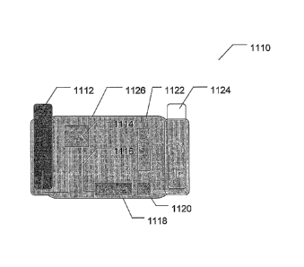
Figure 8 depicts an example module.
Figure 9 is a time series of images generated in accordance with a method of
the
present invention.
6
CA 02703687 2010-04-23
WO 2009/055587
PCT/US2008/080983
Figure 10 is a contour plot depicting the full time course of a detection
experiment
conducted in accordance with a method of the present invention.
Figure 11 is a graphical representation of the movement of an entity through a
conduit
developed in accordance with a method of the present invention.
Figure 12 is a graphical representation of a layered affinity column for use
with the
invention.
Figure 13 is a graphical representation of a layered affinity column
integrated with a
microcoil for use with the invention.
Fig. 14 is a schematic depicting a basic embodiment of an electrical LC
resonating
circuit according to the present disclosure, in which a tuning inductor is
connected in series
with a miniaturized or "micro" sample coil.
Fig. 14A is a schematic depicting the circuit shown in Fig. 14, except with
the single
sample coil replaced by a plurality of coils connected in series.
Fig. 14B is a schematic depicting the circuit shown in Fig. 14, except with
the single
sample coil replaced by a plurality of coils connected in parallel.
Fig. 15 is a schematic diagram indicating how an electrical circuit according
to the
present disclosure can be implemented in a magnetic resonance experiment.
Fig. 16 is a schematic diagram depicting another embodiment of an electrical
circuit
according to the present disclosure, in which the miniaturized sample coil is
connected in
parallel with the tuning inductor via wires or cables of a length
corresponding substantially to
a quarter wavelength of the resonant frequency.
Fig. 17 is a schematic diagram, similar to Fig. 16, of yet another alternative
embodiment of a circuit according to the present disclosure, showing the
transmission line
connected to and across a partial segment of the tuning inductor.
Fig. 18 is a schematic diagram, similar to Fig. 16, of yet another alternative
embodiment of a circuit according to the present disclosure, showing the
connection of an
impedance transformer to the transmission line, between the sample coil and
the tuning
inductor.
Fig. 19 is a schematic depicting a basic embodiment of an electrical LC series
resonating circuit according to the present disclosure, in which a tuning
inductor is connected
in series with a miniaturized or "micro" sample coil.
Fig. 20 is a schematic diagram elaborating somewhat on Fig. 14.
Fig. 21 is a schematic diagram elaborating somewhat on Fig. 16.
7
CA 02703687 2010-04-23
WO 2009/055587
PCT/US2008/080983
Detailed Description of the Invention
In a first aspect, the present invention provides modules, comprising:
(a) a microcoil possessing an inner diameter of between 25 microns and 550
microns;
(b) a conduit disposed proximate to the microcoil, wherein the conduit is
in fluid
communication with a sample reservoir;
(c) an affinity column in fluid communication with the conduit and the
sample
reservoir; and;
(d) a connector for connecting the module to a magnetic resonance detector.
In a second aspect, the present invention provides modules comprising:
(a) a plurality of microcoils each possessing an inner diameter of between
25
microns and 550 microns;
(b) a conduit disposed proximate to each microcoil, wherein the conduit is
in fluid
communication with a sample reservoir; and
(c) a connector for connecting the module to a magnetic resonance detector.
In a third aspect, the present invention provides microcoils comprising an
inner
diameter of between 25 microns and 550 microns, wherein the microcoil is an
effective
magnetic resonance transmitter or receiver coil; and wherein the microcoil is
within or
surrounds an affinity column.
In a fourth aspect, the present invention provides detection devices
comprising
(a) a permanent magnet possessing a field strength of less than or equal to
4
Tesla;
(b) a microcoil disposed proximate to a magnetic field generated by the
permanent magnet, wherein the microcoil possesses an inner diameter of between
25
microns and 550 microns;
(c) a conduit disposed proximate to the microcoil, wherein the conduit is
in fluid
communication with a sample reservoir;
(d) an affinity column in fluid communication with the conduit and the
sample
reservoir.
In a fifth aspect, the present invention provides detection devices comprising
(a) a permanent magnet possessing a field strength of less than or
equal to 4
Tesla;
8
CA 02703687 2010-04-23
WO 2009/055587
PCT/US2008/080983
(b) a plurality of microcoils each possessing an inner diameter of between
25
microns and 550 microns, wherein each microcoils is an effective magnetic
resonance
transmitter or receiver coil; and
(c) a conduit disposed proximate to each microcoil, wherein the conduit is
in fluid
communication with a sample reservoir.
In a sixth aspect, the present invention provides magnetic resonance
detectors,
comprising
(a) a housing comprising a conduit guide;
(b) a permanent magnet possessing a field strength of less than or equal to
4 Tesla
within the housing; and
(b) the module of any embodiment of any of aspect of the
invention, wherein the
connector connects the module to the housing via the conduit guide.
The modules, microcoils, detection devices and magnetic resonance detectors of
the
various aspects and embodiments of the present invention provide, for example,
increased
functionality and detection modes, improved sensitivity and specificity, time
and cost
savings, and a variety of other benefits including those described below.
All of the various embodiments for different components of the modules,
microcoils,
and detection devices of the first through sixth aspects of the invention are
capable of use
together; thus, any embodiment disclosed for one aspect can be combined with
any
embodiment for another aspect, as will be understood by those of skill in the
art.
The modules of the invention can be used, for example, to couple disposable
components of any embodiment detectors of the invention to permanent detector
components
by connecting the module to the detector via, for example, the conduit guide
as discussed
herein. In one example, all fluidic components of the detector are located on
the module,
thus reducing the probability that portions of a fluid sample will leak into
the permanent
detector components. Multiple modules may be used with a single detector, for
example, to
reduce the probability of contamination between detection experiments. In one
embodiment,
different sample fluids can be assigned their own modules which thus do not
come in contact
with other test samples. Removable modules also permit the conduit and
microcoil
characteristics to be adjusted based on the sample fluid used, or other
aspects of a detection
experiment. For example, a longer microcoil may be used in one experiment. In
another
example, a larger-diameter conduit may be used.
To provide structural support or make the module easier to handle, the module
may be
disposed on a surface, such as a card or board for example, or the removable
module may be
9
CA 02703687 2010-04-23
WO 2009/055587
PCT/US2008/080983
disposed in a housing. However, no support or housing means are necessary. For
example,
the module may comprise a section of a conduit with a solenoidal microcoil
wrapped around
a portion of the conduit. An exemplary module is provided in Figure 8.
In an example detector, the module can slide into the detector on conduit
guides that
place the conduit and the microcoil in a uniform region the field generated by
the permanent
magnet. The microcoil can be mounted directly on the conduit, and electrical
leads extending
from the microcoil can extend to electrical contact pads or connector on an
edge of the
module. Any other fluidic channels that are used in the course of the
detection experiment
can also be contained on the module. The conduit guides also place the module
in a selected
alignment with the magnetic gradient coil and a vacuum fluidic drive.
One embodiment of the detector includes a test box and a disposable module.
The
box may comprise the magnet, detection circuitry and interface, fluidic driver
and controls,
user interface, result printer, interface to the clinical data base, module ID
reader, conduit
guide, master processor system and software, and other associated power
supplies and
supporting electronics.
As used herein, an "affinity column" comprises any means to trap a target
entity
capable of separating biochemical mixtures based on specific interactions with
a target,
including but not limited to antigen-antibody interactions; enzyme-substrate
interactions; and
receptor-ligands interactions. The affinity column for use in the devices and
methods of the
invention may comprise the "stationary phase" (the specific substance, or
resin, used to
separate analytes) within the conduit, may comprise a separate affinity column
placed in the
conduit (the stationary phase and column hardware), or any other suitable
variations thereof
An affinity column may be used with the modules of the invention to trap a
target
entity and permit the target entity to be labeled with a labeling bead, such
as a magnetic label.
Any affinity column capable of separating biochemical mixtures based on
specific
interactions with a target can be used. For example, one or more affinity
columns may
comprise a capture agent to immobilize a target entity in a sample fluid as a
way to
concentrate the fluid prior to flowing the fluid through a detection zone of
the conduit. A
"capture agent" is any molecule capable of selectively binding, or being
derivatized to
selectively bind, a target of interest in the sample fluid. Suitable capture
agents include, but
are not limited to, proteins, nucleic acids, antibodies, lectins, enzymes,
mono-, or poly-
saccharides. The capture agent can be attached to the column packing material
using a linker
which binds to the column through a reversible binding reaction. For example,
a hexa-
histidine linker will bind to Ni or Co ions attached to a Ni-, or Co-
nitriloacetate-agarose
CA 02703687 2010-04-23
WO 2009/055587
PCT/US2008/080983
column matrix. The binding of the linker to the column can be reversed through
competition
with a release agent, such as histidine or imidazole, in the case of a hexa-
his linker. This
linker is covalently attached to the recognition molecule via standard
chemical methods.
This use of affinity columns provides a "pre-filter" for the detection device,
and can
be used to both concentrate the targets and remove excess unbound labeling
beads. For larger
samples, or samples suspected of having low concentrations of targeted
entities, the use of a
concentrating mechanism such as an affinity column may facilitate a reduction
in the sample
volume. For example, an environmental sample of 50 milliliters may be
suspected of
containing low concentrations of a target entity. To speed up the detection
process, an
affinity column with a labeled capture agent capable of selectively binding to
the target entity
may be used to isolate and hold the target entity while the remainder of the
sample is washed
away. After the extraneous portion of the sample is removed, and carrier fluid
can be passed
through the affinity column to carry the previously trapped target entities
into the sensitive
volume of the detector.
Alternatively, unlabeled target entities may be trapped by a capture agent on
a solid
phase, such as in an affinity column or other column known from
chromatography. Once
attached to the affinity column and immobilized, a solution of labeled beads
with attached
antibodies selective for the target entities may be introduced so that all of
the attached targets
are labeled with beads. The excess beads that do not label any target may then
be washed out
of the column. The targets, with their labels attached, may then be eluted
from the column
and this eluant can then be processed by the NMR detector.
In another embodiment, the affinity column comprises two or more layers (2, 3,
4, 5,
6, 7, 8, 9, 10, or more layers), and wherein each layer comprises a different
capture agent,
which can be used to trap different targets in different regions of space in
the column, which
can be detected differentially using multiple NMR detectors or MRI techniques,
as described
in more detail below. In one embodiment, overall magnetic resonance signal
changes are
first detected, and then MRI techniques are used to identify which of the
affinity column
layers is the source of the change.
In another embodiment, the microcoil is integrated into the affinity column
(ie: within
or surrounds the affinity column). Affinity columns can be made very small and
typically
contain substances that can produce NMR signals. Thus, a microcoil can be
built into (or
around) a column so that it can directly detect targets trapped inside; for
example, the
microcoil can be wound around the affinity column, or embedded in its walls. A
stationary
magnetic target can be detected many times, confirming the positive detection.
Different
11
CA 02703687 2010-04-23
WO 2009/055587
PCT/US2008/080983
protocols can be applied to a target of known location, improving detection
accuracy and
precision, allowing more definitive detection, differential detection, etc.
The integrated
detector coil eliminates the need for a separate elution step, which reduces
time, target
damage, stripping of magnetic labels from the targets, dilution of targets
into a larger elution
volume, spreading of targets through diffusive processes into a larger region
of sample
volume, etc.
The column material itself can be chosen so as to contribute to the NMR
signal,
including that part of the signal affected by the presence of a magnetic
target. For example,
the material can be a gel, or any other material that is stationary but also
provide a liquid-like
NMR signal. In one embodiment, the material can contain hydrogen or fluorine
atoms
sufficient to produce a detectible NMR signal. The material may also be a
solid material
containing aluminum or any other atom that produces a sufficiently liquid-like
NMR signal,
for example from the central transition of its quadrupole-perturbed NMR
spectrum. The
column material can be modified ("liquefied") so that it contributes to the
detected NMR
signal. For example, the column material may be initially a solid that
produces no usable
NMR signal, but can be transformed so that it produces a usable liquid-like
signal through the
action of chemicals or the changing of the temperature. In the extreme case,
the column
material may be dissolved through the use of an appropriate solvent. The
column material
can be chosen so that it does not alter the magnetic field being applied to
the sample space
(i.e., the column's stationary phase can be chosen so that it has the same
magnetic
susceptibility as the fluid). For example, the column material can be a glass,
ceramic, or gel
that contains an appropriate admixture of impurities, such as paramagnetic
ions (Cu, Mn,
Gd), so that the materials overall magnetic properties match those of the
sample fluid being
processed. The impurities to be mixed and their concentration can be chosen so
that the
presence of the column material does not degrade the homogeneity of the
magnetic field in
the column.
In another embodiment, the solid phase of the affinity column can chosen so
that it
can be dissolved and the column contents passed through the detector,
eliminating the need
for elution and allowing more of the column itself to contribute to the NMR
signal. For
example, the dissolving can be achieved through the use of a solvent chosen to
dissolve the
solid phase, or by changing the temperature of the column in a way that
renders the solid
phase a liquid (such as raising the temperature to melt the solid phase). The
dissolution need
not be complete and the solid phase may simple break apart in to pieces that
are small enough
to pass through the NMR detection coil.
12
CA 02703687 2010-04-23
WO 2009/055587
PCT/US2008/080983
In another embodiment, a plurality of different affinity columns can be used,
in
parallel, or more preferably in series, in order to attach a particular
pathogen type in each
separate column.
The detectors of the invention comprise a microcoil disposed proximate to the
magnetic field generated by the permanent magnet, wherein the microcoil
possesses an inner
diameter between 25 microns and 550 microns. The microcoil is an effective
magnetic
resonance transmitter or receiver coil. In various embodiments, the inner
diameter of the
microcoil can be between 25-500, 25-450, 25-400, 25-350, 25-300, 25-250, 25-
200; 50-550,
50-500, 50-450, 50-400, 50-350, 50-300, 50-250, 50-200, 100-550, 100-500, 100-
450, 100-
400, 100-350, 100-300, 100-250, 148-648, 170-555, and 100-200 microns. In an
example
detector, the microcoil is solenoidal in shape and can be wound around a
section of a conduit
that holds the volume of fluid during a detection experiment. However, other
coil shapes
may be used, including but not limited to planar coils, rectangular coils,
saddle coils, and
meanderline coils. For example, the cylindrical axis a flat or a solenoidal
microcoil may be
capable of being oriented perpendicularly to the axis of a conduit, and the
coil may be filled
with a material, for example but not limited to, ferrites, to enhance the
sensitivity of the coil.
Further, the microcoil may be formed through other construction techniques,
including but
not limited to depositing the coil material on a surface or etching the coil.
The length of the
microcoil can be selected such that the length of the microcoil is coextensive
with the
uniform region of the magnetic field generated by the permanent magnet of the
detection
devices of the invention. Other lengths may also be selected. In various
embodiments, the
length of the microcoil is between 25 gm ¨ 5 cm, 50 gm ¨ 5 cm, 75 gm ¨ 5 cm,
100 gm ¨ 5
cm, 100 gm ¨ 4 cm, 100 gm ¨ 3 cm, 100 gm ¨ 2 cm, 100 gm ¨ 1.5 cm, 100 gm ¨ 1
cm,
1.5mm ¨ 1.5 cm, 2.0 mm¨ 1.5 cm, 3 mm¨ 1.5 cm, 4 mm¨ 1.5 cm, 5 mm¨ 1.5, 6 mm¨
1.5
cm, 7 mm ¨ 2 cm, 8 mm ¨ 1.5 cm, and 9 mm ¨ 1 cm. Generally, the strength of
the signal
produced by a microcoil increases with the length of the coil. Some detectors
may include a
multiplicity of differently-sized microcoils. In an example detector, a first
microcoil with a
larger inner diameter is used to conduct an initial analysis of a sample. If
the presence of an
entity in a fluid is detected by the first microcoil, the fluid may be
diverted to be analyzed at a
higher sensitivity by a second microcoil with a smaller inner diameter.
As used herein, the microcoil being "disposed proximate to the magnetic field"
means
that at least a portion of the microcoil is located within the magnetic field
produced by the
permanent magnet. In an example detector, the entire coiled section of a
solenoidal microcoil
is placed within a magnetic field and oriented such that the magnetic field
observed at all
13
CA 02703687 2010-04-23
WO 2009/055587
PCT/US2008/080983
points on the coiled section of the solenoidal microcoil is uniform. However,
any orientation
of the microcoil within the magnetic field may be used, for example an
orientation that aligns
the microcoil with a non-zero component of the magnetic field in order to
employ MRI
techniques. For example, the microcoil may be angled with respect to the
direction of the
magnetic field, and portions of the microcoil, such as the ends or the
electrical leads from the
microcoil, may extend beyond the magnetic field.
In one embodiment, the microcoil is a closely wound microcoil, with a total
length
according to any embodiment of the invention, including ranges between 100
i_tm and 1500
[tm, 100 [tm and 1100 [tm, or any range in between. In another embodiment, the
microcoil or
closely wound microcoil is wound around a capillary with an outer diameter of
and/or an
inner diameter as discussed below. In another embodiment, the microcoil
comprises wire
with a diameter less than or equal to 2.5 times a skin-depth of the wire
material.
The microcoil is capable of being energized at a frequency that permits
detection of a
magnetic resonance within a volume of fluid within the conduit based on the
strength of the
magnetic field generated by the permanent magnet. The frequency that permits
detection of a
magnetic resonance within a volume of fluid varies with the strength of the
magnetic field
such that f = y'B, where fis the frequency, B is the magnetic field strength,
and y' is a
proportionality constant based on the nuclei examined within the fluid. For
example, y' for
the nuclei of hydrogen atoms is approximately 42.6 MHz / Tesla. In various
examples, the
frequency that permits detection of a magnetic resonance with in a volume of
fluid in the
conduit is between 1-100, 10-100, 20-100, 30-100, 40-100, 50-100, 60-100, 70-
100, 80-100,
90-100, 20-85, 30-85, 40-85, 50-85, 60-85, 70-85, 80-85, 20-65, 30-65, 40-65,
60-65, and 35-
45 MHz.
It is possible to provide for additional throughput by using two, or more,
microcoil
sections in parallel in the modules and/or detectors of the invention, in
order to enhance
throughput of sample fluids. This may be optimized by the use of MRI
techniques, such that
the signal from a particular coil may be discerned by the characteristics of
the signal, for
example frequency content of frequency characteristic. For example, magnetic
field
gradients (discussed below), applied at the appropriate angle oblique to the
microcoils,
separate the signals from each coil into its own unique frequency range,
allowing
simultaneous signal acquisition from all coils. Alternatively, or
additionally, each coil may
be electrically connected individually, and the signal processed in a separate
electronic
channel.
14
CA 02703687 2010-04-23
WO 2009/055587
PCT/US2008/080983
In any embodiment of any aspect of the present invention, the microcoil may be
part
of a resonant circuit, comprising:
(a) the microcoil;
(b) an auxiliary inductor coil electrically connected in series to the
microcoil; and
(c) a tuning capacitor electrically connected to the microcoil and the
auxiliary
inductor coil to form a resonant circuit.
In various preferred embodiments, the microcoil is a closely wound microcoil
and/or
comprises wire with a diameter less than or equal to 2.5 times a skin-depth of
the wire
material.
Resonant circuits of all aspects and embodiments of the present invention
allow for
the design and production of improved microcoil NMR devices that provide, for
example,
improved SNR and line width performance as well as increased functionality as
described in
detail herein, and thus greatly improve detection capabilities, and also allow
further
reductions in sample volume and further miniaturization of microcoil NMR
devices than was
possible in prior methods and devices. In various embodiment of the resonant
circuits, the
microcoil or closely wound microcoil has a total length as disclosed in any of
the
embodiments herein. In another embodiment of the resonant circuits, the
microcoil or closely
wound microcoil is wound around a capillary with an outer diameter and/or an
inner diameter
as disclosed in any of the embodiments herein. In a further embodiment of the
detector
aspects of the invention, the auxiliary or tuning inductor comprises wire of a
larger diameter
than a diameter of the microcoil or closely wound microcoil wire. In a still
further
embodiment of the resonant circuits of the invention, the auxiliary or tuning
inductor coil has
a radius of 0.3 cm to 0.6 cm. In another embodiment, the resonant circuits are
mounted on a
mechanical support; when mounted on a mechanical support, the resonant circuit
may be
installed in one or more shielded probe bodies. In one embodiment, the one or
more shielded
probe bodies comprise two shielded probe bodies, wherein a first shielded
probe body shields
the closely wound microcoil, wherein a second shielded body shields the
shielded probe body
and/or shields the auxiliary inductor and the capacitor. Each of these
embodiments can be
used in combination with other embodiments of all the aspects of the
invention, as well as
further aspects of the invention that involve use of the resonant circuits.
Any conduit capable of receiving a fluid sample may be used, including but not
limited to a capillary tube. In one embodiment, the conduit is hollow and
cylindrical in
shape, with the inner and outer diameters sized to accommodate picoliter-
microliter volumes
within the sensitive volume of the detector. The inner diameter of the conduit
may be
CA 02703687 2010-04-23
WO 2009/055587
PCT/US2008/080983
selected based on the properties of the fluid used in a detection experiment,
as well as other
parameters of a detection experiment such as the strength of the signal
produced by a fluid,
the flow rate of the fluid, and the desired resolution of the experiment, for
example. The
conduit has an inner diameter between 25 and 550 microns. In various
embodiments, the
conduit inner diameter can be between 25-500, 25-450, 25-400, 25-350, 25-300,
25-250, 25-
200; 50-550, 50-500, 50-450, 50-400, 50-350, 50-300, 50-250, 50-200, 100-550,
100-500,
100-450, 100-400, 100-350, 100-300, 100-250, 170-550, and 100-200 microns. The
outer
diameters can be any that may suitably be used with conduits of the inner
diameters disclosed
herein. Further, in embodiments where the microcoil is used to retain the
fluid (microcoil
forms the conduit), the wall thickness of the conduit may be reduced to zero.
The efficiency
of the detector may be improved by reducing the difference between the inner
and outer
diameters of the conduit. An example conduit is a capillary tube with an inner
diameter of
100 microns and an outer diameter of 170 microns. Conduits conforming to
different shapes
may also be used, including but not limited to elliptical conduits. Further,
the conduit may
comprise multiple sections, and may include removable sections. Removable
sections, for
example, may facilitate a reduction in the probability of a contamination of a
sample or may
permit the device to be more readily cleaned or repaired. The conduit itself
may be disposed
on the conduit guide, either directly placed thereon or indirectly with
another component
serving to guide the conduit into the conduit guide. In one embodiment, the
conduit is
disposed on a module, discussed in more detail herein, that the conduit guide
is capable of
receiving.
The conduit can receive fluid from any suitable component, including but not
limited
to a reservoir for providing fluid to the conduit. Such a reservoir can be on
board the device
or on the module discussed herein. The reservoir can simply be a component of
the conduit
in which a valve is placed to control flow from the reservoir to the portion
of the conduit used
for analysis.
As used herein, the conduit being "disposed proximate to the microcoil" means
any
position from which a signal transmitted from the microcoil can reach the
conduit and the
corresponding energy released from the fluid in the sensitive volume of the
conduit can
induce an electrical current in the microcoil. In an example embodiment, a
solenoidal
microcoil is wrapped around the conduit, such that the axis of the microcoil
is parallel with
the axis of the conduit. In another example, a planar coil is located
immediately adjacent to
the conduit and oriented such that the center axis of the planar coil is
perpendicular with the
center axis of the conduit.
16
CA 02703687 2010-04-23
WO 2009/055587
PCT/US2008/080983
Further, the conduit may comprise a plurality (ie: 2, 3, 4, 5, 6, 7, 8, 9, 10,
50, 100, or
more) of branches capable of receiving a volume of fluid. The plurality of
branches may be
disposed proximate to a plurality of microcoils. In one embodiment, each
branch is disposed
proximate to a separate microcoil, where each branch and each microcoil may be
the same or
have different sizes as deemed appropriate for the specific use. For example,
the plurality of
branches may permit the division of a fluid sample into multiple subsamples,
or may permit
assaying of more than one fluid sample at a time. In a further embodiment, one
or more
branches of the conduit are used in other fluidic processes. For example, one
or more
branches may be fluidically coupled to one or more affinity columns. In
experiments that use
labeling beads to aide in the identification of entities in a fluid, different
labels may be added
to each of the subsamples. The plurality of branches may also be coupled to
valves,
sequestration chambers, and/or other fluidic structures as suitable for a
given purpose. Such
fluidic components can be "on board" the detector, or may be provided via a
removable
module, such as one that can be connected to the conduit guide.
The detectors of any embodiment of any aspect of the present invention may
comprise
a conduit guide capable of receiving a conduit for receiving fluid, wherein
the conduit guide
is capable of disposing the conduit proximate to the microcoil and proximate
to the magnetic
field generated by the permanent magnet (and proximate to the magnetic
gradient when the
detector is in use). The conduit guide may comprise any means for orienting
the conduit,
including but not limited to mounting brackets, mechanical guides, and
couplings. The
conduit guide can be made from any material or materials that are capable of
establishing and
maintaining the position of the conduit guide. For example, metals, plastics,
composite
materials, ceramics, and multi-layer materials can be used individually or in
combination to
form the conduit guide. In an example detector, the conduit guide also aligns
the axis of the
portion of the conduit that holds the sample fluid (sensitive volume) during
an experiment
with the direction of the magnetic gradient produced by the magnetic gradient
generator. In
another example detector, the conduit guide is adjustable to permit an
operator to select a
position of the conduit relative to the magnetic gradient that minimizes a
frequency shift in a
signal emitted by the sensitive volume when the magnetic gradient is
energized. In
embodiments that utilize a coil as a magnetic gradient generator, the conduit
guide may align
the long axis of a cylindrical conduit with the center of the gradient coil.
However, the
conduit guide may dispose the conduit in other positions and orientations as
deemed
appropriate by an operator.
17
CA 02703687 2010-04-23
WO 2009/055587
PCT/US2008/080983
The module comprises a connector for connecting the module to a detector, such
as
those disclosed herein. Any connector capable of coupling the module to a
detector may be
used. For example, any of the conduit guides described as part of the detector
or method
aspects of the present invention may be used. Further, any mechanical coupling
capable of
securing the module in place may be used. For example, a connector where
threaded screws
or bolts on the module were coupled to corresponding threaded holes on the
detector may be
used. Other example connectors include mechanical clips, snap fittings,
mortise-and-tenon
connections, pins, socket fittings, and compression fittings.
The module may further comprise electrical contacts capable of establishing an
electrical connection between the microcoil or the module and the detector of
any
embodiment of the invention. The electrical connection between the removable
module and
the detector may permit the microcoil to interface with a tuning circuit,
signal processor, or
any other circuitry on a detector, such as those disclosed herein. In
removable modules that
contain electronics such as signal generators, signal processors, tuning
circuits and other
electronics on the removable module, the electrical connection between the
removable
module and the detector may permit any of the electronic components on the
removable
module to interface with electrical components within the detector. For
example, the
electrical connection can be used to supply power to the removable module or
allow the
removable module to connect to a user interface. Any means of establishing an
electrical
connection may be used. For example, wire leads coupled to the microcoil may
extend from
the module. In embodiments where the microcoil is electrically connected to
the module,
conductive traces may establish a connection between the microcoil and an
electrical
coupling on the module, which may be inserted into a receptacle on a detector.
The module may further comprise a fluidic drive fluidically coupled to the
conduit.
Any suitable fluidic drive may be used with the module. The modules and
detectors of any
embodiment of any aspect of the invention may further comprise a fluidic
drive, capable of
being fluidically coupled to the conduit. The fluidic drive may permit the
purposeful diving
of a fluid in the conduit. Typically, the fluidic drive operates by applying a
change in the
pressure on one end of the conduit. For example, a vacuum may be attached to
one end of
the conduit to draw the fluid through a portion of the conduit. A positive
displacement pump,
such as a syringe pump may also be used to establish fluid flow. The fluid
drive may also use
air pressure or gravity to drive the fluid. Any device that is capable of
imparting a flow to a
fluid in the conduit may be used. The fluidic drive can be "on board" the
detector, or may be
18
CA 02703687 2010-04-23
WO 2009/055587
PCT/US2008/080983
provided via a removable module, such as one that can be connected to the
conduit guide
(described in more detail herein).
Other fluidic components that may be fluidically coupled to the conduit, such
as
valves, sequestration chambers, and affinity columns may also be included on
the removable
module and/or the detector. Any valve that is capable of being fluidically
coupled to a
portion of the conduit may be used with the modules of the invention. A valve
may allow the
flow of a fluid in the conduit to be controlled. For example, in modules with
conduits that
contain a plurality of branches, one or more valves may be used to sequence
the flow of a
fluid through the plurality of branches. A sequestration chamber (which may
be, for
example, a well on a microplate, a separate conduit branch, a reservoir, etc.)
may be used to
hold a portion of a fluid used in a detection experiment. For example, a valve
may be used to
divert a portion of the fluid into a sequestration chamber if an entity is
detected in the fluid.
Any volume sequestration chamber may be used. For example, a small volume of a
few
nanoliters may be enough to allow for a subsequent microscopic evaluation of
the fluid in
which target is detected. In another example, several microliters, or even the
entire volume
of a sample fluid may be held in the sequestration chamber.
The detectors of the invention comprise a permanent magnet. The permanent
magnet
possesses a field strength, wherein the field strength is less than or equal
to 4 Tesla. In
various embodiments, the field strength may be between 0.1-4, 0.1-3.8, 0.1-
3.6, 0.1-3.4, 0.1-
3.2, 0.1-3.0, 0.1-2.8, 0.1-2.6, 0.1-2.4, 0.1-2.2, 0.1-2, 0.1-1.9, 0.1-1.8, 0.1-
1.7, 0.1-1.6, 0.1-1.5,
0.1-1.4, 0.1-1.3, 0.1-1.2, 0.1-1.1, 0.1-1.0, 0.25-4.0, 0.25-3.5, 0.25-3.0,
0.25-2.5, 0.25-2, 0.25-
1.9, 0.25-1.8, 0.25-1.7, 0.25-1.6, 0.25-1.5, 0.25-1.4, 0.25-1.3, 0.25-1.2,
0.25-1.1, 0.25-1.0,
0.5-2, 0.5-1.9, 0.5-1.8, 0.5-1.7, 0.5-1.6, 0.5-1.5, 0.5-1.4, 0.5-1.3, 0.5-1.2,
0.5-1.1, and 0.5-1.0
Tesla. The permanent magnet may be constructed out of a single magnet, or a
plurality of
permanent magnets may be combined. Further, any materials used in the
construction of
permanent magnets may be used to form the permanent magnet for the detector.
For
example, iron, other ferrous and non-ferrous alloys, ceramic magnetic
materials, rare earth
magnets including SmCo and NdFeB, and other magnetic materials may be used.
The
permanent magnet may also be formed into any shape. For example, magnets (or
combinations of magnets) with curved, rectangular, cylindrical, or other
profiles may be used.
In an example detector, a dipole magnet with steel pole pieces is used as the
permanent
magnet. However, other magnets, such as Halbach magnets may also be used. In
an example
detector, the magnetic field produced by the permanent magnet is uniform.
However,
permanent magnets that form magnetic fields possessing gradients, such as the
static
19
CA 02703687 2010-04-23
WO 2009/055587
PCT/US2008/080983
gradients sometimes encountered when constructing magnets, may also be used in
the
detector. The slope of the gradient that may be present from the permanent
magnet may
range between 0 G/cm and 1.0 G/cm. In embodiments where the permanent magnet
possesses a gradient, the gradient generated by the magnetic gradient
generator differs in
strength of type (for example, pulsed vs. static). The detector may also
comprise a
multiplicity of permanent magnets with a multiplicity of magnetic fields. In
an example
detector, two permanent magnets may be used, the first with a magnetic field
strength of 2.0
Tesla, and the second with a magnetic field strength of 1.0 Tesla. The use of
different
magnetic field strengths during the course of a detection experiment may
facilitate the
detection of a variety of different entities with a fluid sample. Further,
since the resonant
frequency of the nuclei in a fluid varies with the field strength, the use of
detectors with a
multiplicity of field strengths may further reduce the likelihood of false
positive or false
negative detections by analyzing a fluid at more than one field strength and
frequency.
In one further embodiment of any other embodiment of the invention, the
module/detection device further comprises an NMR imaging system. Such imaging
systems
are known to those of skill in the art. In another embodiment, the
module/detection device
further comprises a matching capacitor in electrical connection to the
resonant circuit, which
connects the resonant circuit to the detection electronics of the device (ie:
the NMR detection
system).
In an embodiment of all aspects of embodiments of the invention, the detection
devices disclosed herein may also comprise magnetic gradient generators and
other
components, such as a signal processor, to form a microcoil NMR-MRI system. In
this
embodiment, the microcoil(s) is/are disposed proximate to the magnetic
gradient. The
detectors of this embodiment comprise a magnetic gradient generator. The
gradient is used to
differentiate between a plurality of signal detection volumes within a
microcoil. The gradient
may also be used to compensate for a gradient in the magnetic field generated
by the
permanent magnet. Any magnetic gradient generator capable of applying a
magnetic
gradient to the magnetic field generated by the permanent magnet may be used,
including but
not limited to permanent magnets, superconducting electromagnets, or gradient
coils. In an
example detector, the magnetic gradient is linear, though any gradient may be
used,
including, but not limited to non-linear gradients. The gradient generator may
generate
gradients with slopes between 0.01 G/cm and 1.0 G/cm. In various embodiments,
the slope
of a localized area of the gradient generated by the magnetic gradient
generator is between
0.01 ¨ 1.0, 0.015 ¨ 1.0, 0.02 ¨ 1.0, 0.02 ¨ 0.9, 0.02 ¨ 0.8 0.02-0.7, 0.02-
0.65, and 0.02-0.6
CA 02703687 2010-04-23
WO 2009/055587
PCT/US2008/080983
G/cm. As discussed herein, in embodiments where the permanent magnet possesses
a
gradient, the gradient generated by the magnetic gradient generator differs in
strength of type
(for example, pulsed vs. static). A static gradient is used in an example
detector because
static gradients are particularly compatible with miniaturized NMR and MRI
platforms, and
are relatively simple compared to other gradients. However, other gradient
types may be
used, including but not limited to pulsed gradients and combinations of pulsed
and static
gradients. The strength of the gradient used in a detector determines the
spatial resolution of
the detector. Increasing the strength of the magnetic gradient permits the
detector to identify
magnetic resonances in smaller sections of the microcoil. In an example
detector, a magnetic
gradient of 0.07 G/mm is used in conjunction with a microcoil that is 1.1mm in
length. Using
the weakest gradient that still provides the desired spatial resolution may
facilitate more
narrow detection bandwidths and improved signal-to-noise characteristics of
the detector. In
an example detector, a magnetic gradient is selected based on the T2* of the
sample fluid,
without changing the center frequency of the energy emitted by the sample
fluid during a
detection experiment is used. In an example detector, a first magnetic
gradient of
approximately 0.14 G/mm may be applied during a first analysis of a sample. If
the results of
the analysis are inconclusive, or if improved signal-to-noise characteristics
are desired, a
second gradient of 0.07 G/mm may be applied during a second analysis of the
same sample.
The detectors of this embodiment may further comprise a signal processor
electrically
coupled to the microcoil and capable of identifying a plurality of frequency
components and a
plurality of magnitude components within the signal received from the
microcoil, and capable
of correlating the plurality of magnitude components and plurality of
frequency components
to a presence or absence of an entity in a volume of fluid at a plurality of
locations along an
axial length of the conduit. The signal processor may use any method for
identifying
frequency components and magnitude components within a signal. In an example
detector,
the signal processor is capable of performing a fast Fourier transformation on
the signal
received from the microcoil to identify a plurality of frequency components
and a plurality of
magnitude components within the signal from the microcoil.
As used herein, the microcoil being "disposed proximate to the magnetic
gradient"
means that at least a portion of the microcoil is placed within the magnetic
gradient generated
by the magnetic gradient generator. In an example detector, the microcoil is
located within
the magnetic gradient and oriented such that the axis of the microcoil is
aligned to be parallel
to the direction of a linear magnetic gradient. However, any orientation of
the microcoil with
respect to the gradient that aligns the microcoil with a non-zero component of
the magnetic
21
CA 02703687 2015-11-23
gradient may be used. As will be understood by those of skill in the art based
on the
teachings herein, disposition of the microcoil proximate to the gradient and
proximate to the
magnetic field arc separate variables in design of the detectors of the
invention.
The modules and detectors of any embodiment of any aspect of thc invention may
further comprise a tuning circuit electrically coupled to the microcoil. The
tuning circuit
comprises a tuning coil capabl.c of having an inductance at least two times
larger than the
inductance of the microcoil, and a capacitor coupled to the tuning coil to
form a resonant
circuit. In various embodiments, the tuning coil may have an inductance that
is 3, 4, 5, 6, 7,
8, 9, 10, 20, 25, 50, 100, 250, 500, or 1000 times larger than the inductance
of the microcoil.
The tuning circuit comprises a tuning coil that possesses an inductance of at
least 2 nH, and
the tuning coil is coupled to a capacitor to form a resonant circuit. In
various embodiments,
the inductor may have an inductance between 2 itH ¨ .1 ttH, 10 nH ¨ 1 1.tt4,
50 nH ¨ 1 uH,
100 nH ¨ 1 tH, 200 nH ¨ 1 i.tH and 500 nH u1-1. The tuning coil may be
coupled to the
microcoil to form a series or a parallel connection with the microcoil. Such
tuning coils can
be "on board" the detector, may be provided via a removable module, such as
one that can be
connected to thc conduit guide, or a combination thereof. Any method of
coupling the
microcoii to the tuning coil may be used, including but not limited to a
transmission line
between the microcoil and the tuning coil. An example tuning circuit is
disclosed in U.S.
Patent No. 7,405,567. Such tuning coils can be "on board" thc detector, may be
provided via
a removable module, such as onc that can be connected to the conduit guide, or
a
combination thereof.
The tuning coil, which may also be referred to as an auxiliary inductor or a
tuning
inductor may be designed in accordance with a method comprising:
(a) preparing a microcoil with wire of a first diameter;
(b) determining an RF resistance of the microcoil;
(c) winding an auxiliary inductor coil with wire of a second
diameter, where the
second diameter is greater than the first diameter, and wherein a radius of
the auxiliary
inductor coil is determined using the formula:
rcoii
467r
where /,,,i,.õ is the wire length, is the wirc dia.meter, and kcl or, is
thc turn-to-turn. wirc
spacing.
22
CA 02703687 2010-04-23
WO 2009/055587
PCT/US2008/080983
The detectors of any embodiment of any aspect of the present invention may
further
comprise a signal processor electrically coupled to the microcoil and capable
of identifying a
plurality of frequency components and a plurality of magnitude components
within the signal
received from the microcoil, and capable of correlating the plurality of
magnitude
components and plurality of frequency components to a presence or absence of
an entity in a
volume of fluid at a plurality of locations along an axial length of the
conduit. The signal
processor may use any method for identifying frequency components and
magnitude
components within a signal. In an example detector, the signal processor is
capable of
performing a fast Fourier transformation on the signal received from the
microcoil to identify
a plurality of frequency components and a plurality of magnitude components
within the
signal from the microcoil. The signal processor may comprise the computer
programs
disclosed herein.
In a further embodiment, the modules/detector may comprise physical computer
readable storage media, for automatically carrying out the methods of the
invention on a
detector, such as those disclosed herein. As used herein the term "computer
readable
medium" includes magnetic disks, optical disks, organic memory, and any other
volatile (e.g.,
Random Access Memory ("RAM")) or non-volatile (e.g., Read-Only Memory ("ROM"))
mass storage system readable by the CPU. The computer readable medium includes
cooperating or interconnected computer readable medium, which exist
exclusively on the
processing system or be distributed among multiple interconnected processing
systems that
may be local or remote to the processing system.
In a seventh aspect, the present invention provides methods for detecting a
target is a
sample fluid, comprising:
(a) flowing a fluid containing one or more magnetically labeled targets
through
the conduit of the detection device of any embodiment of any aspect of the
invention;
(b) energizing the microcoil at a frequency that permits detection of a
magnetic
resonance within the sample fluid; and
(c) processing a signal received from the microcoil to detect the
magnetically
labeled targets in the sample fluid.
The methods of the present invention allow direct detection of single cells,
molecules,
etc. in a given sample volume. The methods comprise moving a sample fluid
through a
detector and determining if an object of interest goes through, which are
counted. Prior art
detectors are sensitive to minimum concentrations of objects. It is especially
unusual to use
23
CA 02703687 2010-04-23
WO 2009/055587
PCT/US2008/080983
NMR to detect single objects, as traditional NMR detection techniques are low-
sensitivity
and require target numbers many times larger than a single cell or molecule.
The methods of the invention comprise energizing the microcoil at a frequency
that
permits detection of a magnetic resonance within the fluid. The microcoil is
used to transmit
a pulsed electromagnetic signal at a selected resonant frequency towards the
fluid in the
conduit. The frequency of the transmitted signal is selected based on the
properties of the
particular nuclei that are being examined in the detection experiment and the
strength of the
magnetic field. The microcoil is also used to detect the energy that is
absorbed or released by
the nuclei in the magnetic field in response to the transmission of the
resonant frequency
signal. This energy induces an electrical current in the microcoil that
corresponds with the
energy absorbed or released by the nuclei in the flowing fluid. The presence
of an entity
within the fluid causes a change in energy that can be detected by the
microcoil.
Such forms of contrast can arise also from any other interactions of the
labeled entity
and the surrounding fluid. The general form of this interaction is due to the
mismatch in
magnetic property called susceptibility, between the entity and the carrier
solution. Thus, in
principle, the device being disclosed can also be sensitive to almost any
distinct object
including even bubbles. However, this effect scales with the strength of the
magnetic field so
it is not a large effect in our relatively low field permanent magnets
especially compared to
the magnetic particles that are chosen because of their strong magnetic
strength, referred to as
magnetic moments.
When the distortion in the uniform magnetic field in the vicinity of the
object is a
result of ferromagnetism or "super-paramagnetism" of the object itself, the
object is
responding non-linearly to the uniform magnetic field, and hence it is not
described as having
a magnetic susceptibility. Instead, the object is described as having a
magnetic moment,
which may or may not be due to a saturation magnetization of the object. This
magnetic
particle disrupts the field homogeneity in its neighborhood, and this
disruption may be
detected by NMR methods.
The object disturbing the magnetic field in its vicinity may be much too small
to be
detected directly by NMR. The field disturbance may be effective over a volume
very much
larger than the object itself If the volume of material affected by the object
is large enough,
then the presence of the object can be detected by NMR. In this sense, the
magnetic field
disturbance serves to amplify the NMR signal indicating the presence of the
object.
The most basic form of NMR contrast is to detect whether something is present
in the
sample region or not. Using the methods of the present invention when a dilute
solution
24
CA 02703687 2010-04-23
WO 2009/055587
PCT/US2008/080983
containing labeled entities, for example, bacteria, is passed through the NMR
detector, the
labeled entities can be detected individually. The magnetic beads that label
specific targets
affect the NMR signal from the surrounding fluid adversely. Therefore, if the
"sphere of
influence" of the magnetic particles is comparable to the volume being
examined, i.e., the
__ "sphere" should span a significant fraction of the tube through which the
solution is flowing,
the NMR signal that arises from the total cross-section of the sample tube are
attenuated.
Protocols for excitation and detection may also be devised to highlight other
properties of the
sample fluids. Fluids may differ in how long they produce signals in response
to an
excitation, for example. These differences in "relaxation time" may be
exploited to make a
__ differential detection of the two (or more) fluids in a mixture. There are
several different
relaxation times, each appropriate to a particular method for exciting and
detecting the atomic
nuclei, and each related to different chemical or physical properties (or
combination of
properties). The most commonly discussed relation times are denoted T1, T2,
and T2*. Many
other parameters, including the diffusion tensor, flow velocity, elastic
modulus, chemical
__ environment, and temperature, can be encoded into the detected signal in an
NMR
experiment. It is also possible to combine the detection of more than one
physical parameter
into the same NMR experiment. It is common, for example, to combine both T2
detection
and spatial location detection into a "T2-weighted image." In this case, the
detected signal is
affected by both the amount of material located at every position in space and
the T2 of the
__ material located there.
In an eighth aspect, the present invention provides methods for detecting a
target in a
sample fluid, comprising:
(a) introducing a sample fluid into a sample reservoir, wherein
the sample
reservoir is in fluid communication with a conduit;
(b) flowing the sample fluid into an affinity column in the conduit,
wherein the
affinity column comprises one or more capture agents that bind to one or more
targets of
interest;
(c) flowing a fluid comprising magnetic particles into the affinity column,
wherein the magnetic particles are capable of binding selectively to the one
or more targets of
__ interest bound to the affinity column via the one or more capture agents,
and wherein binding
of the magnetic particles to the targets produces magnetic particle-target
complexes;
(d) washing the affinity column to reduce the number unbound magnetic
particles;
(e) eluting bound magnetic particle-target complexes from the affinity
column;
CA 02703687 2010-04-23
WO 2009/055587
PCT/US2008/080983
(0 flowing the fluid comprising the magnetic particle-target
complexes through a
conduit disposed proximate to a microcoil, wherein the microcoil possesses an
inner diameter
of between 25 microns and 550 microns, wherein the microcoil is an effective
magnetic
resonance transmitter or receiver coil; and
(g) energizing the microcoil at a frequency that permits detection of a
magnetic
resonance within the sample fluid; and
(h) processing a signal received from the microcoil to detect
magnetic particle-
target complexes in the sample fluid.
The use of affinity columns as a filtering step before NMR detection can be
used to
concentrate the targets and/or remove the excess unbound labels. The sample
fluid is
pumped through a column in which a stationary phase of an affinity column is
placed. The
stationary phase is functionalized with one or more capture agents to
preferentially trap the
targets of interest. The sample fluid may be circulated through the column
numerous times if
useful in trapping all the targets. Once the column is loaded with one or more
target types, a
fluid containing the magnetic labels is flowed into the column. These labels
are chosen to
selectively bind only to the target(s). The labeling fluid may be
recirculated. Once the
targets are labeled, the column is flushed to remove all magnetic labels that
are not attached
to targets. Then the column is eluted in a manner that released the bond
between the targets
and the stationary phase, without disturbing the bond between the targets and
the magnetic
labels. The elutant thus will contain labeled targets, a reduced number of
excess magnetic
labels, and the total volume is substantially less than the original sample
(ie: the targets will
have been concentrated.) The elutant will then be passed though the NMR
detector, which
will count the magnetically labeled targets. Even if all the excess beads are
not removed, a
reduction in their numbers is very useful. In another embodiment, the
stationary phase of the
column can be liquefied and could pass through the NMR detector. For example,
a solvent
appropriate for the column's solid phase material would dissolve the solid
phase. The
stationary phase can be released (via the removal or induced failure of the
frit on the output
side of the column) and pass through the NMR detector. For example, if the
solid phase is
made up very fine particles that can pass through the detector, they can be
held in place in the
column by a grate-like structure, called a frit, which constrains them
mechanically to stay
within the column. Once the column is loaded, the frit is released, and the
now unconstrained
particles flow through the detector, along with any targets bound thereto.
Concentration of target entities on the affinity column allows processing of
larger
amounts of biological, or other fluids of interest, such as environmental
water, industrial
26
CA 02703687 2010-04-23
WO 2009/055587
PCT/US2008/080983
wastes, process water, or water used to wash foodstuffs, such as vegetables.
The column can
accommodate much higher flow rates than are used in the detection step. It may
not be
otherwise practical to process large sample fluid volumes directly through the
microcoil
because it is understood that the high sensitivity of the method partly
derives from the use of
very small volume microcoils. The coil volume may be measured in, for example,
nanoliters
(10-9 Liter). However, with the use of an affinity column with a volume of,
for example,
microliters (10-6 Liter) one thousand times as much fluid can be processed in
order to
concentrate and trap targets in the same amount of time required to process
the microcoil
volume. This may serve to shorten the analysis cycle markedly; for example in
this case by
1000-fold. It also may reduce the amount of non-specific material introduced
into the NMR
detector so that the rate of false-positive results may be reduced. The use of
an affinity
column may increase the sensitivity of the detection method. For example, if
one is wishing
to detect a single bacterial cell in 10-2 Liter of whole human blood, if the
flow rate is limited,
for example to 10-6 Liter per minute, it would require 10,000 seconds, or
almost 3 hours to
process a single blood sample. However, with a 10-5 Liter affinity column in
place, the flow
may be increased to 10-3 Liter per minute with the result that the blood
sample may be loaded
onto the column in only 10 seconds. Then, the column can be eluted, and only
the fraction
known to contain the void-volume peak (containing the target of interest) need
be sent into
the NMR coil. The column also serves to provide a surface onto which the
specific
biochemistry needed to assemble the unique target complex can take place.
Material
specifically eluted from the column by the release agent (imidazole, glucose,
pH, ionic
strength, etc.) then enters the detection zone and provides a specific signal
characteristic of
the target sought.
In a ninth aspect, the present invention provides methods for detecting a
target is a
sample fluid, comprising:
(a) introducing a sample fluid into a sample reservoirõ wherein the sample
reservoir is in fluid communication with a conduit, wherein the conduit is
disposed proximate
to a microcoil, wherein the microcoil possesses an inner diameter of between
25 microns and
550 microns, wherein the microcoil is an effective magnetic resonance
transmitter or receiver
coil;
(b) flowing the sample fluid into an affinity column in the conduit,
wherein the
affinity column comprises two or more layers, wherein each layer comprises one
or more
capture agents that bind to one or more targets of interest in the sample
fluid, wherein each
27
CA 02703687 2010-04-23
WO 2009/055587
PCT/US2008/080983
layer in the affinity column is capable of binding to molecules distinct from
other layers; and
wherein the affinity column is located at least partially within the
microcoil;
(c) flowing a fluid comprising magnetic particles into the affinity column,
wherein the magnetic particles are capable of binding selectively to the one
or more targets of
interest, and wherein binding of the magnetic particles to the targets
produces magnetic
particle-target complexes;
(d) energizing the microcoil at a frequency that permits detection of a
magnetic
resonance within the affinity column; and
(e) processing a signal received from the microcoil to detect magnetic
particle-
target complexes in one or more layers of the affinity column.
In a tenth aspect, the present invention comprises methods for detecting a
sample in a
target fluid, comprising:
(a) mixing a sample fluid with magnetic particles capable of binding to the
one or
more targets of interest in the sample fluid, wherein binding of magnetic
particles to targets
produces magnetic particle-target complexes
(b) introducing the sample fluid into a sample reservoir, wherein the
sample
reservoir is in fluid communication with a conduit, wherein the conduit is
disposed proximate
to a microcoil, wherein the microcoil possesses an inner diameter of between
25 microns and
550 microns;
(c) flowing the sample fluid into an affinity column in the conduit,
wherein the
affinity column comprises two or more layers, wherein each layer comprises one
or more
capture agents that bind to one or more targets of interest, wherein each
layer in the affinity
column is capable of binding to molecules distinct from other layers; and
wherein the affinity
column is located at least partially within the microcoil;
(d) energizing the microcoil at a frequency that permits detection of a
magnetic
resonance within the affinity column; and
(e) processing a signal received from the microcoil to detect
magnetic particle-
target complexes in one or more layers of the affinity column.
In these aspects, the methods identify target entities by location within a
multi-layer
affinity column. By trapping different targets in different regions of space,
they can be
differentially detected using multiple NMR detectors or MRI techniques. For
example, a
change in overall signal can first be detected, followed by the use of MRI
methods to identify
which of the layers is the source of the signal change. In one embodiment, the
microcoil is
integrated with the affinity column. Affinity columns can be made very small
and typically
28
CA 02703687 2010-04-23
WO 2009/055587
PCT/US2008/080983
contain substances that can produce NMR signals. Hence, a microcoil can be
built into (or
around) a column and used to directly detect targets trapped inside. The coil
can be wound
around the column, or embedded in its walls.
In one embodiment, a fluid containing magnetically labeled targets can be made
to
flow through such a column, with the column functionalized with capture agents
to
preferentially trap the targets. This trapping can be chemical and/or physical
in nature. The
magnetism of the target will affect the detected NMR signal; many different
effects are
possible as described herein, and the NMR signal detection protocol is
optimized for each
one accordingly. In one embodiment, NMR detections might proceed while the
fluid is
flowing through the affinity column, or the fluid flow might be stopped once
the sample has
been loaded onto the affinity column. The NMR protocol (or data analysis
procedure) can be
designed to distinguish between stationary magnetic targets and moving
magnetic particles.
For example, the distinction can be based on two detections of position of the
target, where if
the position changed, the target is be identified as moving. In another
example, a flow-
compensated NMR pulse sequence (such as those used in MRI angiography) is used
to
distinguish between signals detected from moving and non-moving samples.
Techniques for
optimizing the detection of NMR signals from moving sample fluids may be
employed, for
example, by using flow compensating magnetic field gradient wave forms, or by
rapidly
repeated data acquisitions together with small flip angles optimized to give
optimal SNR.
The column material itself can be chosen so as to contribute to the NMR
signal, including
that part of the signal affected by the presence of a magnetic target. The
column material can
be modified ("liquefied") so that it contributes to the detected NMR signal.
The column
material can be chosen so that it does not alter the magnetic field being
applied to the sample
space (i.e., the column's stationary phase can be chosen so that it has the
same magnetic
susceptibility as the fluid). In another embodiment, the affinity column's
solid phase can be
dissolved so that the column's contents pass through the detection zone,
eliminating the need
for elution and allows more of the column itself to contribute to the NMR
signal (for column-
integrated detectors).
The use of these methods provides many advantages, in that stationary magnetic
target can be detected many times, confirming the positive detection.
Different protocols can
be applied to a target of known location, improving detection accuracy and
precision,
allowing more definitive detection, differential detection, etc. The
integrated detector coil
eliminates the need for a separate elution step, which may take too much time,
damage the
targets, strip the magnetic labels from the targets, cause the targets to be
diluted into a larger
29
CA 02703687 2010-04-23
WO 2009/055587
PCT/US2008/080983
elution volume, cause the targets to spread through diffusive processes into a
larger region of
sample volume, etc.
In an eleventh aspect, the present invention provides methods for detecting a
target in
a sample fluid, comprising:
(a) introducing a sample fluid into a sample reservoir, wherein the sample
reservoir is in fluid communication with a conduit, wherein the sample fluid
comprises
magnetic particles capable of binding selectively to one or more targets of
interest in the
sample fluid, and wherein binding of magnetic particles to targets produces
magnetic
particle-target complexes;
(b) flowing the sample fluid through the conduit disposed proximate to a
microcoil, wherein the microcoil possesses an inner diameter of between 25
microns and 550
microns;
(c) energizing the microcoil at a frequency that permits detection
of a magnetic
resonance within the sample fluid;
(d) processing a signal received from the microcoil to detect magnetic
particle-
target complexes in the sample fluid;
(e) flowing a portion of the flowing fluid in which the labeled
entity was detected
through a conduit disposed proximate to a secondary microcoil;
(0 energizing the secondary microcoil at a frequency that permits
detection of a
magnetic resonance within the sample fluid; and
(g) processing a signal received from the secondary microcoil to
determine a
further property of the magnetic particle-target complexes.
In order to enhance signal to noise (S/N), it is possible to have a two step
process,
whereby a fast detection process with poorer S/N is used to determine the
presence of any
pathogen, and then a slower detection process, with better S/N is used to
determine a signal
level with good discrete discrimination between magnetic bead levels used. The
methods of
this aspect of the invention use an initial detection step to trigger a
secondary measurement in
the same device. This can be done with or without sequestration. The
"secondary microcoil"
can be a different microcoil or the same microcoil where the sample fluid has
been
recirculated to it for measurement of a secondary property of the magnetic
particle-target
complex (ie: something other than detection). In one embodiment, the device is
operated in a
high through-put mode initially, with a relatively large volume of sample
being processed.
The initial detection process is optimized for sample throughput, making it
difficult to learn
much more than the simple presence of a target. Once a detection event has
occurred,
CA 02703687 2010-04-23
WO 2009/055587
PCT/US2008/080983
however, we can shift the focus of the device from merely detecting things to
learning more
about them, such as their identity. This is possible without changing the
label on the targets,
but by acquiring data in ways that give more resolution, have more precision,
etc. These
other data collection techniques are slower than the high-through put mode.
The second
measurements can be done, for example, in a separate microcoil. This separate
coil may have
characteristics that optimize signal to noise in addition to the reduced fluid
flow velocity in
the coil. The separate coil and detection process may employ different NMR
excitation and
detection techniques capable of making measurements or achieving measurement
accuracy
distinct from those possible in the initial detector coil.
In one embodiment, the separate microcoil is placed along the conduit flow
path
downstream from the initial microcoil. The fluid flow rate might be stopped or
slowed, or the
geometry of the flow can be changed (larger conduit diameter, e.g.) in a
manner appropriate
for enhanced, reduced-speed detections. The second step can also be done in
the first
microcoil, with the target brought back into that coil by flowing the sample
in the opposition
direction. The second step could occur in a "by line," a parallel flow path in
which the fluid
moves more slowly, or is temporarily stopped. The second step may comprise an
NMR
experiment protocol that is distinct from the initial protocol so that it
highlights a different
aspect of the target's signal.
Alternatively, the second "high S/R" detection process may be applied after
the entire
sample fluid has been processed in the initial step.
These second steps may comprise any useful measurement, including but not
limited
to precise measurements of relaxation times, and spectroscopic identification
of objects or
molecules including the identification of properties or features of the labels
that were not
discernable during the rare entity detection phase of the device's operation.
In a twelfth aspect, the present invention provides methods for detecting a
target in a
sample fluid, comprising:
(a) introducing a sample fluid into a sample reservoir, wherein the sample
reservoir is in fluid communication with a conduit, wherein the sample fluid
comprises
magnetic particles capable of binding selectively to one or more targets of
interest in the
sample fluid, and wherein binding of magnetic particles to targets produces
magnetic
particle-target complexes;
(b) flowing the sample fluid through the conduit disposed proximate to a
microcoil, wherein the microcoil possesses an inner diameter of between 25
microns and 550
microns;
31
CA 02703687 2010-04-23
WO 2009/055587
PCT/US2008/080983
(c) energizing the microcoil at a frequency that permits detection of a
magnetic
resonance within the sample fluid;
(d) processing a signal received from the microcoil to detect magnetic
particle-
target complexes in the sample fluid;
(e) diverting a portion of the flowing fluid in which the magnetic particle-
target
complexes were detected into a sequestration chamber to produce a concentrated
target
solution.
Since the methods of the present invention determine where in space a target
is, an
identified target can then be sequestered for further analysis. The detection
volume can be
very small (a few nanoliters, typically), and this volume can be confined in a
very narrow
conduit, and the sample fluid being measured can be made to flow through this
conduit. We
can, for example, use a detection event to trigger microfluidic valves that
will collect very
small, isolated sections of the flowing sample in a sequestration chamber, so
that the
collected sections contain the objects that were seen by the NMR detector.
Nearly all of the
background fluid flows through the conduit without being sequestered. The end
result is the
sequestration of the objects of interest into a very small total volume. That
is, a dramatic
enhancement of concentration of the targets, making further processing (either
in the NMR
device or in some subsequent device or procedure) much faster, easier, and
more effective.
Further in-device processing might be used to differentially identify targets,
suppress false
positives (especially those due to locally high background bead
concentrations, eg.), etc. In
one embodiment, sequestration can be allowed to complete during an initial
detection run,
with a subsequent processing of the sequestered sample, or the processing the
sequestered
material might commence immediately. In a further embodiment, sequestration
can be used
to reduce false positives from high background concentrations, by running the
sequestered
sample through the detection zone again (same or different microcoil).
In a thirteenth aspect, the present invention provides methods for detecting a
target in
a sample fluid, comprising:
(a) introducing a sample fluid into a sample reservoir, wherein
the sample
reservoir is in fluid communication with a conduit, wherein the sample fluid
comprises two
or more targets of interest, and wherein the sample fluid comprises magnetic
particles
differentially bound to the two or more targets of interest to create at least
a first magnetic
particle-target complex and a second magnetic particle-target complex;
32
CA 02703687 2010-04-23
WO 2009/055587
PCT/US2008/080983
(b) flowing the sample fluid through the conduit disposed proximate to a
microcoil, wherein the microcoil possesses an inner diameter of between 25
microns and 550
microns;
(c) energizing the microcoil at a frequency that permits detection of a
magnetic
resonance within the sample fluid; and
(d) processing a signal received from the microcoil to differentially
detect the at least first
magnetic particle-target complex and the second magnetic particle-target
complex in the
flowing fluid.
In this aspect, the methods permit detection and classification/identification
in a single
data acquisition step. We can differentially detect two or more unique targets
if we can
attach different amounts of magnetic material to each. All such labeled
targets are detected in
detection devices of the invention because they will all cause a decrease in
the detected NMR
signal. The more magnetic targets will cause a larger decrease in signal. We
can determine
the degree of signal decrease (or other modification of the signal) and use
this information to
determine which kind of target was detected.
This method requires different levels of magnetic labeling on the targets to
be
differentiated. These levels of labeling can be achieved in various ways,
including but not
limited to:
(a) Attaching the same number of beads (on average) of the same size to the
unique targets, where the beads for target A have more magnetic strength than
the
beads for target B. Different strength beads may be made from different
magnetic
materials, or contain different amounts of the same material (e.g. 60% Fe304
vs. 90%
Fe304);
(b) Attaching different sized beads to the different unique targets. This
results
in differential labeling because different numbers of beads are attached per
unit target
surface area, and the magnetic moment per bead is different for the different
sized
beads (e.g., beads containing the same fractional amount of the same magnetic
material will differ in magnetic strength due to the total magnetic content);
(c) Attaching a maximal number of the same beads to all of the unique targets,
where the targets themselves differ in total surface area. The unique targets
differ in
total magnetic moment because the total number of beads that can be
accommodated;
(d) Use the same beads for all targets where the particular capture agent
chosen, such antibodies, have different affinities for the different unique
targets,
resulting in a different (average) number of beads attached to each type of
target.
33
CA 02703687 2010-04-23
WO 2009/055587
PCT/US2008/080983
In one embodiment, the level of labeling is measured in a separate step from
the initial
detection. This second step might occur in the same fluid stream (e.g., in a
second microcoil
and detection volume optimized for level detections, or by slowing down the
flow
temporarily to improve level-precision, etc.) or may be in a parallel stream
or a parallel stop-
flow experiment. The level of labeling determination might be done in a
completely separate
apparatus, using sequestration techniques in the initial detector. A different
experimental
magnetic resonance protocol might be employed to differentiate between the
different types
or degrees of labeling.
We have generated data showing the different detection of clusters of small
numbers of
beads. This data demonstrate extraction of signal intensity differences using
the methods and
devices of the invention, and the ability to discern multiple levels of
"labeling."
The following further embodiments can be used with any embodiment/aspect of
the
invention.
To achieve stronger, more-sensitive, faster, or otherwise improved detection,
the
microcoil detector may be immersed in a material that matches the
susceptibility of the metal
used to wind the coil. Alternatively, or in addition, the wire of the coil may
be chosen or
treated such that its magnetic susceptibility matches its surroundings. The
material forming
the tube on which the coil is wound, or in which the coil is wound, and which
contains the
fluid sample may be chosen to match either the susceptibility of the fluid, or
of the wire, or in
any other way to best facilitate detection.
In one embodiment, the processing comprises processing a plurality of signals
received from the microcoil over time (ie: two or more time points), wherein
the processing
comprises identifying a plurality of frequency components and a plurality of
magnitude
components within each signal received from the microcoil, and correlating the
plurality of
magnitude components and plurality of frequency components within each signal
to a
presence or absence of a labeled entity in the flowing fluid at a plurality of
locations along an
axial length of the microcoil. In this embodiment, signals are processed from
two or more
time points (ie: 2, 3, 4, 5, 6, 7, 8, 9, 10, or more), permitting correlation
of signal data from
two or more different time points to identify an appropriate correlation
between signals,
confirming that the signal is not caused by background or other electrical
disturbances. For
example, a first signal is detected in one location and a second signal is
detected at a second
time point at an appropriate distance from the first signal, based on fluid
velocity and other
factors. Those of skill in the art will understand, based on the teachings
herein that
34
CA 02703687 2010-04-23
WO 2009/055587
PCT/US2008/080983
appropriate spacing between time points will depend on a variety of factors,
including but not
limited to microcoil size, fluid velocity and viscosity, conduit size, etc.
The methods of the present invention can be used, for example, in MRI and/or
NMR
detection of one or more labeled molecules ("entity") in a fluid; the methods
disclosed herein
provide dramatic reduction in susceptibility to false positive and false
negative detections.
The methods permit the detection of a labeled entity in a flowing fluid, and
also permit
multiplexing of the detection process, providing the reliability of repeated
single NMR
experiments in a single experiment. The methods also permit the presence and
movement of
a labeled entity within a sample volume to be observed and recorded. Entities
that can be
detected using the devices and methods of the invention, include, but are not
limited to, cells
(such as bacteria, fungi, other parasites, cancer cells, etc.), viruses,
proteins (including
antibodies), prions, nucleic acids, carbohydrates, lipids, small molecules,
antibiotics, toxins,
etc.
In various embodiments, detection via the methods of the invention can
comprise
detection, identification, and/or quantitation of entities in a sample fluid.
Any sample fluid of
interest can be used, including but not limited to bodily fluid samples
(blood, urine, saliva,
semen, vaginal secretions, tears, amniotic fluid, cerebral spinal fluid,
etc.), swab samples
from skin, wound, or other body sites; semi-solid samples, such as fecal
samples (processed
by appropriate sample dilution in a liquid), environmental water, industrial
wastes, process
water, liquid foodstuffs (including but not limited to milk, juice, drinking
water, soda, etc.) or
water used to wash foodstuffs such as vegetables and fruit. The size of the
sample volume
may vary depending on the properties of the fluid being analyzed, the desired
flow rate
through the sensitive volume, and the sizes of the conduit. The sensitive
volume of the
conduit (ie: the volume being analyzed) can vary depending on the size of the
sample
volume, the size of the conduit, the flow rate, the size of the microcoil, and
other factors as
determined by a user. In various embodiments, the sensitive volume ranges
between 100
picoliters and 50 microliters. In various other embodiments, the sensitive
volume is ranges
between 4-7, 2-10, 1-15, 0.5-20, 0.4-50, 0.3-75, 0.2-100, 0.1-150, 0.05-300,
0.04-500, 0.03-
1000, 0.02-2000, 0.01-5000 nanoliters. In an example detector, less than a
nanoliter of fluid
is contained in the sensitive volume of the detector at any given time.
However, nanoliter,
microliter, and even milliliter-sized samples may be used depending on the
overall capacity
of the detector. For larger samples, or samples suspected of having low
concentrations of
targeted entities, the use of a concentrating mechanism such as an affinity
column may
facilitate a reduction in the sample volume. For example, an environmental
sample of 50
CA 02703687 2010-04-23
WO 2009/055587
PCT/US2008/080983
milliliters may be suspected of containing low concentrations of a target
entity. To speed up
the detection process, an affinity column with a moiety capable of selectively
binding to the
target entity may be used to isolate and hold the target entity while the
remainder of the
sample is washed away. After the extraneous portion of the sample is removed,
and carrier
fluid can be passed through the affinity column to carry the previously
trapped target entities
into the sensitive volume of the detector.
Any suitable method for labeling target entities can be employed. In various
embodiments of the methods of the invention can comprise labeling a target
entity with a
detection enhancing label using specific attachment chemistry to influence the
magnetic
resonance properties of the sample fluid. Any label that can be used to
specifically target an
entity of interest can be used, including but not limited to magnetic beads
derivatized for
binding to an entity of interest. In an example use of a label, a magnetic
bead labels a
pathogen by using antibodies bound to the magnetic bead that are selective for
target antigens
on the pathogen. The magnetic bead alters the magnetic resonance of the fluid
surrounding
the bead, causing the fluid surround the bead to behave differently than the
fluid would in the
absence of the magnetic bead. Any bead that causes the target entity or the
fluid surrounding
the target entity to exhibit different magnetic resonant behavior than the
remaining fluid in a
sample can be used as a label. For example, magnetic and non-magnetic beads
can be used.
In another example use of a label, a plurality of different beads with known
magnetic
resonance profiles and the ability to attach to specific target entities are
added to a fluid
sample. If one of the known magnetic resonance profiles is detected during an
experiment,
then the corresponding entity is present within the fluid. By using a
multiplicity of different
beads during a single experiment, a single experiment can be used to detect a
multiplicity of
entities within the fluid. Beads may also be internalized by the target
entity, either singly or
in multiplicity, for example by eukaryote cells.
The use of multiple layers of beads results in an increased magnetic moment.
By
increasing the effective area of the target cell or molecule by attaching a
first layer of beads
(or single bead) or first layer of molecules, and attaching the magnetic
labels to this first
layer, the target object is effectively larger and has more surface area.
Hence, it can bind
more of the magnetic labels that we use for detection. The first layer need
not be magnetic.
The methods for attachment between target and first bead, and between first
bead and
magnetic label, can be chosen to enhance the sensitivity and specificity of
the labeling.
In another embodiment, two (or more) magnetic materials with different chi are
used
to differentially label targets, so that in low fields, one will have a higher
magnetic moment
36
CA 02703687 2010-04-23
WO 2009/055587
PCT/US2008/080983
than the other because it is more highly magnetized. Paramagnetic materials
respond linearly
to the application of an applied field. In particular, they become magnetized
in direct
proportion to the strength of the applied field, with the proportionality
constant often called
"chi", the magnetic susceptibility. This linear response cannot go on forever,
at a some high
value of the field, the magnetism of the material no longer increases. The
material is said to
saturate, and the magnetic moment that it has in this state (Msat) is the
largest it can have. Of
interest here are two materials with different chi, so that in low fields, one
will have a higher
magnetic moment than the other because it is more highly magnetized. However,
if this
material with a higher chi value has a lower Msat value, then as the applied
magnetic field is
increased (by choice of a different or subsequent magnet, say) then the second
material will
eventually have a higher magnetic moment. The strength of the magnetic moment
can be
assessed in our detection process, and we will be able to tell which is
stronger in any given
magnetic field. Then at one field strength bead A has a larger moment, while
at another
strength bead B has a larger moment.
The label may also be one that modifies signal by displacement of the medium
surrounding the object(s), such as through attachment of glass or plastic
beads. Alternatively
a label may be used that enhances or changes the signal, such as by attachment
of a material
whose properties are measured directly, as opposed to the signal from the
medium. The bead
could consist of iron, iron oxide (Fe203, Fe304), Fe:Pt, gadolinium metal or
gadolinium
oxide, iron nitride (Fe4N), or other ferromagnetic, paramagnetic, or super-
paramagnetic
material encapsulated in some manner, for example, encapsulated in glass or in
a plastic. The
bead could also be made up almost entirely of the magnetic material, either
coated or
uncoated. A coating might be applied to guarantee inertness in the fluid of
interest, to
stabilize the magnetic particle against degradation during use or storage, or
to allow the
attachment of other materials or coatings in order to optimize some behavior,
such as
attachment of antibodies for the facilitation of attaching to the target
entity.
Magnetic material may be chosen to provide the best possible magnetic moment,
or
provide the best overall signal performance. As some materials saturate in an
applied field,
another material with a lower magnetic moment might allow for a better overall
signal due to
lack of saturation. The magnetic material can be ferro-magnetic, ferri-
magnetic, para-
magnetic, super-paramagnetic, or diamagnetic. If ferro-magnetic, the beads may
be
magnetized prior to their use in the device, or they may be demagnetized
initially and become
permanently magnetized as they pass through the magnet.
37
CA 02703687 2010-04-23
WO 2009/055587
PCT/US2008/080983
Iron oxide beads may also be of a particular size, i.e. nanoparticles, such
that the
beads are super-paramagnetic, with advantages that the beads are not
magnetized when there
is no strong applied field, while on the other hand developing a very large
magnetic moment
when subjected to a magnetic field. The lack of a permanent magnetic moment
can keep the
beads from aggregating in the absence of an applied magnetic field.
The effect on the surrounding medium of beads with smaller magnetic moments
can
be different from the effect of beads with larger magnetic moments because the
size of the
volume of surrounding fluid affected by the bead is roughly proportional to
the magnetic
moment of the beads. This can be used to facilitate differentiation of targets
to which beads
preferentially attach. To achieve differences in magnetic moment of bead
labels, beads
containing different densities of magnetic material, beads of different sizes,
beads containing
different materials, or any combination of these or other factors may be
employed.
Differentiation may also be achieved by variations in the multiplicity of the
labeling, for
example, with larger targets carrying more beads on their surface.
In an example of one embodiment, two species of bacteria are present in a
fluid, and
each are specifically labeled with beads with the associated antibody
targeting the specific
species of bacteria. The amount of iron oxide in the beads of the label for,
in one example,
bacteria 2 (B2) is larger than that of the label for bacteria 1 (B1).
Including the effect of the
average binding density of the bead labels to the target bacteria, it is
possible to differentiate
the signals from the two species of bacteria based on the differences in the
net magnetic
moment, and therefore the amount of fluid medium the targeted bacteria affects
as it flows
through the detection coil. The detection events are manifested in a reduction
in the signal
from the medium, with the target with larger net magnetic moment producing a
larger
decrease in signal.
It may be advantageous to provide additional labeling to the sample, for
example to
increase the signal associated with a detection event, wherein the initial
label bead itself is
subsequently labeled by additional beads. These secondary beads may be
appropriate for
creating additional signal or entity differentiation. For example, secondary
beads containing
a larger fraction of magnetic material may be constructed to bind to primary
beads which
recognize a particular entity, while other secondary beads, containing a
smaller fraction of
magnetic material may be constructed to bind to primary beads which recognize
a separate
entity. Differentiation of one labeled entity from the other is accomplished
by an analysis of
the resulting signal from the medium. Secondary beads can be used to alter the
signal in
some other way, for example adding ferromagnetic secondary beads to the
initial beads for a
38
CA 02703687 2010-04-23
WO 2009/055587
PCT/US2008/080983
particular bacterial species, and paramagnetic secondary beads to the initial
beads for another
bacterial species, and in this way differentiate between two or more bacterial
species.
The methods may be used for rare entity detection or for entity concentration
measurement device. In rare entity mode, the detector will generally have only
one target
entity in its sensitive volume at any one time. In this mode, the goal of the
device is to find
the rare target in a relatively large volume of fluid that is flowing through
the detector
volume. In concentration mode, the detector will have many targets in the
sensitive volume
and the methods can be used to characterize the concentration in relative or
absolute terms.
Signal differentiation may also be achieved through control of the magnetic
moment
of the bead that is attached to a particular target. This may be achieved
through control of the
amount of magnetic material in a bead, the size of beads at a given
concentration, the type of
magnetic material used to produce different magnetic moments, the number of
beads
attached, and so on. Thus it is possible to provide, for example, multiply
labeled species or
strains of pathogens and have them be uniquely identified by characteristics
of the NMR
signal, such as amplitude.
Differentiation between targets may be achieved via measurements of secondary
effects of the labels on the detected signal. For example, the detection of
the presence of a
target may be achieved by making one kind of measurement, for example T2*,
while the
differentiation between different targets so detected may be achieved via the
measurement of
a second NMR property, for example T1. The differentiation may be enhanced
through the
use of more than one kind of bead that share the same (or nearly the same)
values of some
detection properties while differing in other properties. These different
beads may label
distinct targets, or some combination of beads might label each target, with
the combination
being distinct from one target to the other.
In addition to a magnetic label, further entity differentiation can be
achieved through
the attachment of other material to the target, such as fluorescent, optical-
absorption, or
acoustic labels. An example of an acoustic label is a structure with a
characteristic ultrasonic
signature. Fluorescent labels include fluorophores (fluorescein, rhodamine)
and quantum
dots. This additional labeling may be provided to, for example, do sample
detection and
identification either in conjunction with or as a separate step, before or
after the detection
according to the methods of the invention. For example the detection event can
trigger the
isolation, or sequestration of the detected bacteria into a separate fluid
path, wherein it can be
further interrogated, for example optically using fluorescence or absorption.
The additional
39
CA 02703687 2010-04-23
WO 2009/055587
PCT/US2008/080983
labels may be attached before or after the detection step, or may be
integrated into a single
object as a "multi-modal" label.
The sample fluid in which labeled target are processed may be changed or
modified to
enhance signal. Useful changes may be made to the hydrogen density, viscosity,
or other
chemical or physical properties of the fluid. The changes can be achieved by
replacement of
one fluid by another, by the addition of solid or fluid components to the
matrix fluid, by
changing the temperature or pressure of the fluid, etc. These changes may
affect the T1, T25
or T2* of the fluid in a beneficial way, for example, by yielding higher
detection signals and
allowing faster processing. The T1 of the fluid may be reduced through the
introduction of
Magnevist or any other T1 contrast agent. Shorter Tis are beneficial in that
the detection
measurement can be performed more rapidly, and repeated at higher rate.
The labeling of individual target entities can rely on any suitable labeling
technique,
including but not limited to antibody-based techniques. Antibodies are protein
molecules
which recognize particular chemical sites, called epitopes, on other
molecules. Antibodies
can be attached, either chemically, or biochemically, to the surfaces of
primary or secondary
magnetic beads. A biochemical example is a bead coated with streptavidin, a
protein which
binds very tightly to biotin. Then, a biotinylated antibody for a particular
entity will bind to a
streptavidin-coated bead. A solution of antibody-labeled beads is then mixed
with the sample
fluid in which the target entity (such as a pathogen) may be present, for
example bacteria in
blood. Based on the type of antibody coating of the bead, a bead, or many
beads, will attach
to the target bacteria. The blood containing the labeled bacteria then flows
through the
detection coil, allowing the presence of the labeled bacteria to be
ascertained.
Alternatively, the target entities may be trapped on a solid phase, such as in
an affinity
column or other chromatography column. Once attached to the affinity column
and
immobilized, the solution of label beads with attached antibodies may be
introduced so that
all of the attached targets are labeled with beads. The excess beads that do
not label any
target may then be washed out of the column. The targets, with their labels
attached, may
then be eluted from the column and this eluant can then be processed by the
NMR detector.
If many beads attach to a single bacterium the effect of the labeled bacterium
on the
signal from the medium can be easily observed against the background signal
the medium in
the presence of isolated single beads. Alternatively, when only a single bead
is attached to a
bacterium, the unbound beads are preferably removed. The removal of the excess
beads may
be accomplished by any suitable means. In one example, antibody-beads are
incubated with
the clinical sample; unattached antibody-beads are then washed through a one-
way valve,
CA 02703687 2010-04-23
WO 2009/055587
PCT/US2008/080983
such as a silicon with a pore size on nm scale (ie: too small for any target
pathogens). A
second valve would then open and a second wash would send only bead-antibody-
complexes
and blood components into the detector. Alternatively, the removal of the
excess beads may
be accomplished by a filter column.
The fluid flow may be stopped during the detection, or the flow may be
continuous.
For example, the method may comprise detecting the presence of a target entity
during fluid
flow, followed by a second detection, with fluid flow or with fluid flow
stopped, to determine
the identity of the detected target entity. Either a single microcoil can be
used, with control
of the flow (including reversing the flow to bring the target back into the
coil), or a secondary
coil (placed downstream from the primary microcoil, or in a branch into which
the target is
steered) can be used. The target entities to be detected may pass through the
detector and
into an sequestration chamber without being substantially modified. Living
organisms or
cells retain viability. The output fluid in the sequestration chamber is
available for further
testing or repeat testing in the device, in a similar device optimized for a
different
measurement, or any other device or process. Sequestered target entities can
be sequestered
into a very small volume to enable the rapid location of the targets on a
microscope slide or
the rapid processing of only the small volume in subsequent processes or
devices. The
collected, concentrated targets may be further analyzed in the detector,
either in the coil used
to detect them in the first place, or in a separate detection circuit or
method.
The magnetic field may be produced by a permanent magnet, such as the
permanent
magnets described as part of the detector aspect of the present invention. Any
of the
magnetic gradient generators that may be used in the detector aspect of the
invention may
also be used to apply a magnetic gradient to a magnetic field according to the
methods of this
aspect. Any of the microcoils, conduits, modules, detectors, and combinations
thereof
disclosed herein may also be used according to the methods of the invention.
Further
multiplexing of the methods can be achieved by use of conduits comprising
multiple
branches, which can be coupled with the use of multiple microcoils to provide
a variety of
detection zones, and further coupled with microfluidics to permit, for
example, (i) selective
flow of a fluid sample of interest to multiple detection zones for separate
detection assays (ie:
different fluid flows; different field strengths; different magnetic
gradients; combinations
thereof; etc.); and (ii) selective flow of multiple fluid samples of interest
to separate detection
zones for sample multiplexing. Other variations are apparent to those of skill
in the art based
on the teachings herein.
41
CA 02703687 2010-04-23
WO 2009/055587
PCT/US2008/080983
Any method of causing the fluid to flow may be used. For example, any of the
fluidic
drives disclosed herein may be used to impart a flow on the fluid. The flow
rate used can be
any rate desirable for a given application. As used herein, "flow rate" refers
to the amount of
fluid that moves through the sensitive volume of the detector over time. In
one embodiment,
the flow rate of the fluid may be between 0.01 microliters per minute and 2.0
microliters per
minute. In various other embodiments, the flow rate can be between 0.01-500,
0.01-450,
0.01-400, 0.01-350, 0.01-300, 0.01-250, 0.01-200, 0.01-150, 0.01-100, 0.01-50,
0.01-10,
0.01-8, 0.01-6, 0.01- 4, 0.01-3, 0.01-2, 0.01-1.75, 0.01-1.50, 0.01-1.25, 0.01-
1.0, 0.01-0.9,
0.01-0.8, 0.01-0.7, 0.01-0.6, 0.01-0.5, 0.5-2.5, 0.5-2.25, 0.5-2.0, 0.5-1.9,
0.5-1.8, 0.5-1.7, 0.5-
1.6, 0.5-1.5, 0.5-1.4, 0.5-1.3, 0.5-1.2, 0.5-1.0, 150-250, 175-225, 180-220,
185-215, 190-210,
195-205, 198-202 and 199-201 microliters per minute depending on the
properties of the
fluid and the particular experiment that uses the methods of the present
invention. Lower
flow rates and even static fluids may also be used. Any flow rate that permits
the detection of
a magnetic resonance within the fluid may be used. The flow rates outside the
sensitive
volume of the detector may also vary. For example, fluid that is held in
sequestration
chamber or held behind a valve may be static. For fluid flowing through an
affinity column,
or through a portion of the conduit that distributes fluid samples to other
conduit branches,
the flow rates may be higher. Further, the flow rate through the sensitive
volume of the
detector may be varied. For example, a valve placed before the sensitive
volume may be
used to increase or decrease the flow rate during the course of a detection
experiment. In an
example method, a detection experiment is primarily conducted at 2 microliters
per minute.
However, if a signal received from the microcoil indicates that an entity
might be present in
the fluid, a valve may be activated to slow the flow rate through the
sensitive volume to 1.0
microliters per minute. In another example method, a detection experiment may
commence
at flow rate of 0.5 microliters per minute. If, after a predetermined period
of time no entities
have been detected, a valve may be activated to increase the flow rate through
the sensitive
volume of the detector to complete the detection experiment more quickly.
When a flowing fluid enters the magnetic field, the magnetic field causes some
of the
nuclei in the fluid to align with the field. The magnetic gradient causes the
nuclei within the
fluid to resonate at different frequencies depending on their location
relative to the magnetic
field and the magnetic field gradient. By altering the frequencies at which
the nuclei in the
fluid resonate, the magnetic gradient establishes the spatial resolution of
the detector, which
permits the detection of an entity within an identifiable section of the
microcoil. In an
example implementation of the methods of the invention, the conduit is
positioned such that a
42
CA 02703687 2010-04-23
WO 2009/055587
PCT/US2008/080983
long axis of the conduit is parallel with the direction of the gradient.
However, the conduit
can be disposed elsewhere within the field of the magnetic gradient generator
provided there
is sufficient gradient component along the conduit to spatially resolve
magnetic resonance
events along the conduit.
The methods also comprise processing a signal received from the microcoil to
detect
an entity in the fluid. The combination of the magnetic gradient (if present)
and the magnetic
field causes the field strength to vary along the length of the conduit, which
in turn causes the
nuclei within the fluid to resonate at different frequencies depending on the
location of the
fluid, permitting identification of numerous contiguous signal producing
volume elements
along the microcoil. Consequently, the microcoil receives signals from the
fluid at a plurality
of frequencies, each corresponding to the location of the fluid. By
correlating the frequency
and magnitude components of the signals received by the microcoil to the
corresponding
locations within the conduit, measurements identifying the magnetic resonant
behavior of the
fluid throughout the length of a portion of the conduit can be obtained.
These measurements can be converted into a variety of different
representations of the
movement of an entity though the fluid, including but not limited to data
array of frequency,
and position, vs time. The construction of a data array from the signal
received from the
microcoil can be accomplished using a variety of techniques, including but not
limited to a
Fourier transformation. In an example implementation of the method, a Fast
Fourier
Transform technique is used. Baseline correction techniques and phase
adjustments may also
be used to augment the signal processing. The signal processor may display the
signal
received from the microcoil in processed or unprocessed form on a user
interface such as a
monitor or screen.
Other data manipulations may also be particularly useful for reducing the
susceptibility to false positive and false negative detections. If the data is
acquired in a
manner that allows subsets of the signal detected by the microcoil to be
assigned to different
positions within the conduit, a device implementing the method can become self-
calibrating.
Further, the determination that an entity is present or absent in a portion of
the conduit may
be based on detecting a change in a magnitude component relative to a
plurality of other
magnitude components. Comparing relative magnitudes may reduce the probability
that a
global increase or decrease in a processed signal that is unrelated to the
presence or absence
of an entity in the fluid would trigger a detection event.
The processing of a signal may also include comparing data from successive
signal
acquisitions. In embodiments that impose a unidirectional flow on the fluid,
an entity in the
43
CA 02703687 2010-04-23
WO 2009/055587
PCT/US2008/080983
fluid may pass through the detector from one end of the conduit to the other.
By comparing
successive data acquisitions, the passage of an entity through the conduit may
be tracked.
Further, in embodiments that compare successive data acquisitions, data
analysis techniques
such as correlation analyses may be used to reject false detections due to
electrical noise or
other random fluctuations, because it is unlikely that electrical noise or
other random
fluctuations would mimic the appearance of an entity moving through a fluid in
multiple data
acquisitions.
The processing of a signal may also include accumulating signal peaks or
pertinent
signal features, including but not limited to signal minima associated with a
target, of a data
array representation of the signal received from a microcoil. Accumulating
signal peaks may
allow multiple scans to be correlated and combined to show the movement of an
entity
through the fluid. A characteristic velocity of a signal peak that is related
to a radial position
in a conduit of an entity can be used to accumulate signal peaks across
successive scans.
Motion correction methods may also be utilized to further enhance the accuracy
of the
data acquired. In embodiments where the fluid flow is relatively slow or
static, spin-echo or
gradient-recalled echo images may be formed. In embodiments using pulsed field
gradients,
the signal-to-noise ratio and accuracy of the images may be further improved.
Multi-
dimensional imaging may also be implemented with embodiments that use slow or
static
flows. Embodiments that implement echo-based techniques may allow a plurality
of effects
of a labeled entity's presence in a fluid to be measured and imaged.
In embodiments with multiple branches and multiple microcoils, various methods
may be used to identify the magnetic resonance behaviors within a given
microcoil. For
example, the microcoils may be angled with respect to the magnetic gradient
such that each
spatial element of each microcoil resonates at a different range of
frequencies. In another
example, electrical switching can be used to selectively monitor a microcoil
for a period of
time. In another example, each microcoil may be coupled to a dedicated signal
processor.
In any embodiment of any aspect of the methods of the invention where a
resonant
circuit is used, the methods may comprise resonating the resonant circuit at
low frequency,
and detecting resonance signals from the sample. "Low" frequency is the
frequency
appropriate for detecting the desired resonance in the magnetic field of the
magnet chosen for
the device.
The methods of the invention may further comprise electrically coupling the
microcoil to a tuning circuit, wherein the tuning circuit comprises a tuning
coil having an
inductance at least two times the inductance of the microcoil and a capacitor
coupled to the
44
CA 02703687 2010-04-23
WO 2009/055587
PCT/US2008/080983
tuning coil to form a resonant circuit. Any of the tuning circuits disclosed
herein may be
used as the tuning circuit.
Further, any of the methods of the present invention may be implemented by a
computer program for use with a detector, such as those disclosed herein. The
computer
program may be implemented in software or in hardware, or a combination of
both hardware
and software.
In a further aspect, the present invention provides physical computer readable
storage
media, for automatically carrying out the methods of the invention on a
detector, such as
those disclosed herein. As used herein the term "computer readable medium"
includes
magnetic disks, optical disks, organic memory, and any other volatile (e.g.,
Random Access
Memory ("RAM")) or non-volatile (e.g., Read-Only Memory ("ROM")) mass storage
system
readable by the CPU. The computer readable medium includes cooperating or
interconnected
computer readable medium, which exist exclusively on the processing system or
be
distributed among multiple interconnected processing systems that may be local
or remote to
the processing system.
Examples:
Referring now to the figures, Figure 1 depicts a portion of an exemplary
microcoil
MRI detector 100. A microcoil 102 is solenoidally shaped and wrapped around a
conduit
104. The portion the conduit 104 that is within the microcoil 102 is the
sensitive volume of
the detector 100. The axis of conduit 104 is aligned with a magnetic gradient
106. In a
detection experiment, a fluid flowing in direction 108 passes through the
conduit 104 and the
microcoil 102. The microcoil 102 is electrically coupled to both a tuning
circuit 110, and a
signal processor 112.
Figure 2 depicts a cross-sectional view of a portion of an exemplary MRI
detector
100. The microcoil 102 is wrapped around the conduit 104 and disposed in a gap
in a magnet
206. Magnet pole faces 208 and 210 possess opposite polarity, and a uniform
magnetic field
212 is established across the gap in magnet 206.
Figure 3 depicts three different example microcoil constructions 302, 304, and
306.
The first microcoil 302 is a solenoidal coil wrapped around a conduit 308. The
second
microcoil 304 is a flat coil, positioned adjacent to the conduit and oriented
to place the axis of
the second microcoil 304 perpendicularly to the axis of conduit 308. The third
microcoil 306
is a meanderline coil place adjacent to the conduit 308.
CA 02703687 2010-04-23
WO 2009/055587
PCT/US2008/080983
Figure 4 depicts a portion of an example detector 400 wherein a conduit 402
contains
three conduit branches 404a-404c. Microcoils 406a-406c are solenoidal in shape
and are
wrapped around conduit branches 404a-404c. A fluid can flow into the conduit
at a fluid
input 408, and flow out of the conduit at a fluid output 410. In example
detector 400, each of
conduit branches 404a-404c and microcoils 406a-406c may be independent. For
example,
each of microcoils 406a-406c may be energized at a different frequency or
range of
frequencies, which may permit detection of different entities in each of
conduit branches
406a-406c. However, microcoils 406a-406c may each be used in identical
detection
experiments, which may further reduce the susceptibility of the detector to
false positive or
false negative detections, by allowing for the flow rate through the sensitive
volumes of the
conduits to be reduced.
Figure 5 depicts a portion of example detector 500 comprising additional
fluidic
components fluidically coupled to a conduit 502. An affinity column 504 may be
used to trap
and hold pathogens or other entities until a detection experiment can be
conducted. During a
detection experiment, a fluid containing a removal reagent 506 may flow past
the affinity
column 504, and cause pathogens or other entities to enter the fluid. The
fluid can then flow
through the section of the conduit 502 that is wrapped by a microcoil 508. If
an entity is
detected by microcoil 508, a valve 510 can be activated to divert the fluid
into a sequestration
chamber 512 for storage or further analysis. If no entities are detected, the
valve 510 may
allow the fluid to exit the conduit through fluid output 514.
Figure 6 depicts a portion of example detector 600 comprising multiple
affinity
columns 602a-602c. At the output end of each of the affinity columns,
solenoidal microcoils
604a-604c are wrapped around conduits 606a-606c. A valve 608 controls the flow
of a
removal reagent 610 past the affinity columns 602a-602c. The use of multiple
affinity
columns 602a-602c may permit additional differentiation and increase the
throughput of the
detector. For example, a detection method may provide ten levels of
discernable signal
differentiation and use ten specific beads in each of the affinity columns
602a-602c. By
processing each affinity column 602a-602c separately, the flow through the
microcoils 604a-
604c is multiplexed, permitting thirty distinct potential identifications
using only ten bead
characteristic levels. Further, since three affinity columns 602a-602c are
used, a sample can
be divided into three portions, permitting the sample to be analyzed three
times more quickly
than with a single column.
Figures 7a-7c depict three example schematic arrangements of the tuning
circuit 110
and the microcoil 102. In Figure 7a, the tuning coil 120 and the microcoil 102
are connected
46
CA 02703687 2010-04-23
WO 2009/055587
PCT/US2008/080983
in series, and a resonant circuit is formed with a tuning capacitor 122. In
Figure 7b, the
tuning coil 120 and the microcoil 102 are connected in parallel. In Figure 7c,
the tuning coil
120 and the microcoil 102 are again connected in parallel, but the microcoil
102 is remotely
located from the tuning circuit 110 via a transmission line 124. In an example
detector, the
transmission line possesses a length that is an odd multiple (i.e. lx, 3X,
5X,) of one-fourth of
the wavelength of the alternating current induced in the microcoil during
resonance.
Figure 8 depicts an example module 1100 wherein a fluid is contained in a
sample
chamber 1112. The sample chamber 1112 is fluidically coupled to a bead chamber
1114 that
contains beads that can be affixed to a target entity in the fluid. A conduit
1116 is fluidically
coupled to the bead chamber 1114, passes through a microcoil 1118, and is
fluidically
coupled to a valve 1120 after passing through the microcoil 1118. The valve
1120 is
fluidically coupled to a sequestration chamber 1122 and a exit reservoir 1124.
If during a
detection experiment the microcoil 1118 detects a magnetic resonance in the
fluid that
indicates the presence of a target entity, the valve 1120 can be activated to
divert the fluid
into the sequestration chamber 1122. If no target entity is detected, the
valve 1120 can be
activated to direct the fluid into the exit reservoir 1124. All of components
of module 1100
are mechanically coupled to a substrate 1126, which provides structure support
and maintains
the relative position of the components on the module 1100.
In an exemplary embodiment, a microcoil with an inner diameter of 170 microns
and
a length of approximately 1.1 mm is wound around a conduit. The conduit and
microcoil are
disposed in a magnetic field with a strength of about 1 Tesla, generated by a
permanent
magnet, and a magnetic gradient of 0.07 G/mm is applied along the long axis of
the
microcoil. As fluid is passed through the conduit, the data acquisition
techniques disclosed
herein are applied to detect the presence of a labeled entity in the fluid.
The example device was used in a successful implementation of the method. A
fluid
consisting of Magnevist doped water (T1 ¨430 ms) and dilute magnetic beads (5
micron
Bangs beads), was disposed in the conduit, and by following a method of the
present
invention, a bead was positively identified. Figure 9 contains a time series
of images, offset
for clarity, in which the bead appears as a dip in the profile as indicated by
the arrow.
Figure 10 depicts the full time course of a detection experiment as a contour
plot. In
Figure 2, the positively identified bead appears as a linear feature moving
diagonally up and
to the left across the centrally located band. The centrally located band
depicts the location
of the sample volume.
47
CA 02703687 2010-04-23
WO 2009/055587
PCT/US2008/080983
Figure 11 depicts a data set from a similar detection experiment using the
example
device and a method of the present invention. A positively detected bead is
visible as dark
bands trending to the lower right within the light band.
Example 2
There are many species of bacteria, and within each species there are also
many
strains. Of the virulent bacteria some strains are more virulent than others.
Additionally,
some strains of a particular species can be resistant to antibiotics, for
example MRSA
(Methicillin-resistant staph. aureus) and GBS. Antibodies are available or can
be produced
that provide strain-specific labeling of bacterial pathogens. Information of
this nature may be
useful for the identification of the appropriate antibiotic for clinical use
based on the
identification of species or strain of a bacterium.
There are also antibodies that are 'pan-bacterial' and that are specific for a
particular
antigen that is indicative of some subset of bacteria, for example, all
strains in a species, or all
of a particular group that shares a common antigenic characteristic, or for
example, all Gram-
positive (G+), or all Gram-negative (G-) bacteria. In this way a test using
the methods of the
invention provide for the screening of blood such that the presence of any
Gram-positive or
Gram-negative bacteria is detected. Further variations include, but are not
limited to: [Gram-
positive, Gram-negative, Fungi]; or [Gram-positive, Gram-negative, Fungi,
mycobacteria]; or
some set of these plus a particular protein or multiple proteins, which are
indicative of a
particular disease state or other important clinical information.
A particular label, with a certain signal characteristic, can be applied to
all bacteria or
other pathogens that are susceptible to a particular antibiotic, and thus a
characteristic signal
in a test sample from a patient identifies the patient as one that would
benefit from use of a
particular antibiotic. This labeling approach, to label a set of pathogens
with a particular
property or characteristic, may be accomplished by attaching the several
antibodies to all
beads, or have a single type of antibody attached to a bead, and then mixing
the several
single-target beads into a mixture bead solution. It is possible to apply more
than one
antibody to a label bead, for example pan-[G+] and pan-[G-], or some specific
set of
antibodies that make up a desired set, for example all prevalent G+ bacteria,
or all bacteria
susceptible to a particular antibiotic, or an antibody for each of the most
prevalent fungi.
This may be desirable in order to reduce the background bead concentration, as
compared to
a concentration of beads for each specific targeted organism.
48
CA 02703687 2010-04-23
WO 2009/055587
PCT/US2008/080983
In a further embodiment, the G+ label solution contains beads with multiple
antibodies attached, for example 10 different antibodies on each bead, or 10
types of beads
with each subset of beads having only one of the 10 antibodies attached.
Alternatively, beads
are labeled with a pan-bacterial antibody may be mixed with additional beads
labeled with
other antibodies, to facilitate the labeling of particular targets. This
embodiment can be used,
for example, where the pan-bacteria label does not adequately attach to some
subset of the
pan-bacteria set.
Exemplary sample fluids that can be used include, but are not limited to
biological
specimens such as blood, urine, CSF, or other fluids; swab samples from skin,
wound, or
other body sites may be tested by washing to entrain the sample from the
collector into a
fluid; and semi-solid samples, such as fecal samples, can also be processed by
appropriate
sample dilution in a liquid.
Using the methods of the invention, it is possible to determine if a specific
bacterium
is present in a sample fluid, by attaching antibodies specific for a specific
bacterial strain to a
bead with a particular signal characteristic. For example, a bead with a
particular amount of
iron can be used, which produces a particular signal level. In this way, the
presence of many
different types of bacteria may be determined in a sample, and the presence of
a particular
bacterial type, or many types can be done in a single measurement cycle. The
number of
different types of bacteria that may be detected depends on the number of
discrete detection
levels that may be determined given the signal discrimination of the detection
process,
dictated for example by the signal-to-noise ratio and the dynamic range of the
measurement.
For example 10 individual signal levels may be detected, such that 10 bead
types (or ten
distinct multiplicities of bead labels) may be identified, and thereby 10
different types of
bacteria may be identified. Labeling of different entities may also be
differentiated by the
number of labels attached to each, rather than a selection achieved via
specific binding.
In order to increase the number of levels detected, it is possible to divide
the test
sample, for example 10m1, into several subsamples, for example 2m1 each, and
have 5
separate labeling stages to provide for identification of 50 distinct
individual bacteria, in this
particular example. The separate testing of each individual subset might be
done by having
a separate detection coil for each subset, or have valves that sequence the
subsamples through
the detection coil.
It is also possible to use a multiplicity of differently-sized NMR coils, or
different flow rates,
or different magnetic fields optimized to detect different magnetic labels.
The fluid under
49
CA 02703687 2010-04-23
WO 2009/055587
PCT/US2008/080983
test can be made to pass through each detector sequentially, so that a
multiplicity of distinct
entities can be detected.
One example of a testing scenario on human blood is:
1) Run Screen Module for G+,G-, Fungi
2) Example test result: G+, 4CFU/m1; Fungi, 8CFU/m1
3) Run G+ Identification Module
4) Example Result; MRSA , 4CFU/m1
5) Run Fungi Identification Module
6) Example Test result: Candida albicans, 8CFU/m1
In another example, all tests are run from a single module, wherein the
subtests run are
dependent on the preceding results:
1) Load sample, 10m1
2) Run 2m1 through G+, G-, Fungi subsection
3) Example Test result: G+, 4CFU/m1; Fungi, 8CFU/m1
4) Run 2m1 through G+ subsection
5) Example Result; MRSA , 4CFU/m1
6) Run 2m1 through Fung subsection
7) Example Test result: Candida albicans, 8CFU/m1
In this way the number of possible identified species or pathogens depends on
the
number of subsection tests, and the number of discrete detection levels that
may be produced.
The module may also be designed to perform more than one detection protocol in
parallel.
For example, the device might rapidly test the sample for the presence of high
concentrations
of targets, perhaps sequentially, while at the same time initiating a longer
search for very rare
targets of the same or of a different nature. This parallel processing may
proceed through the
use of multiple coils or a single detector coil, operated in a multiplexed
way.
An additional feature of the detection process is the measurement of the
concentration
of the pathogen, for example bacteria, such that the nature, status, and
progress of the
infection may be determined and monitored using test samples obtained from a
given patient
at different time points.
In addition to detecting pathogens, molecular targets such as proteins may be
detected
in the device. Individual molecules can be detected if they have a bead
attached, since the
detection amplification process for molecules is identical to that of cells or
organisms.
Protein concentrations can also be measured in a concentration mode of
detection. Any other
entity to which an antibody can be raised may also be targeted, for example
viruses (HIV,
CA 02703687 2010-04-23
WO 2009/055587
PCT/US2008/080983
HepA,B,C) or Prion diseases (vCJD, TSE). The device may be used to detect
mammalian
cells, for example cancer cells, in either rare entity or concentration mode.
Thus the
technology may be used to detect, for example, bladder cancer cells in urine,
or circulating
cancer cells in blood, or fetal cells in maternal blood or amniotic fluid.
While operating in rare entity detection mode, the device is able to sequester
the
detected targets into a very small volume (few to 100 nL, or 1 uL, etc.). This
collection of
targets will thereby be concentrated. The collection volume can be small
enough to enable
the rapid location of the targets on a microscope slide, or the rapid
processing of only the
small volume in subsequent processes or devices. The collected, concentrated
targets may be
further analyzed in the device, either in the coil used to detect them in the
first place, or in a
separate detection circuit or method. Alternatively, the collection volume may
be located on
the module in such a way as to facilitate the introduction of that volume into
a second magnet
and detector system optimized to study the volume as a stationary NMR sample.
These
further NMR studies may be precise measurements of relaxation times,
spectroscopic
identification of objects or molecules, including the identification of
properties or features of
the labels that were not discernable during the rare entity detection phase of
the device's
operation.
The clinical application of the methods and devices of the invention may be to
take
the place of blood culture, which is presently the method used to determine if
there are
pathogens in blood. Blood culture takes a significant amount of time to
produce a result,
which nevertheless exhibits a high degree of variability, has a significant
rate of false
negative, and only determines the presence of a pathogen, not the identity of
the pathogen.
Further identification steps must be employed to determine the actual pathogen
type,
including steps performed by a person. For example, a technician will take a
sample from a
positive blood culture bottle, do preparation and staining of the sample, and
do a preliminary
qualitative analysis of the pathogen (ie: "Gram- rod", or "Gram + cocci").
Further
identification and testing for antibiotic susceptibility must be performed in
additional
processing steps and instruments. It is common for the process to take 1 to 2
days, and it is
possible for the process to take more than 5 days in some instances.
A device based on the disclosed technology can provide for the detection of
pathogens and
also the identification of the pathogen. Because there are antibodies specific
to strains that
are indicative of antibiotic resistance, the disclosed technology is capable
of doing detection,
identification, and susceptibility determination (through strain
identification) of pathogens,
51
CA 02703687 2010-04-23
WO 2009/055587
PCT/US2008/080983
e.g. bacteria. Thus the technology can provide a clinician with a result that
allows the timely
identification of the optimal antibiotic for the pathogen that is present in
the sample.
Such a device would be very useful clinically, as the progression of
infectious
diseases is frequently much faster than the time periods associated with blood
culture, or
culturing of other samples. For example, bacterial meningitis is identified
through the
examination of a sample from the CSF. Although these samples may be cultured,
bacterial
meningitis has such a short mortality period that culture is unable to be used
for detection and
identification. Staining and microscope examination of the sample must be
performed, and
this is not an optimal way to detect and identify bacteria. GBS testing of
women about to
experience child birth is another example where the swab sample is not able to
be cultured
before the result is required for clinical use.
In these cases, and many others, the disclosed device is able to produce
detection and
identification in a time that is appropriate to the clinical use.
The disclosed technology is also able to detect proteins that are associated
with
pathogens, or with the response of the host to pathogen or inflammatory
responses that are
associated with a disease, or an infection, or some other indication of the
status of a disease
or health in general. There are for example many host response proteins that
may be
indicative of the development and progress of sepsis. Although the relevance
of these
proteins is being debated at this time, the disclosed technology can detect
these proteins,
through magnetic labeling and detection, and thus identify and quantify the
protein in the
tested fluid. There may therefore be a correlation determined such that a
particular
combination of proteins, or their concentrations are indicative of a
particular disease state or
progress. Or the concentrations of the proteins taken also with the detected
pathogen, may
indicate a particular disease state or progress. Or some set of parameters,
such as other
clinical information and diagnostic assays or measurements, taken with the
concentration of
proteins, and/or with the identified pathogen, may indicate a disease state or
progress. This
may include the time dynamics of said measurements.
The device is be capable of detecting proteins at significantly lower
concentrations than other
techniques. In principle, one molecule in 10mL is possible. If more than one
bead can be
attached to the protein molecule, then a homogeneous assay approach for
protein detection is
possible.
One embodiment of the technology involves the use of affinity chromatography
to
concentrate the target entity, be it bacteria, cells, viruses, proteins,
protozoa, prions, nucleic
acids, or other biological material of interest, on a column modified to
contain a capture agent
52
CA 02703687 2010-04-23
WO 2009/055587
PCT/US2008/080983
which recognizes the target. For example, a protein (bovine serum albumin;
BSA) is
modified with a hexa-histidine peptide tag. This protein can then be bound to
either a nickel-
agarose, or a cobalt-agarose column where the histidine residues recognized
the nickel or
cobalt ions and bind the protein to the column. The column is then probed with
magnetic
particles to which an anti-BSA antibody is attached. The particles bind to the
immobilized
BSA. To release the bound particles, the column is eluted with imidazole,
which
competitively interferes with histidine binding to the metal ions. The result
is that the Bead-
Ab-BSA complex is released from the column in a very small volume. This
released material
then enters the NMR microcoil and can be detected by means of the magnetic
bead's effect
on the transverse (T2*) relaxation time of the background water in the sample.
Similar
performance can be achieved if the column contains a bound target protein that
recognizes,
for example, sugar groups, such as a lectin, of which Concanavalin A is a
prototype. The
column-bound material can then be released by eluting the column with the
sugar to which
the lectin bound, in the case of Concanavalin A, this release agent is
glucose. There are many
lectin/sugar combinations which can be used for this purpose. Other affinity
media include
antibodies from one species which are directed against antibodies from a
different species, for
example, antibodies raised in goats can be recognized by rabbit, anti-goat
antibodies. Other
affinity media include, but are not limited to, streptavidin/biotin, protein
A/antibodies, and
dextran/glucose.
The column may be a matrix of material that has the antibody for the target of
interest
attached to the matrix, for example a specific target such as a protein, a
fungus or bacterium
or a broader class of targets, such as all G+ or G- bacteria. As an example,
once the target
has been attached to the column, the flow of the sample may be stopped, and
the column
washed of excess materials, and then a fluid containing magnetic label beads
may be flowed
through the column. These beads may have the same antibody as the column, or
may have
some other antibody. For example, if the column has G+ antibody, the labels
may have an
antibody for MRSA, and thus only attach to MRSA bacteria, if present. Or there
may be
several different types of beads, for example each specific type with a
different characteristic
signal and an antibody to a different target G+ bacteria. In this way all G+
bacteria are
collected, and then the determination of the type of G+ bacteria is made
through the particular
label antibody that causes specific attachment of a particular bead type that
has a detectable
characteristic signal.
The sample fluid may also be caused to flow through several different affinity
columns, in parallel, or more preferably in series, in order to attach a
particular pathogen type
53
CA 02703687 2010-04-23
WO 2009/055587
PCT/US2008/080983
in each separate column. For example three affinity columns might be used in
series, one for
G+, G-, and fungi. The sample, for example blood, is caused to flow through
each in
succession, and in a loop several times, until the probability of attachment
is acceptable.
Thereby if there is G+, G-, or fungus separately or together in the sample,
the pathogen is
attached to the associated column. After the attachment to the column, the
sample flow may
be turned off, a wash step may be used to remove unbound material, and then a
solution with
label may be flowed through the column. The bead solution flowing through the
column
allows the beads to attach to the pathogen for which the bead has antibody.
Once the bead
solution has flowed through the column sufficiently to have an acceptable
level of labeling
probability, for example in a loop, the bead solution flow is stopped. Then
the column is
washed, and subsequently eluted with a reagent that causes the attached
pathogen, with the
label, to release from the column and pass through the detection circuit.
The label solution, containing magnetic beads, can be flowed through the
columns in
series or in parallel. The magnetic beads may have a specific antibody
attached in order to
target particular bacteria, as described herein. For example, there may be 30
different levels,
10 each of types of G+, G-, and Fungi. In this way the solution containing the
beads may be
flowed through the affinity filters in series, and the labels attaching to the
specific pathogen
for which the bead has antibody.
In another embodiment, there may be 10 levels only of discernable signal
differentiation (for example based on signal to noise of the detection
method), and in this case
10 specific beads may be used in each of the 3 affinity filters. The solution
for each column
is processed separately, and the flow through the detection circuit is
multiplexed, such that
the determination of the type of pathogen includes the signal level from the
bead, for example
level 7, and the column from which the solution originated, for example, the
fungi column.
This provides 30 distinct identifications, using only 10 bead characteristic
levels.
It is also possible to have distinct layers in the affinity column such that
the target
pathogens are attached to a specific region. For example there may be 10
layers in a column
with each layer having a specific antibody associated with it (figure 12). The
bacteria would
then attach to the associated region, and be labeled. The release wash would
then produce a
flow of liquid through the detection coil that has a certain profile, in time,
associated with the
position on the column. A detection event from the fluid associated with a
particular level
would indicate a certain bacteria. Thus the time from wash flow initiation to
the detection
event would associate the fluid to a particular region on the column.
54
CA 02703687 2010-04-23
WO 2009/055587
PCT/US2008/080983
The layers could also be for a general type of pathogen, and the bead labels
provide
further specificity. For example, there might be three layers, one each for
G+, G-,and fungi,
and there may be 10 magnet labeled beads, each with a specific signal
characteristics and
antibody. One level is associated with 3 antibodies, one particular species or
strain of G+, G-
-- , and fungi. With the pathogen attached to the layer, the bead solution is
caused to flow
through the column, labeling the specific bead to the target pathogen. When
released,
identification is achieved for a particular pathogen by the signal level, and
the time from the
release, which is related to the layer from which the bead originated.
Additionally, the layered column might be held within the detection coil, and
thus the
-- identification can be made by the layer associated with the detected signal
(figure 13). Using
MRI based techniques, the location of the signal may be determined, and
thereby the
associated layer. As previously stated, a multi layer affinity column may be
constructed. In
this case the label beads may have a specific antibody, or general antibody,
as long as the
pathogens of each layer are labeled sufficiently for detection. The
specificity is attained by
-- the specific antibody associated with a layer, or a position on the column,
and a signal
strength associated with a particular label that has antibody for a specific
pathogen. For
example, a affinity column layer may have a pan G+ antibody to localize any G+
bacteria,
and then specific label beads with characteristic signal strengths for the
various G+ bacteria
would label the individual targets. Identification is determined by the
location on the column
-- and the signal strength associated with the labels for a particular G+
target bacteria.
If an affinity column step is not used, then it is preferred that the magnetic
beads are
multivalent so that they cross-link targets, or multiple beads are attached to
a single target
entity, and the NMR signal arises from the differential effect of the
aggregated vs. dispersed
magnetic beads on the surrounding water.
Blood Safety
The screening of donated blood in a blood bank requires the detection of small
amounts of a number of potential pathogens. Bacterial contamination is
acknowledged as the
most frequent transfusion-transmitted infection of all blood components.
However, parasitic,
-- viral, host-leukocyte, and prion contamination are also important players
in the transmission
of disease via transfusion. The disclosed methods and devices of the invention
can provide
ultrahigh-sensitivity detection of each and every one of the following blood-
supply threats.
Bacteria. Currently, the human blood supply is subject to several safeguards,
however, despite a battery of tests for pathogens, only a single bacterial
strain, Treponema
CA 02703687 2010-04-23
WO 2009/055587
PCT/US2008/080983
pallidum, the bacterium that causes syphilis, has historically been selected
for routine
screening. Due to the absence of bacterial-monitoring, transfusion-associated
bacterial sepsis
is estimated to occur in 1 case per 6 contaminated units transfused, with
fatality occurring in
up to 25% of these cases. In the UK, data from the 1996-2001 Serious Hazards
of
Transfusion (SHOT) study indicated that 60% of all reported cases of
transfusion-transmitted
infection were attributed to bacterial contamination. In France, data from the
Haemovigilance
Network reported 15% of transfusion-related deaths between 1996-1997 were due
to bacterial
contamination. Although automated culture methods are available for detecting
the presence
of different classes of bacteria, these systems have limitations, including
sampling errors,
false-positive and false-negative results, and long lead times of more than 48
hours for
reporting.
Bacteria are a particular problem in platelet concentrates because of their
storage at
room temperature, which favors rapid bacterial growth. In addition, bacteria
are an under-
reported problem in platelet transfusions. The estimated incidence of
bacterial contamination
is 1 in 2000 random donor or apheresis platelet units.
Parasites. In addition to bacteria, other blood supply threats include
parasites.
Detection tests are not yet developed for the presence of any recognized
parasites in donated
blood. With the increase in global travel and migration over the last 20
years, blood-borne
parasitic diseases such as malaria, Chagas' disease and Leishmaniasis have
become more
common in blood transfusions. Parasite screening tests are not routinely used,
and
notoriously-unreliable donor interviews remain the primary defense against
such infections.
Globally, malaria is estimated to infect more than 400 million people every
year in
tropical regions. This disease, transmitted through blood transfusion,
accounted for 91 cases
in the US between 1963 and 1999. Currently, blood donors who have visited
areas in which
malaria is endemic may be deferred from donating blood for six months.
Trypanosoma cruzi, the causative agent of Chagas' disease, is endemic in much
of
South America. Although the disease can be transmitted through blood
transfusion and has
been documented in the US and Canada, routine screening of blood for T. cruzi
is not
performed. Studies in Los Angeles, California have indicated that in some
regions as many as
1 in 7,500 blood donors show evidence of infection.
Leishmaniasis, caused by the Leishmania sp., is a serious illness endemic to
subtropical regions. Recent studies of Leishmania/HIV co-infection have
revealed that the
true prevalence of Leishmania in Spain and Southern Europe may be under-
reported and the
parasite can pose a serious health risk to immunocompromised patients.
56
CA 02703687 2010-04-23
WO 2009/055587
PCT/US2008/080983
Viruses. Not only bacteria and parasites, but also viruses and prions
contaminate the
human blood supply. Viral safety is also an important
Table 1. Year of detection of
concern for manufacturers of biopharmaceutical
Viruses.
products. Although as many as 25 known viral
Year Virus
pathogens may be present in blood, only a handful of
1980 HTLV-1
viruses, including HIV, HCV, and HBV, are required
1982 HTLV-2
to be screened after collection. The need for scrutiny
1983 HIV-1
has not kept up with the infection incidence. For
1986 HIV-2
example, over the past two decades, Table 1 shows
1986 HDV
the new viral threats to the blood supply that have
1989 HCV
been identified.
1990-91 HEV
One of these, West Nile Virus (WNV),
1994-96 HHV-8
provides a notable example of the ability of pathogens
1995-96 HGV
to migrate across continents and emerge as threats in
1997 TTV
new regions. Over the last 40 years, WNV has caused
1999 SEN-V
sporadic outbreaks of human disease in Europe. The
2002 BDV
most recent European outbreak in humans occurred in
2002 WNV
1996-7 in Bucharest, Romania, and involved more
2003 SARS*
than 500 recorded cases, with a fatality rate of ¨10%.
Moreover, since 2002, a new strain of this virus has spread rapidly through
the United States,
with 12,375 cases of human WNV infection reported by February 18, 2003, and
466
associated deaths. Furthermore, as of April 2003, it had been confirmed that
23 cases of
WNV illness had developed in patients as a result of infection from a blood
transfusion or
organ transplant.
Although most of the routine screening tests in common use are directed
against viral
pathogens, the blood supply still remains vulnerable to many known viral
diseases.
Cytomegalovirus (CMV) is such a common infection that it is not routinely
screened due to
its high prevalence (from 40 - 100% in Western adult populations). However,
CMV can pose
a serious threat to immunocompromised individuals, who receive CMV-
seronegative blood
units from a limited supply. Although leukoreduction has been proposed as an
alternative to
the use of CMV-seronegative units, some studies have indicated that leuko-
filtration is not
completely effective at preventing transmission of CMV. Parvovirus B19 is
another virus
which is widespread (up to 70 - 90% estimated seroprevalence in adults), and
is not included
57
CA 02703687 2010-04-23
WO 2009/055587
PCT/US2008/080983
in the panel of blood bank screening tests. B19 infection can be hazardous for
certain
populations such as those who are immunocompromised, sickle cellithalassemia
patients, and
pregnant women.
Prions. Prion diseases, such as Transmissable Spongiform Encephalopathy (TSE)
and
variant Creutzfeldt-Jacob disease (vCJD ) are caused by the misfolded prion
protein (PrPSc).
Prion diseases, can spread through the food supply, organ transplants,
contaminated medical
instruments, the blood supply, or pharmaceuticals made from animal products.
Recently, two
patients died of variant Creutzfeldt-Jacob disease from blood transmission
from an
asymptomatic infected individual. A rapid blood screening test for prion
diseases is needed.
Prion peptide reagents, that bind the pathogenic prion protein isoform, PrPSc,
have been
coated on magnetic beads to effectively capture and concentrate human PrPSc
from brain
homogenates. Rapid blood screening tests of PrPSc from serum can use the prion-
specific
antibody 15B3. This magnetic prion assay can achieve the sensitivity and
throughput required
for screening human blood samples.
Donor Leukocytes. The presence of viable donor leukocytes in donated blood has
long been recognized as a source of risk for recipients. Transfusion-
associated graft-versus-
host disease (TA-GVHD) caused by proliferation of donor T-cells in the
recipient may carry
a mortality rate as high as 84%. In addition, cytokines released by leukocytes
during unit
storage can cause febrile non-hemolytic transfusion reactions. Leukocytes can
also harbor
latent viruses such as CMV and HTLV. If one detects leukocytes, one can
utilize current
safeguards against leukocytes. These often take the form of gamma irradiation,
which is used
to inactivate donor leukocytes. Leukoreduction is another standard tool for
reducing risk to
recipients, and leukofiltered units contain a much-reduced number of white
cells.
Example 3
Construction methods
Winding coils¨As our goal is to construct small portable NMR systems at
reasonable
cost, we seek a method for manufacturing microcoils that is significantly
simpler than the
focused ion beam techniques used to construct the coil for our first
experiments. The classic
coil construction method is "hand-winding" using standard (usually enameled)
wires. A
simple gear-synchronized device for accurately winding very small coils on
pulled pipette
58
CA 02703687 2010-04-23
WO 2009/055587
PCT/US2008/080983
tips has been previously described [17]. The main requirements of any such
device are
methods for holding the sample tube and rotating it and for controlling the
position of the
very fine wire as it is taken up on the tube. We find that these requirements
are readily met
through the use of a miniature lathe [Taig Tools, Chandler, AZ] in conjunction
with optical
fiber chucks [Newport] appropriately sized to hold micro-capillary tubes
[Vitrocom]. The
tube is mounted to the headstock of the lathe using the fiber chuck. One end
of the wire is
taped to the fiber chuck while the other is taped to a support whose position
is controlled by
the saddle and cross-feed of the lathe. A 7x-30x dissecting microscope is used
to visually
monitor the coils as they are wound by gradually turning the headstock and
repositioning the
cross-feed of the lathe by hand. The capillary tubes are very flexible, and
their deflection
while winding aids in maintaining the proper tension on the wires [typically
50 gauge
enameled copper, California Fine Wire]. When finished, the coil is secured to
the tube using
standard five-minute epoxy. With minimal practice, coils can be wound in about
15 minutes.
One key to this rapid coil construction lies in our choice to close-wind our
coils.
Generally, NMR practitioners assume that coils should be wound with some space
between
the turns [25,27] in order to avoid strong proximity effects that can increase
the high-
frequency resistance of the coil windings, leading to excess electrical noise.
However, we are
operating our coils at rather low frequencies, low enough that the diameter of
our wire is only
about 2.5 times larger than the radio frequency (RF) skin depth. Furthermore,
the enamel
layer on our wire is about 6 um thick, yielding a measured 37 um turn-to-turn
spacing. (The
diameter of 50 gauge bare copper wire is 25 um.) Under these conditions, the
RF resistance
is enhanced over the DC resistance by 4% due to the skin effect and only an
additional 4-11%
due to the proximity effect [1]. On the other hand, a close-wound coil has the
highest
possible pitch and hence the highest signal detection sensitivity. The high
performance of
close-wound micro-coils has been demonstrated experimentally [17]; the SNR was
found to
be maximal for minimal turn spacing. However, as discussed herein, this
practice has been
discouraged by NMR practitioners as leading to strong proximity effects that
ultimately lead
to excess noise [25, 27]. One practical benefit of close winding is that it
greatly reduces the
need to accurately control the wire during coil construction.
Designing the probe circuit¨In our earlier work with microcoils, we introduced
the
counterintuitive idea of using a large, fixed-value auxiliary inductor in the
resonant circuit of
the probe. While not strictly necessary for the "large" (550 um diameter) coil
in our earlier
work, such an auxiliary inductor is a practical necessity for smaller coils
operating at low
59
CA 02703687 2010-04-23
WO 2009/055587
PCT/US2008/080983
frequency. There are at least two benefits of the auxiliary inductor. Very
small sample coils
cannot be resonated at the low operating frequencies typical of permanent
magnet based
NMR devices without the use of very high capacitances. Either a physically
large variable
capacitor (contrary to the goal of miniaturization) or a large amount of fixed
capacitance in
parallel with a variable capacitor (which reduces the tuning range, awkward
given the field
drifts of permanent magnets) would be required. Furthermore, it is difficult
to construct a
resonant circuit for a microcoil without introducing paths for circulating
currents whose
inductances are larger than the branch of the circuit that contains the
microcoil. This is
especially true for circuits that contain more than one tuning capacitor in
parallel. Under
such conditions, the circuit as a whole may resonate at the desired frequency,
but the current
paths in this resonant mode will largely avoid the branch of the circuit
containing the sample
coil. An auxiliary inductor, placed in series with the microcoil, both forces
the resonant
current to flow through the sample coil and reduces the need to include
numerous capacitors
and their multiple, competing current paths.
The auxiliary inductor raises the Q of the probe circuit. For high resistance
microcoils, the resulting Q is still modest (-10-20), rendering the electrical
resonance easy to
detect without presenting difficulties with respect to probe stability or
excessive ring-down.
Proper design of the auxiliary inductor insures that it does not degrade the
SNR
performance of the probe over what theoretically can be achieved without the
auxiliary
inductor. One key idea is that the auxiliary inductor should not contribute to
the resistance of
the resonant circuit. It is stray resistance that degrades the performance of
NMR circuits, not
stray inductance. [27] Once the microcoil has been wound, its RF resistance
can be
measured or calculated, and this value serves as the starting point for
auxiliary inductor
design. Ideally, the RF resistance of the auxiliary inductor should be much
less than that of
the microcoil. If the inductor adds 10% to the circuit's RF resistance, the
SNR is reduced by
5%. To minimize its resistance, the auxiliary inductor is wound using large
diameter wire.
For our probes, we typically choose 14 gauge copper wire (diameter = 1.63 mm).
Given that
the current is carried only at the outer surface of the wire, we can calculate
the longest piece
of such wire consistent with our goal of low resistance. It can be shown that
the maximal
inductance that can be constructed from a wire of length /wire is achieved
with a coil of radius:
1125/,,õ,õkdi,õ,õ
rcoti
(1)
46z-
CA 02703687 2010-04-23
WO 2009/055587
PCT/US2008/080983
where dwire is the wire diameter, and kd,,,õ is the turn-to-turn spacing (k =
1.3, typically). Our
microcoils have RF resistances that are typically 0.2-1.0 S2 at our operating
frequency (44
MHz), and the calculated auxiliary inductors are conveniently sized (radius
0.3-0.6 cm, 2-4
turns).
With the auxiliary inductor properly constructed, we build the remainder of
our NMR
probe circuit. For example, a small rectangle of acrylic sheet (roughly 4 cm x
3 cm x 0.7 cm)
can serve as a mechanical support. Small pieces of adhesive-backed copper foil
are cut and
placed on the acrylic block so that the sample coil, auxiliary inductor, and
tuning and
matching capacitors can all be mounted on the block. The capillary tube on
which the sample
coil is wound is glued into two lengths of flexible tubing (we have used PEEK
and Radel)
using standard five-minute epoxy. This structure is in turn glued to the
acrylic block so that
the fragile capillary tube is isolated from mechanical shocks transmitted
along the flexible
tubing. The auxiliary inductor and capacitors are soldered to the copper foil,
and the acrylic-
block-mounted module is then installed in a shielded probe body (we have used
cast
aluminum boxes, or plastic boxes covered with copper foil). As we show herein,
these very
simple and crude probe construction techniques yield NMR detectors that
achieve optimal
performance.
Results
Using the methods described herein, we have wound a series of microcoils of
different sizes, as detailed in Table 1.
Table 1. Microcoil probe parameters.
Capillary Turns Length Calculated Sample diameter Sample
volume
OD/ID (.1m) (pm) inductance (nH) (jlm) (nL)
170/100 4 148 3 100 1.2
250/150 5 185 7 150 3.3
330/200 6 222 13 200 7.0
550/400 17.5 648 120 400 81
We designate the capillary size with two numbers, the outer diameter and inner
diameter of the tube, in micrometers. The length of the coil is calculated
from the number of
turns and measured total wire diameter of 37 pm. The inductance is calculated
using the
standard formula. [25] The three smallest coils were built into probe circuits
that contained
61
CA 02703687 2010-04-23
WO 2009/055587 PCT/US2008/080983
an auxiliary inductor: 3 turns of 14 gauge bare copper wire with a 14.5 mm
diameter
(calculated inductance 150 nH). The probe circuit for the largest coil did not
employ an
auxiliary inductor.
To determine the SNR, we performed a single-pulse experiment and acquired a
single
two-channel free induction decay (FID) signal. For all of our experiments, we
used a
1.04 Tesla permanent magnet with a 5 cm clear gap equipped with linear
gradient/shim coils.
This is hardly a portable magnet; however it has the same field as our 0.6 kg
1 Tesla magnet,
and the much larger gap facilitates rapid detector prototyping. The NMR
console was a
compact imaging system from MRTechnology (Japan). The sample fluid was
Magnevist(Gd)-doped water (Ti ¨ 430 ms) delivered by syringe though the tubing
attached to
the capillary. To determine the SNR, the detected signal was corrected for
baseline offset
and then mathematically adjusted to be on resonance with all of the signal
power in one of
the channels.
Table 2. Measured performance and resistance of microcoils.
Probe Observed Detector Line width
Resistance Power
SNR bandwidth (Hertz) (f)) (1-LW)
( 10%) (Hertz) ( 10%)
( 0.01 S)) ( 15%)
170/100 8.6 250 1.5 0.43 7.6
250/150 38 250 1.5 0.44 14
330/200 63.5 250 2.8 0.57 20
550/400 485 250 5 1.61 25
The noise value was taken to be the standard deviation of the data in the
baseline
region, the last 50% or so of the data set. The signal was determined by
extrapolating the
FID back to zero time. Extrapolation was necessary because our narrow
bandwidth filters
ring for a significant time after the RF pulse. (Our console, designed for
imaging, is not
optimized for operation at our narrow bandwidths and is not equipped with a
adjustable
receiver muting circuit.) The measured SNR values are given in Table 2 and
have an
estimated uncertainty of 10%, which comes from both the extrapolation and from
the
uncertainty in the standard deviation of the baseline data.
The minimum achieved line width (FWHM of a Lorentzian fit to the Fourier
Transform of FID data) is also given in Table 2. The probe was carefully
positioned in the
most homogeneous location in the magnet, and the linear shims were adjusted to
maximize
the lifetime of the FID (and optimize its shape). The shims typically provided
a 2-5 fold
62
CA 02703687 2010-04-23
WO 2009/055587
PCT/US2008/080983
reduction in the line width. The line widths reported in Table 2 are the
narrowest we
achieved. Typically, careful placement of each probe into the same position in
the magnet
allowed the line width to be reproduced to within 20%. Note that the FID data
used to
determine minimum line widths were not the same as those used to determine
maximum
SNR.
Table 3. Calculated resistance and SNR performance of microcoil probes.
Capillary Rpc Skin depth Proximity RRF Auxiliary Total
Calculated
(Q) factor factor (S2) inductor
(S2) (S2) SNR
170/100 0.43 0.040 0.039 0.45 0.047 0.50 13
250/150 0.44 0.040 0.044 0.47 0.047 0.52 36
330/200 0.57 0.040 0.050 0.61 0.047 0.66 67
550/400 1.61 0.040 0.114 1.82 none 1.82
474
The DC resistance of the microcoils, given in Table 2, was measured with a
benchtop
DMM (Fluke 8840A/AF) operating in two-wire mode. The resistance of the
microcoil
together with the leads connecting it to the rest of the probe circuit was
measured. The DMM
lead resistance of 0.040 0.005 S2 was subtracted from the measured values,
which have an
overall uncertainty of about 0.01 Q. The right-most column in Table 2 gives
the RF power
required to produce the 100 ils 7c/2 pulse in our experiments. This power was
determined by
measuring the peak-to-peak voltage of the pulse seen by the probe circuit by
routing this
pulse to a 50 S2-terminated oscilloscope. The uncertainty in the measurement
comes from the
roughly 10% uncertainty of reading the oscilloscope screen.
Discussion
Comparison to original microcoil results¨We have achieved an SNR of 485 50
with the 50 gauge wire-wound 550/400 microcoil using a detection bandwidth of
250 Hz.
To facilitate comparison to our previous focused ion beam (FIB) coil result,
we re-analyzed
the previous data using the method described herein. (Henceforth, we refer to
this coil as the
"FIB-coil.") The re-analyzed SNR value for the FIB-coil was 38 2 in a
detection
bandwidth of 5000 Hz. At this bandwidth, our wire-wound coil should give an
SNR of
108 12. To account for the different lengths of the sample volumes, we note
that extending
our wire-wound coil to the 2.1 mm length of the FIB-coil would enclose 3.24
time more
63
CA 02703687 2010-04-23
WO 2009/055587
PCT/US2008/080983
volume while increasing the resistance by the same factor. We therefore scale
the SNR by
=N/.4 and calculate that a wire wound coil of the same size as the FIB-coil
should yield an
SNR of 194 20. Hence, the wire-wound coil achieved a 5-fold (5.1 0.8)
improvement in
SNR performance over the FIB-coil. The DC resistances of a wire-wound coil
that is the
same length as the FIB-coil is 5.22 S2, compared with the 5.42 S2 of the FIB
coil. The pitch
of the wire-wound coil is two times finer than the FIB-coil. These two
differences account
for a factor of two improvement in the SNR. The FIB-coil's probe circuit
contained other
sources of resistive losses, including the 45 cm quarterwave cable and the
long (10cm) leads
between components in the two shielded boxes. These resistances are eliminated
in the
present design, which helps account for the observed 5-fold improvement in
SNR.
The line width achieved in the wire-wound 550/400 probe was 115 ppb, a factor
of 2
worse than our FIB-coil result. In our previous paper [26], we speculated that
the narrow line
had been achieved due to the high cylindrical symmetry of the FIB-coil. Due to
the
following experiments, we now believe that the main reason was the relative
lack of metal
present in that coil.
Before winding the 550/400 coil described in Table 1, we wound a 20-turn coil
with
40-gauge enameled copper wire on a 550/400 tube. This coil was 1.5 mm long and
contained
a sample volume of about 190 nL. The turn-turn spacing was ¨80 microns, nearly
the same
as the FIB-coil, while the volume was about 3/4 of the FIB-coil's sample
volume. However,
the narrowest line achieved in this 40-gauge probe was 1 ppm, nearly 20 times
worse than the
FIB-coil. Since a narrower line had been achieved over a larger sample volume
in the FIB-
coil, homogeneity of the background magnetic field can be ruled out as a
factor limiting the
line width. A series of modifications were made to this 40-gauge probe in
order to ascertain
the source of the excess line broadening. Guided by the original FIB-coil
probe, in which no
piece of the probe circuitry or support structure came within 3 mm of the
sample volume,
new versions of the 40-gauge probe were constructed in which the support
structures,
electrical circuitry, and fluid handling tubing were all moved well away from
the sample
volume; no improvement in the line width was observed. The final modification
was to
replace the 40-gauge with 50-gauge wire. This 50-gauge coil achieved the much
smaller line
width given in Table 2.
The 550/400 coil wound with 50-gauge wire yielded a line width a factor of 2
worse
than our original FIB-coil result, while the 40-gauge coil was a factor of 10
worse than the
50-gauge coil. These line width differences correlate very well with the cross-
sectional areas
64
CA 02703687 2010-04-23
WO 2009/055587
PCT/US2008/080983
of the wires in each of the these coils, 4900 1lm2, 490 1lm2, and 325 1lm2 for
the 40-gauge,
50-gauge, and FIB-coils, respectively. (The wire-wound coils were copper. The
FIB coil
was gold, 3 times more diamagnetic than copper, over a very thin chromium
layer, which
would compensate for the gold to some extent.) Due to the skin-depth effect,
only the outer
surface of the 40-gauge wire (an effective area of 250 lm2) carries current,
so most of the
copper in that wire is "wasted." This excess copper, although it would not
affect the SNR
performance of the coil [Peck1995][1], affects the homogeneity of the field in
the sample
volume. It appears that the presence of copper near the sample volume should
be minimized
in order to achieve the highest resolution, indicating that wires much larger
than the skin
depth should be avoided. Of course, susceptibility matching techniques [3,6]
may be
employed to overcome the distortions of the field due to the wires, but these
would
complicate our device.
Performance of smaller coils¨As shown in Table 2, the resolution achieved in
the
three smallest coils is extremely good, approaching 30 ppb using only linear
shims.
Furthermore, the SNR is high enough that the FID is visible in a single shot,
even in the
smallest coil's ¨1 nL volume. It is clear that our probe circuit design and
simple, fast,
inexpensive construction methods produce useable NMR detectors. However, we
may ask if
these detectors are performing optimally.
The SNR value we expect from these coils and probe circuits can be calculated
using
[1]
SNR=
k0 (B1 I i)usNyh2 I(I +1)coo2 13-0.k BT
(2)
114k BTRAf
where ko is a constant that accounts for geometric effects associated with
irregularity of the
B1 field produced by the finite solenoid as well as non-uniform excitation of
the spins in the
sample volume. (13 I / i) is the efficiency of the coil in generating B1, and
for a solenoid it has
the value /ion, where ,u0 = 47c x 10-7 Tm/A and n is the turns per unit
length. vs is the sample
volume, N = 6.7 x 1028/m3 the number density of hydrogen nuclei in our water
samples, y=
2.675 x 10⁸radians/sec/T is the gyromagnetic ratio for hydrogen nuclei, I = 'A
for hydrogen,
wo = 2.78 x 10⁸ radians/s for hydrogen nuclei in our field, T= 297K, R the
high frequency
resistance of the LC resonant circuit, and Afthe detection bandwidth. ko is
the only parameter
that cannot be measured or calculated easily. It should have a value close to
but slightly less
than 1.0, and should not differ substantially for our probes because they all
have similar coil
CA 02703687 2010-04-23
WO 2009/055587
PCT/US2008/080983
and sample geometries. We set ko = 1.0 in our calculations. Our detectors
differ mainly in
their sample volumes and resistances.
To calculate the expected SNR, we need to know the RF resistances of each
element
of the LC resonant circuit. The other parts of the probe, including the
matching capacitor, the
connections to ground, etc., play a much smaller role in contributing to
losses in the circuit
because the currents flowing in these sections of the probe are much smaller
than in the
resonant loop itself. We can calculate the resistances for each element of the
resonant circuit
by measuring their physical dimensions and considering that current will only
flow in the
region within a skin depth (10 um in copper at our frequency) of the surface.
For the 50-
gauge wire, we measure the DC resistance of the microcoil and its leads; this
value is given in
Table 2 and repeated in the 2nd column of Table 3 for convenience. We convert
this DC
resistance to a calculated RF resistance by using Tables 2 and 4 in Peck, et
al., [1]. The RF
resistance of the leads is raised by the skin depth effect, the strength of
which may be found
using Table 2 of Peck, et al. The factional enhancement to the DC resistance
is about 4% for
our coils, as shown in the third column of our Table 3. For the wires in a
coil, the proximity
effect also plays a role. The strength of the proximity effect depends on the
number of turns
and the coil aspect ratio, and can be calculated using Figures 2 and 4 in
Peck, et al. The 4th
column in our Table 3 gives the fractional enhancement to the DC resistance
due to the
proximity effect in each of our coils. The 5th column in our table is the
total RF resistance,
calculated by applying the enhancement factors to the measured DC resistance,
with the
proximity effect applied only to the fraction of the wire length in the
microcoils. The
calculated RF resistance of the auxiliary inductor is given in the 6th column,
and the total RF
resistance of the LC circuit is given in the 7th. We find that the copper foil
and the tuning
capacitors contribute negligibly to the resistive losses.
One is faced with a number of definitional questions in linking measurements
of
absolute SNR to the theory [28,29]. Johnson's measurements were single-channel
incoherent measurements of sub-megahertz voltage signals. Our NMR spectrometer
processes the signal from the NMR coil to yield two channels of voltage data,
each derived
from a coherent combining of the NMR voltage signal with a local oscillator.
In order to
make the most direct connection with the older measurements, we process our
two-channel
signals by frequency- and phase-shifting them so that the entire signal
appears in one of the
channels. From this point forward, we treat this single channel as our entire
signal,
66
CA 02703687 2010-04-23
WO 2009/055587
PCT/US2008/080983
measuring the signal level from its amplitude at zero time and calculating the
noise as the
standard deviation of the values in the baseline, as described earlier.
A second consideration applies to the definition of the bandwidth Af In
Nyquist's
treatment, the rms voltage noise is white (that is, it has equal powers in
every frequency
interval Al). The number 4 in the denominator of Eq. 2 is appropriate for an
ideal filter that
passes (without any weighting) only frequencies within the bandwidth Af and
excludes all
others. A single pole filter, with its Lorentzian transfer function, will
admit more noise, and
the factor 4 should be multiplied by 7c/2, assuming that Af represents the
FWHM of the
Lorentzian. Our filters are eighth order, so they closely approximate the
ideal filter.
The calculated SNR values in Table 3 agree very well with the measured values
in
Table 2, indicating that we accurately accounted for the losses in the probe
circuit.
It is also possible to relate the SNR performance of the system to its
characteristics in
transmit mode. [27] If the current i that gives rise to B1 flows through the
resistance R
responsible for the loss, then the power dissipated is P=iR2 and Eq. (2) may
be recast in the
form:
r Au B2
(3)
SNR j
with A = 2.31 x 10¹⁴ watt'/2 na-3 T-1 (for ko = 1, Af = 500 Hz). A 100 i.IS
7c/2 pulse corresponds
to B1 = 0.59 G = 5.9 x 10⁻⁵ T. Using Eq. (3), the sample volumes vs from Table
1, and the
measured SNR values from Table 2, we calculate expected RF power requirements
of
9.1 W, 14 W, 23 W, and 52 iAW for our four probes. The agreement with the
measured
values given in Table 2 is within uncertainties for the three smallest coils.
Hence, the SNR
measurements are consistent with the transmit performance of the probe,
indicating that the
SNR values are not corrupted by excess noise in our receiver. For the largest
coil, the
agreement is not quite as good, even when the uncertainties in the measured
SNR and power
are taken into account. We do not currently have a detailed explanation for
this, except to
note that the largest coil is substantially larger than the others and has a
correspondingly
much higher SNR.
Table 3 shows that our auxiliary inductor does not decrease the SNR
performance
unacceptably. We see that in the probes tested, the auxiliary inductor
accounts for about 10%
of the resistive losses, leading to a 5% decrease in the SNR that might be
achieved without it.
Removing the inductor therefore will not make a very large improvement in the
SNR
67
CA 02703687 2010-04-23
WO 2009/055587
PCT/US2008/080983
performance of these probes. However, without the auxiliary inductor, the
probe circuit is
much more difficult to construct and operate.
Building and testing a better probe¨The agreement between the observed and
calculated SNR in our first set of probes indicates that we have a good
quantitative
understanding of the factors contributing to the SNR performance. We now ask
if we can
improve the SNR.
This is equivalent to asking if there are stray resistances that can be
removed. The
first set of coils had rather long connections between the microcoils
themselves and the
remainder of the probe circuit, in order to avoid the homogeneity-degrading
effects of large
conductors too near the sample. Since these connections are made with the
narrow gauge
wire, they contribute substantially to the resistance (and hence noise) in our
detector, without
contributing signal. We have measured the lead lengths of our coils and then
used the results
of Peck, et al. [1] to calculate the separate contributions of the microcoils
and the leads to the
resistance of the LC circuit. The values are given in the 2nd and 3rd columns
of Table 4. The
4th column gives the total calculated RF resistance of the coil and its leads,
while the 5th
column gives the value of this same quantity that we calculated based on the
measured DC
resistance (see Table 3). The last column gives the calculated RF resistance
of the auxiliary
inductor, also from Table 3.
Table 4. Contributions to the resistive losses in the microcoil probe
circuits.
Capillary Microcoil Leads Total "Measured" Total Auxiliary
()) ()) ()) ()) inductor (S))
170/100 0.097 0.35 0.44 0.45 0.047
250/150 0.17 0.22 0.39 0.47 0.047
330/200 0.26 0.29 0.55 0.61 0.047
550/400 1.29 0.31 1.60 1.82 none
Table 4 indicates that probes with shorter leads can achieve higher SNR
performance.
Shortening the leads requires bringing larger conductors closer to the sample
volume. Given
our experience with the 40 gauge wire and the 550/400 coil, we expect that the
resolution will
eventually be degraded as larger amounts of copper are placed closer to the
sample volume.
A compromise between resolution and SNR, informed by the requirements of the
measurements to be performed, will govern the optimization of the probe
circuit design.
68
CA 02703687 2010-04-23
WO 2009/055587
PCT/US2008/080983
Conclusions
We have shown that very small microcoils can readily be operated as NMR
detectors
in the low magnetic fields of permanent magnets, a new size and frequency
regime for
miniaturized NMR detectors. The close agreement in the measured SNR and the
values
calculated based on a detailed analysis of the sources of resistive losses in
our NMR probes
indicate that the simple construction methods we employ yield outstanding
probes. In
particular, the SNR performance confirms that the auxiliary "tuning" inductor
does not
degrade SNR performance. The detailed analysis of the resistive losses also
points the way
toward available improvements in SNR performance: reduction of the leads
connecting the
microcoil to the remainder of the probe circuit. However, reduction of these
lead lengths
must respect the fact that bringing more metal closer to the sample volume can
readily
degrade the field homogeneity.
Example 4 Tuning coils
The example includes a LC resonance circuit that is appropriate for performing
magnetic resonance experiments, among other beneficial purposes. The circuit
has a
microcoil, an adjustable tuning capacitance, and a tuning inductor. A
microcoil having an
inductance of 25 nH or less is often much smaller than a tuning inductor. The
circuit
elements are connected by wires, which may be attached to the elements as
"leads," or added
separately during construction using connection techniques known to
practitioners of the art.
The innovation features the inclusion of the tuning inductor in addition to
the microcoil.
One embodiment of the circuit of the present invention is depicted in Fig. 14.
This
circuit is appropriate for magnetic resonance experiments, among others. The
circuit of Fig.
1 may be construed as parallel or series resonant, depending upon how other
MR/NMR
system components are coupled to it. For the discussion immediately following,
Fig. 14 may
be assumed to be a parallel resonant circuit. The circuit has a microcoil 10,
an adjustable
tuning capacitor 12, and a second coil functioning as a tuning inductor 14. A
microcoil 10,
preferably having an inductance of approximately 25 nH or less, is generally
much smaller
than a tuning inductor. The tuning capacitance 12 and the tuning inductance 14
together
constitute the principal elements of the resonant circuit. The circuit
elements are connected
by wires, which may be attached to the elements as "leads," or added
separately during
construction using connection techniques known to practitioners of the art.
The presence in
the circuit of both the tuning inductor 14 and the microcoil 10 is a basic
facet of the present
disclosure.
69
CA 02703687 2010-04-23
WO 2009/055587
PCT/US2008/080983
The microcoil 10 may be any electronically conducting structure intended to
create a
magnetic field at the location of a material under study. The microcoil 10 may
be designed
and built in any manner, using techniques known to practitioners of the art.
For example, the
microcoil 10 can be built by winding copper, silver, gold, or other wire to
form a helical coil.
The wire may be round, rectangular, or elliptical in cross section. Microcoil
10 may also be
constructed by patterning a metallic layer on a non-conducting support
material, so that the
metallic layer has the configuration of a helix. In an experiment that uses
such a coil, the
material being studied is placed inside the helix. Alternatively, the
microcoil 10 may be flat
(or planar) in shape, and the material being studied is placed very close to a
planar face of the
coil. Microcoil 10 may also have the form of a flat planar coil that has been
bent to define a
curve, so that the concavo-convex coil fits around or inside the material
under study. The
microcoil 10 may also be a structure compounded of the shapes described above.
The
microcoil 10 may be designed and built in any manner, using proprietary or
public domain
techniques, as required for any particular experiment. In a first embodiment
of the disclosed
apparatus, the microcoil 10 preferably has an inductance sufficiently low such
that, without
the use of the auxiliary tuning inductor, an inconveniently large tuning
capacitor would have
been required.
The microcoil 10 need not necessarily be a single coil. As particular
applications may
suggest, alterative circuit embodiments may substitute a plurality of
microcoils 10' connected
electrically in series (Fig. 14A) or in parallel (Fig. 14B) for a single coil
10. The assembled
plurality of microcoils 10' is then connected in series or parallel with the
tuning
inductance 14 in the resonating circuit.
Similarly, the adjustable tuning capacitor 12 may be constructed from one or
more
capacitors, which may each be either fixed or adjustable. Any capacitor
technology and any
arrangement of multiple capacitors may be used.
The tuning inductor 14 is constructed using any of the techniques known in the
art.
For example, it may be formed from wire wound in a helix or in a flat spiral,
or it may take
any other form as long as it remains an inductor. The wire may be copper,
silver, gold, or
any other electrically conducting material. The wire may be solid, stranded,
woven, a
specialty wire such at "Litz" wire, or evaporated onto a capillary. The wire
may be operated
at a reduced temperature, and may be operated in a superconducting state. The
tuning
inductor 14 may be compounded of separate inductors, which each separate
inductor having
the same or a different construction or form.
CA 02703687 2010-04-23
WO 2009/055587
PCT/US2008/080983
The tuning inductor 14 very preferably has a sufficiently large inductance
that it can
be resonated with a tuning capacitor that is of a pragmatically convenient
size. In many
cases, the inductance of the tuning inductor 14 preferably is at least 2, 3,
4, 5, 6, 7, 8, 9, or 10
times larger than the inductance of the microcoil 10. Thus, the tuning coil 14
has an
inductance substantially larger than the inductance of the microcoil 10, so
that the microcoil
is an effective magnetic resonance transmitter or receiver coil, and yet
contributes no
substantial inductance needed for resonance.
When the closed series resonance LC circuit of Fig. 14 is in use, its
electrical signals
are communicated to the remainder of a NMR or MRI apparatus. This can be done
in a large
variety of ways. One example, shown in Fig. 15, employs a matching or
"coupling"
capacitor 16 connecting the junction between the tuning capacitance 12 and the
tuning
inductor 14. An additional connection typically is provided to the other
terminal of the
tuning capacitance 12, so that the overall resonance circuit presents two
terminals to the
remainder of the NMR/MRI electronics. A nearly limitless variety of coupling
circuits,
utilizing either capacitive or inductive coupling, are known to practitioners
skilled in the art,
and may be adapted for use in the method. All of such coupling/communication
schemes
apply to the microcoil tuning circuit shown in Fig. 14.
This apparatus and method is most beneficial in circumstances where the use of
a very
small sample coil (the "microcoil") is desired, yet while the sample coil must
be brought into
electrical resonance at a frequency so low that a capacitor of inconveniently
large physical
size otherwise would be required (i.e. if a conventional resonance circuit was
employed). In
describing the operation of the invention, it is assumed that the operator
already possesses a
microcoil, and seeks to resonate that microcoil at an inconveniently low
frequency.
Known circuit theory teaches that in an LC resonant circuit, the total
inductance L and
total capacitance C serve to store electrical energy. The stored energy
alternates between
being stored in the capacitance and being stored in the inductance. The rate
at which the
energy moves back and forth is the electrical resonance frequency, which may
be calculated
using:
1 1
f =
27-c -NtY'
Hence, if the total inductance L has a very low value, a very large value of
capacitance C is
required to achieve a low value of the frequencyf. For example, if the only
inductance in the
circuit is a microcoil of inductance 10 nH and the experiment is carried out
in a field of only
71
CA 02703687 2010-04-23
WO 2009/055587
PCT/US2008/080983
1 Tesla (which sets the proton nuclear magnetic resonance frequency to be 42.6
MHz), the
required capacitance is 1400 pF.
In the circuit of Fig. 14, the total inductance L is the sum of the inductance
of the
microcoil 10 and the inductance of the tuning inductor 14. By introducing the
tuning
inductor 14, we can raise the value of total inductance to the point where the
capacitance
required to achieve the appropriate electrical resonance frequency is a
convenient value. In
fact, the tuning inductor 14 that achieves this convenience is very similar to
a standard-sized
sample coil for operation at the same required frequency. Persons
knowledgeable in the art
of NMR/MRI circuit construction are readily enabled to construct the required
tuning
inductor 14.
It is preferred that the radiofrequency resistance of the tuning inductor is
much less
than the radiofrequency resistance of the microcoil 10. Otherwise, the tuning
inductor 14
may add to the electrical noise of the resonant circuit, and the signal
detection performance of
the circuit is degraded. This occurs, for example, if the tuning inductor 14
is constructed
from the same wire as the microcoil 10. Preferably, the tuning inductor 14 has
less than about
one-tenth of the RF resistance of the microcoil 10. This can be achieved, for
example, by
fabricating the tuning inductor 14 from very thick wire, by using stranded
wire, by using Litz
wire, by cooling the tuning inductor, or by using superconducting wire.
Thus, the microcoil 10 is in series with the second coil 14 which may, but
need not
necessarily, have a substantially larger (e.g., at least a factor of two, or
even a factor of ten or
more) inductance. The connected coils 10 and 14 form a resonant circuit with
the
capacitor 12, and the microcoil 10 functions as an effective magnetic
resonance
transmitter/receiver coil.
Using a separate tuning inductor 14 with the microcoil 10 may as well be
practiced in
a series resonant circuit. Attention is invited to Fig. 19 in this regard.
Accordingly, there is
disclosed a method and apparatus for obtaining magnetic resonance signals from
a
microcoil 10 at low frequency, where the microcoil is electrically connected
in series with a
tuning coil 14 having an inductance substantially larger than the inductance
of the microcoil,
and yet where the microcoil functions effectively as a magnetic resonance
transmitter or
receiver coil while contributing no substantial inductance needed for
resonance, and also
where the resonant circuit of the coils 10, 14 with a capacitor may be either
series resonant
circuit or a parallel resonant circuit.
Attention is invited to Fig. 16, which shows an alternative embodiment of the
disclosed apparatus, in which the microcoil 10 is placed some distance from
the tuning
72
CA 02703687 2010-04-23
WO 2009/055587
PCT/US2008/080983
capacitance 12 and the tuning inductance 14. Such an embodiment permits the
invention to
be practiced under circumstances where the receiving/transmitting "sensor"
microcoil 10
must be remote physically from the remainder of the system, such as may be
required under
certain field or laboratory conditions (e.g., character or shape of the sample
being evaluated,
need to isolate microcoil in extreme conditions such as being placed in a
cryogenic bath, and
the like). The remotely located microcoil 10 is connected to the remainder of
the resonant
circuit by a cable or transmission line 18 that contains two separate
conductors. The ends of
the microcoil 10 are connected to the distal ends of the two conductors in the
transmission
line 18. At the proximate end of the transmission line 18, its conductors are
connected to the
tuning circuit elements, so that the microcoil 10 is disposed in electrical
parallel with the
tuning capacitance 12, the tuning inductance 14, or some portion of the tuning
inductance or
capacitance.
The transmission line 18 may be any electrically conducting structure that can
carry
signals along its length. At the frequencies of most magnetic resonance
experiments, the
transmission line 18 typically is a coaxial cable, although microwave-style
waveguides,
twisted pair wires, and other technologies known to practitioners of the art
may be used. The
length of the transmission line 18 is such that it achieves an impedance
transformation
between its proximate and distal ends (in order that the impedance of the
microcoil 10 is
transformed to different impedance at the point where the line 18 connects to
the tuning
elements 12, 14). This is most simply accomplished by providing a transmission
line 18
having a length that is an odd multiple (i.e., lx, 3X, 5X) of the length of
one-fourth the
wavelength of the alternating current at resonance. In this embodiment
utilizing a
transmission line 18, the microcoil 10 can be operated at any practically any
distance from
the tuning/matching components. However, a remotely operated microcoil
exploits a
transmission line 18 having a length of at least one-fourth the resonant
wavelength. Thus, it
is explicitly understood that the operation potentially may be practiced with
the microcoil at
practically any straight-line distance of physical separation from the
tuning/matching
components, regardless of the presence of the transmission line 18. But the
advantage of
remote operation is realized when the microcoil is functioning at a
significant separation
distance. Thus, in this description and in the claims, "remote" and "remotely"
refer to
operation of a microcoil that is located a substantial distance from the
remaining elements of
the resonance circuit, but remains in communication therewith by means of a
transmission
line. As mentioned, the transmission line 18 preferably has a length
corresponding to at least
one-fourth the resonant wavelength, so the microcoil 18 may function at least
such a physical
73
CA 02703687 2010-04-23
WO 2009/055587
PCT/US2008/080983
separation distance from the other circuit components. A skilled practitioner
of the art
readily understands how to select a transmission line length to achieve an
impedance
transformation that allows a convenient choice for the tuning capacitance 12
and tuning
inductance 14.
To use this alternative embodiment, the electrical signals in the circuit of
Fig. 16 are
communicated to the remainder of the NMR/MRI electronics. This task is the
same as
previously discussed for the preferred embodiment, and may be achieved with
any of the
methods described there, or by other methods known to those skilled in the
art. The
microcoil 10, tuning capacitance 12, and tuning inductance 14 of the
alternative embodiment
operate substantially the same as the corresponding elements explained in the
preferred
embodiment of Fig. 14. The transmission line 18, however, must be constructed
or selected
so that it does not add appreciably to the resistance of the microcoil
circuit.
Fig. 17 illustrates a variation of the embodiment shown in Fig. 16. In this
alternative
embodiment, the microcoil 10 again is located remotely from the
tuning/matching
elements 12, 14. The transmission line 18 preferably has a length
corresponding to an odd
multiple of the length of one-fourth the wavelength of the alternating current
at resonance.
The microcoil 10 has a parallel connection with the tuning inductance 14;
however, the
transmission line 18 is connected to the tuning inductor, across a partial
section thereof
Thus, only a selected segment of the overall length of the tuning coil is
disposed in electrical
parallel with the microcoil 10.
Referring to Fig. 18, yet another embodiment of the "remote microcoil" version
of the
apparatus is disclosed which disposes an impedance transformer 20 between the
microcoil 10
and the tuning inductance 14. This embodiment is substantially similar to the
embodiment of
Fig. 16, except that the microcoil is connected in parallel with the tuning
inductance by
means of the transmission line 18 and a separate impedance transformer 20. The
incorporation of the impedance transformer 20 allows the microcoil 10 to be
presented to the
tuning inductance 14 as a large capacitor, so that the remotely located
microcoil 10 is
presented to the resonant circuit as a large impedance in parallel with the
tuning inductance.
As a result, the microcoil 10 serves as an effective magnetic resonance
transmitter/receiver
coil without making a significant contribution to the inductance needed for
resonance.
In possible applications of the foregoing disclosure, and referring to Figs.
20 and 21, a
circuit contains a variable tuning capacitance CT, in resonance with an
inductance, and
variable coupling (or matching) capacitance Cm used to properly couple the
resonant circuit
74
CA 02703687 2010-04-23
WO 2009/055587
PCT/US2008/080983
to the 50 S2 transmitter and receiver of a NMR spectrometer (not shown). The
capacitances
CT, Cm can consist of single or multiple variable capacitors, combined in
series or parallel,
which may be additionally mounted in parallel or series with fixed capacitors.
The
inductance is the very small sample microcoil together with a (typically
larger) tuning coil L.
Two examples for combining the sample microcoil and "tuning" coil are
contemplated. The circuit of Fig. 20, which is an elaboration on the
information provided in
Fig. 14, adds the two coils in series. The circuit of Fig. 21, expanding on
the disclosure of
Fig. 16, places the sample coil at the distal end of a cable of length roughly
equal to a
quarter-wavelength at the resonant frequency. The other, proximate, end of the
cable is
connected in parallel with the larger coil. The effect is to transform the
reactance of the
sample coil to a much larger value (via the cable) and then place this
reactance in parallel
with the larger coil. (The small inductance of the sample coil is transformed
into a larger
capacitive reactance by this cable.) The value of the large inductance, L, can
be chosen so
that the circuit achieves electrical resonance at the desired frequency
without requiring
awkwardly large values for the capacitances.
The circuits of this example can be used in obtaining magnetic resonance
signals from
a microcoil at low frequency. It includes in most basic procedure the steps of
connecting a
microcoil in series to a second, tuning, coil typically having a substantially
larger inductance,
and forming a resonant circuit of the combined coils with a capacitor, such
that the microcoil
functions as an effective magnetic resonance transmitter/receiver coil, but
without the
microcoil making a significant contribution to the inductance needed for
parallel resonance so
that the capacitor's capacitance is determined primarily by the inductance of
the tuning coil.
It should be appreciated that connecting a microcoil in series with a second,
tuning, coil may
be a step of connecting electrically in mutual series a plurality of
microcoils, so that an
assembly of coils may be substituted for a single microcoil. Alternatively,
such a plurality of
microcoils may be mutually connected in parallel to constitute the assembly of
coils that is
substituted for a single microcoil, and then connecting the plural assembly in
series with the
second or "tuning" coil.
A method according to this disclosure permits obtaining magnetic resonance
signals,
at low frequency, from a microcoil located remotely from the tuning/matching
elements. This
benefit is realized by connecting, in parallel, a microcoil and a second or
tuning coil with a
transmission line that is an odd multiple of the length of one-fourth of the
wavelength of the
alternating current at resonance. The second coil is constructed to provide
with a
CA 02703687 2010-04-23
WO 2009/055587
PCT/US2008/080983
substantially larger inductance and is electrically connected to a capacitor
to form part of a
parallel resonant circuit. The remotely located microcoil presents as large
impedance in
parallel to the second coil to act as an effective magnetic resonance
transmitter/receiver coil,
yet without making a significant contribution to the inductance needed for
parallel resonance.
As with the basic preferred process, connecting a microcoil in parallel with a
second or
"tuning" coil may be the step of connecting in mutual series or in parallel a
plurality of micro
coils, so that an assembly of microcoils may be substituted for a single
microcoil.
In the method for obtaining magnetic resonance signals from a microcoil
located
remotely from the tuning/matching elements and at low frequency, the process
steps may
include connecting in parallel the microcoil with a cable that is an odd
multiple of the length
of one-fourth the wavelength of the alternating current at resonance to only a
partial section
of the large-inductance second coil. Connecting these with a capacitor to
provide a resonant
circuit, an apparatus thereby is provided whereby the remotely located
microcoil will
function as impedance in parallel to the second coil to act as an effective
magnetic resonance
transmitter/receiver, coil without making a significant contribution to the
inductance needed
for parallel resonance.
Alternative method steps include connecting in parallel the microcoil with a
transmission line and an impedance transformer that will present the microcoil
as a large
capacitor to a second coil having substantially larger inductance, and
connecting these to a
capacitor to define part of a resonant circuit, whereby the microcoil at a
distance from the
resonant circuit presents the microcoil as a large impedance in parallel to
the said second coil
to be an effective magnetic resonance transmitter/receiver coil without making
a significant
contribution to the inductance needed for resonance.
In sum, "extra" inductance, in the form of a large coil, is deliberately
included in the
circuit of this example. Such stray inductance is generally and previously
regarded as flaw in
NMR resonant circuit design, due to an assumption that the stray inductance
will decrease the
efficiency of signal detection. However, we have determined that application
of known
circuit design principles avoids degradation in detection efficiency, provided
the extra
inductance does not add appreciably to the electrical resistance of the
circuit. Conventional
circuit design approaches for NMR resonant circuits, which do not allow the
extra
inductance, are not capable of yielding practical, easy-to-tune circuits for
small sample coils
operating at low resonance frequencies, such as below 100MHz. Indeed, no
resonant circuits
for very small sample coils (< 25 nH impedance) are known to be operable at
resonant
frequencies below 100 MHz. According to the present example, the capacitances
and tuning
76
CA 02703687 2015-11-23
inductance can be mounted in a position remote from the "micro" sample coil.
Readily
manipulated knobs or other adjustment aids can be attached to the
capacitances, since they
are not subject to space constraints. A transmission cable can be used to
connect the sample
coil to the main resonant circuit, facilitating remote operation and the
miniaturization of a
N.MR probe.
Various arrangements and embodiments in accordance with the present invention
have been described herein. All embodiments of each aspect of the invention
can be used
with embodiments of other aspects of the invention. It will be appreciated,
however, that
those skilled in the art will understand that changes and modifications -may
be made to these
arrangements and embodiments, as well as combinations of the various
embodiments without
departing from the true scope of the present invention, which is defined by
the
following claims.
References
[1] T.L. Peck, R.L. Magin, and P.C. Lauterbur, "Design and Analysis of
Microcoils for
N-MR Microscopy," J. -Magn. Reson. B 108, 114-124 (1995).
[2] D.L. Olsen, T.L. .Peck, A.G. Webb, R.L. Magin, and J.V. Sweedler, "High-
resolution
microcoil 1H-NMR for mass-limited, nanoliter-volume samples," Science, 270,
1967
(1995).
[3] A.G. Webb and S.C. Grant, "Signal-to-Noise and Magnetic Susceptibility
Trade-offs
in Solenoidal. Microcoils for N-MR," J. Magn. Reson., B 113, 83-87 (1996).
[4] J. A. Rogers, R. J. Jackman, G. M. Whitesides, D. L. Olson, and J. V.
Sweedler,
"Using microcontact printing, to fabricate microcoils on capillaries for high.
resolution
proton nuclear magnetic resonance on nanoliter volumes," Appl. Phys. Lett.,
70,
2464-2466 (1997).
[5] J.E. Stocker, T.L. Peck, A.G. Webb, Wong, R.L.Magin, "Nanoliter Volume,
High-
Resolution NMR Microspectroscopv Using a 60-um Planer Microcoil", IEEE Trans.
Blamed. Eng. 44, .1122-1127 (1997).
[6] B. Behnia and A.G. Webb, "Limited-Sample NMR Using Solenoidal
Microcoils,
Per-fluorocarbon Plugs, and Capillary Spinning," Anal. Chem., 70, 5326-5331
(1998).
[7] R. Subramanian, M.M. Lam., and A.G. Webb, "R.F Microcoil Design -for
Practical
NMR of Mass-Limited Samples," J. Magn. RCS017. .133, 227-231 (.1998).
77
CA 02703687 2010-04-23
WO 2009/055587
PCT/US2008/080983
[8] D. L. Olson, M. E. Lacey, and J. V. Sweedler, "High-resolution
microcoil NMR for
analysis of mass-limited, nanoliter samples," Analytical Chem., vol. 70, pp.
645-650,
1998.
[9] V. Malba, R. Maxwell, L.B. Evans, A.F. Bernhardt, M Cosman, and K. Yan,
"Laser-
lathe Lithography¨A Novel Method for Manufacturing Nuclear Magnetic Resonance
Microcoils," Biomed. Microdev., 5, 21-27 (2003).
[10] D. Djukovic, S. Liu, I. Henry, B. Tobias, and D. Rafferty, "Signal
Enhancement in
HPLC/Microcoil NMR Using Automated Column Trapping," Anal. Chem., 78, 7154-
7160 (2006).
[11] N. Wu, T.L. Peck, A.G. Webb, R.L. Magin, and J.V. Sweedler, "1H-NMR
Spectroscopy on the Nanoliter Scale for Static and On-Line Measurements,"
Anal.
Chem. 66, 3849 (1994).
[12] J. D. Trumbull, I. K. Glasgow, D. J. Beebe, and R. L. Magin, IEEE Trans.
Biomed.
Eng. 47, 3 (2000).
[13] C. Massin, F. Vincent, A. Homsy, K. Ehrmann, G. Boero, P.-A. Besse, A.
Daridon, E.
Verpoorte, N.F. de Rooij, and R.S. Popovic, "Planar microcoil-based
microfluidic
NMR probes," J. Magn. Reson., 164, 242-255 (2003).
[14] B. Sorli, J. F. Chateaux, M. Pitaval, H. Chahboune, B. Favre, A. Briguet,
and P.
Morin, "Micro-spectrometer for NMR: analysis of small quantities in vitro,"
Meas.
Sci. Technol,. 15, 877-880 (2004).
[15] D. A. Seeber, J. H. Hoftiezer, W. B. Daniel, M. A. Rutgers, and C. H.
Pennington,
"Triaxial magnetic field gradient system for microcoil magnetic resonance
imaging,"
Rev. Sci. Inst., 71, 4263-4272 (2000).
[16] S.C. Grant, N.R. Aiken, H.D. Plant, S. Gibbs, T.H. Mareci, A.G. Webb, and
S.J.
Blackband, "NMR Spectroscopy of Single Neurons," Magn. Reson. Med., 44, 19-22
(2000).
[17] D.A. Seeber, R.L. Cooper, L. Ciobanu, and C.H. Pennington, "Design and
testing of
high sensitivity microreceiver coil apparatus for nuclear magnetic resonance
and
imaging," Rev. Sci. Inst. 72, 2171-2179 (2001).
[18] S. C. Grant, L. A. Murphy, R. L. Magin, and G. Friedman, "Analysis of
Multilayer
Radio Frequency Microcoils for Nuclear Magnetic Resonance Spectroscopy," IEEE
Trans. Magn. 37, 2989-2998 (2001).
[19] L. Ciobanu and C.H. Pennington, "3D micron-scale MRI of single biological
cells,"
Solid State Nucl. Magn. Reson. 25, 138-141 (2004).
78
CA 02703687 2010-04-23
WO 2009/055587
PCT/US2008/080983
[20] H. Wensink, D.C. Hermes, A. van den Berg, "High signal to noise ratio in
low-field
NMR on a chip: Simulations and experimental results," 17th IEEE MEMS, 407-410
(2004).
[21] G. Moresi and R.L. Magin, "Miniature permanent magnet for table-top NMR,"
Concept. Magn. Res., 19B, 35-43 (2003).
[22] A. G. Goloshevsky, J. H. Walton, M. V. Shutov, J. S. de Ropp, S. D.
Collins, M. J.
McCarthy, "Development of low field nuclear magnetic resonance microcoils,"
Rev.
Sci. Inst., 76, 024101 (2005).
[23] A. G. Goloshevsky, J. H. Walton, M. V. Shutov, J. S. de Ropp,
"Integration of biaxial
planar gradient coils and an RF microcoil for NMR flow imaging," Meas. Sci.
Technol., 16, 505-512 (2005).
[24] Halbach K, "Design of permanent multipole magnets with oriented rare
earth cobalt
material," Nuclear Instrum Methods, 169, 1-10 (1980).
[25] E. Fukushima and S.B.W. Roeder, Experimental Pulse NMR, (New York:
Addison-
Wesley, 1981).
[26] L. O. Sillerud, A. F. McDowell, N. L. Adolphi, R. E. Serda, D. P. Adams,
M. J.
Vasile, T. M. Alam, "1H NMR Detection of superparamagnetic nanoparticles at 1
T
using a microcoil and novel tuning circuit," J. Magn. Reson., 181, 181-190
(2006).
[27] D.I. Hoult and R. E. Richards, "The Signal-to-Noise Ratio of the Nuclear
Magnetic
Resonance experiment," J. Magn. Reson. 24, 71-85 (1976).
[28] J.B. Johnson, "Thermal Agitation of Electricity in Conductors," Phys.
Rev., 32, 97-
109 (1928).
[29] H. Nyquist, "Thermal Agitation of Electric Charge in Conductors," Phys.
Rev., 32,
110-113 (1928).
79