Note: Descriptions are shown in the official language in which they were submitted.
CA 02703705 2010-04-23
WO 2009/061818 PCT/US2008/082481
METHODS OF TREATING SCLERODERMA
This application claims priority to U.S. Patent Application Nos. 60/996,175
and
61/100,545, each of which is hereby incorporated by reference in its entirety.
FIELD OF THE INVENTION
The present invention provides methods for treating/ameliorating scleroderma
and
associated symptoms.
BACKGROUND OF THE INVENTION
Scleroderma, or systemic sclerosis (SSC), is a progressive, debilitating
autoimmune
disorder characterized by excess protein deposition into the extracellular
matrix by dermal
fibroblasts, also referred to as dermal fibrosis. Patients with diffuse
cutaneous disease often
present unique markers such as upregulation of type I interferon (IFN)-induced
genes in skin
as well as serum antinuclear autoantibodies specific for topoisomerase I.
Supporting the idea
that IFN plays a role in dermal fibrosis are recent reports of scleroderma
arising in patients
receiving IFN therapy for chronic viral infection. For a review of systemic
sclerosis, see
Varga & Abraham, 2007, J. Clin. Invest., 117:557-567.
Type I IFNs, a, 0, 0, x, and co, are cytokines expressed from 13 functional
IFN-a
genes, one IFN-(3 gene, one IFN-0 gene, one IFN- x gene, and one IFN-w gene.
(Theofilopoulos AN, Baccala R, Beutler B, Kono DH. Type I Interferons (a/(3)
in immunity
and autoimmunity. Immunol Rev. 2005 Apr; 204:9-26). There are at least 28
potential IFN-
a subtypes, with the following being a partial listing of these: al, a2a, a2b,
a4, a5, a6, a7,
a8, a10, a16, a17, and a21. In certain instances, reference to interferon
alpha subtype a2
encompasses both a2a and a2b.
All human type I interferons bind to a cell surface receptor (IFN alpha
receptor,
IFNAR) consisting of two transmembrane proteins, IFNAR-1 and IFNAR-2 (Uze et
at.
(1990) Cell 60:225; Novick et at. (1994) Cell 77:391). Binding to this
receptor results in the
activation of intracellular signal transduction pathways (Stark GR, Kerr IM,
Williams BR,
Silverman RH, Schreiber RD. Annu Rev Biochem 1998; 67:227-64), initiated by
the
activation of the Jak kinases, Jak1 and Tyk2. These kinases subsequently
phosphorylate
signal transducer and activator of transcription (STAT) proteins, STATs 1 and
2.
Phosphorylated STAT proteins form the transcription factor complex, IFN-
stimulated gene
factor 3 (ISGF3) that, together with p48, translocates into the nucleus. These
complexes
1
CA 02703705 2010-04-23
WO 2009/061818 PCT/US2008/082481
activate the IFN-stimulated response element (ISRE) that induces the
expression of IFN-
inducible genes.
In addition to specific antiviral functions, Type I IFNs play a critical role
in the
regulation of the immune system. (Theofilopoulos AN, Baccala R, Beutler B,
Kono DH.
Type I Interferons (a/b) in immunity and autoimmunity. Immunol Rev. 2005 Apr;
204:9-26
and Belardelli F, Gresser I. The neglected role of type I interferon in the T-
cell response:
implications for its clinical use. Immunol Today 1996; 17:369-72). Various
types of cells
including monocytes, macrophages, DCs, and lymphocytes, as well as other
hematological
cells, produce Type I IFNs in response to pro-inflammatory cytokines, as well
as components
of various pathogens. These cells also respond to Type I IFN and enhance the
expression of
immunologically important molecules such as MHC class I, CD38, interleukins
(BLyS, IL-6,
IL-10 and IL-15), and chemokines (IL-8, MCP-1, MCP-2, MIG, MIPla, MIPlb, and
IP10).
Moreover, type I IFNs induce multiple biological functions in key components
of the immune
system including dendritic, T, B, and natural killer (NK) cells. For example,
Type I IFNs
promote DC maturation, memory CD8+ T cell proliferation, inhibition of CD4+ T
cell
apoptosis, NK cell activation, and B cell differentiation. (Banchereau J,
Pascual V, Palucka
AK. Autoimmunity through cytokine-induced dendritic cell activiation.
Immunity, Vol. 20,
539-550, May, 2004 and Taki S. Cytokine & Growth Factor Reviews 13 (2002) 379-
391 and
Mailliard RB, Son YI, Redlinger R, Coates PT, Giermasz A, Morel PA, Storkus
WJ, Kalinski
P. J Immunol. 2003 Sep 1; 171(5):2366-73).
While almost all cells can produce Type I IFNs in response to stimulation by
viral and
bacterial components, plasmacytoid dendritic cells (pDCs), or "natural IFN-
producing cells"
produce up to 1000-fold more Type I IFN than other cell types. Type I IFN
production can
be induced by the stimulation of endosomal Toll-Like Receptors (TLR), such as
TLR7 and
TLR9, with single stranded RNA (ssRNA), hypomethylated CpGs in bacterial DNA,
or
autoantigen-antibody immune complexes.
There is a need for a better understanding of the role of type I interferons
in the
pathogenesis scleroderma and for the identification of new treatments for this
disease and its
clinical manifestations.
Citation or discussion of a reference herein shall not be construed as an
admission that
such is prior art to the present invention.
2
CA 02703705 2010-04-23
WO 2009/061818 PCT/US2008/082481
SUMMARY OF THE INVENTION
The present invention provides compositions and methods of treating
scleroderma and
the symptoms associated with scleroderma by administering to a patient in need
of such
treatment, a therapeutically effective amount of an antagonist of type I IFN.
The present
invention further provides methods of inhibiting type I IFN-inducible gene
expression
associated with scleroderma.
BRIEF DESCRIPTION OF THE FIGURES
For the purpose of illustrating the invention, there are depicted in the
drawings certain
embodiments on the invention. However, the invention is not limited to the
precise
arrangements and instrumentalities of the embodiments depicted in the
drawings.
Figure 1 diagrams the production of a model of systemic sclerosis (SSc) in
mice, i.e.,
disease induction, wherein RAG2-/- mice are grafted with minor
histocompatibility (miHag)-
mismatched total splenocytes, and SSc symptoms such as dermal collagen
deposition and
autoantibodies develop over time.
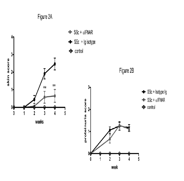
Figure 2A and B are graphs of the clinical signs of scleroderma measured over
time
in mice having no disease induction (control), having disease induction in the
presence of an
anti-interferon alpha receptor (IFNAR) antibody (SSc + aIFNAR), or having
disease
induction in the presence of an Ig isotype control antibody (SSc + Ig Isotype;
Fig. 2A) or
(SSc + Isotype Ig; Fig. 2B). Shown on the Y-axes are the skin score (Fig. 2A)
and the
proteinuria score (Fig. 2B). Figure 2C presents photos of mice following
disease induction in
the presence of an anti-interferon alpha receptor antibody (aIFNAR; top
panel), or in the
presence of an Ig isotype control antibody (Ig Control; top panel).
Figure 3A is a bar graph depicting the histopathological analysis of SSc skin
from
RAG2-/-mice having no disease induction (no graft) (control), having disease
induction in the
presence of an anti-interferon alpha receptor antibody (SSc + aIFNAR), or
having disease
induction in the presence of an Ig isotype control antibody (SSc + Ig
Isotype). Shown on the
Y-axis are the additive scores for inflammation (0 = normal; 1= sparse
cellular infiltrate; 2 =
moderate infiltrate; 3 = pervasive dermal infiltrate) and collagen deposition
(0 = normal, 1 =
mild; 2 = moderate; 3 = severe). Figure 3B shows immunohistochemistry of
representative
H&E (left panel) and Masson's trichrome (right panel) stains of skin from mice
having no
disease induction (control), having disease induction in the presence of an
anti-interferon
3
CA 02703705 2010-04-23
WO 2009/061818 PCT/US2008/082481
alpha receptor antibody (SSc + aIFNAR), or having disease induction in the
presence of an Ig
isotype control antibody (SSc + Ig Isotype).
Figures 4A - 4F show immunohistochemistry of skin sections stained with either
a
goat anti-mouse Ig-FITC (green) or rat anti-mouse Clq-PE polyclonal antibody
(red) and
mounted in DAPI (blue). Skin sections came from syngeneic graft control mice
(Figs. 4A
and 4D), mice with SSc induction in the presence of Ig isotype control (Figs.
4B and 4E), and
mice with SSc induction in the presence of anti-IFNAR antibody (Figs. 4C and
4F).
Figure 5A is a bar graph depicting serum anti-Scl-70 and anti-SSA
autoantibodies
(IgG, IgA, IgM) as detected by ELISA of sera from mice having no disease
induction
(control), having disease induction in the presence of an anti-interferon
alpha receptor
antibody (SSc + aIFNAR), or having disease induction in the presence of an Ig
isotype
control antibody (SSc + Ig Isotype). Figure 5B presents 3 bar graphs depicting
the amounts
of anti-Scl-70 IgGl (top), anti-Scl-70 IgG2 (middle), and anti-SSA IgGl
(bottom), in sera
from mice having no disease induction (no GVH), having disease induction in
the presence of
an anti-interferon alpha receptor antibody (10 mpk A53), or having disease
induction in the
presence of an Ig isotype control antibody (10 mpk 1A7). Figure 5C shows
immunohistochemisty of spleen sections from syngeneic graft control mice
(panels a and d),
mice with SSc induction in the presence of Ig isotype control (panels b and
e), and mice with
SSc induction in the presence of anti-IFNAR antibody (panels c and f), stained
for
CD45R/B220 (brown), and peanut agglutinin (red) to identify germinal centers
(GC).
Figure 6A is a bar graph depicting the quantitative analysis by flow cytometry
cell
sorting (FACS) of the number of splenic plasmacytoid dendritic cell (pDC)
(B220+/ Gr-llo
/CD1 lc+/CD1 lb-) in mismatch graft recipients 2 weeks post-graft (SSc) or
ungrafted RAG2-
/- controls (control). Figure 6B presents time course graphs of skin score
(left) and
proteinuria (right) in RAG2-/- mice that were grafted with total miHag
mismatched
splenocytes (total splenocytes) or with Gr-l-depleted (i.e., pDC-depleted)
miHag mismatched
splenocytes (Gr-l(-) splenocytes).
Figure 7 is a "heatmap" presentation of whole genome array (WGA) data analysis
of
genes that are repressed or induced in the skin of mice having no SSc disease
induction
(control), having SSc disease induction in the presence of an Ig isotype
control antibody (Ig
control), or having SSc disease induction in the presence of an anti-
interferon alpha receptor
antibody (a-IFNAR). Clinical skin scores, determined as described for Figure
3A, are shown
below the columns for each WGA analyzed sample.
4
CA 02703705 2010-04-23
WO 2009/061818 PCT/US2008/082481
Figures 8A - 8D summarize a series of experiments directed to analyzing
temporal
expression of type I IFNs in GVH-SSc skin. Figure 8A summarizes qPCR analysis
of
IFNa2, a5, a9, and R mRNA induction. Figures 8B and 8C summarize qPCR analysis
of
IFNy and IFN,-2, respectively (data are representative of 2 studies,
n=4/timepoint). Figure
8D depicts immunohistochemical staining of IFN,-3 in GVH-SSc (bottom panel)
and non-
SSc (top panel) dermal epithelial cells (magnification, 400x).
Figures 9A - 9C summarize data from the GVH-induced SSc animal model.
Fluidigm (qPCR) analysis was performed on skin samples from mice treated twice
weekly
with 10 mg/kg of body weight of the anti-IFNAR1 murine antibody 5A3 (hatched
bars) or
control Ig (solid bars), and compared to non-SSc skin (open bars). Figure 9A
summarizes the
results of expression of four IFN-inducible genes (IFI44, MX1, OASL, OAS2) at
two
timepoints, and the data indicates that early expression is IFNAR1-independent
and that
chronic expression is IFNAR-1-dependent. Figure 9B summarizes results
demonstrating that
inflammatory gene expression (MPO, TNFa, IL-6, INOS) in skin is reduced with
anti-
IFNAR1 antibody treatment. Figure 9C summarizes results demonstrating that
tissue
remodeling-related gene expression (KLF10, TIMP, EPGN, MMP9) is reduced with
anti-
IFNARl antibody treatment.
DETAILED DESCRIPTION OF THE INVENTION
SCLERODERMA THERAPY
The present invention provides methods of treating scleroderma or systemic
sclerosis
as well as methods of treating the symptoms of scleroderma or systemic
sclerosis by the
administration of an antagonist of type I IFN.
A "therapeutically effective amount" or "therapeutically effective dose" of an
anti-
type I IFN or anti-interferon alpha or anti-IFNaR antibody of the invention
preferably results
in a decrease in severity of disease symptoms, an increase in frequency and
duration of
disease symptom-free periods, or a prevention of impairment or disability due
to the disease
affliction. In the case of, for example, scleroderma, a therapeutically
effective amount or
dose preferably prevents further deterioration of physical symptoms associated
with
scleroderma or systemic sclerosis, such as, for example, dermal fibrosis, skin
lesions,
alopecia, inflammation, dermal thickening, collagen deposition, proteinuria,
autoantibody
production, and complement deposition. A therapeutically effective amount or
dose
5
CA 02703705 2010-04-23
WO 2009/061818 PCT/US2008/082481
preferably also prevents or delays onset of scleroderma or systemic sclerosis,
such as may be
desired when early or preliminary signs of the disease are present. Likewise
it includes
delaying chronic progression associated with scleroderma or systemic
sclerosis. Laboratory
tests utilized in the diagnosis of scleroderma or systemic sclerosis include
chemistries,
hematology, histopathology, serology and radiology. Accordingly, any clinical
or
biochemical assay that monitors any of the foregoing may be used to determine
whether a
particular treatment is a therapeutically effective dose for treating
scleroderma or systemic
sclerosis. One of ordinary skill in the art would be able to determine such
amounts based on
such factors as the subject's size, the severity of the subject's symptoms,
and the particular
composition or route of administration selected.
As used herein, the term "treating" refers to alleviating, ameliorating,
and/or
decreasing the severity of scleroderma or systemic sclerosis and associated
symptoms.
In some embodiments, methods are provided for treating one or more of the
symptoms of scleroderma, which include dermal fibrosis, skin lesions,
alopecia,
inflammation, dermal thickening, collagen deposition, proteinuria,
autoantibody production,
and complement deposition.
The severity, progression, response to treatment, and other clinical measures
of the
symptoms of scleroderma typically include an evaluation of the patient using
the modified
Rodnan skin score, the Raynaud's Condition Score, measurements of the forced
vital capacity
as part of pulmonary function tests, right heart catheterization
haemodynamics,
measurements of serum creatine, blood pressure and complete blood counts, and
measurements of serum creatinine phosphokinase levels (see, for example,
Furst, 2008,
Rheumatology, 47:v29-v30 and Furst et at., 2007, J. of Rheumatology, 34:5,
1194-1200).
The modified Rodnan skin score is a clinical scoring of scleroderma using
established
palpation to estimate skin thickness in a patient, using measurements from
seventeen skin
sites on the patient and grading each site with a 0 = normal skin, 1 =
thickened skin, 2 =
thickened skin and unable to pinch, and 3 = thickened skin and unable to move,
for a total of
51 points (see Czirjak et at., 2007, Ann Rheum Dis.; 66(7):966-9. Epub 2007
Jan 18 and
Brennan et at., 1992, Br J Rheumatol.; 31(7):457-60).
Accordingly, in certain embodiments, provided are methods of treating
scleroderma in
a patient in need of such treatment, comprising administering a
therapeutically effective
amount of an antagonist of type I interferon, wherein said treating results in
an improvement
6
CA 02703705 2010-04-23
WO 2009/061818 PCT/US2008/082481
in symptoms as measured by the modified Rodnan skin score (i.e. a reduction in
the total
modified skin score value).
In one embodiment, the invention provides a method of treating scleroderma in
a
patient in need of such treatment, comprising administering a therapeutically
effective
amount of an antagonist of type I interferon, wherein said patient has a pre-
treatment
modified Rodnan skin score of 1 to 51 and a post-treatment modified Rodnan
skin score of (1
to 51)-x, where x = 1 to 51 and the post-treatment modified Rodnan skin score
is not below 0.
In one embodiment, the invention provides a method of treating scleroderma in
a
patient in need of such treatment, comprising administering a therapeutically
effective
amount of an antagonist of type I interferon, wherein said patient has a pre-
treatment
modified Rodnan skin score of 1, 2, 3, 4, 5, 6, 7, 8, 9, 10, 11, 12, 13, 14,
15, 16, 17, 18, 19,
20, 21, 22, 23, 24, 25, 26, 27, 28, 29, 30, 31, 32, 33, 34, 35, 36, 37, 38,
39, 40, 41, 42, 43, 44,
45, 46, 47, 48, 49, 50, or 51, and a post-treatment modified Rodnan skin score
of 50, 49, 48,
47, 46, 45, 44, 43, 42, 41, 40, 39, 38, 37, 36, 35, 34, 33, 32, 31, 30, 29,
28, 27, 26, 25, 24, 23,
22, 21, 20, 19, 18, 17, 16, 15, 14, 13, 12, 11, 10, 9, 8, 7, 6, 5, 4, 3, 2, 1,
or 0.
In one embodiment, the invention provides a method of treating scleroderma in
a
patient in need of such treatment, comprising administering a therapeutically
effective
amount of an antagonist of type I interferon, wherein said treating reduces
the modified
Rodnan skin score value by at least 1. In one embodiment, the invention
provides a method
of treating scleroderma in a patient in need of such treatment, comprising
administering a
therapeutically effective amount of an antagonist of type I interferon,
wherein said treating
reduces the modified Rodnan skin score value by at least 5. In one embodiment,
the
invention provides a method of treating scleroderma in a patient in need of
such treatment,
comprising administering a therapeutically effective amount of an antagonist
of type I
interferon, wherein said treating reduces the modified Rodnan skin score value
by at least 10.
In one embodiment, the invention provides a method of treating scleroderma in
a patient in
need of such treatment, comprising administering a therapeutically effective
amount of an
antagonist of type I interferon, wherein said treating reduces the modified
Rodnan skin score
value by at least 25.
In one embodiment, the invention provides a method of treating scleroderma in
a
patient in need of such treatment, comprising administering a therapeutically
effective
amount of an antagonist of type I interferon, wherein said treating reduces
the modified
Rodnan skin score value by at least 1, at least 2, at least 3, at least 4, at
least 5, at least 6, at
least 7, at least 8, at least 9, at least 10, at least 11, at least 12, at
least 13, at least 14, at least
7
CA 02703705 2010-04-23
WO 2009/061818 PCT/US2008/082481
15, at least 16, at least 17, at least 18, at least 19, at least 20, at least
21, at least 22, at least
23, at least 24, at least 25, at least 26, at least 27, at least 28, at least
29, at least 30, at least
31, at least 32, at least 33, at least 34, at least 35, at least 36, at least
37, at least 38, at least
39, at least 40, at least 41, at least 42, at least 43, at least 44, at least
45, at least 46, at least
47, at least 48, at least 49, at least 50, or 51.
In certain embodiments, provided are methods of treating scleroderma in a
patient in
need of such treatment, comprising administering a therapeutically effective
amount of an
antagonist of type I interferon, wherein said treating results in an
improvement in symptoms
as measured by the Raynaud's Condition Score (RCS). The RCS is a daily self-
assessment of
Rayaud's Phenomenom activity using a scale of 0-10, with an increasing number
indicating a
worsening of symptoms associated with scleroderma (see, for example, Merkel et
al., 2002,
Arthritis & Rheumatism, 46:9, pp 2410-2420).
In one embodiment, the invention provides a method of treating scleroderma in
a
patient in need of such treatment, comprising administering a therapeutically
effective
amount of an antagonist of type I interferon, wherein said patient has a pre-
treatment RCS
score of 1 to 10 and a post-treatment RCS score of (1 to 10)-x, where x = 1 to
10 and the
post-treatment RCS score is not below 0.
In one embodiment, the invention provides a method of treating scleroderma in
a
patient in need of such treatment, comprising administering a therapeutically
effective
amount of an antagonist of type I interferon, wherein said patient has a pre-
treatment RCS
score of 1, 2, 3, 4, 5, 6, 7, 8, 9, or 10, and a post-treatment modified
Rodnan skin score of 9,
8, 7, 6, 5, 4, 3, 2, 1, or 0.
In one embodiment, the invention provides a method of treating scleroderma in
a
patient in need of such treatment, comprising administering a therapeutically
effective
amount of an antagonist of type I interferon, wherein said treating reduces
the RCS score
value by at least 1. In one embodiment, the invention provides a method of
treating
scleroderma in a patient in need of such treatment, comprising administering a
therapeutically
effective amount of an antagonist of type I interferon, wherein said treating
reduces the RCS
score value by at least 2. In one embodiment, the invention provides a method
of treating
scleroderma in a patient in need of such treatment, comprising administering a
therapeutically
effective amount of an antagonist of type I interferon, wherein said treating
reduces the RCS
score value by at least 3. In one embodiment, the invention provides a method
of treating
scleroderma in a patient in need of such treatment, comprising administering a
therapeutically
effective amount of an antagonist of type I interferon, wherein said treating
reduces the RCS
8
CA 02703705 2010-04-23
WO 2009/061818 PCT/US2008/082481
score value by at least 5. In one embodiment, the invention provides a method
of treating
scleroderma in a patient in need of such treatment, comprising administering a
therapeutically
effective amount of an antagonist of type I interferon, wherein said treating
reduces the RCS
score value by at least 6. In one embodiment, the invention provides a method
of treating
scleroderma in a patient in need of such treatment, comprising administering a
therapeutically
effective amount of an antagonist of type I interferon, wherein said treating
reduces the RCS
score value by at least 7. In one embodiment, the invention provides a method
of treating
scleroderma in a patient in need of such treatment, comprising administering a
therapeutically
effective amount of an antagonist of type I interferon, wherein said treating
reduces the RCS
score value by at least 8. In one embodiment, the invention provides a method
of treating
scleroderma in a patient in need of such treatment, comprising administering a
therapeutically
effective amount of an antagonist of type I interferon, wherein said treating
reduces the RCS
score value by at least 9. In one embodiment, the invention provides a method
of treating
scleroderma in a patient in need of such treatment, comprising administering a
therapeutically
effective amount of an antagonist of type I interferon, wherein said treating
reduces the RCS
score value by 10.
In one embodiment, the invention provides a method of treating scleroderma in
a
patient in need of such treatment, comprising administering a therapeutically
effective
amount of an antagonist of type I interferon, wherein said treating reduces
the RCS score
value by at least 1, at least 2, at least 3, at least 4, at least 5, at least
6, at least 7, at least 8, at
least 9, or 10.
In certain embodiments, provided are methods of treating scleroderma in a
patient in
need of such treatment, comprising administering a therapeutically effective
amount of an
antagonist of type I interferon, wherein said treating results in an
improvement in symptoms
as measured by clinical measurements of serum creatinine.
In certain embodiments, provided are methods of treating scleroderma in a
patient in
need of such treatment, comprising administering a therapeutically effective
amount of an
antagonist of type I interferon, wherein said treating results in an
improvement in symptoms
as measured by measurements of serum creatine phosphokinase levels.
In certain embodiments, provided are methods of treating scleroderma in a
patient in
need of such treatment, comprising administering a therapeutically effective
amount of an
antagonist of type I interferon, wherein said treating results in an
improvement in symptoms
as measured by clinical measurements of the forced vital capacity as part of
pulmonary
function tests.
9
CA 02703705 2010-04-23
WO 2009/061818 PCT/US2008/082481
In certain embodiments, provided are methods of treating scleroderma in a
patient in
need of such treatment, comprising administering a therapeutically effective
amount of an
antagonist of type I interferon, wherein said treating results in an
improvement in symptoms
as measured by right heart catheterization haemodynamics.
In certain embodiments, provided are methods of treating scleroderma in a
patient in
need of such treatment, comprising administering a therapeutically effective
amount of an
antagonist of type I interferon, wherein said treating results in an
improvement in symptoms
as measured by blood pressure and complete blood counts.
In certain embodiments, provided are methods of treating scleroderma in a
patient in
need of such treatment, comprising administering a therapeutically effective
amount of an
antagonist of type I interferon, wherein said treating results in an
improvement in symptoms
as measured by histopathologic analysis of patient skin samples. In one
embodiment, this
improvement is measured as a reduction in inflammation as represented by
inflammatory cell
infiltrate in the tissue sample, collagen deposition, or overall thickening of
the dermis. In one
embodiment, treatment with an antagonist of type I interferon reduces
inflammation by 2-
fold. In another embodiment, treatment with an antagonist of type I interferon
reduces
inflammation by 3-fold. In a further embodiment, treatment with an antagonist
of type I
interferon reduces inflammation by 5-fold.
In certain embodiments, provided are methods of treating scleroderma in a
patient in
need of such treatment, comprising administering a therapeutically effective
amount of an
antagonist of type I interferon, wherein said treating results in an
improvement in symptoms
as measured by qPCR analysis performed on patient skin samples. See, for
example,
International Patent Application Publication WO/08070137A2, entitled
Interferon Alpha-
Induced Pharmacodynamic Markers.
In one embodiment, provided are methods of treating scleroderma in a patient
in need
of such treatment, comprising administering a therapeutically effective amount
of an
antagonist of type I interferon, wherein said treating reduces inflammatory
gene expression
including, but not limited to, MPO, TNFa, IL-6, and INOS. In one embodiment,
provided
are methods of treating scleroderma in a patient in need of such treatment,
comprising
administering a therapeutically effective amount of an antagonist of type I
interferon, wherein
pre-treatment said patient exhibits increased inflammatory gene expression
including, but not
limited to, MPO, TNFa, IL-6, and INOS, and post-treatment said patient
exhibits a reduction
in inflammatory gene expression including, but not limited to, MPO, TNFa, IL-
6, and INOS.
CA 02703705 2010-04-23
WO 2009/061818 PCT/US2008/082481
In one embodiment, provided are methods of treating scleroderma in a patient
in need of such
treatment, comprising administering a therapeutically effective amount of an
antagonist of
type I interferon, wherein said treating yields an at least 2-fold, at least 3-
fold, at least 4-fold,
at least 5-fold, or at least 10-fold reduction in inflammatory gene expression
as measured by
qPCR. In another embodiment, provided are methods of treating scleroderma in a
patient in
need of such treatment, comprising administering a therapeutically effective
amount of an
antagonist of type I interferon, wherein said treating yields a reduction of
at least 2%, at least
3%, at least 4%, at least 5%, at least 7%, at least 8%, at least 10%, at least
15%, at least 25%,
at least 30%, at least 35%, at least 40%, at least 45%, at least 50%, at least
60%, at least 70%,
at least 75%, at least 80%, or at least 90% of inflammatory gene expression as
measured by
qPCR.
In a further embodiment, provided are methods of treating scleroderma in a
patient in
need of such treatment, comprising administering a therapeutically effective
amount of an
antagonist of type I interferon, wherein said treating reduces tissue
remodeling-related gene
expression including, but not limited to, KLF 10, TIMP, EPGN, and MMP9. In one
embodiment, provided are methods of treating scleroderma in a patient in need
of such
treatment, comprising administering a therapeutically effective amount of an
antagonist of
type I interferon, wherein pre-treatment said patient exhibits increased
tissue remodeling-
related gene expression including, but not limited to, KLF10, TIMP, EPGN, and
MMP9, and
post-treatment said patient exhibits a reduction in tissue remodeling-related
gene expression
including, but not limited to, KLF10, TIMP, EPGN, and MMP9. In one embodiment,
provided are methods of treating scleroderma in a patient in need of such
treatment,
comprising administering a therapeutically effective amount of an antagonist
of type I
interferon, wherein said treating yields an at least 2-fold, at least 3-fold,
at least 4-fold, at
least 5-fold, or at least 10-fold reduction in tissue remodeling-related gene
expression as
measured by qPCR. In another embodiment, provided are methods of treating
scleroderma in
a patient in need of such treatment, comprising administering a
therapeutically effective
amount of an antagonist of type I interferon, wherein said treating yields a
reduction of at
least 2%, at least 3%, at least 4%, at least 5%, at least 7%, at least 8%, at
least 10%, at least
15%, at least 25%, at least 30%, at least 35%, at least 40%, at least 45%, at
least 50%, at least
60%, at least 70%, at least 75%, at least 80%, or at least 90% of tissue
remodeling-related
gene expression as measured by qPCR.
Pharmacodynamic (PD) markers can be used in methods of treating patients with
a
therapeutic agent that binds to and antagonizes type I IFN activity, more
particularly, IFNa
11
CA 02703705 2010-04-23
WO 2009/061818 PCT/US2008/082481
activity, or IFNaR activity, methods that identify patients as candidates for
a therapeutic
agent that binds to and antagonizes type I IFN activity, more particularly,
IFNa activity, or
IFNaR activity, methods of diagnosing a patient as having a disorder
associated with
increased type I IFN or, more particularly, IFNa levels, or IFNaR activity,
methods of
monitoring disease progression of a patient receiving treatment with a
therapeutic agent that
binds to and antagonizes type I IFN activity, more particularly, IFNa
activity, or IFNaR
activity, and methods of identifying a candidate therapeutic for treating type
I IFN- and, more
particularly, IFNa -mediated, or IFNAR-mediated, disorders.
In one aspect the present invention provides methods of treating patient
having a type
I IFN-mediated disease or disorder comprising administering an antagonist of
type I IFN
activity; wherein the patient comprises a type I IFN-inducible PD marker
expression profile;
and wherein the antagonist neutralizes the type I IFN -inducible PD marker
expression profile
of the subject. In a specific embodiment, the disease or disorder is
scleroderma or systemic
sclerosis.
The invention encompasses methods of identifying, diagnosing, treating, and
monitoring disease progression in patients. Patients include any animal having
a type I IFN-
or an IFNa-inducible disease, disorder, or condition. The patient may have the
disease,
disorder, or condition as a result of experimental research, e.g., it may be
an experimental
model developed for the disease, disorder, or condition. Alternatively, the
patient may have
the disease, disorder, or condition in the absence of experimental
manipulation. Patients
include humans, mice, rats, horses, pigs, cats, dogs, and any animal used for
research.
Methods of identifying, diagnosing, treating, and monitoring disease
progression in
patients using type I IFN-inducible or IFNa-inducible PD marker expression
profiles and/or
using an antagonist that neutralizes the type I IFN-inducible or IFNa-
inducible PD marker
expression profile of the patient are disclosed in International Patent
Application Publication
No. WO/08070137A2 entitled "Interferon Alpha-induced Pharmacodynamic Markers,"
which
is hereby incorporated herein by reference.
An antagonist of type I IFN that binds to and blocks type I IFN or IFNa or
IFNaR
activity may neutralize a type I IFN or IFNa-inducible profile. Neutralization
of the type I
IFN or IFNa-inducible profile is a reduction in at least one, at least two, at
least three, at least
five, at least seven, at least eight, at least ten, at least twelve, at least
fifteen, at least twenty, at
least twenty five, at least thirty, at least thirty five, at least forty, at
least forty five, or at least
fifty genes induced by type I IFN or IFNa. The genes induced by type I IFN or
IFNa may be
12
CA 02703705 2010-04-23
WO 2009/061818 PCT/US2008/082481
any group of genes in disclosed in International Patent Application
Publication No.
WO/08070137A2 entitled "Interferon Alpha-induced Pharmacodynamic Markers".
Neutralization of the type I IFN or IFNa-inducible profile is a reduction of
at least 2%, at
least 3%, at least 4%, at least 5%, at least 7%, at least 8%, at least 10%, at
least 15%, at least
25%, at least 30%, at least 35%, at least 40%, at least 45%, at least 50%, at
least 60%, at least
70%, at least 75%, at least 80%, or at least 90% of any of the at least one,
at least two, at least
three, at least five, at least seven, at least eight, at least ten, at least
twelve, at least fifteen, at
least twenty, at least twenty five, at least thirty, at least thirty five, at
least forty, at least forty
five, or at least fifty genes in any type I IFN or IFNa-inducible profile.
Alternatively,
neutralization of the type I IFN or IFNa-inducible profile refers to a
reduction of expression
of type I IFN or IFNa-inducible genes that is within at most 50%, at most 45%,
at most 40%,
at most 35%, at most 30%, at most 25%, at most 20%, at most 15%, at most 10%,
at most
5%, at most 4%, at most 3%, at most 2%, or at most 1% of expression levels of
those type I
IFN or IFNa-inducible genes in a control sample. If the agent that binds to
and modulates,
more particularly, antagonizes type I IFN or IFNa activity is a biologic
agent, such as an
antibody, the agent may neutralize the type I IFN or IFNa profile at doses of
0.3 to 30 mg/kg,
0.3 to 10 mg/kg, 0.3 to 3 mg/kg, 0.3 to 1 mg/kg, 1 to 30 mg/kg, 3 to 30 mg/kg,
5 to 30 mg/kg,
10 to 30 mg/kg, 1 to 10 mg/kg, 3 to 10 mg/kg, or 1 to 5 mg/kg.
The agent that binds to and modulates, more particularly, antagonizes type I
IFN or
IFNa activity may further or alternatively neutralize expression of one or
more type I IFN or
IFNa subtypes. The IFNa or type-I IFN subtypes may include any more than one,
more than
two, more than three, more than four, more than five, more than six, more than
seven, more
than eight, more than nine, or more than ten IFNa or type-I IFN subtypes.
These subtypes
may include IFNa1, IFNa2, IFNa4, IFNa5, IFNa6, IFNa7, IFNa8, IFNa10, IFNa14,
IFNa17, IFNa21, IFNI3, or IFNw. These subtypes may include all of IFNa1,
IFNa2, IFNa8,
and IFNa14. Alternatively, these subtypes may include IFNa1, IFNa2, IFNa4,
IFNa5,
IFNa8, IFNa10, IFNa21. Neutralization of the IFNa or type-I IFN subtypes may
be a
reduction of at least 2%, at least 3%, at least 4%, at least 5%, at least 7%,
at least 8%, at least
10%, at least 15%, at least 25%, at least 30%, at least 35%, at least 40%, at
least 45%, at least
50%, at least 60%, at least 70%, at least 75%, at least 80%, or at least 90%
of any of the at
least one, at least two, at least three, at least five, at least seven, at
least eight, or at least ten of
the subtypes. Neutralization of the IFNa or type-I IFN subtypes may be a
reduction in
expression of IFNa or type-I IFN subtype genes that is within at most 50%, at
most 45%, at
13
CA 02703705 2010-04-23
WO 2009/061818 PCT/US2008/082481
most 40%, at most 35%, at most 30%, at most 25%, at most 20%, at most 15%, at
most 10%,
at most 5%, at most 4%, at most 3%, at most 2%, or at most 1% of expression
levels of those
IFNa or type I IFN subtypes in a control sample. If the agent that binds to
and modulates,
more particularly, antagonizes IFNa activity or type I IFN activity is a
biologic agent, such as
an antibody, the agent may neutralize the IFNa or type I IFN subtypes at doses
of 0.3 to 30
mg/kg, 0.3 to 10 mg/kg, 0.3 to 3 mg/kg, 0.3 to 1 mg/kg, 1 to 30 mg/kg, 3 to 30
mg/kg, 5 to 30
mg/kg, 10 to 30 mg/kg, 1 to 10 mg/kg, 3 to 10 mg/kg, or 1 to 5 mg/kg.
The agent that binds to and modulates, more particularly, antagonizes type I
IFN or
IFNa activity may further or alternatively neutralize expression of IFNa
receptors, either
IFNAR1 or IFNAR2, or both, or TNFa, or IFNy, or IFNy receptors (either IFNGR1,
IFNGR2, or both IFNGR1 and IFNGR2). Neutralization of expression of IFNa
receptors,
either IFNAR1 or IFNAR2, or both, or TNFa, or IFNy, or IFNy receptors (either
IFNGRI,
IFNGR2, or both IFNGR1 and IFNGR2) may be a reduction of at least 2%, at least
3%, at
least 4%, at least 5%, at least 7%, at least 8%, at least 10%, at least 15%,
at least 25%, at least
30%, at least 35%, at least 40%, at least 45%, at least 50%, at least 60%, at
least 70%, at least
75%, at least 80%, or at least 90% of any of the at least one, at least two,
at least three, at
least five, or at least six of these genes. Neutralization of expression of
IFNa receptors,
either IFNAR1 or IFNAR2, or TNFa, or IFNy, or IFNy receptors (either IFNGR1,
IFNGR2,
or both IFNGRI and IFNGR2) is a reduction of expression of at most 50%, at
most 45%, at
most 40%, at most 35%, at most 30%, at most 25%, at most 20%, at most 15%, at
most 10%,
at most 5%, at most 4%, at most 3%, at most 2%, or at most 1% of expression
levels of these
genes in a control sample. If the agent that binds to and modulates, more
particularly,
antagonizes type I IFN or IFNa activity is a biologic agent, such as an
antibody, the agent
may neutralize expression of IFNa receptors IFNAR1 or IFNAR2, or TNFa, or
IFNy, or
IFNy receptors IFNGR1 or IFNGR2 at doses of 0.3 to 30 mg/kg, 0.3 to 10 mg/kg,
0.3 to 3
mg/kg, 0.3 to 1 mg/kg, 1 to 30 mg/kg, 3 to 30 mg/kg, 5 to 30 mg/kg, 10 to 30
mg/kg, 1 to 10
mg/kg, 3 to 10 mg/kg, or 1 to 5 mg/kg.
Applicants provide a set of non-limiting embodiments to describe some of the
aspects
of the invention.
1. A method for treating scleroderma in a patient in need of such treatment
comprising
administering a therapeutically effective amount of an antagonist of type I
interferon (IFN).
14
CA 02703705 2010-04-23
WO 2009/061818 PCT/US2008/082481
2. A method for alleviating one or more of the symptoms associated with
scleroderma in a
patient in need of such treatment comprising administering a therapeutically
effective amount
of an antagonist of type I interferon.
3. The method of embodiments 1 or 2, wherein said antagonist is an antibody.
4. The method of embodiment 3, wherein said antibody is an anti-IFNaR
antibody.
5. The method of embodiment 3, wherein said antibody is an anti-IFNa antibody.
6. The method of embodiments 1 or 2, wherein the symptoms are selected from
the group
consisting of. dermal fibrosis, skin lesions, alopecia, inflammation, dermal
thickening,
collagen deposition, proteinuria, and complement deposition.
7. The method of embodiments 1 or 2, wherein the antibody is administered at a
dose
between approximately 0.03 mg/kg and approximately 30 mg/kg.
8. The method of embodiments 1 or 2, wherein said treating results in an
improvement in
symptoms as measured by the modified Rodnan skin score
9. The method of embodiments 1 or 2, wherein said treating results in an
improvement in
symptoms as measured by the Raynaud's Condition Score (RCS).
10. The method of embodiments 1 or 2, wherein said treating results in an
improvement in
symptoms as measured by qPCR analysis performed on patient skin samples.
11. The method of embodiment 10, wherein said treating reduces expression of
inflammatory genes selected from the group consisting of MPO, TNFa, IL-6, and
INOS.
12. The method of embodiment 10, wherein said treating reduces expression of
tissue
remodeling-related genes selected from the group consisting of KLF 10, TIMP,
EPGN, and
MMP9.
CA 02703705 2010-04-23
WO 2009/061818 PCT/US2008/082481
Dosing and Administration
The amount of the composition of the invention which will be effective in the
treatment, prevention or management of scleroderma / systemic sclerosis and
the symptoms
of the disease can be determined by standard research techniques. Selection of
the preferred
effective dose can be determined (e.g., via clinical trials) by a skilled
artisan based upon the
consideration of several factors that will be known to one of ordinary skill
in the art. Such
factors include the symptoms involved, the patient's body mass, the patient's
immune status
and other factors known by the skilled artisan to reflect the accuracy of
administered
pharmaceutical compositions.
The precise dose to be employed in the formulation will also depend on the
route of
administration, and the seriousness of the scleroderma symptoms displayed, and
should be
decided according to the judgment of the practitioner and each patient's
circumstances.
Effective doses may be extrapolated from dose-response curves derived from in
vitro or
animal model test systems.
For antibodies, fusion proteins, or muteins, the dosage administered to a
patient is
typically 0.1 mg/kg to 100 mg/kg of the patient's body weight. In one
embodiment, the
dosage administered to a patient is between 0.1 mg/kg and 20 mg/kg of the
patient's body
weight. In another embodiment, the dosage administered to a patient is between
1 mg/kg and
10 mg/kg of the patient's body weight. In another embodiment, the dosage
administered to a
patient is between 0.3 mg/kg and 30 mg/kg of the patient's body weight. In
another
embodiment, the dosage administered to a patient is between 0.3 mg/kg and 3.0
mg/kg. In
another embodiment, the dosage administered to a patient is 0.1 mg/kg, 0.3
mg/kg, 0.5
mg/kg, 1.0 mg/kg, 2.0 mg/kg, 3.0 mg/kg, 4.0 mg/kg, 5.0 mg/kg, 6.0 mg/kg, 7.0
mg/kg, 8.0
mg/kg, 9.0 mg/kg, 10.0 mg/kg, 11.0 mg/kg, 12.0 mg/kg, 13.0 mg/kg, 14.0 mg/kg,
15.0
mg/kg, 16.0 mg/kg, 17.0 mg/kg, 18.0 mg/kg, 19.0 mg/kg, 20.0 mg/kg, or 25
mg/kg. In a
specific embodiment, the dosage administered to a patient is selected from the
group
consisting of 0.1 mg/kg, 0.3 mg/kg, 1.0 mg/kg, 3.0 mg/kg, 10 mg/kg, and 30
mg/kg of the
patient's body weight.
In specific embodiments, dosages of an antibody (optionally in a
pharmaceutically
acceptable carrier as part of a pharmaceutical composition) are at least about
0.0005, 0.001,
0.05, 0.075, 0.1, 0.25, 0.375, 0.5, 1, 2.5, 5, 10, 20, 37.5, or 50 mg/m2
and/or less than about
500, 475, 450, 425, 400, 375, 350, 325, 300, 275, 250, 225, 200, 175, 150,
125, 100, 75, 60,
16
CA 02703705 2010-04-23
WO 2009/061818 PCT/US2008/082481
50, 37.5, 20, 15, 10, 5, 2.5, 1, 0.5, 0.375, 0.1, 0.075 or 0.01 mg/m2. In
certain embodiments,
the dosage is between about 0.0005 to about 200 mg/m2, between about 0.001 and
150
mg/m2, between about 0.075 and 125 mg/m2, between about 0.375 and 100 mg/m2,
between
about 2.5 and 75 mg/m2, between about 10 and 75 mg/m2, and between about 20
and 50
mg/m2. In related embodiments, the dosage of an anti-IFNa or IFNaR used is at
least about
0. 1, 0.2, 0.3, 0.4, 0.5, 0.6, 0.7, 0.8, 0.9, 1, 1.5, 2, 2.5, 3, 3.5, 4, 4.5,
5, 5.5, 6, 6.5, 7, 7.5, 8, 8.5,
9, 9.5, 10, 10.5, 11, 11.5, 12, 12.5, 13, 13.5, 14, 14.5, 15, 15.5, 16, 16.5,
17, 17.5, 18, 18.5,
19, 19.5, 20, 20.5 mg/kg of body weight of a patient.
In specific embodiments, the dose of an anti-IFNa or anti-IFNaR antibody used
is at
least about 1 to about 10, about 5 to about 15, about 10 to about 20, or about
15 to about 25
mg/kg of body weight of a patient. In certain embodiments, the dose of an anti-
IFNa or
IFNaR antibody used is at least about 1 to about 20, about 3 to about 15, or
about 5 to about
10 mg/kg of body weight of a patient. In other embodiments, the dose of an
anti-IFNa or
IFNaR antibody used is at least about 5, about 6, about 7, about 8, about 9,
or about 10
mg/kg of body weight of a patient. In certain embodiments, a single dosage
unit of the
antibody (optionally in a pharmaceutically acceptable carrier as part of a
pharmaceutical
composition) can be at least about 0.5, about 1, about 2, about 4, about 6,
about 8, about 10,
about 12, about 14, about 16, about 18, about 20, about 22, about 24, about
26, about 28,
about 30, about 32, about 34, about 36, about 38, about 40, about 42, about
44, about 46,
about 48, about 50, about 52, about 54, about 56, about 58, about 60, about
62, about 64,
about 66, about 68, about 70, about 72, about 74, about 76, about 78, about
80, about 82,
about 84, about 86, about 88, about 90, about 92, about 94, about 96, about
98, about 100,
about 102, about 104, about 106, about 108, about 110, about 112, about 114,
about 116,
about 118, about 120, about 122, about 124, about 126, about 128, about 130,
about 132,
about 134, about 136, about 138, about 140, about 142, about 144, about 146,
about 148,
about 150, about 152, about 154, about 156, about 158, about 160, about 162,
about 164,
about 166, about 168, about 170, about 172, about 174, about 176, about 178,
about 180,
about 182, about 184, about 186, about 188, about 190, about 192, about 194,
about 196,
about 198, about 200, about 204, about 206, about 208, about 210, about 212,
about 214,
about 216, about 218, about 220, about 222, about 224, about 226, about 228,
about 230,
about 232, about 234, about 236, about 238, about 240, about 242, about 244,
about 246,
about 248, or about 250 micrograms/m2. In other embodiments, dose is up to
about 1 g per
single dosage unit.
17
CA 02703705 2010-04-23
WO 2009/061818 PCT/US2008/082481
Administration of compositions of the invention to a human patient can be by
any
route, including but not limited to intravenous, intradermal, transdermal,
subcutaneous,
intramuscular, inhalation (e.g., via an aerosol), buccal (e.g., sub lingual),
topical (i.e., both
skin and mucosal surfaces, including airway surfaces), intrathecal,
intraarticular, intraplural,
intracerebral, intra arterial, intraperitoneal, oral, intralymphatic,
intranasal, rectal or vaginal
administration, by perfusion through a regional catheter, or by direct
intralesional injection.
In one embodiment, compositions of the invention are administered by
intravenous push or
intravenous infusion given over defined period (e.g., 0.5 to 2 hours).
Compositions of the
invention can be delivered by peristaltic means or in the form of a depot,
although the most
suitable route in any given case will depend, as is well known in the art, on
such factors as
the species, age, gender and overall condition of the subject, the nature and
severity of the
condition being treated and/or on the nature of the particular composition
(i.e., dosage,
formulation) that is being administered. In particular embodiments, the route
of
administration is via bolus or continuous infusion over a period of time, once
or twice a
week. In other particular embodiments, the route of administration is by
subcutaneous
injection, optionally once or twice weekly. In one embodiment, compositions,
and/or
methods of the invention are administered on an outpatient basis. In another
embodiment,
compositions and/or methods of the invention are administered using pre-filled
syringes.
In certain embodiments, the dose of a composition comprising an anti-IFNa or
IFNaR antibody is measured in units of mg/kg of patient body weight. In other
embodiments, the dose of a composition comprising an anti-IFNa or IFNaR
antibody is
measured in units of mg/kg of patient lean body weight (i.e., body weight
minus body fat
content). In yet other embodiments, the dose of a composition comprising an
anti-IFNa or
IFNaR antibody is measured in units of mg/m2 of patient body surface area. In
yet other
embodiments, the dose of a composition comprising an anti-IFNa or IFNaR
antibody is
measured in units of mg per dose administered to a patient. Any measurement of
dose can be
used in conjunction with compositions and methods of the invention and dosage
units can be
converted by means standard in the art.
Those skilled in the art will appreciate that dosages can be selected based on
a number
of factors including the age, sex, species and condition of the subject (e.g.,
stage of
scleroderma), the desired degree of cellular depletion, the disease to be
treated and/or the
particular antibody or antigen binding fragment being used and can be
determined by one of
skill in the art. For example, effective amounts of compositions of the
invention may be
18
CA 02703705 2010-04-23
WO 2009/061818 PCT/US2008/082481
extrapolated from dose response curves derived in vitro test systems or from
animal model
(e.g., the cotton rat or monkey) test systems. Models and methods for
evaluation of the
effects of antibodies are known in the art (Wooldridge et al., Blood, 89(8):
2994 2998
(1997)), incorporated by reference herein in its entirety).
Examples of dosing regimens that can be used in methods of the invention
include,
but are not limited to, daily, three times weekly (intermittent), weekly, or
every 14 days. In
certain embodiments, dosing regimens include, but are not limited to, monthly
dosing or
dosing every 6-8 weeks, or weekly for a defined period of time. In one
embodiment, dosing
can take place weekly for 1, 2, 3, 4, 5, 6, 7,8, 9, 10, 11, or 12 weeks.
Those skilled in the art will appreciate that dosages are generally higher
and/or
frequency of administration greater for initial treatment as compared with
maintenance
regimens. All of the above doses are exemplary and can be used in conjunction
with
compositions and methods of the invention, however where an an anti-IFNa or
IFNaR
antibody is used in conjunction with an anti-inflammatory agent the lower
doses described
above may be used.
In certain embodiments, the dosage and rate of delivery can be adjusted and/or
the
infusion rate can be reduced based on patient's immunogenic response to
compositions and
methods of the invention. According to another aspect of methods of the
invention, a patient
may be pretreated with compositions and methods of the invention to detect,
minimize
immunogenic response, or minimize adverse effects of compositions and methods
of the
invention.
TYPE I INTERFERON ANTAGONISTS
As used herein, the term "antagonist of type I IFN" refers to any agent that
blocks,
inhibits, suppresses, neutralizes, decreases or otherwise interferes with or
abrogates signaling
through and/or activation of the interferon alpha receptor (IFNAR).
Antagonists of type I
IFN may act by interfering with the interaction between any type I IFN and
IFNAR. Thus
antagonists of type I IFN may act by binding and antagonizing a type I IFN or
by binding an
antagonizing the IFNAR. Antagonists of type I IFN may bind to either the IFNAR
1 chain of
the IFNAR or to the IFNAR 2 chain of the IFNAR, or they may bind to epitopes
resulting
from the combination of the two chains. In particular embodiments, antagonists
of type I IFN
are antibodies that bind to the IFNAR 1 chain of the IFNAR, such as those
disclosed in U.S.
19
CA 02703705 2010-04-23
WO 2009/061818 PCT/US2008/082481
Patent Appl. Publ. Nos. 2006/0029601, 2006/0020118, and U.S Patent No.
6,713,609, which
are incorporated herein by reference in its entirety.
In certain embodiments of the present invention are provided antagonists that
interfere
with type 1 IFN ligand binding such as, for example, soluble receptor chains
(e.g., soluble
IFNAR1 or IFNAR2) or fragments thereof. Other related embodiments provide
antibodies or
antigen binding fragments thereof that selectively bind to one or more type 1
interferon or
bind to the IFNAR in such a way as to interfere with ligand binding, such as,
for example, by
competitive, non-competitive or uncompetitive inhibition. Alternative
embodiments provide
antagonists that interfere with signal transduction by the IFNAR. Still
further embodiments
provide antagonists that antagonize the downstream effects of type 1
interferons.
An antagonist of type I interferon may be administered to a patient or a
patient may be
identified as a candidate for administration of an agent or a therapeutic
agent. In some
embodiments, an antagonist of type I interferon is any molecule that binds to
and blocks type
I IFN, IFNa, or IFNAR activity. The antagonist may be a small molecule or a
biological
agent. If the antagonist is a small molecule it may be synthesized or
identified and isolated
from a natural source.
Type I Interferon Antagonist Antibodies
Within certain embodiments of the present invention, type 1 interferon
antagonists
include anti-IFNAR antibodies and/or fragments thereof that bind to a type 1
interferon
receptor and thereby block the binding of its ligand (i.e., interferon alpha,
interferon beta or
interferon omega). Alternatively or additionally, type 1 interferon
antagonists may be anti-
type 1 interferon antibodies and/or fragments thereof that bind to a type 1
interferon (i.e.,
interferon alpha, interferon beta or interferon omega) and thereby block its
binding to its
receptor (i.e., IFNaR). Antibody-mediated inhibition of ligand binding may
occur through
competitive, non-competitive or uncompetitive inhibition. Alternatively,
antibody-based
antagonists may act by preventing intracellular signaling through the type 1
interferon
receptor.
Thus, included within the scope of the present invention, are chimeric,
primatized,
veneered, humanized, deimmunized and human anti-IFNAR and anti-type 1
interferon
antibodies and/or antigen-binding fragments thereof. Suitable antibody
antagonists for use in
the therapeutic methods of the present invention include monoclonal antibodies
such as, for
example, non-human, chimeric, primatized, humanized, de-immunized and/or fully
human
CA 02703705 2010-04-23
WO 2009/061818 PCT/US2008/082481
antibodies or antigen binding fragments thereof. Antibody antagonists may
further comprise
one or more chemical modifications to increase the circulating half-life of
the antibody, or
antigen binding fragment thereof, such as, for example, crosslinking to
polyethylene glycol
(i.e., PEGylation).
In one embodiment, the antibody may be specific for any subtype(s) of type I
IFN or
IFNa. For instance, the antibody may be specific for any one of IFNa1, IFNa2,
IFNa4,
IFNa5, IFNa6, IFNa7, IFNa8, IFNa10, IFNa14, IFNa17, IFNa21, IFN(3, or IFNw.
Alternatively, the antibody may be specific for any two, any three, any four,
any five, any six,
any seven, any eight, any nine, any ten, any eleven, or any twelve type I IFN
of IFNa
subtypes. If the antibody is specific for more than one type I IFN subtype,
the antibody may
be specific for IFNa1, IFNa2, IFNa4, IFNa5, IFNa8, IFNa10, and IFNa21; or it
may be
specific for IFNa1, IFNa2, IFNa4, IFNa5, IFNa8, and IFNa10; or it may be
specific for
IFNa1, IFNa2, IFNa4, IFNa5, IFNa8, and IFNa21; or it may be specific for
IFNa1, IFNa2,
IFNa4, IFNa5, IFNa10, and IFNa21; or any combinations of these subtypes.
Antibodies
specific for type I IFN or IFNa include MEDI-545, any biologic or antibody
other than
MEDI-545, antibodies described in U.S. patent applications 11/009,410 filed
December 10,
2004 and 11/157,494 filed June 20, 2005, 9F3 (and humanized variants thereof)
and other
antibodies described in U.S. Patent No. 7,087,726, NK-2 and YOK5/19 (WO
84/03105), LO-
22 (U.S. Patent 4,902,618), 144 BS (U.S. Patent 4,885,166), and EBI-1, EBI-2,
and EBI-3
(EP 119476).
In one embodiment, the antibody antagonist is MEDI-545. MEDI-545 is a fully
human, 147,000 Dalton IgGlk monoclonal antibody (Mab) that binds to multiple
interferon-
alpha (IFN-a) subtypes. MEDI-545 is made from 100% human protein sequences,
thereby
making it a fully human monoclonal antibody. Fully human monoclonal antibodies
may have
advantages over other forms of monoclonal antibodies, such as chimeric and
humanized
antibodies, as they may have a more favorable safety profile and may be
eliminated less
rapidly from the human body, thereby possibly reducing the frequency of
dosing. MEDI-545
was derived from an IgG4k antibody, 13H5, which was selected based on
functional assays
as having the most desirable properties for a potential therapeutic agent.
13H5 was
subsequently converted to an IgGI antibody isotype, produced in CHO cells, and
selected for
further characterization and preclinical development with an initial
designation of MDX-
1103, now referred to as MEDI-545. See also U.S. Patent Application
Publication No.
2007/0014724, U.S. Provisional Application No. 60/909,232 entitled "Antibodies
with
21
CA 02703705 2010-04-23
WO 2009/061818 PCT/US2008/082481
Decreased Deamidation Profiles," U.S. Provisional Application No. 60/909,117
entitled
"Antibody Formulation," International Patent Application Publication No.
WO/08070137A2
entitled "Interferon Alpha-induced Pharmacodynamic Markers," and International
Patent
Application No. WO/08070135A2 entitled "Methods of Treating Systemic Lupus
Erythematosus," each of which is hereby incorporated herein by reference. In a
specific
embodiment, the antibody is not MEDI-545.
Antibodies of the invention include, but are not limited to, synthetic
antibodies,
monoclonal antibodies, polyclonal antibodies, recombinantly produced
antibodies,
intrabodies, multispecific antibodies (including bi-specific antibodies),
human antibodies,
humanized antibodies, chimeric antibodies, synthetic antibodies, single-chain
Fvs (scFv)
(including bi-specific scFvs), BiTE molecules, single chain antibodies Fab
fragments, F(ab')
fragments, disulfide-linked Fvs (sdFv), and anti-idiotypic (anti-Id)
antibodies, and epitope-
binding fragments of any of the above. In particular, antibodies of the
present invention
include immunoglobulin molecules and immunologically active portions of
immunoglobulin
molecules. Furthermore, the antibodies of the invention can be of any isotope.
In one
embodiment, antibodies of the invention are of the IgGI, IgG2, IgG3 or IgG4
isotope. The
antibodies of the invention can be full-length antibodies comprising variable
and constant
regions, or they can be antigen-binding fragments thereof, such as a single
chain antibody, or
a Fab or Fab'2 fragment.
The invention also provides an immunoconjugate comprising an antibody of the
invention, or antigen-binding portion thereof, linked to a therapeutic agent,
such as a
cytotoxin or a radioactive isotope. The invention also provides a bispecific
molecule
comprising an antibody, or antigen-binding portion thereof, of the invention,
linked to a
second functional moiety having a different binding specificity than said
antibody, or antigen
binding portion thereof.
Compositions comprising an antibody, or antigen-binding portion thereof, or
immunoconjugate or bispecific molecule of the invention and a pharmaceutically
acceptable
carrier are also provided.
Antibodies that bind IFNaR are known in the art. Nonlimiting examples of these
antibodies can be found in, for example, U.S. Patent Appl. Publ. No.
2006/002960 1, which is
incorporated herein by reference in its entirety. Antibodies that bind type I
interferons are
known to the art. Nonlimiting examples of these antibodies can be found in,
for example,
22
CA 02703705 2010-04-23
WO 2009/061818 PCT/US2008/082481
U.S. Provisional Patent Application Nos. 61/006,962 filed on February 8, 2008,
61/034,618
filed on March 7, 2008, and 61/049,970 filed on May 2, 2008, each of which is
entitled
"Anti-IFNARl Antibodies with Reduced Fc Ligand Affinity." Antibodies that bind
multiple
subtypes of interferon a are known in the art. Nonlimiting examples of these
antibodies can
be found in, for example, U.S. Patent No. 7,087,726 and U.S. Patent Appl.
Publ. No.
2007/0014724, which are incorporated herein by reference in their entirety.
In certain embodiments, as discussed hereinabove, it may be desirable to alter
the
activity of specific subtypes, or combinations of subtypes, of interferon a.
In certain embodiments, it may be desirable to alter the half-life of the anti-
type I
interferon, anti-interferon a antibodies or anti-IFNAR antibodies. In one
embodiment, it may
be desirable to decrease the in vivo half-life of the antibodies. In another
embodiment, it may
be desirable to increase the in vivo half-life of the antibodies. See, for
example, U.S. Patent
Application Publication No. 2006/0198840 Al, which is incorporated herein by
reference in
its entirety.
The antibodies or fragments thereof can be produced by any of numerous well
known
methods in the art for the generation, synthesis, and production of
antibodies, in particular, by
chemical synthesis or by recombinant expression techniques. See, for example,
Brinkman et
at., 1995, J. Immunol. Methods 182:41-50; Ames et al., 1995, J. Immunol.
Methods 184:177-
186; Kettleborough et at., 1994, Eur. J. Immunol. 24:952-958; Persic et at.,
1997, Gene
187:9-18; Burton et at., 1994, Advances in Immunology 57:191-280;
International
Application No. PCT/GB91/0l 134; International Publication Nos. WO 90/02809,
WO
91/10737, WO 92/01047, WO 92/18619, WO 93/11236, WO 95/15982, WO 95/20401,
W097/13844; U.S. Pat. Nos. 5,698,426, 5,223,409, 5,403,484, 5,580,717,
5,427,908,
5,750,753, 5,821,047, 5,571,698, 5,427,908, 5,516,637, 5,780,225, 5,658,727,
5,733,743
5,969,108, International Publication No. WO 92/22324; Mullinax et at., 1992,
BioTechniques
12(6):864-869; Sawai et at., 1995, AJRI 34:26-34, Better et at., 1988, Science
240:1041-
1043, U.S. Pat. Nos. 4,444,887 4,716,111, International Publication Nos. WO
98/46645, WO
98/50433, WO 98/24893, W098/16654, WO 96/34096, WO 96/33735, WO 91/1074,
International Publication Nos. WO 98/24893, WO 96/34096, WO 96/33735, U.S.
Pat. Nos.
5,413,923, 5,625,126, 5,633,425, 5,569,825, 5,661,016, 5,545,806, 5,814,318,
5,939,598,
Morrison, 1985, Science 229:1202, Oi et al., 1986, BioTechniques 4:214;
Gillies et al., 1989,
J. Immunol. Methods 125:191-202, U.S. Pat. Nos. 5,807,715, 4,816,567,
4,816,397,
23
CA 02703705 2010-04-23
WO 2009/061818 PCT/US2008/082481
6,311,415, European Patent No. EP 239,400, International Publication No. WO
91/09967,
U.S. Pat. Nos. 5,225,539, 5,530,101, 5,585,089, European Patent Nos. EP
592,106, EP
519,596, Padlan, 1991, Molecular Immunology 28(4/5):489-498, Studnicka et at.,
1994,
Protein Engineering 7(6):805-814, Roguska et al., 1994, Proc. Natl. Acad. Sci.
USA 91:969-
973), U.S. Pat. No. 5,565,332, U.S. Pat. Nos. 6,407,213, 5,766,886, WO
9317105, Tan et at.,
J. Immunol. 169:1119-25 (2002), Caldas et al., Protein Eng. 13(5):353-60
(2000), Morea et
at., Methods 20(3):267-79 (2000), Baca et at., J. Biol. Chem. 272(16):10678-84
(1997),
Roguska et at., Protein Eng. 9(10):895-904 (1996), Couto et at., Cancer Res.
55 (23
Supp):5973s-5977s (1995), Couto et al., Cancer Res. 55(8):1717-22 (1995),
Sandhu J S,
Gene 150(2):409-10 (1994), and Pedersen et at., J. Mol. Biol. 235(3):959-73
(1994), Queen
et at., U.S. Pat. No. 5,585,089; Riechmann et at., 1988, Nature 332:323,
Kutmejer et at.,
1994, BioTechniques 17:242), International Publication No. WO 86/05807,
International
Publication No. WO 89/01036, and U.S. Pat. No. 5,122,464, and U.S. Pat. No.
5,807,715,
each of which is incorporated by reference herein in its entirety.
Antagonistic Polypeptides and Small Molecules
In some embodiments, the type I IFN antagonist is a polypeptide. In particular
embodiments the antagonist is a short polypeptide or petide that may be a
fragment of a type
I IFN or a fragment of one of the chains of the IFNAR. For example, peptide
fragments of
the IFNAR 1 chain, in particular peptide fragments of the extracellular
portion of the IFNAR
1 chain may be used as type I IFN antagonists. Such IFNAR fragment peptides
are disclosed
in U.S. Patent Appl. Publ. No. 2004/0067888, which is herein incorporated by
reference in its
entirety. In particular embodiments, peptide antagonists are about 9 - 12
amino acid residues
fragments of the IFNAR1 chain. Peptide analogues may be used which are derived
from
IFNAR chains, but which have one or more amino acid substitutions (e.g., one
or more
conservative substitutions) and/or deletions and/or additions which retain the
ability of the
peptide to act as a Type 1-IFN antagonist.
A peptide of the invention may be administered via expression in vivo of a
corresponding nucleic acid encoding the peptide. Thus, in yet another aspect,
the present
invention provides a nucleic acid capable of expressing a peptide or
polypeptide of the
invention in subject/patient appropriate cells for use as a Type 1-IFN
antagonist. Such a
nucleic acid may be a viral vector or a non-viral vector including such
vectors packaged in a
form for delivery of a nucleic acid of the invention to human cells. Thus,
nucleic acids of the
24
CA 02703705 2010-04-23
WO 2009/061818 PCT/US2008/082481
invention include viral vectors in a form suitable for viral vector therapy,
for example, a
recombinant retrovirus, an adenovirus or attenuated influenza virus.
Alternatively, a nucleic
acid of the invention may be a non-viral vector, for example packaged into
liposomes or into
surfactin-containing vector delivery particles.
A peptide or polypeptide of the invention may be prepared by synthesis using
conventional techniques or by expression of a nucleic acid in host cells. It
may be produced
by fragmentation of a longer sequence, e.g., a fusion polypeptide having an
appropriate
protease cleavage site for cleavage to obtain the desired peptide or
polypeptide of the
invention.
In one embodiment, modulators are proteins, often naturally occurring proteins
or
fragments of naturally occurring proteins. In certain embodiments, these
proteins may be
antagonistic muteins of interferons, and such as muteins of interferon alpha
that act to
antagonize the INFaR. Thus, e.g., cellular extracts containing proteins, or
random or
directed digests of proteinaceous cellular extracts, may be used. In this way
libraries of
proteins may be made for screening in the methods of the invention. Particular
embodiments
are libraries of bacterial, fungal, viral, and mammalian proteins, with the
latter being
preferred, and human proteins being especially preferred. Particularly useful
test compounds
will be directed to the class of proteins to which the target belongs, e.g.,
substrates for
enzymes or ligands and receptors.
In some embodiments, modulators are peptides of from about 5 to about 30 amino
acids, more particularly from about 5 to about 20 amino acids, and further
particularly from
about 7 to about 15 amino acids. The peptides may be digests of naturally
occurring proteins
as is outlined above, random peptides, or "biased" random peptides. By
"randomized" or
grammatical equivalents herein is meant that the nucleic acid or peptide
consists of
essentially random sequences of nucleotides and amino acids, respectively.
Since these
random peptides (or nucleic acids, discussed below) are often chemically
synthesized, they
may incorporate any nucleotide or amino acid at any position. The synthetic
process can be
designed to generate randomized proteins or nucleic acids, to allow the
formation of all or
most of the possible combinations over the length of the sequence, thus
forming a library of
randomized candidate bioactive proteinaceous agents.
In one embodiment, the library is fully randomized, with no sequence
preferences or
constants at any position. In a preferred embodiment, the library is biased.
That is, some
positions within the sequence are either held constant, or are selected from a
limited number
CA 02703705 2010-04-23
WO 2009/061818 PCT/US2008/082481
of possibilities. In a preferred embodiment, the nucleotides or amino acid
residues are
randomized within a defined class, e.g., of hydrophobic amino acids,
hydrophilic residues,
sterically biased (either small or large) residues, towards the creation of
nucleic acid binding
domains, the creation of cysteines, for cross-linking, prolines for SH-3
domains, serines,
threonines, tyrosines or histidines for phosphorylation sites, etc.
In addition to antibody-based type 1 interferon antagonists, the present
invention also
contemplates type 1 interferon antagonists, and compositions thereof,
comprising one or
more small molecules such as, for example, those small molecules that
interfere with binding
of a type 1 interferon with its receptor (i.e., IFNAR).
In certain embodiments, combinatorial libraries of potential small molecule
antagonists may be screened for an ability to bind to a type 1 interferon or
to the type 1
interferon receptor. Conventionally, new chemical entities with useful
properties are
generated by identifying a chemical compound (called a "lead compound") with
some
desirable property or activity, e.g., inhibiting activity, creating variants
of the lead compound,
and evaluating the property and activity of those variant compounds. Often,
high throughput
screening (HTS) methods are employed for such an analysis.
In one preferred embodiment, high throughput screening methods involve
providing a
library containing a large number of potential therapeutic compounds
(candidate
compounds). Such "combinatorial chemical libraries" are then screened in one
or more
assays to identify those library members (particular chemical species or
subclasses) that
display a desired characteristic activity. The compounds thus identified can
serve as
conventional "lead compounds" or can themselves be used as potential or actual
IBD
therapeutics.
A combinatorial chemical library is a collection of diverse chemical compounds
generated by either chemical synthesis or biological synthesis by combining a
number of
chemical "building blocks" such as reagents. For example, a linear
combinatorial chemical
library, such as a polypeptide (e.g., mutein) library, is formed by combining
a set of chemical
building blocks called amino acids in every possible way for a given compound
length (i.e.,
the number of amino acids in a polypeptide compound). Millions of chemical
compounds
can be synthesized through such combinatorial mixing of chemical building
blocks. Gallop et
at., J. Med. Chem. 37(9): 1233-1251 (1994).
Preparation and screening of combinatorial chemical libraries is well known to
those
of skill in the art. Such combinatorial chemical libraries include, but are
not limited to,
26
CA 02703705 2010-04-23
WO 2009/061818 PCT/US2008/082481
peptide libraries (see, e.g., U.S. Pat. No. 5,010,175, Furka, Pept. Prot. Res.
37: 487-493
(1991), Houghton et at., Nature, 354: 84-88 (1991)), peptoids (PCT Publication
No. WO
91/19735), encoded peptides (PCT Publication No. WO 93/20242), random bio-
oligomers
(PCT Publication WO 92/00091), benzodiazepines (U.S. Pat. No. 5,288,514),
diversomers
such as hydantoins, benzodiazepines and dipeptides (Hobbs et at., Proc. Nat.
Acad. Sci. USA
90: 6909-6913 (1993)), vinylogous polypeptides (Hagihara et al., J. Amer.
Chem. Soc. 114:
6568 (1992)), nonpeptidal peptidomimetics with a Beta-D-Glucose scaffolding
(Hirschmann
et at., J. Amer. Chem. Soc. 114: 9217-9218 (1992)), analogous organic
syntheses of small
compound libraries (Chen et at., J. Amer. Chem. Soc. 116: 2661 (1994)),
oligocarbamates
(Cho, et at., Science 261: 1303 (1993)), and/or peptidyl phosphonates
(Campbell et al., J.
Org. Chem. 59: 658 (1994)). See, generally, Gordon et at., J. Med. Chem. 37:
1385 (1994),
nucleic acid libraries (see, e.g., Strategene, Corp.), peptide nucleic acid
libraries (see, e.g.,
U.S. Pat. No. 5,539,083), antibody libraries (see, e.g., Vaughn et at., Nature
Biotechnology
14(3: 309-314 (1996), and PCT/US96/10287), carbohydrate libraries (see, e.g.,
Liang et at.,
Science 274: 1520-1522 (1996), and U.S. Pat. No. 5,593,853), and small organic
molecule
libraries (see, e.g., benzodiazepines, Baum, C&EN, January 18, page 33 (1993);
isoprenoids,
U.S. Pat. No. 5,569,588; thiazolidinones and metathiazanones, U.S. Pat. No.
5,549,974;
pyrrolidines, U.S. Pat. Nos. 5,525,735 and 5,519,134; morpholino compounds,
U.S. Pat. No.
5,506,337; benzodiazepines, U.S. Pat. No. 5,288,514; and the like).
Devices for the preparation of combinatorial libraries are commercially
available (see,
e.g., 357 MPS, 390 MPS, Advanced Chem Tech, Louisville Ky., Symphony, Rainin,
Woburn, Mass., 433A Applied Biosystems, Foster City, Calif., 9050 Plus,
Millipore,
Bedford, Mass.).
A number of well-known robotic systems have also been developed for solution
phase
chemistries. These systems include automated workstations like the automated
synthesis
apparatus developed by Takeda Chemical Industries, LTD. (Osaka, Japan) and
many robotic
systems utilizing robotic arms (Zymate II, Zymark Corporation, Hopkinton,
Mass.; Orca,
Hewlett-Packard, Palo Alto, Calif.), which mimic the manual synthetic
operations performed
by a chemist. The above devices, with appropriate modification, are suitable
for use with the
present invention. In addition, numerous combinatorial libraries are
themselves
commercially available (see, e.g., ComGenex, Princeton, N.J., Asinex, Moscow,
Ru, Tripos,
Inc., St. Louis, Mo., ChemStar, Ltd, Moscow, RU, 3D Pharmaceuticals, Exton,
Pa., Martek
Biosciences, Columbia, Md., etc.).
27
CA 02703705 2010-04-23
WO 2009/061818 PCT/US2008/082481
For detection of interferon-receptor interactions, assays that detect IFN-
mediated
signal transduction may be used such as IFN-mediated inhibition of cell
proliferation in
cultured human tumor cell lines. Additionally, reporter gene assays may be
used, for
example, using reporter genes expressed from an IFN-sensitive gene promoter.
Lallemand et
at., J. Leukocyte Biol. 60: 137-146 (1996). Suitable reporter genes include
genes encoding
luciferase and green fluorescent protein. In such an assay, reporter gene
expression is
dependent on IFN activity and the IFN antagonist selectively inhibits IFN-
stimulated gene
expression.
High throughput assays for evaluating the presence, absence, quantification,
or other
properties of particular polypeptides are well known to those of skill in the
art. Similarly,
binding assays and reporter gene assays are similarly well known. Thus, e.g.,
U.S. Pat. No.
5,559,410 discloses high throughput screening methods for proteins, U.S. Pat.
No. 5,585,639
discloses high throughput screening methods for nucleic acid binding (i.e., in
arrays), while
U.S. Pat. Nos. 5,576,220 and 5,541,061 disclose high throughput methods of
screening for
ligand/antibody binding.
In addition, high throughput screening systems are commercially available
(see, e.g.,
Zymark Corp., Hopkinton, Mass.; Air Technical Industries, Mentor, Ohio;
Beckman
Instruments, Inc. Fullerton, Calif.; Precision Systems, Inc., Natick, Mass.,
etc.). These
systems typically automate procedures, including sample and reagent pipetting,
liquid
dispensing, timed incubations, and final readings of the microplate in
detector(s) appropriate
for the assay. These configurable systems provide high throughput and rapid
start up as well
as a high degree of flexibility and customization. The manufacturers of such
systems provide
detailed protocols for various high throughput systems. Thus, e.g., Zymark
Corp. provides
technical bulletins describing screening systems for detecting the modulation
of gene
transcription, ligand binding, and the like.
Combination Therapy
In some embodiments, a second agent other than the agent that binds to and
antagonizes type I IFN activity or, more particularly, IFNa activity may be
administered to
the patient. Second agents include, but are not limited to non-steroidal anti-
inflammatory
drugs such as ibuprofen, naproxen, sulindac, diclofenac, piroxicam,
ketoprofen, diflunisal,
nabumetone, etodolac, and oxaprozin, indomethacin; anti-malarial drugs such as
hydroxychloroquine; corticosteroid hormones, such as prednisone,
hydrocortisone,
methylprednisolone, and dexamethasone; methotrexate; immunosuppressive agents,
such as
28
CA 02703705 2010-04-23
WO 2009/061818 PCT/US2008/082481
azathioprine and cyclophosphamide; and biologic agents that, e.g., target T
cells such as
Alefacept and Efalizumab, or target TNFa, such as, Enbrel, Remicade, and
Humira.
In other embodiments, a second agent that is used to counteract the negative
influence
of the scleroderma vascular disease may be used in combination with the
antagonists of the
invention. For example, calcium channel blockers are reported to help blood
flow to the skin
and heart; angiotensin converting enzyme inhibitors (ACE) inhibitors reverse
the vasospasm
of the scleroderma renal crisis; and bosentan (a new endothelin-1 receptor
inhibitor) or
epoprostenol (prostacyclin) can improve blood flow in the lung. Further, drugs
that reverse
vasospasm (calcium channel blockers, bosentan, prostacyclin, or nitric oxide)
all have the
potential to modify the course of the disease. The final outcome of untreated
scleroderma
vascular disease is occlusion of the vessels by either thrombus formation or
advanced fibrosis
of the intima. Accordingly, anti-platelet therapy in the form of low-dose
aspirin may be used
in combination with the antagonists of the invention. Anti-fibrotic agents
including, but not
limited to, colchicine, para-aminobenzoic acid (PABA), dimethyl sulfoxide, and
D-
penicillamine may be used in combination with the antagonists of the
invention.
It is envisioned that compositions of the present invention comprise one or
more type
1 interferon antagonist, such as, for example, anti-type 1 IFN antibodies or
fragments thereof,
anti-IFNAR antibodies or fragments thereof, proteins, including peptides, and
small chemical
molecules.
Pharmaceutical Compositions
Nonlimiting examples of pharmaceutical compositions comprising anti-interferon
a
antibodies for use in the present invention can be found in U.S. patent
application no.
60/909,117 entitled "Antibody Formulation", which is hereby incorporated by
reference
herein in its entirety. Examples of pharmaceutical compositions comprising
anti-type I
interferon antibodies can be found in Patent Appl. Publ. No. 2006/002960 1,
which is hereby
incorporated by reference herein in its entirety.
In one embodiment, a formulation of the invention is for parenteral
administration. In
one embodiment, a formulation of the invention is an injectable formulation.
In one
embodiment, a formulation of the invention is for intravenous, subcutaneous,
or
intramuscular administration. In a specific embodiment, a formulation of the
invention
comprises an anti-type I IFN or anti-interferon alpha or anti-IFNaR antibody
wherein said
29
CA 02703705 2010-04-23
WO 2009/061818 PCT/US2008/082481
formulation is for subcutaneous injection. In a specific embodiment, an anti-
type I IFN or
anti-interferon alpha or anti-IFNaR antibody is formulated for subcutaneous
administration
in a pre-filled syringe.
In one embodiment, a formulation of the invention is for intravenous
administration
wherein said formulation comprises between about 20 mg/ml and about 40 mg/ml
anti-type I
IFN or anti-interferon alpha or anti-IFNaR antibody or a fragment thereof. In
a specific
embodiment, a formulation of the invention is for intravenous administration
wherein said
formulation comprises between about 20 mg/ml and about 40 mg/ml of an anti-
type I IFN or
anti-interferon alpha or anti-IFNaR antibody.
In one embodiment, a formulation of the invention is for subcutaneous
administration
wherein said formulation comprises between about 70 mg/ml and about 250 mg/ml
of an
anti-type I IFN or anti-interferon alpha or anti-IFNaR antibody or a fragment
thereof. In a
specific embodiment, a formulation of the invention is for subcutaneous
administration
wherein said formulation comprises between about 70 mg/ml and about 250 mg/ml
of an
anti-type I IFN or anti-interferon alpha or anti-IFNaR antibody.
In one embodiment, a formulation of the invention is for aerosol
administration.
The present invention also provides a pharmaceutical unit dosage form suitable
for
parenteral administration to a human which comprises an anti-type I IFN or
anti-interferon
alpha or anti-IFNaR antibody formulation in a suitable container. In one
embodiment, a
pharmaceutical unit dosage of the invention comprises an anti-type I IFN or
anti-interferon
alpha or anti-IFNaR antibody. In one embodiment, a pharmaceutical unit dosage
of the
invention comprises an intravenously, subcutaneously, or intramuscularly
delivered of an
anti-type I IFN or anti-interferon alpha or anti-IFNaR antibody formulation.
In another
embodiment, a pharmaceutical unit dosage of the invention comprises aerosol
delivered anti-
type I IFN or anti-interferon alpha or anti-IFNaR antibody formulation. In a
specific
embodiment, a pharmaceutical unit dosage of the invention comprises a
subcutaneously
delivered anti-type I IFN or anti-interferon alpha or anti-IFNaR antibody
formulation. In
another embodiment, a pharmaceutical unit dosage of the invention comprises an
aerosol
delivered anti-type I IFN or anti-interferon alpha or anti-IFNaR antibody
formulation. In a
further embodiment, a pharmaceutical unit dosage of the invention comprises an
intranasally
administered anti-type I IFN or anti-interferon alpha or anti-IFNaR antibody
formulation.
CA 02703705 2010-04-23
WO 2009/061818 PCT/US2008/082481
In one embodiment, a formulation of the invention is provided in a sealed
container.
The present invention further provides a kit comprising an antagonist of type
I
interferon formulation of the invention.
An anti-IFNa or IFNaR antibody composition may be formulated with a
pharmaceutically acceptable carrier. The term "pharmaceutically acceptable"
means one or
more non toxic materials that do not interfere with the effectiveness of the
biological activity
of the active ingredients. Such preparations may routinely contain salts,
buffering agents,
preservatives, compatible carriers, and optionally other therapeutic agents.
Such
pharmaceutically acceptable preparations may also routinely contain compatible
solid or
liquid fillers, diluents or encapsulating substances which are suitable for
administration into a
human. When used in medicine, the salts should be pharmaceutically acceptable,
but non
pharmaceutically acceptable salts may conveniently be used to prepare
pharmaceutically
acceptable salts thereof and are not excluded from the scope of the invention.
Such
pharmacologically and pharmaceutically acceptable salts include, but are not
limited to, those
prepared from the following acids: hydrochloric, hydrobromic, sulfuric,
nitric, phosphoric,
maleic, acetic, salicylic, citric, boric, formic, malonic, succinic, and the
like. Also,
pharmaceutically acceptable salts can be prepared as alkaline metal or
alkaline earth salts,
such as sodium, potassium or calcium salts. The term "carrier" denotes an
organic or
inorganic ingredient, natural or synthetic, with which the active ingredient
is combined to
facilitate the application. The components of the pharmaceutical compositions
also are
capable of being co mingled with the antibodies of the present invention, and
with each other,
in a manner such that there is no interaction which would substantially impair
the desired
pharmaceutical efficacy.
According to certain aspects of the invention, an anti-IFNa or IFNaR antibody
compositions can be prepared for storage by mixing the antibody or
immunoconjugate having
the desired degree of purity with optional physiologically acceptable
carriers, excipients or
stabilizers (Remington's Pharmaceutical Sciences, 16th edition, Osol, A. Ed.
(1999)), in the
form of lyophilized formulations or aqueous solutions. Acceptable carriers,
excipients, or
stabilizers are nontoxic to recipients at the dosages and concentrations
employed, and include
buffers such as phosphate, citrate, and other organic acids; antioxidants
including ascorbic
acid and methionine; preservatives (such as octadecyldimethylbenzyl ammonium
chloride;
hexamethonium chloride; benzalkonium chloride, benzethonium chloride; phenol,
butyl or
benzyl alcohol; alkyl parabens such as methyl or propyl paraben; catechol;
resorcinol;
31
CA 02703705 2010-04-23
WO 2009/061818 PCT/US2008/082481
cyclohexanol; 3 pentanol; and m cresol); low molecular weight (less than about
10 residues)
polypeptide; proteins, such as serum albumin, gelatin, or immunoglobulins;
hydrophilic
polymers such as polyvinylpyrolidone; amino acids such as glycine, glutamine,
asparagine,
histidine, arginine, or lysine; monosaccharides, disaccharides, and other
carbohydrates
including glucose, mannose, or dextrins; chelating agents such as EDTA; sugars
such as
sucrose, mannitol, trehalose or sorbitol; salt forming counter ions such as
sodium; metal
complexes (e.g., Zn protein complexes); and/or non ionic surfactants such as
TWEEN,
PLURONICSTM or polyethylene glycol (PEG).
Anti-IFNa or IFNaR antibody compositions also may contain, optionally,
suitable
preservatives, such as: benzalkonium chloride; chlorobutanol; parabens and
thimerosal.
Anti-IFNa or IFNaR antibody compositions may conveniently be presented in unit
dosage form and may be prepared by any of the methods well known in the art of
pharmacy.
All methods include the step of bringing the active agent into association
with a carrier which
constitutes one or more accessory ingredients. In general, an anti-IFNa or
IFNaR antibody
compositions are prepared by uniformly and intimately bringing the active
compound into
association with a liquid carrier, a finely divided solid carrier, or both,
and then, if necessary,
shaping the product.
In one embodiment, the compositions are substantially pyrogen free.
Compositions suitable for parenteral administration conveniently comprise a
sterile
aqueous or non aqueous preparation of an anti-IFNa or IFNaR, which is
preferably isotonic
with the blood of the recipient. This preparation may be formulated according
to known
methods using suitable dispersing or wetting agents and suspending agents. The
sterile
injectable preparation also may be a sterile injectable solution or suspension
in a non toxic
parenterally acceptable diluent or solvent, for example, as a solution in 1,3
butanediol.
Among the acceptable vehicles and solvents that may be employed are water,
Ringer's
solution, and isotonic sodium chloride solution. In addition, sterile, fixed
oils are
conventionally employed as a solvent or suspending medium. For this purpose
any bland
fixed oil may be employed including synthetic mono or di glycerides. In
addition, fatty acids
such as oleic acid may be used in the preparation of injectables. Carrier
formulation suitable
for oral, subcutaneous, intravenous, intramuscular, etc. administration can be
found in
Remington's Pharmaceutical Sciences, Mack Publishing Co., Easton, PA. In
certain
embodiments, carrier formulation suitable for various routes of administration
can be the
32
CA 02703705 2010-04-23
WO 2009/061818 PCT/US2008/082481
same or similar to that described for RITUXANTM. See, Physicians' Desk
Reference
(Medical Economics Company, Inc., Montvale, NJ, 2005), pp. 958 960 and 1354
1357,
which is incorporated herein by reference in its entirety. In certain
embodiments of the
invention, an anti-IFNa or IFNaR antibody compositions are formulated for
intravenous
administration with sodium chloride, sodium citrate dihydrate, polysorbate 80,
and sterile
water where the pH of the composition is adjusted to approximately 6.5. Those
of skill in the
art are aware that intravenous injection provides a useful mode of
administration due to the
thoroughness of the circulation in rapidly distributing antibodies.
Intravenous administration,
however, is subject to limitation by a vascular barrier comprising endothelial
cells of the
vasculature and the subendothelial matrix. In certain embodiments, an anti-
IFNa or IFNaR
antibodies of compositions and methods of the invention are self administered
subcutaneously. In such embodiments, the composition is formulated as a
lyophilized drug
or in a liquid buffer (e.g., PBS and/or citrate) at about 50 mg/mL.
The formulation herein may also contain more than one active compound as
necessary
for the particular indication being treated, preferably those with
complementary activities that
do not adversely affect each other. For example, it may be desirable to
further provide an
additional immunosuppressive agent. Such molecules are suitably present in
combination in
amounts that are effective for the purpose intended.
The active ingredients may also be entrapped in microcapsule prepared, for
example,
by coacervation techniques or by interfacial polymerization, for example,
hydroxymethylcellulose or gelatin microcapsule and poly (methylmethacylate)
microcapsule,
respectively, in colloidal drug delivery systems (for example, liposomes,
albumin
microspheres, microemulsions, nano particles and nanocapsules) or in
macroemulsions. Such
techniques are disclosed in Remington's Pharmaceutical Sciences 16th edition,
Osol, A. Ed.
(1980).
The formulations to be used for in vivo administration are typically sterile.
This is
readily accomplished by filtration through sterile filtration membranes. In
one embodiment,
sterile refers to being subtantially pyrogen free.
Sustained release preparations may be prepared. Suitable examples of sustained
release preparations include semipermeable matrices of solid hydrophobic
polymers
containing an an anti-IFNa or IFNaR antibody, which matrices are in the form
of shaped
articles, e.g., films, or microcapsule. Examples of sustained release matrices
include
33
CA 02703705 2010-04-23
WO 2009/061818 PCT/US2008/082481
polyesters, hydrogels (for example, poly(2 hydroxyethyl methacrylate), or
poly(vinylalcohol)), polylactides (U.S. Patent No. 3,773,919), copolymers of L
glutamic acid
and y ethyl L glutamate, non degradable ethylene vinyl acetate, degradable
lactic acid
glycolic acid copolymers such as the LUPRON DEPOTTM (injectable microspheres
composed of lactic acid glycolic acid copolymer and leuprolide acetate), and
poly D O 3
hydroxybutyric acid. While polymers such as ethylene vinyl acetate and lactic
acid glycolic
acid enable release of molecules for over 100 days, certain hydrogels release
proteins for
shorter time periods. When encapsulated antibodies remain in the body for a
long time, they
may denature or aggregate as a result of exposure to moisture at 37 C,
resulting in a loss of
biological activity and possible changes in immunogenicity. Rational
strategies can be
devized for stabilization depending on the mechanism involved. For example, if
the
aggregation mechanism is discovered to be intermolecular S S bond formation
through thio
disulfide interchange, stabilization may be achieved by modifying sulfhydryl
residues,
lyophilizing from acidic solutions, controlling moisture content, using
appropriate additives,
and developing specific polymer matrix compositions. In certain embodiments,
the
pharmaceutically acceptable carriers used in compositions of the invention do
not affect
human ADCC or CDC.
Anti-IFNa or IFNaR antibody compositions disclosed herein may also be
formulated
as immunoliposomes. A "liposome" is a small vesicle composed of various types
of lipids,
phospholipids and/or surfactant which is useful for delivery of a drug (such
as an anti-IFNa
or IFNaR antibodies disclosed herein) to a human. The components of the
liposome are
commonly arranged in a bilayer formation, similar to the lipid arrangement of
biological
membranes. Liposomes containing antibodies of the invention are prepared by
methods
known in the art, such as described in Epstein et al., Proc. Natl. Acad. Sci.
USA, 82:3688
(1985); Hwang et al., Proc. Natl. Acad. Sci. USA, 77:4030 (1980); and U.S.
Patent Nos.
4,485,045 and 4,544,545. Liposomes with enhanced circulation time are
disclosed in U.S.
Patent No. 5,013,556. Particularly useful liposomes can be generated by the
reverse phase
evaporation method with a lipid composition comprising phosphatidylcholine,
cholesterol
and PEG derivatized phosphatidylethanolamine (PEG PE). Liposomes are extruded
through
filters of defined pore size to yield liposomes with the desired diameter. The
antibody of the
present invention can be conjugated to the liposomes as described in Martin et
al., J. Biol.
Chem., 257:286 288 (1982) via a disulfide interchange reaction. A therapeutic
agent can also
34
CA 02703705 2010-04-23
WO 2009/061818 PCT/US2008/082481
be contained within the liposome. See, Gabizon et al., J. National Cancer
Inst., (19)1484
(1989).
EXAMPLES
The invention is now described with reference to the following examples. These
examples are provided for the purpose of illustration only and the invention
should in no way
be construed as being limited to these examples but rather should be construed
to encompass
any and all variations which become evident as a result of the teachings
provided herein.
Example 1. Induction of graft-versus-host (GVH) systemic sclerosis (SSc) mouse
model.
To better understand the role of type I interferons (IFNs) in dermal fibrosis,
we used a
monoclonal antibody directed against IFNAR-1 to block type I IFN signals in a
murine model
of systemic sclerosis as described below. We investigated the impact of anti-
IFNAR on
clinical, histological, serological, and molecular disease endpoints in skin
and kidneys to
identify potential mechanisms by which IFN signaling may promote dermal
fibrosis.
SSc can be induced in mice by transferring into mature B- and T cell-
deficient mice
spleen cells from congenic mice that do not share minor histocompatibility
antigens
(miHags). A single cell suspension of splenocytes is isolated from 6-10 week
old female
B10.D2-HclH2dH2-T18 /nSnJ (B10.D2) mice, and red blood cells lysed by 4 minute
incubation with a 0.8% ammonium chloride solution. Leukocytes are pelletted by
centrifugation at 200xg, washed extensively with phosphate buffered saline
(PBS), and 30 x
10^6 cells are injected via lateral tail vein into recipient host 129S6(B6)-
Rag2tm1F"'aN12
(RAG2-/-) mice. . For IFN signaling blocking studies, 10 mg/kg anti-IFNAR mAb
or
isotype control IgGlisadministered 2x/week in 0.lML PBS vehicle via the
intraperitoneal
route starting 1 day prior to graft, and tissues are harvested for analysis at
2 and 4 weeks.
Example 2. Blockade of type I IFN signaling in SSc.
Scleroderma clinical scores and presentation.
Prophylactic treatment with l0mpk anti-IFNAR mAb 2/weekly significantly
reduced
skin lesions in SSc mice (**p<.001). Skin was scored weekly as follows: 0=
normal; 1 =
lesion <lcm2; 2= lesion 1-2cm2; 3 = lesion > 2cm2. Extremities (ear, tail,
paws) appearing
CA 02703705 2010-04-23
WO 2009/061818 PCT/US2008/082481
scaly were given a score of 0.3, for a maximum total score of 3.9 per animal.
Results
representative of 3 replicate studies are presented in Figure 2A.
SSc-induced proteinuria: Mild proteinuria was observed in both anti-IFNAR and
Ig
control-treated groups, but not in syngeneic graft recipients (see Fig. 2B).
Clinical presentation of SSc: representative mice from anti-IFNAR and control
Ig-
treated groups, 4 weeks post-graft, are shown in Fig. 2C. Pervasive and severe
lesions and
alopecia were evident in all control Ig-treated animals, while only 1/5 anti-
IFNAR treated
animals developed severe lesions.
Example 3. Histopathogical analysis of SSc skin.
Skin was harvested 4 weeks post-graft from identical dorsal locations on SSc
and
control mice and compared to ungrafted RAG2-/- skin samples. 5 m H&E and
Masson's
trichrome stained sections were scored for inflammation (0 = normal; 1= sparse
cellular
infiltrate; 2 = moderate infiltrate; 3 = pervasive dermal infiltrate) and
collagen deposition (0 =
normal, 1 = mild; 2 = moderate; 3 = severe). The inflammation and collagen
deposition
scores were totaled to yield a histopathology score, with a maximum score of
6. Anti-IFNAR
treatment reduced total skin pathology by 75% (p<0.001). The data shown in
Figure 3A are
from 3 replicate studies combined (n= 20 SSc + anti-IFNAR; n= 18 SSc + Ig
control). The
anti-IFNAR antibody decreased inflammation and thickened dermis in SSc skin,
as can be
seen in Figure 3B, which shows representative H&E (left) and Masson's
trichrome (right)
stains of anti-IFNAR and control Ig-treated SSc skin.
Example 4. Ig and complement deposition in SSc dermis.
5 M frozen skin sections were fixed in acetone for 10 minutes and incubated
for 30
minutes with either goat anti-mouse Ig-FITC (green) or rat anti-mouse Clq-PE
polyclonal
antibody (red) and then mounted in DAPI (blue). The results are presented in
Figures 4A -
4F. Ig and Clq were undetectable in syngeneic graft controls (A and D,
respectively), but Ig
was strongly detected on both Ig control (B) and anti-IFNAR (C) dermal
fibroblasts. C l q
deposition was observed on dermal fibroblasts (arrow), epidermis, and other
dermal
structures in animals treated with Ig control (E) but was undetectable in anti-
IFNAR treated
skin (F).
Example 5. Autoantibody production and class switching in SSc animals.
36
CA 02703705 2010-04-23
WO 2009/061818 PCT/US2008/082481
Serum anti-Scl-70 and anti-SSA autoantibodies (IgG, IgA, IgM) were detected by
ELISA in SSc animals but not controls; no significant differences in total Ig
were observed in
anti-IFNAR-treated sera. Results are shown in Figure 5A. We found that Ig
class switching
is intact in anti-IFNAR treated mice. Results shown in Figure 5B reflect that
IgGl was the
predominant class of anti-Scl-70 and anti-SSA autoantibodies, and no defect in
class
switching was observed following IFN blockade. We also examined the splenic
architecture
in SSc animals 4 weeks post-graft. The results are shown in Figure 5C. 5um
frozen spleen
sections were stained for CD45R/B220 (brown), and peanut agglutinin (red) to
identify
germinal centers (GC). GCs were more frequent in SSc than control spleens, but
no
significant difference was seen in GC frequency or size between anti-IFNAR and
Ig control
groups.
Example 6. Donor pDCs are primary source of type I IFN in GVH-SSc.
We examined SSc-induced plasmacytoid dendritic cell expansion by carrying out
quantitative analysis by FACS of splenic pDC (B220+/Gr-llo/CD 1lc+/CD1lb-)
numbers in
mismatch graft recipients 2 weeks post-graft. Results are shown in Figure 6A.
Splenic pDC
numbers in syngeneic graft recipients were similar to ungrafted RAG2-/-
controls (not
shown). The donor pDCs were shown to be the primary source of the type I IFN
in the GVH-
SSc model, by generating grafts of pDC-depleted donor splenocytes. Results are
shown in
Figure 6B. Gr-l(+) splenocytes are removed by labeling cells for 30 minutes
with a rat anti-
mouse anti-Grl antibody (clone RB68C5) and then incubating 30 minutes with
sheep anti-rat
IgG-conjugated magnetic beads according to manufacturer's directions. After
confirming
removal of Gr-1lo cells by FACS, remaining splenocytes are grafted into RAG2-/-
recipients
(30 x 106 cells/mouse). Both total splenocyte and Gr-l(-) graft hosts
developed mild
proteinuria within 4 weeks, but only the total splenocyte grafts induced skin
lesions in host
mice (* *p<0.01; ***p< 0.001).
Example 7. Heatmap representation of overexpressed genes in skin that were
suppressed by anti-IFNAR mAb treatment.
Whole genome array (WGA) data analysis found that the expression of 308 probe
sets
that were upregulated by at least 2-fold (p<0.05) in the Ig isotype control Ab-
treated group,
and were neutralized by anti-IFNAR mAb treatment by at least 50% in skin.
Results are
depicted in Figure 7. Cell adhesion, onconstatin M signaling via MAPK or
Jak/Stat, CCR3
are among the most significant pathways activated in skin and neutralized by
IFNAR Ab. 10
37
CA 02703705 2010-04-23
WO 2009/061818 PCT/US2008/082481
probe sets were upregulated in Ig isotype control Ab-treated group by at least
2 fold with
p<0.05, and were neutralized by IFNAR Ab by at least 50% in kidney. The type I
IFN-
inducible genes suppressed by the anti-IFNAR mAb included RSAD2, Ube216,
Ube2S, Nfi13,
Lysmd2. The type I IFN-inducible genes were determined previously by ex vivo
stimulation
of healthy human donor whole blood with type I IFN family members. Of the
pathways
suppressed by anti-IFNAR mAb treatment were those involved in cell adhesion,
inflammation, cytoskeleton remodeling and apoptosis. By contrast, limited
effect of anti-
IFNAR mAb treatment was observed in SSc kidneys as compared to that of control
Ig
treatment (not shown). All samples were profiled using Affymetrix mouse genome
430v2.0
arrays. Hierarchical clustering analyses were performed with SpotFire
(http://www.spotfire.com/) and Pathway and network analyses of gene expression
data were
conducted with the MetaCoreTM integrated software suite from GeneGo, Inc. (St.
Joseph,
MI).
Example 8. Real time PCR quantitation of SSc gene expression following IFNARI
blockade.
The GVH iduced SSc mouse model as described in Example 1 was used to determine
expression of various genes associated with inflammation and tissue
remodeling. Results of
these experiments are summarized in Figures 9A - 9C. Dorsal skin samples were
snap frozen
and cDNAs generated by reverse transcription of purified mRNAs. Probes
specific for the
IFN-inducible genes IFI44, MX1, OASL, and OAS2, inflammatory genes MPO, TNFa,
IL-6,
and iNOS, and remodeling genes KLF10, TIMP, EPGN, and MMP were plated on a
Biomark
48.48 Dynamic Array chip (Fluidigm Corp.), and cDNAs were assayed for gene
expression.
The IFNAR1-blocking mAb 5A3 significantly inhibited induction of all four IFN-
inducible
genes at the 4 week timepoint (IFI44 by 93%, P<0.006; MX1 by 85%, P<0.0001;
OASL by
57%, P<0.02; OAS2 by 81% P<0.0001), but did not significantly impact
expression at 2
weeks. Similar results were shown in pro-inflammatory gene induction, where
5A3
completely neutralized MPO, TNFa, IL-6 and iNOS expression at 4 weeks,
although a trend
toward inhibition at 2 weeks that did not reach statistical significance was
also observed.
Finally, 5A3 treatment reduced induction of KLF10, a TGF-beta responsive gene,
by 47%
(P<0.06) at 2 weeks and by 91% (P<0.0001) at 4 weeks, indicating strong
inhibition of a
major fibrosis pathway. EPGN, an epithelial cell mitogen, was also neutralized
>95%
(P<0.03) at both timepoints, while the matrix homeostasis-related genes TIMP
and MMP9
38
CA 02703705 2010-04-23
WO 2009/061818 PCT/US2008/082481
were significantly inhibited by 5A3 at 4 weeks (by 100% , P<0.05 and 92%,
P<0.03,
respectively). Taken together, these data demonstrate that, in addition to
inhibiting dermal
inflammation, IFNAR1 blockade has a dramatic impact on epithelial and
fibroblast
remodeling in GVH-SSc
Summary
Our studies demonstrate that type I IFN signaling plays a significant role in
dermal
fibrosis in a murine model of systemic sclerosis. Antagonizing IFN signaling
through IFNaR
significantly reduced both the incidence and severity of skin inflammation and
dermal
remodeling, while proteinuria and scleroderma-associated autoantibody levels
were
equivalent to that seen in disease controls. Supporting our antibody data was
the observation
that prior depletion of pDC-inclusive Gr-l(+) populations from donor
splenocytes failed to
induce skin lesions in host mice, yet did not significantly affect
proteinuria.
Whereas, particular embodiments of the invention have been described above for
purposes of description, it will be appreciated by those skilled in the art
that numerous
variations of the details may be made without departing from the invention as
described in
the appended claims.
All publications, patents and patent applications mentioned in this
specification are
herein incorporated by reference into the specification to the same extent as
if each individual
publication, patent or patent application was specifically and individually
indicated to be
incorporated herein by reference.
39